1 FACIAL NERVE AND ACOUSTIC NEUROMA Possible Damage and Rehabilitation INTRODUCTION At the time most people learn they have an acoustic neuroma, they are also told that this tumor may involve the facial nerve that controls facial movement. The patient may find it difficult to grasp the connection between a benign growth on the nerve of hearing (the eighth, or acoustic nerve) and any compromise of the facial nerve (the seventh). In the 2014 AN patient survey, 35% of the respondents reported facial weakness or paralysis, some of which were before and some were after treatment. This represented a significant improvement from the 1998 patient survey of post-treatment acoustic neuroma patients which revealed that at the time they completed the survey; only 59% were satisfied with the appearance of their face. People scarcely appreciate the exquisite musculature of the face that allows them to express themselves through speech and emotional expression—from wide-mouthed laughter to scowling disapproval. The eyes blink and are precisely moistened. Taste, a sensation that reflects accurately sweet, sour, bitter and bland, is also a function of the facial nerve. Treatment for an acoustic neuroma may damage the facial nerve – either with surgery or radiation. It is usually possible, however, to preserve some degree of facial nerve function even in cases where the nerve is extensively involved. For those with partial nerve regeneration, in whom some facial weakness remains, non-surgical facial rehabilitation therapies, Botox treatment and reconstructive surgery may be available or beneficial. LOCATION AND UNIQUE QUALITIES OF THE FACIAL NERVE The facial nerve is one of a set of twelve cranial nerves originating from each side of the brainstem. Other nerves control hearing, balance, transmission of vision images to the brain, smell, eye movement and other functions. The facial nerve originates from the medulla portion of the brain, emerges from the pons, courses through a small area in the lower back section of the brain where several soft tissue structures meet (the cerebellopontine angle), then runs through a small bony canal (the internal auditory canal) where it closely approximates the eighth cranial nerve (Figure 1).

Welcome message from author
This document is posted to help you gain knowledge. Please leave a comment to let me know what you think about it! Share it to your friends and learn new things together.
Transcript
1
FACIAL NERVE AND ACOUSTIC NEUROMA
Possible Damage and Rehabilitation
INTRODUCTION
At the time most people learn they have an acoustic neuroma, they are also told
that this tumor may involve the facial nerve that controls facial movement. The
patient may find it difficult to grasp the connection between a benign growth on
the nerve of hearing (the eighth, or acoustic nerve) and any compromise of the
facial nerve (the seventh). In the 2014 AN patient survey, 35% of the
respondents reported facial weakness or paralysis, some of which were before
and some were after treatment. This represented a significant improvement
from the 1998 patient survey of post-treatment acoustic neuroma patients which
revealed that at the time they completed the survey; only 59% were satisfied
with the appearance of their face.
People scarcely appreciate the exquisite musculature of the face that allows
them to express themselves through speech and emotional expression—from
wide-mouthed laughter to scowling disapproval. The eyes blink and are
precisely moistened. Taste, a sensation that reflects accurately sweet, sour,
bitter and bland, is also a function of the facial nerve.
Treatment for an acoustic neuroma may damage the facial nerve – either with
surgery or radiation. It is usually possible, however, to preserve some degree of
facial nerve function even in cases where the nerve is extensively involved. For
those with partial nerve regeneration, in whom some facial weakness remains,
non-surgical facial rehabilitation therapies, Botox treatment and reconstructive
surgery may be available or beneficial.
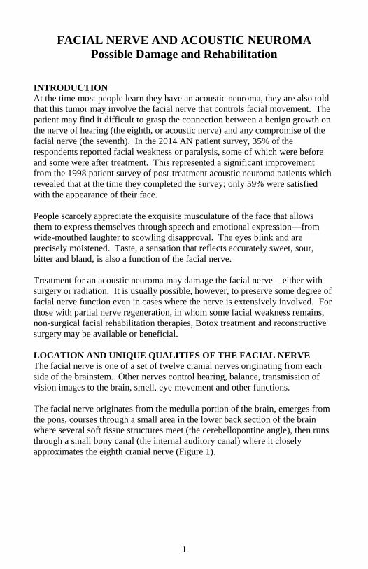
LOCATION AND UNIQUE QUALITIES OF THE FACIAL NERVE The facial nerve is one of a set of twelve cranial nerves originating from each
side of the brainstem. Other nerves control hearing, balance, transmission of
vision images to the brain, smell, eye movement and other functions.
The facial nerve originates from the medulla portion of the brain, emerges from
the pons, courses through a small area in the lower back section of the brain
where several soft tissue structures meet (the cerebellopontine angle), then runs
through a small bony canal (the internal auditory canal) where it closely
approximates the eighth cranial nerve (Figure 1).

2
Figure 1.
Facial Nerve Path
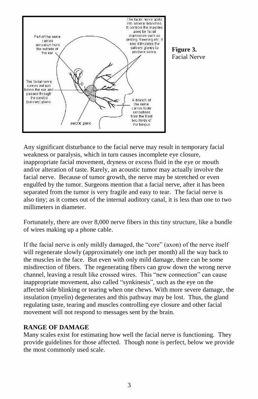
As it courses through the ear (temporal) bone system, the facial nerve sends
branches to the tear glands, the tongue and the salivary glands. After leaving the
ear’s bony structures, it divides again into many branches that serve various facial
muscles. The division after it leaves the mastoid is shown in Figures 2 & 3.
Figure 2.
Facial Nerve
Division
3
Figure 3.
Facial Nerve
Any significant disturbance to the facial nerve may result in temporary facial
weakness or paralysis, which in turn causes incomplete eye closure,
inappropriate facial movement, dryness or excess fluid in the eye or mouth
and/or alteration of taste. Rarely, an acoustic tumor may actually involve the
facial nerve. Because of tumor growth, the nerve may be stretched or even
engulfed by the tumor. Surgeons mention that a facial nerve, after it has been
separated from the tumor is very fragile and easy to tear. The facial nerve is
also tiny; as it comes out of the internal auditory canal, it is less than one to two
millimeters in diameter.
Fortunately, there are over 8,000 nerve fibers in this tiny structure, like a bundle
of wires making up a phone cable.
If the facial nerve is only mildly damaged, the “core” (axon) of the nerve itself
will regenerate slowly (approximately one inch per month) all the way back to
the muscles in the face. But even with only mild damage, there can be some
misdirection of fibers. The regenerating fibers can grow down the wrong nerve
channel, leaving a result like crossed wires. This “new connection” can cause
inappropriate movement, also called “synkinesis”, such as the eye on the
affected side blinking or tearing when one chews. With more severe damage, the
insulation (myelin) degenerates and this pathway may be lost. Thus, the gland
regulating taste, tearing and muscles controlling eye closure and other facial
movement will not respond to messages sent by the brain.
RANGE OF DAMAGE
Many scales exist for estimating how well the facial nerve is functioning. They
provide guidelines for those affected. Though none is perfect, below we provide
the most commonly used scale.
4
HOUSE-BRACKMANN FACIAL NERVE GRADING SCALE
Grade 1. Normal facial function in all areas.
Grade 2. Mild movement weakness, normal symmetry at rest. Slight
weakness noticeable on close inspection; may have very slight synkinesis
(inappropriate movement with voluntary movement of another muscle),
moderate to good forehead motion, complete eye closure with minimum effort,
only slight mouth disturbance.
Grade 3. Moderate dysfunction with noticeable asymmetry, good eye
closure. Obvious but not disfiguring difference between two sides; noticeable
but not severe synkinesis. Normal balance and tone at rest, slight to moderate
movement of forehead, complete eye closure with effort, mouth movement
slightly weak with maximum effort.
Grade 4. Moderately severe dysfunction with gross asymmetry, incomplete
eye closure. Obvious facial weakness and/or disfiguring asymmetry with gross
movement. Normal symmetry and tone at rest. No forehead movement on
affected side, incomplete eye closure, mouth asymmetric with maximum effort.
Grade 5. Severe dysfunction with minimal facial movement. Only barely
perceptible motion with attempted movement. Face unbalanced at rest. No
forehead motion, incomplete eye closure. Slight mouth movement possible.
Grade 6. Complete paralysis. No movement.
Facial weakness is usually noted when the patient awakens following surgery,
and for a few patients it can occur days after surgery. For most people, there
will be some improvement within three to six weeks; however, in others there
may not be significant recovery for several months after surgery. In a few
instances, a part of the facial nerve may have been preserved, but because of
severe damage or disruption of necessary blood supply, loss of function may be
prolonged or even permanent.
SURGICAL REHABILITATION The sooner regeneration is noted following surgery, the more likely it is that
recovery will be complete. However, it is reasonable to wait up to a year for
recovery, before proceeding with facial reanimation, when the surgeon believes
that the nerve integrity has been preserved. On the other hand, if the nerve is
severed during removal of the tumor, then facial reanimation surgery should be
done as soon as the patient’s health permits.
It is of utmost importance that facial reanimation is undertaken in a timely
fashion to get the best possible outcome. Most importantly if an individual has
no movement after one year, they should undergo a thorough evaluation by an
experienced facial nerve surgeon.
5
A. Immediate Repair
1. Direct Nerve Repair
The best time to repair the nerve is at the time of the initial surgery, and at the site
of injury. In a few cases, both ends of the severed nerve can be reattached. Better
nerve function can be achieved from a direct seventh nerve repair than with any
other technique. To perform this, both ends of the delicate nerve must be available
and contain adequate bulk and length to be stitched without tension.
2. Interposition Graft
When the severed facial nerve no longer provides two repairable ends which may be
reattached, a grafted section of another nerve may be positioned between the two
“connectable” ends. A suitable graft may be obtained from the neck, leg, or arm.
B. Later Repair
Sometimes it is not possible to repair the nerve, even with interposition grafts,
because one or both ends may not be suitable for direct attachment. Thus a graft
using both ends of the seventh nerve becomes technically impossible.
There are other techniques available that may be performed shortly after tumor
removal as the situation and the best alternatives for facial nerve rehabilitation
dictate.
1. Hypoglossal – Facial (12/7) Transfer (or Variations like “Jump Grafts”)
This technique has been widely used—either immediately to graft the tongue
nerve (hypoglossal or 12th
facial nerve) on the affected side to the facial nerve
on that side, or after a recommended waiting period (usually 12 months) when
there has been no return of facial nerve function, even though the nerve was
intact at the conclusion of surgery.
To accomplish this “hook-up,” the facial nerve is exposed from an incision in
front of the ear and the hypoglossal nerve is identified further down in the neck.
The 12th
nerve is cut (either partly or completely) and the portion coming from
the brain is attached to the healthy stump of the facial nerve.
After several months, fibers from the 12th
nerve grow within the 7th
nerve to the
muscles of facial expression. Partial voluntary movement can return; facial
motion is initiated by tongue movement. The 12/7 transfer is recommended as
soon as there is no possibility of 7th
nerve function. If a 12/7 is attempted
several years or longer after facial paralysis, it is likely that the facial nerve will
be fibrose—filled with scar tissue and unresponsive.
2. Masseteric Facial (5/7) Transfer
Similar to the 12/7 transfer discussed earlier, the masseteric nerve can be used to
give input to the paralyzed facial nerve. The masseteric (5th cranial nerve) nerve
gets activated when an individual clenches or bites down. It is a natural
movement that can be learned to be used by individuals to help them smile after
this surgery. The other advantage of 5/7 is that it causes less mass movement
6
than 12/7 and a more natural smile. It can also be used for individuals who have
multiple nerve injuries who are not candidates for 12/7.
3. Regional Muscle Transfer
Regional muscle transfers are surgical options for individuals with long-standing
facial paralysis who are not great candidates for 12/7, 5/7, or free muscle
transfers such as gracilis flap. The most common regional muscle used in facial
reanimation is the temporalis. In this procedure the end of the muscle or its
tendon (orthodromic temporalis tendon transfer) is attached to the corner of the
mouth. When the individual bites down or clenches, the patient’s face recreates
a smile. This operation also improves the tone and the static position of the face.
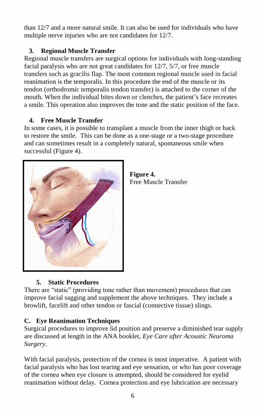
4. Free Muscle Transfer In some cases, it is possible to transplant a muscle from the inner thigh or back
to restore the smile. This can be done as a one-stage or a two-stage procedure
and can sometimes result in a completely natural, spontaneous smile when
successful (Figure 4).
Figure 4.
Free Muscle Transfer
5. Static Procedures
There are “static” (providing tone rather than movement) procedures that can
improve facial sagging and supplement the above techniques. They include a
browlift, facelift and other tendon or fascial (connective tissue) slings.
C. Eye Reanimation Techniques
Surgical procedures to improve lid position and preserve a diminished tear supply
are discussed at length in the ANA booklet, Eye Care after Acoustic Neuroma
Surgery.
With facial paralysis, protection of the cornea is most imperative. A patient with
facial paralysis who has lost tearing and eye sensation, or who has poor coverage
of the cornea when eye closure is attempted, should be considered for eyelid
reanimation without delay. Cornea protection and eye lubrication are necessary
7
for the health of the eye. Both should be attainable without having to sew the
eyelids together (tarsorrhaphy).
Canthoplasty (tightening of the tendons of the corner of the eye), implantation of a
gold or platinum weight or wire spring, and blocking the tear drainage system are
some of the surgical possibilities for eyelid reanimation and tear preservation.
For more information, see the booklet mentioned above, your ophthalmologist or
facial nerve specialist.
D. Surgical Interventions for Synkinesis
When patients have a beneficial response to Botox injections, there are several
interventions, either office-based or in the operating room, that can create a more
long lasting or permanent effect. Among these procedures are platysmectomy
(removal of a strip of platysma to stop excessive neck pulling) and highly selective
neurolysis of the grimacius or frowning muscles to improve upward smile
movement and reduce tension in the neck.
NON-SURGICAL THERAPIES FOR FACIAL MUSCLE
MAINTENANCE AND REHABILITATION When the facial nerve has been damaged or stretched by tumor growth or removal,
a temporary weakness usually results while the nerve regenerates. The facial nerve
must heal before an exercise program can be effective, so intensive therapy is not
recommended during this time (much like a broken bone in a cast has to heal
before beginning an exercise program). Avoid exercising the face too early to
reduce the tendency for the unaffected side to take over and create even more
asymmetry. New research suggests that gently stroking your fingers over the
affected side from the eye toward the mouth, from the ear toward the corner of the
mouth and from the back of the jaw under the earlobe toward the lower lip, may
result in more accurate nerve healing. Pay attention to the sensation as the fingers
slide over the facial skin. Doing so may help the brain “maintain connection” to
the affected side while the movement sensation signals are absent.
Give yourself permission to rest and let the nerve heal. Watch for small facial
movements to become visible as nerve recovery progresses. Several self-
administered therapies could help promote normalized facial movements after
recovery begins. Beware of electrical stimulation therapy which is often
recommended. These stimulations may increase the chance of synkinesis or
aberrant nerve regeneration.
A. Exercises
Patients with facial weakness should be aware of facial “posture.” Therapists
suggest keeping the muscles on the unaffected side relaxed. Doing so will
prevent the unaffected side from “pulling over” the affected side and help
maintain facial symmetry.
8
Among other things, the facial muscles are designed to create facial expressions.
These expressions convey emotional signals that are crucial for non-verbal
communication between human beings. Facial expression is almost always
subconscious and automatic. We rarely think of how we move our face during
spontaneous situations or how many different expressions we use. Typical,
functional, facial expressions are small and subtle, never forced with great
effort. For optimal results, facial “exercises” should mimic the normal functional
usage of the muscles, so they should be soft and gentle. Keeping this concept in
mind, the following movement patterns performed softly and easily (never
forced) may be helpful as facial function returns. Do not let the unaffected side
work harder or go further than the affected side. Exercising in this manner will
create tiny movements at first, but in the long run it will reinforce good facial
symmetry and balance and more accurately mimic the normal, unconscious,
facial movements we use everyday.
Softly raise eyebrows.
Draw eyebrows down and together.
Raise upper lip while wrinkling nose, as though something smells bad.
Watch for development of the crease between the side of the nose and
the corner of the mouth. This is called the nasolabial fold.
Focus eyeballs in downward direction - softly close both eyes.
Softly pucker lips – as though blowing a light feather.
Softly smile - think of smiling from the upper cheek rather than with the
lips – don’t let the unaffected side smile larger than the operated side.
Watch in a mirror while speaking on the telephone to maintain symmetry.
DO NOT chew gum as this promotes asymmetry.
Neuromuscular Re-Education Techniques
A number of therapists in the United States specialize in neuromuscular re-
education techniques which can help improve facial expression and symmetry
once movement begins to return.
The principle underlying this therapy is that the plasticity of the brain (i.e., ability
to learn new movements) allows for retraining abnormalities that result from a
damaged facial nerve. This is especially helpful in cases where incomplete
recovery has resulted in abnormal facial movements known as synkinesis.
Specially trained therapists combine individual evaluation, specific movement
patterns, massage and biofeedback to normalize the abnormal movements that can
occur with faulty nerve regeneration. In many cases of synkinesis, the lack of
function can be attributed to improper coordination as opposed to (or in
combination with) muscle weakness. The main focus with retraining synkinesis is
on reducing the inappropriate, uncoordinated muscle movements that distort
expression, while improving muscle coordination to enhance expression.
9
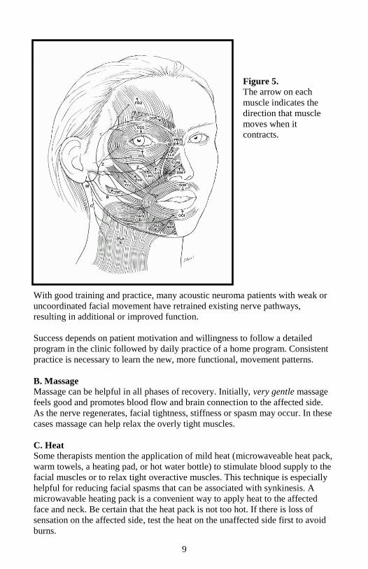
Figure 5.
The arrow on each
muscle indicates the
direction that muscle
moves when it
contracts.
With good training and practice, many acoustic neuroma patients with weak or
uncoordinated facial movement have retrained existing nerve pathways,
resulting in additional or improved function.
Success depends on patient motivation and willingness to follow a detailed
program in the clinic followed by daily practice of a home program. Consistent
practice is necessary to learn the new, more functional, movement patterns.
B. Massage
Massage can be helpful in all phases of recovery. Initially, very gentle massage
feels good and promotes blood flow and brain connection to the affected side.
As the nerve regenerates, facial tightness, stiffness or spasm may occur. In these
cases massage can help relax the overly tight muscles.
C. Heat
Some therapists mention the application of mild heat (microwaveable heat pack,
warm towels, a heating pad, or hot water bottle) to stimulate blood supply to the
facial muscles or to relax tight overactive muscles. This technique is especially
helpful for reducing facial spasms that can be associated with synkinesis. A
microwavable heating pack is a convenient way to apply heat to the affected
face and neck. Be certain that the heat pack is not too hot. If there is loss of
sensation on the affected side, test the heat on the unaffected side first to avoid
burns.
10
D. Vibration and Electrical Stimulation
Some therapists recommend limited use of a small, flat vibrator for short periods
to accompany facial muscle exercises. There is little evidence that vibration is
beneficial. Electrical stimulation should not be used at any time during the
healing phase as it may interfere with normal nerve regeneration and/or
reinforce abnormal movement patterns. It also may increase synkinesis.
E. Acupuncture
The effect of acupuncture on facial recovery is unknown. There is little evidence
that it either assists in or interferes with facial nerve recovery. Many patients
have reported improvements in headaches or a greater sense of well being and
relaxation after undergoing acupuncture.
F. Other techniques
In recent years, several facial exercise programs have been advertised in TV
infomercials. Regardless of the fact that scientific research is lacking, these
techniques were marketed to reduce the signs of aging in normal faces, not to
rehabilitate facial paralysis patients. Their use could promote asymmetry,
synkinesis or facial spasm.
G. Botox® (Botulinum toxin)
Botox has received a great deal of media attention as a cosmetic procedure for
reducing wrinkles. It is also being used increasingly in cases of facial paralysis
with synkinesis, to improve facial coordination. Botox blocks the electrical signal
generated by the facial nerve from entering the muscle. The portion of the muscle
injected becomes paralyzed and cannot move. The effect is temporary, lasting 3 to
6 months. When injected selectively into improperly contracting muscles, Botox
can effectively reduce some of the abnormal contractions and improve
coordination; however, it can cause severe paralysis if injected in the wrong place.
Botox must be administered by a qualified physician who has experience with
facial paralysis and synkinesis.
H. Cosmetic Considerations
Facial appearance and symmetry can be enhanced by hairstyle, cosmetic
application or tinted eyeglasses. A cosmetologist can help in designing
individualized techniques to improve appearance as the facial nerve recovers.
11
WHAT IS THE ACOUSTIC NEUROMA ASSOCIATION (ANA)?
Acoustic Neuroma Association was founded in Carlisle, Pennsylvania, in 1981 by an
acoustic neuroma patient, Virginia Fickel Ehr. She found no patient information or
patient support available when she had surgery for the removal of an acoustic
neuroma in 1977. She resolved that future acoustic neuroma patients should have
easy-to-read medical material about their condition and support and comfort from
each other. With the help of her physician, she contacted eight other patients and
formed the organization.
The association is incorporated and is a 501(c)(3) non-profit organization. The
patient-focused, member organization now serves close to 5,000 members, is
governed by a primarily all-patient Board of Directors and is operated by a small staff
in metropolitan Atlanta, Georgia. Medical information is provided by the ANA
Medical Advisory Board.
ANA membership benefits include receipt of a quarterly newsletter, patient
information booklets, access to a network of Local Support Groups, participation in
our webinars by leading medical professionals, access to a list of acoustic neuroma
patients willing to talk about their experience throughout the country and our website
Member Section. Our exclusive website Member Section includes past webinars,
published medical journal articles on acoustic neuroma and all of our patient
information booklets and newsletters and many symposium presentations. ANA also
maintains an interactive website at www.ANAUSA.org with an ANA Discussion
Forum, along with a social media presence.
ANA is patient-founded, patient-focused and patient-funded. ANA recommends
treatment from a medical team with substantial acoustic neuroma experience.
Although the association cannot recommend specific doctors, medical centers or
medical procedures, guidelines for selecting a qualified medical professional can be
found at the ANA website, www.ANAUSA.org. Now available on our website is a
listing of medical resources. The physicians and organizations listed have self-
reported data to meet criteria established by ANA for having substantial experience in
treating acoustic neuromas. The listings should NOT in any way be construed as an
endorsement or recommendation by ANA. It is every individual’s responsibility to
verify the qualifications, education and experience of any health care professional.
12
ANA PUBLICATIONS
ANA members can view patient information booklets and newsletters online on our
website at www.ANAUSA.org in our Member Section. To order ANA publications,
please contact:
Acoustic Neuroma Association
600 Peachtree Parkway, Suite 108
Cumming, GA 30041
1-877-200-8211
You may also order online in the ANA Store at www.ANAUSA.org using VISA® or
Mastercard®.
Booklets Color Price Each*
Acoustic Neuroma Basic Overview Tan $1.50
Diagnosis: AN – What Next? Peach $2.00
Eye Care after AN Surgery Yellow $3.00
Facial Nerve and AN: Possible Damage Gray $2.00
and Rehabilitation
A Glimpse of the Brain Green $1.50
Headache Associated with AN Treatment Violet $2.00
Improving Balance Associated with AN Red $3.00
Hearing Loss Rehabilitation for AN Patients Teal $3.00
Newsletter Back Issues
Notes (quarterly publication) $10.00 per mailing
*(For non-ANA members and multiple copies)
Please add shipping and handling:
Orders $ 0.25 to $15.00 add $ 4.00
Orders $15.01 to $30.00 add $ 5.00
Orders $30.01 to $45.00 add $ 6.00
Orders $45.01 to $99.00 add $10.00
Orders $99.01 to $200.00 add $12.00
Georgia residents, please add 7% Sales Tax
© Acoustic Neuroma Association, July 2015
Related Documents