Facial Fractures – Mandible and Frontal Bones Dale Reynolds, MD UT Houston Plastic & Reconstructive Surgery

Facial Fractures – Mandible and Frontal Bones Dale Reynolds, MD UT Houston Plastic & Reconstructive Surgery.
Dec 28, 2015
Welcome message from author
This document is posted to help you gain knowledge. Please leave a comment to let me know what you think about it! Share it to your friends and learn new things together.
Transcript
Facial Fractures –Mandible and Frontal Bones
Dale Reynolds, MD
UT Houston
Plastic & Reconstructive Surgery

Facial FracturesPhases
Emergency Treatment Airway
Edema Teeth Blood FB Mandible fracture tongue to pharynx Stridor, hoarseness, retraction, drooling ETT Tracheostomy
Long term IMF Cricothyroidotomy
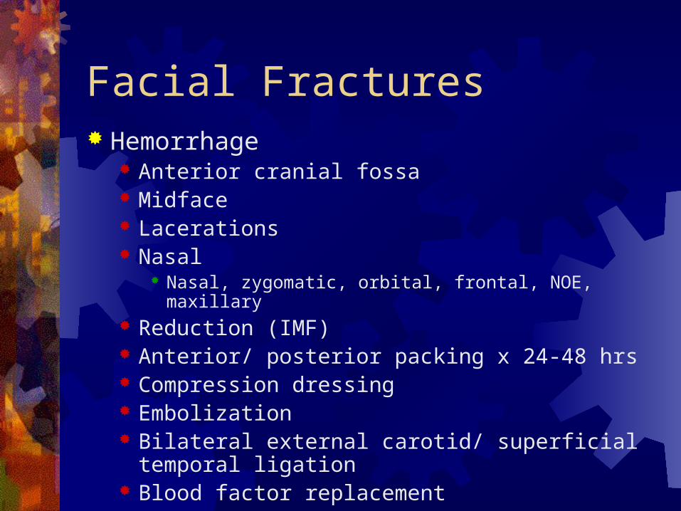
Facial Fractures Hemorrhage
Anterior cranial fossa Midface Lacerations Nasal
Nasal, zygomatic, orbital, frontal, NOE, maxillary Reduction (IMF) Anterior/ posterior packing x 24-48 hrs Compression dressing Embolization Bilateral external carotid/ superficial temporal
ligation Blood factor replacement

Facial Fractures Aspiration
Low threshold for ETT Other
Eye Brain Spine

Facial Fractures Early injury care
History PE
Nerves, vision, intraoral, nasopharyngeal, dentition Radiographs Lacerations IMF Impressions
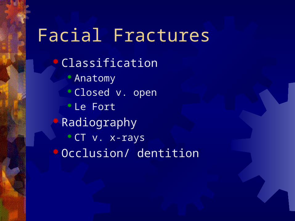
Facial Fractures Classification
Anatomy Closed v. open Le Fort
Radiography CT v. x-rays
Occlusion/ dentition

Facial FracturesMandible
Anatomy

Facial FracturesMandible
Anatomy

Facial FracturesMandible
Anatomy

Facial FracturesMandible
Anatomy

Facial Fractures Mandible
Most common facial fracture after nasal 10-25% of all facial fractures Body> angle> condyle> parasymphysis> other M: F = 2: 1 58% multiple (93% ,
3 fx) Preinjury relationships Stable bony union Facial proportions Avoid complications

Facial FracturesMandible
History Previous trauma Previous baseline Pre-injury photo

Facial FracturesMandible
PE Crepitance Symmetry Tenderness Oral/ dental – missing
teeth Step offs

Facial FracturesMandible
Radiography Panorex CT Plain films
PA, Towne’s, R and L lateral oblique views (mandibular series)

MandibleTreatment
Restore form and function
Occlusion, TMJ function, cosmesis
ORIF Exact anatomic reduction Allows early resumption of mandibular function

Mandible
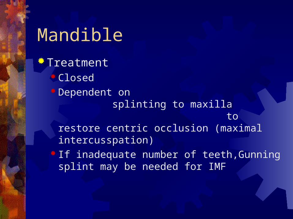
MandibleTreatment
Closed Dependent on
splinting to maxilla to restore centric occlusion (maximal intercusspation)
If inadequate number of teeth,Gunning splint may be needed for IMF

Mandible Treatment
Open Accurate reduction
Within 2 weeks If maxilla cannot be used then mandible first or splints
Avoid prolonged IMF Traumitizes gingiva Impairs oral hygiene periodontal disease Uncomfortable Forces can alter tooth position and periodontal
attachments Great aspiration risk Contraindication in COPD, seizure d/o, impaired MS Articular surfaces under compression cause pressure
necrosis

MandibleORIF
Lag screw – Anterior

MandibleORIF
Reconstruction plate – Comminuted body

MandibleORIF
Two plate/ tension band – Angle

MandibleORIF
Dynamic compression plate - Condyle

MandibleTreatment
Contraindications to open Not required Not candidate
Rarely needed in children Simple Heal quickly Occlusion less established

Facial Fractures
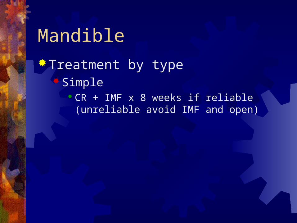
MandibleTreatment by type
Simple CR + IMF x 8 weeks if reliable (unreliable avoid
IMF and open)
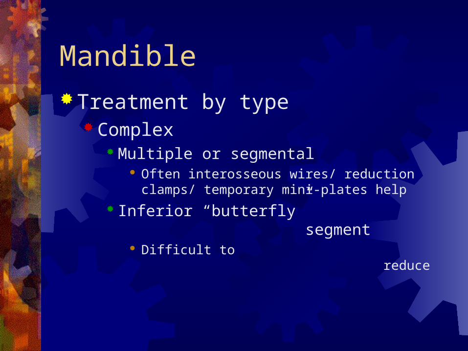
MandibleTreatment by type
Complex Multiple or segmental
Often interosseous wires/ reduction clamps/ temporary mini-plates help
Inferior “butterfly” segment
Difficult to reduce

Mandible Treatment by type
Complex Bilateral fracture each hemi-mandible
Simultaneous reduction may be required to avoid magnification of discrepancy
Arch bars and IMF may worsen Anterior fracture with one or both condyles
Consider reducing one or both condyles first if difficult to control flaring the inferior border
Unilateral segmental fracture in one hemi-mandible Close fractures – two plates Separated fractures – long spanning plate

MandibleTreatment by type
Complex Comminuted
High energy – GSW, SGW, MVC Easy to devitalize small fragments Difficult to accurately reduce Large reconstruction plate may be required Temporary external fixator may be used if condition
of patient or soft tissue requires Bone graft for extensive loss Pre-treatment infection: Debride small fragments Post-treatment infection: FB (bone or screw)
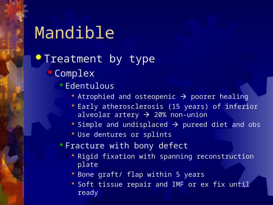
MandibleTreatment by type
Complex Edentulous
Atrophied and osteopenic poorer healing Early atherosclerosis (15 years) of inferior alveolar
artery 20% non-union Simple and undisplaced pureed diet and obs Use dentures or splints
Fracture with bony defect Rigid fixation with spanning reconstruction plate Bone graft/ flap within 5 years Soft tissue repair and IMF or ex fix until ready
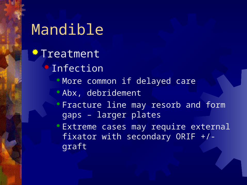
MandibleTreatment
Infection More common if delayed care Abx, debridement Fracture line may resorb and form gaps –
larger plates Extreme cases may require external fixator with
secondary ORIF +/- graft

MandibleTreatment
Children Most need CR + immobilization (single arch bar
or lingual splint) x 2 weeks Conical shape makes arch bars less useful Indications for ORIF
Unstable fractures Not amenable to CR Bilateral fractures with gross instability
Use unicortical plates Remove 6-8 weeks later
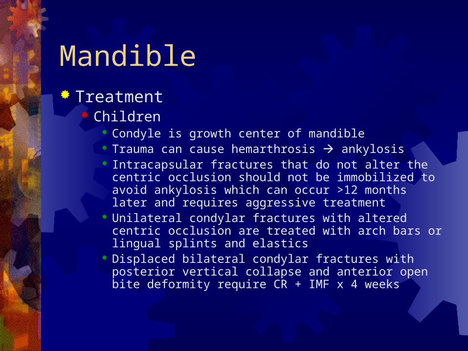
Mandible Treatment
Children Condyle is growth center of mandible Trauma can cause hemarthrosis ankylosis Intracapsular fractures that do not alter the centric occlusion
should not be immobilized to avoid ankylosis which can occur >12 months later and requires aggressive treatment
Unilateral condylar fractures with altered centric occlusion are treated with arch bars or lingual splints and elastics
Displaced bilateral condylar fractures with posterior vertical collapse and anterior open bite deformity require CR + IMF x 4 weeks

Mandible Treatment
By Location Alveolar Process (1%)
Remove if devitalized o/w IMF or splint Symphysis (5.8%)
Often associated with condylar fractures Significant forces cause lateral flaring of posterior
segments (often worse with IMF) Parasymphysis (11.6%)
Often associated with contralateral fractures Mental nerve Burr/ osteotome may help lessen anterior curvature

MandibleTreatment
By Location Body (31.9%)
May require external approach Bi-cortical plates placed beneath mental canal
Angle (27.5%) May require external approach Often associated with contralateral Highest complication rate due to third molar teeth
and displacing forces

MandibleTreatment
By Location Ramus (2.5%)
Usually require extraoral approach Often stable due to splinting effect of masseter-
medial pterygoid muscle sling unless displacement causes vertical shortening (telescoping)
Coronoid process (1.8%) Soft diet usually enough Severe pain may require brief IMF

MandibleTreatment
By Location Condyle (23.8%)
Proximal segment can undergo AVN Intra-articular fractures: Very difficult ORIF, OA is
common outcome, usually brief IMF for malocclusion o/w early mobilization +/- elastics
Condylar neck: Anteromedial displacement of proximal segment by lateral pterygoid, usually treated with IMF x 6 weeks, ORIF if joint capsule is thought to be involved

MandibleTreatment
By Location Condyle
ORIF Displaced in to middle cranial fossa FB within joint Lateral extra-capsular displacement of condyle Displacement blocking opening or closing Posterior vertical shortening of mandible with
open bite after 2 week IMF trial Relative
Bilateral associated with unstable midface fractures
Bilateral edentulous without splint

Mandible Postoperative care
+/- Abx, airway control with IMF (wire cutters), HOB (secretions) + ice pack for edema
Diet CLD blenderized, 48o IVF, 15 lb wt loss
Splints/ IMF Oral hygiene (peridex, H2O2, brush), remove wax
Oral washouts Release IMF q 3-5 days if needed

Mandible Centric occlusion
Remove IMF to assess ORIF
Therapeutic rehabilitation Regain strength and mobility, PT if severe
(prolonged IMF or condyle fracture) Dental treatment (missing teeth)
Complications Malocclusion, malunion, non-union, hardware
exposure, infection, non-compliance

Mandible Teeth in
fracture line

Facial FracturesFrontal bone anatomy – 7 bones

Facial FracturesFrontal bone anatomy

Facial FracturesFrontal sinus anatomy
Middle meatus
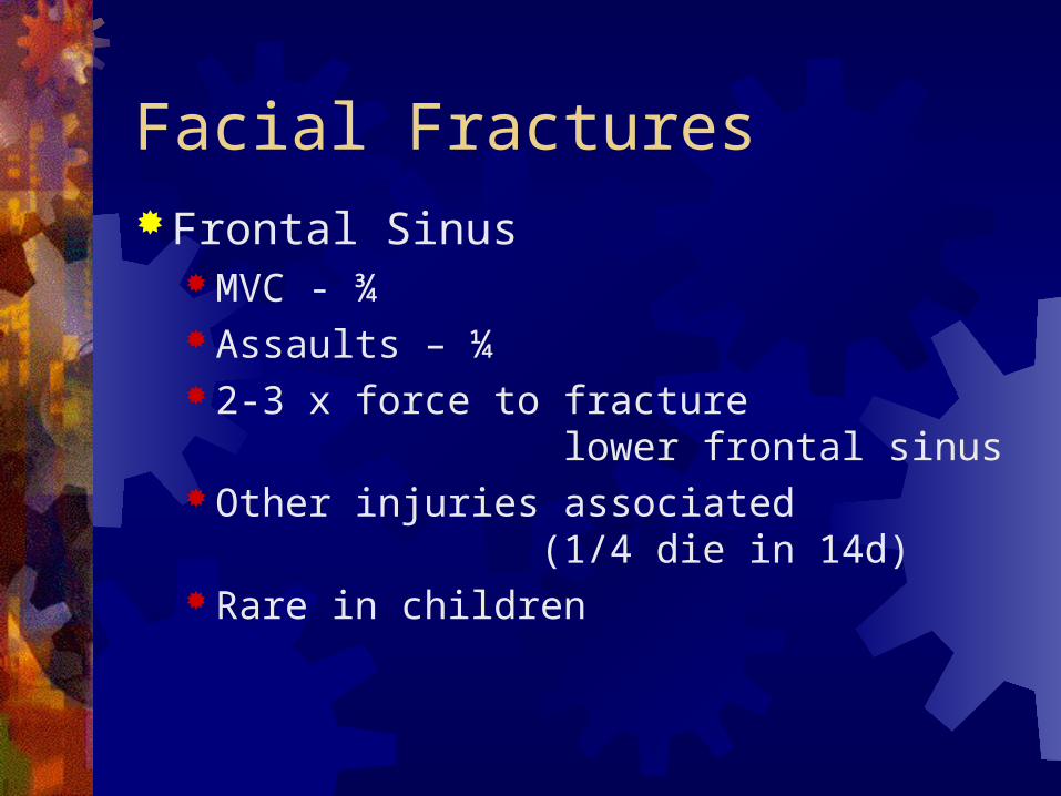
Facial FracturesFrontal Sinus
MVC - ¾ Assaults – ¼ 2-3 x force to fracture
lower frontal sinus Other injuries associated
(1/4 die in 14d) Rare in children

Facial FracturesFrontal Sinus Fracture
Signs Rhinorrhea Step-off Supraorbital anesthesia Subconjunctival hematoma Subcutaneous crepitance

Facial FracturesFrontal Sinus Fracture
Diagnosis Plain films CT

Facial FracturesFrontal sinus fractures
Anterior Table (Thick) Displaced ORIF Blockage of nasofrontal
duct (methylene blue) Remove mucosa Bone graft nasofrontal
ducts, fill space Elevate and fixate bone
Posterior Table (Thin) Comminuted Cranialize Displaced greater than one wall thickness
ORIF

Facial Fractures Frontal Sinus Fracture
Complications (Posterior > anterior) Acute
Epistaxis CSF leak Meningitis Intracranial injury Hematoma
Subacute Mucocele Sinusitis
Chronic Osteomyelitis Abscesses

END
Related Documents