Fabrication of nano-grained Ti–Nb–Zr biomaterials using spark plasma sintering M.A. Hussein a,b , C. Suryanarayana c , N. Al-Aqeeli a, ⁎ a Department of Mechanical Engineering, King Fahd University of Petroleum & Minerals, Dhahran 31261, Saudi Arabia b Department of Mechanical Engineering, Kafrelsheikh University, Kafrelsheikh 33516, Egypt c Department of Mechanical and Aerospace Engineering, University of Central Florida, Orlando, FL 32816-2450, USA abstract article info Article history: Received 10 June 2015 Received in revised form 12 August 2015 Accepted 17 August 2015 Available online 20 August 2015 Keywords: Biomaterials Spark plasma sintering Mechanical alloying Nanomaterials Ti–Nb–Zr alloy Nanostructured near-β Ti–20Nb–13Zr at % alloy with non-toxic elements and enhanced mechanical properties has been synthesized by spark plasma sintering (SPS) of nanocrystalline powders obtained by mechanical alloying. The consolidated bulk product was characterized by density measurements and Vickers hardness (HV), and X-ray diffraction (XRD), field emission scanning electron microscopy (FE-SEM) combined with energy-dispersive spectroscopy (EDX), and transmission electron microscopy (TEM) for structural details. The temperature during spark plasma sintering was varied between 800 and 1200 °C, while the heating rate and holding time of 100°K/min and 10 min were maintained constant in all the experiments. The effect of SPS temperature on the densification, microstructure, and HV was discussed. The results show that a nearly full den- sity structure was obtained after SPS at 1200 °C. The microstructure of the obtained alloy is a duplex structure with the α-Ti (hcp) region having an average size of 70–140 nm, surrounding the β-Ti (bcc) matrix. The obtained alloy was chemically homogenized with a micro hardness value, HV of 660. The developed nanostructured Ti–20Nb–13Zr alloy is suggested for biomedical use as in implant material in dental and orthopedic applications. © 2015 Elsevier Ltd. All rights reserved. 1. Introduction A number of different metallic materials have been used in a variety of applications in the medical field. Specifically, they are used for inter- nal support and biological tissue replacements such as joint replace- ment, dental roots, orthopedic fixation and stents [1]. The common metals and alloys that are being utilized for biomedical applications are: stainless steels, Co-based alloys, and Ti-based alloys. However, Ti and Ti-based alloys have dominated over other alloy systems in the medical and dentistry fields due to their improved biocompatibility [2–4]. Many Ti-based alloys were developed with the addition of multi- ple alloying elements in order to evaluate their individual contributions and arrive at an improved composition that possesses the highest biocompatibility. Different metals like vanadium, nickel, chromium, and aluminum were added via different processing routes and some Ti-based alloys were introduced, e.g., Ti–6Al–4V [5]. However, it was observed that vanadium and aluminum are probably toxic [2] and can cause mutagenic cytology in addition to triggering allergic reactions [3]. Other constituents like nickel were found to result in lower biocompat- ibility [5] and chromium has a major concern due to its genotoxicity [5]. Therefore, both the selection of alloy composition and finding the best processing method to develop Ti-based alloys have been a challenge in the biomedical/biomaterials field. All the three constituents in the Ti–Nb–Zr alloy meet the criteria for biomaterials in terms of biocompatibility, resistance to corrosion, mechanical considerations, and ionic cytotoxicity [5]. The addition of Nb to Ti allows stabilizes the β phase in these alloys and possess im- proved mechanical properties, and also results in improved wear resis- tance, while the addition of Zr helps in obtaining the solid solution required for achieving the hardness [2]. There has been some work reported in the literature on the develop- ment of Ti-based alloys using arc melting and casting techniques [2,3, 6–12]. However, when there is a broad range of melting temperatures be- tween the alloy constituents, utilization of conventional casting techniques in the synthesis of Ti-based alloys does not seem to be an optimal route due to plausible absence of homogeneity in the final alloy. Therefore, mechanical alloying (MA), a completely solid-state powder processing technique, can be considered as a promising alternate tech- nique to combat limitations faced by casting techniques. MA has also been used successfully in the development of nanostructured materials. This technique is known to be simple, versatile, economically viable and can be scaled up to produce large quantities [13]. Powder metallurgy (PM) processing is also considered to be an effective way in reducing the higher machining costs of Ti alloys. It has the advantage to produce a homogenous alloy compared to other techniques, especially when the alloying element has a higher melting temperature such as Nb [4]. Materials and Design 87 (2015) 693–700 ⁎ Corresponding author. E-mail addresses: [email protected] (M.A. Hussein), [email protected] (C. Suryanarayana), [email protected] (N. Al-Aqeeli). http://dx.doi.org/10.1016/j.matdes.2015.08.082 0264-1275/© 2015 Elsevier Ltd. All rights reserved. Contents lists available at ScienceDirect Materials and Design journal homepage: www.elsevier.com/locate/jmad

Welcome message from author
This document is posted to help you gain knowledge. Please leave a comment to let me know what you think about it! Share it to your friends and learn new things together.
Transcript

Materials and Design 87 (2015) 693–700
Contents lists available at ScienceDirect
Materials and Design
j ourna l homepage: www.e lsev ie r .com/ locate / jmad
Fabrication of nano-grained Ti–Nb–Zr biomaterials using sparkplasma sintering
M.A. Hussein a,b, C. Suryanarayana c, N. Al-Aqeeli a,⁎a Department of Mechanical Engineering, King Fahd University of Petroleum & Minerals, Dhahran 31261, Saudi Arabiab Department of Mechanical Engineering, Kafrelsheikh University, Kafrelsheikh 33516, Egyptc Department of Mechanical and Aerospace Engineering, University of Central Florida, Orlando, FL 32816-2450, USA
⁎ Corresponding author.E-mail addresses: [email protected] (M.A. Hus
(C. Suryanarayana), [email protected] (N. Al-Aqeeli).
http://dx.doi.org/10.1016/j.matdes.2015.08.0820264-1275/© 2015 Elsevier Ltd. All rights reserved.
a b s t r a c t
a r t i c l e i n f oArticle history:Received 10 June 2015Received in revised form 12 August 2015Accepted 17 August 2015Available online 20 August 2015
Keywords:BiomaterialsSpark plasma sinteringMechanical alloyingNanomaterialsTi–Nb–Zr alloy
Nanostructured near-β Ti–20Nb–13Zr at % alloy with non-toxic elements and enhanced mechanical propertieshas been synthesized by spark plasma sintering (SPS) of nanocrystalline powders obtained by mechanicalalloying. The consolidated bulk product was characterized by density measurements and Vickers hardness(HV), and X-ray diffraction (XRD), field emission scanning electron microscopy (FE-SEM) combined withenergy-dispersive spectroscopy (EDX), and transmission electron microscopy (TEM) for structural details. Thetemperature during spark plasma sintering was varied between 800 and 1200 °C, while the heating rate andholding time of 100°K/min and 10 min were maintained constant in all the experiments. The effect of SPStemperature on the densification, microstructure, and HVwas discussed. The results show that a nearly full den-sity structure was obtained after SPS at 1200 °C. The microstructure of the obtained alloy is a duplex structurewith theα-Ti (hcp) region having an average size of 70–140 nm, surrounding theβ-Ti (bcc)matrix. The obtainedalloy was chemically homogenized with a micro hardness value, HV of 660. The developed nanostructuredTi–20Nb–13Zr alloy is suggested for biomedical use as in implantmaterial in dental and orthopedic applications.
© 2015 Elsevier Ltd. All rights reserved.
1. Introduction
A number of different metallic materials have been used in a varietyof applications in the medical field. Specifically, they are used for inter-nal support and biological tissue replacements such as joint replace-ment, dental roots, orthopedic fixation and stents [1]. The commonmetals and alloys that are being utilized for biomedical applicationsare: stainless steels, Co-based alloys, and Ti-based alloys. However, Tiand Ti-based alloys have dominated over other alloy systems in themedical and dentistry fields due to their improved biocompatibility[2–4]. Many Ti-based alloys were developedwith the addition of multi-ple alloying elements in order to evaluate their individual contributionsand arrive at an improved composition that possesses the highestbiocompatibility. Different metals like vanadium, nickel, chromium,and aluminum were added via different processing routes and someTi-based alloys were introduced, e.g., Ti–6Al–4 V [5]. However, it wasobserved that vanadium and aluminum are probably toxic [2] and cancausemutagenic cytology in addition to triggering allergic reactions [3].Other constituents like nickel were found to result in lower biocompat-ibility [5] and chromium has amajor concern due to its genotoxicity [5].Therefore, both the selection of alloy composition and finding the best
sein), [email protected]
processing method to develop Ti-based alloys have been a challengein the biomedical/biomaterials field.
All the three constituents in the Ti–Nb–Zr alloy meet the criteriafor biomaterials in terms of biocompatibility, resistance to corrosion,mechanical considerations, and ionic cytotoxicity [5]. The addition ofNb to Ti allows stabilizes the β phase in these alloys and possess im-proved mechanical properties, and also results in improved wear resis-tance, while the addition of Zr helps in obtaining the solid solutionrequired for achieving the hardness [2].
There has been some work reported in the literature on the develop-ment of Ti-based alloys using arc melting and casting techniques [2,3,6–12]. However, when there is a broad range ofmelting temperatures be-tween the alloy constituents, utilization of conventional castingtechniques in the synthesis of Ti-based alloys does not seem to be anoptimal route due to plausible absence of homogeneity in the final alloy.Therefore, mechanical alloying (MA), a completely solid-state powderprocessing technique, can be considered as a promising alternate tech-nique to combat limitations faced by casting techniques. MA has alsobeen used successfully in the development of nanostructured materials.This technique is known to be simple, versatile, economically viable andcan be scaled up to produce large quantities [13]. Powder metallurgy(PM) processing is also considered to be an effective way in reducingthe higher machining costs of Ti alloys. It has the advantage to producea homogenous alloy compared to other techniques, especially whenthe alloying element has a higher melting temperature such as Nb [4].
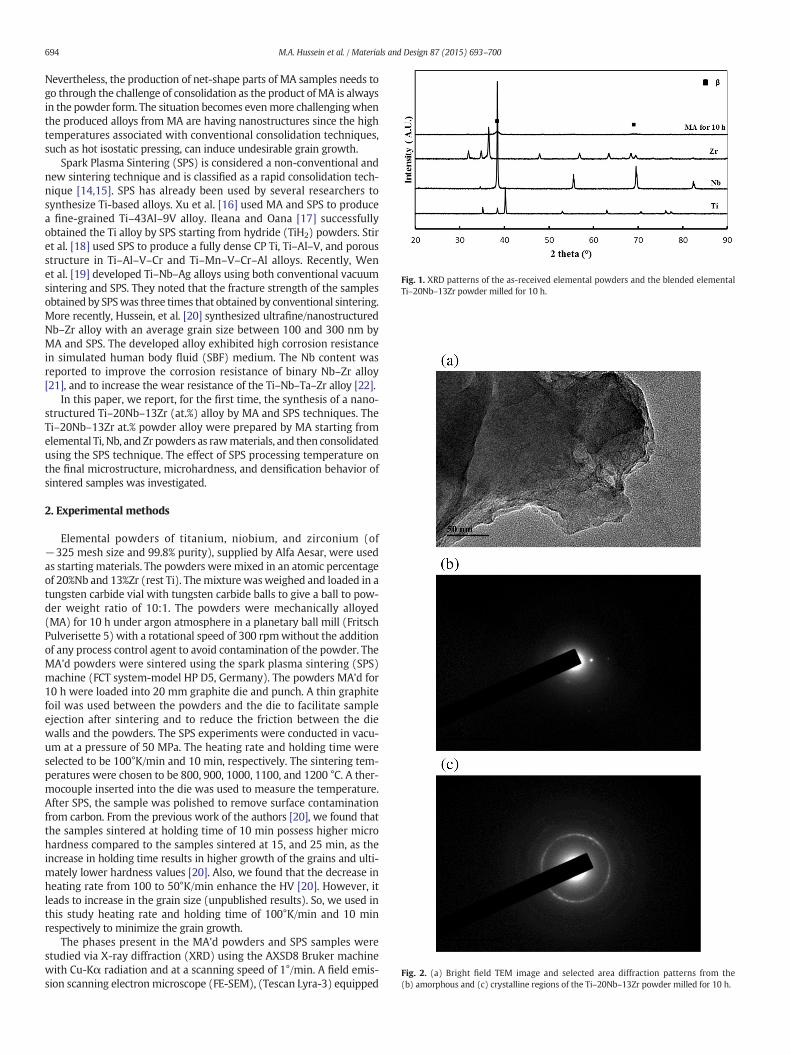
Fig. 1. XRD patterns of the as-received elemental powders and the blended elementalTi–20Nb–13Zr powder milled for 10 h.
Fig. 2. (a) Bright field TEM image and selected area diffraction patterns from the(b) amorphous and (c) crystalline regions of the Ti–20Nb–13Zr powder milled for 10 h.
694 M.A. Hussein et al. / Materials and Design 87 (2015) 693–700
Nevertheless, the production of net-shape parts of MA samples needs togo through the challenge of consolidation as the product of MA is alwaysin the powder form. The situation becomes evenmore challengingwhenthe produced alloys from MA are having nanostructures since the hightemperatures associated with conventional consolidation techniques,such as hot isostatic pressing, can induce undesirable grain growth.
Spark Plasma Sintering (SPS) is considered a non-conventional andnew sintering technique and is classified as a rapid consolidation tech-nique [14,15]. SPS has already been used by several researchers tosynthesize Ti-based alloys. Xu et al. [16] used MA and SPS to producea fine-grained Ti–43Al–9V alloy. Ileana and Oana [17] successfullyobtained the Ti alloy by SPS starting from hydride (TiH2) powders. Stiret al. [18] used SPS to produce a fully dense CP Ti, Ti–Al–V, and porousstructure in Ti–Al–V–Cr and Ti–Mn–V–Cr–Al alloys. Recently, Wenet al. [19] developed Ti–Nb–Ag alloys using both conventional vacuumsintering and SPS. They noted that the fracture strength of the samplesobtained by SPSwas three times that obtained by conventional sintering.More recently, Hussein, et al. [20] synthesized ultrafine/nanostructuredNb–Zr alloy with an average grain size between 100 and 300 nm byMA and SPS. The developed alloy exhibited high corrosion resistancein simulated human body fluid (SBF) medium. The Nb content wasreported to improve the corrosion resistance of binary Nb–Zr alloy[21], and to increase the wear resistance of the Ti–Nb–Ta–Zr alloy [22].
In this paper, we report, for the first time, the synthesis of a nano-structured Ti–20Nb–13Zr (at.%) alloy by MA and SPS techniques. TheTi–20Nb–13Zr at.% powder alloy were prepared by MA starting fromelemental Ti, Nb, and Zr powders as rawmaterials, and then consolidatedusing the SPS technique. The effect of SPS processing temperature onthe final microstructure, microhardness, and densification behavior ofsintered samples was investigated.
2. Experimental methods
Elemental powders of titanium, niobium, and zirconium (of−325 mesh size and 99.8% purity), supplied by Alfa Aesar, were usedas startingmaterials. The powders were mixed in an atomic percentageof 20%Nb and 13%Zr (rest Ti). Themixture wasweighed and loaded in atungsten carbide vial with tungsten carbide balls to give a ball to pow-der weight ratio of 10:1. The powders were mechanically alloyed(MA) for 10 h under argon atmosphere in a planetary ball mill (FritschPulverisette 5) with a rotational speed of 300 rpmwithout the additionof any process control agent to avoid contamination of the powder. TheMA'd powders were sintered using the spark plasma sintering (SPS)machine (FCT system-model HP D5, Germany). The powders MA'd for10 h were loaded into 20 mm graphite die and punch. A thin graphitefoil was used between the powders and the die to facilitate sampleejection after sintering and to reduce the friction between the diewalls and the powders. The SPS experiments were conducted in vacu-um at a pressure of 50 MPa. The heating rate and holding time wereselected to be 100°K/min and 10 min, respectively. The sintering tem-peratures were chosen to be 800, 900, 1000, 1100, and 1200 °C. A ther-mocouple inserted into the die was used to measure the temperature.After SPS, the sample was polished to remove surface contaminationfrom carbon. From the previous work of the authors [20], we found thatthe samples sintered at holding time of 10 min possess higher microhardness compared to the samples sintered at 15, and 25 min, as theincrease in holding time results in higher growth of the grains and ulti-mately lower hardness values [20]. Also, we found that the decrease inheating rate from 100 to 50°K/min enhance the HV [20]. However, itleads to increase in the grain size (unpublished results). So, we used inthis study heating rate and holding time of 100°K/min and 10 minrespectively to minimize the grain growth.
The phases present in the MA'd powders and SPS samples werestudied via X-ray diffraction (XRD) using the AXSD8 Bruker machinewith Cu-Kα radiation and at a scanning speed of 1°/min. A field emis-sion scanning electron microscope (FE-SEM), (Tescan Lyra-3) equipped
695M.A. Hussein et al. / Materials and Design 87 (2015) 693–700
with the energy-dispersiveX-ray (EDX) analysiswas used to characterizethemorphology of the powders, microstructures, and chemical composi-tions for both the MA'd powders and SPS samples. The microstructureswere also examined using a transmission electron microscope (TEM:Tecnai G Series operated at 200 keV). The SPS samples were etchedwith 10%HF solution for 5 s to reveal the microstructural details onFE-SEM. A hardness tester (Buehler, USA) with 500 g load and 10 sdwell time was used to measure the microhardness of the consolidatedsamples after polishing. The density determination kit (Mettler Toledo)was used to measure the density of the sintered samples based onArchimedes principle. The reported values were the average of ten mea-surements for HV and three measurements for the density.
3. Results and discussion
Fig. 1 shows the XRD patterns of the as-received elemental powdersand the Ti–20Nb–13Zr powder blend afterMA for 10 h. All the expected
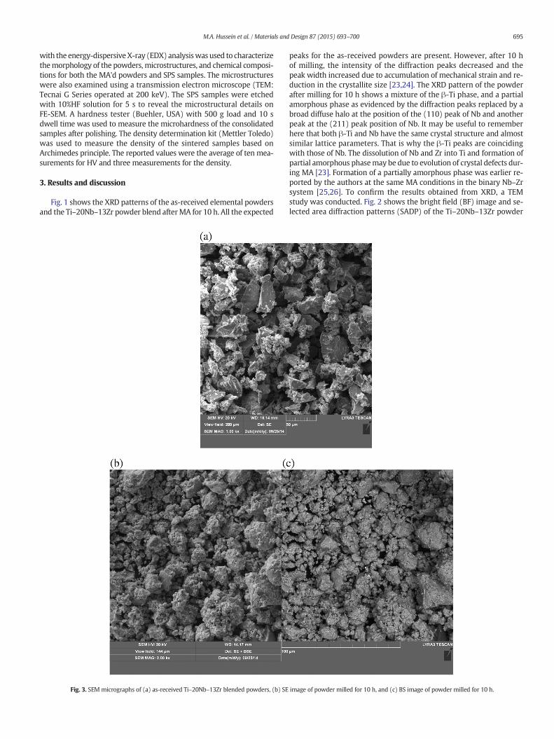
Fig. 3. SEM micrographs of (a) as-received Ti–20Nb–13Zr blended powders, (b) SE
peaks for the as-received powders are present. However, after 10 hof milling, the intensity of the diffraction peaks decreased and thepeak width increased due to accumulation of mechanical strain and re-duction in the crystallite size [23,24]. The XRD pattern of the powderafter milling for 10 h shows a mixture of the β-Ti phase, and a partialamorphous phase as evidenced by the diffraction peaks replaced by abroad diffuse halo at the position of the (110) peak of Nb and anotherpeak at the (211) peak position of Nb. It may be useful to rememberhere that both β-Ti and Nb have the same crystal structure and almostsimilar lattice parameters. That is why the β-Ti peaks are coincidingwith those of Nb. The dissolution of Nb and Zr into Ti and formation ofpartial amorphous phasemay be due to evolution of crystal defects dur-ing MA [23]. Formation of a partially amorphous phase was earlier re-ported by the authors at the same MA conditions in the binary Nb–Zrsystem [25,26]. To confirm the results obtained from XRD, a TEMstudy was conducted. Fig. 2 shows the bright field (BF) image and se-lected area diffraction patterns (SADP) of the Ti–20Nb–13Zr powder
image of powder milled for 10 h, and (c) BS image of powder milled for 10 h.
696 M.A. Hussein et al. / Materials and Design 87 (2015) 693–700
milled for 10 h. Some regions in the powdermilled for 10 h clearly showthe presence of an amorphous phase. This is confirmed from the high-resolution TEM micrograph that shows the salt-and-pepper like con-trast (Fig. 2a) and the presence of a broad diffuse halo in the diffractionpattern (Fig. 2b). The existence of a crystalline phase is also confirmedby the presence of sharp diffraction rings (Fig. 2c). This clearly suggeststhe co-existence of both amorphous and crystalline phases in the Ti–20Nb–13Zr powders milled for 10 h, confirming the XRD observations.
Fig. 3 presents the SEM micrographs of the Ti–20Nb–13Zr powderblend showing the morphology before and after MA. It can be seenthat the raw initial powder blend before MA has different irregularand elongated shapes with an average particle size of 23 μm for Ti andNb and 31 μm for Zr. The secondary electron (SE) and back scattered(BS) images showing the morphology of the Ti–20Nb–13Zr powderafter MA for 10 h are shown in Fig. 3(b) and (c), respectively. By com-parison, it is clear that the average particle size in the MA condition isreduced and that the shape of the powder particles is spherical andmore uniform compared to the irregular shape that existed in the as-received powders. Some large particles have also appeared due toagglomeration of the smaller particles and excessive cold welding.
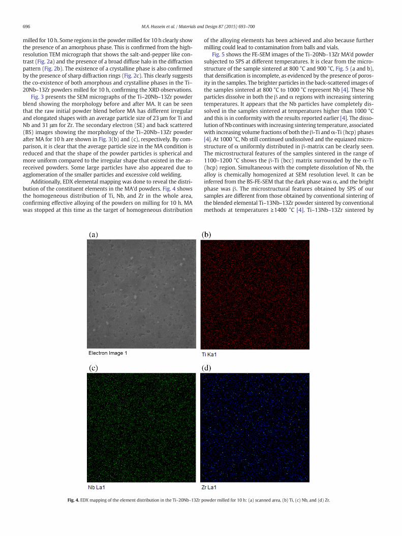
Additionally, EDX elemental mapping was done to reveal the distri-bution of the constituent elements in the MA'd powders. Fig. 4 showsthe homogeneous distribution of Ti, Nb, and Zr in the whole area,confirming effective alloying of the powders on milling for 10 h. MAwas stopped at this time as the target of homogeneous distribution
Fig. 4. EDX mapping of the element distribution in the Ti–20Nb–13Zr p
of the alloying elements has been achieved and also because furthermilling could lead to contamination from balls and vials.
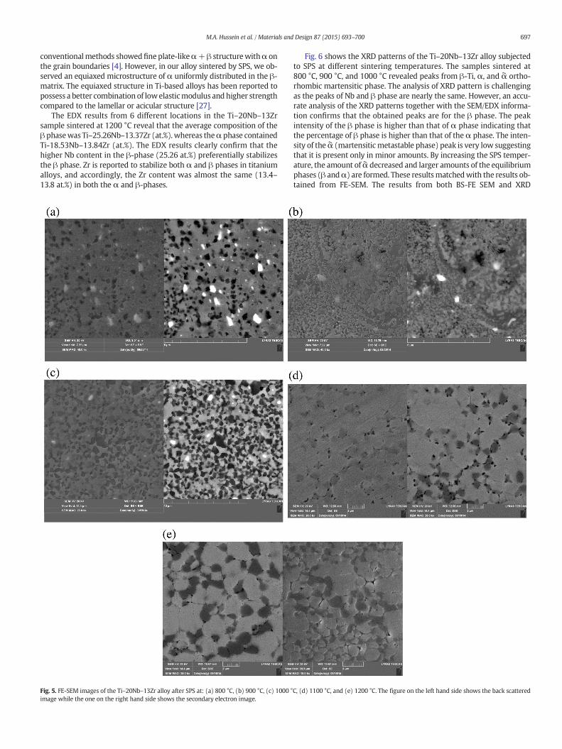
Fig. 5 shows the FE-SEM images of the Ti–20Nb–13Zr MA'd powdersubjected to SPS at different temperatures. It is clear from the micro-structure of the sample sintered at 800 °C and 900 °C, Fig. 5 (a and b),that densification is incomplete, as evidenced by the presence of poros-ity in the samples. The brighter particles in the back-scattered images ofthe samples sintered at 800 °C to 1000 °C represent Nb [4]. These Nbparticles dissolve in both the β and α regions with increasing sinteringtemperatures. It appears that the Nb particles have completely dis-solved in the samples sintered at temperatures higher than 1000 °Cand this is in conformity with the results reported earlier [4]. The disso-lution of Nb continueswith increasing sintering temperature, associatedwith increasing volume fractions of both the β-Ti andα-Ti (hcp) phases[4]. At 1000 °C, Nb still continued undissolved and the equiaxed micro-structure of α uniformly distributed in β-matrix can be clearly seen.The microstructural features of the samples sintered in the range of1100–1200 °C shows the β-Ti (bcc) matrix surrounded by the α-Ti(hcp) region. Simultaneous with the complete dissolution of Nb, thealloy is chemically homogenized at SEM resolution level. It can beinferred from the BS-FE-SEM that the dark phase was α, and the brightphase was β. The microstructural features obtained by SPS of oursamples are different from those obtained by conventional sintering ofthe blended elemental Ti–13Nb–13Zr powder sintered by conventionalmethods at temperatures ≥1400 °C [4]. Ti–13Nb–13Zr sintered by
owder milled for 10 h: (a) scanned area, (b) Ti, (c) Nb, and (d) Zr.
697M.A. Hussein et al. / Materials and Design 87 (2015) 693–700
conventionalmethods showedfineplate-likeα+β structurewithα onthe grain boundaries [4]. However, in our alloy sintered by SPS, we ob-served an equiaxed microstructure of α uniformly distributed in the β-matrix. The equiaxed structure in Ti-based alloys has been reported topossess a better combination of lowelasticmodulus andhigher strengthcompared to the lamellar or acicular structure [27].
The EDX results from 6 different locations in the Ti–20Nb–13Zrsample sintered at 1200 °C reveal that the average composition of theβ phasewas Ti–25.26Nb–13.37Zr (at.%). whereas theα phase containedTi-18.53Nb–13.84Zr (at.%). The EDX results clearly confirm that thehigher Nb content in the β-phase (25.26 at.%) preferentially stabilizesthe β phase. Zr is reported to stabilize both α and β phases in titaniumalloys, and accordingly, the Zr content was almost the same (13.4–13.8 at.%) in both the α and β-phases.
Fig. 5. FE-SEM images of the Ti–20Nb–13Zr alloy after SPS at: (a) 800 °C, (b) 900 °C, (c) 1000 °image while the one on the right hand side shows the secondary electron image.
Fig. 6 shows the XRD patterns of the Ti–20Nb–13Zr alloy subjectedto SPS at different sintering temperatures. The samples sintered at800 °C, 900 °C, and 1000 °C revealed peaks from β-Ti, α, and ἃ ortho-rhombic martensitic phase. The analysis of XRD pattern is challengingas the peaks of Nb and β phase are nearly the same. However, an accu-rate analysis of the XRD patterns together with the SEM/EDX informa-tion confirms that the obtained peaks are for the β phase. The peakintensity of the β phase is higher than that of α phase indicating thatthe percentage of β phase is higher than that of theα phase. The inten-sity of theἃ (martensitic metastable phase) peak is very low suggestingthat it is present only in minor amounts. By increasing the SPS temper-ature, the amount ofἃ decreased and larger amounts of the equilibriumphases (β andα) are formed. These resultsmatchedwith the results ob-tained from FE-SEM. The results from both BS-FE SEM and XRD
C, (d) 1100 °C, and (e) 1200 °C. The figure on the left hand side shows the back scattered

Fig. 6. XRD patterns of the Ti–20Nb–13Zr alloy subjected to SPS at different sinteringtemperatures. Note that the amount of the β-phase increased with increasing SPStemperature.
698 M.A. Hussein et al. / Materials and Design 87 (2015) 693–700
suggested that dissolution of Nb continued till 1000 °C, as indicated bythe existence of Nb peaks in addition to those of β, α, and ἃ phasesin the samples sintered at 800 °C, 900 °C, and 1000 °C. The increasingintensity of the β-phase peaks with increasing sintering temperatureclearly confirmed that the amount of the β phase increased due todissolution of Nb. Above a sintering temperature of 1000 °C, all the Nbhas probably completely dissolved as inferred from the microstructuralanalysis.
To study the effect of SPS temperature on the densification behaviorand Vickers hardness of Ti–20Nb–13Zr alloy, the samples were sinteredat heating rate of 100°K/min and 10 min holding time while thesintering temperature was varied. It is clear from Fig. 7(a) that the rela-tive density increased with increasing sintering temperature due to adecrease in the porosity that existed in the specimen. The rate of pore
Fig. 7. (a) densification, and (b)Vickers hardness of Ti–20Nb–13Zr alloy after SPS at differenttemperatures.
elimination increased by increasing the SPS temperature which leadsto an increase in the relative density of the sample. The samples showa relative density of 96.7% when sintered at 800 °C, which increasedslowly with increasing sintering temperature till 99.5% at 1200 °C. Thesame trend of results was also reported [28,29]. The Vickers micro-hardness of the samples also increased with increasing sintering tem-perature (Fig. 7(b)); this may be due to a decrease in the number ofpores in the resultant sample during heating and the improved contactbetween the powder particles, even though the trend was slightlydifferent from that of densification [30,31]. The HV increased from619 at 800 °C to 670 at 900 °C. As can be seen fromFig. 7(b), thehardnessof the sample sintered at 1000 °C showed a decrease and then the valuesstarted to increase slowly again to 658 at 1200 °C. This latter increase inhardness from 1000 °C to 1200 °C could be attributed to the increasedhomogeneity in themicrostructure. The sample sintered at 900 °C exhib-ited the highest hardness, even though there is substantial variationin the value with a standard error of 24.4 among all the samples.(This deviation may be related to the different features of the micro-structure obtained at 900 °C).
Taddei et al. [32] synthesized the Ti–35Nb–7Zr–5Ta alloy by mixingthe metallic powders and cold isostatic pressing and subsequentlydensifying by conventional sintering. Full densification was achievedat a high sintering temperature of 1700 °C [32]. Also, in the Ti–13Nb–13Zr alloy developed by conventional sintering, the relative densityvaried between 93 and 97% after sintering at 1400 °C [4]. Compared tothese results, it is clear that we were able to obtain by SPS, near fullydensity in the Ti–20Nb–13Zr alloy, at a much lower temperature. Thereason for obtaining a high density in SPS at a relatively lower temper-ature was due to the ionization of the particles by local sparking duringSPS, which causes melting of the titanium oxide films and formation ofneck junctions between powder particles at lower temperatures [33].Additionally, the plasma generated during the SPS process enhancesthe properties of the sintered powders due to acceleration of interdiffu-sion between atoms [34].
The hardness and the types of phases present in the nanograinedTi–20Nb–13Zr alloy investigated in this study aswell as those other alloysdeveloped for biomedical applications are summarized in Table 1. Thelisted compositions are in weight percentage unless mentioned other-wise. The hardness values for the samples sintered by SPS are higherthan those obtained by conventional sintering methods. These resultsare in agreementswith [35,36]. According to [35] among three consolida-tion techniques used (SPS, Microwave Sintering (μWS), and Hot IsotacticPress Sintering (HIS), SPS consolidated samples showed the highest HVvalues. The same conclusion was also drawn by [36], where the HV ofSPS of Al6061 and Al2124 alloys samples was also higher than samplessintered by μWS technique.
The increase in hardness of the nanocrystalline materials may berelated to the effect of higher dislocation density of smaller grain size[37,38]. According to [39] a high dislocation density was observed inthe nanograins sintered by SPS at 950 °C.
Table 1Comparison of HV and phases in titanium alloys studied.
Alloy composition Consolidationtechnique
Phases HV Reference
Ti–13Nb–13Z Conventionalsintering
α′ 290 [3]Ti–13Nb α′ 340Ti–6Al–4 V 500Ti–13Nb–13Zr Conventional
sinteringα + β 300 [4]
Ti–43Al–9V (molar ratio) SPS γ-TiAl + α2Ti3Al + B2
592 [16]
Ti–35Nb–7Zr–5Ta SPS α + β 531–668 [45]NanocrystallineTi–20Nb–13Zr (at.%)alloy developed inthis study
SPS α + β 660
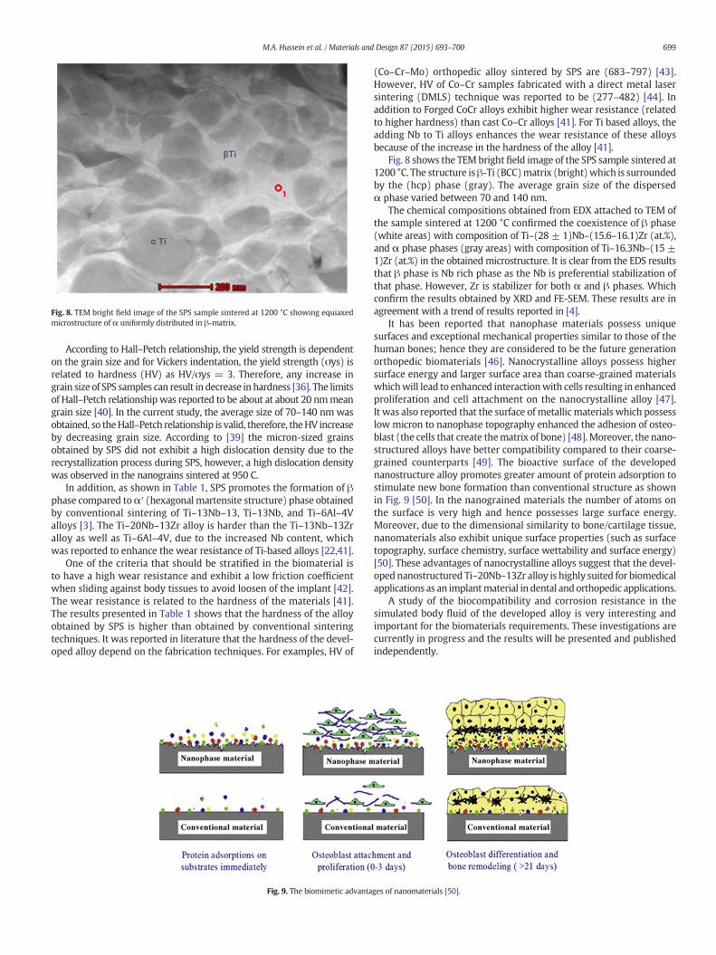
Fig. 8. TEM bright field image of the SPS sample sintered at 1200 °C showing equiaxedmicrostructure of α uniformly distributed in β-matrix.
699M.A. Hussein et al. / Materials and Design 87 (2015) 693–700
According to Hall–Petch relationship, the yield strength is dependenton the grain size and for Vickers indentation, the yield strength (σys) isrelated to hardness (HV) as HV/σys = 3. Therefore, any increase ingrain size of SPS samples can result in decrease in hardness [36]. The limitsof Hall–Petch relationshipwas reported to be about at about 20 nmmeangrain size [40]. In the current study, the average size of 70–140 nm wasobtained, so theHall–Petch relationship is valid, therefore, theHV increaseby decreasing grain size. According to [39] the micron-sized grainsobtained by SPS did not exhibit a high dislocation density due to therecrystallization process during SPS, however, a high dislocation densitywas observed in the nanograins sintered at 950 C.
In addition, as shown in Table 1, SPS promotes the formation of βphase compared toα′ (hexagonal martensite structure) phase obtainedby conventional sintering of Ti–13Nb–13, Ti–13Nb, and Ti–6Al–4Valloys [3]. The Ti–20Nb–13Zr alloy is harder than the Ti–13Nb–13Zralloy as well as Ti–6Al–4V, due to the increased Nb content, whichwas reported to enhance the wear resistance of Ti-based alloys [22,41].
One of the criteria that should be stratified in the biomaterial isto have a high wear resistance and exhibit a low friction coefficientwhen sliding against body tissues to avoid loosen of the implant [42].The wear resistance is related to the hardness of the materials [41].The results presented in Table 1 shows that the hardness of the alloyobtained by SPS is higher than obtained by conventional sinteringtechniques. It was reported in literature that the hardness of the devel-oped alloy depend on the fabrication techniques. For examples, HV of
Fig. 9. The biomimetic advanta
(Co–Cr–Mo) orthopedic alloy sintered by SPS are (683–797) [43].However, HV of Co–Cr samples fabricated with a direct metal lasersintering (DMLS) technique was reported to be (277–482) [44]. Inaddition to Forged CoCr alloys exhibit higher wear resistance (relatedto higher hardness) than cast Co–Cr alloys [41]. For Ti based alloys, theadding Nb to Ti alloys enhances the wear resistance of these alloysbecause of the increase in the hardness of the alloy [41].
Fig. 8 shows the TEMbrightfield image of the SPS sample sintered at1200 °C. The structure isβ-Ti (BCC)matrix (bright)which is surroundedby the (hcp) phase (gray). The average grain size of the dispersedα phase varied between 70 and 140 nm.
The chemical compositions obtained from EDX attached to TEM ofthe sample sintered at 1200 °C confirmed the coexistence of β phase(white areas) with composition of Ti–(28 ± 1)Nb–(15.6–16.1)Zr (at.%),and α phase phases (gray areas) with composition of Ti–16.3Nb–(15 ±1)Zr (at.%) in the obtainedmicrostructure. It is clear from the EDS resultsthat β phase is Nb rich phase as the Nb is preferential stabilization ofthat phase. However, Zr is stabilizer for both α and β phases. Whichconfirm the results obtained by XRD and FE-SEM. These results are inagreement with a trend of results reported in [4].
It has been reported that nanophase materials possess uniquesurfaces and exceptional mechanical properties similar to those of thehuman bones; hence they are considered to be the future generationorthopedic biomaterials [46]. Nanocrystalline alloys possess highersurface energy and larger surface area than coarse-grained materialswhichwill lead to enhanced interactionwith cells resulting in enhancedproliferation and cell attachment on the nanocrystalline alloy [47].It was also reported that the surface of metallic materials which possesslow micron to nanophase topography enhanced the adhesion of osteo-blast (the cells that create thematrix of bone) [48]. Moreover, the nano-structured alloys have better compatibility compared to their coarse-grained counterparts [49]. The bioactive surface of the developednanostructure alloy promotes greater amount of protein adsorption tostimulate new bone formation than conventional structure as shownin Fig. 9 [50]. In the nanograined materials the number of atoms onthe surface is very high and hence possesses large surface energy.Moreover, due to the dimensional similarity to bone/cartilage tissue,nanomaterials also exhibit unique surface properties (such as surfacetopography, surface chemistry, surface wettability and surface energy)[50]. These advantages of nanocrystalline alloys suggest that the devel-opednanostructured Ti–20Nb–13Zr alloy is highly suited for biomedicalapplications as an implantmaterial in dental and orthopedic applications.
A study of the biocompatibility and corrosion resistance in thesimulated body fluid of the developed alloy is very interesting andimportant for the biomaterials requirements. These investigations arecurrently in progress and the results will be presented and publishedindependently.
ges of nanomaterials [50].

700 M.A. Hussein et al. / Materials and Design 87 (2015) 693–700
4. Conclusions
NanostructuredTi–20Nb–13Zr near-β alloywith enhancedmechanicalproperties was fabricated by mechanical alloying and subsequent sparkplasma sintering technique. The main conclusions drawn from the resultsobtained are as follows:
1. A newnanostructured Ti–20Nb–13Zr near-β alloywas developed forbiomedical applications. The newalloy contains non-toxic: Ti, Nb andZr to replace the existing Ti–Al–V which contains toxic elements.
2. Themicrostructure of the obtained alloy contains a two phase region,β-Ti (bcc) andα-Ti (hcp). The equiaxedβ-Ti (bcc)matrix is surroundedby α-Ti (hcp) regions, exhibiting an equiaxed structure and chemicalhomogeneity.
3. The developed nanostructured alloy exhibits a maximum micro-hardness HV of 660, a value higher than reported in the literature.
4. The developed nanostructured Ti–20Nb–13Zr alloy is suggested forbiomedical applications as in implant material in dental and ortho-pedic applications.
5. Solid state synthesis and SPS method is beneficial in prevention ofgrain growth and synthesis of nanostructured alloys for biomedicalapplications.
Acknowledgments
The authors gratefully acknowledge the financial support fromKing Fahd University of Petroleum&Minerals (KFUPM) through Projectno # IN131020.
References
[1] J. Park, R.S. Lakes, Biomaterials an introduction, 3ird ed. Springer, Berlin/Heidelberg,Germany, 2007.
[2] Ana Lu'cia Roselino Ribeiro, Rubens Caram Junior Fla 'via Farias Cardoso, RomeuBelon Fernandes Filho Luı 's Geraldo Vaz, Mechanical, physical, and chemicalcharacterization of Ti–35Nb–5Zr and Ti–35Nb–10Zr casting alloys, J Mater Sci:Mater Med. 20(2009) 1629–1636.
[3] K. Niespodziana, K. Jurczk, M. Jurczk, The synthesis of titanium alloys for biomedicalapplications, Rev. Adv. Mater. Sci. 18 (2008) 236–240.
[4] V.A.R. Henriques, E.T. Galvani, S.L.G. Petroni, M.S.M. Paula, T.G. Lemos, Production ofTi–13Nb–13Zr alloy for surgical implants by powder metallurgy, J. Mater. Sci. 45(2010) 5844–5850.
[5] A. Biesiekierski, J. Wang, M. Abdel-Hady Gepreel, C. Wen, A new look at biomedicalTi-based shape memory alloys, Acta Biomater. 8 (2012) 1661–1669.
[6] L.W. Ma, H.S. Cheng, C.Y. Chung, B. Yuan, Effect of heat treatment time on micro-structure and mechanical properties of Ti–19Nb–9Zr (at.%) shape memory alloy,Mater. Sci. Eng. A 561 (2013) 427–433.
[7] V. Brailovski, S. Prokoshkin, M. Gauthier, K. Inaekyan, S. Dubinskiy, M. Petrzhik, M.Filonov, Bulk and porous metastable beta Ti–Nb–Zr(Ta) alloys for biomedical appli-cations, Mater. Sci. Eng. C 31 (2011) 643–657.
[8] W. Sungtong, A. Khantachawana, Effect of Zr addition on mechanical propertiesof Ti–Nb–Zr alloys for biomedical applications, Adv. Mater. Res. 463–464 (2012)841–844.
[9] L. Wang, G. Yang, H. Yang, J. Cao,W. Lü, D. Zhang, Characterization of microstructureand mechanical properties of TiNbZr alloy during heat treatment, Rare Metal Mater.Eng. 38 (2009) 1136–1140.
[10] L.M. Elias, S.G. Schneider, S. Schneider, H.M. Silva, F. Malvisi, Microstructural andmechanical characterization of biomedical Ti–Nb–Zr(−Ta) alloys, Mater. Sci. Eng.A 432 (2006) 108–112.
[11] P. Majumdar, S.B. Singh, M. Chakraborty, The role of heat treatment on micro-structure and mechanical properties of Ti–13Zr–13Nb alloy for biomedical loadbearing applications, J. Mech. Behav. Biomed. Mater. 4 (2011) 1132–1144.
[12] A.E. Aguilar Maya, D.R. Grana, A. Hazarabedian, G.A. Kokubu, M.I. Luppo, G. Vigna,Zr–Ti–Nb porous alloys for biomedical application, Mater. Sci. Eng. C 32 (2012)321–329.
[13] C. Suryanarayana, N. Al-Aqeeli, Mechanically alloyed nanocomposites, Prog. Mater.Sci. 58 (2013) 383–502.
[14] M. Oghbaei, O. Mirzaee, Microwave versus conventional sintering: a review offundamentals, advantages and applications, J. Alloys Compd. 494 (2010) 175–189.
[15] G. Xie, F. Qin, S. Zhu, A. Inoue, Ni-free Ti-based bulk metallic glass with potential forbiomedical applications produced by spark plasma sintering, Intermetallics 29(2012) 99–103.
[16] L.-J. Xu, S.-L. Xiao, Y.-Y. Chen, J. Wang, Microstructure and mechanical properties ofTi–43Al–9V alloy fabricated by spark plasma sintering, Trans. Nonferrous MetalsSoc. 22 (2012) 768–772.
[17] C. Ileana, P. Oana, Bulk titanium for structural and biomedical applications obtainingby spark plasma sintering (SPS) from titanium hydride powder, J. Therm. Anal.Calorim. 113 (2013) 849–857.
[18] M. Stir, B. Nebe, E. Burkel, R. Nicula, F. Lu, Spark plasma sintering synthesis of porousnanocrystalline titanium alloys for biomedical applications, Biomol. Eng. 24 (2007)564–567.
[19] M. Wen, C. Wen, P. Hodgson, Y. Li, Fabrication of Ti–NbAg alloy via powder metal-lurgy for biomedical applications, Mater. Des. 56 (2014) 629–634.
[20] M.A. Hussein, C. Suryanarayana, A. Madhan Kumar, N. Al-Aqeeli, Effect of sinteringparameters on microstructure, mechanical properties and electrochemical behaviorof Nb–Zr alloy for biomedical applications, Mater. Des. 83 (2015) 344–351.
[21] F.Y. Zhou, B.L.Wang, K.J. Qiu,W.J. Lin, L. Li, Y.B.Wang, F.L. Nie, Y.F. Zheng,Microstructure,corrosion behavior and cytotoxicity of Zr–Nb alloys for biomedical application, Mater.Sci. Eng. C 32 (2012) 851–857.
[22] S.J. Li, R. Yang, S. Li, Y.L. Hao, Y.Y. Cui, M. Niinomi, Z.X. Guo, Wear characteristics ofTi–Nb–Ta–Zr and Ti–6Al–4V alloys for biomedical applications, Wear 257 (2004)869–876.
[23] A. Nouri, X. Chen, Y. Li, Y. Yamada, P.D. Hodgson, C.E. Wen, Synthesis of Ti–Sn–Nballoy by powder metallurgy, Mater. Sci. Eng. A 485 (2008) 562–570.
[24] C. Suryanarayana, Mechanical alloying and milling, Prog. Mater. Sci. 46 (2001)1–184.
[25] N. Al-Aqeeli, C. Suryanarayana, M.A. Hussein, Formation of an amorphous phase andits crystallization in the immiscible Nb–Zr system by mechanical alloying, J. Appl.Phys. 114 (2013) 153512–153514.
[26] N. Al-Aqeeli, M.A. Hussein, C. Suryanarayana, Phase evolution during high energyball milling of immiscible Nb–Zr alloys, Adv. Powder Technol. 26 (2015) 385–391.
[27] M. Niinomi, T. Akahori, Improvement of the fatigue life of titanium alloys for bio-medical devices through microstructural control, Expert Rev. Med. Devices 7(2010) 481–488.
[28] G.M. Le, A. Godfrey, N. Hansen, Structure and strength of aluminum withsub-micrometer/micrometer grain size prepared by spark plasma sintering, Mater.Des. 49 (2013) 360–367.
[29] Z.-F. Liu, Z.-H. Zhang, J.-F. Lu, A.V. Korznikov, E. Korznikova, F.-C. Wang, Effect ofsintering temperature on microstructures and mechanical properties of sparkplasma sintered nanocrystalline aluminum, Mater. Des. 64 (2014) 625–630.
[30] R.M. German, Powder metallurgy science, Metal Powder Industries Federation,Princeton, 1994.
[31] R.M. German, Sintering theory and practice, John Wiley, New York, 1996.[32] E.B. Taddei, V.A.R. Henriques, C.R.M. Silva, C.A.A. Cairo, Production of new titanium
alloy for orthopedic implants, Mater. Sci. Eng. C 24 (2004) 683–687.[33] F. Zhang, A.Weidmann, B.J. Nebe, E. Burkel, Preparation of TiMn alloy by mechanical
alloying and spark plasma sintering for biomedical applications, J. Phys. Conf. Ser.114 (2009) 1–5 (012007).
[34] M. Omori, Sintering, consolidation, reaction and crystal growth by the spark plasmasystem (SPS), Mater. Sci. Eng. 287 (2000) 183–188.
[35] N. Al-Aqeeli, Processing of CNTs reinforced Al-based nanocomposites using differentconsolidation techniques, J. Nanomater. (2013) 1–10.
[36] N. Saheb, Spark plasma and microwave sintering of Al6061 and Al2124 alloys, Int.J. Miner. Metall. Mater. 2 (2013) 152–159.
[37] A.P. Zhilyaev, T.G. Langdon, Using high-pressure torsion for metal processing:fundamentals and applications, Prog. Mater. Sci. 53 (2008) 893–979.
[38] C. Suryanarayana, Nanocrystalline materials, Int. Mater. Rev. 40 (1995) 41–64.[39] Somayeh Pasebani, Indrajit Charit, Darry P. Butt, James I. Cole, Yaqiao Wu, Jatuporn
Burns, Sintering behavior of Lanthana-bearing nanostructured ferritic steel consoli-dated via spark plasma sintering, Adv. Eng. Mater. (2015) 1–9.
[40] S. Yip, Nanocrystals: the strongest size, Nature 391 (1998) 532–533.[41] M.A. Hussein, A.S. Mohammed, N. Al-Aqeeli, Wear characteristics of metallic bioma-
terials: a review, Materials 8 (2015) 2749–2768.[42] J.J. Ramsden, The design and manufacture of biomedical surfaces, CIRP Ann. Manuf.
Technol. 56 (2007) 687–711.[43] Bhairav Patel, Fawad Inam, Mike Reece, Mohan Edirisinghe, William Bonfield, Jie
Huang, Arash Angadji, A novel route for processing cobalt–chromium–molybdenumorthopaedic alloys, J. R. Soc. Interface 7 (2010) 1641–1645.
[44] Simel Ayyıldız, Elif Hilal Soylu, Semra İde, Selim Kılıç, Cumhur Sipahi, Bulent Pişkin,Hasan Suat Gökçe, Annealing of Co–Cr dental alloy: effects on nanostructure andRockwell hardness, J. Adv. Prosthodont. 5 (2013) 471–478.
[45] L.M. Zou, C. Yang, Y. Long, Z.Y. Xiao, Y.Y. Li, Fabrication of biomedical Ti–35Nb–7Zr–5Taalloys by mechanical alloying and spark plasma sintering, Powder Metall. 55 (2012)65–71.
[46] M. Geetha, A.K. Singh, R. Asokamani, A.K. Gogia, Ti based biomaterials, the ultimatechoice for orthopaedic implants — a review, Prog. Mater. Sci. 54 (2009) 397–425.
[47] D. Khang, J. Lu, C. Yao, K.M. Haberstroh, T.J. Webster, The role of nanometer andsub-micron surface features on vascular and bone cell adhesion on titanium, Bio-materials 29 (2008) 970–983.
[48] B.C.Ward, T.J.Webster, Increased functions of osteoblasts onnanophasemetals,Mater.Sci. Eng. C 27 (2007) 575–578.
[49] T.J. Webster, J.U. Ejiofor, Increased osteoblast adhesion on nanophase metals: Ti,Ti6Al4V, and CoCrMo, Biomaterials 25 (2004) 4731–4739.
[50] L. Zhang, T.J. Webster, Nanotechnology and nanomaterials: promises for improvedtissue regeneration, Nano Today 4 (2009) 66–80.
Related Documents