Review Studies on anticancer activities of antimicrobial peptides David W. Hoskin a, ⁎ , Ayyalusamy Ramamoorthy b, ⁎ a Department of Pathology and Department of Microbiology and Immunology, Faculty of Medicine, Dalhousie University, Halifax, Nova Scotia, Canada B3H 1X5 b Department of Chemistry and Biophysics, 930 N. University Avenue, University of Michigan, Ann Arbor, MI 48109-1055, USA Received 5 July 2007; received in revised form 23 October 2007; accepted 5 November 2007 Available online 22 November 2007 Abstract In spite of great advances in cancer therapy, there is considerable current interest in developing anticancer agents with a new mode of action because of the development of resistance by cancer cells towards current anticancer drugs. A growing number of studies have shown that some of the cationic antimicrobial peptides (AMPs), which are toxic to bacteria but not to normal mammalian cells, exhibit a broad spectrum of cytotoxic activity against cancer cells. Such studies have considerably enhanced the significance of AMPs, both synthetic and from natural sources, which have been of importance both for an increased understanding of the immune system and for their potential as clinical antibiotics. The electrostatic attraction between the negatively charged components of bacterial and cancer cells and the positively charged AMPs is believed to play a major role in the strong binding and selective disruption of bacterial and cancer cell membranes, respectively. However, it is unclear why some host defense peptides are able to kill cancer cells when others do not. In addition, it is not clear whether the molecular mechanism(s) underlying the antibacterial and anticancer activities of AMPs are the same or different. In this article, we review various studies on different AMPs that exhibit cytotoxic activity against cancer cells. The suitability of cancer cell-targeting AMPs as cancer therapeutics is also discussed. © 2007 Elsevier B.V. All rights reserved. Keywords: Antimicrobial peptide; Anticancer peptide; Membrane; Lipid bilayer; Drug Contents 1. Introduction .............................................................. 358 2. Antimicrobial peptides ........................................................ 359 3. Membrane differences that contribute to the selectivity of antimicrobial peptides for cancer cells .................. 361 4. α-Helical anticancer peptides ..................................................... 363 4.1. BMAP-27 and BMAP-28 ................................................... 363 4.2. Cecropin A and B ....................................................... 363 4.3. LL-37/hCAP-18 ........................................................ 364 4.4. Magainins and other amphibian-derived anticancer peptides ................................. 364 4.5. Melittin ............................................................ 366 5. β-sheet anticancer peptides ...................................................... 366 5.1. Defensins ........................................................... 366 5.2. Lactoferricin .......................................................... 367 5.3. Tachyplesin I ......................................................... 367 6. Linear anticancer peptides ...................................................... 368 7. Hybrid and Synthetic Anticancer Peptides .............................................. 368 7.1. Cecropin A-based Hybrid Anticancer Peptides ........................................ 368 Available online at www.sciencedirect.com Biochimica et Biophysica Acta 1778 (2008) 357 – 375 www.elsevier.com/locate/bbamem ⁎ Corresponding authors. D.W. Hoskin, tel.: +1 902 494 6509. A. Ramamoorthy, tel.: +1 734 647 6572. E-mail addresses: [email protected] (D.W. Hoskin), [email protected] (A. Ramamoorthy). 0005-2736/$ - see front matter © 2007 Elsevier B.V. All rights reserved. doi:10.1016/j.bbamem.2007.11.008

Welcome message from author
This document is posted to help you gain knowledge. Please leave a comment to let me know what you think about it! Share it to your friends and learn new things together.
Transcript
Available online at www.sciencedirect.com
Biochimica et Biophysica Acta 1778 (2008) 357–375www.elsevier.com/locate/bbamem
Review
Studies on anticancer activities of antimicrobial peptides
David W. Hoskin a,⁎, Ayyalusamy Ramamoorthy b,⁎
a Department of Pathology and Department of Microbiology and Immunology, Faculty of Medicine, Dalhousie University, Halifax,Nova Scotia, Canada B3H 1X5
b Department of Chemistry and Biophysics, 930 N. University Avenue, University of Michigan, Ann Arbor, MI 48109-1055, USA
Received 5 July 2007; received in revised form 23 October 2007; accepted 5 November 2007Available online 22 November 2007
Abstract
In spite of great advances in cancer therapy, there is considerable current interest in developing anticancer agents with a new mode of actionbecause of the development of resistance by cancer cells towards current anticancer drugs. A growing number of studies have shown that some ofthe cationic antimicrobial peptides (AMPs), which are toxic to bacteria but not to normal mammalian cells, exhibit a broad spectrum of cytotoxicactivity against cancer cells. Such studies have considerably enhanced the significance of AMPs, both synthetic and from natural sources, whichhave been of importance both for an increased understanding of the immune system and for their potential as clinical antibiotics. The electrostaticattraction between the negatively charged components of bacterial and cancer cells and the positively charged AMPs is believed to play a majorrole in the strong binding and selective disruption of bacterial and cancer cell membranes, respectively. However, it is unclear why some hostdefense peptides are able to kill cancer cells when others do not. In addition, it is not clear whether the molecular mechanism(s) underlying theantibacterial and anticancer activities of AMPs are the same or different. In this article, we review various studies on different AMPs that exhibitcytotoxic activity against cancer cells. The suitability of cancer cell-targeting AMPs as cancer therapeutics is also discussed.© 2007 Elsevier B.V. All rights reserved.
Keywords: Antimicrobial peptide; Anticancer peptide; Membrane; Lipid bilayer; Drug
Contents
1. Introduction . . . . . . . . . . . . . . . . . . . . . . . . . . . . . . . . . . . . . . . . . . . . . . . . . . . . . . . . . . . . . . 3582. Antimicrobial peptides . . . . . . . . . . . . . . . . . . . . . . . . . . . . . . . . . . . . . . . . . . . . . . . . . . . . . . . . 3593. Membrane differences that contribute to the selectivity of antimicrobial peptides for cancer cells . . . . . . . . . . . . . . . . . . 3614. α-Helical anticancer peptides . . . . . . . . . . . . . . . . . . . . . . . . . . . . . . . . . . . . . . . . . . . . . . . . . . . . . 363
4.1. BMAP-27 and BMAP-28 . . . . . . . . . . . . . . . . . . . . . . . . . . . . . . . . . . . . . . . . . . . . . . . . . . . 3634.2. Cecropin A and B . . . . . . . . . . . . . . . . . . . . . . . . . . . . . . . . . . . . . . . . . . . . . . . . . . . . . . . 3634.3. LL-37/hCAP-18 . . . . . . . . . . . . . . . . . . . . . . . . . . . . . . . . . . . . . . . . . . . . . . . . . . . . . . . . 3644.4. Magainins and other amphibian-derived anticancer peptides . . . . . . . . . . . . . . . . . . . . . . . . . . . . . . . . . 3644.5. Melittin . . . . . . . . . . . . . . . . . . . . . . . . . . . . . . . . . . . . . . . . . . . . . . . . . . . . . . . . . . . . 366
5. β-sheet anticancer peptides . . . . . . . . . . . . . . . . . . . . . . . . . . . . . . . . . . . . . . . . . . . . . . . . . . . . . . 3665.1. Defensins . . . . . . . . . . . . . . . . . . . . . . . . . . . . . . . . . . . . . . . . . . . . . . . . . . . . . . . . . . . 3665.2. Lactoferricin . . . . . . . . . . . . . . . . . . . . . . . . . . . . . . . . . . . . . . . . . . . . . . . . . . . . . . . . . . 3675.3. Tachyplesin I . . . . . . . . . . . . . . . . . . . . . . . . . . . . . . . . . . . . . . . . . . . . . . . . . . . . . . . . . 367
6. Linear anticancer peptides . . . . . . . . . . . . . . . . . . . . . . . . . . . . . . . . . . . . . . . . . . . . . . . . . . . . . . 3687. Hybrid and Synthetic Anticancer Peptides . . . . . . . . . . . . . . . . . . . . . . . . . . . . . . . . . . . . . . . . . . . . . . 368
7.1. Cecropin A-based Hybrid Anticancer Peptides . . . . . . . . . . . . . . . . . . . . . . . . . . . . . . . . . . . . . . . . 368
⁎ Corresponding authors. D.W. Hoskin, tel.: +1 902 494 6509. A. Ramamoorthy, tel.: +1 734 647 6572.E-mail addresses: [email protected] (D.W. Hoskin), [email protected] (A. Ramamoorthy).
0005-2736/$ - see front matter © 2007 Elsevier B.V. All rights reserved.doi:10.1016/j.bbamem.2007.11.008
358 D.W. Hoskin, A. Ramamoorthy / Biochimica et Biophysica Acta 1778 (2008) 357–375
7.2. Diastereomeric and other synthetic anticancer peptides . . . . . . . . . . . . . . . . . . . . . . . . . . . . . . . . . . . . 3698. Summary and outlook . . . . . . . . . . . . . . . . . . . . . . . . . . . . . . . . . . . . . . . . . . . . . . . . . . . . . . . . . 370Acknowledgements . . . . . . . . . . . . . . . . . . . . . . . . . . . . . . . . . . . . . . . . . . . . . . . . . . . . . . . . . . . . . 370References . . . . . . . . . . . . . . . . . . . . . . . . . . . . . . . . . . . . . . . . . . . . . . . . . . . . . . . . . . . . . . . . . 370
1. Introduction
Despite recent advances in treatment modalities, cancerremains a major source of morbidity and mortality throughoutthe world. In the United States, cancer is the leading cause ofdeath for individuals less than 85 years of age [1]. Moreover, theincidence of many cancers, including cancers of the skin,prostate, breast, and kidney, continues to increase [2]. “Cancer”is, in fact, a general term that refers to over 100 distinct diseasesaffecting many different tissues and cell types. However,all forms of cancer are characterized by abnormal cellgrowth resulting from a relatively small number of inheritedor environmentally-induced genetic mutations [3]. Hanahan andWeinberg [4] have argued that in order for a cell to becomecancerous, it must acquire six unique traits as a result of alteredcell physiology. These defining traits of cancer cells are: (1) theability to generate their own growth signals or respond to weakgrowth signals that are ignored by healthy cells; (2) insensitivityto antiproliferative signals; (3) resistance to cellular suicidemechanisms that normally cause aberrant cells to die byapoptosis; (4) the capacity for limitless replication; (5) theability to stimulate new blood vessel development in order toallow for tumour growth; and (6) the capacity to invade tissues,at first locally, and later to spread or metastasize throughout the
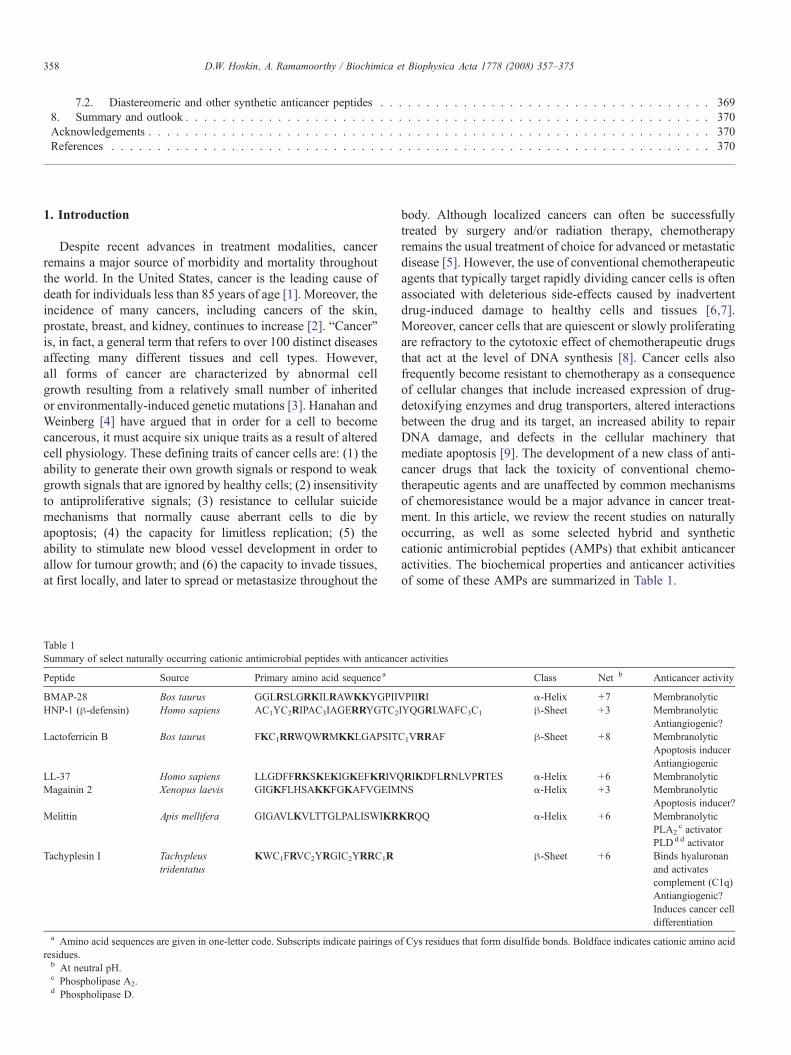
Table 1Summary of select naturally occurring cationic antimicrobial peptides with anticanc
Peptide Source Primary amino acid sequence a
BMAP-28 Bos taurus GGLRSLGRKILRAWKKYGPIIVHNP-1 (β-defensin) Homo sapiens AC1YC2RIPAC3IAGERRYGTC2
Lactoferricin B Bos taurus FKC1RRWQWRMKKLGAPSIT
LL-37 Homo sapiens LLGDFFRKSKEKIGKEFKRIVQMagainin 2 Xenopus laevis GIGKFLHSAKKFGKAFVGEIM
Melittin Apis mellifera GIGAVLKVLTTGLPALISWIKR
Tachyplesin I Tachypleustridentatus
KWC1FRVC2YRGIC2YRRC1R
a Amino acid sequences are given in one-letter code. Subscripts indicate pairings oresidues.b At neutral pH.c Phospholipase A2.d Phospholipase D.
body. Although localized cancers can often be successfullytreated by surgery and/or radiation therapy, chemotherapyremains the usual treatment of choice for advanced or metastaticdisease [5]. However, the use of conventional chemotherapeuticagents that typically target rapidly dividing cancer cells is oftenassociated with deleterious side-effects caused by inadvertentdrug-induced damage to healthy cells and tissues [6,7].Moreover, cancer cells that are quiescent or slowly proliferatingare refractory to the cytotoxic effect of chemotherapeutic drugsthat act at the level of DNA synthesis [8]. Cancer cells alsofrequently become resistant to chemotherapy as a consequenceof cellular changes that include increased expression of drug-detoxifying enzymes and drug transporters, altered interactionsbetween the drug and its target, an increased ability to repairDNA damage, and defects in the cellular machinery thatmediate apoptosis [9]. The development of a new class of anti-cancer drugs that lack the toxicity of conventional chemo-therapeutic agents and are unaffected by common mechanismsof chemoresistance would be a major advance in cancer treat-ment. In this article, we review the recent studies on naturallyoccurring, as well as some selected hybrid and syntheticcationic antimicrobial peptides (AMPs) that exhibit anticanceractivities. The biochemical properties and anticancer activitiesof some of these AMPs are summarized in Table 1.
er activities
Class Net b Anticancer activity
PIIRI α-Helix +7 MembranolyticIYQGRLWAFC3C1 β-Sheet +3 Membranolytic
Antiangiogenic?C1VRRAF β-Sheet +8 Membranolytic
Apoptosis inducerAntiangiogenic
RIKDFLRNLVPRTES α-Helix +6 MembranolyticNS α-Helix +3 Membranolytic
Apoptosis inducer?KRQQ α-Helix +6 Membranolytic
PLA2c activator
PLDd d activatorβ-Sheet +6 Binds hyaluronan
and activatescomplement (C1q)Antiangiogenic?Induces cancer celldifferentiation
f Cys residues that form disulfide bonds. Boldface indicates cationic amino acid

359D.W. Hoskin, A. Ramamoorthy / Biochimica et Biophysica Acta 1778 (2008) 357–375
2. Antimicrobial peptides
Naturally occurring AMPs probably represent one of the firstevolved and successful forms of chemical defense of eukaryoticcells against bacteria, protozoa, fungi, and viruses [10]. AMPshave been found in every species that has been tested, includingbacteria, fungi, plants, and animals. A very large number ofAMPs isolated from nature and thousands of synthetic variantshave been found to have a broad spectrum of antimicrobialactivities. For a regularly updated list of plant and animal AMPs,see the websites: http://www.bbcm.univ.trieste.it/~tossi/antimic.html and http://aps.unmc.edu/AP/main.php. Most AMPs killboth Gram-positive and Gram-negative bacteria, while a sig-nificant number of these bactericidal peptides have been shownto have anticancer and antiviral activities [10,11]. Some of thesepeptides have been found to have lipopolysaccharide (LPS)neutralizing ability and the capacity to recruit the adaptiveimmune response [12]. The growing problem of resistance toconventional antibiotics is a global public health problem andthe need for new antibiotics has stimulated interest in thedevelopment of AMPs as human therapeutics [13]. SeveralAMPs have already entered pre-clinical and clinical trials topromote wound healing and for the treatment of cystic fibrosis,catheter site infections, acne, and patients undergoing stem celltransplantation [14–16].
In addition to antibacterial activities of AMPs, recent studieson their anticancer activities are exciting and are most relevantfor this review [17]. While not all AMPs are able to kill cancercells, those that do can be placed into two broad categories: (a)AMPs that are highly potent against bacteria and cancer cellsbut not against normal mammalian cells and (b) AMPs that arecytotoxic for bacteria, cancer cells, and normal mammaliancells. The structural and functional properties of AMPs arebriefly outlined below.
Although the amino acid sequences of different AMPs arehighly heterogeneous and a great variation in their secondarystructures has been reported. AMPs are generally cationic (i.e.,the net charge at neutral pH varies from +2 to +9) andamphipathic, which enables the peptides to interact with anddisrupt lipid membranes. Most AMPs are very short in length,containing 5 to 40 amino acid residues, while a few of themcontain more than 40 residues. Positively charged residuessuch as Lys and Arg and substantial hydrophobic residues(∼30% or more) are commonly found in these peptides. Animportant set of AMPs, such as tritrpticin, lactoferricins andindolicidin, are rich in Trp and Arg residues [18]. Studies haveshown that all-D-amino-acid analogs have identical activitybut opposite chirality relative to natural all-L-peptides; thisproperty has been utilized in designing AMPs that can resistproteolytic degradation. Unlike currently available conven-tional antibiotics, which typically interact with a specific targetprotein, most of these cationic AMPs target the cell membraneof invading microorganisms, leading to cell lysis and death[10,18,19,20]. Thus, AMPs offer the possibility of a new classof therapeutics agents, which are complementary to existingantibiotics, and to which bacteria may not be able to developresistance.
Most of the linear AMPs are unstructured in solution (anexception is LL37, a human antimicrobial peptide [20,21]),while the cyclic peptides due to the presence of one or moreCys–Cys disulfide bonds form β-sheets [22]. The amphipathi-city of AMPs is enhanced upon the induction of specificsecondary structures, such as α-helices, β-sheets, or extendedpolyproline-like helices; this amphipathicity is thought to play akey role in their antimicrobial mechanism of action. Themechanism by which an AMP executes its function depends ona number of physicochemical properties: the amino acid se-quence, net charge, amphipathicity, hydrophobicity, structuralfolding (includes secondary structure, dynamics and orienta-tion) in membranes, oligomerization, peptide concentration, andmembrane composition [19]. There are several mechanisms ofmembrane-disruption proposed to explain the activity of AMPs(Fig. 1). Some of the models used to identify the membrane-disrupting process are carpet model [23], barrel-stave model[19], toroidal-pore wormhole model [24], and detergent-typemembrane lytic mechanism [25,26]. In the carpet model, AMPsassemble on the surface of the membrane to disrupt themembrane via barrel-stave, toroidal-pore, or detergent-typemechanisms. In the barrel-stave model [19,27–31], AMPsinsert with a transmembrane orientation in membranes andaggregate to form a traditional ion-channel pore. In the toroidal-pore model [24,32], the peptides are located closer to the headgroup region with an initial orientation parallel to the lipidbilayer surface. In this orientation, the hydrophilic side of thehelix is exposed to the hydrophilic lipid head groups and thewater phase outside bilayers, while the hydrophobic face of thehelix is buried in the hydrophobic core of the membrane tominimize the net free energy of folding process. Aggregation ofpeptides to a sufficient local concentration increases the curva-ture strain on the membrane surface to extent that toroidal-poresform. In the detergent-type mechanism [26], the peptides firstcarpet the surface of the lipid bilayer like the beginning stages ofthe toroidal-pore model. Following this step, the peptide aggre-gation leads to a sufficient high local concentration where theamphipathic nature of the peptide allows them to behave likedetergents and break the lipid membrane into small fragments.These fragments can be like bicelles or micelles [20]. There areother models such as the sinking raft model [33] or the mole-cular electroporation [34], which have not received muchattention in the field, but could be useful to explain the anti-microbial activity of certain AMPs. In the sinking raft model,AMPs bound to the cell membrane introduce a large membranecurvature due to a mass imbalance, which make AMPs sink andgenerate transient pores in membranes. On the other hand, in themolecular electroporation model, AMPs create a difference inthe electrical potential across the membrane leading to poreformation through electroporation.
Bacterial membranes are negatively charged with lipids suchas phosphatidylglycerol (PG), cardiolipin (CL), or phosphati-dylserine (PS). Electrostatic interaction between these nega-tively charged lipids and the positively charged AMPs enablethe cationic peptides to bind bacterial membranes. The outermembrane of a Gram-negative bacteria is also negativelycharged as it contains anionic lipopolysaccharides (LPS); LPS

Fig. 1. Models of AMP-induced membrane permeabilization. Cationic linear antimicrobial peptides are initially unstructured monomers (most AMPs) or helical andaggregated (for example, LL-37) in aqueous solution. They bind to negatively charged membrane surface by electrostatic interactions. In the carpet model, peptidesbind to phospholipid head groups and align themselves parallel to the membrane surface in a carpet-like fashion until a critical threshold concentration is reached. Inthe barrel stave model, peptides self aggregate in the membrane once a critical threshold concentration of peptide is reached, resulting in the formation of atransmembrane pore lined by peptide oriented such that the hydrophilic face forms the inner channel while the hydrophobic face is on the outside. Since an AMP has tospan the entire thickness of the lipid bilayer in this model, a minimum of ∼20 residues for α-helical peptide and ∼8 residues for a β-sheet peptide is required tofunction via the barrel stave mechanism. The toroidal pore model is an extension of the barrel stave model and postulates that at some critical concentration of peptidecurvature strain induces the membranes to curve inward, resulting in the formation of a pore that is lined by both peptide and lipid headgroups. Repulsive interactionsbetween the positively charged residues of the peptide are minimized due to the presence of the negatively charged phospholipids in the pore-lining. While theformation of these pores depend on the lipid:peptide ration and the ionic selectivity depend on the membrane composition, the lifetime of these pores seem to vary. Thedetergent-like model proposes that peptides intercalate in between the phospholipid head groups in a cone-like fashion causing curvature strain and micellization atlocal regions of high peptide density or when preformed peptide aggregates interact with lipid membranes. Recent NMR studies suggested that after sufficient time(may be a month or longer) AMPs fragment the lipid bilayers (even those containing toroidal pores) to form bicelle or micelle-like structures. Other membrane-disruptive and non-membrane disruptive mechanisms of some AMPs are discussed in the text.
360 D.W. Hoskin, A. Ramamoorthy / Biochimica et Biophysica Acta 1778 (2008) 357–375
is normally stabilized by the divalent cations like Ca2+ andMg2+
but AMPs displaces them to interact with the outer membrane.On the other hand, mammalian cell membranes consist largelyof zwitterionic phospholipids (neutral in net charge) such asphosphatidylethanolamine (PE), phosphatidylcholine (PC),or sphingomyelin (SM) and are therefore less attractive tocationic AMPs. In addition, cholesterol present in mammalianmembranes makes it harder for AMPs to disrupt lipid bi-layer structures. Therefore, the AMPs are selectively toxic tobacteria.
Solid-state NMR approaches have been developed andsuccessfully utilized to determine the secondary structure andlocation of the peptide in lipid bilayers, and also to determinethe structure of the lipid bilayers. For example, solid-state NMRstudies have determined the structure and bilayer-surfaceorientation of several linear AMPs including MSI-78 [32,35–37], MSI-594 [35], magainin2 [38], MSI-843 [39], PGLa[40–43], subtilosin A [44], KIAGAKI [45], fragments ofgranulysin [46], and other peptides [47–50]. These studiessupport the carpet mechanism and rule out the barrel-stavemechanism as the mode of action for these AMPs. Thus, thesepeptides have been shown to induce positive curvature strainthat leads to toroidal-type membrane disruption [24,32], whichis consistent with other biophysical studies on magainin pep-tides. These peptides also induce significant disorder in thehydrophobic core of lipid bilayers. On the other hand, peptides
like pardaxin [27] and alamethicin [29–31] prefer a transmem-brane orientation and disrupt membranes via the barrel-stavemechanism. Interestingly, pardaxin changes its orientationdepending on the membrane composition, which is consistentwith its multiple biological activities. A recent solid-state NMRstudy has reported a change in the orientation of PGLa when theconcentration of the peptide was increased [41–43]. Similarstudies have also been reported for non-linear AMPs. Solid-state NMR studies on non-helical peptides like protegrin haverevealed their mechanism of action as well [51].
While the above-mentioned mechanisms explain the bacter-icidal activity of AMPs due to the disruption of the membraneintegrity that lead to leakage of ions and metabolites anddepolarization, there are AMPs that act on putative key intra-cellular targets in bacteria without disrupting the cell membrane[Hwang and Vogel, 1998]. These AMPs inhibit the synthesis ofprotein or cell-wall, inhibit the activities of certain enzymes,interfere with the metabolic processes of microbes, or interactwith DNA or RNA.
Certain AMPs have been shown to have antiviral activities:they inhibit the replication of enveloped viruses such asinfluenza A virus [52], vesicular stomatitis virus (VSV) andhuman immunodeficiency virus (HIV-1) [53,54]. The generallyaccepted antiviral mechanism of action is a direct interaction ofthese AMPs with the envelope of the virus, leading to per-meation of the envelope and, eventually, lysis of the virus
361D.W. Hoskin, A. Ramamoorthy / Biochimica et Biophysica Acta 1778 (2008) 357–375
particle, analogous to the pore-formation mechanism mentionedabove for antibacterial activity of AMPs. However, othermechanisms for antiviral activities have also been proposed:T22 ([(Tyr5,12, Lys7)-polyphemusin II]) a 18 amino acidspeptide analogue of polyphemusin II, an AMP isolated from thehemocyte debris of American horseshoe crabs (Limuluspolyphemus), was found to specifically inhibit the ability of Tcell line-tropic HIV-1 to induce cell fusion [55]. On the otherhand, lactoferricin, which is an N-terminal fragment oflactoferrin, inhibits the binding and uptake of human papillomavirus, human cytomegalovirus, and herpes simplex virus intohuman cells [56–58].
3. Membrane differences that contribute to the selectivity ofantimicrobial peptides for cancer cells
Changes in the membrane of a cell have important impli-cations in the progression of cancer, as they play a key role inthe cell's response to its environment. The cell membrane of amalignant tumor cell may influence its ability to grow evenwithout the signals that would normally inspire growth, as wellas to attach and respond to neighboring cells differently. Thecell membrane may also affect a cancer cell's motility, aiding ininvasion and metastasis. Therefore, it is important to look at thedifferences between normal cells, non-malignant tumor cells,and malignant tumor cells, as well as how these differences ariseduring the progression from a normal cell to a tumor cell. Thesedifferences likely give insight into possible treatments andprevention. The cell membrane is a site of many suchdifferences, and will be further examined at the molecular level.
Fundamental differences exist between the cell membranesof malignant cells and normal cells that likely account for theability of certain AMPs to kill cancer cells (hereafter referred toas anticancer peptides; ACPs) while sparing healthy cells. Inthis regard, electrostatic interactions between cationic ACPs andanionic cell membrane components are believed to be a majorfactor in the selective killing of cancer cells by ACPs. Cancercell membranes typically carry a net negative charge due to ahigher than normal expression of anionic molecules such as PS(b9% of the total phospholipids of membranes) [59,60] and O-glycosylated mucins [61,62]. In addition, the negative mem-brane potential of cancer cells may also contribute to the selec-tive cytotoxic activity of ACPs [63]. In contrast to neoplasticcells, electrostatic interactions between ACPs and untrans-formed cells are not favored because of the overall neutralcharge conferred on healthy cells by the zwitterionic nature oftheir major membrane components, e.g., SM, PE, and PC [64].Therefore, it is believed that the ACPs could kill cancer cells viaone of the aforementioned mechanisms to disrupt the cellmembrane. Another possibility is the induction of apoptosis incancer cells via mitochondrial membrane disruption followingACP uptake into the cytoplasm. Other factors that contribute tothe preferential killing of cancer cells by ACPs includemembrane fluidity and cell-surface area. The membrane fluidityof cancer cells is greater than that of untransformed cells[65,66], which may enhance the lytic activity of ACPs byfacilitating membrane destabilization. Leuschner and Hansel
[67] have suggested that cholesterol, which is a major com-ponent of eukaryotic cell membranes [68], may protecteukaryotic cells from the cytolytic effect of antimicrobialpeptides by altering membrane fluidity and thereby interferingwith the membrane insertion of lytic peptides. In this regard, therate of membrane insertion by the AMP cecropin and itsanalogues is reduced when the cholesterol content of themembrane is increased [69,70]. Interestingly, some breast andprostate cancer cell lines have recently been shown to possesselevated levels of cholesterol-rich lipid rafts [71], which maymake these neoplastic cells less susceptible to killing by ACPs.Finally, cancer cells have a greater cell surface area than normalcells due to the presence of higher numbers of microvilli[72,73], which are minute projections of the cell membrane, thatmay allow cancer cells to bind increased numbers of ACPmolecules. The microvilli are also more irregular in size andshape, which may be due to changes in structural proteins[73,74]. These irregularities could potentially affect receptoraccessibility, cell adhesion, and other communications betweenthe cancer cell and its environment, and could also play a role inthe binding of ACPs selectively to the cancer cells. Therefore,differences in cell membrane composition, fluidity, and surfacearea between cells from different types of cancer may accountfor the variability in killing efficacy observed when a givenACP is tested against different types of cancer cells [67,75].
Glycosylation of membrane-associated glycoproteins andglycolipids undergo changes when a cell becomes cancerous,which could play a role in determining the susceptibility of agiven cancer cell to the cytotoxic activity of ACPs. Therefore, itwould be useful to understand these important changes at themolecular level. These changes in glycoproteins are most oftendue to activation of certain glycosyltransferases, which catalyzebiosynthesis of glycoproteins. Less often, the changes can beattributed to over-expression of glycosidases, which catalyzedegradation processes [76].
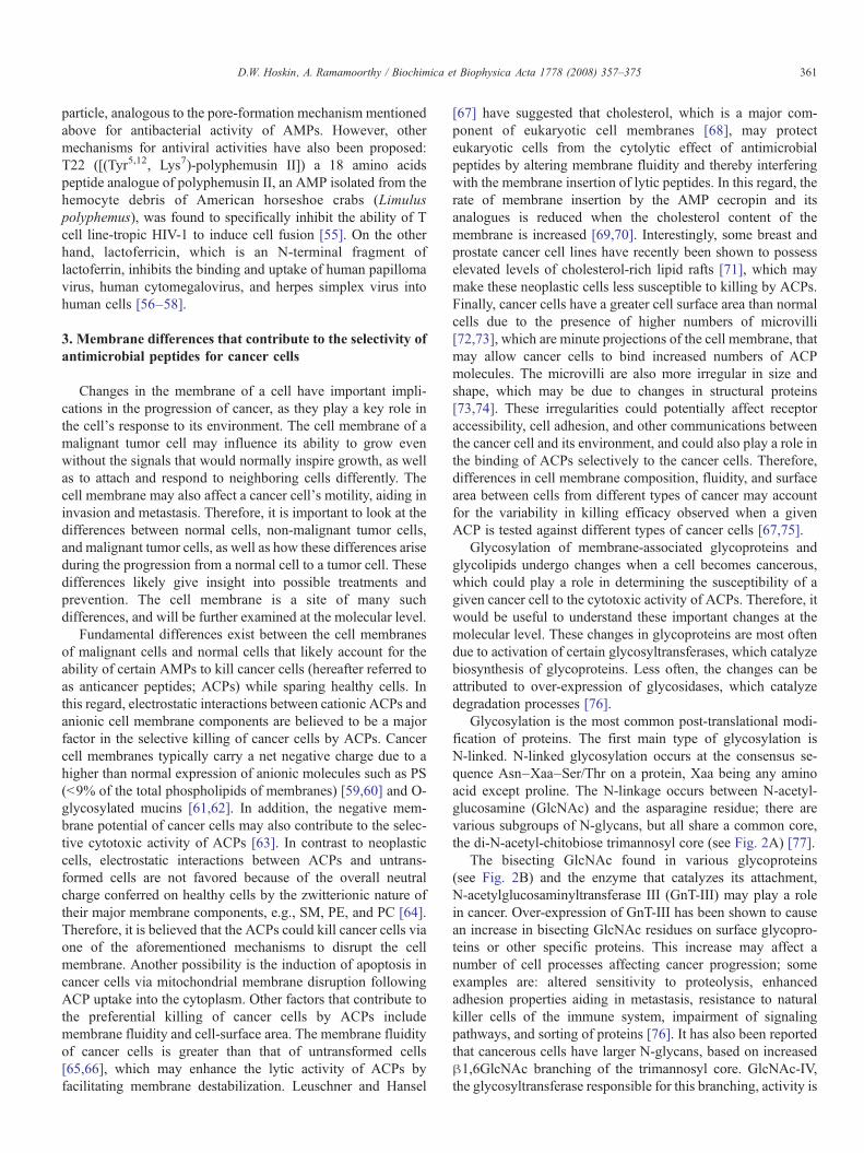
Glycosylation is the most common post-translational modi-fication of proteins. The first main type of glycosylation isN-linked. N-linked glycosylation occurs at the consensus se-quence Asn–Xaa–Ser/Thr on a protein, Xaa being any aminoacid except proline. The N-linkage occurs between N-acetyl-glucosamine (GlcNAc) and the asparagine residue; there arevarious subgroups of N-glycans, but all share a common core,the di-N-acetyl-chitobiose trimannosyl core (see Fig. 2A) [77].
The bisecting GlcNAc found in various glycoproteins(see Fig. 2B) and the enzyme that catalyzes its attachment,N-acetylglucosaminyltransferase III (GnT-III) may play a rolein cancer. Over-expression of GnT-III has been shown to causean increase in bisecting GlcNAc residues on surface glycopro-teins or other specific proteins. This increase may affect anumber of cell processes affecting cancer progression; someexamples are: altered sensitivity to proteolysis, enhancedadhesion properties aiding in metastasis, resistance to naturalkiller cells of the immune system, impairment of signalingpathways, and sorting of proteins [76]. It has also been reportedthat cancerous cells have larger N-glycans, based on increasedβ1,6GlcNAc branching of the trimannosyl core. GlcNAc-IV,the glycosyltransferase responsible for this branching, activity is
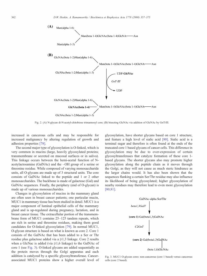
Fig. 3. MUC1 O-glycan cores: non-cancerous (core 1 based) versus cancerouscells (core 2 based).
Fig. 2. (A) N-glycan di-N-acetyl-chitobiose trimannosyl core; (B) bisecting GlcNAc via addition of GlcNAc by GnT-III.
362 D.W. Hoskin, A. Ramamoorthy / Biochimica et Biophysica Acta 1778 (2008) 357–375
increased in cancerous cells and may be responsible forincreased malignancy by altering regulation of growth andadhesion properties [78].
The second major type of glycosylation is O-linked, which isvery common in mucins (large, heavily glycosylated proteins;transmembrane or secreted on mucosal surfaces or in saliva).This linkage occurs between the hemi-acetal function of N-acetylactosamine (GalNAc) and the –OH group of a serine orthreonine residue. While composed of varying monosaccharideunits, all O-glycans are made up of 3 structural units. The coreconsists of GalNAc linked to the peptide and 1 or 2 othermonosaccharides. The backbone is made of galactose (Gal) andGalNAc sequences. Finally, the periphery (end of O-glycan) ismade up of various monosaccharides.
Changes in glycosylation of mucins in the mammary glandare often seen in breast cancer patients; one particular mucin,MUC1 in mammary tissue has been studied in detail. MUC1 is amajor component of luminal epithelial cells of the mammarygland and is up-regulated during pregnancy, lactation, and inbreast cancer tissue. The extracellular portion of the transmem-brane form of MUC1 contains 25–125 tandem repeats, whichare rich in serine and threonine residues, making them goodcandidates for O-linked glycosylation [79]. In normal MUC1,O-glycan structure is based on what is known as core 2. Core 1consists of the GalNAc that has been added to a Ser or Thrresidue plus galactose added via a β1,3 linkage. Core 2 resultswhen a GlcNac is added (via β1,6 linkage) to the GalNAc ofcore 1 (see Fig. 3). O-linked glycans are added sequentially asthe protein moves through the Golgi apparatus and eachaddition is catalyzed by a specific glycosyltransferase. Cancer-associated MUC1 proteins show a higher overall level of
glycosylation, have shorter glycans based on core 1 structure,and feature a high level of sialic acid [80]. Sialic acid is aterminal sugar and therefore is often found at the ends of thetruncated core 1 based glycans of cancer cells. This difference inglycosylation may be due to over-expression of certainglycosyltransferases that catalyze formation of these core 1-based glycans. The shorter glycans also may promote higherglycosylation along the peptide chain as it moves throughthe Golgi, as they will not cause as much steric hindrance asthe larger chains would. It has also been shown that thesequences flanking a certain Ser/Thr residue may also influenceits likelihood of being glycosylated; higher glycosylation ofnearby residues may therefore lead to even more glycosylation[80,81].

363D.W. Hoskin, A. Ramamoorthy / Biochimica et Biophysica Acta 1778 (2008) 357–375
While the exact molecular mechanism by which alteredglycosylation occurs as a cell becomes malignant is still unclear,such changes could play an important role in the selectivity ofACPs. It is known that glycosylation alters the secondarystructure and dynamics of a membrane-associated peptide orprotein. Moreover, differential branching and sialic acid contentof N-linked glycans associated with transmembrane glycopro-teins are believed to contribute to the net negative charge that iscarried by the cell membrane of many cancer cells [82].Interestingly, glycosylation has also been shown to increase thepotency of the AMP drosocin [83]. It is therefore likely that thebinding affinity of cationic AMPs (with and without glycosyla-tion) for cancer cells and the subsequent permeabilization ofmembranes depend at least in part on altered glycosylation ofcancer cell membrane proteins. Future investigations on theeffect of ACP glycosylation on cytotoxic activity might yieldmore effective anticancer compounds.
The general membrane-targeted method of cell lysis showspotential for synergy with current cancer treatments and ACPshave, in fact, shown additional killing of cancer cells whentested in concert with antineoplastic drug treatments. AMPswith cytotoxic activity against cancer cells also show potentialfor overcoming common problems with multiple-drug resistant(MDR) proteins since AMPs should not select for resistantcancer cells. MDR proteins cause problems for current treat-ments because they give a cancerous cell the ability to resisttreatment by simply pumping antineoplastic drugs out of thecell where they can do no harm. An AMP, however, will kill thecell simply by membrane disruption and avoid this resistancemechanism. Other beneficial traits of AMPs as anticanceragents include their wide range of activity, their ability to killcancer cells quickly, their ability to destroy primary tumors aswell as prevent metastasis, and the fact that they do not harmvital organs [84].
4. α-Helical anticancer peptides
4.1. BMAP-27 and BMAP-28
BMAP-27 (amino acid sequence: GRFKRFRKKFKKLFK-KLSPVIPLLHL) and BMAP-28 (GGLRSLGRKILRAWK-KYGPIIVPIIRI) are bovine cathelicidin-derived AMPs withsequences of 27 and 28 amino acid residues, respectively, thatwere originally deduced from their cDNAs [85]. The cationicNH2-terminal portion (residues 1 to 18) of these peptides ispredicted to form an amphipathic α-helix that is followed by ahydrophobic tail (residues 19 to 27 or 28) at the COOH-terminus. Interestingly, a truncated analogue of BMAP-28composed of only the 18 NH2-terminal amino acid residues, aswell as a modified form of full-length BMAP-28 in whichhighly hydrophobic amino acids in the COOH-terminal regionwere replaced with more hydrophilic amino acids, both showeda dramatic reduction in membrane-permeabilizing activity. Thisfinding indicated that the hydrophobic tail is important forBMAP peptides to mediate their cytotoxic effect. The cytotoxicactivity of BMAP-27 and -28 against neoplastic cells wasrevealed by the observation that freshly isolated human leuke-
mia cells and various human leukemia cell lines exposed to1.5–6 μM concentrations of the BMAP peptides exhibitedmembrane permeabilization and an influx of Ca++, followed byDNA fragmentation characteristic of apoptosis [86]. CEM-CCRF human T leukemia cells with a vinblastine-resistantphenotype were also susceptible to killing by BMAP-27 and-28. A subsequent study demonstrated that BMAP-28 treatmentof U937 and K562 human leukemia cell lines caused a rapidreduction in mitochondrial membrane potential in situ that wasrelated to a BMAP-28-induced opening of the mitochondrialpermeability transition pore, which resulted in cytochrome crelease and the initiation of cell death by apoptosis [87].Although non-proliferating lymphocytes are not affected by theBMAP peptides at concentrations that are cytotoxic for humanleukemia cells, at these concentrations both BMAP-27 and -28are cytotoxic for activated human lymphocytes [86]. Lowmicromolar concentrations of BMAP-27 and -28 are thereforeonly active against cells that are actively proliferating. How-ever, significant lysis of normal human neutrophils and erythro-cytes occurs following exposure to higher concentrations(30 μM and greater) of the BMAP peptides [85]. The relativelynarrow dosage range for BMAP-27- and BMAP-28-mediatedkilling of cancer cells without deleterious hemolytic activity,combined with the predicted inability of BMAP peptides toeffectively kill slowly proliferating or dormant neoplastic cells,place severe restraints on the development of BMAP-27 and -28as therapeutic anticancer agents.
4.2. Cecropin A and B
Cecropins are a class of antimicrobial peptides that were firstdescribed in insects, including the giant silk moth Hyalophoracecropia [88], but were later also found to be present inmammals [89]. Cecropins derived from insect sources consist of34–39 amino acid residues [90,91]. The best studied of theseantimicrobial peptides are cecropin A (KWKLFKKIEKV-GQNIRDGIIKAGPAVAVVGQATQIAK) and cecropin B(KWKVFKKIEKMGRNIRNGIVKAGPAIAVLGEAKAL),both of which assume a secondary structure that is characterizedby the presence of two α helices [92,93]. The NH2-terminal α-helix of cecropin A and cecropin B is highly amphipathic whilethe COOH-terminal α-helix is hydrophobic. Patch-clampanalysis of the effect of cecropin B on the Ags human stomachcarcinoma cell line revealed that peptide treatment caused shortoutward currents that were consistent with the formation oftransient channel-like pores [94]. In contrast, the cecropin B3analogue, which consists of two hydrophobic α helices, failedto induce pore formation. Interestingly, the cecropin B1analogue, which possesses two amphipathic α helices, showspotent cytotoxic activity against several human leukemia celllines at peptide concentrations that do not lyse normalfibroblasts or erythrocytes [95]. In fact, cecropin B1 is a morepotent cytolytic agent than cecropin B against HL-60 humanpromyelocytic leukemia cells [96]. Collectively, these findingsindicate that the amphipathic NH2-terminal α-helix of cecropinB, which is believed to interact with anionic membranecomponents via its basic amino acid residues, is also able to
364 D.W. Hoskin, A. Ramamoorthy / Biochimica et Biophysica Acta 1778 (2008) 357–375
mediate cytotoxic activity against cancer cells, whereas thehydrophobic COOH-terminal α-helix is dispensable in thisregard. Nevertheless, the hydrophobic COOH-terminal α-helixmay promote membrane insertion by the peptide, causingpositive curvature strain on the membrane that creates an ion-permeable toroidal pore [93].
Cecropin A and B are able to lyse different types of humancancer cells at peptide concentrations that are not harmful tonormal eukaryotic cells [91,97,98]. Cecropin B may havepotential for use in the treatment of human cancers since thisACP exhibits in vivo antitumor activity in mice bearing asciticcolon adenocarcinoma cells, as well as in vitro cytotoxic acti-vity against multidrug-resistant human breast and ovariancancer cell lines [97]. Interestingly, the combination of cecropinA and the conventional chemotherapeutic agents 5-fluorouraciland cytarabine, at certain doses, shows a synergistic cytotoxiceffect on CCRF-SB human lymphoblastic leukemia cells [98].Conventional antineoplastic drugs might therefore be used incombination with ACPs such as cecropin A in order to lower thedosage of the drug that is required to have a therapeutic effectand thereby reduce chemotherapy-induced side effects. How-ever, therapeutic use of cecropins or other ACPs will requirerepeated administration in order to maintain systemic levels ofthe peptide that are equivalent to the high concentrationsrequired for ACPs to kill human cancer cells in culture or inmice. One attractive alternative is to transfer genes encodingACPs directly into cancer cells or to the vicinity of solid tumors.In this regard, the introduction of expression constructscontaining the cecropin A gene into EJ human bladder carci-noma cells prevented or reduced the growth of the tumor cells inimmune-deficient mice [99]. Gene therapy using ACPs such ascecropin A may therefore 1 day be possible for the treatment ofhuman cancers.
4.3. LL-37/hCAP-18
LL-37/hCAP-18 (human cationic AMP of 18 kDa) is theonly human cathelicidin-derived AMP that has been identifiedto date [100]. This cathelicidin is initially synthesized as apreproprotein (hCAP-18) that is subsequently processed to itsactive form (LL-37) by proteinase 3-mediated extracellularcleavage [101]. hCAP-18 is expressed in a variety of cell types,including neutrophils [102] and squamous epithelial cells [103].LL-37 (LLGDFFRKSKEKIGKEFKRIVQRIKDFLRNLV-PRTES) is a 4.5 kDa AMP that has antimicrobial activity atlow micromolar concentrations against a variety of Gram-positive and Gram-negative bacteria. Unlike other AMPs,LL-37 is toxic to eukaryotic cells at a slightly higher con-centration (25–30 mM, which is 3–5 times its MIC value).LL-37 has been found to have additional defensive roles such asregulating the inflammatory response and chemo-attractingcells of the adaptive immune system to wound or infectionssites, stimulation of angiogenesis and chemotaxis of neutro-phils, monocytes and T-cells. This peptide is found throughoutthe body: in epithelial cells of testis, respiratory tract andgastrointestinal, in leukocytes and in skin. In addition, LL-37has been expressed in Escherichia coli and purified for
biophysical studies [104]. CD and NMR studies have shownthat LL-37 is unstructured in pure water but forms helicaloligomers in solution either at a higher peptide concentration orin the presence of ions [105]. This property of the peptide isbelieved to be important as the oligomer can escape theproteolytic degradation [106]. Solid-state NMR studies haveshown that LL-37 also assumes an α-helical conformation in amembrane environment and its oligomeric nature depends onthe membrane composition. These studies revealed that the α-helical structure is comprised of both cationic and hydrophobicfaces that orient in a parallel manner with lipid membranes,suggesting a carpet-like rather than channel-forming mechan-ism of cytotoxicity [105]. In addition, solid-state NMR andDSC experiments have shown that LL-37 is highly sensitive tothe membrane composition, upon binding to membraneschanges the head group conformation of phospholipids, inducespositive curvature strain on lipid bilayers, and significantlydisorders the hydrophobic core of the membrane [107]. Moredetails on the structural and functional properties of thisintriguing molecule can be found in a recent review article[20,21].
A COOH-terminal fragment of hCAP-18 consisting ofamino acid residues 109–135 (hCAP18109–135), which corre-sponds to amino acid residues 6–32 of LL-37, was recentlyfound to induce apoptosis in a human oral squamous carcinomacell line via a mechanism involving mitochondrial depolariza-tion without any detectable activation of caspase-3 [108]. Thisfinding is in line with a report that the core cytotoxic activityof LL-37 resides within a 13 amino acid residue fragment ofthe COOH-terminal region corresponding to amino acidresidues 17–29, designated LL-37(17–29) [109]. AlthoughLL-37(17–29) is equally cytotoxic to drug-sensitive and drug-resistant variants of the KB human squamous cancer cell line,LL-37(17–29) also kills untransformed human endothelialcells. Native LL-37 is similarly cytotoxic to human peripheralblood lymphocytes [110] and has significant hemolytic activity[106]. LL-37(17–29) and native LL-37 therefore lack theselectivity for cancer cells required of an ACP. In contrast, atumoricidal concentration of hCAP18109–135 did not have anycytotoxic effect on either human gingival fibroblasts or theHaCaT human keratinocyte cell line [108]. The apparent se-lective cytotoxic activity of hCAP18109–135 against humancancer cells indicates that hCAP18109–135 warrants furtherinvestigation as a potential therapeutic ACP.
4.4. Magainins and other amphibian-derived anticancerpeptides
The skin secretions of amphibians are a rich source of AMPs,several of which (aurein 1.2, citropin 1.1, gaegurins, magaininsand analog peptides of magainins) have been reported to beselectively cytotoxic for human cancer cells [111–116]. Thebest studied of these ACPs are the magainins, which are AMPsisolated from the skin of the African clawed frog Xenopuslaevis [117]. Magainins are comprised of 21 to 27 amino acidresidues that create an α-helical secondary structure character-ized by separate cationic and hydrophobic faces. Magainin 2

365D.W. Hoskin, A. Ramamoorthy / Biochimica et Biophysica Acta 1778 (2008) 357–375
(GIGKFLHSAKKFGKAFVGEIMNS) and its more potentsynthetic analogues (magainins A, B, and G) cause the rapidlysis of both hematopoietic and solid tumor cell lines at con-centrations that are 5–10 fold lower than magainin concentra-tions that are lytic for normal human peripheral bloodlymphocytes or neutrophils [115,118]. Magainin 2 also showsselective cytotoxic activity against several human bladder celllines with an average IC50 of approximately 200 μM [119].Fluorescence spectrophotometric measurement of cancer cellmembrane potential following exposure to magainin 2 or syn-thetic magainin analogues indicates that magainins lyse tumorcells by forming ion-conducting α-helical channels in thecancer cell membrane [115]. However, there is some evidencethat, at lower concentrations, magainins destabilize the mem-brane bilayer via the carpet model [120]. A more recent studyshows that a magainin 2 derivative is able to permeabilize andcross the cell membrane of HeLa human cervical carcinomacells via an energy- and receptor-independent mechanism [121].Magainins that gain access to the cytosolic compartment ofcancer cells may trigger the mitochondrial pathway of apoptosissince magainins have been shown to form channels in themembranes of isolated rat liver mitochondria [122]. Interest-ingly, 10–50 nM concentrations of magainin 1 have recentlybeen shown to induce apoptosis in HL-60 human promyelocyticleukemia cells via a mechanism that involves cytochrome crelease from mitochondria and an increase in proteosomeactivity [116]. At the present time it is not clear whethermagainins kill human cancer cells primarily through membranelysis and/or apoptosis. A large number of recent biophysicalstudies focused on the mechanism of membrane-disruption bymagainins (e.g., magainin 2 [38], PGLa [40–43], MSI-78(commercially known as pexiganan) [32,35,36], MSI-594 [35],MSI-843 [39] and other synthetic variants) on model mem-branes. These peptides are unstructured in solution, form a α-helical structure in membranes, bind to the membrane with thehelical axis parallel to the membrane surface and with a highaffinity for negatively charged membranes, form oligomers,induce positive curvature strain, and disrupt membranes viacarpet-toroidal-pore mechanism, which lead to fragmentation ofmembranes into bicelles and micelles after a sufficient time(N1 month). Some of these peptides like magainin2 [123],PGLa [43] and MSI-78 [35,36] form antiparallel α-helicaldimers in membranes.
A number of synthetic magainin analogues exhibit superiorcytotoxic activity against neoplastic cells in comparison tonative magainins [115,124–126]. Superior selectivity forhuman cancer cells is exhibited by magainin G whereasmagainin B is the most potent of the synthetic magaininanalogues in terms of cytotoxicity [115]. Magainins A and G,which were designed to have increased α-helical potential anddecreased hemolytic activity in comparison to native magainin1 and 2, inhibit the growth of human small cell lung cancer celllines, including drug-resistant tumor cell variants, with anaverage IC50 of approximately 9 μM [124]. In contrast, twicethe concentration of magainin A or G is needed to inhibit thegrowth of normal human fibroblast cell lines. Magainin A and Galso enhance the effectiveness of the chemotherapeutic agents
DDP and VP-16, suggesting that these synthetic magaininanalogues might be used in combination with conventionalanticancer drugs in order to reduce chemotherapy-induced sideeffects. Two additional synthetic magainin analogues (MSI-136, comprised of L-amino acids and MSI-238, comprised of D-amino acids) designed to have an enhanced amphipathic α-helical structure, were found to have superior in vitro cytotoxicactivity against human lung carcinoma cells in comparison tonative magainin 2 and to increase survival of ovarian teratoma-bearing mice [125]. All-D-amino acid MSI-238 was moreeffective in vivo than all L-amino acid MSI-136 or magainin 2,most likely because of decreased susceptibility to proteolyticdegradation. An additional synthetic all-D-amino acid magaininanalogue designated MSI-511 showed selective killing humanmelanoma cells in vitro at concentrations that do not harmnormal melanocytes [126]. Moreover, intratumoral injection ofMSI-511 completely eradicated human melanoma cells grownas subcutaneous xenografts in immune-deficient mice, provid-ing evidence that locoregional therapy with protease-resistantmagainin peptides may be useful for the treatment of malignantmelanoma in humans.
Gaegurins are a class of six related AMPs that have beenisolated from the skin of a Korean frog Rana rugosa [127]. Thegaegurins assume a random-coil conformation in aqueoussolution but adopt an amphipathic α-helical structure in mem-brane environments, allowing these AMPs to mediate cytolysisby the barrel stave and/or carpet mechanism [128]. More recentstudies have revealed that gaegurin 5 (FLGALFKVASKVLPS-VKCAITKKC) and gaegurin 6 (FLPLLAGLAANFLPTIICFI-SYKC) have selective cytotoxic activity against neoplastic cells[113,114]. Gaegurin 5 and 6 are each comprised of 24 aminoacid residues. Gaegurin 5 and two synthetic peptide analoguesare able to selectively kill a range of human tumor cell types,including HCT116 colon and MCF-7 breast carcinoma cells,while showing only minimal hemolytic activity [113]. Gaegurin6 and a synthetic peptide analogue (PTP7) have a similar broadspectrum of cytotoxic activity against human cancer cells withno detectable cytotoxicity against peripheral blood mono-nuclear cells and minimal hemolytic activity [114]. In addition,gaegurin 6 and PTP7 are active against a multidrug-resistantvariant of the MCF-7 breast cancer cell line. Gaegurin 6- andPTP7-mediated cytotoxicity may involve apoptosis since DNAfragmentation was detected in MCF-7 breast cancer cells thatwere exposed to these ACPs.
Aurein 1.2 (GLFDIIKKIAESF) is a small cationic AMPcomprised of 13 amino acid residues that has been isolated fromthe Australian bell frog Litoria raniformis [111]. NMR studiesindicate that aurein 1.2 adopts an α-helical structure in solution.Aurein 1.2 is moderately cytotoxic for 57 of 60 human tumorcell lines but does not lyse erythrocytes at concentrations ashigh as 100 μg/ml, which is sufficient to kill most human cancercells. Citropin 1.1 (GLFDVIKKVASVIGGL) is a relativelysmall AMP isolated from the tree frog Litoria citropa. ThisAMP is able to kill a wide range of human hematopoietic andnon-hematopoietic tumor cell lines at concentrations that do notcause significant lysis of red blood cells [112]. Citropin 1.1,which is comprised of 16 amino acid residues, has an α-helical

366 D.W. Hoskin, A. Ramamoorthy / Biochimica et Biophysica Acta 1778 (2008) 357–375
structure characterized by well-defined hydrophobic and hydro-philic regions. The relatively small size of aurein 1.2 andcitropin 1.1 implies that these ACPs mediate their cytotoxiceffect via the carpet mechanism since ACPs need to becomprised of at least 20 amino acid residues in order to spaneukaryotic cell membranes and cause cytolysis by the barrel-stave mechanism [129,130]. However, it is important to notethat peptides comprised of fewer than 20 amino acid residuesmay dimerize end-on in order to effect complete penetration ofbiological membranes [131].
4.5. Melittin
Melittin (GIGAVLKVLTTGLPALISWIKRKRQQ), an alka-line polypeptide comprised of 26 amino acid residues, is themajor component of European honeybee (Apis mellifera)venom [132]. The NH2-terminal region of melittin is largelyhydrophobic whereas the region at the COOH-terminuscontains positively-charged amino acid residues and is hydro-philic [133]. Melittin forms channels in lipid bilayers [134,135]and is lytic for both cancer cells and normal, healthy cells,including erythrocytes [133,135,136]. Nevertheless, murineL1210 leukemia cells have been reported to be several-foldmore sensitive to melittin-mediated cytotoxicity than are normalmouse splenocytes or bone-marrow cells [137]. At low mem-brane concentrations, melittin assumes an α-helical structurethat lies parallel to the bilayer plane [138]. However, recentstudies indicate that monomeric melittin has little effect on amembrane mimetic bilayer structure whereas a dimeric form ofmelittin causes destabilization of membrane mimetic bilayersformed from dioleoylphosphatidylcholine [139]. Self associa-tion of amphipathic α-helical monomers of melittin is thereforesuggested to be responsible for the large perturbations inmembrane integrity that result in cellular lysis. Melittin isbelieved to cause damage to cell membranes via the barrel-stavemechanism [140]. However, melittin has additional effects oncancer cells. Melittin counter-selects for ras-over expressingcancer cells through a mechanism that involves the hyper-activation of phospholipase A2, an influx of Ca++, and thesubsequent destruction of the transformed cells [141,142].Furthermore, melittin-mediated cytolysis of U937 humanmonocytic leukemia cells is associated with the transientactivation of endogenous phospholipase D, which has beensuggested to participate in an uncharacterized signal transduc-tion pathway involved in the permeabilization of cancer cellmembranes by this ACP [143].
Because of the relative lack of selectivity displayed bymelittin for cancer cells, efforts have been focused on targetingmelittin to neoplastic cells and/or tumor vasculature. Onestrategy is based on the use of matrix metalloproteinase-2,which is over expressed by human cancer cells and tumor-associated endothelium [144], to selectively cleave a melittin–avidin conjugate, thereby restoring the lytic function of melittinat the site of matrix metalloprotease-2 expression [145]. DU 145prostate carcinoma cells and SKOV3 ovarian carcinoma cellsthat exhibit high levels of matrix metalloproteinase-2 activityare killed by the melittin–avidin conjugate whereas normal
mouse L-cell fibroblasts that possess little matrix metallopro-tease-2 activity are unaffected by the melittin–avidin conjugate.In addition, intratumoral administration of the melittin–avidinconjugate causes a significant reduction in the growth of B16murine melanoma cells in syngeneic mice. Alternatively, tumor-specific antibodies can be used to target melittin to tumor cells.In this regard, administration of an immunoconjugate contain-ing a melittin-like peptide improved the survival of immune-deficient mice bearing subcutaneous human prostate carcinomaxenografts [146]. Melittin-based gene therapy of human cancersis also a possibility, as demonstrated by the adenovirus-mediated transfer of the melittin gene under the control of theα-fetoprotein promoter to BEL-7402 human hepatocellularcarcinoma cells, which resulted in a dramatic inhibition of the invitro and in vivo (in immune-deficient mice) growth of thehepatocellular carcinoma cells [147]. Intratumoral injection ofthe adenovirus–melittin construct also caused subcutaneousBEL-7402 tumors in immune-deficient mice to shrink andeventually disappear. Clearly, the targeted delivery of melittinand other ACPs to tumor sites has exciting possibilities as atherapeutic approach.
5. β-sheet anticancer peptides
5.1. Defensins
Defensins are a group of closely-related, Cys–Arg-rich,cationic AMPs comprised of 29 to 45 amino acid residues [148].The six conserved Cys residues that are a characteristic featureof defensins form three intramolecular disulfide bridgesbetween the NH2-terminal and COOH-terminal regions of thepeptide, creating a cyclic, triple-stranded, amphiphilic β-sheetstructure, making up the characteristic “defensin-like” fold andspatially separated hydrophobic and hydrophilic regions[149,150]. Although defensins have been isolated from manydifferent species, α- and β-defensins of human origin remainthe best studied [10,22,151]. The disulfide connectivities inα-defensins are Cys1–Cys6, Cys2–Cys4 and Cys3–Cys5 (thenumber indicates the location of the Cys residue in the aminoacid sequence from the N-terminus), while in β-defensin areCys1–Cys5, Cys2–Cys4 and Cys3–Cys6.
Human neutrophil peptides (HNPs)-1 (ACYCRIPACIA-GERRYGTCIYQGRLWAFCC), HNP-2 (CYCRIPACIAGE-RRYGTCIYQGRLWAFCC), and HNP-3 (DCYCRIPACI-AGERRYGTCIYQGRLWAFCC) are α-defensins that wereoriginally purified from the azurophilic granules of neutrophils[152] and were later found to have a cytotoxic effect on severaldifferent types of human and mouse tumor cells, including Rajihuman B-lymphoma cells, human oral squamous carcinomacells, and MOTmouse teratocarcinoma cells [153–155]. Higherconcentrations (N25 μg/ml) of HNP-1, -2, and -3 suppress DNAsynthesis in renal cell carcinoma lines, as well as reducingcancer cell viability [156]. Rabbit macrophage-associateddefensins that are homologues of HNP-1 and -2 are also ableto lyse murine tumor cells [157]. Tumor cell killing by HNP-1,-2, and -3 involves a membrane binding event, most likelymediated by electrostatic interactions, followed by rapid col-
367D.W. Hoskin, A. Ramamoorthy / Biochimica et Biophysica Acta 1778 (2008) 357–375
lapse of the membrane potential and loss of membrane integrity[154,158]. Membrane permeabilization by HNPs has beenattributed to the channel-forming ability of these peptidesbecause HNP-1 has been shown to form voltage-dependent,ion-permeable channels in planar phospholipid bilayer mem-branes [159]. HNP-mediated cytotoxicity may also involveDNA damage since single strand DNA breaks were detected inHNP-treated target cells, although nucleosome-sized fragmentsof DNA that are characteristic of apoptosis were not observed[160]. In addition to their cytotoxic activity, HNP-1 and -3 maybe able to interfere with neovascularization during tumordevelopment because these α-defensins inhibit the α5β1
integrin-dependent migration and adhesion of endothelial cellsto fibronectin in response to vascular endothelial growth factor(VEGF) [161]. In addition, HNP-1 and -3 inhibited VEGF-induced proliferation of endothelial cells via the inductionof apoptosis. Unfortunately, the clinical utility of human α-defensins is limited by the fact that HNPs are not tumor-selective, causing the lysis of normal human leukocytes,epithelial cells, and fibroblasts [153,162]. In addition, serumstrongly inhibits HNP-mediated cytotoxicity [154], which posesan obstacle to the systemic administration of these humanα-defensins.
5.2. Lactoferricin
Lactoferricin is a cationic AMP produced by acid-pepsinhydrolysis of mammalian lactoferrin [163], an iron-bindingprotein that is present in the secretory granules of neutrophilsand in secreted fluids that include milk and saliva [164–166].Bovine lactoferricin (LfcinB) isolated from cow's milk consistsof 25 amino acid residues (FKCRRWQWRMKKLGAP-SITCVRRAF), including two Cys residues that create adisulfide bond linking the highly positively-charged NH2-terminal region and the COOH-terminal region of the peptide[167]. In aqueous solution, LfcinB assumes a twisted β-sheetconfiguration with the basic amino acid residues arranged onone face and most of the hydrophobic amino acid residuesarranged on the other face [168]. LfcinB exhibits in vitrocytotoxic activity against many different types of mouse andhuman cancer cell lines, including leukemia cells, fibrosarcomacells, various carcinomas, and neuroblastoma cells [169–172],at concentrations that do not substantially affect the viability ofnormal fibroblasts, lymphocytes, epithelial cells, endothelialcells, or erythrocytes [171,173]. The cytotoxic activity ofLfcinB against cancer cells is very much dependent on theamphipathic structure and high net positive charge of thepeptide since cytotoxic activity is increased in LfcinB deriva-tives with clear cationic and hydrophobic sectors, while aglutamic acid-containing homologue of murine lactoferricinlacks the ability to kill cancer cells [174–176]. LfcinB binds tocancer cell membranes, causing membrane integrity to be lostdue to the formation of transmembrane pores that allow thepeptide to enter the cytoplasmic compartment of the cancercell and co-localize with negatively-charged mitochondria[172,177]. Although mouse fibrosarcoma cells and humanneuroblastoma cells exposed to LfcinB die primarily via
necrosis caused by a cell membrane lytic effect [172], LfcinBkills human leukemia and breast carcinoma cells by a processthat involves the sequential generation of reactive oxygenspecies, loss of mitochondrial transmembrane potential, andactivation of the caspase cascade culminating in cell death byapoptosis [171,178]. Whether LfcinB-treated cancer cells die bynecrosis or apoptosis may ultimately be determined by thedegree of irreparable LfcinB-mediated damage to the cytoplas-mic membrane relative to mitochondrial membrane damagecaused by internalized LfcinB. Although the cytotoxic activityof LfcinB is reduced in the presence of high concentrations ofserum [178], systemic or intratumoral administration of LfcinBis nonetheless able to inhibit the in vivo growth and/or meta-stasis of several different tumor types in mice [170,172,178].
Recently, LfcinB has been shown to suppress both basicfibroblast growth factor (bFGF)- and VEGF-driven prolifera-tion and migration of human endothelial cells in vitro, as well asinterfering with bFGF- and VEGF-induced angiogenesis insubcutaneous Matrigel plugs implanted in mice, by competingwith bFGF and VEGF for growth factor receptor-associatedheparan sulfate proteoglycans on the endothelial cell surface[179]. The antiangiogenic activity of LfcinB is dependent on theprimary structure of the peptide since a scrambled peptidecomprised of the same amino acid residues failed to effectivelycompete with bFGF or VEGF for heparin-like binding sites onendothelial cells. These findings are consistent with an earlierreport that subcutaneous administration of LfcinB inhibitedblood vessel development in B16–BL6 melanoma tumors im-planted in syngeneic mice [178]. However, the relativecontributions of the cytotoxic and antiangiogenic effects ofLfcinB to the inhibition of tumor progression in vivo are notknown at the present time.
5.3. Tachyplesin I
Hemocytes of the horseshoe crab (Tachypleus tridentatus)are the source of the cationic AMP tachyplesin I [180], whichconsists of 17 amino acid residues (KWCFRVCYRGI-CYRRCR) arranged in two anti-parallel β-sheets that are heldin place by two disulfide bonds [181]. This configuration allowsall six of the basic amino acid residues (Arg, Lys) of tachyplesinI to be exposed on the surface of the peptide, resulting in anamphipathic structure. While it is believed that a disulfide bondprovides stability to the structure and also against proteolyticdegradation, a recent showed that tachyplesin I without Cysresidues retained the antimicrobial activity of the wild-typepeptide, but may not be stable in serum [182]. Tachyplesin I hasa unique method of killing cancer cells. In a recent study,tachyplesin I was shown to bind to hyaluronan on hyaluronan-over expressing human TSU prostate carcinoma cells, as well asto the C1q component of complement in human serum, leadingto activation of the classical complement pathway andcomplement-mediated lysis of tachyplesin I-coated cancercells [183]. The C1q-binding activity of tachyplesin I wasdependent on the secondary structure of the peptide sincedenatured tachyplesin I bound significantly less C1q. Inter-actions between tachyplesin I and hyaluronan are believed to
368 D.W. Hoskin, A. Ramamoorthy / Biochimica et Biophysica Acta 1778 (2008) 357–375
contribute to the selective killing of cancer cells by tachyplesin Ibecause many tumor cells tend to express hyaluronan at levelsthat are much higher than those found on normal tissues [184].Hyaluronan is also present in large amounts on the surface ofendothelial cells involved in neovascularization [185], such asoccurs during tumor development, suggesting that tachyplesin Imight cause complement-mediated destruction of tumor-associated vasculature, as well as neoplastic cells. Interestingly,heat-inactivation of serum complement only partially protectedtachyplesin I-treated TSU prostate cancer cells from lysis,suggesting that tachyplesin I has additional effects on cancercells that can lead to cell death. In this regard, RGD-targeting ofsynthetic tachyplesin I to integrins on the surface of TSUprostate cancer cells and endothelial cells in vitro results in theinhibition of cell growth due to cell membrane damage and theinduction of caspase-dependent apoptosis [186]. Administrationof RGD-tachyplesin I also inhibits the in vivo growth of B16melanoma cells in mice. It is possible that the overall positivecharge of tachyplesin I allows internalized RGD-tachyplesin I totarget and disrupt negatively-charged mitochondrial membranesin the cancer cells, thereby triggering the intrinsic pathway ofapoptosis. However, at the present time it is not clear whetherthe hyaluronan-binding activity of tachyplesin I contributes tothe cytotoxic effect of RGD-tachyplesin I on cancer cells.Although RGD-tachyplesin I-treated mice did not experienceany obvious side-effects, it is important to note that, at higherconcentrations, tachyplesin I interacts with neutral lipids in theplasma membrane of erythrocytes, resulting in membranepermeabilization and hemolysis [187].
Tachyplesin I also inhibits the growth of cancer cells througha non-cytolytic mechanism. Tachyplesin I treatment of SMMC-7721 human hepatoma cell and BGC-823 human gastric adeno-carcinoma cell cultures leads to a decrease in the proliferativecapacity of these cancer cells [188,189]. Reduced growth ofSMMC-7721 cells in the presence of tachyplesin I wasassociated with the reversal of malignant morphological andultrastructural features, decreased expression of tumor-asso-ciated antigens (α-fetoprotein, proliferating cell nuclear anti-gen), modulation of differentiation-associated enzyme (γ-glutamyltransferase, tyrosine aminotransferase) expression,decreased expression of the c-myc oncogene, and increasedexpression of the tumor suppressor gene p21WAF1/CIP1 [188].Tachyplesin I treatment of BGC-823 cells also altered themorphology and ultrastructure of these cancer cells [190], aswell as inhibiting their in vitro proliferation, decreasing c-myc,c-erbB-2 and mtp53 oncogene expression, and increasing p16tumor suppressor gene expression [189]. Taken together, thesefindings indicate that tachyplesin I is capable of inducing tumorcell differentiation, thereby reversing the malignant phenotype.
6. Linear anticancer peptides
PR-39 is a Pro–Arg-rich member of the cathelicidin familythat was originally isolated from porcine small intestine [191]and, later, from porcine neutrophils [192]. This linear AMP iscomprised of 39 amino acid residues (RRRPRPPYLPRPRP-PPFFPPRLPPRIPPGFPPRFPPRFP) and lacks the secondary
structure that is characteristic of other ACPs. The mechanism bywhich PR-39 exerts anticancer activity is also unique amongACPs. Treatment of human hepatocellular carcinoma cell lineswith PR-39 results in the induction of syndecan-1 expression[193]. Similar results were obtained when HLF hepatocellularcarcinoma cells were transfected with the PR-39 gene. PR-39might therefore be able to prevent or reduce the invasion andmetastasis of tumor cells because heightened syndecan-1expression by human myeloma and hepatocellular carcinomacells has been associated with a reduction in the invasiveactivity of these cancer cells [193,194]. Suppressed motileactivity and altered actin structure in PR-39-transfectedhepatocellular carcinoma cells might also contribute to thePR-39-mediated inhibition of cancer cell invasion [193]. Inaddition, ras-transformed cells transfected with the PR-39 genewere suppressed in their ability to proliferate both in vitro andin vivo [195].
PR-39 binds to eukaryotic cells in a receptor-dependentmanner and subsequently rapidly gains access to the cytosoliccompartment, without permeabilizing the plasma membrane[196]. Once inside the cell, SH3-binding motifs allow PR-39 tocomplex with multiple SH3-containing cytoplasmic proteins,including the signaling adaptor protein p130Cas and the p85αregulatory subunit of phosphatidylinositol 3-kinase [195,196].Recent studies on variants of the PR-39 peptide have shown thatthe SH3-binding and syndecan induction activities of PR-39 aredependent on the presence of NH2-terminal arginine residues[197]. The anticancer activity of PR-39 is most likely aconsequence of the peptide interacting with and affectingthe activity of SH3-bearing proteins that are involved in keycellular signaling processes. For example, endothelial cellstreated with PR-39 show altered localization of p130Cas whilephosphatidylinositol 3-kinase activity in ras-transformedNIH3T3 cells is suppressed as a consequence of PR-39 bindingto p85α [195,197].
7. Hybrid and Synthetic Anticancer Peptides
7.1. Cecropin A-based Hybrid Anticancer Peptides
In recent years, a number of attempts have been made toimprove upon naturally occurring ACPs by creating hybridACPs that incorporate the best qualities of individual ACPs.Research on hybrid ACPs has been largely focused on com-bining the NH2-terminal positively-charged α-helical region ofcecropin Awith the NH2-terminal α-helical hydrophobic regionof either melittin (CA-ME) or magainin 2 (CA-MA) [198–200].Both CA(1–8)–ME(1–12) and CA(1–8)–MA(1–12) exhibitcytolytic activity against human small cell lung cancer cell lines,although at the highest peptide concentrations CA(1–8)–ME(1–12) has modest hemolytic activity whereas CA(1–8)–MA(1–12) has little or no lytic effect on erythrocytes [198]. Studieson CA(1–8)–ME(1–12) and CA(1–8)–MA(1–12) analoguescontaining amino acid substitutions suggested that flexibilityin the hinge region of these hybrid peptides is an importantfactor in their cytolytic activity [199]. In this regard, a com-parison between the three-dimensional structures of CA(1–8)–

369D.W. Hoskin, A. Ramamoorthy / Biochimica et Biophysica Acta 1778 (2008) 357–375
ME(1–12) and CA(1–8)–MA(1–12) revealed that the centralhinge region (Gly–Ile–Gly) dictates whether these hybridpeptides have sufficient conformational flexibility to allow theircationic NH2-terminal α-helix to interact with and align in aparallel fashion with the cell membrane whilst their hydro-phobic COOH-terminal α-helix inserts into and spans the cellmembrane, thereby triggering cytolysis [200]. Thus, omissionof the Gly–Ile–Gly hinge region resulted in a dramatic re-duction in the anticancer and hemolytic activity of CA(1–8)–ME(1–12) and CA(1–8)–MA(1–12). The α-helical content andamphipathic properties of these hybrid peptides were also foundto be important determinants of hemolytic activity since, incomparison to CA(1–8)–MA(1–12), hemolysis-inducing CA(1–8)–ME(1–12) has a longer and more stable α-helix and is amore hydrophobic molecule. P18 is an α-helical ACP that wasdesigned from CA(1–8)–MA(1–12) [201]. This hybrid ACP isselectively cytotoxic for human cancer cells, including Jurkat Tleukemia cells, K562 chronic myeloid leukemia cells, andMDA-MB-361 breast carcinoma cells, without causing anyhemolysis. Structure–function analysis showed that the COOH-terminal α-helical region of P18 is important for cytolyticactivity against human cancer cells whereas the NH2-terminalα-helical region is not. The design of hybrid ACPs such as P18and its analogues that exert potent cytotoxic activity againstneoplastic cells without any appreciable hemolytic activity orcytotoxicity against healthy cells represents an importantadvance in the quest for new anticancer agents.
7.2. Diastereomeric and other synthetic anticancer peptides
Synthetic diastereomeric lytic peptides are of considerableinterest as ACPs because of their ability to selectively perme-abilize negatively-charged phospholipid membranes, includingthose of cancer cells, while resisting enzymatic degradation byserum proteins [202,203]. D-K4R2L9 is a diastereomericamphipathic peptide comprised of Leu, Lys and Arg residues,totaling 15 amino acid residues, with one third of its sequencecomprised of D-amino acids [204]. The D-K4R2L9 peptide bindsto and lyses B16-F10 mouse melanoma cells in culture atconcentrations that do not harm normal 3T3 fibroblasts orerythrocytes. Moreover, intravenous administration of D-K4R2L9 was effective in preventing intravenous-injected
Fig. 4. Reduction of tumor size by a synthetic anticancer peptide, D-K6L9:ma
D122 lung carcinoma cells from forming lung tumors inmice. A subsequent study revealed that it is possible to obtainsimilar selective in vitro cytotoxic activity against humanprostate cancer cell lines using a modified 15-amino aciddiastereomeric amphipathic peptide designated D-K6L9, whichcontains D-Lys and D-Leu residues in one third of its sequence[205]. Interestingly, an analogue of the D-K6L9 peptide that iscomposed entirely of L-amino acids (L-K6L9) had similaranticancer activity but was also lytic for normal fibroblast anderythrocytes. This highlights another important advantage ofemploying D-amino acids in the construction of synthetic ACPs.In addition, the D-K6L9 peptide showed synergistic cytotoxicactivity with the conventional anticancer drug doxorubicinagainst both androgen-dependent and androgen-independentprostate cancer cells. Importantly, intratumoral injections of D-K6L9 caused 22RV1 prostate carcinoma xenografts in immune-deficient mice to decrease in size and, in some cases, completelydisappear. In contrast, L-K6L9 lacked in vivo antitumor activitydue to its complete inactivation by serum proteins. Recently,systemic administration of D-K6L9 was shown to inhibit thegrowth and metastatic spread of 22RV1 prostate carcinoma cellsand MB-231 breast carcinoma cells in immune-deficient mice,as shown in Fig. 4 [206]. The selective cytotoxicity of D-K6L9
for human cancer cells was due to the binding of cationicD-K6L9 to anionic surface-exposed PS, followed by cyto-plasmic membrane depolarization and necrosis.
Targeting or membrane-penetrating sequences have alsobeen used to enhance the selectivity or activity, respectively, ofsynthetic ACPs. Targeting domains (cyclic CNGRC or double-cyclic RGD-4C) have been coupled to the pro-apoptotic peptide(KLAKLAK)2, which is comprised of 14 D-amino acid residuesand kills by disrupting mitochondrial membranes following itsuptake into the cytosolic compartment [207]. These targetedsynthetic peptides were selectively cytotoxic for endothelialcells under angiogenic conditions, i.e. tumor vasculature, butdid not harm endothelial cells under angiostatic conditions. As aresult, systemic administration of targeted (KLAKLAK)2peptides significantly prolonged the survival of immune-deficient mice bearing human breast carcinoma xenografts, aswell as dramatically reducing tumor volume without anyapparent treatment-related toxicity. In another approach, the(KLAKLAK)2 peptide has been fused to the PTD-5 protein
le mice without (left) and with treatment using the peptide (right) [206].
370 D.W. Hoskin, A. Ramamoorthy / Biochimica et Biophysica Acta 1778 (2008) 357–375
transduction domain in order to promote rapid internalization ofthe peptide [208]. The DP-1 peptide created in this way triggersapoptosis in cultures of mouse fibrosarcoma cells and humanhead and neck tumor isolates. Moreover, intratumoral injectionsof DP-1 caused established MCA205 mouse fibrosarcomatumors to regress and, in some cases, totally disappear withoutany apparent side effects. In a similar approach, a cell-pene-trating domain consisting of seven D-arginine residues (r7) wascoupled to the D-amino acid configuration synthetic mitochon-drial membrane-disrupting peptide kla [209]. In vitro treatmentwith r7-kla resulted in rapid peptide uptake and the induction ofapoptosis in different types of human cancer cells, includingbreast, colorectal, and prostate carcinoma cell lines. In addition,a single intratumoral injection of r7-kla triggered apoptosis,along with significant tumor tissue loss and extensive necrosis,in HT1080 human fibrosarcoma xenografts grown in immune-deficient mice. However, it is important to note that syntheticACPs such as DP-1 and r7-kla that have been engineered forrapid cellular uptake will also need to be selectively targeted totumor cells in order to avoid unacceptable toxicity followingsystemic administration.
8. Summary and outlook
Current cancer treatments have many harmful side effects,since commonly used antineoplastic drugs target all rapidlydividing cells, rather than solely cancerous cells. In contrast,certain AMPs seem to show specificity towards malignant cells.It is clear that a great deal of further study must be devoted tostudying the exact mechanism of interaction of ACPs withcancer cells and which of the characteristics of a cancer cellmake some AMPs preferentially target them. In particular,biophysical studies dealing with the structure, dynamics,topology, mechanism of membrane disruption, and the role ofindividual components of membranes using a variety of modelmembranes mimicking cancer and normal cells would provideinsights into the function of an ACP at a very high-resolution.For example, recently developed solid-state NMR approachesand model membranes to mimick various types of cellmembranes [20] can be utilized to solve the high-resolutionstructure and oritentation of ACPs and also possible interactionwith glycosylated proteins. Since NMR structures of most ofACPs are known, it would be relatively easy to interpret theexperimental data that lead to membrane disruption by ACPs. Itwould also be worthwhile to design glycosylated ACPs todetermine if glycosylation increases their potency. In addition,cross-linking and/or the use of fluorinated-amino acids toincrease the stability of oligomeric structures as recently shownfor MSI-78 could be useful. Such studies would greatlyfacilitate the design of more potent and highly selective ACPswith a variety of modes of action against human cancer cells, aswell as notable advantages over current drug-based cancertreatments.
Also, current anticancer drugs must be taken into the cell inorder to be effective, which makes it possible for cancer cellsdevelop resistance to these drugs by pumping them out of thecell. Again, ACPsmay have an advantage here, as they act on the
cell membrane without having to be taken into the cancer cell.Major requirements for clinical usage of ACPs in cancertreatment include a very high specificity (low cytolytic activityto normal mammalian cells and red blood cells) and killingability, as well as stability in serum. Serum has often been shownto affect the potency of AMPs, including ACPs; for example,LDL (low density lipoprotein), a type of cholesterol, is a stronginhibitor of peptide-mediated cell lysis. However, certainnaturally occurring ACPs are more stable in serum. SyntheticACPs have been specifically designed for this purpose as well.In this regard, recent studies have shown that ACPs containingD-amino-acids are more stable against proteolytic degradation.ACPs that are foreign to the human body might also elicittreatment-neutralizing antibodies and/or potentially dangerousallergic responses by cancer patients. One approach to avoidgenerating deleterious anti-ACP immune responses is to usepeptides that are of human origin or to co-administer foreignACPs with immunosuppressive drugs. Alternatively, immuno-genic ACPs could be encapsulated in liposomes engineered todeliver their cargo to tumor sites, thereby minimizing theopportunity to develop anti-ACP immunity. Cost is an additionalobstacle that will need to be overcome before ACPs can beemployed in cancer treatment. Although the cost of isolatingnaturally occurringACPs or producing synthetic ACPs currentlyexceeds that of manufacturing conventional chemotherapeuticagents, massive investment by pharmaceutical companies in thedevelopment of ACPs as cancer therapeutics will inevitablyresult in economies of scale and innovative strategies for ACPisolation/synthesis. In the relatively near future, AMPs that havebeen optimized for anticancer activity may be an economicallyviable and therapeutically superior alternative to the currentgeneration of chemotherapeutic drugs.
Acknowledgements
We thank Dr. Yechiel Shai for providing Fig. 4, AngelaRichardson (Dalhousie University) for assistance with artwork,and Amy Krukemeyer (University of Michigan) for preparingFigs. 2 and 3. Dr. David Hoskin's research (Dalhousie Uni-versity) was funded by the Natural Sciences and EngineeringResearch Council of Canada, the Dairy Farmers of Canada,the Canadian Breast Cancer Foundation, and the Leukemiaand Lymphoma Society of Canada. Research in Dr. A.Ramamoorthy's lab (University of Michigan) is funded by theNational Institutes of Health (AI054515), American HeartAssociation, and Eli Lilly Co.
References
[1] A. Jemal, R. Siegel, E. Ward, T. Murray, J. Xu, C. Smigal, M.J. Thun,Cancer statistics, 2006, CA Cancer J. Clin. 56 (2006) 106–130.
[2] B.K. Edwards, M.L. Brown, P.A. Wingo, H.L. Howe, E. Ward, L.A.G.Ries, D. Schrag, P.M. Jamison, A. Jemal, X.C. Wu, C. Friedman, L.Harlan, J. Warren, R.N. Anderson, L.W. Pickle, Annual report to thenation on the status of cancer, 1975–2002, featuring population-basedtrends in cancer treatment, J. Natl. Cancer Inst. 97 (2005) 1407–1427.
[3] M.J. Renan, How many mutations are required for tumorigenesis?Implications from human cancer cells, Mol. Carcinog. 7 (1993) 139–146.
371D.W. Hoskin, A. Ramamoorthy / Biochimica et Biophysica Acta 1778 (2008) 357–375
[4] D. Hanahan, R.A. Weinberg, The hallmarks of cancer, Cell 100 (2000)57–70.
[5] E. Espinosa, P. Zamora, J. Feliu, m. Gonzalez Baron, Classification ofanticancer drugs—A new system based on therapeutic targets, CancerTreat. Rev. 29 (2003) 515–523.
[6] J. Cassidy, J.L. Misset, Oxaliplatin-related side effects: characteristics andmanagement, Semin. Oncol. 29 (Suppl. 15) (2002) 11–20.
[7] B. Kalyanaraman, J. Joseph, S. Kalivendi, S. Wang, E. Konorev, S.Kotamraju, Doxorubidin-induced apoptosis: implications in cardiotoxi-city, Mol. Cell. Biochem. 234–235 (2002) 119–124.
[8] G.N. Naumov, J.L. Townson, I.C. MacDonald, S.M. Wilson, V.H.Bramwell, A.C. Groom, A.F. Chambers, Ineffectiveness of doxorubicintreatment on solitary dormant mammary carcinoma cells or late-developing metastases, Breast Cancer Res. Treat. 82 (2003) 199–206.
[9] L. Gatti, F. Zunino, Overview of tumor cell chemoresistance mechanisms,Methods Mol. Med. 111 (2005) 127–148.
[10] M. Zasloff, Antimicrobial peptides of multicellular organisms, Nature415 (2002) 389–395.
[11] R.E. Hancock, G. Diamond, The role of cationic antimicrobial peptides ininnate host defences, Trends Microbiol. 8 (2000) 402–410.
[12] Y. Rosenfeld, Y. Shai, Lipopolysaccharide (Endotoxin)-host defenseantibacterial peptides interactions: role in bacterial resistance andprevention of sepsis, BBA. Biomembranes 1758 (2006) 1513–1522.
[13] D.A. Devine, R.E.W. Hancock, Cationic peptides: distribution andmechanism of resistance, Curr. Pharm. Des. 8 (2002) 703–714.
[14] A. Mor, Peptide-based antibiotics: a potential answer to ragingantimicrobial resistance, Drug Dev. Res. 50 (2000) 440–447.
[15] A.R. Koczulla, R. Bals, Antimicrobial peptides—Current status andtherapeutics potential, Drugs 63 (2003) 389–406.
[16] A. Giuliani, G. Pirri, S.F. Nicoletto, Antimicrobial peptides: an overviewof a promising class of therapeutics, Cen. Eur. J. Biol. 2 (2007) 1–33.
[17] J.S. Mader, D.W. Hoskin, Cationic antimicrobial peptides as novelcytotoxic agents for cancer treatment, Expert Opin. Investig. Drugs 15(2006) 933–946.
[18] D.I. Chan, E.J. Prenner, H.J. Vogel, Tryptophan- and arginine-rich anti-microbial peptides: structures and mechanisms of action, BBA.Biomembranes 1758 (2006) 1184–1202.
[19] Y. Shai, Mechanism of the binding, insertion and destabilization ofphospholipid bilayer membranes by alpha-helical antimicrobial and cellnon-selective membrane-lytic peptides, Biochim. Biophys. Acta 1462(1999) 55–70.
[20] U.H.N. Dürr, U.S. Sudheendra, A. Ramamoorthy, LL-37, the only humanmember of the cathelicidin family of antimicrobial peptides, BBA.Biomembranes 1758 (2006) 1408–1425.
[21] K.A.H. Wildman, PhD Thesis on “The mechanism of lipid bilayerdisruption by the human antimicrobial peptides, LL-37, University ofMichigan (2003).
[22] V. Dhople, A. Krukemeyer, A. Ramamoorthy, The human beta-defensin-3, an antibacterial peptide with multiple biological functions, BBA.Biomembranes 1758 (2006) 1499–1512.
[23] Z. Oren, Y. Shai, Mode of action of linear amphipathic a? Helicalantimicrobial peptides, Biopolymers 47 (1998) 451–463.
[24] K. Matsuzaki, O. Murase, N. Fujii, K. Miyajima, An antimicrobial peptide,magainin 2, induced rapid flip-flop of phospholipids coupled with poreformation and peptide translocation, Biochemistry 35 (1996) 11361–11368.
[25] Y. Shai, Mode of action of membrane active antimicrobial peptides,Biopolymers 66 (2002) 236–248.
[26] B. Bechinger, K. Lohner, Detergent-like actions of linear amphipathic cati-onic antimicrobial peptides, BBA. Biomembranes 1758 (2006) 1529–1539.
[27] K.J. Hallock, H.I. John Omnaas, A. Mosberg, Membrane compositiondetermines pardaxin's mechanism of lipid bilayer disruption, Biophys. J.83 (2002) 1004–1013.
[28] F. Porcelli, B. Buck, D.K. Lee, K.J. Hallock, A. Ramamoorthy, G. Veglia,Structure and orientation of pardaxin determined by NMR experiments inmodel membranes, J. Biol. Chem. 279 (2004) 46815–46823.
[29] D.S. Cafisco, Alamethicin: a peptide model for voltage gating andprotein–membrane interaction, Annu. Rev. Biophys. Biomol. Struct. 23(1994) 141–165.
[30] P.C. Dave, E. Billington, Y.L. Pan, S.K. Straus, Interaction of alamethicinwith ether-linked phospholipid bilayers: oriented circular dichroism, 31Psolid-state NMR, and differential scanning calorimetry studies, Biophys.J. 89 (2005) 2434–2442.
[31] L. Stella, M. Burattini, C. Mazzuca, A. Palleschi, M. Venanzi, I. Coin, C.Peggion, C. Toniolo, B. Pispisa, Chem. Biodivers. 4 (2007) 1299.
[32] K.J. Hallock, D.K. Lee, A. Ramamoorthy, MSI-78, an analogue of themagainin antimicrobial peptides, disrupts lipid bilayer structure viapositive curvature strain, Biophys. J. 84 (2003) 3052.
[33] A. Pkorny, P.F.F. Almeida, Kinetics of dye efflux and lipid flip-flopinduced by delta-lysin in phosphatidylcholine vesicle and the mechanismof graded release by amphipathic, alpha-helical peptides, Biochemistry43 (2004) 8846–8857.
[34] M. Miteva, M. Anderson, A. Karshikoff, G. Otting, Molecularelectrophoration: a unifying concept for the description of membranepore formation by antibacterial peptides, exemplified with NK-lysin,FEBS Lett. 462 (1999) 155–158.
[35] A. Ramamoorthy, S. Thennarasu, D.K. Lee, A. Tan, Lee Maloy, Solid-state NMR investigation of the membrane-disrupting mechanism ofantimicrobial peptides MSI-78 and MSI-594 derived from magainin 2and melittin, Biophys. J. 91 (2006) 206–216.
[36] F. Porcelli, B. Buck-Koehntop, S. Thennarasu, A. Ramamoorthy, G.Veglia, Structures of the dimeric and monomeric variants of magaininantimicrobial peptides (MSI-78 and MSI-594) in micelles and bilayersby NMR spectroscopy, Biochemistry 45 (2006) 5793–5799.
[37] A. Mecke, D.K. Lee, A. Ramamoorthy, B.G. Orr, M.M.B. Holl,Membrane thinning due to antimicrobial peptide binding: An atomicforce microscopy study of MSI-78 in lipid bilayers, Biophys. J. 99 (2005)4043–4050.
[38] B. Bechinger, M. Zasloff, S.J. Opella, Structure and orientation of theantibiotic peptide magainin in membranes by solid-state Nuclear Mag-netic Resonance Spectroscopy, Prot. Sci. 2 (1993) 2077–2084.
[39] S. Thennarasu, D.K. Lee, A. Tan, U.P. Kari, A. Ramamoorthy,Antimicrobial activity and membrane selective interactions of a syntheticlipopeptide MSI-843, Biochim. Biophys. Acta 1711 (2005) 49–58.
[40] B. Bechinger, M. Zasloff, S.J. Opella, Structure and dynamics of theantibiotic peptide PGLa in membranes by solution and solid-state NuclearMagnetic Resonance Spectroscopy, Biophys. J. 74 (1998) 981–987.
[41] E. Strandberg, P. Wadhwani, P. Tremouilhac, U.H.N. Durr, A.S. Ulrich,Solid-state NMR analysis of the PGLa peptide orientation in DMPCbilayers: Structural fidelity of 2H-labels versus high sensitivity of19F-NMR, Biophys. J. 90 (2006) 1676–1686.
[42] P. Tremouilhac, E. Strandberg, P. Wadhwani, A.S. Ulrich, Conditionsaffecting the re-alignment of the antimicrobial peptide PGLa in mem-branes as monitored by solid-state 2H NMR, BBA. Biomembranes 1758(2006) 1330–1342.
[43] P. Tremouilhac, E. Strandberg, P. Wadhwani, A.S. Ulrich, Synergistictransmembrane alignment of the antimicrobial heterodimer PGLa/magainin, J. Biol. Chem. 281 (2006) 32089–32094.
[44] S. Thennarasu, D.K. Lee, A. Poon, K.E. Kawulka, J.C. Vederas, A.Ramamoorthy, Membrane permeabilization, orientation, and antimicro-bial mechanism of subtilosin A, Chem. Phys. Lipids 137 (2005) 38–51.
[45] J.X. Lu, J. Blazyk, G.A. Lorigan, Exploring membrane selectivity of theantimicrobial peptide KIGAKI using solid-state NMR spectroscopy,BBA. Biomembranes 1758 (2006) 1303–1313.
[46] A. Ramamoorthy, S. Thennarasu, A. Tan, D.K. Lee, C. Clayberger, A.M.Krensky, Cell selectivity correlates with membrane interactions: a casestudy on the antimicrobial peptide G15 derived from granulysin, BBA.Biomembranes 1758 (2006) 154–163.
[47] M.P. Boland, F. Separovic, Membrane interactions of antimicrobialpeptides from Australian tree frogs, BBA. Biomembranes 1758 (2006)1178–1183.
[48] Y.L. Pan, J.T.J. Cheng, J. Hale, J.H. Pan, R.E.W. Hancock, S.K. Straus,Characterization of the structure and membrane interaction of theantimicrobial peptides aurein 2.2 and 2.3 from Australian southern bellfrogs, Biophys. J. 92 (2007) 2854–2864.
[49] E.Y. Chekmenev, B.S. Vollmar, K.T. Forseth, M.N. Manion, S.M. Jones,T.J. Wagner, R.M. Endicott, B.P. Kyriss, L.M. Homem, M. Pate, J. He, J.
372 D.W. Hoskin, A. Ramamoorthy / Biochimica et Biophysica Acta 1778 (2008) 357–375
Raines, P.L. Gor'kov, W.W. Brey, D.J. Mitchell, A.J. Auman, M.J. Ellard-Ivey, J. Blazyk, M. Cotton, Investigating molecular recognition andbiologicl function at interfaces using piscidins, antimicrobial peptidesfrom fish, BBA. Biomembranes 1758 (2006) 1359–1372.
[50] I. Marcotte, K.L. Wegener, Y.H. Lam, B.C.S. Chia, MRR de Planque,J.H. Bowie, M. Auger, F. Separovic, Interaction of antimicrobial peptidesfrom Australian amphibians with lipid membranes, Chem. Phys. Lipids122 (2003) 107–120.
[51] R. Mani, J.J. Buffy, A.J. Waring, R.I. Lehrer, M. Hong, Solid-state NMRinvestigation of the selective disruption of lipid membranes by protegrin-1, Biochemistry 43 (2004) 13839–13848.
[52] T. Murakami, M. Niwa, F. Tokunaga, T. Miyata, S. Iwanaga, Direct virusinactivation of tachyplesin I and its isopeptides from horseshoe crabhemocytes, Chemotherapy 37 (1991) 327–334.
[53] M. Morimoto, H. Mori, T. Otake, N. Ueba, N. Kunita, M. Niwa, T.Murakami, S. Iwanaga, Inhibitory effect of tachyplesin I on the pro-liferation of human immunodeficiency virus in vitro, Chemotherapy 37(1991) 206–211.
[54] M. Masuda, H. Nakashima, T. Ueda, H. Naba, R. Ikoma, A. Otaka, et al.,A novel anti-HIV synthetic peptide T-22 ([Tyr5,12,Lys7]-polyphemusinII), Biochem. Biophys. Res. Commun. 189 (1992) 845–850.
[55] T. Murakami, T. Nakajima, Y. Koyanagi, K. Tachibana, N. Fujii, H.Tamamura, N. Yoshida, M. Waki, A. Matsumoto, O. Yoshie, T. Kishimoto,N. Yamamoto, T. Nagasawa, A small molecule CXCR4 inhibitor that blocksT cell line-tropic HIV-1 infection, J. Exp. Med. 186 (1997) 1389–1393.
[56] J.H. Andersen, S.A. Osbakk, L.H. Vorland, T. Traavik, T.J. Gutteberg,Lactoferrin and cyclic lactoferricin inhibit the entry of human cyto-megalovirus into human fibroblasts, Antivir. Res. 51 (2001) 141–149.
[57] J.H. Andersen, H. Jenssen, K. Sandvik, T.J. Gutteberg, Anti-HSVactivityof lactoferrin and lactoferricin is dependent on the presence of heparansulfate at the cell surface, J. Med. Virol. 74 (2004) 262–271.
[58] N. Mistry, P. Drobi, J. Näslund, V.G. Sunkari, H. Jenssen, M. Evander,The anti-papillomavirus activity of human and bovine lactoferricin,Antivir. Res. 75 (2007) 258–265.
[59] T. Utsugi, A.J. Schroit, J. Connor, C.D. Buccana, I.J. Fidler, Elevatedexpression of phosphatidylserine in the outer leaflet of human tumor cellsand recognition by activated human blood monocytes, Cancer Res. 51(1991) 3062–3066.
[60] J. Dobrzynska, B. Szachowicz-Petelska, S. Sulkowski, Z. Figaszewski,Changes in electric charge and phospholipids composition in humancolorectal cancer cells, Mol. Cell. Biochem. 276 (2005) 113–119.
[61] W.H. Yoon, H.D. Park, K. Lim, B.D. Hwang, Effect of O-glycosylatedmucin on invation and metastasis of HM7 human colon cancer cells,Biochem. Biophys. Res. Commun. 222 (1996) 694–699.
[62] M.D. Burdick, A. Harris, C.J. Reid, T. Iwamura, M.A. Hollingsworth,Oligosaccharides expressed on MUC1 by pancreatic and colon tumor celllines, J. Biol. Chem. 272 (1997) 24198–24202.
[63] R.A. Cruciani, J.L. Barker, M. Zasloff, H.-C. Chen, O. Colamonici,Antibiotic magainins exert cytolytic activity against transformed cell linesthrough channel formation, Proc. Natl. Acad. Sci. U. S. A. 88 (1991)3792–3796.
[64] A. Zachowski, Phospholipids in animal eukaryotic membranes: trans-verse asymmetry and movement, Biochem. J. 294 (Pt. 1) (1993) 1–14.
[65] K. Kozlowska, J. Nowak, B. Kwiatkowski, M. Cichorek, ESR study ofplasmatic membrane of the transplantable melanoma cells in relation totheir biological properties, Exp. Toxicol. Pathol. 51 (1999) 89–92.
[66] M. Sok, M. Sentjurc, M. Schara, Membrane fluidity characteristics ofhuman lung cancer, Cancer Lett. 139 (1999) 215–220.
[67] C. Leuschner, W. Hansel, Membrane disrupting lytic peptides for cancertreatments, Curr. Pharm. Des. 10 (2004) 2299–2310.
[68] K. Simons, E. Ikonen, How cells handle cholesterol, Science 290 (2000)1721–1726.
[69] H. Steiner, D. Andreu, R.B. Merrifield, Binding and action of cecropinand cecropin analogues: antibacterial peptides from insects, Biochim.Biophys. Acta 939 (1988) 260–266.
[70] L. Silvestro, K. Gupta, J.N. Weiser, P.H. Axelson, The concentration-dependent membrane activity of cecropin A, Biochemistry 36 (1997)11452–11460.
[71] Y.C. Li, M.J. Park, S.K. Ye, C.W. Kim, Y.N. Kim, Elevated levels ofcholesterol-rich lipid rafts in cancer cells are correlated with apoptosissensitivity induced by cholesterol-depleting agents, Am. J. Pathol. 168(2006) 1107–1118.
[72] W. Domagala, L.G. Koss, Surface configuration of human tumor cellsobtained by fine needle aspiration biopsy, Scan. Electron. Microsc. 1(1980) 101–108.
[73] J. Chaudhary, M. Munshi, Scanning electron microscopic analysis ofbreast aspirates, Cytopathology 6 (1995) 162–167.
[74] L.E. Allred, K.R. Porter, Morphology of Normal and Transformed Cells,in: R. Hynes (Ed.), Surfaces of Normal and Malignant Cells, John Wiley& Sons, New York, NY, 1979, pp. 21–61.
[75] J.S. Mader, J. Salsman, D.M. Conrad, D.W. Hoskin, Bovine lactoferricinselectively induces apoptosis in human leukemia and carcinoma celllines, Mol. Cancer Ther. 4 (2005) 612–624.
[76] N. Taniguchi, C.X. Gao, Y. Ihara, E. Miyoshi, M. Yoshimura, Y. Sheng,A.S. Sultan, Y. Ikeda, The Involvement of Bisecting N-Acetylglucosa-mine in Cancer, in: M. Aubery (Ed.), Glycans in Cell Interaction andRecognition: Therapeutic Aspects, Harwood Academic Publishers,Japan, 2001, pp. 73–88.
[77] J.C. Michalski, Structure and Analysis of Glycoprotein-AssociatedOligosaccharides, in: M. Aubery (Ed.), Glycans in Cell Interactionand Recognition: Therapeutic Aspects, Harwood Academic Publishers,Japan, 2001, pp. 17–35.
[78] J.W. Dennis, M. Granovsky, β1,6 N-Acetylglucosaminyltransferase V isa Determinant of Cancer Growth and Metastasis, in: M. Aubery (Ed.),Glycans in Cell Interaction and Recognition: Therapeutic Aspects,Harwood Academic Publishers, Japan, 2001, pp. 89–104.
[79] J. Taylor-Papadimitriou, J.M. Burchell, T. Plunkett, R. Graham, I. Correa,D. Miles, M. Smith, J.Mammary Gland Biol. Neoplasia 7 (2002) 209–221.
[80] J.M. Burchell, A. Mungul, J. Taylor-Papadimitriou, J. Mammary GlandBiol. Neoplasia 6 (2001) 335–364.
[81] U. Karsten, S. von Mensdorff-Pouilly, S. Goletz, Tumor Biol. 26 (2005)217–220.
[82] J.W. Dennis, N-linked oligosaccharide processing and tumor cell biology,Semin. Cancer Biol. 2 (1991) 411–420.
[83] A.M. McManus, L. Otvos, R. Hoffmann, D.J. Craik, Conformationalstudies by NMR of the antimicrobial peptide, drosocin, and its non-glycosylated derivatives: effects of glycosylation on solution conforma-tion, Biochemistry 38 (1999) 705–714.
[84] N. Papo, Y. Shai, Host defense peptides as new weapons in cancertreatment, Cell. Mol. Life Sci. 62 (2005) 784–790.
[85] B. Skerlavaj, R. Gennaro, L. Bagella, L. Merluzzi, A. Risso, M. Zanetti,Biological characterization of two novel cathelicidin-derived peptidesand identification of structural requirements for their antimicrobial andcell lytic activities, J. Biol. Chem. 271 (1996) 28375–28381.
[86] A. Risso, M. Zanetti, R. Gennaro, Cytotoxicity and apoptosis mediatedby two peptides of innate immunity, Cell. Immunol. 189 (1998) 107–115.
[87] A. Risso, E. Braidot, M.C. Sordano, A. Vianello, F. Macri, B. Skerlavaj,M. Zanetti, R. Gennaro, P. Bernardi, BMAP-28, an antibiotic peptide ofinnate immunity, induces cell death through opening of the mitochondrialpermeability transition pore, Mol. Cell. Biol. 22 (2002) 1926–1935.
[88] H. Steiner, H. Bennich, H.G. Boman, A. Engstrom, D. Hultmark,Sequence and specificity of 2 anti-bacterial proteins involved in insectimmunity, Nature 292 (1981) 246–248.
[89] . J.Y. Lee, A. Boman, C.X. Sun, M. Andersson, H. Jornvall, V. Mutt,H.G. Boman, Antibacterial peptides from pig intestine: isolation ofa mammalian cecropin, Proc. Natl. Acad. Sci. U. S. A. 86 (1989)19159–19162.
[90] H.G. Bowman, D. Hultmark, Cell-free immunity in insects, Annu. Rev.Microbiol. 41 (1987) 103–126.
[91] H.M. Chen, W. Wang, D. Smith, S.C. Chan, Effects of the anti-bacterialpeptide cecropin B and its analogs, cecropins B-1 and B-2, on lipo-somes bacteria, and cancer cells, Biochim. Biophys. Acta. 1336 (1997)171–179.
[92] T.A. Holak, A. Engstrom, P.J. Kraulis, G. Lindeberg, H. Bennich, T.A.Jones, A.M. Gronenborn, G.M. Clore, The solution conformation of theantibacterial peptide cecropin A: a nuclear magnetic resonance and

373D.W. Hoskin, A. Ramamoorthy / Biochimica et Biophysica Acta 1778 (2008) 357–375
dynamical simulated annealing study, Biochemistry 27 (1988)7620–7629.
[93] S.C. Hung, W. Wang, S.I. Chan, H.M. Chen, Membrane lysis by theantibacterial peptides cecropin B1 and B3: a spin-label electron spinresonance study on phospholipids bilayers, Biophys. J. 77 (1999)3120–3133.
[94] J.-S. Ye, X.-J. Zheng, K.W. Leung, H.M. Chen, F.-S. Sheu, Inductionof transient ion channel-like pores in a cancer cell by antibiotic peptide,J. Biochem. 136 (2004) 255–259.
[95] S. Srisailam, T.K.S. Kumar, A.I. Arunkumar, K.W. Leung, C. Yu, H.M.Chen, Crumpled structure of the custom hydrophobic lytic peptidececropin B3, Eur. J. Biochem. 268 (2001) 4278–4284.
[96] S.C. Chan, W.L. Yau, W. Wang, D.K. Smith, F.-S. Sheu, H.M. Chen,Microscopic observations of the different morphological changes causedby anti-bacterial peptides on Klebsiella pneumoniae and HL-60 leukemiacells, J. Pept. Sci. 4 (1998) 413–425.
[97] A.J. Moore, D.A. Devine, M.C. Bibby, Preliminary experimentalanticancer activity of cecropins, Pept. Res. 7 (1994) 265–269.
[98] L. Hui, K. Leung, H.M. Chen, The combined effects of antibacterialpeptide cecropin A and anti-cancer agents on leukemia cells, AnticancerRes. 22 (2002) 2811–2816.
[99] D. Winder, W.H. Günzburg, V. Erfle, B. Salmons, Expression ofantimicrobial peptides has an antitumour effect in human cells, Biochem.Biophys. Res. Commun. 242 (1998) 608–612.
[100] J.W. Larrick, H. Michimasa, R.F. Balint, J. Lee, J. Zhong, S.C. Wright,Human CAP18: a novel antimicrobial lipopolysaccharide-bindingprotein, Infect. Immun. 63 (1995) 1291–1297.
[101] O.E. Sørensen, P. Follin, A.H. Johnsen, J. Calafat, G.S. Tjabringa, P.S.Hiemstra, N. Borregaard, Human cathelicidin, hCAP-18, is processed tothe antimicrobial peptide LL-37 by extracellular cleavage with proteinase3, Blood 97 (2001) 3951–3959.
[102] O. Sørensen, K. Arnljots, J.B. Cowland, D.F. Bainton, N. Borregaard,The human antibacterial cathelicidin, hCAP-18, is synthesized inmyelocytes and metamyelocytes and localized to specific granules inneutrophils, Blood 90 (1997) 2796–2803.
[103] M.F. Nilsson, B. Sandstedt, O. Sørensen, G. Weber, N. Borregaard, M.Ståhle-Bäckdahl, The human cationic antimicrobial protein (hCAP18), apeptide antibiotic, is widely expressed in human squamous epithelia andco-localizes with interleukin 6, Infect. Immun. 67 (1999) 2561–2566.
[104] J.Y. Moon, K.A. Henzler-Wildman, A. Ramamoorthy, Expression andpurification of a recombinant LL-37 from Escherichia coli, BBA.Biomembranes 1758 (2006) 1351–1358.
[105] K.A. Henzler-Wildman, D.K. Lee, A. Ramamoorthy, Mechanism oflipid bilayer disruption by the human antimicrobial peptide, LL-37,Biochemistry 42 (2003) 6545–6558.
[106] Z. Oren, J.C. Lerman, G.H. Gudmundsson, B. Agerberth, Y. Shai,Structure and organization of the human antimicrobial peptide LL-37 inphospholipid membranes: relevance to the molecular basis for its non-selective activity, Biochem. J. 341 (1999) 501–513.
[107] K.A. Henzler-Wildman, G.V. Martinez, M.F. Brown, A. Ramamoorthy,Perturbation of the hydrophobic core of lipid bilayers by the humanantimicrobial peptide LL-37, Biochemistry 43 (2004) 8459–8469.
[108] K. Okumura, A. Itoh, E. Isogai, K. Hirose, Y. Hosokawa, Y. Abiko, T.Shibata, M. Hirata, H. Isogai, C-terminal domain of human CAP18antimicrobial peptide induces apoptosis in oral squamous cell carcinomaSAS-H1 cells, Cancer Lett. 212 (2004) 185–194.
[109] X. Li, Y. Li, H. Han, D.W. Miller, G. Wang, Solution structures of humanLL-37 fragments and NMR-based identification of a minimal membrane-targeting antimicrobial and anticancer region, J. Am. Chem. Soc. 128(2006) 5776–5785.
[110] J. Johansson, G.H. Gudmundsson, M.E. Rottenberg, K.D. Berndt, B.Agerberth, Conformation-dependent antibacterial activity of the naturallyoccurring human peptide LL-37, J. Biol. Chem. 273 (1998) 3718–3724.
[111] T. Rozek, K.L. Wegener, J.H. Bowie, I.N. Olver, J.A. Carver, J.C.Wallace, M.J. Tyler, The antibiotic and anticancer active aurein peptidesfrom the Australian Bell Frogs Litoria aurea and Litoria raniformis. Thesolution structure of aurein 1.2, Eur. J. Biochem. 267 (2000) 5330–5341.
[112] J. Doyle, C.S. Brinkworth, K.L. Wegener, L.E. J.A. Carver, I.N.
Llewellyn, J.H. Olver, P.A. Bowie, M.J. Wabnitz, nNOS inhibition,antimicrobial and anticancer activity of the amphibian skin peptide,citropin 1.1 and synthetic modifications. The solution structure of amodified citropin 1.1, Eur. J. Biochem. 270 (2003) 1141–1153.
[113] H.-S. Won, M.-D. Seo, S.-J. Jung, S.-J. Lee, S.-J. Kang, W.-S. Son, H.-J.Kim, T.-K. Park, S.-J. Park, B.-J. Lee, Structural determinants for themembrane interaction of novel bioactive undecapeptides derived fromgaegurin 5, J. Med. Chem. 49 (2006) 4886–4895.
[114] S. Kim, S.S. Kim, Y.-J. Bang, S.-J. Kim, B.J. Lee, In vitro activities ofnative and designed peptide antibiotics against drug sensitive andresistant tumor cell lines, Peptides 24 (2003) 945–953.
[115] R.A. Cruciani, J.L. Barker, M. Zasloff, H.-C. Chen, O. Colamonici,Antibiotic magainins exert cytolytic activity against transformed cell linesthrough channel formation, Proc. Natl. Acad. Sci. U. S. A. 88 (1991)3792–3796.
[116] L. Cruz-Chamoro, M.A. Puertollano, E. Puertollano, G.A. de Cienfuegos,M.A. de Pablo, In vitro biological activities of magainin 1 alone or incombination with nisin, Peptides 27 (2006) 1201–1209.
[117] M. Zasloff, Magainins, a class of antimicrobial peptides from Xenopusskin: isolation, characterization of two active forms, and partial DNAsequence of a precursor, Proc. Natl. Acad. Sci. U. S. A. 84 (1987)5449–5453.
[118] L. Jacob, M. Zasloff, Potential therapeutic applications of magainins andother antimicrobial agents of animal origin, Ciba Found. Symp. 186(1994) 197–216.
[119] J. Lehmann, M. Retz, S.S. Sidhu, H. Suttmann, M. Sell, F. Paulsen, J.Harder, G. Unteregger, M. Stöckle, Antitumor activity of the anti-microbial peptide magainin II against bladder cancer cell lines, Eur. Urol.50 (2006) 141–147.
[120] S.J. Ludtke, K. He, Y. Wu, H.W. Huang, Cooperative membrane insertionof magainin correlated with its cytolytic activity, Biochim. Biophys. Acta1190 (1994) 181–184.
[121] K. Takeshima, A. Chikushi, K.-K. Lee, S. Yonehara, K. Matsuzaki,Translocation of analogues of the antimicrobial peptides magainin andbuforin across human cell membranes, J. Biol. Chem. 278 (2003)1310–1315.
[122] H.V. Westerhoff, R.W. Hendler, M. Zasloff, D. Juretic, Interactions bet-ween a new class of eukaryotic antimicrobial agents and isolated ratmitochondria, Biochim. Biophys. Acta 975 (1989) 361–369.
[123] K. Wakamatsu, A. Takeda, T. Tachi, K. Matsizaki, Dimer structure ofmagainin 2 bound to phospholipid vesicles, Biopolymers 64 (2002)314–327.
[124] Y. Ohsaki, A.F. Gazdar, H.-C. chen, B.E. Johnson, Antitumor activity ofmagainin analogues against human lung cancer cell lines, Cancer Res. 52(1992) 3534–3538.
[125] M.A. Baker, W.L. Maloy, M. Zasloff, L.S. Jacob, Anticancer efficacy ofmagainin 2 and analogue peptides, Cancer Res. 53 (1993) 3052–3057.
[126] P.W. Soballe, W.L. Maloy, M.L. Myrgra, L.S. Jacob, M. Herlyn,Experimental local therapy with lytic magainin peptides, Int. J. Cancer 60(1995) 280–284.
[127] J.M. Park, J.E. Jung, B.J. Lee, Antimicrobial peptides from the skin of aKorean frog, Rana rugosa, Biochem. Biophys. Res. Commun. 205(1994) 948–954.
[128] Z. Oren, Y. Shai, Mode of action of linear amphipathic α-helicalantimicrobial peptides, Biopolymers 47 (1998) 451–463.
[129] Y.C. Shai, Molecular recognition between membrane-spanning peptides,Trends Biochem. Sci. 20 (1995) 460–464.
[130] R.M. Epand, Y.C. Shai, J.P. Segrest, G.M. Anantharamaiah, Mechanismsfor the modulation of membrane bilayer properties by amphipathic helicalpeptides, Biopolymers 37 (1995) 319–338.
[131] D. Andreu, J. Ubach, A. Boman, B. Wåhlin, D. Wade, R.B. Merrifield,H.G. Boman, Shortened cecropin A-melittin hybrids. Significant size re-duction retains potent antibiotic activity, FEBS Lett. 296 (1992) 190–194.
[132] J. Gauldie, J.M. Hanson, R.A. Shipolini, C.A. Vernon, The structures ofsome peptides from bee venom, Eur. J. Biochem. 83 (1978) 405–410.
[133] E. Habermann, J. Jentsch, Sequenzanalyse des Melittins aus den trypti-schen und peptischen Spaltstucken. Hope-Seyler's Z, Physiol. Chem. 348(1967) 37–50.

374 D.W. Hoskin, A. Ramamoorthy / Biochimica et Biophysica Acta 1778 (2008) 357–375
[134] Primary structure of peptides and ion channels. Role of amino acidsside chains in voltage gating melittin channels, Biophys. J. 58 (1990)1367–1375.
[135] M.T. Tosteson, D.C. Tosteson, The sting melittin forms channels in lipidbilayers, Biophys. J. 36 (1981) 109–116.
[136] M.T. Tosteson, S.J. Holmes, M. Razin, D.C. Tosteson, Melittin lysis ofred cells, J. Membr. Biol. 87 (1985) 35–44.
[137] J.J. Killion, J.D. Dunn, Differential cytolysis of murine spleen, bone-marrow and leukemia cells by melittin reveals differences in membranetopography, Biochem. Biophys. Res. Commun. 139 (1986) 222–227.
[138] S. Frey, L.K. Tamm, Orientation of melittin in phospholipid bilayers: apolarized attenuated total reflection infrared study, Biophys. J. 60 (1991)922–930.
[139] K. Hristova, C.E. Dempsey, S.H. White, Structure, location, and lipidperturbations of melittin at the membrane interface, Biophys. J. 80 (2001)801–811.
[140] S.F. Sui, H. Wu, Y. Guo, K.S. Chen, Conformational changes of melittinupon insertion into phospholipids monolayer and vesicles, J. Biochem.116 (1994) 482–487.
[141] S.V. Sharma, Melittin resistance: a counterselection for ras transforma-tion, Oncogene 7 (1992) 193–201.
[142] S.V. Sharma, Melittin-induced hyperactivation of phospholipase A2
activity and calcium influx in ras-transformed cells, Oncogene 8 (1993)939–947.
[143] S.S. Saini, A.K. Chopra, J.W. Peterson, Melittin activates endogenousphospholipase D during cytolysis of human monocytic leukemia cells,Toxicon 37 (1999) 1605–1619.
[144] P. Vihinen, V.M. Kahari, Matrix metalloproteinases in cancer: prognosticmarkers and therapeutic targets, Int. J. Cancer 10 (2002) 155–167.
[145] L. Holle, W. Song, F. Holle, Y. Wei, T. Wagner, X. Yu, A matrixmetalloproteinase 2 cleavable melittin/avidin conjugate specificallytargets tumor cells in vitro and in vivo, Int. J. Oncol. 22 (2003) 93–98.
[146] P.J. Russell, D. Hewish, T. Carter, K. Sterling-Levis, K. Ow, M. Hattarki,L. Doughty, R. Guthrie, D. Shapira, P.L Molloy, J.A. Werkmeister, A.A.Kortt, Cytotoxic properties of immunoconjugates containing melittin-likepeptide 101 against prostate cancer: in vitro and in vivo studies, CancerImmunol. Immunother. 53 (2004) 411–421.
[147] C.-Q. Ling, B. Li, C. Zhang, D.-Z. Zhu, X.-Q. Huang, W. Gu, S.-X. Li,Inhibitory effect of recombinant adenovirus carrying melittin gene onhepatocellular carcinoma, Ann. Oncol. 16 (2005) 109–115.
[148] R.I. Lehrer, A.K. Lichtenstein, T. Ganz, Defensins: antimicrobial andcytotoxic peptides of mammalian cells, Annu. Rev. Immunol. 11 (1993)105–128.
[149] M.E. Selsted, S.S.L. Harwig, Determination of the disulfide array in thehuman defensin HNP-2: a covalently cyclized peptide, J. Biol. Chem. 264(1989) 4003–4007.
[150] P.C. Hill, J. Yee, M.E. Selsted, D. Eisenberg, Crystal structure of defensinHNP-3, an anphiphilic dimmer: mechanisms of membrane permeabiliza-tion, Science 251 (1991) 1481–1485.
[151] E. Martin, T. Gantz, R. Lehrer, Defensins and other endogenous peptideantibiotics of vertebrates, J. Leukoc. Biol. 58 (1995) 128–136.
[152] T. Ganz, M.E. Selsted, D. Sklarek, S.S.L. Harwig, K. Daher, D.F.Bainton, R.I. Lehrer, Defensins: natural peptide antibiotics of humanneutrophils, J. Clin. Invest. 76 (1985) 1427–1435.
[153] A. Lichtenstein, T. Ganz, M.E. Selsted, R.I. Lehrer, In vitro tumor cellcytolysis mediated by peptide defensins of human and rabbit granulo-cytes, Blood 68 (1986) 1407–1410.
[154] A.K. Lichtenstein, T. Ganz, T.-M. Nguyen, M.E. Selsted, R.I. Lehrer,Mechanism of target cytolysis by peptide defensins. Target cell metabolicactivities, possibly involving endocytosis, are crucial for expression ofcytotoxicity, J. Immunol. 140 (1988) 2686–2694.
[155] S.T.W. McKeown, F.T. Lundy, J. Nelson, D. Lockhart, C.R. Irwin, C.G.Cowan, J.J. Marley, The cytotoxic effects of human neutrophil peptide-1(HNP-1) and lactoferrin on oral squamous cell carcinoma (OSCC) invitro, Oral Oncol. 42 (2006) 685–690.
[156] C.A. Müller, J. Markovic-Lipkovski, T. Klatt, J. Gamper, G. Schwarz, H.Beck, M. Deeg, H. Kalbacher, S. Widmann, J.T. Wessels, V. Becker, G.A.Müller, T. Flad, Human α-defensins HNPs-1, -2, and-3 in renal cell
carcinoma. Influences on tumor cell proliferation, Am. J. Pathol. 160(2002) 1311–1324.
[157] J.-T. Sheu, W.W. Baldwin, K.W. Brunson, Cytotoxicity of rabbitmacrophage peptides MCP-1 and MCP-2 for mouse tumor cells,Antimicrob. Agents Chemother. 28 (1985) 626–629.
[158] A. Lichtenstein, Mechanism of mammalian cell lysis mediated by peptidedefensins. Evidence for an initial alteration of the plasma membrane,J. Clin. Invest. 88 (1991) 93–100.
[159] B.L. Kagan, M.E. Selsted, T. Ganz, R.I. Lehrer, Antimicrobial defensinpeptides form voltage-dependent ion-permeable channels in planar lipidbilayer membranes, Proc. Natl. Acad. Sci. U. S. A. 87 (1990) 210–214.
[160] J.F. Gera, A. Lichtenstein, Human neutrophil peptide defensins inducesingle strand DNA breaks in target cells, Cell. Immunol. 138 (1991)108–120.
[161] T. Chavakis, D.B. Cines, J.-S. Rhee, O.D. Liang, U. Schubert, H.-P.Hammes, A. Al-Roof Higazi, P.P. Nawroth, K.T. Preissner, K. Bdeir,Regulation of neovascularization by human neutrophil peptides(α-defensins): a link between inflammation and angiogenesis, FASEBJ. 18 (2004) 1306–1308.
[162] M. Nishimura, Y. Abiko, Y. Kurashige, M. Takeshima, M. Yamazaki, K.Kusano, M. Saitoh, K. Nakashima, T. Inoue, T. Kaku, Effect of defensinpeptides on eukaryotic cells: primary epithelial cells, fibroblasts andsquamous cell carcinoma cell lines, J. Dermatol. Sci. 36 (2004) 87–95.
[163] M. Tomita, W. Bellamy, M. Takase, K. Yamauchi, H. Wakabayashi, K.Kawase, Potent antibacterial peptides generated by pepsin digestion ofbovine lactoferrin, J. Dairy Sci. 74 (1991) 4137–4142.
[164] P.L. Masson, J.F. Heremans, E. Schonne, Lactoferrin, an iron-bindingprotein in neutrophilic leukocytes, J. Exp. Med. 130 (1969) 643–658.
[165] P.L. Masson, J.F. Heremans, Lactoferrin in milk from different species,Comp. Biochem. Physiol., B 39 (1971) 119–129.
[166] C. Dipaola, I.D. Mandel, Lactoferrin concentration in human parotidsaliva as measured by an enzyme-linked immunosorbent assay (ELISA),J. Dent. Res. 59 (1980) 1463–1465.
[167] W. Bellamy, M. Takase, K. Yamauchi, H. Wakabayashi, K. Kawase, M.Tomita, Identification of the bactericidal domain of lactoferrin, Biochim.Biophys. Acta. 1121 (1992) 130–136.
[168] P.M. Hwang, N. Zhou, X. Shan, C.H. Arrowsmith, H.J. Vogel, Three-dimensional solution structure of lactoferricin B, an antimicrobial peptidederived from bovine lactoferrin, Biochemistry 37 (1998) 4288–4298.
[169] Y.-C. Yoo, R. Watanabe, Y. Koike, M. Mitobe, K. Shimazaki, S.Watanabe, I. Azuma, Apoptosis in human leukemic cells induced bylactoferricin, a bovine milk protein-derived peptide: involvement ofreactive oxygen species, Biochem. Biophys. Res. Commun. 237 (1997)624–628.
[170] L.T. Eliassen, G. Berge, B. Sveinbjørnsson, J.S. Svendsen, L.H. Vorland,Ø. Rekdal, Evidence for a direct antitumour mechanism of action ofbovine lactoferricin, Anticancer Res. 22 (2002) 2703–2710.
[171] J.S. Mader, J. Salsman, D.M. Conrad, D.W. Hoskin, Bovine lactoferricinselectively induces apoptosis in human leukemia and carcinoma celllines, Mol. Cancer Ther. 4 (2005) 612–624.
[172] L.T. Eliassen, G. Berge, A. Leknessund, M.Wikman, I. Lindin, C. Løkke,F. Ponthan, J.I. Johnsen, B. Sveinbjørnsson, P. Kogner, T. Flæstad, Ø.Rekdal, The antimicrobial peptide, Lactoferricin B, is cytotoxic toneuroblastoma cells in vitro and inhibits xenograft growth in vivo, Int. J.Cancer 119 (2006) 493–500.
[173] S.J. Furlong, J.S. Mader, D.W. Hoskin, Lactoferricin-induced apoptosisin estrogen-nonresponsive MDA-MB-435 breast cancer cells is enhancedby C6 ceramide or tamoxifen, Oncol. Rep. 15 (2006) 1385–1390.
[174] L.T. Eliassen, B.E. Haug, G. Berge, Ø. Rekdal, Enhanced antitumouractivity of 15-residue bovine lactoferricin derivatives containing bulkyaromatic amino acids and lipophilic N-terminal modifications, J. Pept.Sci. 9 (2003) 510–517.
[175] N. Yang, M.B. Strøm, S.M. Mekonnen, J.S. Svendsen, Ø. Rekdal, Theeffects of shortening lactoferrin derived peptides against tumour cells,bacteria and normal human cells, J. Pept. Sci. 10 (2004) 37–46.
[176] J.L. Gifford, H.N. Hunter, H.J. Vogel, Lactoferricin: a lactoferrin-derivedpeptide with antimicrobial, antiviral, antitumor and immunologicalproperties, Cell. Mol. Life Sci. 62 (2005) 2588–2598.
375D.W. Hoskin, A. Ramamoorthy / Biochimica et Biophysica Acta 1778 (2008) 357–375
[177] J.S. Mader, A. Richardson, J. Salsman, D. Top, R. de Antueno, R.Duncan, D.W. Hoskin, Bovine lactoferricin causes apoptosis in JurkatT-leukemia cells by sequential permeabilization of the cell membrane andtargeting of mitochondria, Exp. Cell Res. 313 (2007) 2634–2650.
[178] Y.-C. Yoo, S. Watanabe, R. Watanabe, K. Hata, K. Shimazaki, I. Azuma,Bovine lactoferrin and lactoferricin, a peptide derived from bovinelactoferrin, inhibit tumor metastasis in mice, Jpn. J. Cancer Res. 88(1997) 184–190.
[179] J.S. Mader, D. Smyth, J.S. Marshall, D.W. Hoskin, Bovine lactoferricininhibits basic fibroblast growth factor- and vascular endothelial growthfactor165-induced angiogenesis by competing for heparin-like bindingsites on endothelial cells, Am. J. Pathol. 169 (2006) 1753–1766.
[180] T. Nakamura, H. Furunaka, T. Miyata, F. Tokunaga, T. Muta, S.Iwanaga, M. Niwa, T. Takao, Y. Shimonishi, Tachyplesin, a class ofantimicrobial peptide from the hemocytes of the horseshoe crab(Tachypleus tridentatus). Isolation and chemical structure, J. Biol.Chem. 263 (1988) 16709–16713.
[181] A.G. Rao, Conformation and antimicrobial activity of linear derivativesof tachyplesin lacking disulfide bonds, Arch. Biochem. Biophys. 361(1999) 127–134.
[182] A. Ramamoorthy, S. Thennarasu, A. Tan, K. Gottipati, S. Sreekumar,D.L. Heyl, F.Y.P. An, C.E. Shelburne, Deletion of all cycteines inTachyplesin I abolishes hemolytic activity and retains antimicrobialactivity and LPS selective binding, Biochemistry 45 (2006) 6529–6540.
[183] J. Chen, X.-M. Xu, C.B. Underhill, S. Yang, L. Wang, Y. Chen, S. hong,K. Creswell, L. Zhang, Tachyplesin activates the classic complementpathway to kill tumor cells, Cancer Res. 65 (2005) 4614–4622.
[184] S. Adamia, C.A. Maxwell, L.M. Pilarski, Hyaluronan and hyaluronansynthase: potential therapeutic targets in cancer, Curr. Drug TargetsCardivasc, Haematol. Disord. 5 (2005) 3–14.
[185] P. Rooney, S. Kumar, J. Ponting, M. Wang, The role of hyaluronan intumour neovascularization (review), Int. J. Cancer 60 (1995) 632–636.
[186] Y. Chen, X. Xu, S. Hong, J. Chen, N. Liu, C.B. Underhill, K. Creswell, L.Zhang, RGD-tachyplesin inhibits tumor growth, Cancer Res. 61 (2001)2434–2438.
[187] T. Katsu, S.Nakao, S. Iwanaga,Mode of action of an anti-microbial peptide,tachyplesin I, on biomembranes, Biol. Pharm. Bull. 16 (1993) 178–181.
[188] G.-L. Ouyang, Q.-F. Li, X.-X. Peng, Q.-R. Liu, S.-G. Hong, Effectsof tachyplesin on proliferation and differentiation of human hepato-cellular carcinoma SMMC-7721 cells, World J. Gastroenterol. 8 (2002)1053–1058.
[189] S.-L. Shi, Y.-Y. Wang, Y. Liang, Q.-F. Li, Effects of tachyplesin andn-sodium butyrate on proliferation and gene expression of human gastricadenocarcinoma cell line BGC-823, World J. Gastroenterol. 12 (2006)1694–1698.
[190] Q.F. Li, G.L. Ou Yang, C.Y. Li, S.G. Hong, Effects of tachyplesin on themorphology and ultrastructure of human gastric carcinoma cell line BGC-823, World J. Gastroenterol. 6 (2000) 676–680.
[191] B. Agerberth, J.-Y. Lee, T. Bergman, M. Carlquist, H.G. Bowman, V.Mutt, H. Jornvall, Amino acid sequence of PR-39: isolation from pigintestine of a new member of the family of proline-arginine-richantibacterial peptides, Eur. J. Biochem. 202 (1991) 849–854.
[192] J. Shi, C.R. Ross, M.M. Chengappa, M.J. Sylte, D.S. McVey, F. Blecha,Antibacterial activity of a synthetic peptide (PR-26) derived from PR-39,a proline–arginine-rich neutrophil antimicrobial peptide, Antimicrob,Agents Chem. 40 (1996) 115–121.
[193] T. Ohtake, Y. Fujimoto, K. Ikuta, H. Saito, M. Ohhira, M. Ono, Y. Kohgo,
Proline-rich antimicrobial peptide, PR-39 gene transduction alteredinvasive activity and actin structure in human hepatocellular carcinomacells, Br. J. Cancer 81 (1999) 393–403.
[194] B.F. Liebersbach, R.D. Sanderson, Expression of syndecan-1 inhibitcell invasion into type I collagens, J. Biol. Chem. 269 (1994)20013–20019.
[195] K. Tanaka, Y. Fujimoto, M. Suzuki, Y. Suzuki, T. Ohtake, H. Saito, Y.Kohgo, PI3-kinase p85α is a target molecule of proline-rich antimicrobialpeptide to suppress proliferation of ras-transformed cells, Jpn. J. CancerRes. 92 (2001) 959–967.
[196] Y.R. Chan, R.L. Gallo, PR-39, a syndecan-inducing antimicrobialpeptide, binds and affects p130Cas, J. Biol. Chem. 273 (1998)28978–28985.
[197] Y.R. Chan, M. Zanetti, R. gennaro, R.L. Gallo, Anti-microbial activityand cell binding are controlled by sequence determinants in the anti-microbial peptide PR-39, J. Invest. Dermatol. 116 (2001) 230–235.
[198] S.Y. Shin, M.K. Lee, K.L. Kim, K.-S. Hahm, Structure-antitumor andhemolytic activity relationships of synthetic peptides derived fromcecropin A-magainin 2 and cecropin A-melittin hybrid peptides, J. Pept.Res. 50 (1997) 279–285.
[199] S.Y. Shin, J.H. Kang, K.-S. Hahm, Structure-antibacterial, antitumor andhemolytic activity relationships of cecropin A-magainin 2 and cecropinA-melittin hybrid peptides, J. Pept. Res. 53 (1999) 82–90.
[200] D. Oh, S.Y. Shin, J.H. Kang, K.-S. hahm, K.L. Kim, Y. Kim, NMRstructural characterization of cecropin A(1–8)–magainin 2 (1–12) andcecropin A (1–8)–melittin (1–12) hybrid peptides, J. Pept. Res. 53 (1999)578–589.
[201] S.Y. Shin, S.-H. Lee, S.-T. Yang, Antibacterial, antitumor and hemolyticactivities of α-helical antibiotic peptide, P18 and its analogues, J. Pept.Res. 58 (2001) 504–514.
[202] Z. Oren, J. Hong, Y. Shai, A repertoire of novel antibacterial dia-stereomeric peptides with selective cytolytic activity, J. Biol. Chem. 272(1997) 14643–14649.
[203] N. Papo, Y. Shai, New lytic peptides based on the D,L-amphipathichelix motif preferentially kill tumor cells compared to normal cells,Biochemistry 42 (2003) 9346–9354.
[204] N. Papo, M. Shahar, L. Eisenbach, Y. Shai, A novel lytic peptidecomposed of D,L-amino acids selectively kills cancer cells in culture andin mice, J. Biol. Chem. 278 (2003) 21018–21023.
[205] N. Papo, A. Braunstein, Z. Eshhar, Y. Shai, Suppression of humanprostate tumor growth in mice by a cytolytic D-amino acid peptide:membrane lysis, increased necrosis, and inhibition of prostate-specificantigen secretion, Cancer Res. 64 (2004) 5779–5786.
[206] N. Papo, D. Seger, A. Makovitzki, V. Kalchenko, Z. Eshhar, H. Degani,Y. Shai, Inhibition of tumor growth and elimination of multiplemetastases in human prostate and breast xenografts by systemicinoculation of a host defense-like lytic peptide, Cancer Res. 66 (2006)5371–5378.
[207] H.M. Ellerby, W. Arap, L.M. Ellerby, R. Kain, R. Andrusiak, G. Del Rio,S. Krajewski, C.R. Lombardo, R. Rao, E. Ruoslahti, D.E. Bredesen, R.Pasqualini, Anti-cancer activity of targeted pro-apoptotic peptides, Nat.Med. 5 (1999) 1032–1038.
[208] J.C. Mai, Z. Mi, S.-H. Kim, B. Ng, P.D. Robbins, A proapoptotic peptidefor the treatment of solid tumors, Cancer Res. 61 (2001) 7709–7712.
[209] B. Law, L. Quinti, Y. Choi, R. Weissleder, C.-H. Tung, A mitochondrialtargeted fusion peptide exhibits remarkable cytotoxicity, Mol. CancerTher. 5 (2006) 1944–1949.
Related Documents