Exuberant Thalamocortical Axon Arborization in Cortex-Specific NMDAR1 Knockout Mice LI-JEN LEE, 1 TAKUJI IWASATO, 2,3 SHIGEYOSHI ITOHARA, 3 AND REHA S. ERZURUMLU 1 * 1 Department of Cell Biology and Anatomy, Louisiana State University Health Sciences Center, New Orleans, Louisiana 70112 2 Precursory Research for Embryonic Science and Technology, Japan Science and Technology Agency, Saitama 332-0012, Japan 3 Laboratory for Behavioral Genetics, RIKEN Brain Science Institute, Saitama 351-0198, Japan ABSTRACT Development of whisker-specific neural patterns in the rodent somatosensory system requires NMDA receptor (NMDAR)-mediated activity. In cortex-specific NR1 knockout (CxNR1KO) mice, while thalamocortical afferents (TCAs) develop rudimentary whisker- specific patterns in the primary somatosensory (barrel) cortex, layer IV cells do not develop barrels or orient their dendrites towards TCAs. To determine the role of postsynaptic NMDARs in presynaptic afferent development and patterning in the barrel cortex, we examined the single TCA arbors in CxNR1KO mice between postnatal days (P) 1–7. Sparsely branched TCAs invade the cortical plate on P1 in CxNR1KO mice as in control mice. In control animals, TCAs progressively elaborate patchy terminals, mostly restricted to layer IV. In CxNR1KO mice, TCAs develop far more extensive arbors between P3–7. Their lateral extent is twice that of controls from P3 onwards. By P7, CxNR1KO TCAs have significantly fewer branch points and terminal endings in layers IV and VI but more in layers II/III and V than control mouse TCAs. Within expansive terminal arbors, CxNR1KO TCAs develop focal terminal densities in layer IV, corresponding to the rudimentary whisker-specific patches. Given that thalamic NMDARs are spared in CxNR1KO mice, the present results show that postsynaptic NMDARs play an important role in refinement of presynaptic afferent arbors and whisker-specific patterning in the developing barrel cortex. J. Comp. Neurol. 485: 280 –292, 2005. © 2005 Wiley-Liss, Inc. Indexing terms: barrels; barreloids; trigeminal system; somatosensory cortex; region-specific gene knockout A conspicuous feature of the rodent primary somatosen- sory cortex is the presence of neural patterns within face and digit representation regions of the body map. Patches of thalamocortical afferent (TCA) arbors and their postsynaptic partners form discrete modules that repli- cate the patterned distribution of whiskers on the con- tralateral snout. These cellular modules are termed “bar- rels” (Woolsey and Van der Loos, 1970). Whisker and digit-related patterns are first established in the brain- stem somatosensory nuclei, then in the ventrobasal nu- clear complex (VB) of the dorsal thalamus, and finally in the layer IV of the neocortex. Such cellular modules are called “barreloids” in the VB, and “barrelettes” in the brainstem (Woolsey and Van der Loos, 1970; Van der Loos, 1976; Ma and Woolsey, 1984). The instructive role of the sensory periphery in sculpting central neural patterns has been demonstrated by lesion studies performed in perinatal Grant sponsor: National Institutes of Health; Grant number: NS 039050 (to R.S.E.); Grant sponsor: Ministry of Education, Science, Sports and Culture of Japan (Grant-in-Aid for Scientific Research to T.I.). *Correspondence to: Reha S. Erzurumlu, Dept. Cell Biology and Anat- omy, LSUHSC, 1901 Perdido St., New Orleans, LA, 70112. E-mail: [email protected] Received 8 October 2004; Revised 30 November 2004; Accepted 22 De- cember 2004 DOI 10.1002/cne.20481 Published online in Wiley InterScience (www.interscience.wiley.com). THE JOURNAL OF COMPARATIVE NEUROLOGY 485:280 –292 (2005) © 2005 WILEY-LISS, INC.

Welcome message from author
This document is posted to help you gain knowledge. Please leave a comment to let me know what you think about it! Share it to your friends and learn new things together.
Transcript
Exuberant Thalamocortical AxonArborization in Cortex-Specific NMDAR1
Knockout Mice
LI-JEN LEE,1 TAKUJI IWASATO,2,3 SHIGEYOSHI ITOHARA,3
AND REHA S. ERZURUMLU1*1Department of Cell Biology and Anatomy, Louisiana State University Health Sciences
Center, New Orleans, Louisiana 701122Precursory Research for Embryonic Science and Technology, Japan Science and
Technology Agency, Saitama 332-0012, Japan3Laboratory for Behavioral Genetics, RIKEN Brain Science Institute,
Saitama 351-0198, Japan
ABSTRACTDevelopment of whisker-specific neural patterns in the rodent somatosensory system
requires NMDA receptor (NMDAR)-mediated activity. In cortex-specific NR1 knockout(CxNR1KO) mice, while thalamocortical afferents (TCAs) develop rudimentary whisker-specific patterns in the primary somatosensory (barrel) cortex, layer IV cells do not developbarrels or orient their dendrites towards TCAs. To determine the role of postsynapticNMDARs in presynaptic afferent development and patterning in the barrel cortex, weexamined the single TCA arbors in CxNR1KO mice between postnatal days (P) 1–7. Sparselybranched TCAs invade the cortical plate on P1 in CxNR1KO mice as in control mice. Incontrol animals, TCAs progressively elaborate patchy terminals, mostly restricted to layerIV. In CxNR1KO mice, TCAs develop far more extensive arbors between P3–7. Their lateralextent is twice that of controls from P3 onwards. By P7, CxNR1KO TCAs have significantlyfewer branch points and terminal endings in layers IV and VI but more in layers II/III andV than control mouse TCAs. Within expansive terminal arbors, CxNR1KO TCAs developfocal terminal densities in layer IV, corresponding to the rudimentary whisker-specificpatches. Given that thalamic NMDARs are spared in CxNR1KO mice, the present resultsshow that postsynaptic NMDARs play an important role in refinement of presynaptic afferentarbors and whisker-specific patterning in the developing barrel cortex. J. Comp. Neurol. 485:280–292, 2005. © 2005 Wiley-Liss, Inc.
Indexing terms: barrels; barreloids; trigeminal system; somatosensory cortex; region-specific
gene knockout
A conspicuous feature of the rodent primary somatosen-sory cortex is the presence of neural patterns within faceand digit representation regions of the body map. Patchesof thalamocortical afferent (TCA) arbors and theirpostsynaptic partners form discrete modules that repli-cate the patterned distribution of whiskers on the con-tralateral snout. These cellular modules are termed “bar-rels” (Woolsey and Van der Loos, 1970). Whisker anddigit-related patterns are first established in the brain-stem somatosensory nuclei, then in the ventrobasal nu-clear complex (VB) of the dorsal thalamus, and finally inthe layer IV of the neocortex. Such cellular modules arecalled “barreloids” in the VB, and “barrelettes” in thebrainstem (Woolsey and Van der Loos, 1970; Van der Loos,
1976; Ma and Woolsey, 1984). The instructive role of thesensory periphery in sculpting central neural patterns hasbeen demonstrated by lesion studies performed in perinatal
Grant sponsor: National Institutes of Health; Grant number: NS 039050(to R.S.E.); Grant sponsor: Ministry of Education, Science, Sports andCulture of Japan (Grant-in-Aid for Scientific Research to T.I.).
*Correspondence to: Reha S. Erzurumlu, Dept. Cell Biology and Anat-omy, LSUHSC, 1901 Perdido St., New Orleans, LA, 70112.E-mail: [email protected]
Received 8 October 2004; Revised 30 November 2004; Accepted 22 De-cember 2004
DOI 10.1002/cne.20481Published online in Wiley InterScience (www.interscience.wiley.com).
THE JOURNAL OF COMPARATIVE NEUROLOGY 485:280–292 (2005)
© 2005 WILEY-LISS, INC.

rodents, or in mice selectively bred for aberrant numbers ofwhiskers (Welker and Van der Loos, 1986; Woolsey, 1990;O’Leary et al., 1994; Killackey et al., 1995). Several lines ofevidence also indicate that somatosensory periphery-relatedneural maps and patterns are conveyed to target cells by theafferents at each synaptic relay station (Erzurumlu andJhaveri, 1990, 1992a, b; Senft and Woolsey, 1991).
In recent years, a number of transgenic and knockout(KO) mouse models have begun yielding clues to the un-derlying mechanisms of pattern formation in the rodentsomatosensory system, with profound implications forother sensory pathways in the mammalian brain. Currentunderstanding is that topographically organized projec-tions (somatotopic maps) along the somatosensory systemare established via a number of axon guidance cues (Er-zurumlu et al., 1990; Maier et al., 1999; Grove andFukuchi-Shimogori, 2003; Lopez-Bendito and Molnar,2003; Vanderhaeghen and Polleux, 2004). On the otherhand, patterning of neural connections within “somato-topic” maps is controlled by neural activity-mediatedmechanisms (Erzurumlu and Kind, 2001). In this phase ofsomatosensory system development, glutamatergic trans-mission occupies the center stage. The results of numer-ous pharmacological blockade and genetic manipulationstudies support the conclusion that N-methyl-D-aspartatereceptor (NMDAR)-mediated neural activity plays a majorrole in refinement of neural connections and plasticity ofvertebrate sensory pathways. In the mouse somatosen-sory system, whisker-specific neural patterns are absentfollowing mutations of the NMDAR subunits NR1 (Li etal., 1994; Iwasato et al., 1997) or NR2B (Kutsuwada et al.,1996). Cortex-specific NR1KO (CxNR1KO) mice were gen-erated using the Cre/loxP system; the NR1 gene is deletedby expression of Cre recombinase in the Emx1-positiveexcitatory cortical neurons but not in the GABAergic in-hibitory neurons (Iwasato et al., 2000). In these animals,while subcortical neural patterns representing the whis-kers are normal, layer IV stellate cells do not orient theirdendrites or form barrels (Iwasato et al., 2000; Datwani etal., 2002a). Only those TCAs representing the large whis-kers form rudimentary patterns and display critical pe-riod plasticity similar to that seen in wildtype (WT) mice(Iwasato et al., 2000; Datwani et al., 2002b).
We performed detailed analyses of TCA development inthe barrel cortex of CxNR1KO mice. Single axon analysesbetween postnatal days (P) 1–7 revealed that whilewhisker-specific TCAs target proper cortical layers at firstand begin arborization similar to that seen in controls,their growth is not confined to layer IV. These axonsdevelop exuberant arbors in multiple cortical layers andfail to consolidate their terminal arbors into patcheswithin layer IV. In addition to branching and formingterminals in multiple layers, TCAs develop wider arborsalong the mediolateral extent of the mutant barrel cortex.Despite these large arbors, zones of terminal condensa-tions are seen in layer IV. These terminal condensationscorrespond to the rudimentary patterning seen with his-tochemical and immunohistochemical markers.
MATERIALS AND METHODS
Generation and genotyping of CxNR1KOmice
CxNR1KO mice were generated as described previously(Iwasato et al., 2000). These mice were obtained by cross-
ing Emx1Cre/�NR1�/- or Emx1Cre/CreNR1�/- mice withNR1flox/flox mice. Out of four types of offspring, Emx1Cre/
�NR1flox/- mice were CxNR1KO, others (Emx1�/
�NR1flox/-, Emx1Cre/�NR1 flox/�, or Emx1�/�NR1 flox/�)were used as controls. Mice were genotyped by tail samplePCR as described before (Iwasato et al., 2000). All animalhandling was in accordance with the protocols approvedby the Animal Use and Care Committees of the LouisianaState University Health Sciences Center and RIKEN BSI.
Histology and axonal labeling
Postnatal (P1, P3, P5, and P7) mice were given anoverdose of sodium pentobarbital and perfused transcar-dially with 4% paraformaldehyde in 0.1 M phosphatebuffer (PB), pH 7.4. Brains were removed and cut trans-versely between the inferior and superior colliculi andkept in the same fixative for 1–2 days. Cortices of somebrains were removed and flattened between two glassslides.
For serotonin (5-HT) transporter (5-HTT) immunohis-tochemistry, 50 �m (P1), 60 �m (P3), or 100 �m (�P5)thick tangential sections were taken from flattened corti-ces. After 1 hour 4% normal goat serum blocking, thesections were incubated overnight in anti 5-HTT rabbitpolyclonal antibody (1:10,000, Diasorin, Stillwater, MN)in PB with 3% Triton X-100. Sections were then rinsed inPB, incubated in a secondary antibody solution (biotinyl-ated goat antirabbit, 1:200; Sigma, St. Louis, MO), andprocessed using an ABC kit (Vector Laboratories, Burlin-game, CA) and 3,3�-diaminobenzidine (DAB, Sigma). Inthis part of the study, a minimum of five animals wereused for each age and genotype.
Previous studies have used a nonconventional plane offorebrain sectioning in order to obtain slices that containVB, its projections, and the barrel cortex (Agmon andConnors, 1991; Senft and Woolsey, 1991; Agmon et al.,1995; Rebsam et al., 2002). Here, we further optimizedcutting angles for each age to obtain single brain sectionsthat contain largely intact thalamocortical pathway in thedeveloping brain (Fig. 1, Table 1). For this purpose, a totalof 128 WT, control, and CxNR1KO mice ages P1, P3, P5,and P7 were used. First, the forebrains were split in halfalong the sagittal plane and embedded in 2% agar withthe medial side down. Second, the specimen was placed ona transparency with an X-Y coordinate. The posterior poleof the cortex was placed on the transparency contactingthe Y-axis and the plane along the olfactory bulb and thebase of the forebrain was aligned with the X-axis as illus-trated in Figure 1B. Different angles (15, 20, 25, or 30°away from the X-axis) of trimming the agar block weretested (Fig. 1). The specimen blocked in agar was liftedand cut in oblique angles (45, 50, 55, or 60° away from theY-axis were tested). Finally, the block was mounted withthe rostral pole of the brain down onto the stage of thevibratome (VT1000S, Leica, Nussloch, Germany) and sec-tioned.
Alternate sections of 400 and 100 �m thickness were cutand reacted for cytochrome oxidase (CO) histochemistry(0.5 mg/ml Type III cytochrome C, Sigma, 0.5 mg/ml DAB,and 40 mg/ml sucrose in PB, pH 7.4). After the barreloidsin the VB were visualized, the 400-�m section was rinsedin PB and a tiny (15–20 �m in diameter) crystal of DiI(1,1�-dioctodecyl-3,3,3�,3�-tetramethylindocarbocyanineperchlorate, Molecular Probes, Eugene, OR) was pickedwith a glass pipette with a tip of 20 �m and inserted into
281NMDAR-DEPENDENT THALAMOCORTICAL DEVELOPMENT
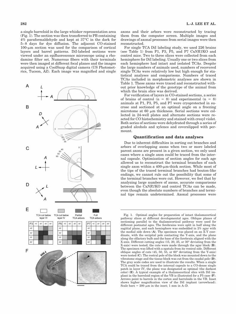
a single barreloid in the large whisker representation area(Fig. 1). The section was then transferred to PB containing4% paraformaldehyde and kept at 37°C in the dark for5–8 days for dye diffusion. The adjacent CO-stained100-�m section was used for the comparison of corticallayers and barrel patterns. DiI-labeled sections wereviewed under an epifluorescence microscope using a rho-damine filter set. Numerous fibers with their terminalswere then imaged at different focal planes and the imagesacquired using a CoolSnap digital camera (US Photomet-rics, Tucson, AZ). Each image was magnified and single
axons and their arbors were reconstructed by tracingthem from the computer screen. Multiple images anddrawings of axonal processes from serial images were thenreconstructed.
For single TCA DiI labeling study, we used 226 brains(see Table 1) from P1, P3, P5, and P7 CxNR1KO andcontrol mice. Two to three slices were collected from eachhemisphere for DiI labeling. Usually one or two slices fromeach hemisphere had intact and isolated TCAs. Despitethe large numbers of animals used, numbers of recoveredsingle TCAs were relatively low but high enough for sta-tistical analyses and comparisons. Numbers of tracedTCAs included in morphometric analyses are shown inTable 1. These axons were traced and reconstructed with-out prior knowledge of the genotype of the animal fromwhich the brain slice was derived.
For verification of layers in CO-stained sections, a seriesof brains of control (n � 8) and experimental (n � 8)animals at P1, P3, P5, and P7 were cryoprotected in su-crose and sectioned at an optimal angle on a freezingmicrotome at 60 �m thickness. Serial sections were col-lected in 24-well plates and alternate sections were re-acted for CO histochemistry and stained with cresyl violet.Both series of sections were dehydrated through a series ofgraded alcohols and xylenes and coverslipped with per-mount.
Quantification and data analyses
Due to inherent difficulties in sorting out branches andarbors of overlapping axons when two or more labeledparent axons are present in a given section, we only usedcases where a single axon could be traced from the inter-nal capsule. Optimization of section angles for each ageallowed us to reconstruct the terminal branches of eachsingle axon within a 400-�m-thick section. While most ofthe tips of the traced terminal branches had bouton-likeendings, we cannot rule out the possibility that some ofthe terminal branches were cut. However, we feel that byanalyzing large numbers of axons, accurate comparisonsbetween the CxNR1KO and control TCAs can be made,even though the absolute numbers of branches and termi-nal tips remain undetermined. Axonal processes were
Fig. 1. Optimal angles for preparation of intact thalamocorticalpathway slices at different developmental ages. Oblique planes ofsectioning parallel to the thalamocortical pathway were used fordifferent postnatal ages. The forebrains were split in half along thesagittal plane, and each hemisphere was embedded in 2% agar withthe medial side down (A). The specimen was placed on an X-Y coor-dinate, with the occipital pole contacting the Y-axis, and the planealong the olfactory bulb and the base of the forebrain aligned with theX-axis. Different cutting angles (15, 20, 25, or 30° deviating from theX-axis) were tested; the cuts were made through the agar block (B).The specimen was lifted with a spatula from its ventral side. Differentoblique angles of cuts (45, 50, 55, or 60° deviating from the Y-axis)were tested (C). The rostral pole of the block was mounted down to thevibratome stage and the tissue block was cut from the caudal pole (D).The gray scale codes are used to illustrate the results. When a singleTCA could be traced from the internal capsule to a CO-dense singlepatch in layer IV, the plane was designated as optimal (the darkestcolor) (E). A typical example of a thalamocortical slice with DiI im-plant in the barreloid region of the VB is illustrated for a P5 case (F).Arrows point to barrels in the cortex and barreloids in the VB. Insetshows higher magnification view of the DiI implant (arrowhead).Scale bars � 200 �m in the inset; 1 mm in A–D.
282 L.-J. LEE ET AL.

measured from the reconstructed 2D display of singleTCAs in both control and CxNR1KO animals ages P1, P3,P5, and P7. The mediolateral extent of each individualafferent arbor was measured. Bifurcation points and ter-minal tips of each reconstructed single TCA were countedin each cortical layer determined by CO histochemistry.Additional quantitative analyses and comparisons weredone by calculating the total number of branch points,terminal tips, and total axonal length after the firstbranch point of the terminal field of all reconstructedsingle axons for each age between experimental and con-trol groups. Arbor density was determined by countingintersections across the two diagonals of every 25 �25-�m square. Student’s t-test was used for testing thevariation of mean values.
Photomicrographs
All samples were analyzed with a Nikon DIAPHOT 300microscope. Photomicrographs were captured with a Cool-Snap digital camera using MetaVue image software (Uni-versal Imaging, Downingtown, PA). The images were ad-justed for brightness and contrast using Adobe PhotoShop7.0 (San Jose, CA) software on a PC computer. For Figure4, images from consecutive optical sections of the samepreparation were merged to form montages. No other al-terations were made.
RESULTS
The emergence of whisker-specific patterning in thecontrol and CxNR1KO mice barrel cortex was visualizedwith 5-HTT immunohistochemistry as early as P3 (Fig. 2).Because developing TCAs transiently express 5-HTT, ithas been used as a reliable marker for developing TCAsfor the somatosensory, visual, and auditory systems (Leb-rand et al., 1996, 1998; Rebsam et al., 2002). This markerfor TCAs and a common barrel pattern marker, CO histo-chemistry, show the emergence and consolidation ofwhisker-specific TCA patterns in the mouse barrel cortexbetween P3–7 (Figs. 2, 3). In CxNR1KO mice, these pat-terns emerge during the same period as in control mice,but are present for only the large whisker TCAs. In addi-tion, these individual patches are much smaller and in-terpatch distances are wider than controls.
At the morphological and physiological levels, thethalamocortical pathway of mice is best analyzed usingbrain slices that contain VB and its projections to thecortical target fields (Agmon and Connors, 1991; Senft andWoolsey, 1991). These slices are cut at an oblique angleparallel to the trajectory of the thalamocortical axons.However, since the forebrain structures change in size andshape during the postnatal period, we first modified the
parameters of oblique sectioning of the brain to obtain anintact thalamocortical pathway slice for P1, 3, 5, and 7mouse brains. These ages in mice correspond to invasionof the cortical plate by TCAs, their initial phases of pat-terning, consolidation of barrel patterns, and after the endof the critical period for structural plasticity, respectively.In order to obtain an optimal angle for thalamocorticalslices, several different angle combinations were tested foreach age group (Fig. 1). For P1, 20–50, 20–55, 20–60,25–50, 25–55, and 30–55; for P3, 20–50, 20–55, and 25–50; for P5, 25–55, 30–55, and 30–60; for P7, 25–55 and25–60 (in degrees, deviated from X-axis and Y-axis, re-spectively) are good angle combinations and are summa-rized in Figure 1E. The optimal angles were similar forboth control and KO brains.
In order to determine whether small patches of TCAsseen with 5-HTT immunohistochemistry and CO histo-chemistry correspond to small terminal arbors of TCAs inCxNR1KO mice, we examined single TCAs by labelingthem from optimized thalamocortical slices. A separateseries of alternate sections from thalamocortical slices ofcontrol and CxNR1KO mice were prepared and stained forCO and Nissl. Comparison of cytoarchitecture of the de-veloping control and CxNR1KO cortex with CO staining(Fig. 3) allowed us to delineate specific cortical layers inDiI-labeled material. We visualized whisker barreloids inthe thalamus by CO staining of thalamocortical slices, andinserted a tiny DiI crystal in a single barreloid at P3, 5,and 7 (Fig. 1F). At P1, barreloids are not visible with COstaining yet, but the outline of the VB is already clear.Therefore, at this age we placed small DiI crystals into thepresumptive barreloid region of the VB visualized by COstaining. Typical examples of DiI-labeled single TCAsagainst a backdrop of CO-stained patches are illustratedin Figure 4. Since these thalamocortical slices are in anoblique coronal plane, our analyses and reconstructions ofsingle axons were restricted to the mediolateral axis ofTCA terminal fields. We were not able to assess the extentof the TCA terminal fields along the rostrocaudal axis ofthe barrel field. Typical examples of single axons recon-structed from control and CxNR1KO groups, for each age,are shown in Figure 5.
At P1, TCAs invade the cortical plate as simple axonswith few small branches. Their morphological appearanceis similar in both control and CxNR1KO mice (Fig. 5A).On P3, TCAs of the control animals display focalizedbranches in layer IV and to a lesser extent in layer VI.Very few branches are seen in other layers. In CxNR1KOcortex, TCAs display a wider terminal territory and morebranches in other layers (Fig. 5B). At P5 and on, as thenormal TCAs consolidate their focal terminal arbors inlayer IV and fewer terminal arbors in layer VI, TCAs of
TABLE 1. Numbers of Animals Used and TCAs Traced
For optimal angle study For TCAs development study
C57 BL/6(wildtype)
EmxICre/� NRIflox/�
(control)EmxICre/� NRIflox/�
(CxNRI KO)EmxICre/� NRIflox/�
(control)EmxICre/� NRIflox/�
(CxNRI KO)
Mice used Mice used TCAs traced Mice used TCAs traced
P1 12 5 4 11 28 12 20P3 24 8 4 38 20 30 17P5 23 7 5 55 19 22 17P7 24 6 6 24 13 34 12Total 83 26 19 128 80 98 66
283NMDAR-DEPENDENT THALAMOCORTICAL DEVELOPMENT

CxNR1KO mice continue their expansion and branchingin other layers (Figs. 4, 5C,D). Quantitative analyses ofnumbers of bifurcation points and terminal tips of TCAs indifferent cortical layers in CxNR1KO and control micerevealed significant differences (Fig. 6). In control ani-mals, bifurcation points and terminal tips were mostlydistributed in layer IV (about 75–80% of the total num-ber), with some in layer VI (10–15%). However, inCxNR1KO cases from P5 and on, significantly (P � 0.01,Student’s t-test) reduced numbers of both bifurcationpoints and terminal tips were counted in layer IV. On theother hand, greatly (P � 0.05, Student’s t-test) increasedbifurcations of TCAs, as well as their terminals, werefound in layers II/III and V in CxNR1KO cortices at P3and older ages. The mediolateral extent of the TCAs in theKO cortex is significantly (P � 0.001, Student’s t-test)larger than controls from P3 and on (Fig. 7). Interestingly,total numbers of bifurcation points and terminal tips in allcortical layers for each age did not show any significantdifferences (Table 2). On the other hand, when we deter-mined the total axonal branch length within the terminalfield of all reconstructed single axons for each age, wefound that this number was significantly higher inCxNR1KO cases beginning on P5 (Table 2). This increaseindicates that as the terminal arbors begin shaping, ter-minal branch segments get longer, thereby contributing to
the wider span of terminal arbors seen in CxNR1KO an-imals.
Despite their exuberance, CxNR1KO mouse TCAs de-velop condensations of terminals, albeit smaller than con-trols, in layer IV (Fig. 8). These focal terminals, within anexpansive terminal arbor, correspond to CO dense regionsseen in the KO cortex, which are smaller than those seenin control mice (Fig. 8C).
DISCUSSION
In the present study, we examined detailed morphologyof single TCAs in the CxNR1KO somatosensory cortexduring the first postnatal week. TCA arbor and terminaldevelopment of CxNR1KO mice appears similar to that ofcontrol mice at P1, but by P3 significant differences beginemerging. We found that CxNR1KO TCAs have longerterminal branch lengths (at P5 and P7) and wider spanslaterally and radially (after P3) than WT TCAs. Thesedifferences in mediolateral extent of arbor fields and num-ber of branch points and terminals in different corticallaminae are further accentuated between P5 and P7,when barrels normally appear as cytoarchitectonic unitsand become distinct (Rice et al., 1985). Previously, weshowed that in CxNR1KO mice NR1 expression in the VBand all other subcortical structures are not altered, but it
Fig. 2. Development of TCA patches in normal and CxNR1KObarrel cortex as revealed with 5-HTT immunohistochemistry. Tan-gential sections of flattened cortices at ages P3 (A and D), P5 (B andE), and P7 (C and F) are collected from control (A–C) and CxNR1KO(D–F) mice. Whisker-specific patterns of 5-HTT-positive TCAs appear
in the normal somatosensory cortex as early as P3 (A), and becomemore distinctive as the brain develops (B, C). However, the patternsare less discernible in the CxNR1KO cortex at P3 (D). At later ages,smaller TCA patches become visible in the major whisker represen-tation area (E, F). Scale bar � 100 �m in F (applies to A–F)..
284 L.-J. LEE ET AL.
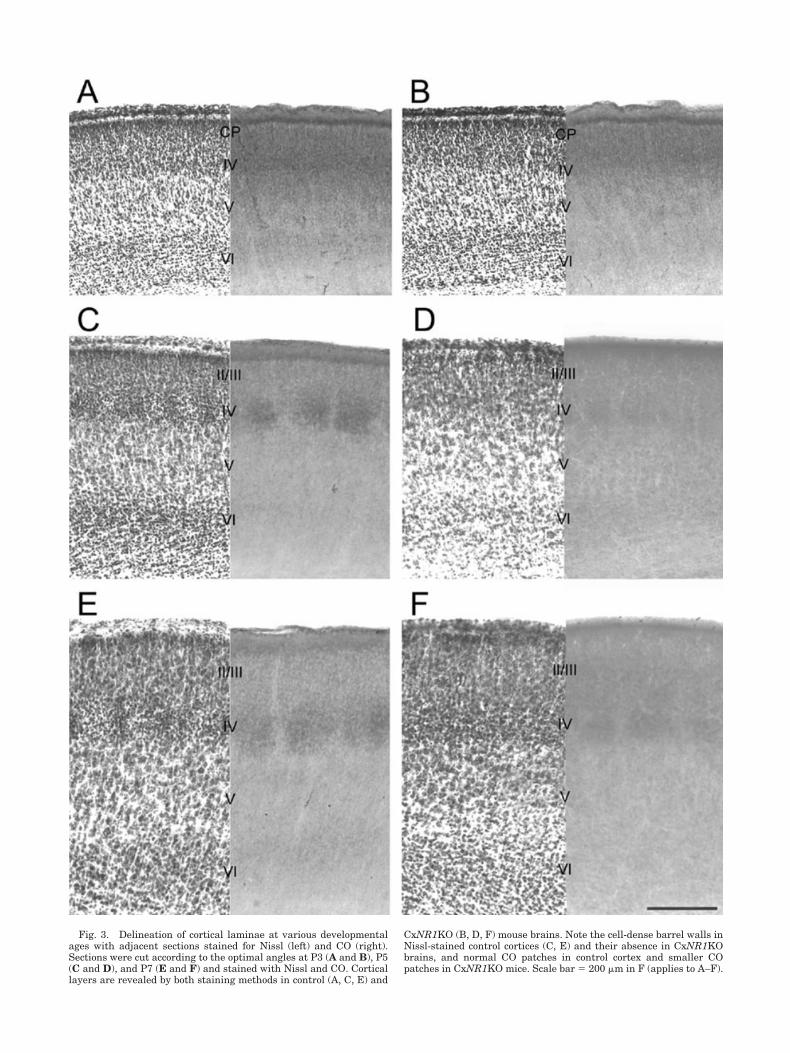
Fig. 3. Delineation of cortical laminae at various developmentalages with adjacent sections stained for Nissl (left) and CO (right).Sections were cut according to the optimal angles at P3 (A and B), P5(C and D), and P7 (E and F) and stained with Nissl and CO. Corticallayers are revealed by both staining methods in control (A, C, E) and
CxNR1KO (B, D, F) mouse brains. Note the cell-dense barrel walls inNissl-stained control cortices (C, E) and their absence in CxNR1KObrains, and normal CO patches in control cortex and smaller COpatches in CxNR1KO mice. Scale bar � 200 �m in F (applies to A–F).

Fig. 4. DiI-labeled single axons in CO-stained thalamocorticalslices. A small crystal of DiI was inserted into the barreloid region ofa slightly CO stained thalamocortical section; single TCAs were thenvisualized and traced. Examples of control (A–C) and CxNR1KO(D–F) P7 TCA are presented. Cortical layers and barrel patterns arerevealed by CO staining (A, D, asterisks). Single TCAs in the somato-sensory cortex are labeled with DiI. Images from different focal planes
were collected and superimposed, then converted into black-and-white for better resolution (B, E). Tracings of single TCAs were mademanually based on the reconstructed images (C, F). Single patches(gray in C, F) from CO-stained sections (A, D) were pasted on theafferent arbor indicating the relative position of the CO-dense patcheswith respect to TCA terminal arbor fields. Scale bar � 100 �m in F(applies to A–F).
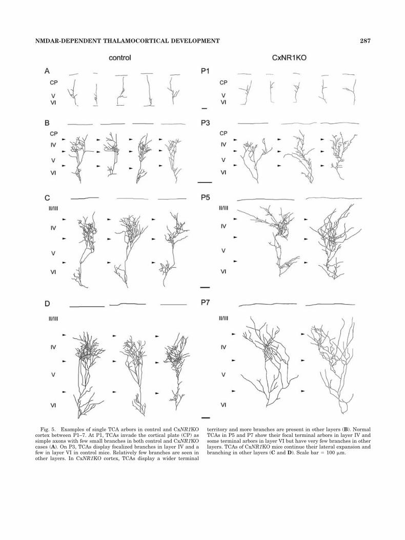
Fig. 5. Examples of single TCA arbors in control and CxNR1KOcortex between P1–7. At P1, TCAs invade the cortical plate (CP) assimple axons with few small branches in both control and CxNR1KOcases (A). On P3, TCAs display focalized branches in layer IV and afew in layer VI in control mice. Relatively few branches are seen inother layers. In CxNR1KO cortex, TCAs display a wider terminal
territory and more branches are present in other layers (B). NormalTCAs in P5 and P7 show their focal terminal arbors in layer IV andsome terminal arbors in layer VI but have very few branches in otherlayers. TCAs of CxNR1KO mice continue their lateral expansion andbranching in other layers (C and D). Scale bar � 100 �m.
287NMDAR-DEPENDENT THALAMOCORTICAL DEVELOPMENT

is abolished in the excitatory neurons of the neocortex,hippocampus, and the olfactory bulb (Iwasato et al., 2000).Exuberant terminal arbor development from VB projec-tions to the NR1 impaired neocortex indicates thatpostsynaptic NMDARs play an important role in refine-ment of presynaptic TCA arbors in the barrel cortex.
In an earlier study we showed that cellular modules anddendritic field orientation never develop in the CxNR1KObarrel cortex (Datwani et al., 2002a). In that study, wealso presented preliminary evidence from very few singleaxons (7 and 8 axons at P5), which suggested that
CxNR1KO TCAs developed smaller and rudimentary ar-bors. However, those few axons were reconstructed fromthalamocortical slices that had not been optimized for theage-specific planes of section. Detailed analyses performedin the present study and examination of more than adozen axons with more complete fills for each age groupnow indicate that CxNR1KO TCAs have much widerspans laterally and radially. In addition, branch segmentsand total axonal length within a terminal field are longer.Our present results coupled with our previous report onthe dendritic defects in layer IV spiny stellate cells of theCxNR1KO cortex (Datwani et al., 2002a) show that corti-cal NMDAR activity plays a major role in sculpting bothTCA terminal arbors and dendrites of barrel cells.
Normal development of whisker-specific TCA arbors inmice has been described (Senft and Woolsey, 1991; Agmonet al., 1995; Rebsam et al., 2002), and the present resultsfrom control cases confirm these findings. Briefly, topo-graphically organized TCAs invade the cortical plateabove layer V by P1 as simple axons with very fewbranches. As barreloids develop in the thalamus, theiraxonal terminal fields and arbors partition into “Gauss-ian” patches in the newly formed layer IV by P3 (Senft andWoolsey, 1991), before the barrels appear as cytoarchitec-tonic modules. Barrels then form around the patchy TCAterminal fields by P5. Between P3–7, there is selectivepruning of overshooting collaterals, while more branchesare added and consolidated within the foci of terminalpatches. As noted by Senft and Woolsey earlier (1991),focal arborization is pronounced within the center of eachwhisker-specific Gaussian distribution of TCA terminals.Here we show by single TCA labeling that focalization ofwhisker-specific TCA terminal fields still takes place, al-beit to a limited extent, when subcortical NMDAR func-tion is intact and cortical NMDAR function is impaired.These patches correspond to the rudimentary patternsseen with CO histochemistry or 5-HTT immunostainingthat are commonly used as markers of barrel/TCA pat-terns. CxNR1KO mice were generated using the Cre/loxPsystem. The expression of Cre recombinase gene is drivenby the promoter of homeobox gene Emx1, which is ex-pressed specifically in the dorsal telencephalon. GABAer-gic inhibitory interneurons migrate into the neocortexfrom the Emx1 negative ganglionic eminence of the ven-tral telencephalon (Anderson et al., 1997). Therefore, inthese mice GABAergic cortical neurons escape the NR1gene excision. GABAergic cells reportedly comprise 13–15% of barrel cells and are located along the barrel walls,with their dendrites oriented toward barrel hollows (Lin etal., 1985). Therefore, it is possible that spared NMDARfunction in GABAergic barrel cortex cells might contributeto rudimentary segregation of large whisker TCA termi-nals within an expansive terminal arbor field.
Our findings that the total number of branch points andterminal tips do not significantly differ betweenCxNR1KO and control mice suggest that these parame-ters might be intrinsically programmed in VB cells andtheir axons. However, we do not have any direct evidencefor this scenario. Similarly, increased total branch lengthin CxNR1KO TCA terminal arbors indicates that postsyn-aptic NMDAR signaling might serve a regulatory functionin terminal elaboration and axon growth vigor. Postsyn-aptic NMDARs have been demonstrated to limit the affer-ent extension in vitro (Baird et al., 1996). During earlydevelopment, NMDARs may function as axonal sprouting
Fig. 6. Bifurcation points and terminal tips of TCAs in differentcortical layers at different postnatal ages. Numbers of bifurcationpoints (A) and terminal tips (B) of each reconstructed single TCA werecounted in different cortical layers in CxNR1KO and control mice. Forbifurcation points (A), from P3 and on, TCAs of CxNR1KO mice havea greater number in layers II/III and layer V than control TCAs. Onthe other hand, in layer IV, CxNR1KO mice have fewer bifurcationpoints than control mice on P5 and P7. For terminal tips (B) from P3and on, CxNR1KO TCAs have a greater number in layers II/III andlayer V but a lower number in layer IV than control TCAs.
288 L.-J. LEE ET AL.

suppressors when AMPARs are not yet recruited (Lin andConstantine-Paton, 1998). At present, we do not know theprecise molecular chain of events that restrict axonal/terminal branch elongation and induce localized elabora-tion through activation of postsynaptic NMDARs. Previ-ously it was reported that calcium/calmodulin dependentprotein kinase II (CaMKII), a key player downstream ofthe NMDAR-mediated signaling, might regulate thegrowth of tadpole retinotectal afferents. When postsynap-
tic CaMKII is inhibited, the growth restriction of presyn-aptic afferents is released, leading to the increased axonalextension (Zou and Cline, 1999).
Because in CxNR1KO mice functional NMDARs arelacking in excitatory postsynaptic cells, while the parentcells of TCAs in VB have normal levels of NMDAR func-tion, there must be retrograde signals generated down-stream of NMDAR activation and consequent Ca2� entryat the postsynaptic site. In fact, many lines of evidence
Fig. 7. Mediolateral extent of single TCA arbors between P1–7.Lateral extent of TCAs from control (black) and CxNR1KO (gray)animals were measured and plotted in groups. At P1, there is nodifference between control and CxNR1KO cases. However from P3
and on the mediolateral extent of the TCAs in the mutant cortex issignificantly greater than those in controls. Mean value of each groupis demonstrated by a horizontal bar. Significant differences are indi-cated by asterisks (***P � 0.001, Student’s t-test).
TABLE 2. Numbers of Terminal Endpoints, Branch Points, and Total Arbor Segment Length Between P1 and P71
Age
Total branch points Total terminal points Total arbor segment length (�m)
control CxNRI KO control CxNRI KO control CxNRI KO
P1 2.45 � 2.48 2.75 � 2.53 3.58 � 2.56 3.64 � 2.28 652 � 211 602 � 228P3 33.4 � 10.6 34.94 � 10.26 34.75 � 10.93 36.88 � 10.26 2241 � 578.8 2489 � 427.4P5 42.53 � 10.01 47.76 � 11.4 44.31 � 10.29 49.29 � 11.93 3707 � 976.3 5389 � 1621.8**P7 42.83 � 11.5 40.91 � 9.32 44.18 � 11.02 42.17 � 9.24 6333 � 1750 8195 � 1542.1*
1Results are mean � SD: *, P � 0.05; **, P � 0.01, Student’s t-test).
289NMDAR-DEPENDENT THALAMOCORTICAL DEVELOPMENT
indicate the involvement of retrograde signals in shapingafferent morphology in the vertebrate central nervous sys-tem (see reviews: Fitzsimonds and Poo, 1998; Debski andCline, 2002). Currently, there are numerous candidateretrograde signals and synaptic molecules that could par-ticipate in this highly complex, activity-driven communi-cation between pre- and postsynaptic elements that even-tually influence their morphological and consequentlyfunctional differentiation. Nitric oxide (NO), brain-derived neurotrophic factor (BDNF), and arachidonic acid(AA) have all been implicated as potential retrograde sig-nals that could modulate stabilization and consolidation ofsynapses, thereby sculpting presynaptic terminal andpostsynaptic dendritic patterns (see Schmidt, 2004, for areview).
Spatial and temporal distribution of NMDARs hasdrawn attention in recent years (see reviews: Carroll andZukin, 2002; Wenthold et al., 2003). As transmembraneproteins, NMDARs anchor other cell surface proteins tomany other cytoplasmic molecules (reviewed by Scheiffele,2003). Pharmacological blockade studies mainly examin-ing the channel properties of NMDARs might not coverthis aspect of NMDAR function. NR2 subunit of NMDARbinds to PDZ domains of PSD-95 (Kornau et al., 1995), themajor scaffolding protein for assembly of signaling com-plex and cell surface molecules (Garner et al., 2000), suchas neuroligins. Neuroligins interact with neurexins toform calcium-dependent heterophilic adhesion molecules(Nguyen and Sudhof, 1997), and induce presynaptic dif-
ferentiation (Scheiffele et al., 2000). In addition, EphB2can interact directly with NR1 subunit and regulate theformation of excitatory synapses through the Ephrin-EphB signaling pathway (Dalva et al., 2000).
To date, several mutant mice have been described withspecific deficits in TCA terminal patterning, cellular pat-terning, or both, in the somatosensory cortex. In some ofthese mice, TCAs form partial or full whisker-specific pat-terns, but layer IV stellate cells do not. In others, bothTCAs and stellate cells fail to develop patterns (reviewedin Erzurumlu and Kind, 2001; Lopez-Bendito and Molnar,2003). These mutations involve various aspects of gluta-matergic synaptic communication and its regulation by5-HT (Cases et al., 1996; Iwasato et al., 1997, 2000; Vitaliset al., 1998; Hannan et al., 1998, 2001; Salichon et al.,2001; Datwani et al., 2002a). In vitro recordings fromthalamocortical slices indicated that 5-HT has a strongpresynaptic inhibitory effect on glutamatergic transmis-sion between somatosensory thalamic axons and barrelcortex cells via the activation of 5-HT1B receptors(Rhoades et al., 1994). Collectively, these data indicatethat thalamocortical axons autoregulate their glutamater-gic transmission via presynaptic mechanisms using 5-HTpresent in their target field (Persico et al., 2001). Deletionsof several intracellular signaling molecules downstreamfrom glutamate and 5-HT also show defects in barrelformation (reviewed in Erzurumlu and Kind, 2001; Gas-par et al., 2003). Rebsam et al. (2002) described the TCAarbors in monoamine oxidase A (MAOA) KO and MAOA/
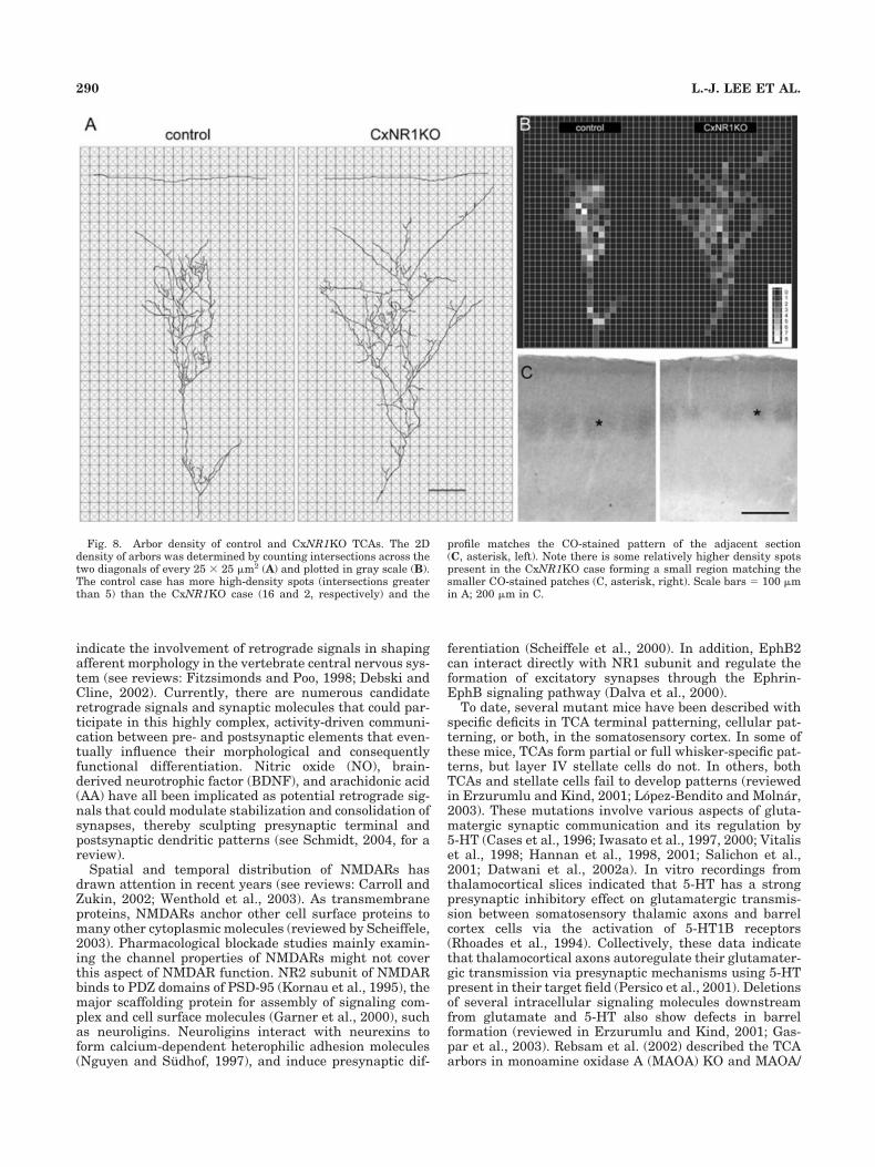
Fig. 8. Arbor density of control and CxNR1KO TCAs. The 2Ddensity of arbors was determined by counting intersections across thetwo diagonals of every 25 � 25 �m2 (A) and plotted in gray scale (B).The control case has more high-density spots (intersections greaterthan 5) than the CxNR1KO case (16 and 2, respectively) and the
profile matches the CO-stained pattern of the adjacent section(C, asterisk, left). Note there is some relatively higher density spotspresent in the CxNR1KO case forming a small region matching thesmaller CO-stained patches (C, asterisk, right). Scale bars � 100 �min A; 200 �m in C.
290 L.-J. LEE ET AL.

5-HT1B receptor double KO mice. They found that inMAOA KO and MAOA/5-HT1B double KO mice, TCAsreach the proper region and laminae of the somatosensorycortex by P1 as in normal mice. At P7, however, whilewhisker-specific TCA terminals are restricted to a singlebarrel in control animals, there was a significant reduc-tion of terminal branches (by half) in layer IV and a 40%increase in the mediolateral span of TCA arbors in MAOAKO mice. In double KO animals, on the other hand, thesedefects were corrected and their TCAs attained “normal”span and terminal distribution by P7. Our present resultscorroborate their observations in normal/control mice, andfurther show that when excitatory cortical cells lack func-tional NMDARs, TCAs also develop expansive arbors andform terminals in multiple cortical layers instead of clus-tering their terminals into patches in layer IV. Intracel-lular signaling pathways downstream from NMDARs (ormetabotropic glutamate receptors and serotonergic mod-ulation of glutamatergic transmission), which lead to spe-cific communication between pre- and postsynaptic ele-ments in the barrel cortex during whisker-specific patternformation, are largely unknown, while adenylyl cyclasetype I, phospholipase C1, cAMP response element-binding protein (CREB), and BDNF have been implicated(reviewed in Erzurumlu and Kind, 2001; Gaspar et al.,2003).
In recent years, some groups have attempted to identifycortical lamina-specific molecular signals that direct TCAsto layer IV (and to a lesser degree to layer VI), and inducethem to stop there, branch, and arborize (see Yamamoto,2002, for a review). While several in vitro models haveindicated the presence of targeting and “stop” signals inlayer IV (Molnar and Blakemore, 1991; Emerling andLander, 1994; Yamamoto et al., 1997), the molecular iden-tity of this signal and its in vivo function remains to besolved. Our present results indicate that in the absence ofpostsynaptic NMDAR function in excitatory cortical cells,TCA terminals explore other cortical layers, branch, andterminate therein, “ignoring” lamina specific “stop” sig-nals. Not only in CxNR1KO mice, but also in other mousemutants with TCA terminal arbor expansions, such as thebarrelless (brl or adenylyl cyclase Type 1KO) andMAOAKO, TCA terminals expand beyond layer IV(Welker et al., 1996; Rebsam et al., 2002). On the otherhand, in all these mutants, and in cortical molecular map-ping studies, it is evident that TCAs from different tha-lamic nuclei respect cortical areal boundaries defined bymolecular markers (Rubenstein et al., 1999; Grove andFukuchi-Shimogori, 2003; Vanderhaeghen and Polleux,2004).
In CxNR1KO mice, the absence of NMDAR function inspiny stellate cells of layer IV and other excitatory neu-rons in the barrel field most likely impairs several down-stream signals that enable postsynaptic cells to detectincoming correlated activity from TCAs and lead to aber-rant dendritic differentiation as well as exuberant spineformation that could not be stabilized (Datwani et al.,2000a). Lack of NMDAR-dependent retrograde signalsthat regulate growth potential and focalization of TCAterminal branches would lead to expansion of TCA termi-nals into other layers and laterally wider areas in searchof such signals. If this scenario were correct, then onewould expect a high turnover of synaptic contacts (tran-sient synapses), and a dynamic attempt at the postsynap-tic site to offer more dendritic spines with relatively little
success in synapse consolidation. Further studies at theultrastructural and physiological levels would resolve thisissue.
ACKNOWLEDGMENTS
We thank Ms. Barri King for help with breeding andgenotyping mice, Baris Yigit for help with axon recon-structions, and Ryan Couvillion for histology.
LITERATURE CITED
Agmon A, Connors BW. 1991. Thalamocortical responses of mouse somato-sensory (barrel) cortex in vitro. Neuroscience 41:365–379.
Agmon A, Yang LT, Jones EG, O’Dowd DK. 1995. Topological precision inthe thalamic projection to neonatal mouse barrel cortex. J Neurosci15:549–561.
Anderson SA, Eisenstat DD, Shi L, Rubensterin JLR. 1997. Interneuronmigration from basal forebrain to neocortex: dependence on Dlx genes.Science 278:474–476.
Baird DH, Trehkner E, Mason CA. 1996. Arrest of afferent axon extensionby target neurons in vitro is regulated by the NMDA receptor. J Neu-rosci 16:2642–2648.
Carroll RC, Zukin RS. 2002. NMDA-receptor trafficking and targeting:implications for synaptic transmission and plasticity. Trends Neurosci25:571–577.
Cases O, Vitalis T, Seif I, De Maeyer E, Sotelo C, Gaspar P. 1996. Lack ofbarrels in the somatosensory cortex of monoamine oxidase A-deficientmice: role of a serotonin excess during the critical period. Neuron16:297–307.
Dalva MB, Takasu MA, Lin MZ, Shamah SM, Hu L, Gale NW, GreenbergME. 2000. EphB receptors interact with NMDA receptors and regulateexcitatory synapse formation. Cell 103:945–956.
Datwani A, Iwasato T, Itohara S, Erzurumlu RS. 2002a. NMDA receptor-dependent pattern transfer from afferents to postsynaptic cells anddendritic differentiation in the barrel cortex. Mol Cell Neurosci 21:477–492.
Datwani A, Iwasato T, Itohara S, Erzurumlu RS. 2002b. Lesion-inducedthalamocortical axonal plasticity in the S1 cortex is independent ofNMDA receptor function in excitatory cortical neurons. J Neurosci22:9171–9175.
Debski EA, Cline HT. 2002. Activity-dependent mapping in the retinotectalprojection. Curr Opin Neurobiol 12:93–99.
Emerling DE, Lander AD. 1994. Laminar specific attachment and neuriteoutgrowth of thalamic neurons on cultured slices of developing cerebralcortex. Development 125:3399–3410.
Erzurumlu RS, Jhaveri S. 1990. Thalamic axons confer a blueprint of thesensory periphery onto the developing rat somatosensory cortex. DevBrain Res 56:229–234.
Erzurumlu RS, Jhaveri S. 1992a. Trigeminal ganglion cell processes arespatially ordered prior to the differentiation of the vibrissa pad. J Neu-rosci 12:3946–3955.
Erzurumlu RS, Jhaveri S. 1992b. Emergence of connectivity in the embry-onic rat parietal cortex. Cereb Cortex 2:336–352.
Erzurumlu RS, Kind PC. 2001. Neural activity: sculptor of “barrels” in theneocortex. Trends Neurosci 24:589–595.
Erzurumlu RS, Jhaveri S, Benowitz LI. 1990. Transient patterns ofGAP-43 expression during the formation of barrels in the rat somato-sensory cortex. J Comp Neurol 292:443–456.
Fitzsimonds RM, Poo MM. 1998. Retrograde signaling in the developmentand modification of synapses. Physiol Rev 78:143–170.
Garner CC, Nash J, Huganir RL. 2000. PDZ domains in synapse assemblyand signaling. Trends Cell Biol 10:274–280.
Gaspar P, Cases O, Maroteaux L. 2003. The developmental role of seroto-nin: news from mouse molecular genetics. Nat Rev Neurosci 4:1002–1012.
Grove EA, Fukuchi-Shimogori T. 2003. Generating the cerebral corticalarea map. Annu Rev Neurosci 26:355–380.
Hannan AJ, Kind PC, Blakemore C. 1998. Phospholipase C-beta1 expres-sion correlates with neuronal differentiation and synaptic plasticity inrat somatosensory cortex. Neuropharmacology 7:593–605.
Hannan AJ, Blakemore C, Katsnelson A, Vitalis T, Huber KM, Near M,
291NMDAR-DEPENDENT THALAMOCORTICAL DEVELOPMENT

Roder J, Kim D, Shin HS, Kind PC. 2001. PLC-1, activated viamGluRs, mediates activity-dependent differentiation in cerebral cor-tex. Nat Neurosci 4:282–288.
Iwasato T, Erzurumlu RS, Huerta PT, Chen DF, Sasaoka T, Ulupinar E,Tonegawa S. 1997. NMDA receptor-dependent refinement of somato-topic maps. Neuron 19:1201–1210.
Iwasato T, Datwani A, Wolf AM, Nishiyama H, Taguchi Y, Tonegawa S,Knopfel T, Erzurumlu RS, Itohara S. 2000. Cortex-restricted disrup-tion of NMDAR1 impairs neuronal patterns in the barrel cortex. Na-ture 406:726–731.
Killackey HP, Rhoades RW, Bennett-Clarke CA. 1995. The formation of acortical somatotopic map. Trends Neurosci 18:402–407.
Kornau H-C, Schenker LT, Kennedy MB, Seeburg PH. 1995. Domaininteraction between NMDA receptor subunits and postsynaptic densityprotein PSD-95. Science 269:1737–1740.
Kutsuwada T, Sakimura K, Manabe T, Takayama C, Katakura N, KushiyaE, Natsume R, Watanabe M, Inoue Y, Yagi T, Aizawa S, Arakawa M,Takahashi T, Nakamura Y, Mori H, Mishina M. 1996. Impairment ofsuckling response, trigeminal neuronal pattern formation, and hip-pocampal LTD in NMDA receptor epsilon 2 subunit mutant mice.Neuron 16:333–344.
Lebrand C, Cases O, Adelbrecht C, Doye A, Alvarez C, El Mestikawy S, SeifI, Gaspar P. 1996. Transient uptake and storage of serotonin in devel-oping thalamic neurons. Neuron 17:823–835.
Lebrand C, Case, O, Wehrle R, Blakely RD, Edwards RH, Gaspar P. 1998.Transient developmental expression of monoamine transporters in therodent forebrain. J Comp Neurol 401:506–524.
Li Y, Erzurumlu RS, Chen C, Jhaveri S, Tonegawa S. 1994. Whisker-related neuronal patterns fail to develop in the trigeminal brainstemnuclei of NMDAR1 knockout mice. Cell 76:427–437.
Lin CS, Lu SM, Schmechel DE. 1985. Glutamic acid decarboxylase immu-noreactivity in layer IV of barrel cortex of rat and mouse. J Neurosci5:1934–1939.
Lin SY, Constantine-Paton M. 1998. Suppression of sprouting: an earlyfunction of NMDA receptors in the absence of AMPA/Kainate receptoractivity. J Neurosci 18:3725–3737.
Lopez-Bendito G, Molnar Z. 2003. Thalamocortical development: how arewe going to get there? Nat Rev Neurosci 4:276–289.
Ma PM, Woolsey TA. 1984. Cytoarchitectonic correlates of the vibrissae inthe medullary trigeminal complex of the mouse. Brain Res 306:374–379.
Maier DL, Mani S, Donovan SL, Soppet D, Tessarollo L, McCasland JS,Meiri KF. 1999. Disrupted cortical map and absence of cortical barrelsin growth-associated protein (GAP)-43 knockout mice. Proc Natl AcadSci U S A 96:9397–9402.
Molnar Z, Blakemore C. 1991. Lack of regional specificity for connectionsformed between thalamus and cortex in coculture. Nature 351:475–477.
Nguyen T, Sudhof TC. 1997. Binding properties of neuroligin 1 and neur-exin 1 beta reveal function as heterophilic cell adhesion molecules.J Biol Chem 272:32–39.
O’Leary DDM, Ruff NL, Dyck RH. 1994. Development, critical periodplasticity, and adult reorganizations of mammalian somatosensorysystems. Curr Opin Neurobiol 4:535–544.
Persico AM, Mengual E, Moessner R, Hall FS, Revay RS, Sora I, ArellanoJ, DeFelipe J, Gimenez-Amaya JM, Conciatori M, Marino R, Baldi A,Cabib S, Pascucci T, Uhl GR, Murphy DL, Lesch KP, Keller F, Hall SF.2001. Barrel pattern formation requires serotonin uptake by thalamo-cortical afferents, and not vesicular monoamine release. J Neurosci21:6862–6873.
Rebsam A, Seif I, Gaspar P. 2002. Refinement of thalamocortical arborsand emergence of barrel domains in the primary somatosensory cortex:
a study of normal and monoamine oxidase a knock-out mice. J Neurosci22:8541–8552.
Rhoades RW, Bennett-Clarke CA, Shi MY, Mooney RD. 1994. Effects of5-HT on thalamocortical synaptic transmission in the developing rat.J Neurophysiol 72:2438–2450.
Rice FL, Gomez C, Barstow C, Burnet A, Sands P. 1985. A comparativeanalysis of the development of the primary somatosensory cortex: in-terspecies similarities during barrel and laminar development. J CompNeurol 236:477–495.
Rubenstein JLR, Anderson S, Shi L, Miyashita-Lin E, Bulfone A, HevnerR. 1999. Genetic control and regionalization and connectivity. CerebCortex 9:524–532.
Salichon N, Gaspar P, Upton AL, Picaud S, Hanoun N, Hamon M, DeMaeyer E, Murphy DL, Mossner R, Lesch KP, Hen R, Seif I. 2001.Excessive activation of serotonin (5-HT) 1B receptors disrupts theformation of sensory maps in monoamine oxidase A and 5-HT trans-porter knock-out mice. J Neurosci 21:884–896.
Scheiffele P. 2003. Cell-cell signaling during synapse formation in theCNS. Annu Rev Neurosci 26:485–508.
Scheiffele P, Fan J, Choih J, Fetter R, Serafini T. 2000. Neuroligin ex-pressed in nonneuroal cells triggers presynaptic development in con-tacting axons. Cell 101:657–669.
Schmidt JT. 2004. Activity-driven sharpening of the retinotectal projec-tion: the search for retrograde synaptic signaling pathways. J Neuro-biol 59:114–133.
Senft SL, Woolsey TA. 1991. Growth of thalamic afferents into mousebarrel cortex. Cereb Cortex 1:308–335.
Van der Loos H. 1976. Barreloids in mouse somatosensory thalamus.Neurosci Lett 2:1–6.
Vanderhaeghen P, Polleux F. 2004. Developmental mechanisms pattern-ing thalamocortical projections: intrinsic, extrinsic and in between.Trends Neurosci 27:384–391.
Vitalis T, Cases O, Callebert J, Launay JM, Price DJ, Seif I, Gaspar P.1998. Effects of monoamine oxidase A inhibition on barrel formation inthe mouse somatosensory cortex: determination of a sensitive develop-mental period. J Comp Neurol 393:169–184.
Welker E, Van der Loos H. 1986. Quantitative correlation between barrel-field size and the sensory innervation of the whiskerpad: a comparativestudy in six strains of mice bred for different patterns of mystacialvibrissae. J Neurosci 6: 3355–3373.
Welker E, Armstrong-James M, Bronchti G, Ourednik W, Gheorghita-Baechler F, Dubois R, Guernsey DL, Van der Loos H, Neumann PE.1996. Altered sensory processing in the somatosensory cortex of themouse mutant barrelless. Science 271:1864–1867.
Wenthold RJ, Prybylowski K, Standley S, Sans N, Petralia RS. 2003.Trafficking of NMDA receptors. Annu Rev Pharmacol Toxicol 43:335–358.
Woolsey TA. 1990. Peripheral alteration and somatosensory development.In: Coleman EJ, editor. Development of sensory systems in mammals.New York: John Wiley & Sons. p 461–516.
Woolsey TA, Van der Loos H. 1970. The structural organization of layer IVin the somatosensory region (SI) of mouse cerebral cortex. The descrip-tion of a cortical field composed of discrete cytoarchitectonic units.Brain Res 17:205–242.
Yamamoto N. 2002. Cellular and molecular basis for the formation oflamina-specific thalamocortical projections. Neurosci Res 42:167–173.
Yamamoto N, Higashi S, Toyama K. 1997. Stop and branch behaviors ofgeniculocortical axons: a time-lapse study in organotypic cocultures.J Neurosci 17:3653–3663.
Zou DJ, Cline HT. 1999. Postsynaptic calcium/calmodulin-dependent pro-tein kinase II is required to limit elaboration of presynaptic andpostsynaptic neuronal arbors. J Neurosci 19:8909–8918.
292 L.-J. LEE ET AL.
Related Documents