Extracranial Carotid Artery Stenting in Surgically High-Risk Patients Using the Carotid Wallstent Endoprosthesis: Midterm Clinical and Ultrasound Follow-Up Results Geert Maleux, 1 Pauwel Bernaerts, 2 Vincent Thijs, 3 Johan Vaninbroukx, 1 Kim Daenens, 2 Inge Fourneau, 2 Andre ´ Nevelsteen 2 1 University Hospitals, Department of Interventional Radiology, Leuven, Belgium 2 University Hospitals, Department of Vascular Surgery, Leuven, Belgium 3 University Hospitals, Department of Neurology, Leuven, Belgium Abstract The purpose of this study was to evaluate the feasibility, safety and midterm outcome of elective implantation of the Carotid Wallstent in patients considered to be at high surgical risk. In a prospective study, 54 carotid artery steno- ses in 51 patients were stented over a 24-month period. Three patients underwent bilateral carotid artery stenting. Institutional inclusion criteria for invasive treatment of ca- rotid occlusive disease (carotid endarterectomy or carotid artery stenting) are patients presenting with a 70% or more symptomatic stenosis and those with an 80% or more asymp- tomatic stenosis having a life-expectancy of more than 1 year. All patients treated by carotid artery stenting were considered at high risk for carotid endarterectomy because of a hostile neck (17 patients—31.5%) or because of severe comorbidities (37 patients— 68.5%). No cerebral protection device was used. Of the 54 lesions, 33 (61.1%) were symp- tomatic and 21 (38.8%) were asymptomatic. Follow-up was performed by physical examination and by duplex ultra- sonography at 1 month, 6 months, 1 year and 2 years after the procedure. All 54 lesions could be stented successfully without periprocedural stroke. Advert events during fol- low-up (mean 13.9 5.7 months) were non-stroke-related death in 6 patients (11.1%), minor stroke in 4 stented hemi- spheres (7.4%), transient ipsilateral facial pain in 1 patient (1.8%), infection of the stented surgical patch in 1 patient (1.8%) and asymptomatic instent restenosis in 4 patients (7.4%). The percutaneous implantation of the Carotid Wall- stent, even without cerebral protection device, appears to be a safe procedure with acceptable clinical and ultrasono- graphic follow-up results in patients at high surgical risk. But some late adverse events such as ipsilateral recurrence of non-disabling (minor) stroke or instent restenosis still remain real challenging problems. Key words: Angioplasty—Carotid stenosis—Carotid stent—High-risk carotid surgery—Stroke Carotid angioplasty and stenting (CAS) has been recom- mended by some clinicians as a real alternative to carotid endarterectomy (CEA) in a limited subgroup of patients [1]. This subgroup includes patients with carotid restenosis after CEA, radiation-induced stenosis, anatomically inaccessible lesion and severe medical co-morbidities. Recently, several non-controlled series [2–12] assessing this patient popula- tion were published and show acceptable short- and mid- term results which are probably better than the results after medical therapy [13]. In most of these (retrospective, non- randomized) studies, balloon- and/or self-expanding stents like the Palmaz stent or iliac Wallstent were inserted, some- times after placement of a cerebral protection device. The purpose of this prospective study was to evaluate the short- and mid-term clinical and ultrasound follow-up of patients treated with the Carotid Wallstent (Boston Scientific, Paris La Garenne, France) for carotid occlusive disease in surgi- cally high-risk patients. This stent (Fig. 1a), mounted on a low profile, monorail catheter system (Fig. 1b), can be con- sidered one of the first stents specifically designed for carotid use. Material and Methods Study Population Between April 2000 and April 2002, 54 CAS procedures (22 right carotid and 32 left carotid lesions) were performed in 51 patients Correspondence to: G. Maleux; email: [email protected] Cardio V ascular and Interventional Radiology © Springer-Verlag New York, Inc. 2003 Cardiovasc Intervent Radiol (2003) 26:340 –346 Published Online: 20 June 2003 DOI: 10.1007/s00270-003-0039-4

Welcome message from author
This document is posted to help you gain knowledge. Please leave a comment to let me know what you think about it! Share it to your friends and learn new things together.
Transcript
Extracranial Carotid Artery Stenting inSurgically High-Risk Patients Using the CarotidWallstent Endoprosthesis: Midterm Clinical andUltrasound Follow-Up ResultsGeert Maleux,1 Pauwel Bernaerts,2 Vincent Thijs,3 Johan Vaninbroukx,1
Kim Daenens,2 Inge Fourneau,2 Andre Nevelsteen2
1University Hospitals, Department of Interventional Radiology, Leuven, Belgium2University Hospitals, Department of Vascular Surgery, Leuven, Belgium3University Hospitals, Department of Neurology, Leuven, Belgium
AbstractThe purpose of this study was to evaluate the feasibility,safety and midterm outcome of elective implantation of theCarotid Wallstent� in patients considered to be at highsurgical risk. In a prospective study, 54 carotid artery steno-ses in 51 patients were stented over a 24-month period.Three patients underwent bilateral carotid artery stenting.Institutional inclusion criteria for invasive treatment of ca-rotid occlusive disease (carotid endarterectomy or carotidartery stenting) are patients presenting with a 70% or moresymptomatic stenosis and those with an 80% or more asymp-tomatic stenosis having a life-expectancy of more than 1year. All patients treated by carotid artery stenting wereconsidered at high risk for carotid endarterectomy because ofa hostile neck (17 patients—31.5%) or because of severecomorbidities (37 patients—68.5%). No cerebral protectiondevice was used. Of the 54 lesions, 33 (61.1%) were symp-tomatic and 21 (38.8%) were asymptomatic. Follow-up wasperformed by physical examination and by duplex ultra-sonography at 1 month, 6 months, 1 year and 2 years afterthe procedure. All 54 lesions could be stented successfullywithout periprocedural stroke. Advert events during fol-low-up (mean 13.9� 5.7 months) were non-stroke-relateddeath in 6 patients (11.1%), minor stroke in 4 stented hemi-spheres (7.4%), transient ipsilateral facial pain in 1 patient(1.8%), infection of the stented surgical patch in 1 patient(1.8%) and asymptomatic instent restenosis in 4 patients(7.4%). The percutaneous implantation of the Carotid Wall-stent�, even without cerebral protection device, appears tobe a safe procedure with acceptable clinical and ultrasono-graphic follow-up results in patients at high surgical risk. But
some late adverse events such as ipsilateral recurrence ofnon-disabling (minor) stroke or instent restenosis still remainreal challenging problems.
Key words: Angioplasty—Carotid stenosis—Carotidstent—High-risk carotid surgery—Stroke
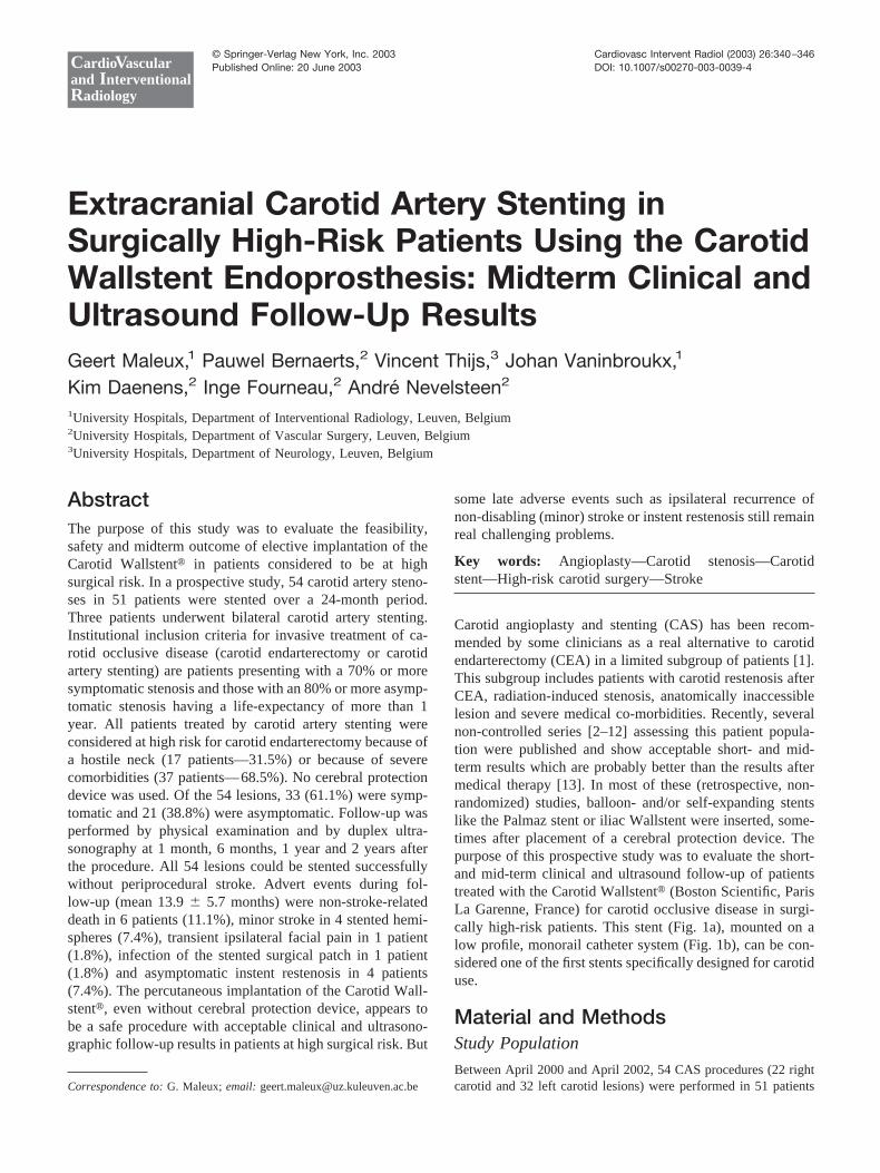
Carotid angioplasty and stenting (CAS) has been recom-mended by some clinicians as a real alternative to carotidendarterectomy (CEA) in a limited subgroup of patients [1].This subgroup includes patients with carotid restenosis afterCEA, radiation-induced stenosis, anatomically inaccessiblelesion and severe medical co-morbidities. Recently, severalnon-controlled series [2–12] assessing this patient popula-tion were published and show acceptable short- and mid-term results which are probably better than the results aftermedical therapy [13]. In most of these (retrospective, non-randomized) studies, balloon- and/or self-expanding stentslike the Palmaz stent or iliac Wallstent were inserted, some-times after placement of a cerebral protection device. Thepurpose of this prospective study was to evaluate the short-and mid-term clinical and ultrasound follow-up of patientstreated with the Carotid Wallstent� (Boston Scientific, ParisLa Garenne, France) for carotid occlusive disease in surgi-cally high-risk patients. This stent (Fig. 1a), mounted on alow profile, monorail catheter system (Fig. 1b), can be con-sidered one of the first stents specifically designed for carotiduse.
Material and MethodsStudy Population
Between April 2000 and April 2002, 54 CAS procedures (22 rightcarotid and 32 left carotid lesions) were performed in 51 patientsCorrespondence to: G. Maleux;email: [email protected]
CardioVascularand InterventionalRadiology
© Springer-Verlag New York, Inc. 2003 Cardiovasc Intervent Radiol (2003) 26:340–346Published Online: 20 June 2003 DOI: 10.1007/s00270-003-0039-4

(36 male, 15 female) (mean age 72.7 years, range 51–88 years). Inthe same period of time, 199 CEA were performed in our institu-tion. According to a strict institutional protocol for the treatment ofcarotid occlusive disease, we include for invasive (non-medical)treatment of carotid occlusive disease (CEA or CAS) patientspresenting with a 70% or more symptomatic stenosis and alsopatients presenting with an 80% or more asymptomatic stenosis,having a life-expectancy of more than 1 year. Informed consent wasobtained for each patient. Indications in favor of CAS versus CEAwere hostile neck in 17 patients (31.5%) and various severe co-morbidities in 37 patients (68.5%) (Table 1). We treated 33 symp-tomatic stenoses (61.1%) and 21 asymptomatic stenoses (38.8%);their etiology was atherosclerosis in 40 lesions, radio-arteritis in 8and post-CEA fibrosis in 6 lesions. The degree of carotid arterystenosis, measured by duplex ultrasound and angiography follow-ing the NASCET-criteria, was �90% in 24 carotids and 80% in 29carotids. Despite our strict institutional protocol, it was also decidedto stent one patient presenting with a 50–70% symptomatic com-mon carotid artery stenosis.
Antithrombotic Protocol
A combination of a subcutaneous injection of tinzaparinum (175IU/kg/24 hours) and a peroral dose of clopidogrel 75 mg wasadministered 1 day before and the day of the procedure, andclopidogrel 75 mg was continued for 1 month after the procedure.Afterwards, only aspirin 160 mg/day was continued for life. Duringthe intervention, 5000 IU of heparin were given intraarterially oncethe sheath was placed in the groin.
Stenting Technique
Before the intervention can be started a vascular imaging work-upis needed. It consisted of a duplex ultrasound of both carotidbifurcations and a vascular mapping of the aortic arch and allsupra-aortic vessels as well as of the intracerebral circulation. Thismapping was done by contrast-enhanced MR angiography and/orby conventional 4-vessel angiography. A team consisting of aninterventional radiologist and a vascular surgeon performed theCAS procedure in the angiography suite (Angiostar Plus, Siemens,Erlangen, Germany). All patients were monitored by noninvasiveblood pressure monitoring, oxymetry and continuous electrocardi-ography. No sedation was administered.
After local anesthesia of lidocaine, vascular access was made byplacing a 7 or 8 French sheath into the femoral artery. Afterselective catheterization of the ipsilateral common or external ca-rotid artery, a 7 or 8 Fr guiding catheter was placed into thecommon carotid artery. Under road-mapping, the stenosis wascrossed by a 0.014 floppy-tipped guidewire (Patriot, Boston Scien-tific, Paris La Garenne, France), followed by predilatation with alow profile monorail balloon catheter (Gazelle, Boston Scientific,Paris La Garenne, France) 3 mm in diameter. Before the predila-tation, 0.125 mg of atropine was injected intravenously. Then theself-expanding carotid stent (Carotid Wallstent�, Boston Scientific,Paris La Garenne, France), mounted on a 6 Fr monorail catheter,was placed under road mapping. After a second intravenous injec-tion of 0.125 mg of atropine, the post-dilatation, using the sametype of monorail balloon catheter (Gazelle, Boston Scientific, ParisLa Garenne or Viatrac, Guidant Europe, Diegem, Belgium) wasperformed to create a luminal stent diameter of 5, 6 or 7 mm,depending on the diameter of the vessel proximal and distal to thestented segment. A completion angiography of the cervical carotidartery was than done before removing the guiding catheter andsheath.
Follow-Up and Study Endpoints
All patients underwent a complete neurological examination by anindependent neurologist and a cerebral CT scan the day before andafter the procedure and a carotid duplex ultrasound at discharge. Afollow-up physical examination and a carotid duplex US were done1 month, 6 and 12 months after the procedure and thereafter eachyear. Repeat invasive angiography was done only if a re-interven-tion was planned.
Statistical Analysis
Life table analysis: cumulative survival rate free of death, restenosisand neurological events [14].
Fig. 1. A, B The Carotid Wallstent is delivered by withdrawal of the outer sheath (arrowheads) of monorail stent-deliverycatheter. The arrow shows the point where the 0.014 wire comes out of the monorail-catheter.
Table 1. Indications in favor of CAS vs CEA
No. of patients
Hostile neckPost-radiation stenosis 8Post-CEA stenosis 6Ankylosing spondilytis 1Short, obese neck 2
Severe co-morbiditiesArterial hypertension 20Ischemic cardiomyopathy 24Dilated cardiomyopathy 6Atrial flutter 3Angor pectoris 5Unstable angina 1Contralateral carotid occlusion 3COPD 8
G. Maleux et al.: Extracranial Carotid Artery Stenting 341

ResultsIn 51 patients, 54 high-grade carotid occlusive lesions werestented. Three patients were stented bilaterally. In only 1patient a bilateral carotid artery stenosis was treated at thesame sitting. Nine lesions (16.6%) were located in the com-mon carotid artery (CCA) (Fig. 2a, b), 36 lesions (66.6%)involved the carotid bifurcation (CB) (Fig. 3a, b) and 9stenoses (16.6%) were located in the post-bulbar portion ofthe internal carotid artery (ICA) (Fig. 4a, b). In these patientsthe stenosis did not extend into the bifurcation. Thirty-threestenoses (61.1%) were symptomatic and 21 (38.8%) wereasymptomatic. In 2 lesions, 2 stents were needed to entirelycover the stenosis and the adjacent kinking. The kinkingoriginated above the first stent as a result of poor placementof the initial stent. Overall, no periprocedural stroke orcardiac disorder was noted. Six patients developed unilateralor bilateral shortlasting convulsive movements that resolvedimmediately and completely after deflation of the post-dila-tation balloon.
Cerebral CT scan as well as neurological examinationafter the procedure did not reveal any new neurologicaldeficit or silent cerebral infarction compared to the neuro-logical status or cerebral CT scan before the stenting proce-dure. One patient presented with transient ipsilateral facialpain immediately after the procedure, but no cerebral lesion(in the stented hemisphere) could be detected by CT scan orby extensive neurological examination. Although the exter-nal carotid artery was still patent after stent placement, aresidual ostial high-grade stenosis was depicted, whereas
before stenting, only a low-grade stenosis was noted. Al-though we have no real explanation for this event, we hy-pothesize that a periprocedural embolization occurred intothe bridged external carotid artery.
Apart from the patients who died during follow-up, allpatients presented for the follow-up physical examinationsand underwent the scheduled duplex ultrasound examina-tions. During follow-up (mean 13.9 � 5.7 months) 6 patients(11.6%) died of different non-stroke-related causes: 4 ofmyocardial infarction 3 days, 1.5, 6 and 9 months, respec-tively, after the procedure and 2 died of pneumonia anddeterioration of the general status 2 and 3 months, respec-tively, after the intervention. In all these patients no neuro-logical deficit was detected up to their exitus.
Clinical follow-up revealed 2 recurrent (minor) strokesoccurring 3 and 5 months after stenting. Duplex ultrasoundin these 2 patients, symptomatic before CAS, showed a fullypatent stent without a recurrent stenosis below or above thestenosis. The patients were treated conservatively and recov-ered without sequelae of this new stroke. Two other patients,also symptomatic prior to CAS, developed an ipsilateralhazy view 2 weeks and 1 month, respectively, after theprocedure; ophthalmologic examination confirmed the pres-ence of small emboli in the ipsilateral ophthalmic arteryprovoking a visual field cut. These symptoms also disap-peared almost entirely after several weeks. Overall, we de-tected 4 (7.4%) minor (ipsilateral) strokes during follow-up.
Ultrasound follow-up detected 4 asymptomatic in-stentrestenoses (7.4%) in 2 patients after 6 months (50% and90%, respectively, restenosis of the stent diameter) and inanother 2 patients after 1 year (50 and 90% restenosis,respectively). The 50% restenoses were followed up withoutother surgical or interventional procedure; the 2 high-gradestenoses were successfully restented (Fig. 5a, b, c).
Finally 1 patient, who had undergone carotid stentingbecause of a tight post patch-CEA restenosis, presented withan infected cutaneous fistula at the site of the previous CEA5 months after CAS. Ultrasound and angiography revealeddiscrete instent neointima without significant stenosis, butinfection of the surgical Dacron patch required surgicalresection of the stent and interposition of a venous graft.
The cumulative survival rate free of death, restenosis andneurological events is graphically shown by a failure timeanalysis (Fig. 6).
DiscussionAlthough CAS becomes more and more accepted as analternative to CEA in specific clinical subgroups of patientswith post-CEA restenosis [2–4], radiation-induced stenosis[5–7] surgically inaccessible (high, low or tandem) lesions,substantial coexistent morbidities [8–10] or contralateralocclusion [11, 12], there is still continuous and intensiveresearch ongoing to optimize the stent design, to decrease theprofile of guidewires, guiding catheters and stent deliverycatheters, all in order to reduce potential complications dur-
Fig. 2. A, B Selective angiography of the right CCA showsthe radiation-induced high-grade stenosis (arrows) of the leftCCA before and after stenting.
342 G. Maleux et al.: Extracranial Carotid Artery Stenting

ing or after the CAS procedure. Historically, the Palmazstent was the first stent extensively studied in the treatmentof carotid artery stenosis [15, 16]. The most important dis-advantages of this type of stent were the rigidity and uncon-formability to the internal and common carotid artery wall,potentially provoking vessel kinking or vessel wall dissec-tion below or above the stented segment, and the tendency tobe compressed or deformed irreversibly during follow-up[17–19]. Subsequently, the more flexible stainless steelWallstent (Boston Scientific) [6, 8, 10, 20, 21] and nitinolSmart stent (Cordis) [22, 23] were used without the men-
tioned drawbacks. Using these stents, periprocedural strokeswere uncommon, but still noted probably due to the rigidityof the balloon and stent delivery catheter which have asubstantial profile (7 Fr) and the tendency to provoke em-bolization of plaques during stent positioning and deploy-ment. In order to make the stent and stent-delivery systemmore flexible, less profiled and easier to manipulate, theCarotid Wallstent� was one of the first stents specificallydesigned for carotid occlusive disease. Its design is compa-rable with that of the iliac Wallstent although the stainlesssteel meshwork is more flexible and narrower than its coun-
Fig. 3. A, B Profile angiogramof the right carotid bifurcationdemonstrates the ostial topostostial high-grade stenosis(arrows) of the right ICA in apatient with ischemiccardiomyopathy before andafter stent implantation. Notethat the stent (arrowheads) isbridging the ostium of theexternal carotid artery.
Fig. 4. A, B Profile angiogramof the right carotid bifurcationshows the bulbar to post-bulbarhigh-grade and ulceratedstenosis (arrows) of the rightICA as a result of radiation-induced arteritis, before andafter stenting. The CarotidWallstent ends (arrowheads) atthe ostium of the ICA withoutbridging the external carotidartery.
G. Maleux et al.: Extracranial Carotid Artery Stenting 343

terpart to prevent embolic complications, but on the otherhand a more important foreshortening is noted.
As opposed to most other published series on CAS, noperiprocedural stroke occurred even though no cerebropro-tection device was used. It can be hypothesized that thereduced size of the guidewire, balloon- and stent-deliverysystem used in this series are less traumatic to the stenotic
plaque and therefore less embolic. Nevertheless 4 patientshad a recurrent ipsilateral stroke during follow-up: 2 pre-sented with ipsilateral visual problems within the first monthafter stenting caused by embolization in the ipsilateral oph-thalmic artery. Two other patients presented with a contralat-eral paresis, respectively 3 and 5 months after the procedure.The symptoms of late stroke in these 4 patients disappeared
Fig. 5. A–C Intimal hyperplasiadetected 6 months after stentplacement and treated by bal-loon angioplasty (B) and restent-ing (C).
Fig. 6. Failure time analysis.
344 G. Maleux et al.: Extracranial Carotid Artery Stenting

almost completely after several weeks and repeat duplexultrasound of the stented carotid artery at the moment ofstroke was within normal limits.
These late recurrences of stroke, already noted by otherauthors [9, 19, 24, 25], are not yet extensively studied, but inour opinion, can be an important issue when CAS is evalu-ated as an alternative to CEA in surgically low-risk patients.The etiology of these recurrent strokes, occurring late afterthe procedure, is not very clear, but different hypotheses canbe made: small atherosclerotic debris can cross the sidemeshes of the stent; thrombi can be formed on the inner stentsurface even with adequate anticoagulant therapy; orthrombi or debris of another atherosclerotic plaque situatedbelow or above the stented segment might cause the distalembolization. In addition, some cardiac diseases such asatrial flutter can provoke thrombus embolizing to the brain.Last but not least, non-embolic stroke is also a potentialcause of acute brain ischemia in a previously stented hemi-sphere. In contrast to other published CAS series, we have avery high death ratio (11.6%), but this is most likely due tothe treated study population: 37 of the stented patients(68.5%) were at high-risk for CEA because of severe con-comitant co-morbidities and 8 patients (15.6%) had an on-cologic past medical history and were treated by radiationtherapy.
Another issue of concern for CAS seems to be the instentrestenosis due to intimal hyperplasia. In our series using theCarotid Wallstent� we found recurrent stenosis in 7.4%which is in the same range as several other published series[9, 21, 26] and this percentage seems to be independent ofthe type of stent implanted [23]. No real long-term data existon the outcome of restenting a recurrent instent stenosiscompared to the outcome of conservative treatment; weprefer to perform a restenting procedure.
The potential benefit for treating asymptomatic patientsby stent placement remains unclear, although the periproce-dural event rate in the present study is very low. But byanalogy with the ACAS-trial [27] and meta-analysis studiesfrom the Cochrane database [28], which demonstrated aslight long-term reduction of ipsilateral stroke after carotidendarterectomy (if periprocedural complication rate was�3%), it can be suggested that carotid artery stenting inasymptomatic patients can have a potential benefit, but thishas to be confirmed by large, multicenter randomized trialscomparing CAS to CEA or medical therapy. The overallclinical and US follow-up results of the treatment of carotidocclusive disease with CAS using the Carotid Wallstents arecertainly better than the results after medical therapy forsymptomatic patients [13]. We conclude that CAS can bepromoted in symptomatic patients considered at high surgi-cal risk.
The limitation of the trial is, of course, the limited numberof patients included and the fact that this is only a feasi-bility study without randomized comparison to carotidendarterectomy. These preliminary results with this spe-cific carotid stent are promising especially with regard to
the absence of periprocedural stroke complications evenwithout the use of a cerebroprotection device, but latecomplications such as ipsilateral recurrent stroke and in-stent restenosis remain the major concerns of the newtreatment modality.
Using the Carotid Wallstent� for the percutaneous treat-ment of carotid occlusive disease, encouraging clinical, ul-trasonographic and excellent periprocedural results arefound for the treatment of patients at high surgical riskalthough some late complications such as recurrent ipsilat-eral (minor) stroke and instent restenosis can occur.
References1. Veith FJ, Amor M, Ohki T, et al. (2001) Current status of carotid
bifurcation angioplasty and stenting based on a consensus of opinionleaders. J Vasc Surg 33:S111–116
2. Vitek JJ, Roubin GS, New G, et al. (2001) Carotid angioplasty withstenting in post-carotid endarterectomy restenosis. J Invasive Cardiol13:123–125
3. New G, Roubin GS, Iyer SS, et al. (2000) Safety, efficacy and durabilityof carotid artery stenting for restenosis following carotid artery endar-terectomy: A multicenter study. J Endovasc Ther 7:354–352
4. Lanzino G, Mericle RA, Lopes DK, et al. (1998) Percutaneous trans-luminal angioplasty and stent placement for recurrent carotid arterystenosis. J Neurosurg 90:688–694
5. Alric P, Branchereau P, Berthet JP, et al. (2002) Carotid artery stentingfor stenosis following revascularization or cervical irradiation. J Endo-vasc Ther 9:14–19
6. Houdart E, Mounayer C, Chapot R, et al. (2001) Carotid stenting forradiation-induced stenoses: A report of 7 cases. Stroke 32:118–121
7. Al-Mubarak N, Roubin GS, Iyer SS, et al. (2000) Carotid stenting forsevere radiation-induced extracranial carotid artery occlusive disease. JEndovasc Ther 7:36–40
8. Waigand J, Gross CM, Kramer J, et al. (1998) Elective stenting ofcarotid artery stenosis in patients with severe coronary artery disease.Eur Heart J 19:1365–1370
9. Paniagua D, Howell M, Strickman N, et al. (2001) Outcomes followingextracranial carotid artery stenting in high-risk patients. J InvasiveCardiol 13:375–381
10. Teitelbaum GP, Lefkowitz MA, Giannotta SL (1998) Carotid angio-plasty and stenting in high-risk patients. Surg Neurol 50:300–312
11. Mathur A, Roubin GS, Gomez CR, et al. (1998) Elective carotid arterystenting in the presence of a contralateral occlusion. Am J Cardiol81:1315–1317
12. Mericle RA, Kim SH, Lanzino G, et al. (1999) Carotid artery angio-plasty and use of stents in high-risk patients with contralateral occlu-sions. J Neurosurg 90:1031–1036
13. North American Symptomatic Carotid Endartectomy Trial Collabora-tors (1991) Beneficial effect of carotid endartectomy in symptomaticpatients with high-grade carotid stenosis. N Engl J Med 325:445–453
14. Stat Soft, Inc. (2002) Electronic Statistics Textbook. Tulsa. Web:http://www.Statsoft.com/textbook/stathome.html
15. Bergeron P, Becquemin JP, Jausseran JM, et al. (1999) Percutaneousstenting of the internal carotid artery: The European CAST I Study—Carotid Artery Stent Trial. J Endovasc Surg 6:155–159
16. Dietrich EB, Ndiaye M, Reid DB (1996) Stenting in the carotid artery:Initial experience in 110 patients. J Endovasc Surg 3:42–62
17. Mathur A, Dorros G, Iyer SS, et al. (1997) Palmaz stent compression inpatients following carotid artery stenting. Cathet Cardiovasc Diagn41:137–140
18. Calvey TA, Gough MJ (1998) A late complication of internal carotidartery stenting. J Vasc Surg 27:753–755
19. Wholey MH, Wholey MH, Tan WA, et al. (2001) Management ofneurological complications of carotid artery stenting. J Endovasc Ther8:341–353
20. Brooks WH, McClure RR, Jones MR, et al. (2001) Carotid angioplastyand stenting versus carotid endarterectomy: Randomized trial in acommunity hospital. JACC 38:1589–1595
21. D’Audiffret A, Desgranges P, Kobeiter H, et al. (2001) Technical
G. Maleux et al.: Extracranial Carotid Artery Stenting 345

aspects and current results of carotid stenting. J Vasc Surg 33:1001–1007
22. Drescher R, Mathias KD, Jaeger JH, et al. (2002) Clinical results ofcarotid artery stenting with a nitinol self-expanding stent (SMARTstent). Eur Radiol 12:2451–2456
23. Mukherjee D, Kalahasti V, Roffi M, et al. (2001) Self-expanding stentsfor carotid interventions: Comparison of nitinol versus stainless-steelstents. J Invasive Cardiol 13:732–735
24. Yadav JS, Roubin GS, Iyer SS, et al. (1997) Elective stenting of theextracranial carotid arteries. Circulation 95:376–381
25. Parodi JC, Schonholz CJ, Ferreira M, et al. (2002) Parodi antiembolism
system in carotid stenting: The first 100 patients (abstract). J VascIntervent Radiol 13:S27
26. Chaktoura EY, Hobson RW II, Goldstein J, et al. (2001) In-stentrestenosis after carotid angioplasty-stenting: Incidence and manage-ment. J Vasc Surg 33:220–226
27. ACAS-Investigators (1995) Endarterectomy for asymptomatic carotidartery stenosis. Executive Committee for the Asymptomatic CarotidAtherosclerotic Study (1995). JAMA 273:1421–1428
28. Chambers BR, You RX, Donnan GA (2000) Carotid endarterectomyfor asymptomaticcarotid stenosis. Carotid Database Syst Rev (2) CD001923
346 G. Maleux et al.: Extracranial Carotid Artery Stenting
Related Documents