ORIGINAL PAPER Extracellular vesicles shed by Trypanosoma cruzi are linked to small RNA pathways, life cycle regulation, and susceptibility to infection of mammalian cells Maria R. Garcia-Silva & Roberta Ferreira Cura das Neves & Florencia Cabrera-Cabrera & Julia Sanguinetti & Lia C. Medeiros & Carlos Robello & Hugo Naya & Tamara Fernandez-Calero & Thais Souto-Padron & Wanderley de Souza & Alfonso Cayota Received: 10 May 2013 /Accepted: 14 October 2013 /Published online: 17 November 2013 # Springer-Verlag Berlin Heidelberg 2013 Abstract The protozoan parasite Trypanosoma cruzi has a complex life cycle characterized by intracellular and extracellular forms alternating between invertebrate and mammals. To cope with these changing environments, T. cruzi undergoes rapid changes in gene expression, which are achieved essentially at the posttranscriptional level. At present, expanding families of small RNAs are recognized as key players in novel forms of posttranscriptional gene regulation in most eukaryotes. However, T. cruzi lacks canonical small RNA pathways. In a recent work, we reported the presence of alternate small RNA pathways in T. cruzi mainly represented by a homogeneous population of tRNA- derived small RNAs (tsRNAs). In T. cruzi epimastigotes submitted to nutrient starvation, tsRNAs colocalized with an argonaute protein distinctive of trypanosomatids (TcPIWI-tryp) and were recruited to particular cytoplasmic granules. Using epifluorescence and electronic microscopy, we observed that tsRNAs and the TcPIWI-tryp protein were recruited mainly to reservosomes and other intracellular vesicles including endosome-like vesicles and vesicular structures resembling the Golgi complex. These data suggested that, in T. cruzi , tsRNA biogenesis is probably part of endocytic/exocytic routes. We also demonstrated that epimastigotes submitted to nutrient starvation shed high levels of vesicles to the extracellular medium, which carry small tRNAs and TcPIWI- tryp proteins as cargo. At least a fraction of extracellular vesicle cargo was transferred between parasites and to mammalian susceptible cells. Our data afford experimental evidence, indicating that extracellular vesicles shed by T. cruzi promote not only life cycle transition of epimastigotes to trypomastigote forms but also infection susceptibility of mammalian cells Introduction Trypanosoma cruzi , which is responsible of Chagas’ disease, has a complex life cycle including intracellular and extracellular forms, which alternate between invertebrate insect vectors belonging to the subfamily Triatominae and mammalian hosts including humans (Barrett et al. 2003; de Souza 2008). Life Electronic supplementary material The online version of this article (doi:10.1007/s00436-013-3655-1) contains supplementary material, which is available to authorized users. M. R. Garcia-Silva : F. Cabrera-Cabrera : J. Sanguinetti : A. Cayota (*) Functional Genomics Unit, Institut Pasteur de Montevideo, Mataojo 2020, Montevideo CP 11400, Uruguay e-mail: [email protected] R. F. Cura das Neves : T. Souto-Padron Laboratório de Biologia Celular e Ultraestrutura, Instituto de Microbiologia Paulo de Góes, Universidade Federal do Rio de Janeiro, Rio de Janeiro, Brazil L. C. Medeiros : W. de Souza Laboratório de Ultraestrutura Celular Hertha Meyer, Instituto de Biofísica Carlos Chagas Filho, Universidade Federal do Rio de Janeiro, Rio de Janeiro, Brazil C. Robello Molecular Biology Unit, Institut Pasteur de Montevideo, Montevideo, Uruguay H. Naya : T. Fernandez-Calero Bioinformatics Unit, Institut Pasteur de Montevideo, Montevideo, Uruguay A. Cayota Department of Medicine, Faculty of Medicine, Montevideo, Uruguay Parasitol Res (2014) 113:285–304 DOI 10.1007/s00436-013-3655-1

Welcome message from author
This document is posted to help you gain knowledge. Please leave a comment to let me know what you think about it! Share it to your friends and learn new things together.
Transcript
ORIGINAL PAPER
Extracellular vesicles shed by Trypanosoma cruzi are linkedto small RNA pathways, life cycle regulation, and susceptibilityto infection of mammalian cells
Maria R. Garcia-Silva & Roberta Ferreira Cura das Neves & Florencia Cabrera-Cabrera &
Julia Sanguinetti & Lia C. Medeiros & Carlos Robello & Hugo Naya &
Tamara Fernandez-Calero & Thais Souto-Padron & Wanderley de Souza & Alfonso Cayota
Received: 10 May 2013 /Accepted: 14 October 2013 /Published online: 17 November 2013# Springer-Verlag Berlin Heidelberg 2013
Abstract The protozoan parasite Trypanosoma cruzi has acomplex life cycle characterized by intracellular andextracellular forms alternating between invertebrate andmammals. To cope with these changing environments, T.cruzi undergoes rapid changes in gene expression, which areachieved essentially at the posttranscriptional level. Atpresent, expanding families of small RNAs are recognized
as key players in novel forms of posttranscriptional generegulation in most eukaryotes. However, T. cruzi lackscanonical small RNA pathways. In a recent work, we reportedthe presence of alternate small RNA pathways in T. cruzimainly represented by a homogeneous population of tRNA-derived small RNAs (tsRNAs). In T. cruzi epimastigotessubmitted to nutrient starvation, tsRNAs colocalized with anargonaute protein distinctive of trypanosomatids (TcPIWI-tryp)and were recruited to particular cytoplasmic granules. Usingepifluorescence and electronic microscopy, we observed thattsRNAs and the TcPIWI-tryp protein were recruited mainly toreservosomes and other intracellular vesicles includingendosome-like vesicles and vesicular structures resemblingthe Golgi complex. These data suggested that, in T. cruzi ,tsRNA biogenesis is probably part of endocytic/exocyticroutes. We also demonstrated that epimastigotes submitted tonutrient starvation shed high levels of vesicles to theextracellular medium, which carry small tRNAs and TcPIWI-tryp proteins as cargo. At least a fraction of extracellularvesicle cargo was transferred between parasites and tomammalian susceptible cells. Our data afford experimentalevidence, indicating that extracellular vesicles shed by T.cruzi promote not only life cycle transition of epimastigotesto trypomastigote forms but also infection susceptibility ofmammalian cells
Introduction
Trypanosoma cruzi , which is responsible of Chagas’ disease,has a complex life cycle including intracellular and extracellularforms, which alternate between invertebrate insect vectorsbelonging to the subfamily Triatominae and mammalian hostsincluding humans (Barrett et al. 2003; de Souza 2008). Life
Electronic supplementary material The online version of this article(doi:10.1007/s00436-013-3655-1) contains supplementary material,which is available to authorized users.
M. R. Garcia-Silva : F. Cabrera-Cabrera : J. Sanguinetti :A. Cayota (*)Functional Genomics Unit, Institut Pasteur de Montevideo,Mataojo 2020, Montevideo CP 11400, Uruguaye-mail: [email protected]
R. F. Cura das Neves : T. Souto-PadronLaboratório de Biologia Celular e Ultraestrutura,Instituto de Microbiologia Paulo de Góes,Universidade Federal do Rio de Janeiro, Rio de Janeiro, Brazil
L. C. Medeiros :W. de SouzaLaboratório de Ultraestrutura Celular Hertha Meyer,Instituto de Biofísica Carlos Chagas Filho,Universidade Federal do Rio de Janeiro, Rio de Janeiro, Brazil
C. RobelloMolecular Biology Unit, Institut Pasteur de Montevideo,Montevideo, Uruguay
H. Naya : T. Fernandez-CaleroBioinformatics Unit, Institut Pasteur de Montevideo,Montevideo, Uruguay
A. CayotaDepartment of Medicine, Faculty of Medicine,Montevideo, Uruguay
Parasitol Res (2014) 113:285–304DOI 10.1007/s00436-013-3655-1

cycle transitions start when the noninfective epimastigote formsproliferate in the midgut of the reduvii bug and advance to thehind gut of the vector. Under these circumstances,epimastigotes suffer a severe nutritional stress, which drivesits differentiation into the mammalian-infective metacyclictrypomastigotes (Contreras et al. 1985; Figueiredo et al.2000). To cope with these changing environments, T. cruzimust undergo rapid and significant changes in gene expression,which are achieved essentially at the posttranscriptional levelthrough modulation of messenger RNA (mRNA) stability andtranslational control mechanisms (Clayton and Shapira 2007).Nevertheless, the precise mechanisms of gene expressionregulation in these parasites remain to be completelyelucidated.
Over the last decade, an expanding family of smallregulators RNAs (i.e., microRNAs, small interfering RNAs,and Piwi-interacting RNAs) were recognized as key players innovel forms of posttranscriptional gene regulation in mosteukaryotes (Ghildiyal and Zamore 2009). Soon after its initialdescription, the presence and relevance of small RNApathways in the control of posttranscriptional gene regulationwere reported for virus, bacteria, and most eukaryotes.However, the machinery associated with small RNAbiogenesis was thought to be either entirely lost or extensivelysimplified in some unicellular organisms, including T. cruzi ,Saccharomyces cerevisiae , Leishmania major , andPlasmodium falciparum (Cerutti and Casas-Mollano 2006).
In a recent work aimed to identify the presence of alternatesmall RNA pathways that could contribute to theposttranscriptional control in T. cruzi , we reported the presenceof a homogeneous population of small RNAs derived frommature transfer RNAs (tRNAs) representing from 25 to 30% ofthe small RNA population. The levels of these tRNA-derivedhalves (tsRNAs) were more pronounced in parasitesundergoing nutritional stress and more than 98 % of clonedtsRNAs derived from the 5′ halves of a restricted group oftRNA issoaceptors (Garcia-Silva et al. 2010a). Surprisingly,tsRNAs were recruited to particular cytoplasmic organelles inT. cruzi epimastigotes, which colocalized at least partially withreservosomes. In trypomastigotes, tsRNAs represented about62 % of small RNAs and were mainly derived from the 3′ armof precursors tRNAs (Reifur et al. 2012). Of note, small RNAsderived from tRNAs have been recently reported by severalgroups as a novel population of small regulators RNAs (Coleet al. 2009; Haussecker et al. 2010; Thompson and Parker2009b), which appear as the most broadly conserved pathwayof small RNAs regulating gene expression and frequentlyinitiated after nutritional, biological, or physicochemical stressin prokaryotes as well as in eukaryotes.
The present work was aimed to characterize the cytoplasmicgranular structures recruiting tRNA fragments and to gaininsights in the biological significance of this small RNApopulation in the protozoan parasite T. cruzi .
Here, we report that tsRNAs were actively produced inepimastigotes undergoing nutritional stress and recruited tocytoplasmic organelles associated with clathrin and with theT. cruzi argonaute protein TcPIWI-tryp (Garcia-Silva et al.2010b). Transmission electron microscopy further confirmedthat tsRNAs colocalized with TcPIWI-tryp in reservosomes,uncharacterized intracellular vesicles, and Golgi-like vesicularstructures. Vesicles carrying small tRNAs and TcPIWI-trypproteins were secreted to the extracellular medium and actedas vehicles for the transfer of these molecules to otherparasites and to mammalian susceptible cells but not tononsusceptible cells. Our data provide experimental evidenceindicating that vesicles shed by T. cruzi were not onlyassociated with life cycle transition of epimastigotes towardthe infective trypomastigote forms but associated to infectionsusceptibility of mammalian cells.
Materials and methods
Parasites and infection assays Epimastigote forms of the T.cruzi Dm 28c clone (Contreras et al. 1988) were cultured andexpanded axenically at 28 °C by weekly transfers in liverinfusion tryptose (LIT) medium supplemented with 10 %heat-inactivated fetal bovine serum. Parasites at the late-stationary phase of growth (1×10⁹ parasites ml−1) werewashed twice and resuspended in serum free or 1 % fetalbovine serum (FBS) RPMI-1640 medium at 5.107×ml−1 andfurther cultured for 24 or 48 h. Parasite viability wasmonitored by light microscopy through the analysis ofmotility and by flow cytometry with propidium iodide, whichshowed >98 % viable parasites within over a period of 72 h.
As we have previously reported (Garcia-Silva et al. 2010a),the optimal conditions to induce small tRNA production andvesicle secretion are achieved when parasites were cultured inserum free or up to 1 % FBS RPMI medium. For extendedperiods of cultures media were supplemented with 1% FBS inorder to assure viability higher than 98 %. Parasites underexponential growing or late-stationary phase do not efficientlyproduce and secrete adequate amounts of tRNA halves andvesicles. As reported (Hernandez et al. 2012), this nutrientstarvation has been recognized as an important conditioninducing a strong nutritional stress promoting the emergenceof a significant fraction of infective trypomastigotes. Thedynamic of cytoplasmic accumulation of tsRNA in unstressedand stressed epimastigotes compared to trypomastigotes wasassessed by immunofluorescence (Supplementary Fig. 1). Asdepicted in Supplementary Fig. 2, epimastigotes becomeprogressively transformed in trypomastigotes over timerepresenting about 30 % of parasites after 48 h, which wereinfective for the Vero (monkey kidney epithelial cells) andHeLa (human cervical cancer cells) cell lines. Parasitessubmitted to nutrient starvation for 24 h (sE24) were used
286 Parasitol Res (2014) 113:285–304

mainly for subcellular localization assays (where accumulationof signals for tRNA halves reach to a maximum), and thosecultured for 48 h (sE48) were used selectively for infectionassays (highly infective with a high ratio of trypomastigotes).Infection assays were performed by incubating sE48 parasiteswith cells at 1:20 cell/parasite ratio for 4 h at 37 °C in serum-free RPMI and washed twice with phosphate-buffered saline(PBS). For nonsusceptible wild-type or electroporated K-562cells (electroporation control and treated cells) infection assayswere prolonged up to 24 h.
Labeling of parasites by transfection with fluorescentsynthetic tsRNAs Epimastigotes at the late-stationary phase ofgrowth (1×10⁹ ml−1) were transfected with synthetic Cy3-labeled oligoribonucleotides (IDT Inc., IA, USA) chemicallymodified to avoid degradation by ribonucleases (terminalphosphorothioate bonds and 2′ O -methyl ribonucleotides).Probes used were the sense 5′-derived tRNA halves fromtRNAAsp
GUC (5′ CTC GGT AGT ATA GTG GTA AGT ATACCC 3′) and from tRNAGlu
CUC (5′ CGG TGTGGTATAGTGGTTAGAACAAGCGGCT 3′). About 1×10⁸ parasites wereresuspended in 0.4 ml of Hepes buffer solution (21 mMHepes,137 mM NaCl2, 5 mM KCl, and 6 mM glucose, pH 7.4) andelectroporated in 0.4-cm cuvettes in the presence of 100 nM ofthe fluorescent probe using an ECM-600 ElectroporationSystem (BTX CA, USA). Parasites were submitted to two450 V/500 μF pulses and allowed to recuperate for 24 h inLIT 10%FBS before use. Nonelectroporated parasites culturedin identical conditions and exposed to equal concentrations ofthe fluorescent probe were used as controls to exclude theuptake of the free probe (soaking). Nonelectroporated controlparasites were followed over a period of 2–24 h withoutevidences of detectable intracellular fluorescent signals.
Immuno f l uore scence and f l uo re scence in s i t uhybridization Indirect immunofluorescence microscopy(IIFM) was used to visualize clathrin and TcPIWI-trypproteins using unconjugated specific antibodies andsecondary antibodies conjugated to either FITC or Alexa-Fluor 488 dyes. The monoclonal anticlathrin antibody (kindlygifted by Dr. Maurilio Soares, ICC, Curitiba Brazil) was usedat 1:300, and the anti-TcPIWI-tryp antibody was used at1:200. The rabbit anti-TcPIWI-tryp polyclonal antibodieswere produced and characterized elsewhere (Garcia-Silvaet al. 2010b). For immunofluorescence, harvested parasiteswere washed and fixed in 4 % of buffered formaldehyde,freshly prepared from paraformaldehyde, for 10 min at roomtemperature, washed twice with PBS, and blocked byincubating in 25 mM NH4Cl for 10 min. Parasites wereallowed to adhere to 0.1 % poly-L-lysine-coated microscopeslides and permeabilized with 0.2 % Triton X-100 in PBS for5 min. Slides were then incubated with primary antibodies atthe indicated dilutions in PBS containing 0.2 % bovine serum
albumin (BSA) and 0.1 % Tween 20 (PBS-T) in for 2 h atroom temperature. After extensive washing in PBS-T, theslides were further incubated with fluorescent secondaryantibodies diluted 1:500 in PBS-T, for 1 h at roomtemperature. After washing, slides were incubated with 4,6-diamidino-2-phenylindole (DAPI) at 1 mg ml−1 for 5 min atroom temperature. Slides were mounted with 5 μl of FluorSave Reagent (DAKO) or 0.2 M N -propyl-gallate in glycerol-PBS (9:1). IIFM assays were performed alone or coupled tofluorescence in situ hybridization (FISH) to identify tRNAAsp-or tRNAGlu-derived 5′ halves by incubating the respectiveantisense oligoprobes directly on IIFM slides as previouslyreported (Garcia-Silva et al. 2010a). Briefly, parasites werefixed in 4 % para-formaldehyde and allowed to adhere topolylysine-coated slides. Samples were further permeabilizedwith 0.2 % Triton X-100 for 5 min. Slides were thenprehybridized at room temperature in 2 % BSA, 5×Denhardt, 4× saline-sodium citrate (SSC), and 35% deionizedformamide for 2 h. Hybridization was performed overnight atroom temperature in a humid chamber by the addition of1 ng ml−1 of the indicated oligonucleotide conjugated tofluorescein isothiocyanate (FITC). After hybridization, slideswere washed once in 2× SSC plus 50% deionized formamide,once in 2× SSC, once in 1× SSC plus 4,6-diamidino-2-phenylindole (DAPI) at 1 mg ml−1, and twice in 0.5× SSC.Merged images were obtained by superimposing the indicatedimages files. The hybridization probes used were 5′ CGGGTA TAC TTA CCA CTA TAC TAC CGA 3 ′(tRNAAsp
GUC) and 5′CGC TTG TAG CCA CTA TAC CAC3′ (tRNAGlu
CUC). The respective scramble probes were usedas control probes. As previously reported (Garcia-Silva et al.2010a), the specificity and characterization of oligoprobesused in FISH assays were performed by using scramble and“bridging” control probes (recognizing only the anticodonloop of the respective full-length tRNA) without detectablereactivity with 5′ or 3′ halves. All hybridization wasperformed in nondenaturating conditions to avoid cross-hybridization with the respective full-length tRNA. FISHassays for TcPIWI mRNA were performed similarly totRNA halves detection except for the addition of aprehybridization (denaturing step) by heating slides at 75 °Cfor 15 min. Hybridization probes were antisense (AAG ATTTAC CCC ATG AGC GGG GTC CGT CTC AAATTT CG)and sense (CGAAAT TTGAGACGGACCCCGCTCATGGGG TAA ATC TTC). Control assays to evaluate thespecificity and reactivity of probes used to detect TcPIWI-tryp mRNA are depicted in Supplementary Fig. 3. Analyseswere performed in an Olympus IX 81 microscope, ZEISSAxioplan II microscope equipped with a Collor View XScamera, and in a Confocal Leica TCS SP5 AOBS coupled toa Hamamatsu Orca-ER camera (Diagnostic Instruments).Merged images were obtained by superimposing the indicatedimages files.
Parasitol Res (2014) 113:285–304 287

Clathrin and TcPIWI-tryp immunoprecipitation: TcPIWI-trypWestern blot For cell extracts, 1×10⁹ sE48 parasites werecollected by centrifugation, washed twice with PBS, andresuspended in 1 ml of hypotonic lysis buffer (50 mM Trisat pH 8.0, 100 mM NaCl, 3 mM MgCl2, 0.5 % NP-40, andprotease inhibitor cocktail). The lysates were centrifuged at15,000×g for 15 min at 4 °C to eliminate debris andaggregates. The total protein concentration of supernatantswas determined using the bicinchoninic acid protein assay(Sigma-Aldrich Co.) according to the manufacturer’sprotocol. Immunoprecipitation was performed by incubating30 μg of cell lysates with either anticlathrin antibodies diluted1:100 anti-TcPIWItryp serum diluted 1/100 or an isotype-matched control antibody for 2 h at 4 °C. After addition ofsecondary antibody-coated magnetic beads (Sigma-AldrichCo.), samples were further incubated overnight (Sigma) at4 °C with gentle agitation. The bound fraction was purifiedby immunomagnetic separation and washed three times in ice-cold hypotonic lysis buffer. The fraction of proteins and totalRNA bound to magnetic bead were isolated simultaneouslywith the Trizol reagent (Sigma-Aldrich Co.) according to themanufacturer’s protocol. The extracted proteins werefractionated by an 8% sodium dodecyl sulfate polyacrylamidegel electrophoresis (PAGE) and electrophoretically transferredto 0.45-μm nitrocellulose membranes. Western blot was doneas previously described (Garcia-Silva et al. 2010b). Thespecificity of anti-TcPIWI-tryp antibodies used was verifiedusing preimmune sera as negative control and by competitionwith the recombinant peptide used as immunogen. Theabsence of reactivity shown by the “unbound fraction” inFig. 1c further support the anti-TcPIWI-tryp specificity.Additionally, irrelevant primary isotype-matchedimmunoglobulins were used as controls for anticlathrin andanti-TcPIWI-tryp antibodies (data not shown).
RT-PCR of tRNA halves Twenty micrograms of total RNAobtained after clathrin and TcPIWItryp immunoprecipitationwas size-fractionated by denaturating 15 % PAGE, and thefraction from 20 to 60 nt was eluted from the gel andsubmitted to reverse transcription using the miScript kit(Qiagen) following the manufacturer’s recommendations.Amplification was performed using a universal reverse primerrecognizing a 3′ adaptor and specific locked nucleic acid(LNA)-enhanced forward primers (Exiqon) speciallydesigned for tRNAhalves. The primers usedwere (LNA basesrepresented by uppercase characters): tRNAAsp 5 ′ctcGgtaGtataGtg, tRNAAsp 3′ acGcgGgtga, and tRNAGlu 5′cGgtGtGgtata tRNAGlu 3′ tCacCcGctaga. Amplification wasperformed with 40 cycles of 15″ at 95 °C and 50″ at 56 °C.Visualization of amplicons was performed after migration in a15 % PAGE and stained with SYBRGold. In all cases, theidentity of amplicons was verified by direct sequencing ofpurified PCR products.
Exchange of tsRNAs and TcPIWI-tryp protein by extracellularvesicles These assays were designed to verify the eventualtransfer of small vesicle cargo from parasite to parasite and fromparasites to susceptible mammalian cells. For parasite-to-parasite exchange 2×10⁶ ml−1 unlabeled sE48 parasites wereincubated in serum-free RPMI supplemented with 0.24 ng ml−1
of isolated extracellular vesicles (EVs) derived from labeledsE48 parasites for 24 h at 28 °C. The EVs to parasite ratio usedwas designed in order to add an amount equivalent to thatestimated to be eventually produced by the target parasites tokeep a range of physiological concentrations. In ourexperimental conditions, we obtained an equivalent of 0.12 ngof protein in the purified vesicular fraction per million of sE48parasites. To further validate this transfer and to exclude theeventual presence of unwanted labeled parasites or cellulardebris in the purified vesicular fraction a series of transwellexperiments were performed. For this, the bottom chambers oftranswell plates (0.4 and 1 μm pore size HTS Transwell-24,Corning Inc.) were loaded with 2×10⁶ unlabeled sE48 parasitesin 1 ml of RPMI-1640 medium supplemented with 1 % ofvesicle-depleted FBS, while upper chambers were loaded with2×106 of labeled sE48 in 1 ml of the same medium. The sE48parasites were labeled with synthetic tRNA 5′ halves fromtRNAAsp conjugated to Cy3 as indicated above. Scrambled 5′halves from tRNAAsp were used as irrelevant control probes. Todeplete FBS of all potential vesicular content, FBS wassubmitted to two round of ultracentrifugation at 110,000×g for70 min at 4 °C and filtered through 1μm pore diameter filters toeventually exclude remnant cells or cellular debris. For parasite-to-cell assays, we replaced unlabeled parasites in the bottomchambers by 2×10⁵ Vero cells in 1 ml of the same medium.Transwell plates were then incubated at 28 °C for 24 h.
The transfer of labeled tRNA halves from the upper to thebottom chamber was monitored by epifluorescencemicroscopy of unlabeled parasites or Vero cells. As controls,unlabeled parasites at 2×10⁶ ml−1 or Vero cells at 2×10⁵ ml−1
were cultured in the presence of free Cy3-labeled tRNAhalves at 100 nM in RPMI-1640 medium supplemented with1 % of vesicle-depleted FBS.
Small vesicle purification The supernatants of 1×10¹¹ sE48parasites cultured for 24 h were collected and centrifuged at2,000×g for 15 min to eliminate remnant cells. The 2,000×gsupernatants were collected and centrifuged at 15,000×g at4 °C for 30 min to remove cell debris and eventual apoptoticblebs. The 15,000×g supernatant was ultracentrifuged at 110,000×g 4 °C for 70 min to pellet small vesicles. The pellet waswashed once by resuspending it in PBS and furtherultracentrifuged at 110,000×g for 1 h. The isolation procedureswere evaluated by transmission electron microscopy, andquantification of MVs was done by determining the totalprotein concentration by the Bradford protein quantificationassay (Pierce). By these procedures, the total protein yield of
288 Parasitol Res (2014) 113:285–304

the small vesicular fraction was about 1.2 μg per 1×10¹⁰
parasites.
Small RNA deep sequencing of secreted vesicles Total RNAswere extracted from purified EVs obtained in supernantantsfrom 1×10¹¹ sE48 parasites using the Trizol reagent (Sigma-Aldrich Co.). Small RNA libraries were prepared using thesmall RNA sample preparation kit (v1.0, Illumina Inc.), which
is specifically designed to isolate small RNAs having 5′phosphate and 3′ hydroxyl ends. The sequencing wasperformed using a Genome Analyzer II (Illumina Inc.).Adaptor sequences were identified and trimmed from eachread using a customized Python script. Reads in which theadaptor could not be identified, reads shorter than 16 nt long,and homopolymers were discarded. The sequence data werestripped of the “CCA” extension and aligned with the T. cruzi
Fig. 1 Colocalization of tRNA-derived fragments with clathrin andTcPIWI-tryp proteins. a Upper panel Immunofluorescence coupled toFISH showing colocalization of tRNAAsp
GUC 5′ halves (red ) andTcPIWI-tryp (green); bottom panel Immunofluorescence coupled toFISH showing colocalization of tRNAAsp
GUC 5′ halves (red) with clathrin(green). Merged images were counterstained with DAPI for visualizationof nuclei and kinetoplasts. The respective differential interference contrastimages are depicted (DIC) in order to identify the parasite cellular bodyand the respective flagellum origin (scale bars=5 μm). The parasites usedin these experiments were submitted to nutrient starvation for 24 h (sE24)where intracellular accumulation of analyzed signals was optimal. Theembedded scatter grams in the bottom right corners of merged images are
depicted to estimate the degree of overlapping signals of FITC (green , y-axis) and Cy3 (red , x-axis) channels. Overlapping pixels of yellow colorare depicted along the diagonal of the scatter gram. b RT-PCR assays for5′ and 3′ halves of tRNAAsp and tRNAGlu, respectively, were performedfrom equivalent amounts of total cellular RNA (total) and clathrin (anti-clathrin) or TcPIWI-tryp (anti-TcPIWI-tryp) immunoprecipitates.Relative levels of amplicons in each immunoprecipitated fraction werequantified by qRT-PCR. The relative abundance of each tsRNAassociated to clathrin is expressed as the percentage of the respective totalintracellular tsRNAs. c TcPIWI-tryp detection byWestern blot in clathrinimmunoprecipitates (clathrin fraction) and the respective supernatant(unbound fraction). Total cell extracts were used as controls
Parasitol Res (2014) 113:285–304 289
strain CL Brenner genome assembly (Esmeraldo and non-Esmeraldo) (El-Sayed et al. 2005;Weatherly et al. 2009) usingNovoalign V2.07.08 (http://www.novocraft.com), whichfinds global optimum alignments using full Needleman–Wunsch algorithm with affine gap penalties. Novoalignparameters were configured to allow up to three mismatches.Only sequence data showing a single genome location wereused for subsequent analysis. Classification of reads infunctional classes was realized according to annotationprovided in the TriTrypDB database (Aslett et al. 2010). Incases where a small RNAs overlapped protein coding genes,the reads were discarded.
In vitro infection and electroporation of K562 cells with T.cruzi-derived small vesicles The myelogenous leukemia cellline K562 was artificially fused with labeled vesicles byelectropration using an ECM-600 Electroporation System(BTX CA, USA) to transfect 1×10⁸ cells in 0.4-cm cuvetteswith 1.2 μg of small vesicle preparation (amounts obtainedfrom 10.10 sE48 parasites). Cells were submitted to two 350 Vpulses of 130 ms and allowed to recuperate for 24 h in RPMI-1640 supplemented with 20 % FBS before use.Nonelectroporated K562 cells soaked for 24 h in 1.2 μg ofsmall vesicle preparation and K562 cells electroporated invesicle-depleted media were used as controls using identicalexperimental conditions. After 24 h K562 cells were coculturewith sE48 parasites in a cell/parasite ratio of 1:20 at 37 °C in a5 % CO2 incubator for the indicated times. Productiveinfection was assessed at 2, 6, and 12 h postinfection bytransmission electron microscopy and after 72 h byfluorescence microscopy.
Reservosomes prelabeling sE48 parasites were washed threetimes in PBS (pH 7.2) at room temperature, counted in aNeubauer hemocytometer, and suspended at 5 ×10⁶ cells ml−1 in serum-free RPMI medium. Samples wereincubated for 4 h at 28 °Cwith 10μgml−1 of Alexa Fluor 594-labeled human transferrin. Human transferrin samplesobtained from Molecular Probes (Invitrogen) wereanonymized. Finally, cells were rinsed and processed forimmunofluorescence microscopy.
Scanning electron microscopy Parasites harvested from theculture medium as previously described were washed twicewith PBS and fixed for 1 h with 2.5 % glutaraldehyde (GA) in0.1M cacodylate buffer (pH 7.2), 5 mM calcium chloride, and2 % sucrose. The parasites were then washed with the samebuffer and adhered to glass coverslips coated with 0.1 % poly-L-lysine (M.W. 70,000, Sigma-Aldrich Co.). Afterpostfixation for 15 min with 1 % osmium tetroxide (OsO4)containing 0.8 % potassium ferrocyanide and 5 mM calciumchloride in cacodylate buffer 0.1 M (pH 7.2), cells werewashed, dehydrated in graded ethanol, and then critical point
dried with CO2. Samples were adhered to scanning electronmicroscopy stubs, coated with a 20-nm-thick gold layer in asputtering device and then observed in a FEI Quanta 250scanning electron microscope operating at 25 kV.
Transmission electron microscopy For routine analysis,samples (parasites and host cells) were washed twice inDulbecco’s PBS and fixed for 1 h at room temperature with2.5 % GA in 0.1 M cacodylate buffer (pH 7.2), 5 mM calciumchloride, and 2 % sucrose. All samples were postfixed in 1 %osmium tetroxide (OsO4) containing 0.8 % potassiumferrocyanide and 5 mM calcium chloride in 0.1 M cacodylatebuffer (pH 7.2) for 1 h at room temperature, dehydrated ingraded acetone, embedded in PolyBed812 (Polysciences Inc.,Warrington, PA, USA), and then polymerized for 3 days at60 °C. Ultrathin sections obtained with a Leica (Nussloch,Germany) ultramicrotome were stained with uranyl acetateand lead citrate. For negative staining, purified MVs wereabsorbed onto formvar/carbon-coated copper grids for10 min, fixed in 4 % formaldehyde in 0.1 M cacodylate buffer(pH 7.2) for 10 min at room temperature, washed in PBS, andstained with 2 % aqueous uranyl acetate for 30 s. Forimmunolabeling, grids were blocked after fixation with50 mM NH4Cl in PBS for 5 min, washed and incubated for10 min PBS, pH 7.2, 0.2 % gelatin, 0.1 % azide (PGN), andthen permeabilized with 0.1 % saponine in PGN for 5 min.Grids were sequentially incubated with anti-TcPIWI-tryp1:500 or equivalent amounts of rabbit IgGs as controls dilutedin PGN for 30 min, washed in PGN, and incubated again with10 nm gold-labeled antirabbit secondary antibody (Sigma-Aldrich Co.) for 30 min. Samples were washed with PGN.For in situ hybridization, permeabilized samples were washedtwice with 2× SSC 50 % formamide and prehybridized for20 min at room temperature in 2 % BSA, 5× Denhardt, 4×SSC, and 35 % deionized formamide (hybridization solution).Hybridization was performed overnight at room temperaturein a humid chamber in the presence of 1 ng ml−1 of the probeconjugated to biotin. At the end of hybridization, grids werewashed twice in 2× SSC plus 50 % deionized formamide,once in 2× SSC, once in 1× SSC, once in 0.5× SSC, and oncein 0.1× SSC and incubated with 20 nm gold ExtrAvidin(Sigma) for 30 min. After washes with 2× SSC, grids werestained with 2 % aqueous uranyl acetate for 30 s.
Cryoultramicrotomy and immunolabeling After incubationwith specific or irrelevant primary antibodies as describedabove, parasites were loaded with 1:50 dilution of antirabbitIgG–gold complex 10 nm in diameter (Sigma-Aldrich Co.) inserum-free RPMI and, after washing in PBS, were fixed in0.1 % glutaraldehyde, 4 % formaldehyde in 0.1 M sodiumcacodylate buffer supplemented with 3.7 % sucrose and 5mMCaCl2 (pH 7.2) for 60min at room temperature. Parasites werethen washed three times in PBS, infiltrated in 10 % gelatin in
290 Parasitol Res (2014) 113:285–304
0.1 M sodium cacodylate buffer, cut into cubes of 1 mm, andinfiltrated with 25 % poly-vinylpyrrolidone in 2.3 M sucroseovernight in a cold room. After that, cells were mounted oncryoultramicrotome stubs flash frozen by immersion in liquidnitrogen. Cryosections were obtained in a temperature range of−70 to −90 using an Ultracut UCT cryo-ultramicrotome(Reichert). The material was collected in 2.3 M sucrose inPBS loop and transferred to Formvar-carbon-coated 400meshsilver grids and stored in PBS, pH 8.0, containing 3 % BSA.For in situ hybridization, samples were washed with 2× SSC50 % formamide and prehybridized for 20 min at roomtemperature in hybridization solution. Hybridization wasperformed with 1 ng ml−1 of the Cy3-conjugated probe in freshhybridization solution overnight at room temperature in ahumid chamber at 37 °C. Then, grids were washed twice in2× SSC 50 % formamide in a decreasing concentration of thisbuffer. After hybridization, grids were washed twice in PBS andincubated for 1 h at room temperature with a mouse anti-Cy3antibody (1:100) in PBS, pH 8.0, containing 1.5 % BSA and0.01 % Tween20. Samples were then washed and incubated for1 h at room temperature with an antimouse IgG–gold complex10 nm in diameter ( Sigma-Aldrich Co., 1:100 dilution) in PBS,pH 8.0, containing 1.5%BSA and 0.01%Tween20. At the endof both preparations, samples were washed twice in PBS. Gridswere thinly embedded in 3 % polyvinyl alcohol–uranile acetate(9:1). All TEM samples were observed in a FEIMorgagni F268(Eindhoven, The Netherlands) transmission electronmicroscope operating at 80 kV.
Dose response curves analyzing the influence of small vesicleson T. cruzi metacyclogenesis in axenic culture Epimastigotesat the late-stationary phase were harvested and cultured bytriplicate in 12-well plates at 4×10⁶ ml−1parasites per well inserum-free RPMI. Parasites were exposed to different levelsof exogenous EVs for 4 days. Parasites incubated without theaddition of exogenous EVs were used as controls. At indicatedtimes, aliquots of cultures were fixed and the absolute numberof trypomastigotes was counted by light microscopy.
Results
TcPIWI-tryp and tRNA halves are recruited to reservosomesand intracellular vesicles
In a previous work (Garcia-Silva et al. 2010a), we reported thatT. cruzi epimastigotes submitted to nutrient starvation producean abundant population of tRNA-derived halves, which wererecruited to cytoplasmic granular structures. Colocalizationstudies by epifluorescence microscopy showed that thesestructures were partially associated with reservosomes anddid not significantly associate with other known organelles.Reservosomes are T. cruzi storage organelles where a
significant proportion of nutrients from the extracellular mediaare delivered through endocytic vesicles originated in thecytostome and the flagellar pocket (Cunha-e-Silva et al.2006; Porto-Carreiro et al. 2000). As in other eukaryotes,trypanosomatids have a clathrin-mediated endocytosismachinery where clathrin-coated vesicles were demonstratedon both early endocytic vesicles and membranes of the trans-Golgi network (Allen et al. 2003; Correa et al. 2007).
To gain insights into the intracellular distribution of tsRNAsand its eventual association with clathrin-coated vesicles, weper formed coloca l iza t ion assays by combin ingimmunofluorescence with specific antibodies recognizingclathrin and TcPIWI-tryp proteins and FISH to detectrepresentative tsRNAs. Results depicted in Fig. 1a showed acolocalization of tsRNAs with the TcPIWI-tryp protein, whichsignificantly overlapped with clathrin-containing intracellularstructures. A minor fraction of weak fluorescent signals forTcPWI-tryp and tsRNAs were located at the anterior region ofthe parasite, which corresponds with the regions of flagellarpocket and cytostome. As depicted in Fig. 1b and c, 5′-derivedtRNA halves from tRNAGlu and tRNAAsp (most abundantintracellular fragments) as well as the TcPIWI-tryp protein wereenriched in immunoprecipitated clathrin fractions asdemonstrated by quantitative reverse transcription PCR (qRT-PCR) and Western blot, respectively. These results suggestedthat at least half of tsRNA could be directly associated toclathrin or indirectly through intracellular structures containingclathrin. Nevertheless, the fact that only two main tRNA halveswere used as representative small tRNAs do not exclude thatother tRNA halves could be also associated to clathrin. Thesedata suggest that tRNA halves are not restricted to the clathrinfraction. This eventuality is supported by data indicatingthat tRNA halves have a wider distribution inside the cellincluding fractions that do not colocalize with clathrin.Immunoprecipitation assays of TcPIWItryp were alsoperformed to verify its association with representative tsRNAs(Fig. 1b). As assessed by qRT-PCR, a significant fraction oftotal tsRNAs derived from Asp and Glu including 5′ and 3′halves were effectively associated with TcPIWItryp.
Further immunogold and gold-labeled streptavidinhybridization assays were performed to detect TcPIWI-trypprotein and tRNA-halves from tRNAGlu and tRNAAsp bytransmission electron microscopy. Results clearlydemonstrated their localization in intracellular vesiclesincluding principally reservosomes, and Golgi-like vesicularstructures and other vesicles dispersed in the cytoplasm(Fig. 2). As depicted in Fig. 2h, the tsRNAs included inintracellular vesicles seem to originate from intracellularcomplexes or uncharacterized structures carrying tsRNAsapparently by a fusion process with intracellular vesicles.When sE24 parasites were preloaded with transferrin-AlexaFluor 594 to label the reservosomes (Fig. 2a–d), we couldobserve a partial but significant colocalization with TcPIWI-
Parasitol Res (2014) 113:285–304 291

tryp. The values of overlapping fluorescent intensities fromthe green channel over the red channel by confocalmicroscopy revealed an overlap coefficient (R ) of 0.708indicating that about 70 % of fluorescent signals fromTcPIWI-tryp colocalized with transferin.
Transferrin is a classical cargo entering by endocytosisthrough the cytostome and early endosomal networks before
its storage in reservosomes. These results confirmed that atleast in part both molecules were recruited to reservosomesand endosomal networks, but they do not necessarily share thesame intracellular fates.
Of note, we could never observe high density of signalswithin the flagellar pocket and the cytostome that could be theconsequence of a very fast clearance in this region. These
Fig. 2 Subcellular localization of TcPIWI-tryp and tRNAGlu and RNAAsp
halves in sE24 parasites. a–d sE24 parasites were preloadedwith transferrin-Alexa Fluor 594 to label the reservosomes and later incubated in thepresence of anti-TcPIWI-tryp antibodies (b and c, respectively) followedby a secondary goat–antirabbit Alexa Fluor 488. The overlay of both b andc images identified some double-labeled vesicles (arrows in d) located atthe posterior region of the parasite. The embedded scatter gram in thebottom left corner of merged images in d is depicted to estimate the degree
of overlapping signals of FITC (green, y-axis) and Cy3 (red , x-axis).Overlapping pixels of yellow color are depicted along the diagonal of thescatter gram. e–i In situ hybridization of tRNA-derived halves with 20 nmgold ExtrAvidin; representative micrographs showing recruitment oftRNAAsp
GUC halves (arrows) in reservosomes (R), intracellular vesicles(V), and in Golgi-like structures (G). Some uncharacterized cytoplasmiccomplexes carrying tsRNA are showed in a process of fusion withintracellular vesicles (arrows in h)
292 Parasitol Res (2014) 113:285–304

results represent the first description of recruitment of tRNA-derived halves and the trypanosomatid argonaute proteinTcPIWI-tryp protein in intracellular vesicles, suggesting theirinvolvement in endocytic and/or exocytic pathways of T. cruzi .
The argonaute TcPIWI-tryp protein and tRNA-derived halvesfrom T. cruzi are exchanged between parasites through shedvesicles
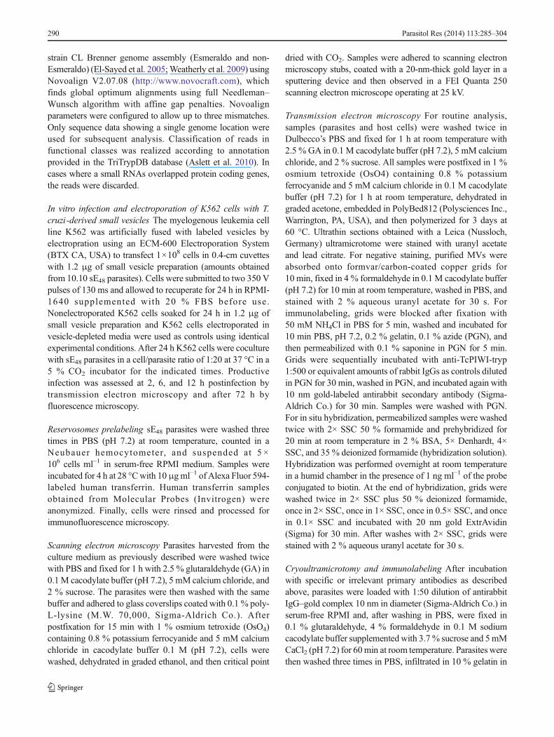
The recruitment of tsRNAs and TcPIWI-tryp protein inintracellular vesicles suggested to us that these molecules couldbe part of an endocytic/exocytic pathway in T. cruzi (Allenet al. 2003; Correa et al. 2007; Ungewickell and Hinrichsen2007). Indeed, a growing body of evidence indicates thatsecreted vesicles (i.e., exosomes and ectosomes) frommammalian cells serve as a mean of delivering geneticinformation and proteins to recipient cells playing pivotal rolesin cell-to-cell communication (Camussi et al. 2010). To tracesmall tRNA transfer parasites were labeled by electroporationwith synthetic fluorescent 5′ halves from tRNAGlu andtRNAAsp. Electroporation efficiency and intracellulardistribution of the fluorescent probe (Fig. 3a) showed that after2 h the fluorescent signal had a diffuse cytoplasmic pattern inmore than 95 % of parasites which adopted the typical granulardistribution observed in stressed parasites beyond 24 h.Fluorescent signals remained detectable beyond 72 h ofcultures with a slow but progressive decline after 4 days (datanot shown). Of note, it was previously reported that T. cruzi areable to endocytose free synthetic probes when exposed for 24 hto concentrations of 20 μM (Malaga and Yoshida 2001).However, in our experimental conditions, nonelectroporatedcontrol parasites incubated with 0.1–2.0 μM of the freefluorescent probe for a period of 2–24 h did not incorporatethe free probe, indicating that the parasite could not incorporatedetectable amounts of free probe when using morephysiological concentrations (Fig. 3 and data not shown). Weconsidered that exposition of wild-type parasites to smallvesicles secreted by electroporated parasites represented a morenatural condition when compared with high nonphysiologicalconcentrations of the free probe.
Next, small secreted vesicles from labeled parasites werecollected after 48 h of culture in serum-free RPMI byultracentrifugation and subsequently filtered through 1- and0.4-μm pore diameter filters to eventually exclude remnantparasites or cellular debris. When unlabeled parasites wereincubated in supernatants depleted from labeled small vesicles,they did not incorporate any detectable fluorescent signal(Fig. 3b). In contrast, parasites incubated with serum-freeRPMI enriched with purified small vesicles from sE48 rapidlyincorporate the fluorescent signals adopting the typical granulardistribution in the posterior region of the cell. To exclude aneventual contamination with intact or fragmented cells, labeledas well as unlabeled parasites were cocultivated in separated
chambers of transwells with a pore diameter of 0.4μm (Fig. 3c).A rapid incorporation of fluorescent signals was observedwithin 2 h in unlabeled parasites located in the bottom chamberof transwells, which adopted the typical granular patternobserved in sE48 parasites (upper panel in Fig. 3c). Of note,identical experiments performed with control fluorescentscrambled probes showed that they were also transferredbetween parasites, indicating a sequence-independentphenomenon. These data strongly suggested that at leasttRNA-derived small RNAs were released to the mediumincluded in vesicles, which deliver their cargo to other parasitesin a homotypic manner.
To verify whether sE48 parasites effectively shed smallvesicles carrying tRNA-derived small RNAs and TcPIWI-trypprotein, we performed both scanning and transmission electronmicroscopy of intact parasites and immune detection ofTcPIWI-tryp coupled to in situ hybridization for 5′halvesderived from tRNAGlu and tRNAAsp on purified extracellularvesicles. Of note, differentiating epimastigotes as well astrypomastigote forms (Fig. 4a and b, respectively) are releasingvesicles from the plasma membrane. Results depicted inFig. 4a–f revealed the presence of small vesicles being releasedthroughout the cell body as well as from the flagellar pocketand at the posterior end of the parasite. In the specific case ofFig. 4e, we noted that the vesicle attached to the outer side ofthe parasite cell membrane has a similar size to that foundwithin the reservosome (asterisk), suggesting that theseorganelles could participate in the generation and release/uptake of vesicles to/from the extracellular compartment.Another important observation is that this reservosome showeda sharp region that seems to touch the cytoplasmic face of theplasma membrane, between the subpellicular microtubules,suggesting that some kind of fusion process could occur at thisregion. As depicted in Fig. 4h, the diameter of vesicles washeterogeneous and ranged from 20 to 200 nm. Additionally,gold labeling (Fig 4h–j) confirmed that 5′derived small tRNAs,and the TcPIWI-tryp protein were included by sE48 parasites insecreted vesicles.
Small vesicles shed from the surface of many mammaliancells both in vitro and in vivo are a mixed population ofvesicles with different origins including exosomes (derivedfrom the endosomal membrane compartments throughmultivesicular bodies), ectosomes (shedding microvesicles)originated by direct budding from the cell plasma membraneand apoptotic blebs (Camussi et al. 2010; Mathivanan et al.2010). In this respect, it was recently reported (Bayer-Santoset al. 2013) that T. cruzi epimastigotes as well as therespective trypomastigote forms release two classes ofvesicles including large vesicles budding from the plasmamembrane resembling ectosomes and a second population ofsmaller vesicles mainly derived from the exocytic fusion ofmultivesicular bodies with the flagelar pocket membrane.Thus, in our experimental conditions the secreted vesicular
Parasitol Res (2014) 113:285–304 293
fraction represents a mixed population of both classes ofvesicles. For this reason, the vesicular fraction studied in thiswork will be referred as extracellular vesicles (EVs).
Small RNA cargo from T. cruzi shed vesicles is transferredto infection susceptible Vero cells.
A similar set of experiments were performed to analyze if smallvesicles shed by sE48 parasites also deliver their cargo in aheterotypic manner to mammalian cells. As depicted in Fig. 5(upper panel), Vero cells cultured in the presence of the freetracing probe added to vesicle-depleted supernatants from
labeled parasites did not incorporate detectable fluorescentsignals for a period of 24 h. When Vero cells were coincubatedwith labeled parasites in separated chambers of 0.4 μm poretranswells, they showed a clear incorporation of fluorescentsignals adopting a perinuclear pattern (Fig. 5, middle panel).As depicted, both the tracing probe (tRNAAsp) and the TcPIWI-tryp argonaute protein were transferred to Vero cells. Wespeculated about the possibility that the incorporation ofTcPIWI-tryp by Vero cells could be the consequence of eitherthe transfer of the protein itself or through a functional mRNAcarried out by shed vesicles. As observed in Fig. 5 (middle andbottom panels), when Vero cells were pretreated with
Fig. 3 Exchange of small tRNAs between parasites through shedvesicles. a Epifluorescence microscopy of parasites labeled withfluorescent tRNAAsp halves after soaking for 2 h in the presence of thefree probe (left panel) and after 2 (middle panel) or 24 h (right panel)after electroporation. In all cases, only merged images are depicted. bUnlabeled parasites cultured for 24 h in serum-free RPMI depleted fromsmall vesicles (upper panel) and in serum-free RPMI enriched withlabeled small vesicles purified from supernatants of labeled parasites
(bottom panel). c Experimental evidences for transfer of small RNAsfrom labeled parasites toward unlabeled parasites in separated chambersof transwells with 0.4μmof pore diameter.Upper panel Parasites labeledwith fluorescent tRNAAsp-derived 5′ halves in the upper chamber; bottomparasites labeled with the respective fluorescent scrambled probe in theupper chamber. Unlabeled parasites were added to bottom chambers andincubated for 24 h at 28 °C
294 Parasitol Res (2014) 113:285–304

cycloheximide to inhibit protein synthesis, the appearance offluorescent signals remained unchanged. Additionally, theabsence of TcPIWI-tryp mRNA inside the treated Vero cells(bottom panel in Fig. 5) further validated the assumption that theTcPIWI-tryp protein was effectively transferred through smallvesicles. Taken together, these data indicated that the TcPIWI-tryp protein and specific small tRNAswere effectively deliveredinto Vero cells by shed small vesicles derived fromdifferentiating forms of T. cruzi . It is noteworthy that, incontrast to their localization inside parasites, the TcPIWI-tryp
and tsRNAs do not colocalize inside Vero cells. One canspeculate that both species follow different intracellular routesreaching distinct subcellular compartments. However, otherexperimental approaches are needed to validate this assumption.
Secreted small vesicles contain a highly representedpopulation of small RNAs derived from tRNAs and rRNAs
In order to know the whole spectrum of small RNAs containedin secreted vesicles, we performed a deep sequencing of an
Fig. 4 TcPIWI-tryp andtRNA-derived halves in shedsmall vesicles from T. cruziepimastigotes. a , b Scanningelectron microscopy of intact sE48parasites showing a parasite undera process of differentiation (a)and a differentiatedtrypomastigote (b) with vesiclesemerging from the flagellum andthe cell body. c–j Transmissionelectron micrographs: c–flongitudinal sections of SE48
parasites showing small vesiclesinside the flagellar pocket(small arrows in c and f), inreservosomes (arrowheads in e)and other smaller vesicles beingliberated from the cell bodymembrane (arrows in d and e).Note that the two vesicles in e(arrowhead and black arrow)present the same size and that themembrane of the reservosome(asterisk) are in close contact withthe cell membrane (white arrow).g Purified secreted small vesicles.h In situ hybridization of purifiedshed vesicles to detect tRNAAsp 5′halves labeled with 20 nm goldExtrAvidin streptavidin. iImmuno-electron microscopywith gold-labeled anti-TcPIWI-tryp antibodies (10 nm. j)Double-labeling staining revealing thepresence of TcPIWI-tryp protein(arrowheads) and tRNAAsp 5′halves (arrows) in thesame vesicle. F flagellum,K kinetoplast, R reservosome,N nucleus
Parasitol Res (2014) 113:285–304 295

18–60-nt size-fractionated complementary DNA library of EVspurified from supernatants of starved sE48 parasites (Fig. 6). Thesequence data were aligned separately with annotated genomesfrom the non-Esmeraldo and Esmeraldo haplotypes andadditionally with a collection of unassigned contigs fromT.cruzi , which represents the sequences that could not beassigned to either haplotype. Sequence analysis resulted in atotal of 10,107,119 aligned reads with a fraction of 6,100,251reads (60 %) with a single alignment location in the genome(single mappers). The length distribution of unique reads(Fig. 6a) was clearly segregated into two main populations withmedian sequence length of ∼20 and ∼34 nt. Categorization inclasses using genome annotations revealed that small RNAsderived from rRNA and tRNA represented about 45.5 % foreach (Fig. 6b). Of note, more than 90 % of reads within ∼33 ntpopulation belonged to the tRNA category. Additionally, morethan 90 % of tsRNAs derived from a restricted group of six
tRNAs: Leu, Thr, Glu, Gly, and Arg (Fig. 5c). The remainingreads were derived from CDS (≈5 %), sno/snRNAs (≈1.5 %),and intergenic regions (≈3 %).
Electrofusion of vesicles from T. cruzi with infection resistantK562 cells confers susceptibility to infection
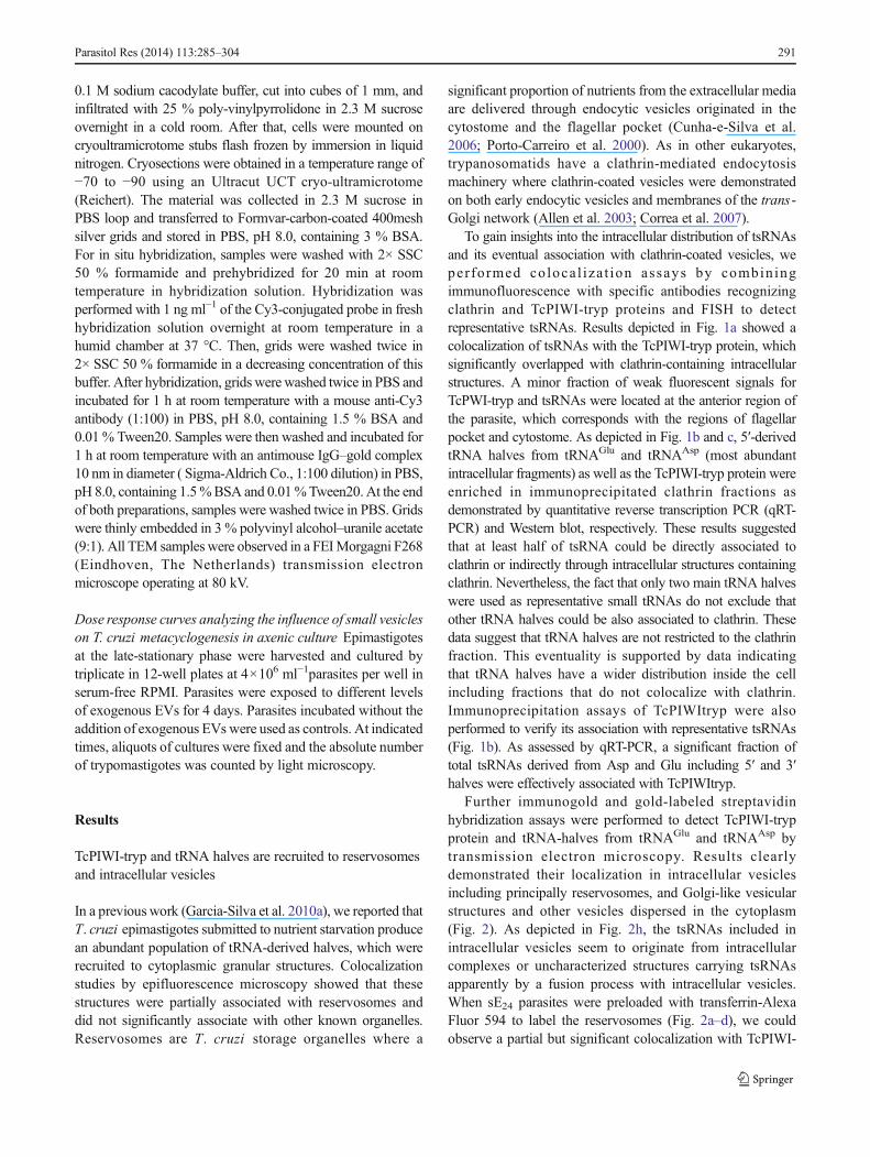
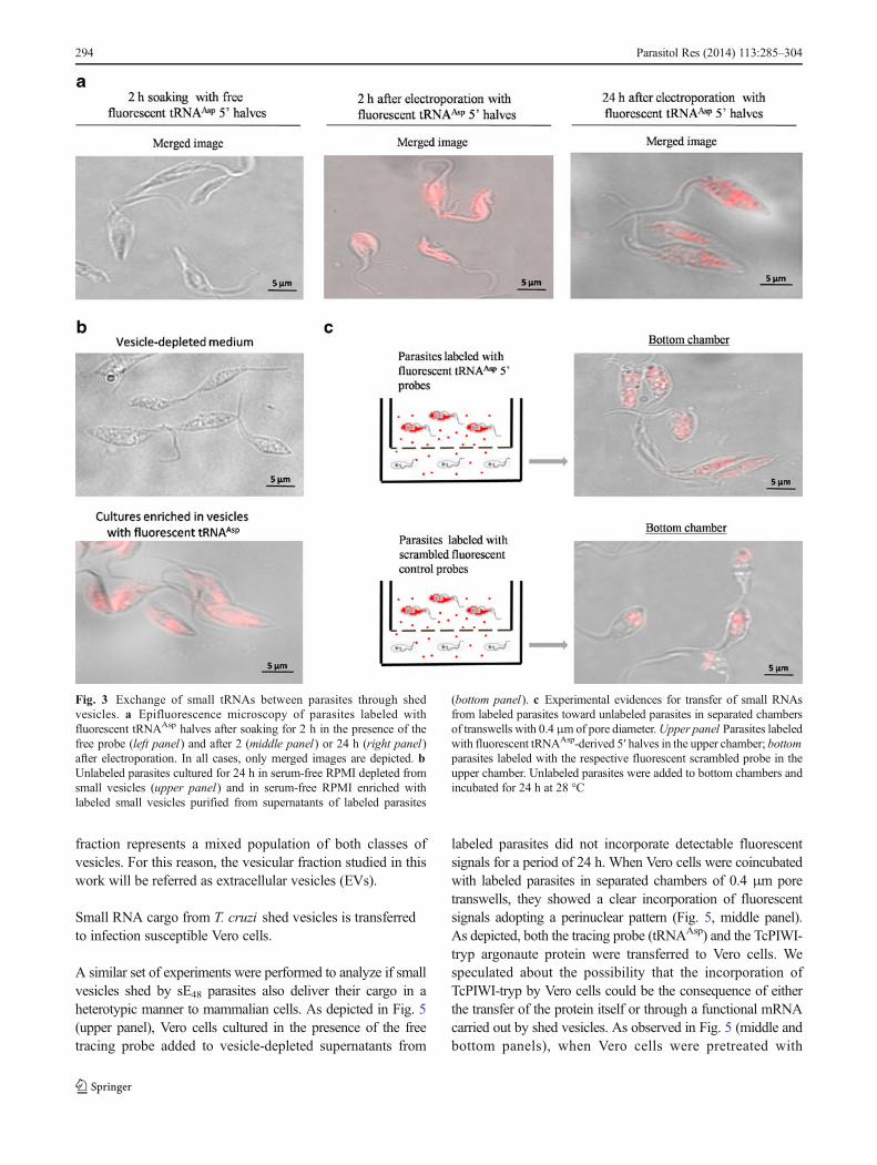
In order to gain insight about the biological significance on thetransfer of genetic information and proteins to mammaliansusceptible cells through secreted vesicles, we performed a setof experiments using as recipient cells the humanerythroleukemic K562 cell line, which is known to resistinfection by T. cruzi (Magdesian et al. 2001; Ruiz et al.1998). In contrast to susceptible cells, K562 cells cultured inthe presence of labeled sE48-derived vesicles for 24 h did notspontaneously incorporate detectable amounts of labeled tRNAhalves (Fig. 7a). Speculating that fusion of T. cruzi-derived EVs
Fig. 5 Transfer of vesicle cargo from T. cruzi to mammalian susceptiblecells. Visualization of fluorescent tRNAAsp 5′ halves secreted from sE48parasites coupled to immunodetection of TcPIWI-tryp in the infectionsusceptible Vero cells. Upper panel Control Vero cells cultured for 24 hin the presence of free tRNAAsp 5′ halves and counterstained with DAPI;middle panel untreated Vero cells cocultured for 24 h in the bottom
chamber of transwells with sE48 parasites labeled with synthetic fluorescenttRNAAsp 5′ halves in the upper chamber (Vero cells+labeled parasites)compared to the same experiment with cycloheximide pretreated Vero cellsfor 24 h (Vero cells+labeled parasites+cycloheximide); bottom panel fishassay to detect TcPIWI-tryp mRNA in Vero cells cocultured with labeledparasites
296 Parasitol Res (2014) 113:285–304

with target cells could be required to achieve a productiveinfection, we performed a series of in vitro infection assaysaimed to evaluate the susceptibility of electroporated K562 cellsto T. cruzi infection either in the absence or the presence ofsE48-derived vesicles. Twenty-four hours after electroporationin vesicle-depleted media, K562 cells do not incorporatedetectable fluorescent signals as assessed by fluorescencemicroscopy (upper panel in Fig. 7b). In contrast, when K562cells were electroporated in the presence of labeled EVs, theyshowed a clear incorporation of fluorescent signals, indicatingthat electroporation was successful to deliver vesicle cargo toK562 cells (bottom panel in Fig. 7b). The ratio of K562 cells toEVs used to obtain this effect was optimal at 1×10⁶ cells for12 ng of vesicle preparation (equivalent to vesicles produced by1×108 parasites cultured for 24 h). To exclude a nonspecificeffect of irrelevant vesicles, we performed an additional controlexperiment by electroporating K562 cells with microvesiclespurified from HeLa cells. In these conditions, K562 cellsremained resistant to infection by the parasite (data not shown).Fluorescent signals remained virtually unchanged for the first48–72 h with a slow decline of signals over the following dayswhen the signals became undetectable at day 5 afterelectroporation.
When electroporated K562 cells were infected with sE48parasites and observed 48–72 h after infection, only k562 cellselectroporated with EVs appeared to become susceptible toinfection as indicated by the presence of rounded forms ofparasites (presumably amastigotes) inside and outside thecell (bottom panel in Fig. 7c). However, in three independentassays, we could not evidence the emergence of trypomastigote
forms, suggesting that in spite of become infected T. cruzicould not complete its life cycle in K562 cells. A plausibleexplanation could be that K562 cells possess a high nucleus/cytoplasm ratio, which limits the extent of amastigotereplication inside the cell with the subsequent prematuredisruption of plasma membrane hampering the emergence oftrypomastigote forms. When control K562 cells wereincubated with EVs 24 h after electroporation, they didnot become infected (upper panel in Fig. 7c), indicatingthat infection susceptibility was not the consequence ofelectroporation.
To verify the entry of infective parasites in electroporatedK562 cells, we followed the infectious process by transmissionelectron microscopy in the first 12 h after infection (Fig. 8).These results revealed that, although control K562 cellselectroporated in vesicle-depleted supernatants were able tointeract with infection-competent parasites by surface-to-surface contact, we never could observe the presence ofintracellular parasites in this period. Surprisingly, K562 cellselectroporated with small labeled vesicles from sE48 parasitesbecame susceptible to infection as revealed by the presence ofseveral intracellular parasites in the first 12 h afterinfection. Images from Fig. 8b–d are representatives of thefirst 2, 6, and 12 h after infection, respectively, depicting aparasite penetrating the cell, and intracellular parasitesin a process of differentiation toward round forms characterizingthe intracellular replicative phase (amastigotes). Theseresults suggested that fusion of T. cruzi-derived small vesicleswith target cells could be a relevant step in the infectiousprocess.
Fig. 6 Deep sequencing of smallRNAs included in shed smallvesicles from T. cruzi . aGraphical representation of lengthdistribution vs number of readswith a single alignment locationin the genome. b Categorizationof reads into known RNA classesusing genome annotations. cDistribution of reads derivedfrom tRNAs according toprecursor tRNAs
Parasitol Res (2014) 113:285–304 297
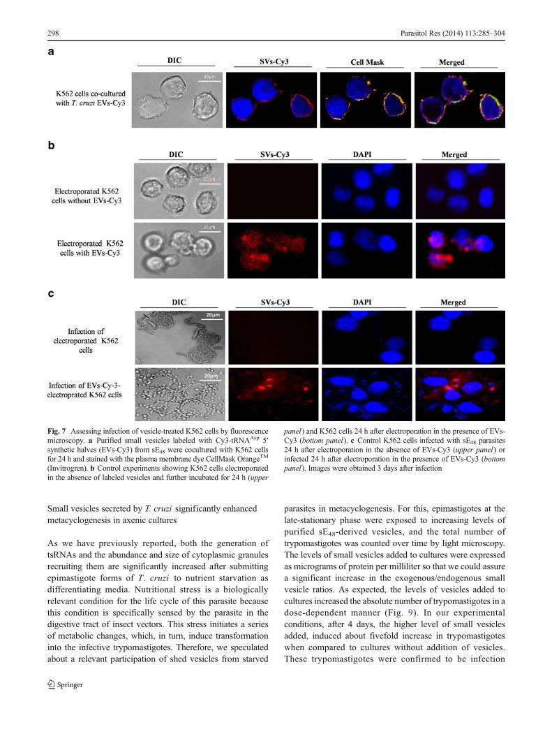
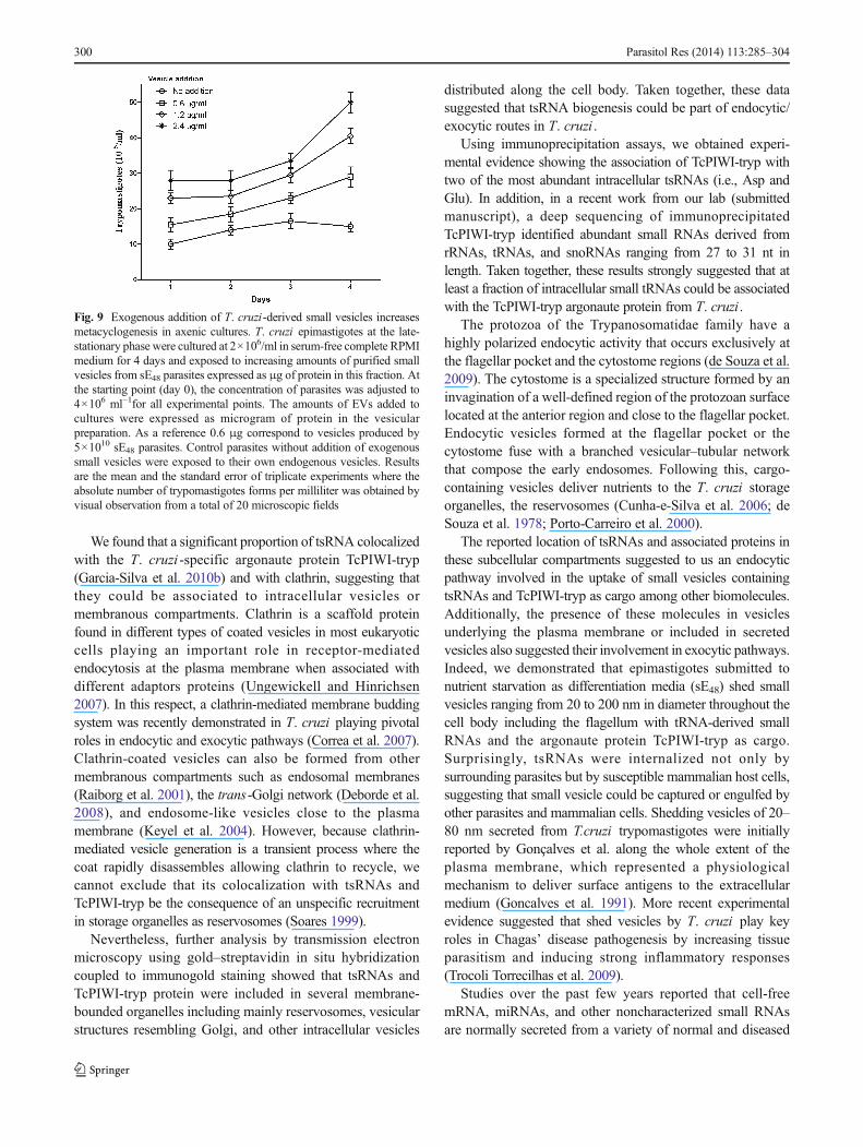
Small vesicles secreted by T. cruzi significantly enhancedmetacyclogenesis in axenic cultures
As we have previously reported, both the generation oftsRNAs and the abundance and size of cytoplasmic granulesrecruiting them are significantly increased after submittingepimastigote forms of T. cruzi to nutrient starvation asdifferentiating media. Nutritional stress is a biologicallyrelevant condition for the life cycle of this parasite becausethis condition is specifically sensed by the parasite in thedigestive tract of insect vectors. This stress initiates a seriesof metabolic changes, which, in turn, induce transformationinto the infective trypomastigotes. Therefore, we speculatedabout a relevant participation of shed vesicles from starved
parasites in metacyclogenesis. For this, epimastigotes at thelate-stationary phase were exposed to increasing levels ofpurified sE48-derived vesicles, and the total number oftrypomastigotes was counted over time by light microscopy.The levels of small vesicles added to cultures were expressedas micrograms of protein per milliliter so that we could assurea significant increase in the exogenous/endogenous smallvesicle ratios. As expected, the levels of vesicles added tocultures increased the absolute number of trypomastigotes in adose-dependent manner (Fig. 9). In our experimentalconditions, after 4 days, the higher level of small vesiclesadded, induced about fivefold increase in trypomastigoteswhen compared to cultures without addition of vesicles.These trypomastigotes were confirmed to be infection
Fig. 7 Assessing infection of vesicle-treated K562 cells by fluorescencemicroscopy. a Purified small vesicles labeled with Cy3-tRNAAsp 5′synthetic halves (EVs-Cy3) from sE48 were cocultured with K562 cellsfor 24 h and stained with the plasma membrane dye CellMask OrangeTM
(Invitrogren). b Control experiments showing K562 cells electroporatedin the absence of labeled vesicles and further incubated for 24 h (upper
panel) and K562 cells 24 h after electroporation in the presence of EVs-Cy3 (bottom panel). c Control K562 cells infected with sE48 parasites24 h after electroporation in the absence of EVs-Cy3 (upper panel) orinfected 24 h after electroporation in the presence of EVs-Cy3 (bottompanel). Images were obtained 3 days after infection
298 Parasitol Res (2014) 113:285–304

competent in further in vitro assays on Vero cells (data notshown).
Discussion
As reported, tRNA cleavage appeared as a highly conservedsmall RNA pathway from prokaryotes to higher eukaryotes,frequently initiated after nutritional, biological, or physico-chemical stress (Haiser et al. 2008; Jochl et al. 2008; Kawajiet al. 2008; Lee and Collins 2005; Li et al. 2008; Zhang et al.2009). Although the biological significance remains to becompletely elucidated, recent experimental evidence suggestedthat tRNA-derived small RNAs emerges as an evolutionary
conserved pathway with a putative role in the regulation of geneexpression under certain survival pressures (Elbarbary et al.2009a, b; Haussecker et al. 2010; Li and Zhou 2009;Nakashima et al. 2007).
The stress-induced tRNA cleavage is mediated byanticodon nucleases in bacteria (Ogawa et al. 1999),Rny1p in yeasts (Thompson and Parker 2009a), andangiogenin (ANG) in humans (Fu et al. 2009). Thesestress-induced tRNA cleavage products are 30–40 nt inlength and seem to form related RNA classes, termedgenerically tsRNAs. An additional class of small RNAs thatare ∼20–30 nt long derived from 5′ or 3′ ends of tRNAs wasalso reported and broadly termed tRNA fragments (Lee et al.2009).
Fig. 8 Monitoring infection ofEVs-treated K562 cells bytransmission electron microscopy.a Representative micrographs ofcontrol electroporated K562 cellscocultured with infective parasites(sE48) for 24 h in a 1:20cell/parasite ratio at 37 °C inserum-free RPMI. Representativemicrographs show residualparasites adhered to the plasmamembranes (P). b–d K562 cellselectroporated with extracellularvesicles (EVs) from sE48parasites. Representativemicrographs showing parasites(P) in early stages of interactionwith host cells. b After 2 h ofinfection. d After 6 h of infection.c After 12 h of infection. Themembranes surrounding parasitesresembling parasitophorousvacuoles are indicated by smallarrows (c and inset of d)
Parasitol Res (2014) 113:285–304 299
We found that a significant proportion of tsRNA colocalizedwith the T. cruzi -specific argonaute protein TcPIWI-tryp(Garcia-Silva et al. 2010b) and with clathrin, suggesting thatthey could be associated to intracellular vesicles ormembranous compartments. Clathrin is a scaffold proteinfound in different types of coated vesicles in most eukaryoticcells playing an important role in receptor-mediatedendocytosis at the plasma membrane when associated withdifferent adaptors proteins (Ungewickell and Hinrichsen2007). In this respect, a clathrin-mediated membrane buddingsystem was recently demonstrated in T. cruzi playing pivotalroles in endocytic and exocytic pathways (Correa et al. 2007).Clathrin-coated vesicles can also be formed from othermembranous compartments such as endosomal membranes(Raiborg et al. 2001), the trans-Golgi network (Deborde et al.2008), and endosome-like vesicles close to the plasmamembrane (Keyel et al. 2004). However, because clathrin-mediated vesicle generation is a transient process where thecoat rapidly disassembles allowing clathrin to recycle, wecannot exclude that its colocalization with tsRNAs andTcPIWI-tryp be the consequence of an unspecific recruitmentin storage organelles as reservosomes (Soares 1999).
Nevertheless, further analysis by transmission electronmicroscopy using gold–streptavidin in situ hybridizationcoupled to immunogold staining showed that tsRNAs andTcPIWI-tryp protein were included in several membrane-bounded organelles including mainly reservosomes, vesicularstructures resembling Golgi, and other intracellular vesicles
distributed along the cell body. Taken together, these datasuggested that tsRNA biogenesis could be part of endocytic/exocytic routes in T. cruzi .
Using immunoprecipitation assays, we obtained experi-mental evidence showing the association of TcPIWI-tryp withtwo of the most abundant intracellular tsRNAs (i.e., Asp andGlu). In addition, in a recent work from our lab (submittedmanuscript), a deep sequencing of immunoprecipitatedTcPIWI-tryp identified abundant small RNAs derived fromrRNAs, tRNAs, and snoRNAs ranging from 27 to 31 nt inlength. Taken together, these results strongly suggested that atleast a fraction of intracellular small tRNAs could be associatedwith the TcPIWI-tryp argonaute protein from T. cruzi .
The protozoa of the Trypanosomatidae family have ahighly polarized endocytic activity that occurs exclusively atthe flagellar pocket and the cytostome regions (de Souza et al.2009). The cytostome is a specialized structure formed by aninvagination of a well-defined region of the protozoan surfacelocated at the anterior region and close to the flagellar pocket.Endocytic vesicles formed at the flagellar pocket or thecytostome fuse with a branched vesicular–tubular networkthat compose the early endosomes. Following this, cargo-containing vesicles deliver nutrients to the T. cruzi storageorganelles, the reservosomes (Cunha-e-Silva et al. 2006; deSouza et al. 1978; Porto-Carreiro et al. 2000).
The reported location of tsRNAs and associated proteins inthese subcellular compartments suggested to us an endocyticpathway involved in the uptake of small vesicles containingtsRNAs and TcPIWI-tryp as cargo among other biomolecules.Additionally, the presence of these molecules in vesiclesunderlying the plasma membrane or included in secretedvesicles also suggested their involvement in exocytic pathways.Indeed, we demonstrated that epimastigotes submitted tonutrient starvation as differentiation media (sE48) shed smallvesicles ranging from 20 to 200 nm in diameter throughout thecell body including the flagellum with tRNA-derived smallRNAs and the argonaute protein TcPIWI-tryp as cargo.Surprisingly, tsRNAs were internalized not only bysurrounding parasites but by susceptible mammalian host cells,suggesting that small vesicle could be captured or engulfed byother parasites and mammalian cells. Shedding vesicles of 20–80 nm secreted from T.cruzi trypomastigotes were initiallyreported by Gonçalves et al. along the whole extent of theplasma membrane, which represented a physiologicalmechanism to deliver surface antigens to the extracellularmedium (Goncalves et al. 1991). More recent experimentalevidence suggested that shed vesicles by T. cruzi play keyroles in Chagas’ disease pathogenesis by increasing tissueparasitism and inducing strong inflammatory responses(Trocoli Torrecilhas et al. 2009).
Studies over the past few years reported that cell-freemRNA, miRNAs, and other noncharacterized small RNAsare normally secreted from a variety of normal and diseased
Fig. 9 Exogenous addition of T. cruzi-derived small vesicles increasesmetacyclogenesis in axenic cultures. T. cruzi epimastigotes at the late-stationary phase were cultured at 2×10⁶/ml in serum-free complete RPMImedium for 4 days and exposed to increasing amounts of purified smallvesicles from sE48 parasites expressed as μg of protein in this fraction. Atthe starting point (day 0), the concentration of parasites was adjusted to4×106 ml−1for all experimental points. The amounts of EVs added tocultures were expressed as microgram of protein in the vesicularpreparation. As a reference 0.6 μg correspond to vesicles produced by5×1010 sE48 parasites. Control parasites without addition of exogenoussmall vesicles were exposed to their own endogenous vesicles. Resultsare the mean and the standard error of triplicate experiments where theabsolute number of trypomastigotes forms per milliliter was obtained byvisual observation from a total of 20 microscopic fields
300 Parasitol Res (2014) 113:285–304
cells to the extracellular media either through membranousvesicles (Camussi et al. 2010) or included in ribonucleo-protein complexes (Arroyo et al. 2011; Vickers et al. 2011;Wang et al. 2010).
It is now accepted that secreted exosomes and shedmicrovesicles (ectosomes) (Cocucci and Meldolesi 2011) serveas a mean of delivering genetic information and proteinsbetween cells playing pivotal roles in cell-to-cellcommunication (Camussi et al. 2010). In an elegant study,Valadi et al. (2007) reported that exosomes from differentmammalian cells carry at least 1,300 mRNAs and more than120 known microRNAs and other noncharacterized smallRNAs. Interestingly, these exosomal mRNAs and microRNAswere completely functional in recipient cells.
Overall, these data strongly suggest that shedding ofvesicles by T. cruzi represents an integral part of an endo-/exocytic pathway involved in cell-to-cell communication,where tsRNAs and TcPIWI-tryp are at least one of severalinvolved molecules. Other proteins and glycoconjugates werealso demonstrated to be included in shed vesicles includingmembers of the gp85/transialidase family, proteases ascruzipain, cytoskeleton proteins, mucin-associated surfaceproteins, and a variety other proteins with known andunknown functions (Aparicio et al. 2004; Frasch 2000;Tribulatti et al. 2005). A recent proteomic analysis of thetwo major fractions of extracellular vesicles shed by T. cruzirevealed the presence of more than 300 proteins belonging torelevant pathways, including trafficking and membranefusion, host–parasite interaction, signaling, and metabolismamong others (Bayer-Santos et al. 2013).
Surprisingly, using synthetic small RNAs derived fromtRNAGlu and tRNAAsp as tracer molecules, we demonstratedthat tsRNAs contained in EVs were efficiently transferred toinfection susceptible Vero cells associated to the TcPIWI-trypargonaute protein but not to the nonsusceptible K562 cells.Therefore, we speculated that vesicles shed by T. cruzi couldhave a role in facilitating or promoting infection susceptibilityof mammalian cells. Indeed, electroporation-mediated fusionof T. cruzi small vesicles with K562 cells turned themsusceptible to infection by infective sE48 parasites. However,parasites internalized by K562 cells do not appear to completetheir life cycle due to the premature disruption of the plasmamembrane with the subsequent release of rounded formsresembling amastigotes. Nevertheless, the amastigote formshave proven to be relevant in the infective cycle of thisparasite. In this respect, it is known that extracellularamastigotes are infective for mammalian cells, indicating thatthey can initiate an alternative subcycle within the life cycle ofthis parasite in mammalian cells (Ley et al. 1988).
We also describe a cross-kingdom transfer of tRNA-derivedsmall RNAs through secreted vesicles from T. cruzi tomammalian susceptible cells. In this respect, recent evidencesdemonstrated another type of cross-kingdom transfer of a
functional microRNA from plants to humans (Zhang et al.2012). Some recent reports afforded experimental evidencedemonstrating the transfer of transposons across phyla(Gilbert et al. 2010), including the transfer of mitochondrialminicircles from T. cruzi to host cells (Hecht et al. 2010;Teixeira et al. 2011).
The idea that the transfer of T. cruzi small vesicle cargo tomammalian cells could be involved in susceptibility toinfection could afford novel avenues to elucidate themolecular mechanisms involved in T. cruzi infection of targetcells. In agreement with this idea, it was recently reported thatin both phagocytic and nonphagocytic cells, the process of T.cruzi entry into the host cell is drastically diminished whenhost cells are treated with dynasore, a potent inhibitor ofdynamin-dependent endocytosis at the plasma membrane(Barrias et al. 2010). In agreement with this idea, in a recentwork, Torrecilhas et al. (2012) demonstrated that shed vesiclesfrom T. cruzi are engulfed by susceptible mammalian cells inthe absence of viable parasites. However, the structures orbiomolecules (e.g., membranes, lipids, proteins and nucleicacids other than small tRNAs) involved in these processesremain to be elucidated.
Additionally, we afford experimental evidences stronglysuggesting that shed vesicles facilitated or significantlyenhanced metacyclogenesis. These results suggested thatEVs and their cargo represent a route of intercellularcommunication delivering “molecular messages” to otherscells aimed to induce coordinated responses to cope withadverse environmental conditions and thus assuring parasitesurvival through the emergence of the infective forms. Indeed,recent unpublished results from Torrecillhas et al. (2012)revealed that T. cruzi trypomastigotes invade fivefold asmuchsusceptible cells when cells were preincubated with purifiedparasite small vesicles. These results suggest that secretedvesicles from T. cruzi and their cargo could act as virulencefactors by either promoting metacyclogenesis, enhancing hostcell susceptibility or both. However, these assumptions remainto be further validated.
In order to know the total spectrum of small RNA includedin shed vesicles, we performed a deep sequencing of shortRNAs included in purified small vesicles. Our data revealedthat rRNA- and tRNA-derived small RNAs equallycontributed to more than 90 % of short RNA cargo of EVs.Of note ∼90 % of tsRNAs were derived from a restrictedgroup of tRNA precursors including tRNALeu, tRNAThr,tRNAGlu, tRNAGly, and tRNAArg, representing about one halfof small RNAs included in secreted vesicles where ∼56 %derived from the 3′ arm of precursor tRNAs. The shortintracellular noncoding RNAs from the Dm28c clonesubmitted to nutrient starvation was recently analyzed by ourgroup (Garcia-Silva et al. 2010a) using low-scale sequencing.These results revealed that tsRNAs represented about 25 % ofintracellular small RNAs and more than 90 % of them derived
Parasitol Res (2014) 113:285–304 301

from the 5′ arm of tRNAAspGUC, tRNAGlu
CUC, andtRNAGlu
UUC. These results indicated that tsRNAs includedin small vesicles are not only quantitatively enriched butqualitatively different from its intracellular counterpart,suggesting a selective recruiting process. However, one canspeculate that the relative abundance of 5′ halves could not bethe consequence of their preferential association with TcPIWI-tryp but the consequence of the higher instability of 3′ halves.
Although small tRNAs derived from tRNAGlu andtRNAAsp were the most abundant intracellular fragments,deep sequencing of EVs revealed that tRNAAsp halvesrepresent a minor fraction of small tRNAs included inextracellular vesicles. Even though this phenomenon couldbe the consequence of a preferential recruitment of certaintRNA-derived species in EVs, we cannot exclude a biasinduced by different sequencing methods used to analyze thepattern of intracellular and secreted tsRNAs. Whatever thecase, these eventualities do not invalidate the use of tRNAAsp
as a tracer molecule in some experiments. Recent results fromour lab (manuscript in preparation) confirmed a selectiverecruitment of tsRNAs in secreted vesicles. Recently, Franzenet al. (2011) reported the intracellular short non-codingtranscriptome of the CL-Brener clone, which was quali- andquantitatively different from that reported for the Dm28c clone.However, we cannot elucidate if these differences are theconsequence of species-specific profiles or they reflectdifferences induced by nutritional stress when compared tonormal growing conditions.
These data extended previous results indicating thatbesides microRNAs and mRNAs, tsRNAs, and other smallRNAs are also included in vesicles and secreted to themedium by certain cells. In recent years, various studies havesuggested that tRNA-derived small RNAs could act aseffector molecules to regulate global gene expression (Liand Zhou 2009; Thompson and Parker 2009b). It is temptingto speculate that similarly to the vesicle-mediated epigeneticreprogramming of cells through either exosome- or ectosometransfer of mRNAs and microRNAs as a novel mechanism ofgenetic exchange between cells, these tsRNAs could alsoperform pivotal roles in cell-to-cell communication in T. cruzi(Camussi et al. 2011; Valadi et al. 2007).
The endo-/exocytic pathways involved in small vesicleuptake/release by T. cruzi epimastigotes are presentlyunknown. However, the presence of tsRNA within vesicularorganelles as reservosomes, Golgi-like structures, intracellularvesicles, and plasma membrane strongly suggest thatendocytosis of EVs cargo uses the highly polarized endocyticprocess characteristic of the Trypanosomatidae family (Soares2006; Souza 2009).
Experimental evidence presented here and in previousreports from our lab (Garcia-Silva et al. 2010a) indicated thata major fraction of tsRNAs are recruited to reservosomes.Reservosomes are big compartments present at the postnuclear
region of epimastigote forms of parasites belonging to thegenus Trypanosoma , subgenus Schizotrypanum, describedinitially as multivesicular bodies (de Souza et al. 1978).Reservosomes deserved their name because they store tracermacromolecules ingested by the parasite through an endocyticprocess (Soares and de Souza 1991), which were reported aslate endosomal or endosomal compartments in T. cruziepimastigotes (Cunha-e-Silva et al. 2006; Souto-Padron et al.1990). In addition, studies on reservosome biogenesis revealedthat vesicles originated from the Golgi complex also fuse withcomponents of the endocytic pathway. Indeed, the cysteinprotease cruzipain, a major protease synthesized by T. cruzi ,is mainly recruited to reservosomes (Souto-Padron et al. 1990).The fact that cruzipain as well as tsRNAs are simultaneouslyobserved in reservosomes and shed vesicles strongly suggestedthe idea that reservosomes could be organelles with exocyticfunctions (Aparicio et al. 2004).
Today, it is accepted that reservosomes do not constitute auniform organelle population. Indeed, different reservosomesmight have storing, recycling, or lysosome typical functions(Cunha-e-Silva et al. 2006). Taken into account theseexperimental evidences, we hypothesized that accumulationof tsRNAs and TcPIWI-tryp protein in reservosomes couldresult from two different pathways: one representing thecontribution of small vesicles through the uptake via endocyticpathways (exogenous origin) and other representing thecontribution of vesicles from the trans-Golgi network of theown cell (endogenous origin). In this respect, it was recentlyreported (Bayer-Santos et al. 2013) that T. cruzi epimastigotesas well as trypomastigotes forms use different populations ofextracellular vesicles resembling ecto- and exosomes to excret/secret proteins and eventually deliver cargo into host cells.
Overall, our results afford new relevant pieces of evidence onthe role of secreted vesicles in parasite biology especially in lifecycle transitions, infectivity, and cell-to-cell communication,which undoubtedly should contribute to gain insights in themolecular mechanisms involved in host–parasite interactions.Additionally, the transfer of proteins and genetic material fromprotists to mammalian cells is an emerging field, which couldconduct us to rethink some concepts in biology.
Acknowledgments This work was supported by grants from theNational Agency of Research and Innovation (ANII, Uruguay), InstitutPasteur de Montevideo (Uruguay) and CNPq (Prosul), CAPEs andFAPERJ from Brazil.
References
Allen CL, Goulding D, Field MC (2003) Clathrin-mediated endocytosisis essential in Trypanosoma brucei . Embo J 22(19):4991–5002
Aparicio IM, Scharfstein J, Lima AP (2004) A new cruzipain-mediatedpathway of human cell invasion by Trypanosoma cruzi requirestrypomastigote membranes. Infect Immun 72(10):5892–5902
302 Parasitol Res (2014) 113:285–304

Arroyo JD, Chevillet JR, Kroh EM, Ruf IK, Pritchard CC, Gibson DF,Mitchell PS, Bennett CF, Pogosova-Agadjanyan EL, Stirewalt DL,Tait JF, Tewari M (2011) Argonaute2 complexes carry a populationof circulatingmicroRNAs independent of vesicles in human plasma.Proc Natl Acad Sci U S A 108(12):5003–5008
Aslett M, Aurrecoechea C, Berriman M, Brestelli J, Brunk BP, CarringtonM, Depledge DP, Fischer S, Gajria B, Gao X, Gardner MJ, Gingle A,Grant G, Harb OS, Heiges M, Hertz-Fowler C, Houston R,Innamorato F, Iodice J, Kissinger JC, Kraemer E, Li W, Logan FJ,Miller JA, Mitra S, Myler PJ, Nayak V, Pennington C, Phan I, PinneyDF, Ramasamy G, Rogers MB, Roos DS, Ross C, Sivam D, SmithDF, Srinivasamoorthy G, Stoeckert CJ Jr, Subramanian S, ThibodeauR, Tivey A, Treatman C, Velarde G, Wang H (2010) TriTrypDB: afunctional genomic resource for the Trypanosomatidae. NucleicAcids Res 38(Database issue):D457–D462
Barrett MP, Burchmore RJ, Stich A, Lazzari JO, Frasch AC, Cazzulo JJ,Krishna S (2003) The trypanosomiases. Lancet 362(9394):1469–1480
Barrias ES, Reignault LC, De SouzaW, Carvalho TM (2010) Dynasore, adynamin inhibitor, inhibits Trypanosoma cruzi entry into peritonealmacrophages. PLoS One 5(1):e7764
Bayer-Santos E, Aguilar-Bonavides C, Rodrigues SP, Cordero EM,Marques AF, Varela-Ramirez A, Choi H, Yoshida N, da SilveiraJF, Almeida IC (2013) Proteomic analysis of Trypanosoma cruzisecretome: characterization of two populations of extracellularvesicles and soluble proteins. J Proteome Res 12(2):883–897
Camussi G, Deregibus MC, Bruno S, Cantaluppi V, Biancone L (2010)Exosomes/microvesicles as a mechanism of cell-to-cellcommunication. Kidney Int 78(9):838–848
Camussi G, Deregibus MC, Bruno S, Grange C, Fonsato V, Tetta C(2011) Exosome/microvesicle-mediated epigenetic reprogrammingof cells. Am J Cancer Res 1(1):98–110
Cerutti H, Casas-Mollano JA (2006) On the origin and functions of RNA-mediated silencing: from protists to man. Curr Genet 50(2):81–99
Clayton C, Shapira M (2007) Post-transcriptional regulation of geneexpression in trypanosomes and leishmanias. Mol BiochemParasitol 156(2):93–101
Cocucci E, Meldolesi J (2011) Ectosomes. Curr Biol 21(23):R940–R941Cole C, Sobala A, Lu C, Thatcher SR, Bowman A, Brown JW, Green PJ,
Barton GJ, Hutvagner G (2009) Filtering of deep sequencing datareveals the existence of abundant Dicer-dependent small RNAsderived from tRNAs. Rna 15(12):2147–2160
Contreras VT, Salles JM, Thomas N, Morel CM, Goldenberg S (1985) Invitro differentiation of Trypanosoma cruzi under chemically definedconditions. Mol Biochem Parasitol 16(3):315–327
Contreras VT, Araujo-Jorge TC, Bonaldo MC, Thomaz N, Barbosa HS,Meirelles Mde N, Goldenberg S (1988) Biological aspects of theDm 28c clone of Trypanosoma cruzi after metacyclogenesis inchemically defined media. Mem Inst Oswaldo Cruz 83(1):123–133
Correa JR, Atella GC, Menna-Barreto RS, Soares MJ (2007) Clathrin inTrypanosoma cruzi : in silico gene identification, isolation, andlocalization of protein expression sites. J Eukaryot Microbiol54(3):297–302
Cunha-e-Silva N, Sant’Anna C, Pereira MG, Porto-Carreiro I, JeovanioAL, de Souza W (2006) Reservosomes: multipurpose organelles?Parasitol Res 99(4):325–327
de Souza W (2008) An introduction to the structural organization ofparasitic protozoa. Curr Pharm Des 14(9):822–838
de Souza W, de Carvalho TU, Benchimol M, Chiari E (1978)Trypanosoma cruzi : ultrastructural, cytochemical and freeze-fracture studies of protein uptake. Exp Parasitol 45(1):101–115
de Souza W, Sant’Anna C, Cunha-e-Silva NL (2009) Electronmicroscopy and cytochemistry analysis of the endocytic pathwayof pathogenic protozoa. Prog Histochem Cytochem 44(2):67–124
Deborde S, Perret E, Gravotta D, Deora A, Salvarezza S, Schreiner R,Rodriguez-Boulan E (2008) Clathrin is a key regulator of basolateralpolarity. Nature 452(7188):719–723
Elbarbary RA, Takaku H, Uchiumi N, Tamiya H, Abe M, Nishida H,Nashimoto M (2009a) Human cytosolic tRNase ZL candownregulate gene expression through miRNA. FEBS Lett583(19):3241–3246
Elbarbary RA, Takaku H, Uchiumi N, Tamiya H, Abe M, Takahashi M,Nishida H, NashimotoM (2009b)Modulation of gene expression byhuman cytosolic tRNase Z(L) through 5′-half-tRNA. PLoS One4(6):e5908
El-Sayed NM,Myler PJ, Bartholomeu DC, Nilsson D, Aggarwal G, TranAN, Ghedin E, Worthey EA, Delcher AL, Blandin G, WestenbergerSJ, Caler E, Cerqueira GC, Branche C, Haas B, Anupama A, ArnerE, Aslund L, Attipoe P, Bontempi E, Bringaud F, Burton P, Cadag E,Campbell DA, Carrington M, Crabtree J, Darban H, da Silveira JF,de Jong P, Edwards K, Englund PT, Fazelina G, Feldblyum T,Ferella M, Frasch AC, Gull K, Horn D, Hou L, Huang Y,Kindlund E, Klingbeil M, Kluge S, Koo H, Lacerda D, Levin MJ,Lorenzi H, Louie T, Machado CR, McCulloch R, McKenna A,Mizuno Y, Mottram JC, Nelson S, Ochaya S, Osoegawa K, Pai G,Parsons M, Pentony M, Pettersson U, Pop M, Ramirez JL, Rinta J,Robertson L, Salzberg SL, Sanchez DO, Seyler A, Sharma R, ShettyJ, SimpsonAJ, Sisk E, TammiMT, Tarleton R, Teixeira S, Van AkenS, Vogt C,Ward PN,Wickstead B,Wortman J,White O, Fraser CM,Stuart KD, Andersson B (2005) The genome sequence ofTrypanosoma cruzi , etiologic agent of Chagas disease. Science309(5733):409–415
Figueiredo RC, Rosa DS, Soares MJ (2000) Differentiation ofTrypanosoma cruzi epimastigotes: metacyclogenesis and adhesionto substrate are triggered by nutritional stress. J Parasitol 86(6):1213–1218
Franzen O, Arner E, Ferella M, Nilsson D, Respuela P, Carninci P,Hayashizaki Y, Aslund L, Andersson B, Daub CO (2011) The shortnon-coding transcriptome of the protozoan parasite Trypanosomacruzi. PLoS Negl Trop Dis 5(8):e1283
Frasch AC (2000) Functional diversity in the trans-sialidase and mucinfamilies in Trypanosoma cruzi . Parasitol Today 16(7):282–286
Fu H, Feng J, Liu Q, Sun F, Tie Y, Zhu J, Xing R, Sun Z, Zheng X (2009)Stress induces tRNA cleavage by angiogenin in mammalian cells.FEBS Lett 583(2):437–442
Garcia-Silva MR, Tosar JP, Frugier M, Pantano S, Bonilla B, Esteban L,Serra E, Rovira C, Robello C, Cayota A (2010a) Cloning,characterization and subcellular localization of a Trypanosoma cruziargonaute protein defining a new subfamily distinctive oftrypanosomatids. Gene 466(1–2):26–35
Garcia-Silva MR, Frugier M, Tosar JP, Correa-Dominguez A, Ronalte-Alves L, Parodi-Talice A, Rovira C, Robello C, Goldenberg S,Cayota A (2010b) A population of tRNA-derived small RNAs isactively produced in Trypanosoma cruzi and recruited to specificcytoplasmic granules. Mol Biochem Parasitol 171(2):64–73
Ghildiyal M, Zamore PD (2009) Small silencing RNAs: an expandinguniverse. Nat Rev Genet 10(2):94–108
Gilbert C, Schaack S, Pace JK 2nd, Brindley PJ, Feschotte C (2010) Arole for host-parasite interactions in the horizontal transfer oftransposons across phyla. Nature 464(7293):1347–1350
Goncalves MF, Umezawa ES, Katzin AM, de Souza W, Alves MJ,Zingales B, Colli W (1991) Trypanosoma cruzi: shedding of surfaceantigens as membrane vesicles. Exp Parasitol 72(1):43–53
Haiser HJ, Karginov FV, Hannon GJ, Elliot MA (2008) Developmentallyregulated cleavage of tRNAs in the bacterium Streptomycescoelicolor. Nucleic Acids Res 36(3):732–741
Haussecker D, Huang Y, Lau A, Parameswaran P, Fire AZ, Kay MA(2010) Human tRNA-derived small RNAs in the global regulationof RNA silencing. Rna 16(4):673–695
Hecht MM, Nitz N, Araujo PF, Sousa AO, Rosa Ade C, Gomes DA,Leonardecz E, Teixeira AR (2010) Inheritance of DNA transferredfrom American trypanosomes to human hosts. PLoS One 5(2):e9181
Parasitol Res (2014) 113:285–304 303

Hernandez R, Cevallos AM, Nepomuceno-Mejia T, Lopez-Villasenor I(2012) Stationary phase in Trypanosoma cruzi epimastigotes as apreadaptive stage for metacyclogenesis. Parasitol Res 111(2):509–514
Jochl C, Rederstorff M, Hertel J, Stadler PF, Hofacker IL, Schrettl M,Haas H, Huttenhofer A (2008) Small ncRNA transcriptome analysisfrom Aspergillus fumigatus suggests a novel mechanism forregulation of protein synthesis. Nucleic Acids Res 36(8):2677–2689
Kawaji H, Nakamura M, Takahashi Y, Sandelin A, Katayama S, FukudaS, Daub CO, Kai C, Kawai J, Yasuda J, Carninci P, Hayashizaki Y(2008) Hidden layers of human small RNAs. BMCGenomics 9:157
Keyel PA, Watkins SC, Traub LM (2004) Endocytic adaptor moleculesreveal an endosomal population of clathrin by total internal reflectionfluorescence microscopy. J Biol Chem 279(13):13190–13204
Lee SR, Collins K (2005) Starvation-induced cleavage of the tRNAanticodon loop in Tetrahymena thermophila . J Biol Chem280(52):42744–42749
Lee YS, Shibata Y, Malhotra A, Dutta A (2009) A novel class of smallRNAs: tRNA-derived RNA fragments (tRFs). Genes Dev 23(22):2639–2649
Ley V, Andrews NW, Robbins ES, Nussenzweig V (1988) Amastigotesof Trypanosoma cruzi sustain an infective cycle inmammalian cells.J Exp Med 168(2):649–659
Li Y, ZhouH (2009) tRNAs as regulators in gene expression. Sci China CLife Sci 52(3):245–252
Li Y, Luo J, Zhou H, Liao JY, Ma LM, Chen YQ, Qu LH (2008) Stress-induced tRNA-derived RNAs: a novel class of small RNAs in theprimitive eukaryote Giardia lamblia . Nucleic Acids Res 36(19):6048–6055
Magdesian MH, Giordano R, Ulrich H, Juliano MA, Juliano L,Schumacher RI, Colli W, Alves MJ (2001) Infection byTrypanosoma cruzi . Identification of a parasite ligand and its hostcell receptor. J Biol Chem 276(22):19382–19389
Malaga S, Yoshida N (2001) Targeted reduction in expression ofTrypanosoma cruzi surface glycoprotein gp90 increases parasiteinfectivity. Infect Immun 69(1):353–359
Mathivanan S, Ji H, Simpson RJ (2010) Exosomes: extracellularorganelles important in intercellular communication. J Proteomics73(10):1907–1920
Nakashima A, Takaku H, Shibata HS, Negishi Y, Takagi M, Tamura M,Nashimoto M (2007) Gene silencing by the tRNAmaturase tRNaseZL under the direction of small-guide RNA. Gene Ther 14(1):78–85
Ogawa T, Tomita K, Ueda T,Watanabe K, Uozumi T, Masaki H (1999) Acytotoxic ribonuclease targeting specific transfer RNA anticodons.Science 283(5410):2097–2100
Porto-Carreiro I, Attias M, Miranda K, De Souza W, Cunha-e-Silva N(2000) Trypanosoma cruzi epimastigote endocytic pathway: cargoenters the cytostome and passes through an early endosomalnetwork before storage in reservosomes. Eur J Cell Biol 79(11):858–869
Raiborg C, Bache KG, Mehlum A, Stang E, Stenmark H (2001) Hrsrecruits clathrin to early endosomes. EMBO J 20(17):5008–5021
Reifur L, Garcia-Silva MR, Poubel SB, Alves LR, Arauco P, Buiar DK,Goldenberg S, Cayota A, Dallagiovanna B (2012) Distinctsubcellular localization of tRNA-derived fragments in the infectivemetacyclic forms of Trypanosoma cruzi. Mem Inst Oswaldo Cruz107(6):816–819
Ruiz RC, Favoreto S Jr, DortaML, OshiroME, Ferreira AT,Manque PM,Yoshida N (1998) Infectivity of Trypanosoma cruzi strains isassociated with differential expression of surface glycoproteins withdifferential Ca2+ signalling activity. Biochem J 330(Pt 1):505–511
Soares MJ (1999) The reservosome of Trypanosoma cruzi epimastigotes:an organelle of the endocytic pathwaywith a role onmetacyclogenesis.Mem Inst Oswaldo Cruz 94(Suppl 1):139–141
Soares MJ (2006) Endocytic portals in Trypanosoma cruzi epimastigoteforms. Parasitol Res 99(4):321–322
Soares MJ, de Souza W (1991) Endocytosis of gold-labeled proteins andLDL by Trypanosoma cruzi. Parasitol Res 77(6):461–468
Souto-Padron T, Campetella OE, Cazzulo JJ, de Souza W (1990)Cysteine proteinase in Trypanosoma cruzi: immunocytochemicallocalization and involvement in parasite-host cell interaction. J CellSci 96(Pt 3):485–490
SouzaW (2009) Structural organization of Trypanosoma cruzi . Mem InstOswaldo Cruz 104(Suppl 1):89–100
Teixeira AR, Gomes C, Nitz N, Sousa AO, Alves RM, Guimaro MC,Cordeiro C, Bernal FM, Rosa AC, Hejnar J, Leonardecz E, HechtMM (2011) Trypanosoma cruzi in the chicken model: Chagas-likeheart disease in the absence of parasitism. PLoS Negl Trop Dis 5(3):e1000
Thompson DM, Parker R (2009a) The RNase Rny1p cleaves tRNAs andpromotes cell death during oxidative stress in Saccharomycescerevisiae . J Cell Biol 185(1):43–50
Thompson DM, Parker R (2009b) Stressing out over tRNA cleavage.Cell 138(2):215–219
Torrecilhas AC, Schumacher RI, Alves MJ, Colli W (2012) Vesicles ascarriers of virulence factors in parasitic protozoan diseases.Microbes Infect 14:1465–1474
Tribulatti MV, Mucci J, Van Rooijen N, Leguizamon MS, Campetella O(2005) The trans-sialidase from Trypanosoma cruzi inducesthrombocytopenia during acute Chagas’ disease by reducing theplatelet sialic acid contents. Infect Immun 73(1):201–207
Trocoli Torrecilhas AC, Tonelli RR, Pavanelli WR, da Silva JS,Schumacher RI, de Souza W, NC ES, de Almeida Abrahamsohn I,Colli W, Manso Alves MJ (2009) Trypanosoma cruzi : parasite shedvesicles increase heart parasitism and generate an intenseinflammatory response. Microbes Infect 11(1):29–39
Ungewickell EJ, Hinrichsen L (2007) Endocytosis: clathrin-mediatedmembrane budding. Curr Opin Cell Biol 19(4):417–425
Valadi H, Ekstrom K, Bossios A, Sjostrand M, Lee JJ, Lotvall JO (2007)Exosome-mediated transfer of mRNAs and microRNAs is a novelmechanism of genetic exchange between cells. Nat Cell Biol 9(6):654–659
Vickers KC, Palmisano BT, Shoucri BM, Shamburek RD, Remaley AT(2011) MicroRNAs are transported in plasma and delivered torecipient cells by high-density lipoproteins. Nat Cell Biol 13(4):423–433
Wang K, Zhang S, Weber J, Baxter D, Galas DJ (2010) Export ofmicroRNAs and microRNA-protective protein by mammalian cells.Nucleic Acids Res 38(20):7248–7259
Weatherly DB, Boehlke C, Tarleton RL (2009) Chromosome levelassembly of the hybrid Trypanosoma cruzi genome. BMCGenomics 10:255
Zhang S, Sun L, Kragler F (2009) The phloem-delivered RNA poolcontains small noncoding RNAs and interferes with translation.Plant Physiol 150(1):378–387
Zhang L, Hou D, Chen X, Li D, Zhu L, Zhang Y, Li J, Bian Z, Liang X,Cai X, Yin Y,Wang C, Zhang T, Zhu D, Zhang D, Xu J, Chen Q, BaY, Liu J,Wang Q, Chen J, Wang J, WangM, Zhang Q, Zhang J, ZenK, Zhang CY (2012) Exogenous plant MIR168a specifically targetsmammalian LDLRAP1: evidence of cross-kingdom regulation bymicroRNA. Cell Res 22(1):107–126
304 Parasitol Res (2014) 113:285–304
Related Documents