RESEARCH Open Access Extracellular Matrix Metalloproteinase Inducer (EMMPRIN) promotes lung fibroblast proliferation, survival and differentiation to myofibroblasts Nadia A. Hasaneen 1,2,3 , Jian Cao 2 , Ashleigh Pulkoski-Gross 2 , Stanley Zucker 1,2 and Hussein D. Foda 1,2,3* Abstract Background: Idiopathic pulmonary fibrosis (IPF) is a chronic progressively fatal disease. Extracellular Matrix Metalloproteinase Inducer (EMMPRIN) is a glycosylated transmembrane protein that induces the expression of some matrix metalloproteinase (MMP) in neighboring stromal cells through direct epithelial–stromal interactions. EMMPRIN is highly expressed in type II alveolar epithelial cells at the edges of the fibrotic areas in IPF lung sections. However, the exact role of EMMPRIN in IPF is unknown. Methods: To determine if EMMPRIN contributes to lung fibroblast proliferation, resistance to apoptosis, and differentiation to myofibroblasts, normal Human lung fibroblasts (NHLF) transiently transfected with either EMMPRIN/GFP or GFP were treated with TGF- β1 from 0 to 10 ng/ml for 48 h and examined for cell proliferation (thymidine incorporation), apoptosis (FACS analysis and Cell Death Detection ELISA assay), cell migration (Modified Boyden chamber) and differentiation to myofibroblasts using Western blot for α–smooth actin of cell lysates. The effect of EMMPRIN inhibition on NHLF proliferation, apoptosis, migration and differentiation to myofibroblasts after TGF- β1 treatment was examined using EMMPRIN blocking antibody. We examined the mechanism by which EMMPRIN induces its effects on fibroblasts by studying the β-catenin/canonical Wnt signaling pathway using Wnt luciferase reporter assays and Western blot for total and phosphorylated β-catenin. Results: Human lung fibroblasts overexpressing EMMPRIN had a significant increase in cell proliferation and migration compared to control fibroblasts. Furthermore, EMMPRIN promoted lung fibroblasts resistance to apoptosis. Lung fibroblasts overexpressing EMMPRIN showed a significantly increased expression of α- smooth muscle actin, a marker of differentiation to myofibroblasts compared to control cells. TGF-β1 increased the expression of EMMPRIN in lung fibroblasts in a dose-dependent manner. Attenuation of EMMPRIN expression with the use of an EMMPRIN blocking antibody markedly inhibited TGF-β1 induced proliferation, migration, and differentiation of fibroblasts to myofibroblasts. EMMPRIN overexpression in lung fibroblasts was found to induce an increase in TOPFLASH luciferase reporter activity when compared with control fibroblasts. Conclusion: These findings indicate that TGF-β1 induces the release of EMMPRIN that activates β-catenin/canonical Wnt signaling pathway. EMMPRIN overexpression induces an anti-apoptotic and pro-fibrotic phenotype in lung fibroblasts that may contribute to the persistent fibro-proliferative state seen in IPF. Keywords: EMMPRIN, TGF- β1: lung fibroblast, Apoptosis, WNT/ β-catenin * Correspondence: [email protected] 1 Department of Medicine and Research, Veterans Administration Medical Center, Northport, USA 2 Department of Medicine, Stony Brook Medicine, Stony Brook, New York, USA Full list of author information is available at the end of the article © 2016 Hasaneen et al. Open Access This article is distributed under the terms of the Creative Commons Attribution 4.0 International License (http://creativecommons.org/licenses/by/4.0/), which permits unrestricted use, distribution, and reproduction in any medium, provided you give appropriate credit to the original author(s) and the source, provide a link to the Creative Commons license, and indicate if changes were made. The Creative Commons Public Domain Dedication waiver (http://creativecommons.org/publicdomain/zero/1.0/) applies to the data made available in this article, unless otherwise stated. Hasaneen et al. Respiratory Research (2016) 17:17 DOI 10.1186/s12931-016-0334-7

Welcome message from author
This document is posted to help you gain knowledge. Please leave a comment to let me know what you think about it! Share it to your friends and learn new things together.
Transcript

RESEARCH Open Access
Extracellular Matrix MetalloproteinaseInducer (EMMPRIN) promotes lungfibroblast proliferation, survival anddifferentiation to myofibroblastsNadia A. Hasaneen1,2,3, Jian Cao2, Ashleigh Pulkoski-Gross2, Stanley Zucker1,2 and Hussein D. Foda1,2,3*
Abstract
Background: Idiopathic pulmonary fibrosis (IPF) is a chronic progressively fatal disease. Extracellular MatrixMetalloproteinase Inducer (EMMPRIN) is a glycosylated transmembrane protein that induces the expression of somematrix metalloproteinase (MMP) in neighboring stromal cells through direct epithelial–stromal interactions.EMMPRIN is highly expressed in type II alveolar epithelial cells at the edges of the fibrotic areas in IPF lung sections.However, the exact role of EMMPRIN in IPF is unknown.
Methods: To determine if EMMPRIN contributes to lung fibroblast proliferation, resistance to apoptosis, anddifferentiation to myofibroblasts, normal Human lung fibroblasts (NHLF) transiently transfected with eitherEMMPRIN/GFP or GFP were treated with TGF- β1 from 0 to 10 ng/ml for 48 h and examined for cell proliferation(thymidine incorporation), apoptosis (FACS analysis and Cell Death Detection ELISA assay), cell migration (ModifiedBoyden chamber) and differentiation to myofibroblasts using Western blot for α–smooth actin of cell lysates. Theeffect of EMMPRIN inhibition on NHLF proliferation, apoptosis, migration and differentiation to myofibroblasts afterTGF- β1 treatment was examined using EMMPRIN blocking antibody. We examined the mechanism by whichEMMPRIN induces its effects on fibroblasts by studying the β-catenin/canonical Wnt signaling pathway using Wntluciferase reporter assays and Western blot for total and phosphorylated β-catenin.Results: Human lung fibroblasts overexpressing EMMPRIN had a significant increase in cell proliferation andmigration compared to control fibroblasts. Furthermore, EMMPRIN promoted lung fibroblasts resistance toapoptosis. Lung fibroblasts overexpressing EMMPRIN showed a significantly increased expression of α- smoothmuscle actin, a marker of differentiation to myofibroblasts compared to control cells. TGF-β1 increased theexpression of EMMPRIN in lung fibroblasts in a dose-dependent manner. Attenuation of EMMPRIN expression withthe use of an EMMPRIN blocking antibody markedly inhibited TGF-β1 induced proliferation, migration, anddifferentiation of fibroblasts to myofibroblasts. EMMPRIN overexpression in lung fibroblasts was found to induce anincrease in TOPFLASH luciferase reporter activity when compared with control fibroblasts.
Conclusion: These findings indicate that TGF-β1 induces the release of EMMPRIN that activates β-catenin/canonicalWnt signaling pathway. EMMPRIN overexpression induces an anti-apoptotic and pro-fibrotic phenotype in lungfibroblasts that may contribute to the persistent fibro-proliferative state seen in IPF.
Keywords: EMMPRIN, TGF- β1: lung fibroblast, Apoptosis, WNT/ β-catenin
* Correspondence: [email protected] of Medicine and Research, Veterans Administration MedicalCenter, Northport, USA2Department of Medicine, Stony Brook Medicine, Stony Brook, New York,USAFull list of author information is available at the end of the article
© 2016 Hasaneen et al. Open Access This article is distributed under the terms of the Creative Commons Attribution 4.0International License (http://creativecommons.org/licenses/by/4.0/), which permits unrestricted use, distribution, andreproduction in any medium, provided you give appropriate credit to the original author(s) and the source, provide a link tothe Creative Commons license, and indicate if changes were made. The Creative Commons Public Domain Dedication waiver(http://creativecommons.org/publicdomain/zero/1.0/) applies to the data made available in this article, unless otherwise stated.
Hasaneen et al. Respiratory Research (2016) 17:17 DOI 10.1186/s12931-016-0334-7

BackgroundIdiopathic pulmonary fibrosis (IPF) is a progressive inter-stitial lung disease with median survival time of about3 years from initial diagnosis [1]. IPF is characterized bythe progressive and ultimately excessive accumulation ofmesenchymal cells (including fibroblasts and their dif-ferentiated phenotype, myofibroblasts) and extracellularmatrix in the lung [2]. An important feature of IPF is thepresence of fibroblastic foci throughout the entire lung[3]. Several studies have demonstrated a paucity of apop-tosis of the fibroblasts within the fibroblastic foci in IPF[4, 5]. Moreover, fibroblasts from fibrotic lungs are resist-ant to a variety of apoptotic stimuli [6, 7]. IPF fibroblastsalso acquire an invasive phenotype and are able to invadeartificial basement membranes more readily than normalfibroblasts [8]. TGF-β1 is strongly implicated in the pa-thogenesis of lung fibrosis. TGF-β1 has been shown topromote the induction of an apoptosis resistant phenotypeof fibroblasts [9, 10]. Furthermore, it is a critical regulatorof fibroblast differentiation into activated myofibroblast[11, 12]. TGF- β1 is secreted by several cells includingtype II alveolar epithelial cells (AEC), and usually secretedin an inactive form attached to a latency-associatedpeptide [13]. Certain MMPs such as MMP-2, MMP-9,and MT1-MMP have been shown to activate latentTGF- β1 [14–18].Extracellular matrix metalloproteinase inducer (EMM-
PRIN), also known as CD147 or basigin is a trans-membrane glycoprotein expressed on epithelial cells that isresponsible for the induction of MMPs in neighboringstromal cells through direct epithelial–stromal interactions[19–22]. EMMPRIN is expressed in type II alveolar epithe-lial cells at the edges of the fibrotic areas in IPF lungsections [23] and its expression is associated with aninduction of MMP-1, -2, -3, -9 as well as α-SMA in theneighboring stromal area [24] implying a role of EMM-PRIN in IPF epithelial-stromal interactions [22, 23, 25]. Re-cently, more direct evidence demonstrated the involvementof EMMPRIN in fibroblast differentiation to myofibroblastsby inducing α-SMA expression in an MMP independentmanner [22, 25]. Furthermore, EMMPRIN has been shownto have a regulatory role over the Wnt/β -catenin signalingpathway [26]. Silencing EMMPRIN inhibited β -catenin sig-naling, cell migration, proliferation, anchorage-independentgrowth and tumor growth in a mouse tumor xenograftmodel [26]. WNT/ β -catenin signaling pathway has beenshown to play a major role in the development of experi-mental and human pulmonary fibrosis [27].In this study we demonstrate that EMMPRIN
overexpression in human lung fibroblasts contributesto a pro-fibrotic phenotype of fibroblasts by inducingfibroblast proliferation, survival, migration and differ-entiation to myofibroblast possibly through activatingβ-catenin/canonical Wnt signaling pathway.
MethodsCell cultureNormal human lung fibroblasts (NHLF) were purchasedfrom Lonza Walkersville Inc. (Walkersville, MD) andwere cultured in Dulbecco’s modified Eagle medium(DMEM) (Invitrogen, Carlsbad, CA, USA) with 10 %fetal bovine serum, and 1 % penicillin/streptomycin(Invitrogen). Cells from passages 5 to10 were used in allexperimental studies.
Transfection of EMMPRINTransient transfection of NHLF with EMMPRIN wasachieved as previously described [28].
a) Construction of the plasmids.
A 1.6 kb length of cDNA representing the entire EMM-PRIN sequence encoding 269 amino acid residues wasplaced in an EcoRI site under the control of the cyto-megalovirus (CMV) promoter in pcDNA3 (a mammalianexpression vector used for efficient expression; Invi-trogen). To facilitate identification of transfected cells invitro, green fluorescent protein (GFP) GFPmut1variantcDNA (Clontech Laboratory, Inc., Palo Alto, CA, USA)was inserted into the EMMPRIN-containing plasmid. TheGFP cDNA along with a separate upstream CMV pro-moter from enhanced GFP plasmid-C1 were inserted intoEMMPRIN expression vectors. An additional polyadenyla-tion signal from pSG5 (Stratagene, La Jolla, CA, USA) wasplaced downstream of the EMMPRIN gene to providebalanced expression of both recombinant genes undercontrol of CMV promoters. The resulting plasmidswere named EMMPRIN/GFP. As a control plasmid,GFP cDNA alone was subcloned into pcDNA3 with-out EMMPRIN cDNA.
b) Transient transfection of human lung fibroblasts.
Human lung fibroblasts grown in 35 mm tissue-cultureplates were cultured in 2 ml 10 % serum-DMEM mediaand incubated for 24 h. with 150nM NaOH containingpolyethylenimine (PEI), and pcDNA, expressing EMM-PRIN complexes, or control vector. The cells werethen washed with PBS and cultured in DMEM mediacontaining 10 % FBS. The efficiency of transfectionwas based on immunoblotting assay using EMMPRINmonoclonal antibodies (Chemicon International, Temecula,CA) and MMP release using gelatin zymography to assessMMP-2 and -9.
TGF-β1 and EMMPRIN functional blocking antibodytreatmentNHLF were plated in 6 well plates in DMEM supple-mented with 10 % FCS and cultured until 70 – 80 %
Hasaneen et al. Respiratory Research (2016) 17:17 Page 2 of 14

confluent. Cells were subjected to starvation by washingcells twice with serum free DMEM for 2 h, followed bythe addition of DMEM and 0.01 % bovine serum albu-min (BSA) to each well. Cells were treated with TGF- β1(R&D, Minneapolis, MN) in a dose from 0 to 10 ng/mlfor 24 h. In some experiments, NHLF were induced toundergo programmed cell death by incubation with50nM staurosporine (STS) for 3 h prior to the end ofthe experiment. Additionally, NHLF were cultured inthe presence of either the EMMPRIN functional block-ing antibody (100 ng/ml) or IgG control antibody(Research Diagnostics, Inc., Flanders, NJ) to confirm theeffects of EMMPRIN on NHLF cells.
Apoptosis assays
a) Annexin V-FITC/propidium iodine (PI) staining:NHLF were treated for 24 h in serum-free mediumunder the described conditions. Detached cells werecollected by centrifugation. Cells were resuspendedin Annexin V binding buffer (BD Biosciences, SanDiego, CA, USA). Cells were then incubated withAnnexin V-FITC and PI for 15 min at roomtemperature. Cells were analyzed and quantified byflow cytometry.
b) Cell Death Detection ELISA assay: Fragmentation ofDNA after cell death was determined by photometricenzyme immunoassay (Cell Death DetectionELISAPLUS, Roche Applied Science) per manufacturersinstruction. Briefly, NHLF from the different treatmentconditions were lysed using lysis buffer and centrifugedat 200 × g, and cytoplasmic fractions were transferredto streptavidin-coated plates that had been incubatedwith a biotinylated, monoclonal anti-histone antibody.The amount of fragmented DNA of nucleosomesbound to anti-histone antibody was evaluated byperoxidase-conjugated monoclonal anti-DNAantibody and the plates were read at 405 nmon a spectrophotometer.
Proliferation assaysNHLF from different treatment conditions were exam-ined for cell proliferation using [3H] Thymidine incorp-oration to assess DNA synthesis rates. Eight hours priorto the end of the experiment, 1 μCi/ml [3H] thymidinewas added to NHLF. After 8 h of incubation at 37 °C,the conditioned media were aspirated. The cells werewashed twice with PBS at 4 °C, and cold 5 %trichloroacetic acid was added for 30 min to precipitateprotein and DNA. The precipitates were washed withcold water and re-suspended in 0.5 ml 1 M NaOH, andthen 0.4 ml aliquots were added to 4 ml scintillationfluid and counted in a scintillation counter (PackardInstrument, Downers Grove, IL, USA).
Cell viability assaysNHLF from each experimental group were examined forcell viability using Cell Titer-Glo® Luminescent CellViability assay (Promega Corporation, Madison, WI)following the manufacturer’s instructions.
Migration assay using a modified Boyden chamber assayNHLF overexpressing EMMPRIN or GFP were exam-ined for their ability to migrate in the presence TGF-β1using a modified Boyden chamber assay. The migrationassays were performed with transwell (Costar, Corning,NY, USA) 24-well tissue-culture plates composed ofpolycarbon membranes with 8 μm pores. EMMPRIN orGFP lung fibroblasts were seeded on the upperchambers of the transwells at 1 × 10⁵ cells in 100 μlDMEM media containing 0.1 % BSA. TGF-β1 in dosesof 0 to10 ng/ml was added to the lower chambers. Thetranswells were incubated for 24 h at 37 °C in a CO2
incubator. The number of cells that migrated to thelower surface of the membrane was counted under 200×magnification. Ten high-power, random fields werecounted per sample. Each group was run in triplicate.
Western blottingProtein concentration of cell lysates was determined byBCA. Equal amounts of proteins (20 μg) of cell lysateswere resolved in 8-16 % SDS-PAGE and transferred ontoa nitrocellulose membrane (Amersham Biosciences,Pittsburgh, PA). After blocking with 5 % milk, themembranes were incubated with primary antibodiesovernight at 4 °C followed by incubation with horserad-ish peroxidase-conjugated secondary antibodies anddetection by use of an enhanced chemiluminescencedetection system (Amersham Biosciences, Pittsburgh,PA). Primary antibodies included mouse monoclonalanti- α-SMA (1:10000 dilution, Sigma Aldrich), anti-human EMMPRIN (1:1000 dilution, Santa Cruz Bio-technology), rabbit polyclonal anti-Caspase 3 (1:500dilution, Santa Cruz Biotechnology), rabbit polyclonalanti β-Catenin (1:1000 dilution, Abcam, Cambridge, MA)and β-Tubulin (1:200 dilution; Santa Cruz Biotechnology).Band densities were digitalized and quantified using imageanalysis software. Results were expressed as a ratio ofband density to total β-tubulin.
Gelatin zymographyGelatin zymography of conditioned media was performedas described previously [28]. Briefly, conditioned mediafrom the different treatment conditions were diluted 1:1in non-reducing sample buffer and separated on 10 % SDSpolyacrylamide gels containing 0.1 % gelatin (Invitrogen)for 150 min at 125 V. SDS was removed by incubationwith renaturing buffer (Triton X-100, 2.5 % diluted inwater) for 30 min at room temperature. The gels were
Hasaneen et al. Respiratory Research (2016) 17:17 Page 3 of 14
washed for 30 min in developing buffer (Invitrogen) andthen incubated overnight at 37 °C in fresh developingbuffer. Finally, gels were stained with Coomassie blue.Zones of enzymatic (gelatinolytic) activity were character-ized by the absence of Coomassie blue.
Wnt luciferase reporter assaysNHLF overexpressing EMMPRIN/GFP or GFP alone wereplated at a density of 5x10⁴ cells/well in a 12-well plate.Cells were transfected with TOPFLASH or FOPFLASHand a renilla luciferase plasmid (Promega, Madison, WI)as a control for transfection efficiency. Transfection ofplasmid DNA into cells was achieved using polyethyleni-mine (PEI) and NaCl (Biosciences, San Jose, CA). Cellswere cultured in DMEM containing 10 % FBS. TGF-β1was added to the fibroblasts one day after transfection.Cells were harvested 72 h after transfection, and luciferaseactivity was assayed using the Dual Luciferase Assay kit(Promega, Madison, WI) according to the user’s manual.Luciferase activity was normalized to renilla activity.
Statistical analysisAll results were reported as mean ± SEM. ANOVA forrepeated measures was used to assess differencesamongst conditions when multiple time-points werecompared. Students’ t- test for unpaired data was usedto assess the difference between conditions. P < 0.05 wasconsidered to be significant.
ResultsEMMPRIN overexpression in human lung fibroblastsinduces a pro-fibrotic phenotype of fibroblastsEMMPRIN overexpression was confirmed in NHLF cellsby detecting GFP using fluorescent microscopy, proteinexpression of EMMPRIN via western blot, and MMP-2activation using gelatin zymography (Fig. 1a-c). Asexpected, NHLF cells overexpressing EMMPRIN ex-hibited a significant increase in cell proliferation asevidenced by increased thymidine incorporation at 48 hcompared to control GFP cells (p < 0.001; Fig. 2a). TGF-β1,in a dose-dependent manner, significantly increased thymi-dine incorporation into both EMMPRIN overexpressingfibroblasts and GFP control cells (p < 0.001; Fig. 2a).To examine the effects of EMMPRIN on the migratory
capacity of the lung fibroblasts, the cell migration assaywas carried out using a modified Boyden chambers with8-μm pore membrane inserts. EMMPRIN overexpressionin NHLF induced a significant increase in cell migrationwhen compared to control GFP stable cells (p < 0.001;Fig. 2b). TGF-β1, in a dose-dependent manner, significantlyincreased cell migration of both EMMPRIN overexpressingfibroblasts and GFP control cells (p < 0.05; Fig. 2b).Differentiation of lung fibroblasts to myofibroblasts was
assessed by analyzing α- smooth actin. Our results demon-strate that NHLF overexpressing EMMPRIN had a signifi-cant increase in α- smooth actin expression compared toGFP control cells. Further, TGF-β1 significantly increased
β-Tubulin
55 kDa
55 kDa
EMMPRIN
GFP EMMPRIN-GFP GFP EMMPRIN-GFP
Active MMP-2
72 kDa
62 kDaPro-MMP-2
CB
GFP EMMPRIN-GFPA
Fig. 1 EMMPRIN overexpression in normal human lung fibroblasts (NHLF): NHLF were transiently transfected with either EMMPRIN/GFP or GFPalone and assessed for EMMPRIN expression using (a) Fluorescent microscopy demonstrating the distribution of GFP throughout the cell (leftpanel) in GFP control cells, while EMMPRIN-GFP cells primarily display cell-surface GFP expression (right panel). White Bar = 50 um. b Western blotanalysis and (c) Gelatin zymography for MMP-2 release and activation (n = 10)
Hasaneen et al. Respiratory Research (2016) 17:17 Page 4 of 14

expression of α- smooth actin in EMMPRIN overexpressingNHLF compared to GFP transfected control cells (Fig. 2c).
EMMPRIN induces resistance of human lung fibroblasts toapoptosisTo determine the effect of EMMPRIN on NHLF survival,EMMPRIN/GFP and GFP transfected NHLF treated with
TGF- β1 were examined for apoptosis using FACS analysisand a cell death detection ELISA assay. As shown inFig. 3a-c, EMMPRIN overexpression in NHLF inhibitedboth (Annexin V+/PI-) early and (Annexin V+/PI+) lateapoptosis as compared to GFP control NHLF. There wasno significant difference in (Annexin V-/PI +) cell necrosisbetween EMMPRIN overexpressing NHLF and GFP
2000
4000
6000
8000
10000
0
Control TGF-β 5ng/ml TGF-β 10ng/mlTh
ym
idin
eIn
co
rpo
ratio
n(C
PM
)
GFPEMMPRIN
**
**
**#
#
Control TGF-β 5ng/ml TGF-β 10ng/ml
Nu
mb
er
of
Mig
ratin
gC
ells
20
40
60
0
GFPEMMPRIN
**
**
**
#
#
55 kDa
42 kDaα-SMA
β-Tubulin
TGF-β 10ng/ml – + – +
GFP EMMPRIN
A
B
C
Fig. 2 EMMPRIN overexpression induces phenotypic changes in normal human lung fibroblasts. Growth arrested NHLF transiently transfectedwith either EMMPRIN/GFP or GFP were treated with TGF- β1 5 and 10 ng/ml for 48 h. a Cell proliferation was assessed by thymidineincorporation. b Cell migration using Modified Boyden chamber. Each bar represents means ± SEM of 3 independent experiments each run induplicates. ** p < 0.001 comparing GFP to EMMPRIN/GFP transfected cells, # p < 0.01 Comparing TGF- β1 treated cells to control. c Arepresentative Western blot for α – smooth actin (α-SMA) of cell lysates from EMMPRIN/GFP and GFP control cells in the presence and absenceof TGF-β1. β-Tubulin was used as the loading control. Image is representative of 3 independent experiments each run in duplicate
Hasaneen et al. Respiratory Research (2016) 17:17 Page 5 of 14

GFP EMMPRIN
GFP + STSEMMPRIN + STS
%o
fC
ell
Apo
ptos
is/N
ecro
sis
GFP EMMPRIN EMMPRIN GFP EMMPRINGFPEarly Apoptosis Late Apoptosis Necrosis
0 5 10 0 5 10 0 5 10 0 5 10 0 5 100 5 10TGF-β ng/ml
2
4
6
8
10
0
****
**
****
**
NS
A
B
15
20
Apop
tosis
%of
Assa
y Pos
itive
Cont
rol
10
5
0
TGF-β ng/ml 0 5 10 0 5 10 0 5 10 0 5 10GFP EMMPRIN GFP +STS EMMPRIN + STS
**
**
**
****
**
C
Fig. 3 (See legend on next page.)
Hasaneen et al. Respiratory Research (2016) 17:17 Page 6 of 14

control cells (Fig. 3a & b). TGF- β1 significantlyinhibited both early and late apoptosis only in GFPtransfected NHLF (p < 0.05; Fig. 3b & c). Next we ex-amined the effect of EMMPRIN on fibroblast survivalduring STS induction of apoptosis. STS is a broadspectrum protein kinase inhibitor that inhibits numer-ous Ser/Thr and Tyr kinases, which triggers celldeath. STS has been shown to induce fibroblast apop-tosis in cells from patients with pulmonary fibrosis[29, 30]. Treatment of NHLF overexpressing EMM-PRIN with STS resulted in a significant decrease inthe percentage of apoptotic cells compared to GFPcontrol cells treated with STS (Fig. 3a and c). TGF-β1treatment had no significant effect on the percentageof apoptosis of both GFP and EMMPRIN transfectedNHLF in the presence of STS (Fig. 3c). This findingshows that EMMPRIN inhibits fibroblast apoptosis inthe presence of apoptotic stimulus independent ofTGF- β1.
TGF–β1 increases EMMPRIN expression and MMP-2activation in human lung fibroblastsWe examined in these experiments whether TGF-β1 in-duced NHLF proliferation, migration and differentiationwas associated with EMMPRIN expression. Our resultsdemonstrated that exposing NHLF to TGF-β1 was asso-ciated with an increase in the expression of EMMPRIN(Fig. 4a), MMP-2 activation (Fig. 4b).
EMMPRIN functional blocking antibody induces apoptosisin human lung fibroblastsFinding that TGF-β1 induces EMMPRIN expression inNHLF, we sought to determine if EMMPRIN inhibitioncould enhance NHLF susceptibility to apoptosis. NHLFwere treated with TGF- β1 in the presence of eitherEMMPRIN functional blocking antibody or IgG anti-body for 24 h. TGF-β1 inhibited apoptosis of NHLF in adose dependent manner. EMMPRIN functional blockingantibody also abrogated the effect of TGF-β1 on NHLF.
(See figure on previous page.)Fig. 3 EMMPRIN overexpression induces resistance of normal human lung fibroblasts to apoptosis: Growth arrested NHLF overexpressing eitherEMMPRIN/GFP or GFP were treated with TGF- β1 from 0 to 10 ng/ml for 24 h in the presence and absence of 0.5 mM of Staurosporine (STS)added for 3 h prior to the end of the experiments. Apoptosis was measured by FACS analysis (a-b) using annexin V/PI staining and cell deathdetection ELISA assay (c). Relative apoptosis is expressed as a percentage of the assay-positive control that was run on the ELISA platefor each experiment. All samples were run in triplicate for each ELISA and FACS analysis. ** p < 0.001 comparing GFP to EMMPRIN/GFPtransfected cells (n = 3 independent experiments)
55 kDaEMMPRIN
T G F -β n g /m l
Control 1 5 1 0
55 kDaβ−Tubulin
TGF-β 10ng/ml
72 kDa62 kDa
Pro-MMP-2Active MMP-2
– – + +IgG Abs Anti-EMMPRIN Abs IgG Abs Anti-EMMPRIN Abs
A
B
Fig. 4 TGF-β1 induces EMMPRIN Expression and MMP-2 activation in normal human lung fibroblasts; inhibition of MMP-2 activation by EMMPRINfunctional blocking antibody: (a) A representative Western blot showing the expression of EMMPRIN in the cell lysates from NHLF stimulated withTGF-β1 compared to control untreated cells. β-Tubulin was used as the loading control. b A representative zymography demonstrating MMP-2activation in the conditioned media from NHLF stimulated with TGF-β1 compared to the control untreated cells and inhibition of MMP-2activation by EMMPRIN functional blocking antibody (n = 3 independent experiments, each run in duplicate)
Hasaneen et al. Respiratory Research (2016) 17:17 Page 7 of 14

**
N S
**
**N . S .
* *
N.S.
5
1 0
1 5
2 0
0
%of
Cel
lApo
ptos
is/N
ecro
sis
TGF-β ng/ml 0 5 10 0 5 10 0 5 10 0 5 10 0 5 10 0 5 10IgG Abs Anti-EMMPRIN IgG Abs Anti-EMMPRIN IgG Abs Anti-EMMPRIN
Early Apoptosis Late Apoptosis Cell Necrosis
Apo
ptos
is%
o fA
ssay
Pos
itive
Con
trol
2
4
6
8
0
Control TGF-β 5ng/ml TGF-β 10ng/ml
****
N.S.
Anti-EMMPRIN Abs
IgG Abs
33kDa
17kDa
Pro-caspase
Cleaved Caspase
TGP-β ng/ml 0 5 10 0 5 10
IgG Abs Anti-EMMPRIN Abs
55kDaβ-Tubulin
B
C
A
Fig. 5 (See legend on next page.)
Hasaneen et al. Respiratory Research (2016) 17:17 Page 8 of 14

The EMMPRIN functional blocking antibody induced anincrease in the percentage of early and late apoptoticNHLF only after TGF- β1 treatment (Fig. 5a and b).EMMPRIN blocking antibody treatment of NHLF abro-gated the survival effect of TGF- β1 on NHLF andrestored the expression of caspase 3 protein close tocontrol levels (Fig. 5c). To confirm that the EMMPRINfunctional blocking antibody inhibits EMMPRIN induc-tion by TGF- β1, we examined MMP-2 release and activa-tion in the conditioned media using gelatin zymography.Our results showed that TGF- β1 treatment induced anincrease in MMP-2 release/activation and EMMPRINfunctional blocking antibody inhibited MMP-2 activitiesreturned it to control levels (Fig. 4b).
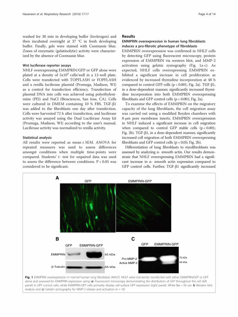
TGF-β1 induces human lung fibroblast proliferation,migration and differentiation to myofibroblast: Inhibitionby EMMPRIN functional blocking antibodyAs shown in Fig. 6a, TGF-β1 significantly induced an in-crease in NHLF proliferation as evidenced by increasedthymidine incorporation (p < 0.05). The EMMPRINblocking antibody significantly inhibited TGF- β1 in-duced fibroblast proliferation. Using a transwell migra-tion assay, TGF-β1 significantly induced an increase inNHLF cell migration and EMMPRIN functional blockingantibody significantly inhibited TGF- β1 induced NHLFmigration (Fig. 6b). TGF-β 1 significantly induced anincrease in NHLF differentiation to myofibroblasts asevidenced by an increase in α-smooth muscle actin.Similar to proliferation and migration, EMMPRIN block-ing antibody significantly inhibited TGF- β1 inducedfibroblast differentiation to myofibroblasts (Fig. 6c).These results imply that the effects of TGF- β1 onNHLF behavior can be attributed to an increase inEMMPRIN expression.
Overexpression of EMMPRIN in NHLF results in activationof a Wnt/β-catenin signaling pathwayWNT/ β -catenin signaling pathway has been shownto play a major role in the development of experi-mental and human pulmonary fibrosis [27]. To ex-plore the mechanism by which EMMPRIN enhancesNHLF proliferation, migration, and differentiation, weexamined the active status of β-catenin, which is animportant factor in the canonical Wnt signaling path-way that can be induced by TGF- β1. Immunoblotting
studies demonstrated an increased expression of phos-phorylated β-catenin in cell lysate from EMMPRIN over-expressing NHLF compared to GFP transfected controlcells. TGF-β1 treatment of EMMPRIN overexpressingNHLF further increased the expression of β- catenin(Fig. 7a). To validate the functional significance ofincreased β-catenin expression, Wnt signaling activity wasmeasured by luciferase expression controlled by TOP-FLASH. EMMPRIN overexpression in NHLF was foundto induce an increase in TOPFLASH luciferase reporteractivity when compared with GFP control-transfected cells(Fig. 7b). TGF-β1 treatment of EMMPRIN overexpressingcells induced an increase in TOPFLASH luciferasereporter activity when compared with GFP controlcells (Fig. 7b).
DiscussionEMMPRIN, which is usually expressed on tumor cells ofepithelial origin, is responsible not only for induction ofMMPs in neighboring stromal cells but also for trans-formation of quiescent fibroblasts to cancer-associatedfibroblasts through direct tumor–stromal interactions[25]. Apart from its role on cancer cells, EMMPRINexpression is increased not only in lung fibrosis [23], butalso in other types of tissue fibrosis including renalfibrosis [31], hepatic fibrosis [32] and corneal woundhealing response [33]. EMMPRIN expression in all thesesituations is mainly localized in actively differentiatingbasal epithelial layer implying a role of EMMPRIN inepithelial stromal interactions [22]. The precise regulatorymechanism of EMMPRIN in lung fibrosis has not beenelucidated. In this study, we examined EMMPRIN’s rolein the regulation of pro-fibrotic phenotypic changes in hu-man lung fibroblast by studying fibroblasts proliferation,migration, differentiation, and resistance to apoptosis. Wepresent evidence that EMMPRIN overexpression signifi-cantly increases lung fibroblast proliferation, migrationand differentiation to myofibroblasts. These findings wereassociated with activation of the Wnt/β-catenin signalingpathway. Additionally, we present evidence that TGF-β1induces EMMPRIN expression in human lung fibroblasts.Finally, we demonstrated that inhibition of EMMPRINusing an EMMPRIN blocking antibody enhanced theapoptotic susceptibility of lung fibroblasts. Further, block-ing EMMPRIN functionally inhibited lung fibroblast pro-liferation, migration, and differentiation to myofibroblasts
(See figure on previous page.)Fig. 5 EMMPRIN functional blocking antibody induces apoptosis in normal human lung fibroblasts: NHLF treated for 24 h with TGF- β1 in thepresence of either EMMPRIN functional blocking antibody or IgG control antibody. Apoptosis was measured by (a) FACS analysis using annexin V/PIstaining; (b) Cell Death Detection ELISA assay, each bar represents means ± SEM of 3 independent experiments, each run in duplicate, ** p < 0.001comparing EMMPRIN functional blocking antibody to IgG control antibody treated cells; and (c) Western blot for caspase- 3 of cell lysates from NHLFtreated with TGF-β1 (5 and 10 ng/ml) in the presence of EMMPRIN functional blocking antibody or IgG control antibody. β-Tubulin was used as theloading control. Image is a representative of 3 independent experiments, each run in duplicate
Hasaneen et al. Respiratory Research (2016) 17:17 Page 9 of 14
1000
2000
3000
0
Control TGF-β5ng/ml TGF-β10ng/ml
Thy
mid
ine
inco
rpor
atio
n(C
PM
)
Anti-EMMPRIN Abs
IgG Abs**
**
N.S.
10
20
30
40
50
0
Nu
mb
er
of
Mig
ratin
gC
ells
Control TGF-β 5ng/ml TGF-β 10ng/ml
Anti-EMMPRIN Abs
IgG Abs
#
**
N.S.
42 kDa
55 kDa
TGF-β ng/ml Control 5 10 5 10
Ig G Abs Anti-EMMPRIN Abs
α−SMA
β−Tubulin
A
B
C
Fig. 6 (See legend on next page.)
Hasaneen et al. Respiratory Research (2016) 17:17 Page 10 of 14

in response to TGF-β1. Collectively, these findings indi-cate that increased EMMPRIN expression in the lung ofIPF patients represents one mechanism by which lungfibroblasts acquire a proliferative, migratory and anti-apoptotic phenotype.The data presented here suggests that TGF- β1 increases
lung fibroblast proliferation, migration, and differentiationinto myofibroblasts via upregulation of EMMPRIN expres-sion, which may be another potential mechanism to explainthe increased number of lung fibroblasts in pulmonaryfibrosis. Our results are in agreement with other recentstudies [22, 24] demonstrating that TGF-β1 contributes tothe upregulation of EMMPRIN in normal epithelial cellsand fibroblasts. Moreover, EMMPRIN, by inducing MMPs
releases activate TGF- β1, which further upregulates EMM-PRIN and creates a positive feedback regulation thatpotentially amplifies EMMPRIN’s effects [24, 34]. MMPs,specifically MMP-2, -9 and MMP-14, have been reportedto activate TGF- β1 [14–18]. MMP-2 deficiency suppressedthe activation of latent TGF-β and the Smad2/3 pathway invivo and in vitro [17].We also provide evidence that EMMPRIN overexpres-
sion induces resistance of NHLF to the apoptotic effectsof Staurosporine, a known inducer of apoptosis. TheEMMPRIN blocking antibody induced apoptosis ofNHLF treated with TGF- β1. These data suggest thatEMMPRIN acts as a fibroblast survival factor and TGF-β1 increases survival of lung fibroblasts via upregulation
(See figure on previous page.)Fig. 6 EMMPRIN blocking antibody attenuates TGF-β1 induced lung fibroblast proliferation, migration and differentiation to myofibroblast: NHLFwere stimulated with TGF-β1 (0-10 ng/ml) in the presence of either EMMPRIN functional blocking antibody or IgG control antibody examined for (a)Cells proliferation using [3H] thymidine incorporation; (b) Cell migration using a modified Boyden chamber assay. Each bar represents means ± SEM of3 independent experiments, each run in duplicate, # p = 0.002 or ** p < 0.001 comparing EMMPRIN functional blocking antibody to IgG controlantibody treated cells; and (c) Cell differentiation using Western blot for α-SMA of cell lysates from NHLF treated with TGF-β1 (5 and 10 ng/ml) in thepresence of either EMMPRIN functional blocking antibody or IgG antibody. β-Tubulin was used as the loading control. Image is a representative of 3independent experiments, each run in duplicate
85 kDa
TGF- - β 10 ng/ml – + – +
β−Tubulin
β−Catenin
55 kDa
GFP EMMPRIN
A
B
Luci
fera
se A
ctiv
ity (
Top
flash
/Fop
flash
rat
io) **
**
GFP EMMPRIN GFP/TGF-β EMMPRIN/TGF-β
2
4
6
8
Fig. 7 EMMPRIN Overexpression Increases Wnt/β-catenin signaling: (a) Representative Western blot for phosphorylated β-catenin of cells lysatesfrom NHLF overexpressing either EMMPRIN/GFP or GFP treated with and without TGF-β1 (10 ng/ml). β-Tubulin was used as the loading control.Image is representative of 3 independent experiments, each run in duplicate. b Transient transfection of TOPFLASH, FOPFLASH, and control renillaluciferase reporter constructs into NHLF overexpressing either EMMPRIN/GFP or GFP then treated with TGF-β1 10 ng/ml. Each bar representsmeans ± SEM of 3 independent experiments, each run in duplicate, ** p < 0.001comparing GFP to EMMPRIN/GFP transfected cells
Hasaneen et al. Respiratory Research (2016) 17:17 Page 11 of 14

of EMMPRIN expression in these cells. These findingsare supported by the report of Ma et al. [35] demon-strating that cell–cell contact mediated by EMMPRINconferred apoptosis resistance in an E-cadherindependent manner and down regulation of EMMPRINinhibited cell–cell contact formation, which induced cellapoptosis. Xie et al also demonstrated that EMMPRINoverexpression protects human umbilical vein endothe-lial cells (HUVECs) from apoptosis [36]. Other studieshave shown that EMMPRIN inhibits apoptosis by acti-vating the FAK-PI3K-calcium (Ca2+) signaling pathwayby interacting with α3β1 integrin and disrupts the nitricoxide /cGMP-mediated negative regulation of store-operated calcium entry, thus increasing the intracellularlevel of calcium [37]. The disruption of intracellularcalcium homeostasis can disturb endoplasmic reticulum(ER) function and induce ER stress (ERS) [38].We report in this study that EMMPRIN overexpres-
sion in NHLF induces fibroblast migration and EMM-PRIN blocking antibody inhibits NHLF migration inresponse to TGF- β1. These results are attributed to therole of EMMPRIN in inducing MMPs and its interactionwith a number of binding partners on the cell surface,including the hyaluronan receptor CD44 [39–43]. Theseinteractions are critical for regulating cell migration,survival and proliferation, and events required in differ-ent processes such as wound healing [44]. A study byToole and Slomiany [45] demonstrated that EMMPRINoverexpression plays an important role in cell migrationand invasion via inducing hyaluronan (HA) production[45]. Overexpression of HA synthase 2 (HAS2) by lungfibroblasts produced an aggressive invasive phenotypeleading to severe lung fibrosis and death after bleomycin-induced injury. Fibroblasts isolated from patients with IPFexhibited an invasive phenotype that was also dependenton HAS2 and CD44. EMMPRIN interaction with CD44has been reported to potentiate cell survival pathways andthe invasive phenotype of epithelial cells. EMMPRIN alsopromotes assembly of signaling complexes containingEMMPRIN, CD44, and EGFR in lipid raft like domainsforming a positive feedback loop that may amplify in-vasiveness of epithelial cells [46].Moreover, we provide evidence that EMMPRIN over-
expression in NHLF promotes the differentiation offibroblasts into myofibroblasts by inducing α-SMA ex-pression and EMMPRIN blocking antibody inhibitsTGF- β1 induced differentiation of fibroblasts to myofi-broblasts. These data are supported by a previouslyreported study showing that EMMPRIN induced cornealfibroblast differentiation into myofibroblasts by inducingα-SMA through MMP-independent mechanisms [22].Blocking EMMPRIN expression by small interferingRNA inhibited α-SMA and collagen gel contractioninduced not only by EMMPRIN but also by TGF-β1, a
major mediator of myofibroblast differentiation [22].EMMPRIN and α- SMA were co-localized to the samecells in the stroma of pathological tissue, thus support-ing a role for EMMPRIN in the differentiation of myofi-broblasts in vivo. These data suggest that in addition toregulating the degradative potential of the myofibro-blasts, EMMPRIN can also influence the contractilephenotype of these cells in an MMP independentmanner and expand on the mechanism by whichEMMPRIN remodels ECM during wound healing andtissue fibrosis.Finally, we demonstrate that EMMPRIN overexpres-
sion in NHLF increased Wnt/β –catenin signaling, asshown by increased β -catenin expression in EMMPRINoverexpressing NHLF and increased TOPFLASH lucifer-ase reporter activity. The Wnt/β catenin pathway isthought to play a major role in pulmonary fibrosis.Abnormal activation of Wnt/ β catenin pathway has beendemonstrated in lung tissue of patients with IPF [47] andin an experimental model of bleomycin induced pulmon-ary fibrosis [48]. MMPs especially MMP-2, -7, -9, -14 andTGF – β1 have been known as Wnt/ β- catenin targetgenes [49–51]. Supporting our data is a study by Sidhuet al [26] demonstrating that EMMPRIN has a regula-tory role over the Wnt/β -catenin signaling pathway.Increasing EMMPRIN expression upregulated the β-catenin signaling pathway and silencing EMMPRINinhibited β -catenin signaling, cell migration, prolifera-tion, anchorage-independent growth and tumor growthin a mouse tumor xenograft model [26].
ConclusionThe present study demonstrates that TGF-β1 induces therelease of EMMPRIN that in turn activates the β-catenin/canonical Wnt signaling pathway. EMMPRIN over ex-pression in human lung fibroblasts contributes to theproliferative, migratory, and anti-apoptotic phenotype oflung fibroblasts that may contribute to the persistentfibro-proliferative state seen IPF. Further studies areneeded to explain the mechanism by which EMM-PRIN mediates the phenotypic changes in humanlung fibroblasts and regulates TGF- β1 signaling.
Competing interestsThe authors declare that they have no competing interests.
Authors’ contributionsNH Designed and performed the experiment, analyzed the data, wrote thepaper. JC, AP provided support with cells transfection, performedtransfection experiment and edited the manuscript and Figures. SZ, revieweddata, help edit the manuscript; lent expertise in some EMMPRIN experiments.HF Designed and supervised the experiments, reviewed and helped analyzethe data, edited and helped write the manuscript and provided funding forthe study. All authors read and approved the final manuscript.
AcknowledgementsThanks to all of the members of Jian Cao laboratory for full cooperation andvaluable advice.
Hasaneen et al. Respiratory Research (2016) 17:17 Page 12 of 14

Author details1Department of Medicine and Research, Veterans Administration MedicalCenter, Northport, USA. 2Department of Medicine, Stony Brook Medicine,Stony Brook, New York, USA. 3Division of Pulmonary, Critical Care and SleepMedicine, Stony Brook University Medical Center, Stony Brook, NY11794-8172, USA.
Received: 8 October 2015 Accepted: 10 February 2016
References1. Raghu G, Weycker D, Edelsberg J, Bradford WZ, Oster G. Incidence and
prevalence of idiopathic pulmonary fibrosis. Am J Respir Crit Care Med.2006;174:810–6.
2. King Jr TE, Pardo A, Selman M. Idiopathic pulmonary fibrosis. Lancet.2011;378:1949–61.
3. Katzenstein AL, Myers JL. Idiopathic pulmonary fibrosis: clinical relevance ofpathologic classification. Am J Respir Crit Care Med. 1998;157:1301–15.
4. Cha SI, Groshong SD, Frankel SK, Edelman BL, Cosgrove GP, Terry-Powers JL,et al. Compartmentalized expression of c-FLIP in lung tissues of patientswith idiopathic pulmonary fibrosis. Am J Respir Cell Mol Biol. 2010;42:140–8.
5. Korfei M, Ruppert C, Mahavadi P, Henneke I, Markart P, Koch M, et al.Epithelial endoplasmic reticulum stress and apoptosis in sporadic idiopathicpulmonary fibrosis. Am J Respir Crit Care Med. 2008;178:838–46.
6. Horowitz JC, Ajayi IO, Kulasekaran P, Rogers DS, White JB, Townsend SK, etal. Survivin expression induced by endothelin-1 promotes myofibroblastresistance to apoptosis. Int J Biochem Cell Biol. 2012;44:158–69.
7. Huang SK, White ES, Wettlaufer SH, Grifka H, Hogaboam CM, Thannickal VJ,et al. Prostaglandin E(2) induces fibroblast apoptosis by modulatingmultiple survival pathways. FASEB J. 2009;23:4317–26.
8. Dancer RC, Wood AM, Thickett DR. Metalloproteinases in idiopathicpulmonary fibrosis. Eur Respir J. 2011;38:1461–7.
9. Horowitz JC, Lee DY, Waghray M, Keshamouni VG, Thomas PE, Zhang H, etal. Activation of the pro-survival phosphatidylinositol 3-kinase/AKT pathwayby transforming growth factor-beta1 in mesenchymal cells is mediated byp38 MAPK-dependent induction of an autocrine growth factor. J Biol Chem.2004;279:1359–67.
10. Kulasekaran P, Scavone CA, Rogers DS, Arenberg DA, Thannickal VJ,Horowitz JC. Endothelin-1 and transforming growth factor-beta1independently induce fibroblast resistance to apoptosis via AKT activation.Am J Respir Cell Mol Biol. 2009;41:484–93.
11. Selman M, King TE, Pardo A, American Thoracic S, European Respiratory S,American College of Chest P. Idiopathic pulmonary fibrosis: prevailing andevolving hypotheses about its pathogenesis and implications for therapy.Ann Intern Med. 2001;134:136–51.
12. Lee CG, Kang HR, Homer RJ, Chupp G, Elias JA. Transgenic modeling oftransforming growth factor-beta(1): role of apoptosis in fibrosis and alveolarremodeling. Proc Am Thorac Soc. 2006;3:418–23.
13. Wilson MS, Wynn TA. Pulmonary fibrosis: pathogenesis, etiology andregulation. Mucosal Immunol. 2009;2:103–21.
14. Yu Q, Stamenkovic I. Cell surface-localized matrix metalloproteinase-9proteolytically activates TGF-beta and promotes tumor invasion andangiogenesis. Genes Dev. 2000;14:163–76.
15. Kobayashi T, Kim H, Liu X, Sugiura H, Kohyama T, Fang Q, et al.Matrix metalloproteinase-9 activates TGF-beta and stimulatesfibroblast contraction of collagen gels. Am J Physiol Lung CellMol Physiol. 2014;306:L1006–1015.
16. Dayer C, Stamenkovic I. Recruitment of Matrix Metalloproteinase-9 (MMP-9)to the fibroblast cell surface by Lysyl Hydroxylase 3 (LH3) TriggersTransforming Growth Factor-beta (TGF-beta) activation and fibroblastdifferentiation. J Biol Chem. 2015;290:13763–78.
17. Shen M, Lee J, Basu R, Sakamuri SS, Wang X, Fan D, et al. Divergent roles ofmatrix metalloproteinase 2 in pathogenesis of thoracic aortic aneurysm.Arterioscler Thromb Vasc Biol. 2015;35:888–98.
18. Karsdal MA, Larsen L, Engsig MT, Lou H, Ferreras M, Lochter A, et al. Matrixmetalloproteinase-dependent activation of latent transforming growthfactor-beta controls the conversion of osteoblasts into osteocytes byblocking osteoblast apoptosis. J Biol Chem. 2002;277:44061–7.
19. Toole BP. Emmprin (CD147), a cell surface regulator of matrixmetalloproteinase production and function. Curr Top Dev Biol.2003;54:371–89.
20. Zucker S, Hymowitz M, Rollo EE, Mann R, Conner CE, Cao J, et al.Tumorigenic potential of extracellular matrix metalloproteinase inducer.Am J Pathol. 2001;158:1921–8.
21. Suzuki S, Sato M, Senoo H, Ishikawa K. Direct cell-cell interaction enhancespro-MMP-2 production and activation in co-culture of laryngeal cancercells and fibroblasts: involvement of EMMPRIN and MT1-MMP. Exp CellRes. 2004;293:259–66.
22. Huet E, Vallee B, Szul D, Verrecchia F, Mourah S, Jester JV, et al.Extracellular matrix metalloproteinase inducer/CD147 promotesmyofibroblast differentiation by inducing alpha-smooth muscle actinexpression and collagen gel contraction: implications in tissueremodeling. FASEB J. 2008;22:1144–54.
23. Guillot S, Delaval P, Brinchault G, Caulet-Maugendre S, Depince A, Lena H, etal. Increased extracellular matrix metalloproteinase inducer (EMMPRIN)expression in pulmonary fibrosis. Exp Lung Res. 2006;32:81–97.
24. Gabison EE, Mourah S, Steinfels E, Yan L, Hoang-Xuan T, Watsky MA, et al.Differential expression of extracellular matrix metalloproteinase inducer(CD147) in normal and ulcerated corneas: role in epithelio-stromalinteractions and matrix metalloproteinase induction. Am J Pathol.2005;166:209–19.
25. Xu J, Lu Y, Qiu S, Chen ZN, Fan Z. A novel role of EMMPRIN/CD147 intransformation of quiescent fibroblasts to cancer-associated fibroblasts bybreast cancer cells. Cancer Lett. 2013;335:380–6.
26. Sidhu SS, Nawroth R, Retz M, Lemjabbar-Alaoui H, Dasari V, BasbaumC. EMMPRIN regulates the canonical Wnt/beta-catenin signalingpathway, a potential role in accelerating lung tumorigenesis.Oncogene. 2010;29:4145–56.
27. Konigshoff M, Balsara N, Pfaff EM, Kramer M, Chrobak I, Seeger W, et al.Functional Wnt signaling is increased in idiopathic pulmonary fibrosis.PLoS One. 2008;3:e2142.
28. Hasaneen NA, Zucker S, Cao J, Chiarelli C, Panettieri RA, Foda HD. Cyclicmechanical strain-induced proliferation and migration of human airwaysmooth muscle cells: role of EMMPRIN and MMPs. FASEB J. 2005;19:1507–9.
29. Moodley YP, Caterina P, Scaffidi AK, Misso NL, Papadimitriou JM, McAnultyRJ, et al. Comparison of the morphological and biochemical changes innormal human lung fibroblasts and fibroblasts derived from lungs ofpatients with idiopathic pulmonary fibrosis during FasL-induced apoptosis.J Pathol. 2004;202:486–95.
30. Buhling F, Wille A, Rocken C, Wiesner O, Baier A, Meinecke I, et al.Altered expression of membrane-bound and soluble CD95/Fascontributes to the resistance of fibrotic lung fibroblasts to FasLinduced apoptosis. Respir Res. 2005;6:37.
31. Kato N, Kosugi T, Sato W, Ishimoto T, Kojima H, Sato Y, et al. Basigin/CD147promotes renal fibrosis after unilateral ureteral obstruction. Am J Pathol.2011;178:572–9.
32. Zhang DW, Zhao YX, Wei D, Li YL, Zhang Y, Wu J, et al. HAb18G/CD147promotes activation of hepatic stellate cells and is a target for antibodytherapy of liver fibrosis. J Hepatol. 2012;57:1283–91.
33. Gabison EE, Huet E, Baudouin C, Menashi S. Direct epithelial-stromalinteraction in corneal wound healing: Role of EMMPRIN/CD147 in MMPsinduction and beyond. Prog Retin Eye Res. 2009;28:19–33.
34. Tang Y, Kesavan P, Nakada MT, Yan L. Tumor-stroma interaction: positivefeedback regulation of extracellular matrix metalloproteinase inducer(EMMPRIN) expression and matrix metalloproteinase-dependent generationof soluble EMMPRIN. Mol Cancer Res. 2004;2:73–80.
35. Ma XK, Wang L, Li Y, Yang XM, Zhao P, HaoTang, Zhu P, Li L, Chen ZN.HAb18G/CD147 cell-cell contacts confer resistance of a HEK293subpopulation to anoikis in an E-cadherin-dependent manner. BMC CellBiol. 2010, 11:27.
36. Xie W, Xie H, Liu F, Li W, Dan J, Mei Y, et al. Propranolol induces apoptosisof human umbilical vein endothelial cells through downregulation ofCD147. Br J Dermatol. 2013;168:739–48.
37. Tang J, Wu YM, Zhao P, Yang XM, Jiang JL, Chen ZN. Overexpressionof HAb18G/CD147 promotes invasion and metastasis via alpha3beta1integrin mediated FAK-paxillin and FAK-PI3K-Ca2+ pathways. Cell MolLife Sci. 2008;65:2933–42.
38. Sammels E, Parys JB, Missiaen L, De Smedt H, Bultynck G. IntracellularCa2+ storage in health and disease: a dynamic equilibrium. CellCalcium. 2010;47:297–314.
39. Toole BP. Hyaluronan: from extracellular glue to pericellular cue.Nat Rev Cancer. 2004;4:528–39.
Hasaneen et al. Respiratory Research (2016) 17:17 Page 13 of 14

40. Tang W, Chang SB, Hemler ME. Links between CD147 function,glycosylation, and caveolin-1. Mol Biol Cell. 2004;15:4043–50.
41. Arora K, Gwinn WM, Bower MA, Watson A, Okwumabua I, MacDonald HR, etal. Extracellular cyclophilins contribute to the regulation of inflammatoryresponses. J Immunol. 2005;175:517–22.
42. Kirk P, Wilson MC, Heddle C, Brown MH, Barclay AN, Halestrap AP. CD147 istightly associated with lactate transporters MCT1 and MCT4 and facilitatestheir cell surface expression. EMBO J. 2000;19:3896–904.
43. Philp NJ, Wang D, Yoon H, Hjelmeland LM. Polarized expression ofmonocarboxylate transporters in human retinal pigment epithelium andARPE-19 cells. Invest Ophthalmol Vis Sci. 2003;44:1716–21.
44. Berditchevski F, Chang S, Bodorova J, Hemler ME. Generation of monoclonalantibodies to integrin-associated proteins. Evidence that alpha3beta1complexes with EMMPRIN/basigin/OX47/M6. J Biol Chem. 1997;272:29174–80.
45. Toole BP, Slomiany MG. Hyaluronan, CD44 and Emmprin: partners in cancercell chemoresistance. Drug Resist Updat. 2008;11:110–21.
46. Grass GD, Tolliver LB, Bratoeva M, Toole BP. CD147, CD44, and theepidermal growth factor receptor (EGFR) signaling pathway cooperate toregulate breast epithelial cell invasiveness. J Biol Chem. 2013;288:26089–104.
47. Chilosi M, Poletti V, Zamo A, Lestani M, Montagna L, Piccoli P, et al.Aberrant Wnt/beta-catenin pathway activation in idiopathic pulmonaryfibrosis. Am J Pathol. 2003;162:1495–502.
48. Liu L, Carron B, Yee HT, Yie TA, Hajjou M, Rom W. Wnt pathway inpulmonary fibrosis in the bleomycin mouse model. J Environ Pathol ToxicolOncol. 2009;28:99–108.
49. Brabletz T, Jung A, Dag S, Hlubek F, Kirchner T. beta-catenin regulates theexpression of the matrix metalloproteinase-7 in human colorectal cancer.Am J Pathol. 1999;155:1033–8.
50. Wu B, Crampton SP, Hughes CC. Wnt signaling induces matrixmetalloproteinase expression and regulates T cell transmigration. Immunity.2007;26:227–39.
51. Karow M, Popp T, Egea V, Ries C, Jochum M, Neth P. Wnt signalling inmouse mesenchymal stem cells: impact on proliferation, invasion and MMPexpression. J Cell Mol Med. 2009;13:2506–20.
• We accept pre-submission inquiries
• Our selector tool helps you to find the most relevant journal
• We provide round the clock customer support
• Convenient online submission
• Thorough peer review
• Inclusion in PubMed and all major indexing services
• Maximum visibility for your research
Submit your manuscript atwww.biomedcentral.com/submit
Submit your next manuscript to BioMed Central and we will help you at every step:
Hasaneen et al. Respiratory Research (2016) 17:17 Page 14 of 14
Related Documents