SHORT COMMUNICATION Extracellular chitinase production by some members of the saprophytic Entomophthorales group P. Mishra • S. K. Singh • S. S. Nilegaonkar Received: 28 May 2010 / Accepted: 29 October 2010 Ó The Mycological Society of Japan and Springer 2010 Abstract Thirteen strains were isolated from different habitats, belonging to two genera, namely Conidiobolus and Basidiobolus, related to saprophytic Entomophthorales. Chitin flake colonization and agar-well diffusion tests were used to screen potential extracellular chitinase-producing strains in plate assays. Preliminary screening resulted in five chitinase producers that were further studied quantitatively. Results indicated that studied isolates of this group produced chitinase at different levels in chitin-containing as well as non-chitin-containing medium. Conidiobolus coronatus was found to be the most significant chitinase producer, giving 0.261 U/ml using colloidal chitin as a carbon source, among the isolates under study. This communication also reports the chitinolytic activity of Basidiobolus haptoporus, the effect of environmental and nutritional parameters on chitinase pro- duction, and utilization of fungal biomass as a carbon source, which hitherto had not been elaborated from this genus. Keywords Basidiobolus haptosporus Á Colloidal chitin Á Conidiobolus coronatus Á Native chitin Introduction Chitin, a tough and pliable b (1–4) homopolymer of N-acetyl-D-glucosamine (GlcNAc), is the most abundant polysaccharide, after cellulose, existing in nature. It con- stitutes a major structural component of many biological systems, particularly those of mollusks, insects, crusta- ceans, fungi, algae, and marine invertebrates (Shaikh and Deshpande 1993). More than 10 11 tonnes of chitinous waste is obtained per year from the aquatic biosphere alone (Tsujibo et al. 1998). Chitin and its derivatives are of commercial and biotechnological interest because of their wider range of biological activities (Bhushan and Hoondal 1998; Gohel et al. 2006). The enzymatic hydrolysis of chitin to free N-acetylglucosamine units is performed by a chitinolytic system, which is found in a variety of organ- isms such as actinomycetes, bacteria, fungi, yeasts, plants, insects, and also in human beings (Bhattacharya et al. 2007; Mellor et al. 1994; Royer et al. 2002; Tjoelker et al. 2000). In recent years, chitinases (EC 3.2.1.14) have received greater attention for reasons of their wider range of biotechnological applications, especially in the biocon- trol of fungal phytopathogens (Mathivanan et al. 1998) and harmful insect pests (Mendonsa et al. 1996; Pinto et al. 1997). Chitinases have also been used in the preparation of sphaeroplasts and protoplasts from yeast and fungal species (Peberdy 1985; Mizuno et al. 1997). Some other signifi- cant applications of chitinases include bioconversions of chitin waste to single-cell protein and ethanol (Vyas and Deshpande 1991) and fertilizers (Sakai et al. 1986). The saprophytic Entomophthorales group has been studied by different workers for its extracellular enzyme profile. However, meager data are available about the extracellular secretion of chitinase from the fungal mem- bers of this group. Keeping this in mind, we studied some All the strains have been deposited and accessioned in National Fungal Culture Collection of India (NFCCI) (WDCM-932) At MACS’s Agharkar Research Institute, Pune, India. Electronic supplementary material The online version of this article (doi:10.1007/s10267-010-0090-3) contains supplementary material, which is available to authorized users. P. Mishra Á S. K. Singh Mycology and Plant Pathology Group, Agharkar Research Institute, G G Agarkar Road, Pune 411 004, India S. S. Nilegaonkar (&) Microbial Sciences Division, Agharkar Research Institute, G G Agarkar Road, Pune 411 004, India e-mail: [email protected] 123 Mycoscience DOI 10.1007/s10267-010-0090-3

Welcome message from author
This document is posted to help you gain knowledge. Please leave a comment to let me know what you think about it! Share it to your friends and learn new things together.
Transcript
SHORT COMMUNICATION
Extracellular chitinase production by some membersof the saprophytic Entomophthorales group
P. Mishra • S. K. Singh • S. S. Nilegaonkar
Received: 28 May 2010 / Accepted: 29 October 2010
� The Mycological Society of Japan and Springer 2010
Abstract Thirteen strains were isolated from different
habitats, belonging to two genera, namely Conidiobolus and
Basidiobolus, related to saprophytic Entomophthorales.
Chitin flake colonization and agar-well diffusion tests were
used to screen potential extracellular chitinase-producing
strains in plate assays. Preliminary screening resulted in five
chitinase producers that were further studied quantitatively.
Results indicated that studied isolates of this group produced
chitinase at different levels in chitin-containing as well as
non-chitin-containing medium. Conidiobolus coronatus was
found to be the most significant chitinase producer, giving
0.261 U/ml using colloidal chitin as a carbon source, among
the isolates under study. This communication also reports the
chitinolytic activity of Basidiobolus haptoporus, the effect of
environmental and nutritional parameters on chitinase pro-
duction, and utilization of fungal biomass as a carbon source,
which hitherto had not been elaborated from this genus.
Keywords Basidiobolus haptosporus � Colloidal chitin �Conidiobolus coronatus � Native chitin
Introduction
Chitin, a tough and pliable b (1–4) homopolymer of
N-acetyl-D-glucosamine (GlcNAc), is the most abundant
polysaccharide, after cellulose, existing in nature. It con-
stitutes a major structural component of many biological
systems, particularly those of mollusks, insects, crusta-
ceans, fungi, algae, and marine invertebrates (Shaikh and
Deshpande 1993). More than 1011 tonnes of chitinous
waste is obtained per year from the aquatic biosphere alone
(Tsujibo et al. 1998). Chitin and its derivatives are of
commercial and biotechnological interest because of their
wider range of biological activities (Bhushan and Hoondal
1998; Gohel et al. 2006). The enzymatic hydrolysis of
chitin to free N-acetylglucosamine units is performed by a
chitinolytic system, which is found in a variety of organ-
isms such as actinomycetes, bacteria, fungi, yeasts, plants,
insects, and also in human beings (Bhattacharya et al.
2007; Mellor et al. 1994; Royer et al. 2002; Tjoelker et al.
2000). In recent years, chitinases (EC 3.2.1.14) have
received greater attention for reasons of their wider range
of biotechnological applications, especially in the biocon-
trol of fungal phytopathogens (Mathivanan et al. 1998) and
harmful insect pests (Mendonsa et al. 1996; Pinto et al.
1997). Chitinases have also been used in the preparation of
sphaeroplasts and protoplasts from yeast and fungal species
(Peberdy 1985; Mizuno et al. 1997). Some other signifi-
cant applications of chitinases include bioconversions of
chitin waste to single-cell protein and ethanol (Vyas and
Deshpande 1991) and fertilizers (Sakai et al. 1986).
The saprophytic Entomophthorales group has been
studied by different workers for its extracellular enzyme
profile. However, meager data are available about the
extracellular secretion of chitinase from the fungal mem-
bers of this group. Keeping this in mind, we studied some
All the strains have been deposited and accessioned in National
Fungal Culture Collection of India (NFCCI) (WDCM-932) At
MACS’s Agharkar Research Institute, Pune, India.
Electronic supplementary material The online version of thisarticle (doi:10.1007/s10267-010-0090-3) contains supplementarymaterial, which is available to authorized users.
P. Mishra � S. K. Singh
Mycology and Plant Pathology Group, Agharkar Research
Institute, G G Agarkar Road, Pune 411 004, India
S. S. Nilegaonkar (&)
Microbial Sciences Division, Agharkar Research Institute,
G G Agarkar Road, Pune 411 004, India
e-mail: [email protected]
123
Mycoscience
DOI 10.1007/s10267-010-0090-3
members of this group from soil and plant litter, including
two strains of Basidiobolus haptosporus from intestinal
contents of frog, and screened them for extracellular chi-
tinase production.
Leaf detritus collected from different locations was
screened for saprophytic Entomophthorales by canopying
moistened litter on MGYP (malt extract-glucose-yeast
extract-peptone agar) plates (Drechsler 1952; Srinivasan
and Thirumalachar 1968). Isolates found in this study
were obtained after 7 days, developing from forcibly
discharged conidia in isolation plates. Excreta was col-
lected from two frogs captured from the institute’s garden
and used for the isolation of fungi by the aforementioned
technique.
The morphological details of isolates were studied and
measurements of different fungal structures were under-
taken following the procedure described earlier to confirm
the identity of the fungal isolates of saprophytic Entom-
ophthorales (Waingankar et al. 2008). All the fungal strains
have been deposited and accessioned in the National
Fungal Culture Collection of India (WDCM-932) at MACS
Agharkar Research Institute, Pune, India.
Fungal cultures were inoculated onto 0.2% yeast extract
agar, and sterile chitin flakes were placed near the fungal
inoculum aseptically and incubated at 288 ± 28C. Flakes
were microscopically examined for the presence of fungal
mycelia after 24 and 48 h of incubation.
One percent (1%) colloidal chitin medium with 0.2%
yeast extract powder was prepared for agar plate assays. An
agar punch from the 48-h-old culture growing on MGYP
plates was placed in the center of the plate. After incuba-
tion at 288 ± 28C for 3–4 days, plates were observed for
the zones of clearance. To enhance the visibility and clarity
of zones of clearance, the dye Remazol Brilliant Blue
(0.85%) was also added into the medium. For the agar-well
diffusion test, crude enzyme was obtained by cultivating
fungal isolates on MGYP broth containing 1% colloidal
chitin at 288 ± 28C at 200 rev/min for 48 h. The cell-free
supernatant (crude enzyme) was used on 1% colloidal
chitin agar.
The production medium for the chitinase contained (in
g/l) polypeptone, 1.25; yeast extract, 1.25; malt extract,
0.75; glucose, 2.5; chitin (native/colloidal), 10.0, with pH
6.5–6.8. Flasks were inoculated with spore suspension
containing 1 9 106 cfu and incubated at 288 ± 28C at
200 rev/min. Acid-swollen chitin, containing about 7 mg
chitin/ml, was prepared according to Hackman (1962) and
used for the quantitative estimations. For the chitinase
assay, crude enzyme was assayed using the colorimetric
method with slight modifications (Elson and Morgan
1933). The reaction mixture contained 1 ml each acid-
swollen chitin, 0.05 M acetate buffer (pH 5.0), and crude
enzyme preparation. N-Acetyl-D-glucosamine (NAG) units
released were measured at A585. A calibration curve was
obtained by using the range of 10–100 lg/ml of standard
NAG. A unit of chitinase activity was defined as the
amount of enzyme that produced 1 lmole of NAG equiv-
alent per hour under the assay conditions.
Basidiobolus was grown on MGYP agar plates. After
sporulation, it was transferred onto MGYP broth contain-
ing 1% NH4Cl for production of darmform cells, up to
48 h, which were used as inoculum (Ingale et al. 2002).
Production medium contained MGYP with 1% colloidal
chitin or 1% native chitin. Flasks were inoculated with
broth containing darmform cells 10% (v/v) and incubated
at 288 ± 28C at 200 rev/min. The effect of different
environmental and nutritional parameters such as inoculum
size (5–25%), pH (4.0, 6.5, 8.0, 10.0, and 12.0), tempera-
tures (208, 258, 308, and 358C), sugars (glucose, sucrose,
maltose, lactose, and galactose), and types of chitin (col-
loidal chitin and flakes) on chitinase production by
Basidiobolus haptosporus was studied using a one factor at
a time approach.
Dried fungal mycelia as an alternate carbon source were
used for the chitinase production. Different fungi, viz.
Aspergillus niger (NFCCI 663), Aspergillus flavus (NFCCI
664), Penicillium sp. (NFCCI 672), Paecilomyces variotii
(NFCCI 671), and Acremonium sp. (NFCCI 662), were
grown in potato dextrose broth for 1 week. Mycelium was
dried and powdered in a blender. From production med-
ium, chitin was replaced by dried and powdered fungal
mycelia. The comparative chitinase production was cal-
culated against chitin-containing medium.
The morphotaxonomic identification of recovered fun-
gal isolates resulted in a total 13 isolates contributing nine
Conidiobolus and four Basidiobolus species (eight Conid-
iobolus and one Basidiobolus from plant litter, one isolate
each from soil, and two Basidiobolus isolates from frog
excreta).
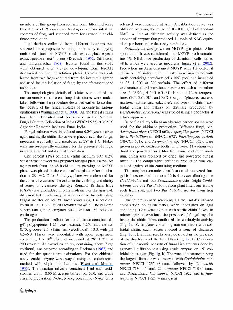
During preliminary screening all the isolates showed
colonization on chitin flakes when inoculated on agar
containing 0.2% yeast extract with sterile chitin flakes. In
microscopic observations, the presence of fungal mycelia
inside the chitin flakes confirmed the chitinolytic activity
(Fig. 1a, b). In plates containing nutrient media with col-
loidal chitin, each isolate showed a zone of clearance
(Fig. 1c, d). Similar results were observed in the presence
of the dye Remazol Brilliant Blue (Fig. 1e, f). Confirma-
tion of chitinolytic activity of fungal isolates was done by
agar-well diffusion test using crude enzyme on 1% col-
loidal chitin agar (Fig. 1g, h). The zone of clearance having
the largest diameter was observed with Conidiobolus cor-
onatus NFCCI 1235 (8 mm), followed by C. couchii
NFCCI 719 (4.5 mm), C. coronatus NFCCI 718 (4 mm),
and Basidiobolus haptosporus NFCCI 1922 and B. hap-
tosporus NFCCI 1923 (4 mm each)
Mycoscience
123
Isolates that produced zones of clearance of more than
4 mm were considered significant producers of extracel-
lular chitinase and were selected for flask level studies.
During preliminary quantitative estimations, maximum
chitinase production was 0.261 U/ml in crude cell-free
supernatant of C. coronatus NFCCI 1235, followed by
Fig. 1 a, b Chitin flake
colonization by Basidiobolushaptosporus NFCCI 1922 and
NFCCI 1923 (b stained with
cotton blue). White arrows,
chitin flake; black arrows,
presence of fungal structures.
Bars a, b 40 lm; 940.
c, d Zone of clearance on
colloidal chitin by Conidioboluscoronatus NFCCI 1235 and
C. couchii NFCCI 719.
e, f Zone of clearance on
colloidal chitin supplemented
with dye formed by B.haptosporus NFCCI 1922 and
C. coronatus NFCCI 1235.
g, h Agar-well diffusion test of
crude chitinase by C. coronatusNFCCI 1235 and C. couchiiNFCCI 719
Mycoscience
123
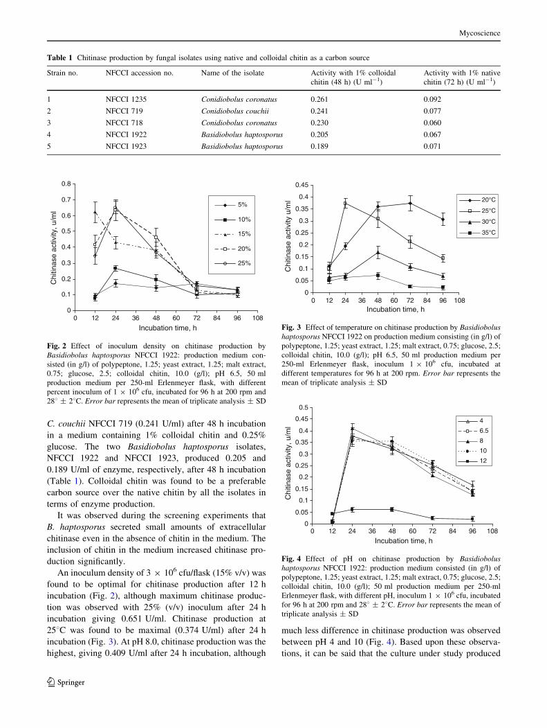
C. couchii NFCCI 719 (0.241 U/ml) after 48 h incubation
in a medium containing 1% colloidal chitin and 0.25%
glucose. The two Basidiobolus haptosporus isolates,
NFCCI 1922 and NFCCI 1923, produced 0.205 and
0.189 U/ml of enzyme, respectively, after 48 h incubation
(Table 1). Colloidal chitin was found to be a preferable
carbon source over the native chitin by all the isolates in
terms of enzyme production.
It was observed during the screening experiments that
B. haptosporus secreted small amounts of extracellular
chitinase even in the absence of chitin in the medium. The
inclusion of chitin in the medium increased chitinase pro-
duction significantly.
An inoculum density of 3 9 106 cfu/flask (15% v/v) was
found to be optimal for chitinase production after 12 h
incubation (Fig. 2), although maximum chitinase produc-
tion was observed with 25% (v/v) inoculum after 24 h
incubation giving 0.651 U/ml. Chitinase production at
258C was found to be maximal (0.374 U/ml) after 24 h
incubation (Fig. 3). At pH 8.0, chitinase production was the
highest, giving 0.409 U/ml after 24 h incubation, although
much less difference in chitinase production was observed
between pH 4 and 10 (Fig. 4). Based upon these observa-
tions, it can be said that the culture under study produced
Table 1 Chitinase production by fungal isolates using native and colloidal chitin as a carbon source
Strain no. NFCCI accession no. Name of the isolate Activity with 1% colloidal
chitin (48 h) (U ml-1)
Activity with 1% native
chitin (72 h) (U ml-1)
1 NFCCI 1235 Conidiobolus coronatus 0.261 0.092
2 NFCCI 719 Conidiobolus couchii 0.241 0.077
3 NFCCI 718 Conidiobolus coronatus 0.230 0.060
4 NFCCI 1922 Basidiobolus haptosporus 0.205 0.067
5 NFCCI 1923 Basidiobolus haptosporus 0.189 0.071
0
0.1
0.2
0.3
0.4
0.5
0.6
0.7
0.8
0 12 24 36 48 60 72 84 96 108
Incubation time, h
Chi
tinas
e ac
tivity
, u/m
l
5%
10%
15%
20%
25%
Fig. 2 Effect of inoculum density on chitinase production by
Basidiobolus haptosporus NFCCI 1922: production medium con-
sisted (in g/l) of polypeptone, 1.25; yeast extract, 1.25; malt extract,
0.75; glucose, 2.5; colloidal chitin, 10.0 (g/l); pH 6.5, 50 ml
production medium per 250-ml Erlenmeyer flask, with different
percent inoculum of 1 9 106 cfu, incubated for 96 h at 200 rpm and
288 ± 28C. Error bar represents the mean of triplicate analysis ± SD
0
0.05
0.1
0.15
0.2
0.25
0.3
0.35
0.4
0.45
0 12 24 36 48 60 72 84 96 108Incubation time, h
Chi
tinas
e ac
tivity
u/m
l 20°C
25°C
30°C
35°C
Fig. 3 Effect of temperature on chitinase production by Basidiobolushaptosporus NFCCI 1922 on production medium consisting (in g/l) of
polypeptone, 1.25; yeast extract, 1.25; malt extract, 0.75; glucose, 2.5;
colloidal chitin, 10.0 (g/l); pH 6.5, 50 ml production medium per
250-ml Erlenmeyer flask, inoculum 1 9 106 cfu, incubated at
different temperatures for 96 h at 200 rpm. Error bar represents the
mean of triplicate analysis ± SD
0
0.05
0.1
0.15
0.2
0.25
0.3
0.35
0.4
0.45
0.5
0 12 24 36 48 60 72 84 96 108
Incubation time, h
Chi
tinas
e ac
tivity
, u/m
l
4
6.5
8
10
12
Fig. 4 Effect of pH on chitinase production by Basidiobolushaptosporus NFCCI 1922: production medium consisted (in g/l) of
polypeptone, 1.25; yeast extract, 1.25; malt extract, 0.75; glucose, 2.5;
colloidal chitin, 10.0 (g/l); 50 ml production medium per 250-ml
Erlenmeyer flask, with different pH, inoculum 1 9 106 cfu, incubated
for 96 h at 200 rpm and 288 ± 28C. Error bar represents the mean of
triplicate analysis ± SD
Mycoscience
123
chitinase that is active in a broad range of pH. It was
observed that chitinase activity declined after a particular
time period, which may occur because of autolysis of the
enzyme or unavailability of the substrate in the production
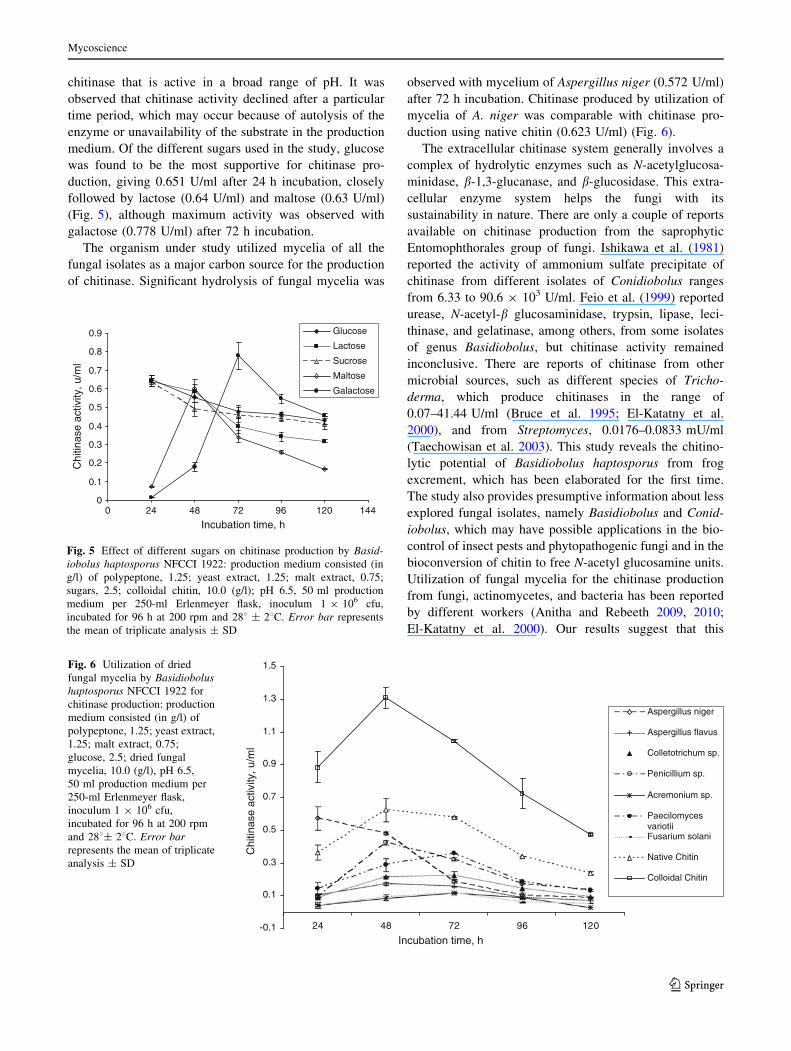
medium. Of the different sugars used in the study, glucose
was found to be the most supportive for chitinase pro-
duction, giving 0.651 U/ml after 24 h incubation, closely
followed by lactose (0.64 U/ml) and maltose (0.63 U/ml)
(Fig. 5), although maximum activity was observed with
galactose (0.778 U/ml) after 72 h incubation.
The organism under study utilized mycelia of all the
fungal isolates as a major carbon source for the production
of chitinase. Significant hydrolysis of fungal mycelia was
observed with mycelium of Aspergillus niger (0.572 U/ml)
after 72 h incubation. Chitinase produced by utilization of
mycelia of A. niger was comparable with chitinase pro-
duction using native chitin (0.623 U/ml) (Fig. 6).
The extracellular chitinase system generally involves a
complex of hydrolytic enzymes such as N-acetylglucosa-
minidase, b-1,3-glucanase, and b-glucosidase. This extra-
cellular enzyme system helps the fungi with its
sustainability in nature. There are only a couple of reports
available on chitinase production from the saprophytic
Entomophthorales group of fungi. Ishikawa et al. (1981)
reported the activity of ammonium sulfate precipitate of
chitinase from different isolates of Conidiobolus ranges
from 6.33 to 90.6 9 103 U/ml. Feio et al. (1999) reported
urease, N-acetyl-b glucosaminidase, trypsin, lipase, leci-
thinase, and gelatinase, among others, from some isolates
of genus Basidiobolus, but chitinase activity remained
inconclusive. There are reports of chitinase from other
microbial sources, such as different species of Tricho-
derma, which produce chitinases in the range of
0.07–41.44 U/ml (Bruce et al. 1995; El-Katatny et al.
2000), and from Streptomyces, 0.0176–0.0833 mU/ml
(Taechowisan et al. 2003). This study reveals the chitino-
lytic potential of Basidiobolus haptosporus from frog
excrement, which has been elaborated for the first time.
The study also provides presumptive information about less
explored fungal isolates, namely Basidiobolus and Conid-
iobolus, which may have possible applications in the bio-
control of insect pests and phytopathogenic fungi and in the
bioconversion of chitin to free N-acetyl glucosamine units.
Utilization of fungal mycelia for the chitinase production
from fungi, actinomycetes, and bacteria has been reported
by different workers (Anitha and Rebeeth 2009, 2010;
El-Katatny et al. 2000). Our results suggest that this
0
0.1
0.2
0.3
0.4
0.5
0.6
0.7
0.8
0.9
0 24 48 72 96 120 144
Incubation time, h
Chi
tinas
e ac
tivity
, u/m
l
Glucose
Lactose
Sucrose
Maltose
Galactose
Fig. 5 Effect of different sugars on chitinase production by Basid-iobolus haptosporus NFCCI 1922: production medium consisted (in
g/l) of polypeptone, 1.25; yeast extract, 1.25; malt extract, 0.75;
sugars, 2.5; colloidal chitin, 10.0 (g/l); pH 6.5, 50 ml production
medium per 250-ml Erlenmeyer flask, inoculum 1 9 106 cfu,
incubated for 96 h at 200 rpm and 288 ± 28C. Error bar represents
the mean of triplicate analysis ± SD
-0.1
0.1
0.3
0.5
0.7
0.9
1.1
1.3
1.5
24 48 72 96 120
Incubation time, h
Chi
tinas
e ac
tivity
, u/m
l
Aspergillus niger
Aspergillus flavus
Colletotrichum sp.
Penicillium sp.
Acremonium sp.
PaecilomycesvariotiiFusarium solani
Native Chitin
Colloidal Chitin
Fig. 6 Utilization of dried
fungal mycelia by Basidiobolushaptosporus NFCCI 1922 for
chitinase production: production
medium consisted (in g/l) of
polypeptone, 1.25; yeast extract,
1.25; malt extract, 0.75;
glucose, 2.5; dried fungal
mycelia, 10.0 (g/l), pH 6.5,
50 ml production medium per
250-ml Erlenmeyer flask,
inoculum 1 9 106 cfu,
incubated for 96 h at 200 rpm
and 288± 28C. Error barrepresents the mean of triplicate
analysis ± SD
Mycoscience
123
enzyme can be used for utilization of fungal biomass
produced during different fermentation processes and thus
N-acetyl glucosamine units can be produced for various
biomedical applications such as in the treatment of osteo-
arthritis and autoimmune encephalomyelitis (Felson and
McAlindon 2000; Reginster et al. 2001; Zhang et al. 2005).
Perusal of the literature indicates that interest in the
biotechnological potential of Conidiobolus has centered on
the production of high-activity alkaline protease (Phadtare
et al. 1993; Srinivasan 2007) and the production of poly-
unsaturated fatty acids (Idemitsu-Petrochem 1990). This is
the first systematic study of the chitinase profile of sapro-
phytic Entomophthorales, in particular from Basidiobolus
haptosprous. As such, the isolation of different isolates in
the present study will serve as a valuable gene pool. Fur-
ther, it attests to the fact that there is an urgent need for
sustained exploration of a wide variety of substrates for
selective isolation of fungi of one of the most neglected
group of saprophytic Entomophthorales and their screening
for extracellular chitinase and other biotechnological
potential.
Acknowledgments The authors are thankful to Dr. M.C. Srinivasan
(Scientist Emeritus, Agharkar Research Institute, Pune) for his expert
opinions and guidance and Mrs. V.M. Waingankar for help in the
isolation work. The authors are also thankful to the Director,
Agharkar Research Institute, for providing facilities.
References
Anitha A, Rebeeth M (2009) In vitro antifungal activity of
Streptomyces griseusa against phytopathogenic fungi of tomato
field. Acad J Plant Sci 2(2):119–123
Anitha A, Rebeeth M (2010) Degradation of fungal cell walls of
phytopathogenic fungi by lytic enzyme of Streptomyces griseus.
Afr J Plant Sci 4(3):61–66
Bhattacharya D, Nagpure A, Gupta RK (2007) Bacterial chitinases:
properties and potential. Crit Rev Biotechnol 27(1):21–28
Bhushan B, Hoondal GS (1998) Isolation, purification and properties
of a thermostable chitinase from an alkalophilic Bacillus sp. BG-
11. Biotechnol Lett 20(2):157–159
Bruce A, Srinivasan U, Staines HJ, Highley TL (1995) Chitinase and
laminarinase production in liquid culture by Trichoderma spp.
and their role in biocontrol of wood decay fungi. Int Biodeterior
Biodegrad 35:337–353
Drechsler C (1952) Widespread distribution of Delacroixia coronataand other saprophytic Entomophthoraceae in plant detritus.
Science 115:575–576
El-Katatny MH, Somitsch W, Robra KH, El-Katatny MS, Gubitz GM
(2000) Production of chitinase and b-1,3-glucanase by Tricho-derma harzianum for control of the phytopathogenic fungus
Sclerotium rolfsii. Food Technol Biotechnol 38(3):173–180
Elson LA, Morgan WTJ (1933) A colorimetric method for the
determination of glucosamine and chondrosamine. Biochem J
27:114–118
Feio CL, Bauwens L, Swinne D, De Meurichy W (1999) Isolation of
Basidiobolus ranarum from ectotherms in Antwerp zoo with
special reference to characterization of the isolated strains.
Mycoses 42:291–296
Felson DT, McAlindon TE (2000) Glucosamine and chondroitin for
osteoarthritis: to recommend or not to recommend? Arthritis
Care Res 13:179–182
Gohel V, Singh A, Maisuria V, Phadnis A, Chhatpar HS (2006)
Bioprospecting and antifungal potential of chitinolytic microor-
ganisms. Afr J Biotechnol 5(2):54–72
Hackman RH (1962) Studies on chitin. V. The effect of mineral acids
on chitin. Aust J Biol Sci 15:526–537
Idemitsu-Petrochem (1990) Preparation of lipid containing bish-
omogammalinolenic acid by culturing Conidiobolus sp. in
sesame oil culture medium JP-089982. Biotechnol Abstr
91:02817
Ingale SS, Rele MV, Srinivasan MC (2002) Alkaline protease
production by Basidiobolus (N.C.L. 97.1.1): effect of ‘darm-
form’ morphogenesis and cultural conditions on enzyme pro-
duction and preliminary enzyme characterization. World J
Microbiol Biotechnol 18(5):403–408
Ishikawa F, Oishi K, Ko A (1981) Chitinase production by
Conidiobolus lamprauges and other Conidiobolus species. Agric
Biol Chem 45(10):2361–2362
Mathivanan N, Kabilan V, Murugesan K (1998) Purification,
characterization and antifungal activity of chitinase from
Fusarium chlamydosporum, a mycoparasite to groundnut rust,
Puccinia arachidis. Can J Microbiol 44:646–651
Mellor KJ, Nicholasb RO, Adams DJ (1994) Purification and
characterization of chitinase from Candida albicans. FEMS
Microbiol Lett 119(1-2):111–117
Mendonsa ES, Vartak PH, Rao JU, Deshpande MV (1996) An
enzyme from Myrothecium verrucaria that degrades insect
cuticles for biocontrol of Aedes aegypti mosquito. Biotechnol
Lett 18:373–376
Mizuno K, Kimura O, Tachiki T (1997) Protoplast formation from
Schizophyllum commune by a culture filtrate of Bacilluscirculans KA-304 grown on a cell-wall preparation of S.commune as a carbon source. Biosci Biotechnol Biochem
61:852–857
Peberdy JF (1985) Mycolytic enzymes. In: Peberdy JF, Ferenczy L
(eds) Fungal protoplast applications in biochemistry and genet-
ics. Dekker, New York, pp 31–44
Phadtare SU, Deshpande VV, Srinivasan MC (1993) High activity
alkaline protease from Conidiobolus coronatus (NCL 86-8-20):
enzyme production and compatibility with commercial deter-
gents. Enzyme Microb Technol 5:72–76
Pinto AS, Barreto CC, Schrank A, Ulhao CJ, Vainstein MH (1997)
Purification and characterization of an extracellular chitinase
from the entomopathogen Metarhizium anisopliae. Can J
Microbiol 43:322–327
Reginster JY, Deroisy R, Rovati LC, Lee RL, Lejeune E, Bruyere O,
Giacovelli G, Henrotin Y, Dacre JE, Gossett C (2001) Long-term
effects of glucosamine sulphate on osteoarthritis progression: a
randomized, placebo-controlled clinical trial. Lancet
357:251–256
Royer V, Fraichard S, Bouhin H (2002) A novel putative insect
chitinase with multiple catalytic domains: hormonal regulation
during metamorphosis. Biochem J 366:921–928
Sakai T, Koo K, Saitoh K (1986) Use of the protoplast fusion for the
development of rapid starch-fermenting strains of Saccharomy-ces diastaticus. Agric Biol Chem 50(2):297–306
Shaikh SA, Deshpande MV (1993) Chitinolytic enzymes: their
contribution to basic and applied research. World J Microbiol
Biotechnol 9(4):468–475
Srinivasan MC (2007) Biology and biotechnology potential of
saprophytic Entomophthorales. In: Ganguli BN, Deshmukh SK
Mycoscience
123
(eds) Fungi: multifaceted microbes. Anamaya, New Delhi,
pp 16–27
Srinivasan MC, Thirumalachar MJ (1968) Two new species of
Conidiobolus from India. J Elisha Mitchell Sci Soc
84:211–212
Taechowisan T, Peberdy JF, Lumyong S (2003) Chitinase production
by endophytic Streptomyces aureofaciens CMUAc130 and its
antagonism against phytopathogenic fungi. Ann Microbiol
53(4):447–461
Tjoelker LW, Gosting L, Frey S, Hunter CL, Trong HL, Steine B,
Brammer H, Gray PW (2000) Structural and functional defini-
tion of the human chitinase chitin-binding domain. J Biol Chem
275(1):514–520
Tsujibo H, Orikoshi H, Shiotani K (1998) Characterization of
chitinase C from marine bacterium Alteromonas sp. strain 0–7,
and its corresponding gene and domain structure. Appl Environ
Microbiol 64:472–478
Vyas P, Deshpande MV (1991) Enzymatic hydrolysis of chitin by
Myrothecium verrucaria chitinase complex and its utilization to
produce SCP. J Gen Appl Microbiol 37:267–275
Waingankar VM, Singh SK, Srinivasan MC (2008) A new thermo-
philic species of Conidiobolus from India. Mycologia
165:173–177
Zhang GX, Yu S, Gran B, Rostami A (2005) Glucosamine abrogates
the acute phase of experimental autoimmune encephalomyelitis
by induction of Th2 response. J Immunol 175:7202–7208
Mycoscience
123
Related Documents


![Expression of a Bacterial Chitinase (ChiB) Gene Enhances ... · Rahman (2012) [14]. Tolerance potential of the transgenic black gram carrying Bacterial chitinase gene was evaluated](https://static.cupdf.com/doc/110x72/5e8e4c7f862d6a32fc34abea/expression-of-a-bacterial-chitinase-chib-gene-enhances-rahman-2012-14.jpg)