ORIGINAL ARTICLE Extracellular ATP reduces tumor sphere growth and cancer stem cell population in glioblastoma cells Pítia Flores Ledur & Emilly Schlee Villodre & Romela Paulus & Lavinia Almeida Cruz & Débora Gazzana Flores & Guido Lenz Received: 22 September 2010 / Accepted: 18 July 2011 / Published online: 5 August 2011 # Springer Science+Business Media B.V. 2011 Abstract Glioblastoma is the most aggressive tumor in the CNS and is characterized by having a cancer stem cell (CSC) subpopulation essential for tumor survival. The purinergic system plays an important role in glioma growth, since adenosine triphosphate (ATP) can induce proliferation of glioma cells, and alteration in extracellular ATP degradation by the use of exogenous nucleotidases dramatically alters the size of gliomas in rats. The aim of this work was to characterize the effect of the purinergic system on glioma CSCs. Human U87 glioma cultures presented tumor spheres that express the markers of glioma cancer stem cells CD133, Oct-4, and Nanog. Messenger RNA of several purinergic receptors were differently expressed in spheres when com- pared to a cell monolayer not containing spheres. Treatment of human gliomas U87 or U343 as well as rat C6 gliomas with 100 μM of ATP reduced the number of tumor spheres when grown in neural stem cell medium supplemented with epidermal growth factor and basic fibroblast growth factor. Moreover, ATP caused a decline in the number of spheres observed in culture in a dose-dependent manner. ATP also reduces the expression of Nanog, as determined by flow cytometry, as well as CD133 and Oct-4, as analyzed by flow cytometry and RT-PCR in U87 cells. The differential expression of purinergic receptor in tumor spheres when compared to adherent cells and the effect of ATP in reducing tumor spheres suggest that the purinergic system affects CSC biology and that ATP may be a potential agonist for differentiation therapy. Keywords Glioma . Cancer stem cells . CD133 . Purinergic system . ATP Abbreviations CSC Cancer stem cell Oct-4 Octamer-4 GLAST Glutamate–aspartate transporter CAMKII Calcium-calmodulin-dependent kinase II NMDA N-methyl-D-aspartate FGF Fibroblast growth factor EGF Epidermal growth factor Introduction Glioblastomas are the most common brain tumors, charac- terized by its high invasivity and recurrence, with patients presenting a median survival that does not exceed 15 months, despite multiple treatments [1]. The concept of cancer stem cells (CSCs) brought a change of paradigm in cancer biology and therapeutics [2, 3], suggesting that specifically targeting these cells for destruction [4] or Electronic supplementary material The online version of this article (doi:10.1007/s11302-011-9252-9) contains supplementary material, which is available to authorized users. P. F. Ledur : E. S. Villodre : R. Paulus : L. A. Cruz : G. Lenz (*) Biophysics Department, IB, Federal University of Rio Grande do Sul (UFRGS), Av Bento Gonçalves, 9500, Prédio 43431, sala 107, Campus do Vale, CEP 91501-970, Porto Alegre, Rio Grande do Sul, Brazil e-mail: [email protected] D. G. Flores Cancer Research Laboratory, Federal University of Rio Grande do Sul (UFRGS), Porto Alegre, Brazil G. Lenz Center of Biotechnology, Federal University of Rio Grande do Sul (UFRGS), Porto Alegre, Brazil Purinergic Signalling (2012) 8:39–48 DOI 10.1007/s11302-011-9252-9

Welcome message from author
This document is posted to help you gain knowledge. Please leave a comment to let me know what you think about it! Share it to your friends and learn new things together.
Transcript

ORIGINAL ARTICLE
Extracellular ATP reduces tumor sphere growth and cancerstem cell population in glioblastoma cells
Pítia Flores Ledur & Emilly Schlee Villodre &
Romela Paulus & Lavinia Almeida Cruz &
Débora Gazzana Flores & Guido Lenz
Received: 22 September 2010 /Accepted: 18 July 2011 /Published online: 5 August 2011# Springer Science+Business Media B.V. 2011
Abstract Glioblastoma is the most aggressive tumor in theCNS and is characterized by having a cancer stem cell (CSC)subpopulation essential for tumor survival. The purinergicsystem plays an important role in glioma growth, sinceadenosine triphosphate (ATP) can induce proliferation ofglioma cells, and alteration in extracellular ATP degradationby the use of exogenous nucleotidases dramatically alters thesize of gliomas in rats. The aim of this work was tocharacterize the effect of the purinergic system on gliomaCSCs. Human U87 glioma cultures presented tumor spheresthat express the markers of glioma cancer stem cells CD133,Oct-4, and Nanog. Messenger RNA of several purinergicreceptors were differently expressed in spheres when com-pared to a cell monolayer not containing spheres. Treatment ofhuman gliomas U87 or U343 as well as rat C6 gliomas with100 μM of ATP reduced the number of tumor spheres whengrown in neural stem cell medium supplemented with
epidermal growth factor and basic fibroblast growth factor.Moreover, ATP caused a decline in the number of spheresobserved in culture in a dose-dependent manner. ATP alsoreduces the expression of Nanog, as determined by flowcytometry, as well as CD133 and Oct-4, as analyzed by flowcytometry and RT-PCR in U87 cells. The differentialexpression of purinergic receptor in tumor spheres whencompared to adherent cells and the effect of ATP in reducingtumor spheres suggest that the purinergic system affects CSCbiology and that ATP may be a potential agonist fordifferentiation therapy.
Keywords Glioma . Cancer stem cells . CD133 . Purinergicsystem . ATP
AbbreviationsCSC Cancer stem cellOct-4 Octamer-4GLAST Glutamate–aspartate transporterCAMKII Calcium-calmodulin-dependent kinase IINMDA N-methyl-D-aspartateFGF Fibroblast growth factorEGF Epidermal growth factor
Introduction
Glioblastomas are the most common brain tumors, charac-terized by its high invasivity and recurrence, with patientspresenting a median survival that does not exceed15 months, despite multiple treatments [1]. The concept ofcancer stem cells (CSCs) brought a change of paradigm incancer biology and therapeutics [2, 3], suggesting thatspecifically targeting these cells for destruction [4] or
Electronic supplementary material The online version of this article(doi:10.1007/s11302-011-9252-9) contains supplementary material,which is available to authorized users.
P. F. Ledur : E. S. Villodre : R. Paulus : L. A. Cruz :G. Lenz (*)Biophysics Department, IB,Federal University of Rio Grande do Sul (UFRGS),Av Bento Gonçalves, 9500, Prédio 43431, sala 107,Campus do Vale,CEP 91501-970, Porto Alegre, Rio Grande do Sul, Brazile-mail: [email protected]
D. G. FloresCancer Research Laboratory,Federal University of Rio Grande do Sul (UFRGS),Porto Alegre, Brazil
G. LenzCenter of Biotechnology,Federal University of Rio Grande do Sul (UFRGS),Porto Alegre, Brazil
Purinergic Signalling (2012) 8:39–48DOI 10.1007/s11302-011-9252-9
differentiation [5] may be the best therapeutic strategy tofollow for several aggressive types of cancers. This,however, may not be easy, since CSCs have shown topresent less reactive oxygen species [6] and to be moreresistant to ionizing radiation [7], vincristine [8], hypoxia,and other chemotherapeutics [9] when compared to non-CSCs. Treatment with some of these agents increased theproportion of CSCs, which may be an explanation for moreaggressive recurrence [7]. On the other hand, preferentialelimination of CSC population may be a part of theeffectiveness of temozolomide, the most effective pharma-cologic agent used in glioma treatment [10].
Extracellular purines have been implicated in severalaspects of glioblastoma biology, such as proliferation[11], migration [12], and death [13]. Models of gliomatumor growth suggest that endogenous adenosine triphos-phate (ATP) is liberated and plays important tumorigenicroles. We have previously shown that injection of theglioma cells together with apyrase, an enzyme thatdegrades ATP and adenosine diphosphate (ADP) toadenosine monophosphate (AMP), significantly reducedtumor growth [14], whereas expression of NTPDase2, anenzyme that degrades ATP to produce mainly ADP,strongly increased tumor size due to the pro-angiogeniceffects of ADP [15].
ATP-mediated signaling was shown to be important inthe differentiation of the murine embryonal carcinoma cellline P19 [16], but no study has addressed the impact ofnucleotides and nucleosides in glioma CSCs. Here we showthat ATP reduces the sphere formation in human and ratglioma CSCs and that purinergic receptors are differentlyexpressed in tumor spheres when compared to adherentcells.
Material and methods
Cell culture
Human glioma cell line U87 was obtained from theAmerican Type Culture Collection (Rockville, MD, USA)and maintained in Dulbecco’s modified Eagle’s medium(DMEM) low glucose (Gibco BRL) containing 2% (w/v) L-glutamine and 5% (v/v) fetal bovine serum (FBS—GibcoBRL), 5% CO2/95% air at 37°C. Fungizone and penicillin/streptomycin (Gibco BRL) were added to the medium.Additionally, cells were also maintained in defined, serum-free, neural stem cell (NSC) medium containing DMEM/F12 supplemented with human recombinant epidermalgrowth factor (EGF) (20 ng/ml; Sigma), basic fibroblastgrowth factor (FGF) (20 ng/ml; Upstate), leukemia inhib-itory factor (LIF) (10 ng/ml; Sigma), and B27 (1×; Gibco).Spheres of cells grown on FBS were obtained from cells
grown on 1% agar layer for 7–10 days with medium changeevery 2–3 days and isolated from single cells by sedimen-tation. In order to evaluate the number of spheres formed asan indication of the presence of colony-forming cells, asphere formation assay was performed [17]. For cellsgrown on FBS, cells were plated at 250, 500, 750, and1,000 cells per well in a 96-well plate with 200 μl/well ofDMEM+5% FBS and allowed to grow for 7 days. ATP ortemozolomide (Sigma Chemical Co., St. Louis, MO, USA)were added at the moment of plating, and spheres werecounted from days 3 until 7. Groups of cells looselyattached that presented a diameter of more than 60 μm wereconsidered spheres (Fig. 1a, arrows). For the ATP titrationassay, a sphere formation assay was performed, with 1,000cells plated per well in a 96-well plate with 200 μl/well ofNSC medium, and ATP was added to the final concen-trations of 1, 10, and 100 μM for 7 days [8].
Flow cytometry
For CD133 flow cytometry, cells were mechanicallydissociated, washed twice in a solution of ethylenediaminetetraacetic acid (EDTA) 2 mM in phosphate-buffered saline(PBS) containing 0.5% bovine serum albumin (BSA), andthen incubated with anti-CD133/1 (AC133)-PE antibody(Miltenyi Biotec, Germany) 1:10 for 10 min at 4°C in thedark. For Oct-4 and Nanog flow cytometry, cells wereharvested, washed twice in PBS, and fixed with formalde-hyde to a final concentration of 4%. Cells were washed andpermeabilized with methanol to a final concentration of90%. For immunostaining, cells were incubated in incuba-tion buffer (5 mg/ml BSA in PBS), and Nanog (D73G4)XP™ rabbit mAb or Oct-4 Rabbit Ab primary antibodies(Cell Signaling Technology, MA, USA) were added to afinal concentration of 1:200 and incubated for 2 h. A newwashing step followed, and secondary antibody anti-rabbitIgG Fab2 Alexa Fluor® 488 was added to a finalconcentration of 1:1,000. Cells were incubated for 1 h,washed, and resuspended in PBS for analysis. Cells werethen analyzed by flow cytometry in a PCA-96 Systemmachine (Guava Technologies, Hayward, CA, USA).
RT-PCRs
Messenger RNA (mRNA) was extracted from the two U87populations (spheres and adherent cells) using Trizol LS®reagent (Life Technologies). Complementary DNA wassynthesized with oligo(dT) primers and MMLV-RT® reversetranscriptase enzyme (Promega). Primers are shown inSupplementary Table 1. Products of the PCR reaction wereanalyzed on a 1% agarose gel stained with 0.5× SYBRGreen (Molecular Probes). β-Actin was used as the internalcontrol gene.
40 Purinergic Signalling (2012) 8:39–48

Immunoblotting analysis
For western blot, cells were lysed with lysis buffer(50 mM Tris pH 7.4, 100 mM NaCl, 1 mM DTT, 1 mMEGTA, 10 mM β-glycerophosphate, 1% Triton-X 100,and protease inhibitors without EDTA from Roche,Germany). Protein content was assessed by the Lowry
method, and equal loading was accessed by staining thePVDF membrane with Coomassie blue. The primaryantibody used was anti-CD133 (Cell Signaling, 1:1,000)and the secondary antibody was horseradish peroxidase-conjugated anti-rabbit antibody (1:2,000, Jackson Im-mune Research) both incubated at RT for 1 h anddetection was as described [18].
0
20
40
60
80
100
CAMKII
OCT4
GLAST
βactin
D
WB: CD133
E
Mon
olaye
r
Spher
es
Mon
olaye
r
Spher
es
82kDa
115kDa
LC
CA
0
Nanog
U343
B
Oct4
FBS NSC FBS NSC FBS NSC FBS NSC
U87 U343U87
F
% o
f pos
itive
cel
ls
Fig. 1 Characterization of tumor spheres. U87 human gliomas formspheres in culture under adherent conditions (arrows) (a), when grownfor 7 days on soft agar in medium with 5% fetal bovine serum (b) andwhen grown on NSC medium (c). Spheres grown on soft agar andadhered cells, obtained from subconfluent cultures which did notcontain spheres, were analyzed by western blotting with anti-CD133
(d) and RT-PCR for markers of undifferentiated (Oct-4) anddifferentiated cells (GLAST and CaMKII) (e). LC loading control.Bar size=100 μm. Proportion of Oct-4 and Nanog-positive cells wasevaluated by flow cytometry in U87 (n=3) and U343 (n=2) humangliomas grown on 5% FBS and on NSC medium. Paired experimentsare linked by lines. Average ± SEM are shown as small lines (f)
Purinergic Signalling (2012) 8:39–48 41

Methylthiazolyltetrazolium bromide viability assay
After 7 days of treatment, culture medium in the 96-well plateused for the sphere formation assay was replaced by themethylthiazolyltetrazolium bromide (MTT) solution dis-solved in PBS, and the plate was incubated at 37°C for3 h. The MTT solution was then aspirated and the formazancrystals formed were dissolved in DMSO. Absorbance wasread at 570 nm and the growth inhibition in treated cells wasexpressed as the percentage of the untreated control cells.
Protein determination
For assessing the amount of protein in samples, Pierce ® BCAprotein assay kit (Thermo Scientific, Rockford, IL, USA) wasused according to instructions from the manufacturer.
Statistical analysis
The level of significance was determined by a paired two-tailed Student’s t test (GraphPad InStat3 software). Allquantitative data presented are the mean ± standard error ofthe mean (SEM).
Results
Characterization of a tumor stem cell population in the U87cell line
The U87 human glioma cells grown on high density presenttwo morphologically distinct populations: one that consists
of cells attached to the plate and a population that grows asspheres that resemble neurospheres (Fig. 1a). Induction oftumor sphere growth in vitro is normally achieved by usingspecific growth factors in the culture medium instead ofserum. In our hands, though, spheres were readily observedeven when grown in the presence of 10% FBS. In order tooptimize sphere formation in culture, we plated U87 cellswith different concentrations of serum and observed thatthe amount of spheres formed was the highest at 5%FBS, whereas serum concentrations lower than 5%restricted cell culture. Spheres were isolated by growingcells on agar (Fig. 1b), and were separated from singlecells by differential sedimentation. When U87 cells weregrown on a neural stem cell medium, they formed sphereseven when plated on normal culture dishes. The size of thespheres was smaller, and the number was much higherwhen compared to serum-containing medium (Fig. 1c—see also Figs. 2 and 3).
Spheres were described to be richer in tumor stem cellswhen compared to cells directly attached to the culture flask(referred as monolayer). Accordingly, spheres presentedmore CD133+ cells when compared to attached cells, whenanalyzed by western blotting (Fig. 1d) or flow cytometry(Supplementary Fig. 1). The mRNA expression of markersof differentiation supported this observation, since expres-sion of octamer-4 (Oct-4), a marker of embryonic stem cellsand a transcription factor that is able to induce pluripotent cells[19], was observed only in spheres, whereas the attachedpopulation presented more expression of GLAST, a markerfor differentiated glial cells and of CAMKII, a neuronalmarker (Fig. 1e). Despite high inter-experiment variability,both U87 and U343 glioma cell lines grown in NSC medium
Table 1 Number of spheres and the amount of cells from U87, C6, and U343 cells at day 7
Cell line Cells plated 750 cells 1,000 cells
Medium Treatment Spheres % cellsa Spheres % cellsa
U87 D+FCS Control 7.7±1.5 (6) 100 11.6±1.4 (6) 100
U87 D+FCS ATP 3.7±0.9* (6) 79±9 (3) 3.9±0.8* (6) 61±14 (3)
U87 D+FCS ADP 5.6±1.2 (4) 83±10 (3) 11.5±1.2 (4) 92±20 (3)
U87 D+FCS ADO 9.2±0.4 (3) 89±19 (3) 6.7±1.0 (3) 66±7 (3)
U87 D+FCS UTP 8.9±1.1 (4) 75±2 (3) 11.1±1.1 (4) 73±5 (3)
U343 NSC Control 39.3±1.8 (5) 100
U343 NSC ATP 36.4±4.22 (5) 91.9±7.6 (5)
C6 NSC Control 290.4±53.3 (2) 100
C6 NSC ATP 100.7±3.8 (2) 66.8±1 (2)
Values expressed as average ± SEM (n)
D+FSC DMEM+5% FCS, NSC neural stem cell medium supplemented with EGF, FGF, LIF, and B27
*p<0.05 testa C6 and U343 cells were grown in NSC and cell amount was evaluated at day 7 using protein assay. U87 were grown in DMEM plus 5% FCSand cell amount was evaluated by MTT. Number of experiments are in parenthesis
42 Purinergic Signalling (2012) 8:39–48

presented higher proportion of Oct-4 and Nanog-positivecells when compared to cells grown in serum-containingmedium in the same experiment (Fig. 1f).
ATP reduces sphere formation
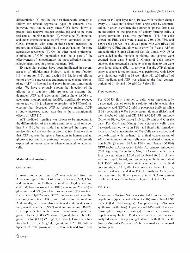
In the sphere formation assay, cells were plated with andwithout agonist, and the number of spheres formed wascounted from days 3 to 7 after plating. In the presence of100 μM ATP, there were significantly fewer spheres bothwhen 750 and 1,000 cells were plated (Fig. 2a). Anotherway of accessing the relative presence of sphere-formingcells in a population is to calculate a linear regression of thepercentage of wells without spheres [17]. The meanx-intercept value of the graph indicates the number of cellsneeded to form one sphere per well, which was higher inthe ATP-treated cells (1,270) when compared to the control(1,014), suggesting that ATP reduces the relative number ofcells capable of forming spheres in the population (Fig. 2b).
Although U87 gliomas have a very low rate ofectonucleotide degradation [20], long treatments mayproduce some degradation of ATP to ADP or adenosinewhich could be responsible for the biological effect.Therefore, we tested ADP and adenosine, which had nosignificant effect on sphere formation (Fig. 2c, d). UTP, anagonist of P2Y2, P2Y4, and P2Y6 receptors [21] also didnot lead to any change in tumor sphere formation (Table 1).
We have previously shown that 24 h of treatment withATP induces proliferation of U87 gliomas in serum-deprived conditions [11]. Since the number of cells directlyaffects the formation of spheres, we measured the amountof viable cells at the end of the sphere-forming assay.Chronic treatment with all purinergic agonists reduced theamount of viable cells at the end of a 7-day treatment. Evencorrecting for this reduction in proliferation induced by ATP,the number of spheres was reduced by 45% and 40% with750 cells and 1,000 cells, respectively (Table 1). It isimportant to mention that the relation between cells plated
0
20
40
60
80
100
0 500 1000 1500
A B
C D
0
2
4
6
8
10
12
14
16
3 4 5 6 7
Nu
mb
er o
f S
ph
eres
per
wel
l
% W
ells
wit
ho
ut
sph
eres
Nu
mb
er o
f S
ph
eres
per
wel
l
Nu
mb
er o
f S
ph
eres
per
wel
l
0
2
4
6
8
10
12
14
16
3 4 5 6 70
2
4
6
8
10
12
14
16
3 4 5 6 7
Control ATP750 750
1000 1000
Control ADP750 750
1000 1000
Control ADO750 750
1000 1000
Days after plating
Days after plating Days after plating
Cell per well
1014 1270
*
*
*
*
*
*
*
*
#
#
#
#
Fig. 2 Treatment with ATP butnot ADP or adenosine reducesthe tumor spheres and stem cellpopulation. U87 cells (750 or1,000) were grown in a 96-wellplate in the presence or absenceof 100 μM of ATP for 7 days,and spheres (average ± SEM)were counted from days 3 to 7(n=6). *p<0.05 for 750 platedcells and #p<0.05 for 1,000plated cells (a). Regressionanalysis of the wells withoutspheres—numbers indicate thevalue of the X-axis intercept ofthe best linear fit (b). U87 cellswere treated with 100 μM ofADP (c) or adenosine (d)and evaluated as in a. Barsize=50 μm
Purinergic Signalling (2012) 8:39–48 43

and spheres formed is linear with an R2 of 0.98, considering250, 500, 750, and 1,000 cells plated per well, indicating
that there is no saturation occurring due to an excess ofcells.
A
B
C
C
ATP
C
ATP
C
ATP
Control ATP
Control ATP
Control ATP
U87
U343
C6
Num
ber
of S
pher
es p
er w
ell
*
*
0
5
10
15
20
25
30
35
40
45
Num
ber
of S
pher
es p
er w
ell
0
50
100
150
200
250
300
350
400
Num
ber
of S
pher
es p
er w
ell
0
1
20
30
40
50
60
70
80
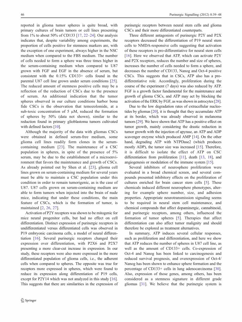
Fig. 3 Treatment with ATPreduces the tumor spheresgrown in neural stem cell (NSC)medium in different glioma celllines. One thousand cells weregrown in a 96-well plate for7 days in the presence orabsence of 100 μM of ATP inNSC medium. The number ofspheres is shown as average ±SEM at day 7 in U87 (n=4) (a),C6 (n=2) (b), or U343 (n=5)(c). Bars represent SEM. t testvalues: *p<0.05. Right panel:representative images of tumorspheres of control and culturestreated for 7 days with ATP
44 Purinergic Signalling (2012) 8:39–48

There are several evidence pointing to the importance ofgrowing CSCs in a defined medium that does not containserum. When experiments were performed in NSC medium,ATP presented a much larger effect in inhibiting U87 spheregrowth when compared to serum-containing medium(Fig. 3a). It is also important to observe that for the samenumber of cells plated, much more spheres where observed inNSC medium when compared to serum-containing mediumsince the former contains factors that favor CSC proliferation.Additionally, another human glioma cell line, U343 as well asthe C6 rat glioma cell line, which is widely used in animalstudies and was recently shown to contain CSCs [22, 23],presented a drastic reduction in sphere formation whentreated with 100 μM ATP in NSC medium (Fig. 3b, c).
In order to estimate from which concentration ATP wouldexert its effect in decreasing sphere formation, anATP titrationassay was performed. U87 and U343 cells were plated at adensity of 1,000 cells per well in ATP concentrations rangingfrom 1 to 100 μM in NSC medium. ATP 1 μM alreadyreduced the sphere number in U87, but not in U343(Supplementary Fig. 2a). Sphere size was already reducedwith 10 μM, and with 100 μM, only a few small spheresremained after 7 days (Supplementary Fig. 2b).
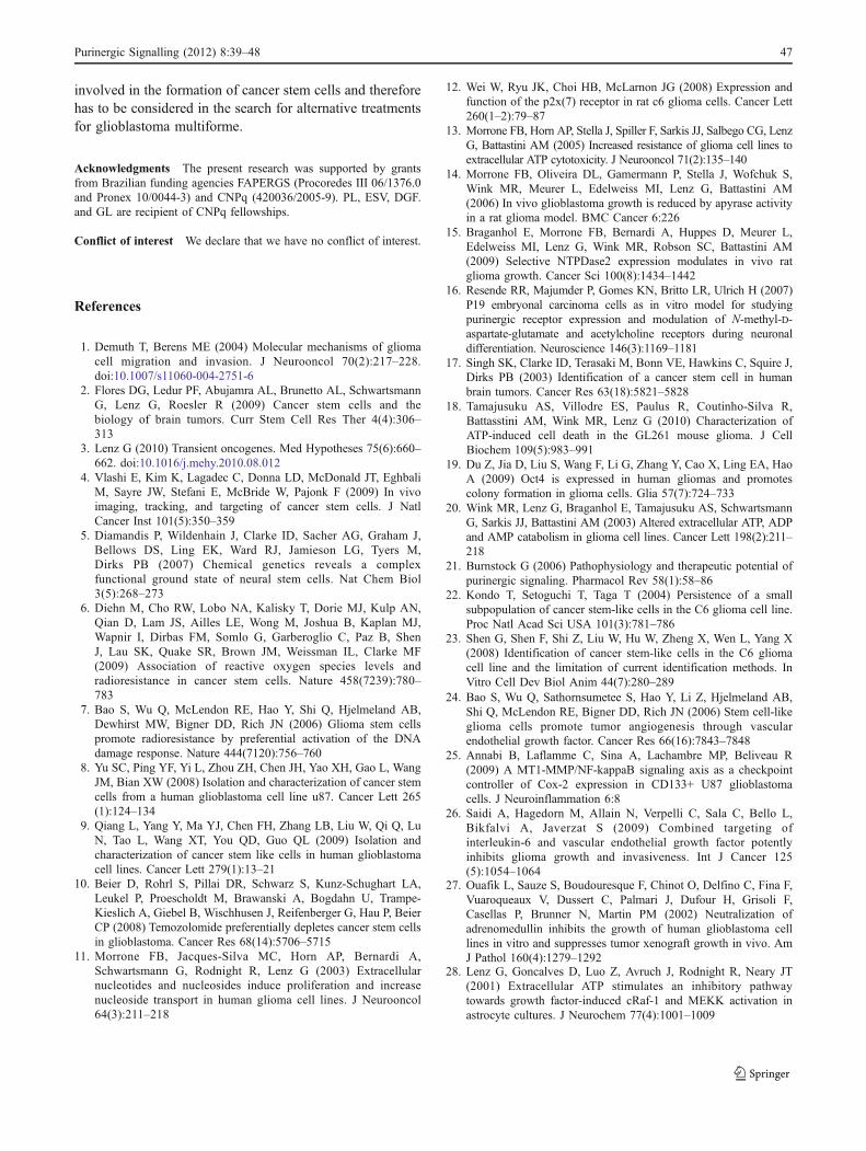
Cells treated with ATP presented less expression of thestem cell markers CD133 and Oct-4 as analyzed by RT-PCR
(Fig. 4a), and the CD133, Oct-4, and Nanog staininganalyzed by flow cytometry was lower in cells treated withATP when compared to untreated cells (Fig. 4c andSupplementary Fig. 1b, c). Next, we wondered whethercomponents of the purinergic system were differentlyexpressed in glioma cells present in spheres or attached tothe culture dish. As expected, different glioma cell linespresent a heterogeneous expression of purinergic receptors(Supplementary Table 2). When comparing adherent versusspheres, expression of P2X6 and P2X7 was found increasedin cells from the adherent population, whereas P2X4,P2Y1, and P2Y14 were more expressed in spheres(Fig. 4b). The other purinergic receptors expressed byU87 cells (Supplementary Table 2) were not differentiallyexpressed between spheres and attached cells.
Discussion
U87 glioma cells grown in the presence of serum formtumor spheres with characteristics that are similar togliomas grown on selected growth factors with regard togrowth of spheres and increased expression of markers ofundifferentiation such as CD133, Nanog, and Oct-4. Thepercentage of cells positive for different stemness marker
P2XP2X
P2X
46
7
Mon
olay
er
Spher
es
P2Y1
P2Y14
B
Mon
olay
er
Spher
esβ-actin
CD133
OCT4
lortnoC
PTA
A
β-actin
0
20
40
60
80
100
Oct4 Nanog
U87
Oct4 Nanog
U343
Eve
nts
(% o
f Con
trol
)
C
Fig. 4 Purinergic systemand tumor sphere formation:expression of CD133 and Oct-4mRNA as analyzed by RT-PCRof cells treated with 100 μMATP for 7 days (a). Expressionof mRNA of purinergicreceptors in spheres and adher-ent cells as analyzed by RT-PCR(representative of fiveindependent experiments) (b).Proportion of Oct-4 and Nanog-positive cells in U87 and U343lines grown on NSC mediumand treated with ATP 100 μMfor 7 days, when compared tocontrol. Average ± SEM areshown as small lines (c)
Purinergic Signalling (2012) 8:39–48 45
reported in glioma tumor spheres is quite broad, withprimary cultures of brain tumors or cell lines presentingfrom 1% to about 50% of CD133 [17, 22–24]. Our analysisindicates that, despite variability among experiments, theproportion of cells positive for stemness markers are, withthe exception of one experiment, always higher in the NSCmedium when compared to the FBS medium. The numberof cells needed to form a sphere was three times higher inthe serum-containing medium when compared to U87grown with FGF and EGF in the absence of serum, butconsistent with the 0.15% CD133+ cells found in theparental U87 cell line grown under serum conditions [25].The reduced amount of stemness positive cells may be areflection of the reduction of CSCs due to the presenceof serum. An additional indication that the tumorspheres observed in our culture conditions harbor bonafide CSCs is the observation that temozolomide, at asub-toxic concentration of 5 μM, reduced the formationof spheres by 50% (data not shown), similar to thereduction found in primary glioblastoma tumors cultivatedwith defined factors [10].
Although the majority of the data with gliomas CSCswere obtained in defined serum-free medium, someglioma cell lines readily form clones in the serum-containing medium [23]. The maintenance of a CSCpopulation in spheres, in spite of the presence of 5%serum, may be due to the establishment of a microenvi-ronment that favors the maintenance and growth of CSCs.As already pointed out by Shen et al. [23], glioma celllines grown on serum-containing medium for several yearsmust be able to maintain a CSC population under thiscondition in order to remain tumorigenic, as is the case ofU87. U87 cells grown on serum-containing medium areable to form tumors when injected into the brain of nudemice, indicating that under these conditions, the mainfeature of CSCs, which is the formation of tumor, ismaintained [2, 26, 27].
Activation of P2Y receptors was shown to be mitogenic formice neural progenitor cells, but had no effect on celldifferentiation. Distinct expression of purinergic receptors inundifferentiated versus differentiated cells was observed inP19 embryonic carcinoma cells, a model of neural differen-tiation [16]. Several purinergic receptors changed theirexpression over differentiation, with P2X6 and P2X7presenting a more clear-cut increase in expression. In ourstudy, these receptors were also more expressed in the moredifferentiated population of glioma cells, i.e., the adherentcells when compared to spheres. The opposite was true forreceptors more expressed in spheres, which were found toreduce its expression along differentiation of P19 cells,except for P2Y14 which was not analyzed in this study [16].This suggests that there are similarities in the expression of
purinergic receptors between neural stem cells and gliomaCSCs and their more differentiated counterparts.
Three different antagonists of purinergic P2Y and P2Xreceptors decreased the differentiation of P19 neural stemcells to NMDA-responsive cells suggesting that activationof these receptors is pro-differentiative for neural stem cells[16]. Here we observed that ATP, which can activate P2Yand P2X receptors, reduces the number and size of spheres,increases the number of cells needed to form a sphere, anddecreases the number of CD133, Nanog and Oct-4 positiveCSCs. This suggests that in CSCs, ATP also has a pro-differentiative role. Accordingly, proliferation during thecourse of the experiment (7 days) was also reduced by ATP.FGF is a growth factor fundamental for the maintenance andgrowth of glioma CSCs, and ATP may act by blocking theactivation of the ERK by FGF, as was shown in astrocytes [28].
Due to the low degradation rates of extracellular nucleo-tides by gliomas [20], it is thought that they accumulate ATPat its border, which was already observed in melanomatumors [29]. We have shown that ATP has a positive effect ontumor growth, mainly considering the drastic reduction intumor growth with the injection of apyrase, an ATP and ADPscavenger enzyme which produced AMP [14]. On the otherhand, degrading ATP with NTPDase2 (which producesmostly ADP), the tumor size was increased [15]. Therefore,it is difficult to isolate the effect of ATP on CSCdifferentiation from proliferation [11], death [13, 18], andangiogenesis or modulation of the immune system [15].
Several inhibitors of neurosphere proliferation wereevaluated in a broad chemical screen, and several com-pounds presented inhibitory effects on the proliferation ofcultures enriched for brain cancer stem cells [5]. Thesechemicals induced different neurosphere phenotypes, alter-ing for example sphere number, size, and adhesionproperties. Appropriate neurotransmission signaling seemsto be required in neural stem cell maintenance, andchemical compounds that affect dopaminergic, cannabinoid,and purinergic receptors, among others, influenced theformation of tumor spheres [5]. Therapies that affectdifferentiation can also affect tumor malignity and shouldtherefore be explored as treatment alternatives.
In summary, ATP induces several cellular responses,such as proliferation and differentiation, and here we showthat ATP reduces the number of spheres in U87 cell line, aswell as the amount of CD133+ cells. Co-expression ofOct-4 and Nanog has been linked to carcinogenesis andreduced survival prognosis, and overexpression of Oct-4/Nanog has been shown to enhance sphere formation and thepercentage of CD133+ cells in lung adenocarcinoma [30].Also, expression of those genes, among others, has beenconsidered as a stemness signature in different gradegliomas [31]. We believe that the purinergic system is
46 Purinergic Signalling (2012) 8:39–48
involved in the formation of cancer stem cells and thereforehas to be considered in the search for alternative treatmentsfor glioblastoma multiforme.
Acknowledgments The present research was supported by grantsfrom Brazilian funding agencies FAPERGS (Procoredes III 06/1376.0and Pronex 10/0044-3) and CNPq (420036/2005-9). PL, ESV, DGF.and GL are recipient of CNPq fellowships.
Conflict of interest We declare that we have no conflict of interest.
References
1. Demuth T, Berens ME (2004) Molecular mechanisms of gliomacell migration and invasion. J Neurooncol 70(2):217–228.doi:10.1007/s11060-004-2751-6
2. Flores DG, Ledur PF, Abujamra AL, Brunetto AL, SchwartsmannG, Lenz G, Roesler R (2009) Cancer stem cells and thebiology of brain tumors. Curr Stem Cell Res Ther 4(4):306–313
3. Lenz G (2010) Transient oncogenes. Med Hypotheses 75(6):660–662. doi:10.1016/j.mehy.2010.08.012
4. Vlashi E, Kim K, Lagadec C, Donna LD, McDonald JT, EghbaliM, Sayre JW, Stefani E, McBride W, Pajonk F (2009) In vivoimaging, tracking, and targeting of cancer stem cells. J NatlCancer Inst 101(5):350–359
5. Diamandis P, Wildenhain J, Clarke ID, Sacher AG, Graham J,Bellows DS, Ling EK, Ward RJ, Jamieson LG, Tyers M,Dirks PB (2007) Chemical genetics reveals a complexfunctional ground state of neural stem cells. Nat Chem Biol3(5):268–273
6. Diehn M, Cho RW, Lobo NA, Kalisky T, Dorie MJ, Kulp AN,Qian D, Lam JS, Ailles LE, Wong M, Joshua B, Kaplan MJ,Wapnir I, Dirbas FM, Somlo G, Garberoglio C, Paz B, ShenJ, Lau SK, Quake SR, Brown JM, Weissman IL, Clarke MF(2009) Association of reactive oxygen species levels andradioresistance in cancer stem cells. Nature 458(7239):780–783
7. Bao S, Wu Q, McLendon RE, Hao Y, Shi Q, Hjelmeland AB,Dewhirst MW, Bigner DD, Rich JN (2006) Glioma stem cellspromote radioresistance by preferential activation of the DNAdamage response. Nature 444(7120):756–760
8. Yu SC, Ping YF, Yi L, Zhou ZH, Chen JH, Yao XH, Gao L, WangJM, Bian XW (2008) Isolation and characterization of cancer stemcells from a human glioblastoma cell line u87. Cancer Lett 265(1):124–134
9. Qiang L, Yang Y, Ma YJ, Chen FH, Zhang LB, Liu W, Qi Q, LuN, Tao L, Wang XT, You QD, Guo QL (2009) Isolation andcharacterization of cancer stem like cells in human glioblastomacell lines. Cancer Lett 279(1):13–21
10. Beier D, Rohrl S, Pillai DR, Schwarz S, Kunz-Schughart LA,Leukel P, Proescholdt M, Brawanski A, Bogdahn U, Trampe-Kieslich A, Giebel B, Wischhusen J, Reifenberger G, Hau P, BeierCP (2008) Temozolomide preferentially depletes cancer stem cellsin glioblastoma. Cancer Res 68(14):5706–5715
11. Morrone FB, Jacques-Silva MC, Horn AP, Bernardi A,Schwartsmann G, Rodnight R, Lenz G (2003) Extracellularnucleotides and nucleosides induce proliferation and increasenucleoside transport in human glioma cell lines. J Neurooncol64(3):211–218
12. Wei W, Ryu JK, Choi HB, McLarnon JG (2008) Expression andfunction of the p2x(7) receptor in rat c6 glioma cells. Cancer Lett260(1–2):79–87
13. Morrone FB, Horn AP, Stella J, Spiller F, Sarkis JJ, Salbego CG, LenzG, Battastini AM (2005) Increased resistance of glioma cell lines toextracellular ATP cytotoxicity. J Neurooncol 71(2):135–140
14. Morrone FB, Oliveira DL, Gamermann P, Stella J, Wofchuk S,Wink MR, Meurer L, Edelweiss MI, Lenz G, Battastini AM(2006) In vivo glioblastoma growth is reduced by apyrase activityin a rat glioma model. BMC Cancer 6:226
15. Braganhol E, Morrone FB, Bernardi A, Huppes D, Meurer L,Edelweiss MI, Lenz G, Wink MR, Robson SC, Battastini AM(2009) Selective NTPDase2 expression modulates in vivo ratglioma growth. Cancer Sci 100(8):1434–1442
16. Resende RR, Majumder P, Gomes KN, Britto LR, Ulrich H (2007)P19 embryonal carcinoma cells as in vitro model for studyingpurinergic receptor expression and modulation of N-methyl-D-aspartate-glutamate and acetylcholine receptors during neuronaldifferentiation. Neuroscience 146(3):1169–1181
17. Singh SK, Clarke ID, Terasaki M, Bonn VE, Hawkins C, Squire J,Dirks PB (2003) Identification of a cancer stem cell in humanbrain tumors. Cancer Res 63(18):5821–5828
18. Tamajusuku AS, Villodre ES, Paulus R, Coutinho-Silva R,Battasstini AM, Wink MR, Lenz G (2010) Characterization ofATP-induced cell death in the GL261 mouse glioma. J CellBiochem 109(5):983–991
19. Du Z, Jia D, Liu S, Wang F, Li G, Zhang Y, Cao X, Ling EA, HaoA (2009) Oct4 is expressed in human gliomas and promotescolony formation in glioma cells. Glia 57(7):724–733
20. Wink MR, Lenz G, Braganhol E, Tamajusuku AS, SchwartsmannG, Sarkis JJ, Battastini AM (2003) Altered extracellular ATP, ADPand AMP catabolism in glioma cell lines. Cancer Lett 198(2):211–218
21. Burnstock G (2006) Pathophysiology and therapeutic potential ofpurinergic signaling. Pharmacol Rev 58(1):58–86
22. Kondo T, Setoguchi T, Taga T (2004) Persistence of a smallsubpopulation of cancer stem-like cells in the C6 glioma cell line.Proc Natl Acad Sci USA 101(3):781–786
23. Shen G, Shen F, Shi Z, Liu W, Hu W, Zheng X, Wen L, Yang X(2008) Identification of cancer stem-like cells in the C6 gliomacell line and the limitation of current identification methods. InVitro Cell Dev Biol Anim 44(7):280–289
24. Bao S, Wu Q, Sathornsumetee S, Hao Y, Li Z, Hjelmeland AB,Shi Q, McLendon RE, Bigner DD, Rich JN (2006) Stem cell-likeglioma cells promote tumor angiogenesis through vascularendothelial growth factor. Cancer Res 66(16):7843–7848
25. Annabi B, Laflamme C, Sina A, Lachambre MP, Beliveau R(2009) A MT1-MMP/NF-kappaB signaling axis as a checkpointcontroller of Cox-2 expression in CD133+ U87 glioblastomacells. J Neuroinflammation 6:8
26. Saidi A, Hagedorn M, Allain N, Verpelli C, Sala C, Bello L,Bikfalvi A, Javerzat S (2009) Combined targeting ofinterleukin-6 and vascular endothelial growth factor potentlyinhibits glioma growth and invasiveness. Int J Cancer 125(5):1054–1064
27. Ouafik L, Sauze S, Boudouresque F, Chinot O, Delfino C, Fina F,Vuaroqueaux V, Dussert C, Palmari J, Dufour H, Grisoli F,Casellas P, Brunner N, Martin PM (2002) Neutralization ofadrenomedullin inhibits the growth of human glioblastoma celllines in vitro and suppresses tumor xenograft growth in vivo. AmJ Pathol 160(4):1279–1292
28. Lenz G, Goncalves D, Luo Z, Avruch J, Rodnight R, Neary JT(2001) Extracellular ATP stimulates an inhibitory pathwaytowards growth factor-induced cRaf-1 and MEKK activation inastrocyte cultures. J Neurochem 77(4):1001–1009
Purinergic Signalling (2012) 8:39–48 47

29. Pellegatti P, Raffaghello L, Bianchi G, Piccardi F, Pistoia V, DiVirgilio F (2008) Increased level of extracellular ATP at tumorsites: in vivo imaging with plasma membrane luciferase. PLoSOne 3(7):e2599
30. Chiou SH, Wang ML, Chou YT, Chen CJ, Hong CF, Hsieh WJ,Chang HT, Chen YS, Lin TW, Hsu HS, Wu CW (2010)Coexpression of Oct4 and Nanog enhances malignancy in lung
adenocarcinoma by inducing cancer stem cell-like properties andepithelial–mesenchymal transdifferentiation. Cancer Res 70(24):10433–10444
31. Clement V, Sanchez P, de Tribolet N, Radovanovic I, Ruiz I,Altaba A (2007) HEDGEHOG-GLI1 signaling regulates humanglioma growth, cancer stem cell self-renewal, and tumorigenicity.Curr Biol 17(2):165–172
48 Purinergic Signalling (2012) 8:39–48
Related Documents