Extracellular amastigotes of Trypanosoma cruzi are potent inducers of phagocytosis in mammalian cells Maria Cecilia Fernandes, 1† Andrew R. Flannery, 1† Norma Andrews 1 and Renato A. Mortara 2 * 1 Department of Cell Biology and Molecular Genetics, University of Maryland, College Park, MD, USA. 2 Departamento de Microbiologia, Imunologia e Parasitologia, Escola Paulista de Medicina, Universidade Federal de São Paulo, São Paulo, Brazil. Summary The protozoan parasite Trypanosoma cruzi, the aetiological agent of Chagas’ disease, has two infective life cycle stages, trypomastigotes and amastigotes. While trypomastigotes actively enter mammalian cells, highly infective extracellular amastigotes (type I T. cruzi) rely on actin-mediated uptake, which is generally inefficient in non- professional phagocytes. We found that extra- cellular amastigotes (EAs) of T. cruzi G strain (type I), but not Y strain (type II), were taken up 100-fold more efficiently than inert particles. Mammalian cell lines showed levels of parasite uptake comparable to macrophages, and extensive actin recruitment and polymerization was observed at the site of entry. EA uptake was not dependent on parasite- secreted molecules and required the same molecu- lar machinery utilized by professional phagocytes during large particle phagocytosis. Transcriptional silencing of synaptotagmin VII and CD63 signifi- cantly inhibited EA internalization, demonstrating that delivery of supplemental lysosomal mem- brane to form the phagosome is involved in parasite uptake. Importantly, time-lapse live imag- ing using fluorescent reporters revealed phago- some-associated modulation of phosphoinositide metabolism during EA uptake that closely resem- bles what occurs during phagocytosis by macro- phages. Collectively, our results demonstrate that T. cruzi EAs are potent inducers of phagocytosis in non-professional phagocytes, a process that may facilitate parasite persistence in infected hosts. Introduction Foreign particles, apoptotic bodies and most invading microorganisms are eliminated from the body by phago- cytic leucocytes, which play a crucial role in the innate immune response (Geissmann et al., 2010). The inges- tion of extracellular objects, also known as phagocytosis, is receptor-mediated and actin-dependent (Aderem and Underhill, 1999). Although it is known that several other mammalian cells are capable of phagocytosis, the effi- ciency of the process varies greatly. To emphasize this variation in phagocytic capacity between different cell types, Rabinovitch coined the terms ‘professional’ and ‘non-professional’ phagocytes (Rabinovitch, 1995). The unique ability of professional phagocytes to effi- ciently internalize a variety of targets is linked to the expression of an array of specialized phagocytic recep- tors such as those for immunoglobulins (Fc receptors), complement and mannose (Aderem and Underhill, 1999). The intracellular vacuoles generated in this receptor- mediated uptake process, designated as phagosomes, undergo a series of fusion and fission events that modify the composition of their limiting membrane and intravacu- olar contents. This process is referred to as phagosome maturation, and grants the vacuole with degradative properties, which are central to its microbicidal function (Berón et al., 1995; Tjelle et al., 2000). Lipid-mediated signalling and phosphoinositides play an important role in this process, from the initial formation of the phagocytic cup until the final steps of phagosome matu- ration (Botelho et al., 2000; Vieira et al., 2001; Yeung et al., 2006; Botelho and Grinstein, 2011). Chimeric constructs consisting of fluorescent proteins fused to pleckstrin homology (PH) domains that recognize the headgroups of phosphatidyilinositol-4,5-bisphosphate (PI(4,5)P 2) or phosphatidylinositol-3,4,5-trisphosphate (PI(3,4,5)P3) have been exploited as very useful tools to characterize the dynamics of these molecules in cells during particle uptake in macrophages (Botelho et al., 2000; Vieira et al., 2001). The vast majority of studies on phagocytosis have been performed in primary macrophages or macrophage- derived cell lines. However, working with these cell lines imposes numerous challenges and technical barriers, which are easily overcome when working with epithelial or fibroblast cell lines. Unfortunately, the limited repertoire of particles (mainly pathogens) that can be internalized by Received 6 August, 2012; revised 26 November, 2012; accepted 11 December, 2012. *For correspondence. E-mail ramortara@ unifesp.br; Tel. (+55) 11 5579 8306; Fax (+55) 11 5571 1095. † These authors contributed equally to this work. Cellular Microbiology (2013) 15(6), 977–991 doi:10.1111/cmi.12090 First published online 9 January 2013 © 2012 John Wiley & Sons Ltd cellular microbiology

Welcome message from author
This document is posted to help you gain knowledge. Please leave a comment to let me know what you think about it! Share it to your friends and learn new things together.
Transcript
Extracellular amastigotes of Trypanosoma cruzi arepotent inducers of phagocytosis in mammalian cells
Maria Cecilia Fernandes,1† Andrew R. Flannery,1†
Norma Andrews1 and Renato A. Mortara2*1Department of Cell Biology and Molecular Genetics,University of Maryland, College Park, MD, USA.2Departamento de Microbiologia, Imunologia eParasitologia, Escola Paulista de Medicina,Universidade Federal de São Paulo, São Paulo, Brazil.
Summary
The protozoan parasite Trypanosoma cruzi, theaetiological agent of Chagas’ disease, has twoinfective life cycle stages, trypomastigotes andamastigotes. While trypomastigotes actively entermammalian cells, highly infective extracellularamastigotes (type I T. cruzi) rely on actin-mediateduptake, which is generally inefficient in non-professional phagocytes. We found that extra-cellular amastigotes (EAs) of T. cruzi G strain (typeI), but not Y strain (type II), were taken up 100-foldmore efficiently than inert particles. Mammalian celllines showed levels of parasite uptake comparableto macrophages, and extensive actin recruitmentand polymerization was observed at the site ofentry. EA uptake was not dependent on parasite-secreted molecules and required the same molecu-lar machinery utilized by professional phagocytesduring large particle phagocytosis. Transcriptionalsilencing of synaptotagmin VII and CD63 signifi-cantly inhibited EA internalization, demonstratingthat delivery of supplemental lysosomal mem-brane to form the phagosome is involved inparasite uptake. Importantly, time-lapse live imag-ing using fluorescent reporters revealed phago-some-associated modulation of phosphoinositidemetabolism during EA uptake that closely resem-bles what occurs during phagocytosis by macro-phages. Collectively, our results demonstrate thatT. cruzi EAs are potent inducers of phagocytosis innon-professional phagocytes, a process that mayfacilitate parasite persistence in infected hosts.
Introduction
Foreign particles, apoptotic bodies and most invadingmicroorganisms are eliminated from the body by phago-cytic leucocytes, which play a crucial role in the innateimmune response (Geissmann et al., 2010). The inges-tion of extracellular objects, also known as phagocytosis,is receptor-mediated and actin-dependent (Aderem andUnderhill, 1999). Although it is known that several othermammalian cells are capable of phagocytosis, the effi-ciency of the process varies greatly. To emphasize thisvariation in phagocytic capacity between different celltypes, Rabinovitch coined the terms ‘professional’ and‘non-professional’ phagocytes (Rabinovitch, 1995).
The unique ability of professional phagocytes to effi-ciently internalize a variety of targets is linked to theexpression of an array of specialized phagocytic recep-tors such as those for immunoglobulins (Fc receptors),complement and mannose (Aderem and Underhill, 1999).The intracellular vacuoles generated in this receptor-mediated uptake process, designated as phagosomes,undergo a series of fusion and fission events that modifythe composition of their limiting membrane and intravacu-olar contents. This process is referred to as phagosomematuration, and grants the vacuole with degradativeproperties, which are central to its microbicidal function(Berón et al., 1995; Tjelle et al., 2000). Lipid-mediatedsignalling and phosphoinositides play an importantrole in this process, from the initial formation of thephagocytic cup until the final steps of phagosome matu-ration (Botelho et al., 2000; Vieira et al., 2001; Yeunget al., 2006; Botelho and Grinstein, 2011). Chimericconstructs consisting of fluorescent proteins fusedto pleckstrin homology (PH) domains that recognizethe headgroups of phosphatidyilinositol-4,5-bisphosphate(PI(4,5)P2) or phosphatidylinositol-3,4,5-trisphosphate(PI(3,4,5)P3) have been exploited as very useful tools tocharacterize the dynamics of these molecules in cellsduring particle uptake in macrophages (Botelho et al.,2000; Vieira et al., 2001).
The vast majority of studies on phagocytosis have beenperformed in primary macrophages or macrophage-derived cell lines. However, working with these cell linesimposes numerous challenges and technical barriers,which are easily overcome when working with epithelial orfibroblast cell lines. Unfortunately, the limited repertoire ofparticles (mainly pathogens) that can be internalized by
Received 6 August, 2012; revised 26 November, 2012; accepted11 December, 2012. *For correspondence. E-mail [email protected]; Tel. (+55) 11 5579 8306; Fax (+55) 11 5571 1095.†These authors contributed equally to this work.
Cellular Microbiology (2013) 15(6), 977–991 doi:10.1111/cmi.12090First published online 9 January 2013
© 2012 John Wiley & Sons Ltd
cellular microbiology
non-professional phagocytes restricts the type of studiesthat can be done in these cells. While intracellular bacte-rial pathogens have evolved mechanisms to perturbcytoskeleton dynamics and stimulate their uptake by non-professional phagocytes (Galan and Bliska, 1996; Finlayand Cossart, 1997; Alonso and Garcia-del Portillo, 2004),their small size also renders them a poor tool for the studyof phagocytosis. Larger protozoan parasites, which canbe much more readily visualized by light microscopy,usually infect non-phagocytic cells by active invasion, asis the case for Toxoplasma gondii (Morisaki et al., 1995;Dobrowolski and Sibley, 1996), or the tissue culture try-pomastigote stage of Trypanosoma cruzi (Tardieux et al.,1992; Fernandes et al., 2011b). In both instances, thehost cell actin cytoskeleton does not play a major role inthe host cell invasion process. Curiously, however, meta-cyclic trypomastigote forms of T. cruzi appear to haveevolved a strain-dependent requirement on actin polym-erization to invade mammalian cells (Ferreira et al.,2006).
Trypanosoma cruzi amastigote forms are usually foundin the cytoplasm of infected cells, but can also be gener-ated by extracellular differentiation of trypomastigotes.These extracellular amastigotes share morphological andimmunochemical markers with their intracellular counter-parts, and are also capable of invading and sustaininginfections in mammalian cells (Nogueira and Cohn, 1976;Ley et al., 1988). However, differently from the infectivetrypomastigote stages, the extracellularly generatedamastigotes (henceforth referred to as EAs) were shownto promptly aggregate actin filaments after attaching toHeLa cells (Mortara, 1991; Procópio et al., 1999).
EAs from group I T. cruzi strains (such as the G strain)were found to enter mammalian cells much more effi-ciently than group II (Y strain) or VI (CL strain) parasites(Fernandes and Mortara, 2004; Mortara et al., 2005; daSilva et al., 2006; Fernandes et al., 2006; Bambino-Medeiros et al., 2011; Rodrigues et al., 2012). Furtherstudies revealed that EAs of either G or Y strains arecapable of initiating infections in a wide range of mamma-lian cells and, as the flagellated trypomastigotes, elicitresponses from the host cell (Fernandes and Mortara,2004; Fernandes et al., 2006; Fernandes et al., 2007a,b;Cruz et al., 2012). Therefore, for this study we havechosen the G strain as a model for type I T. cruzi.
The precise mechanism of EA internalization is not yetfully characterized. Here we describe that EAs are effec-tive inducers of classical phagocytosis in non-professionalphagocytes. We demonstrate that EA forms induce vigor-ous localized host cell actin polymerization during theearly steps of invasion, and are capable of being engulfedby various mammalian cells at levels comparable to mac-rophages. Furthermore, we show that these parasitesrecruit to their phagocytic cup and parasitophorous
vacuole the same host cell markers that were shown to berecruited during classical particle phagocytosis in profes-sional phagocytes. This remarkable ability to induce effi-cient phagocytosis is likely to be a specific adaptation ofthese parasites to infect various cell types and propagatethe infection in the mammalian host.
Results and discussion
T. cruzi EAs trigger formation of structures rich inpolymerized actin which mediate their internalization innon-professional phagocytes
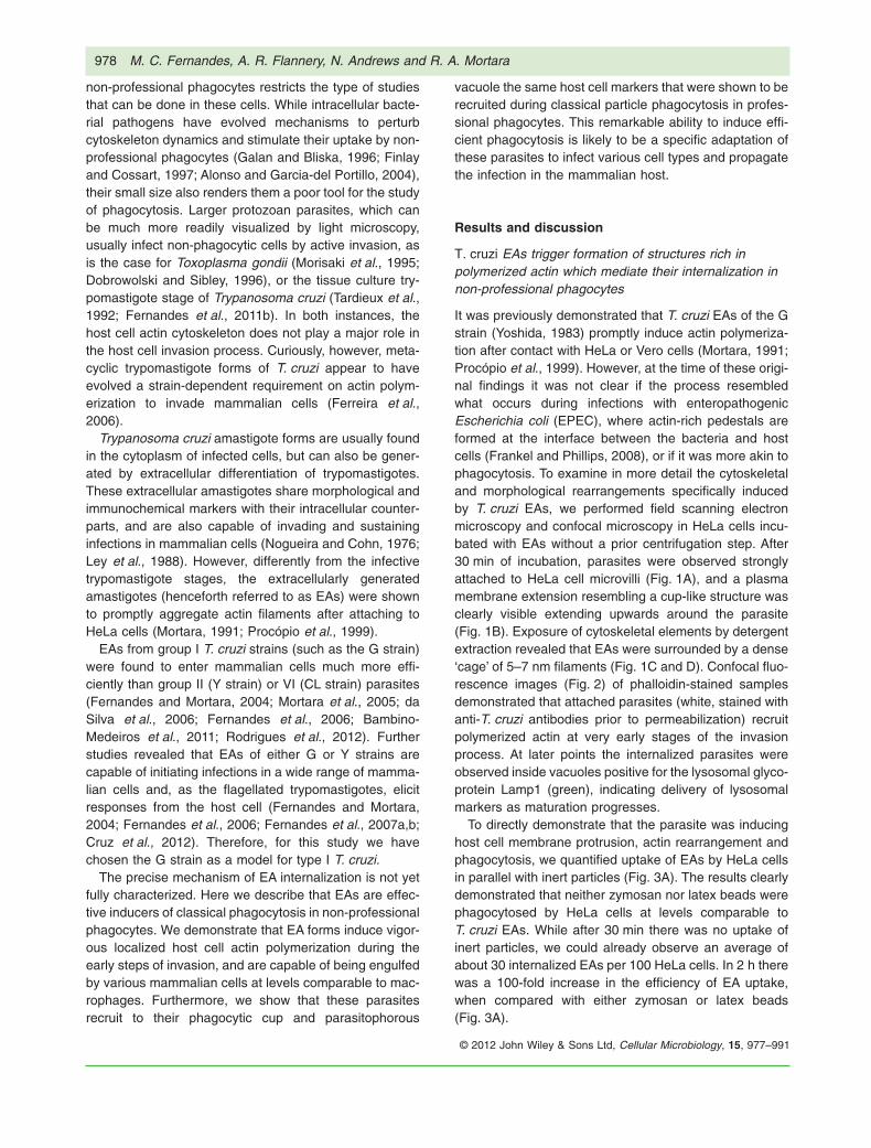
It was previously demonstrated that T. cruzi EAs of the Gstrain (Yoshida, 1983) promptly induce actin polymeriza-tion after contact with HeLa or Vero cells (Mortara, 1991;Procópio et al., 1999). However, at the time of these origi-nal findings it was not clear if the process resembledwhat occurs during infections with enteropathogenicEscherichia coli (EPEC), where actin-rich pedestals areformed at the interface between the bacteria and hostcells (Frankel and Phillips, 2008), or if it was more akin tophagocytosis. To examine in more detail the cytoskeletaland morphological rearrangements specifically inducedby T. cruzi EAs, we performed field scanning electronmicroscopy and confocal microscopy in HeLa cells incu-bated with EAs without a prior centrifugation step. After30 min of incubation, parasites were observed stronglyattached to HeLa cell microvilli (Fig. 1A), and a plasmamembrane extension resembling a cup-like structure wasclearly visible extending upwards around the parasite(Fig. 1B). Exposure of cytoskeletal elements by detergentextraction revealed that EAs were surrounded by a dense‘cage’ of 5–7 nm filaments (Fig. 1C and D). Confocal fluo-rescence images (Fig. 2) of phalloidin-stained samplesdemonstrated that attached parasites (white, stained withanti-T. cruzi antibodies prior to permeabilization) recruitpolymerized actin at very early stages of the invasionprocess. At later points the internalized parasites wereobserved inside vacuoles positive for the lysosomal glyco-protein Lamp1 (green), indicating delivery of lysosomalmarkers as maturation progresses.
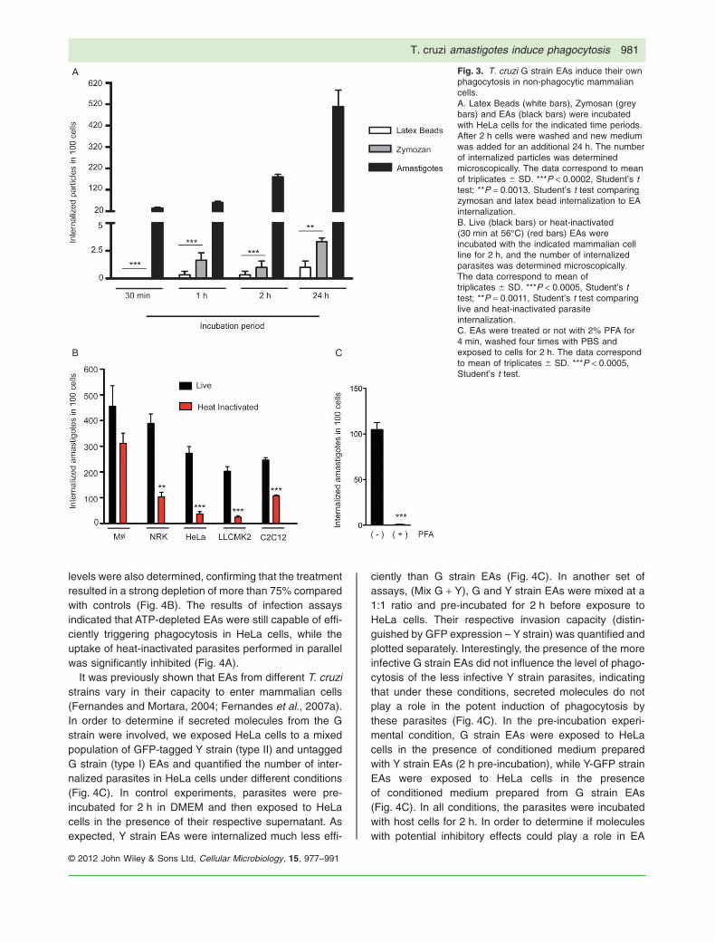
To directly demonstrate that the parasite was inducinghost cell membrane protrusion, actin rearrangement andphagocytosis, we quantified uptake of EAs by HeLa cellsin parallel with inert particles (Fig. 3A). The results clearlydemonstrated that neither zymosan nor latex beads werephagocytosed by HeLa cells at levels comparable toT. cruzi EAs. While after 30 min there was no uptake ofinert particles, we could already observe an average ofabout 30 internalized EAs per 100 HeLa cells. In 2 h therewas a 100-fold increase in the efficiency of EA uptake,when compared with either zymosan or latex beads(Fig. 3A).
978 M. C. Fernandes, A. R. Flannery, N. Andrews and R. A. Mortara
© 2012 John Wiley & Sons Ltd, Cellular Microbiology, 15, 977–991

To further demonstrate the ability of EAs to inducephagocytosis in other non-professional phagocyte celllines, we compared the number of internalized parasites,live and heat-inactivated, in bone marrow derived mousemacrophages and NRK, HeLa, LLCMK2 and C2C12 cells(Fig. 3B). The results demonstrated that live EAs wereefficiently internalized by non- professional phagocytesat levels that were similar or only slightly reducedwhen compared with macrophages. Interestingly, heat-inactivated (Fig. 3B) or lightly fixed (Fig. 3C) parasiteswere significantly less internalized by the non-professional phagocyte cell lines, indicating that the effi-cient uptake process characteristic of T. cruzi EAs wasinduced by the live parasites. In contrast, heat-killed andlive EAs were equally taken up by macrophages (Fig. 3B).
It was previously demonstrated that wortmannin, a clas-sical inhibitor of PI3 kinase and a potent inhibitor ofphagocytosis, decreases EA invasion (Fernandes et al.,
2006). Earlier studies also showed that pre-treatment ofhost cells with the actin filament disrupting agent cytocha-lasin D also significantly impairs EA invasion (Procópioet al., 1998). Our results corroborate these previousfindings and further demonstrate that phagocytosis canbe induced by T. cruzi EA in several different types ofnon-professional phagocytic cells. The fact that onlyprofessional phagocytes were able to internalize heat-inactivated EAs at levels comparable to control parasitessuggests that intact surface molecules are necessary topromote efficient uptake and to trigger efficiently the sig-nalling cascade that leads to internalization.
These results showed that T. cruzi EAs trigger a mark-edly different invasion pathway than the flagellated tissueculture trypomastigotes (Procópio et al., 1998), which donot rely on host cell actin polymerization to enter host cells(Schenkman et al., 1991; Tardieux et al., 1992; Fern-andes et al., 2011a). Trypomastigotes were shown to
Fig. 1. T. cruzi G strain EAs interact with microvilli on the surface of HeLa cells.A and B. HeLa cells and parasites imaged by Field Emission-SEM; (A) parasites (arrows) attached to surface microvilli; (B) a cup-like plasmamembrane extension (yellow arrowheads) is clearly visible surrounding the parasite (arrow).C and D. Detergent-extracted cells reveal cytoskeletal elements (black arrowheads) surrounding internalized parasites (white arrows); in (D) abasket-like array of thin (5–7 nm) filaments (yellow arrowheads) is clearly visible around the parasite (white arrow).Bars: (A) 5 mm, (B) 2 mm, (C) 5 mm, (D) 2 mm.
T. cruzi amastigotes induce phagocytosis 979
© 2012 John Wiley & Sons Ltd, Cellular Microbiology, 15, 977–991
wound the host cell and to take advantage of the plasmamembrane repair mechanism which involves lysosomalexocytosis and subsequent endocytosis (Fernandeset al., 2011b). Although EAs ultimately also reside inLamp1 enriched compartments intracellularly (Procópioet al., 1998), the results presented in this work clearlyemphasize the different invasion strategies used by thesetwo infective developmental forms of T. cruzi, with theability to induce phagocytosis emerging as a unique pro-perty of EAs.
Phagocytic uptake induced by T. cruzi EAs requiresintact surface molecules but is not dependent onparasite energy
To determine if EAs ability to trigger phagocytosis wasenergy dependent, we performed ATP depletion in theparasites with azide and 2-deoxyglucose (Schenkmanet al., 1991) and performed invasion assays in HeLa cells,comparing EA internalization efficiency of control andheat-inactivated parasites (Fig. 4A). The parasite ATP
Fig. 2. T. cruzi G strain EAs are internalized within actin-rich plasma membrane extensions. After 30 min (upper panels), 1 h (middle panels)and 2 h (lower panels) of infection, coverslips were fixed and processed for fluorescence microscopy. Samples were stained with phalloidin(red), anti-Lamp1 (green) and recently attached parasites (not fully internalized) were stained with anti-T. cruzi polyclonal (grey). The yellowarrows (30 min and 1 h) indicate actin rich structures that resemble phagocytic cups, which are more abundant at earlier time points ofinfection. Over time, more parasites were observed inside Lamp1 positive vacuoles (yellow arrows, 2 h).
980 M. C. Fernandes, A. R. Flannery, N. Andrews and R. A. Mortara
© 2012 John Wiley & Sons Ltd, Cellular Microbiology, 15, 977–991
levels were also determined, confirming that the treatmentresulted in a strong depletion of more than 75% comparedwith controls (Fig. 4B). The results of infection assaysindicated that ATP-depleted EAs were still capable of effi-ciently triggering phagocytosis in HeLa cells, while theuptake of heat-inactivated parasites performed in parallelwas significantly inhibited (Fig. 4A).
It was previously shown that EAs from different T. cruzistrains vary in their capacity to enter mammalian cells(Fernandes and Mortara, 2004; Fernandes et al., 2007a).In order to determine if secreted molecules from the Gstrain were involved, we exposed HeLa cells to a mixedpopulation of GFP-tagged Y strain (type II) and untaggedG strain (type I) EAs and quantified the number of inter-nalized parasites in HeLa cells under different conditions(Fig. 4C). In control experiments, parasites were pre-incubated for 2 h in DMEM and then exposed to HeLacells in the presence of their respective supernatant. Asexpected, Y strain EAs were internalized much less effi-
ciently than G strain EAs (Fig. 4C). In another set ofassays, (Mix G + Y), G and Y strain EAs were mixed at a1:1 ratio and pre-incubated for 2 h before exposure toHeLa cells. Their respective invasion capacity (distin-guished by GFP expression – Y strain) was quantified andplotted separately. Interestingly, the presence of the moreinfective G strain EAs did not influence the level of phago-cytosis of the less infective Y strain parasites, indicatingthat under these conditions, secreted molecules do notplay a role in the potent induction of phagocytosis bythese parasites (Fig. 4C). In the pre-incubation experi-mental condition, G strain EAs were exposed to HeLacells in the presence of conditioned medium preparedwith Y strain EAs (2 h pre-incubation), while Y-GFP strainEAs were exposed to HeLa cells in the presenceof conditioned medium prepared from G strain EAs(Fig. 4C). In all conditions, the parasites were incubatedwith host cells for 2 h. In order to determine if moleculeswith potential inhibitory effects could play a role in EA
Fig. 3. T. cruzi G strain EAs induce their ownphagocytosis in non-phagocytic mammaliancells.A. Latex Beads (white bars), Zymosan (greybars) and EAs (black bars) were incubatedwith HeLa cells for the indicated time periods.After 2 h cells were washed and new mediumwas added for an additional 24 h. The numberof internalized particles was determinedmicroscopically. The data correspond to meanof triplicates � SD. ***P < 0.0002, Student’s ttest; **P = 0.0013, Student’s t test comparingzymosan and latex bead internalization to EAinternalization.B. Live (black bars) or heat-inactivated(30 min at 56°C) (red bars) EAs wereincubated with the indicated mammalian cellline for 2 h, and the number of internalizedparasites was determined microscopically.The data correspond to mean oftriplicates � SD. ***P < 0.0005, Student’s ttest; **P = 0.0011, Student’s t test comparinglive and heat-inactivated parasiteinternalization.C. EAs were treated or not with 2% PFA for4 min, washed four times with PBS andexposed to cells for 2 h. The data correspondto mean of triplicates � SD. ***P < 0.0005,Student’s t test.
T. cruzi amastigotes induce phagocytosis 981
© 2012 John Wiley & Sons Ltd, Cellular Microbiology, 15, 977–991

Fig. 4. Induced phagocytosis is a property unique to T. cruzi G strain EAs, is not dependent on parasite energy or secreted molecules, butrequires parasite heat-sensitive surface molecules.A. Invasion kinetics of control, ATP depleted (100 mM sodium azide + 100 mM 2-deoxyglucose) or heat-inactivated parasites (30 min at 56°C).The data correspond to mean of triplicates � SD. ***P < 0.0001, Student’s t test; **P = 0.0022, Student’s t test comparing control andheat-inactivated parasite internalization at the indicated time points.B. ATP levels measured in EAs used in (A). The data correspond to mean of triplicates � SD. ***P < 0.0002, Student’s t test comparingcontrol to ATP depleted and to heat-inactivated parasites.C. Invasion of G strain (black bars) and Y strain (green bars) EAs. Y strain EAs were tagged with GFP to allow distinction between thestrains. In the control condition, each strain was pre-incubated 2 h in DMEM before exposure to HeLa cells. In Mix G + Y condition, G and Ystrain EAs were mixed and pre-incubated for 2 h before simultaneously exposed to HeLa cells. Their invasion efficiency was quantified andplotted separately. In the pre-incubation condition, G strain EAs were exposed to HeLa cells in the presence of conditioned medium from Ystrain EAs (2 h pre-incubation), while Y strain EAs were exposed to HeLa cells in the presence of conditioned medium from G strain EAs.Parasites were exposed to cells for 2 h. The data correspond to mean of triplicates � SD. ***P < 0.0004, Student’s t test comparing control Gstrain EA internalization and Y strain internalization.D. Live or heat-inactivated parasites were incubated for 2 h, their supernatants (conditioned medium) were collected and swapped betweenthe samples during exposure to host cells. Black bar: EAs not pre-incubated; dark grey bar: EAs taken up in the presence of heat-inactivatedparasites’ supernatant; light grey, heat-inactivated EAs taken up in the presence of live parasites’ supernatant. The data correspond to meanof triplicates � SD. ***P < 0.0001, Student’s t test.E. Live and heat-inactivated parasites were mixed together and exposed to HeLa cells for 2 h at the indicated MOI (pink, maroon and whitebars). As control, live (black bar) and heat-inactivated (red bar) parasites were separately exposed to HeLa cells. The data correspond tomean of triplicates � SD. ***P < 0.0001, Student’s t test.
982 M. C. Fernandes, A. R. Flannery, N. Andrews and R. A. Mortara
© 2012 John Wiley & Sons Ltd, Cellular Microbiology, 15, 977–991
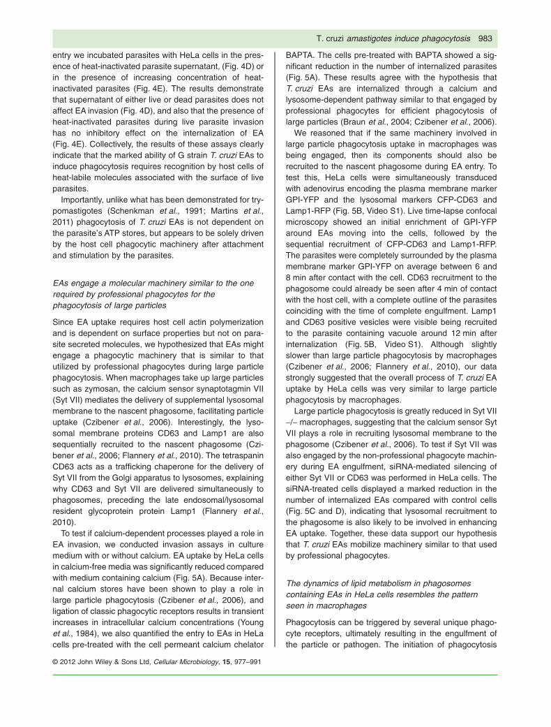
entry we incubated parasites with HeLa cells in the pres-ence of heat-inactivated parasite supernatant, (Fig. 4D) orin the presence of increasing concentration of heat-inactivated parasites (Fig. 4E). The results demonstratethat supernatant of either live or dead parasites does notaffect EA invasion (Fig. 4D), and also that the presence ofheat-inactivated parasites during live parasite invasionhas no inhibitory effect on the internalization of EA(Fig. 4E). Collectively, the results of these assays clearlyindicate that the marked ability of G strain T. cruzi EAs toinduce phagocytosis requires recognition by host cells ofheat-labile molecules associated with the surface of liveparasites.
Importantly, unlike what has been demonstrated for try-pomastigotes (Schenkman et al., 1991; Martins et al.,2011) phagocytosis of T. cruzi EAs is not dependent onthe parasite’s ATP stores, but appears to be solely drivenby the host cell phagocytic machinery after attachmentand stimulation by the parasites.
EAs engage a molecular machinery similar to the onerequired by professional phagocytes for thephagocytosis of large particles
Since EA uptake requires host cell actin polymerizationand is dependent on surface properties but not on para-site secreted molecules, we hypothesized that EAs mightengage a phagocytic machinery that is similar to thatutilized by professional phagocytes during large particlephagocytosis. When macrophages take up large particlessuch as zymosan, the calcium sensor synaptotagmin VII(Syt VII) mediates the delivery of supplemental lysosomalmembrane to the nascent phagosome, facilitating particleuptake (Czibener et al., 2006). Interestingly, the lyso-somal membrane proteins CD63 and Lamp1 are alsosequentially recruited to the nascent phagosome (Czi-bener et al., 2006; Flannery et al., 2010). The tetraspaninCD63 acts as a trafficking chaperone for the delivery ofSyt VII from the Golgi apparatus to lysosomes, explainingwhy CD63 and Syt VII are delivered simultaneously tophagosomes, preceding the late endosomal/lysosomalresident glycoprotein protein Lamp1 (Flannery et al.,2010).
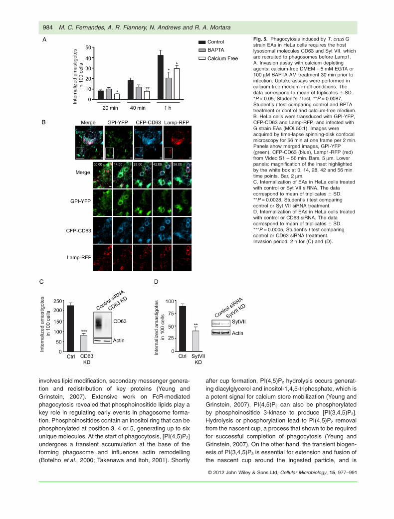
To test if calcium-dependent processes played a role inEA invasion, we conducted invasion assays in culturemedium with or without calcium. EA uptake by HeLa cellsin calcium-free media was significantly reduced comparedwith medium containing calcium (Fig. 5A). Because inter-nal calcium stores have been shown to play a role inlarge particle phagocytosis (Czibener et al., 2006), andligation of classic phagocytic receptors results in transientincreases in intracellular calcium concentrations (Younget al., 1984), we also quantified the entry to EAs in HeLacells pre-treated with the cell permeant calcium chelator
BAPTA. The cells pre-treated with BAPTA showed a sig-nificant reduction in the number of internalized parasites(Fig. 5A). These results agree with the hypothesis thatT. cruzi EAs are internalized through a calcium andlysosome-dependent pathway similar to that engaged byprofessional phagocytes for efficient phagocytosis oflarge particles (Braun et al., 2004; Czibener et al., 2006).
We reasoned that if the same machinery involved inlarge particle phagocytosis uptake in macrophages wasbeing engaged, then its components should also berecruited to the nascent phagosome during EA entry. Totest this, HeLa cells were simultaneously transducedwith adenovirus encoding the plasma membrane markerGPI-YFP and the lysosomal markers CFP-CD63 andLamp1-RFP (Fig. 5B, Video S1). Live time-lapse confocalmicroscopy showed an initial enrichment of GPI-YFParound EAs moving into the cells, followed by thesequential recruitment of CFP-CD63 and Lamp1-RFP.The parasites were completely surrounded by the plasmamembrane marker GPI-YFP on average between 6 and8 min after contact with the cell. CD63 recruitment to thephagosome could already be seen after 4 min of contactwith the host cell, with a complete outline of the parasitescoinciding with the time of complete engulfment. Lamp1and CD63 positive vesicles were visible being recruitedto the parasite containing vacuole around 12 min afterinternalization (Fig. 5B, Video S1). Although slightlyslower than large particle phagocytosis by macrophages(Czibener et al., 2006; Flannery et al., 2010), our datastrongly suggested that the overall process of T. cruzi EAuptake by HeLa cells was very similar to large particlephagocytosis by macrophages.
Large particle phagocytosis is greatly reduced in Syt VII-/- macrophages, suggesting that the calcium sensor SytVII plays a role in recruiting lysosomal membrane to thephagosome (Czibener et al., 2006). To test if Syt VII wasalso engaged by the non-professional phagocyte machin-ery during EA engulfment, siRNA-mediated silencing ofeither Syt VII or CD63 was performed in HeLa cells. ThesiRNA-treated cells displayed a marked reduction in thenumber of internalized EAs compared with control cells(Fig. 5C and D), indicating that lysosomal recruitment tothe phagosome is also likely to be involved in enhancingEA uptake. Together, these data support our hypothesisthat T. cruzi EAs mobilize machinery similar to that usedby professional phagocytes.
The dynamics of lipid metabolism in phagosomescontaining EAs in HeLa cells resembles the patternseen in macrophages
Phagocytosis can be triggered by several unique phago-cyte receptors, ultimately resulting in the engulfment ofthe particle or pathogen. The initiation of phagocytosis
T. cruzi amastigotes induce phagocytosis 983
© 2012 John Wiley & Sons Ltd, Cellular Microbiology, 15, 977–991
involves lipid modification, secondary messenger genera-tion and redistribution of key proteins (Yeung andGrinstein, 2007). Extensive work on FcR-mediatedphagocytosis revealed that phosphoinositide lipids play akey role in regulating early events in phagosome forma-tion. Phosphoinositides contain an inositol ring that can bephosphorylated at position 3, 4 or 5, generating up to sixunique molecules. At the start of phagocytosis, [PI(4,5)P2]undergoes a transient accumulation at the base of theforming phagosome and influences actin remodelling(Botelho et al., 2000; Takenawa and Itoh, 2001). Shortly
after cup formation, PI(4,5)P2 hydrolysis occurs generat-ing diacylglycerol and inositol-1,4,5-triphosphate, which isa potent signal for calcium store mobilization (Yeung andGrinstein, 2007). PI(4,5)P2 can also be phosphorylatedby phosphoinositide 3-kinase to produce [PI(3,4,5)P3].Hydrolysis or phosphorylation lead to PI(4,5)P2 removalfrom the nascent cup, a process that shown to be requiredfor successful completion of phagocytosis (Yeung andGrinstein, 2007). On the other hand, the transient biogen-esis of PI(3,4,5)P3 is essential for extension and fusion ofthe nascent cup around the ingested particle, and is
Fig. 5. Phagocytosis induced by T. cruzi Gstrain EAs in HeLa cells requires the hostlysosomal molecules CD63 and Syt VII, whichare recruited to phagosomes before Lamp1.A. Invasion assay with calcium depletingagents: calcium-free DMEM + 5 mM EGTA or100 mM BAPTA-AM treatment 30 min prior toinfection. Uptake assays were performed incalcium-free medium in all conditions. Thedata correspond to mean of triplicates � SD.*P < 0.05, Student’s t test; **P = 0.0087,Student’s t test comparing control and BPTAtreatment or control and calcium-free medium.B. HeLa cells were transduced with GPI-YFP,CFP-CD63 and Lamp-RFP, and infected withG strain EAs (MOI 50:1). Images wereacquired by time-lapse spinning-disk confocalmicroscopy for 56 min at one frame per 2 min.Panels show merged images, GPI-YFP(green), CFP-CD63 (blue), Lamp1-RFP (red)from Video S1 – 56 min. Bars, 5 mm. Lowerpanels: magnification of the inset highlightedby the white box at 0, 14, 28, 42 and 56 mintime points. Bar, 2 mm.C. Internalization of EAs in HeLa cells treatedwith control or Syt VII siRNA. The datacorrespond to mean of triplicates � SD.**P = 0.0028, Student’s t test comparingcontrol or Syt VII siRNA treatment.D. Internalization of EAs in HeLa cells treatedwith control or CD63 siRNA. The datacorrespond to mean of triplicates � SD.***P = 0.0005, Student’s t test comparingcontrol or CD63 siRNA treatment.Invasion period: 2 h for (C) and (D).
984 M. C. Fernandes, A. R. Flannery, N. Andrews and R. A. Mortara
© 2012 John Wiley & Sons Ltd, Cellular Microbiology, 15, 977–991

inducible within 2–3 min of phagosomal sealing in FcR-mediated phagocytosis (Marshall et al., 2001). Thismarked but short-lived burst of PI(3,4,5)P3 recruits pro-tein kinases, adaptor proteins and guanine nucleotideexchange factors to activate small GTPases, whichare important for phagosome development (Marshallet al., 2001; Botelho and Grinstein, 2011). Finally,phosphatidylinositol-3-phosphate [PI(3)P] is found onearly endosomes and newly formed phagosomes at about1 min after phagosome sealing, leading to phagosomematuration (Fratti et al., 2001; Vieira et al., 2001).
To test whether EA-containing phagosomes sequen-tially acquired and lost phosphoinositide phagocytosisreporter molecules during internalization, we followedphosphoinositide metabolism during parasite invasionwith GFP-labelled probes and live cell spinning disk con-focal microscopy. The first lipid to be enriched at thenascent phagosome was PI(4,5)P2, which can be moni-tored with the PH domain from phospholipase C ∂ (PH-PLC∂) (Botelho et al., 2000). HeLa cells were transfectedwith plasmids encoding PH-PLC∂-GFP and LifeAct-RFP(a 17-amino-acid peptide that labels F-actin) and exposedto G strain EAs. After contact of the parasite with the cell,phagocytosis was initiated as seen by PH-PLC∂-GFP andactin recruitment (Fig. 6A, Video S2). This indicated thatPI(4,5)P2 was enriched at the site of phagocytosis and themarker, PH-PLC∂-GFP, was retained at the base of thenascent phagosome for approximately 7–8 min and thenlost as the parasite was internalized. To follow the nextstage of phosphoinositide production, HeLa cells weretransfected with constructs encoding the PH domain ofAKT fused with GFP, which binds to PI(3,4,5)P3 andLifeAct-RFP. As expected, PI(3,4,5)P3 monitored byPH-AKT-GFP began to be recruited approximately 4 minafter the beginning of phagocytosis and persisted on thenascent phagosome for 20–30 min (Fig. 6B, Video S3).This was an interesting observation, since it contrasts withthe fast PI(3,4,5)P3 disappearance that was reported tooccur as early as 1 min after phagosome sealing in mac-rophages (Vieira et al., 2001), coinciding with loss of theYFP-GPI signal resulting from quenching in an acidicenvironment (Fig. 4, Video S1). Following phagosomesealing in macrophages, PI(3)P becomes apparent onsealed phagosomes (Vieira et al., 2001). To monitorPI(3)P levels in HeLa cells, a plasmid encoding twotandem FYVE domains fused to GFP (2FYVE-GFP) weretransfected into HeLa cells and exposed to G-strain EAs.Approximately 20–30 min after initiation of phagocytosis,the nascent phagosome recruited 2FYVE-GFP indicatingthe formation of PI(3)P (Fig. 6C, Video S4). The signalpersisted for approximately 50–70 min until it dissipated.This timing was significantly longer than what is observedin classic phagocytosis by macrophages, with PI(3)P per-sisting for approximately 10 min after phagosome sealing
(Yeung and Grinstein, 2007). Thus, while the kinetics ofthe PI metabolism appears to be delayed when comparedwith professional phagocytes, the same sequential eventsare in place.
In order to determine if the delayed kinetics in therecruitment of 2FYVE-GFP in HeLa cells reflects an intrin-sic characteristic of non-professional phagocytes, wecompared the uptake of EAs to the internalization of inertparticles. As expected, coating latex beads with fibronec-tin (FN-beads) (Ozeri et al., 1998) promoted uptake atlevels comparable to EAs, while uncoated beads showedinsignificant uptake (Fig. 7A). Comparison of uptakekinetics of FN-beads and EA revealed that internalizationprocess in HeLa cells, in both cases, presents a delay inmarker recruitment (Fig. 7B) when compared with macro-phages (Yeung and Grinstein, 2007). This result stronglysuggests that while epithelial cells contain the machineryfor particle uptake by phagocytosis, the duration of theprocess is significantly extended. Interestingly, our resultsalso revealed that during EA-induced phagocytic cup for-mation, the PI(4,5)P2 recruitment period is longer, whencompared with inert particles (Fig. 7B). However, it hasbeen demonstrated that the geometry of the phagocy-tosed particle greatly influences phagocytic responses(Doshi and Mitragotri, 2010), and differences in shapebetween round latex beads and the oval EAs mayaccount for this variation. Altogether our results revealthat although the kinetics seems to be delayed, non-professional phagocytes are capable of classical phago-cytosis and that T. cruzi EAs are not responsible fordelaying the process.
Although the precise role of extracellularly generatedamastigotes in the pathogenesis of Chagas’ disease hasonly been hypothesized (Scharfstein and Morrot, 1999) itis conceivable that the potent phagocytic properties ofT. cruzi amastigotes play a role in parasite persistencewithin the mammalian host. Immune lysis of infected cellsis predicted to release viable amastigotes into the extra-cellular medium, and a potent induction of re-uptake bynearby cells can promote parasite persistence in tissues,a hallmark of the chronic infection that is characteristic ofChagas’ disease.
Experimental procedures
Host cells and parasites
HeLa cells CCL-2.1 (ATCC), NRKs, C2C12 and LLC-MK2 weregrown in Dulbecco’s modified Eagle’s medium (DMEM – Sigma)supplemented with 10% of fetal bovine serum (FBS), at 37°C with5% CO2. Trypomastigotes from T. cruzi G strain (Yoshida, 1983)were obtained from the supernatant of infected monolayers ofHeLa cells as previously described (Fernandes et al., 2007b).Trypomastigotes from the T. cruzi Y strain were obtained fromthe supernatant of infected monolayers of LLC-MK2 cells, as
T. cruzi amastigotes induce phagocytosis 985
© 2012 John Wiley & Sons Ltd, Cellular Microbiology, 15, 977–991

Fig. 6. The phosphoinositide markers PH-PLC∂, PH-AKT and FYVE domain are sequentially recruited to nascent phagosomes containingT. cruzi G strain EAs, with a slower kinetics than what was previously shown for zymosan particles in macrophages. HeLa cells weretransfected with plasmids encoding PH-PLC∂-GFP, PH-AKT-GFP or 2-FYVE-GFP together with LifeAct-RFP, and infected with T. cruzi Gstrain EAs (MOI 50:1).A. Images were acquired by time-lapse spinning-disk confocal microscopy for 16 min at one frame per 30 s.B. Images were acquired by time-lapse spinning-disk confocal microscopy for 41 min at one frame per 30 s.C. Images were acquired by time-lapse spinning-disk confocal microscopy for 90 min at one frame per 30 s.A–C. Panels show differential interference contrast (DIC), PI-binding-domain-GFP (green), LifeAct-RFP (red) and merged fluorescenceimages. For the fluorescence images, the left panels represent the X–Z plane, the top panel represents the Y–Z plane, and the middle panelrepresents the X–Y plane centred at the amastigote. Time is displayed in minutes : seconds. Bars, 1 mm.
986 M. C. Fernandes, A. R. Flannery, N. Andrews and R. A. Mortara
© 2012 John Wiley & Sons Ltd, Cellular Microbiology, 15, 977–991

previously described (Andrews et al., 1987). For EA differentia-tion, cell derived trypomastigotes (Y or G strain) were isolatedfrom culture supernatants of infected cells by centrifugation at1200 g for 10 min. The pellet was resuspended in LIT medium,pH 5.8 and incubated for 24 h at 37°C, and at least 95% pure EAswere obtained (Ley et al., 1988; Mortara, 1991).
Y strain-GFP parasites were obtained after transfection ofp-TREX–GFP vector (DaRocha et al., 2004) in epimastigotesforms of T. cruzi. Briefly, epimastigotes were harvested from cul-tures grown in liver infusion tryptose (LIT) medium containing10% FBS at 28°C (Nogueira and Cohn, 1976), washed once withPBS and resuspended to 2 ¥ 108 parasites ml-1 in electroporationbuffer (137 mM NaCl, 21 mM HEPES, 5 mM KCl, 5.5 mMNa2HPO4, 0.77 mM glucose, pH 7.0). Aliquots (0.7 ml) of parasitesuspension were mixed with 25 mg of DNA in 0.4 cm cuvettes andelectroporated using a Bio-Rad Gene Pulser® set at 0.3 kV and500 mF with two pulses. The transfected cells were transferred to5 ml of LIT with 10% FCS and incubated at 28°C for 48 h beforeadding G418 (500 mg ml-1). Plasmid DNA used in electroporationexperiments was obtained by alkaline lysis using Qiagencolumns (Qiagen, USA).
Cell invasion assays
Cells (1.8 ¥ 105 per well) were plated on glass coverslips placedin 35 mm wells, 24 h before experiments. EAs were washed
twice with phosphate-buffered saline (PBS) and resuspended in10% FBS–DMEM and incubated for 1 h at 37°C to recover fromdifferentiation medium. Semiconfluent cells were then infectedwith G strain EAs (MOI 15:1) or Y strain EAs (MOI 50:1) in 2%FBS–DMEM for the indicated period of times.
For calcium chelation, invasion assays were performed incalcium-free DMEM (Invitrogen) in all conditions. HeLa cells werepre-treated for 30 min prior invasion with either DMEM contain-ing 5 mM EGTA, or calcium-free DMEM containing 100 mMBAPTA-AM (Invitrogen). Samples were fixed for 5 min at roomtemperature with Bouin solution (71.4% saturated picric acid,23.8% formaldehyde and 4.8% acetic acid), stained with Giemsaand sequentially dehydrated in acetone followed by a gradedseries of acetone : xylol (9:1, 7:3, 3:7) and finally xylol. Thistechnique allows microscopic distinction between intracellularparasites, which are seen surrounded by a halo, from attachedparasites. The number of intracellular parasites was determinedby counting at least 300 cells per coverslip, in triplicate in a NikonE200 microscope with a 100¥ N.A. 1.3 oil immersion objective.For immunofluorescence, coverslips were fixed in 4% parafor-maldehyde (PFA) diluted in PBS for 15 min and processed asdescribed below. For inert particle internalization assays, poly-styrene beads (Spherotech, 5 mm) were washed three times withcold PBS without calcium or magnesium (PBS -/-). After the finalwash, the beads were resuspended in 1 ml of PBS -/- at 5 ¥ 107
beads ml-1 with 50 mg ml-1 fibronectin (BD Biosciences) and
Fig. 7. EAs display similar uptake kinetics as fibronectin-coated latex beads.A. HeLa cells were incubated with EAs (Amastigotes), fibronectin-coated beads (FN-beads) or uncoated beads (ratio 20:1) and exposed toHeLa cells for 2 h. The data correspond to mean of triplicates � SD. ***P = 0.0005, Student’s t test.B. HeLa cells were transfected with plasmids encoding PH-PLC∂-GFP, PH-AKT-GFP or 2-FYVE-GFP together with LifeAct-RFP, and infectedwith T. cruzi G strain EAs (MOI 50:1) or fibronectin-coated beads (ratio 3:1). The timing of lipid metabolism was recorded as the resident timeof lipid markers on the phagosomal membrane, based on the relative abundance of PI(4,5)P2 (blue), PI(3,4,5)P3 (red) and PI(3)P (green). Thecurves for the T. cruzi G strain and fibronectin-coated beads represent idealized profiles derived from experimental data with heights reflectingrelative changes and not absolute concentrations. Representative images are shown to the right of the graphs for T. cruzi G strain andfibronectin-coated beads.
T. cruzi amastigotes induce phagocytosis 987
© 2012 John Wiley & Sons Ltd, Cellular Microbiology, 15, 977–991
incubated overnight at 4°C. The beads were then washed threetimes with PBS (-/-) and incubated in 1 ml of BSA (10 mg ml-1)for 3 h at 4°C. The beads were washed three times with cold PBS-/- and suspended in DMEM culture media for use in uptakeassays and live cell microscopy. Zymosan was purchased fromSigma.
Immunofluorescence
PFA fixed cells were washed with PBS, quenched with 15 mMNH4Cl for 15 min, and incubated with PBS containing 2% bovineserum albumin (BSA) for 1 h. When processed for an inside/outside immunofluorescence assay (Tardieux et al., 1992)samples were incubated with rabbit anti-T. cruzi polyclonal anti-bodies for 1 h followed by 1 h anti-rabbit IgG conjugated to Alex-aFluor™ 647 (Invitrogen, for confocal images) or AlexaFluor™594 (for quantification) secondary antibodies, to stain extracellu-lar parasites. Cells were then permeabilized with 0.1% saponinfor 30 min and incubated with H4A3 anti-human Lamp1 mousemonoclonal antibodies. (Developmental Studies HybridomaBank) for 1 h followed by 1 h incubation with anti-mouse Alex-aFluor™ 488 secondary antibodies and Phalloidin-594 (Invitro-gen) to stain actin filaments. The images shown in Fig. 1 (allpanels) maximum projections images of optical sections(0.13 mm Z step) acquired on a Leica SPX5 confocal system witha 63¥ N.A. 1.4 oil objective.
In order to quantify internalized G strain or Y strain-GFP EAs orinert particles (Fig. 3C), inside/outside fluorescence assay wasperformed as described above and slides were analysed usingNikon E200 epifluorescence microscope with a 100¥ N.A. 1.3 oilimmersion objective. The number of internalized parasites (nega-tive for anti-T. cruzi antibody staining) was determined in at least300 cells. All samples were incubated with 10 mM DAPI (Sigma)for nuclei and kinetoplasts staining.
ATP depletion, ATP assays and heat inactivation
For ATP depletion, parasites were treated with 100 mM sodiumazide and 100 mM 2-deoxyglucose in glucose free DMEM(Gibco) for 1 h (Schenkman et al., 1991). Invasion assays wereperformed subsequently for the indicated time period.
Parasites were washed twice with PBS and resuspended at1e105 parasites per 100 ml. Amount of ATP was determined withthe ATPlite assay kit (PerkinElmer) as outlined by manufacturer’sprotocol. The data were acquired on SpectraMax m5e platereader (Molecular Devices) and analysed with the Softmax Prosoftware package (Molecular Devices). Parasites were heat-killed at 56°C in a water bath for 20 min.
Electron microscopy
HeLa cells were grown in glass coverslips and allowed to interactwith G strain EAs at a 10–20:1 parasites per cell ratio. After30 min when parasites are mostly initiating contact, cells werewashed with 0.1 M phosphate-buffered saline (37°C) five times toremove unattached parasites and fixed with 2% glutaraldehyde,4% paraformaldehyde (both from Electron Microscopy Sciences– EMS, Hatfield, PA, USA), 140 mM NaCl, 2 mM CaCl2, 1 mMMgCl2 in 0.1 M HEPES (pH 7.2) for 30 min. To reveal cytoskeletalelements, after 30 min of interaction, samples were treated with
a membrane extraction solution containing 1% Triton X-100,100 mM PIPES (pH 7.2), 4% sucrose, 1 mM MgCl2, 10 mM taxol(Invitrogen) and 10 mM phalloidin (Sigma) – to stabilize microtu-bules and microfilaments respectively (Sant’Anna et al., 2005) –for 10 min under gentle rocking at room temperature thenwashed twice for 10 min in the same solution without the deter-gent, then fixed as described above. Samples were post-fixedwith 1% OsO4 (EMS) and 1% tannic acid (EMS), both for 1 h,dehydrated with ethanol and critical point-dried from CO2 (CPD020 Balzers-Tec). The slides were coated with a thin carbon layer(8 nm) (BAF 300, Balzers) and the glass coverslips detachedfrom the cell layer by immersion in 20% hydrofluoric acid (Sigma).Small pieces of the cell layer were transferred for distilled waterfor 10 min and finally collected onto copper grids (300 mesh).Images were acquired on a Hitachi S-4800 field emission scan-ning electron microscope operated at 5 kV.
Plasmids and adenoviral constructs
Constructs encoding the PH domain of PLC-d fused to GFP(PLC-d-GFP), the PH domain of Akt fused to GFP (AKT-GFP)and the tandem FYVE domains fused to GFP (2FYVE-GFP) weredescribed previously (Stauffer et al., 1998; Haugh et al., 2000;Vieira et al., 2001). These plasmids were generous gifts fromSergio Grinstein (Program in Cell Biology, The Hospital for SickChildren, 555 University Avenue, Toronto, Ontario M5G 1X8,Canada). The mammalian expression vector encoding a17-amino-acid peptide fused to TagRFP, pCMVLifeAct-TagRFP(LifeAct-RFP) for labelling actin filaments was purchased fromibidi. The adenoviral production and purification of virons encod-ing CFP-CD63, Lamp-RFP and GPI-YFP were described previ-ously (Keller et al., 2001; Flannery et al., 2010).
Transfection, adenoviral transduction, live cell imagingand image analysis
Before live cell imaging experiments, HeLa cells were seeded at1.8 ¥ 105 cells per dish in DMEM media at 37°C and 5% CO2 ina 35 mm glass-bottom dish (MatTek Corporation). For adenoviraltransduction, cells were transduced for 18 h by replacing theculture media with HeLa imaging medium (DMEM without phenolred, 10% FBS, 1% penicillin/streptomycin) and containing puri-fied adenovirus encoding CFP-CD63, Lamp-RFP and GPI-YFPat a 10:1 MOI. For plasmid transfection, cells were transfectedwith plasmids encoding either PLC-d-GFP, AKT-GFP or 2FYVE-GFP and LifeACT-RFP with Fugene HD (Roche) according to themanufacturer’s protocol. After 18 h, the cells were washed,medium containing G strain EAs (50:1 MOI) was added, and thedishes were imaged.
For imaging, dishes were placed in a LiveCell System chamber(Pathology Devices) at 37°C with 5% CO2 attached to an EclipseTi inverted microscope with a 60¥ NA 1.4 objective (Nikon).Spinning disk confocal images were acquired using theUltraVIEW VoX system (PerkinElmer) equipped with a Hama-matsu C9100-50 camera, analysed and edited using the VolocitySoftware Suite (PerkinElmer). Final image gamma settings werechanged to 1.4.
CD63 and Syt VII transcriptional silencing
HeLa cells were transfected with Lipofectamine RNAiMAX and160 pmol of medium GC content control (12935300), CD63
988 M. C. Fernandes, A. R. Flannery, N. Andrews and R. A. Mortara
© 2012 John Wiley & Sons Ltd, Cellular Microbiology, 15, 977–991
(HSS101615) or Syt VII (HSS113401) Stealth siRNA duplexes,according to the manufacturer’s instructions (Invitrogen). At 24 hafter transfection, the cells were submitted to invasion assay.
Western blot analysis
To generate whole-cell lysates, HeLa cells treated with siRNA asoutlined above, were lysed in WCL buffer (25 mM HEPES pH 7.4,150 mM NaCl, 1% Triton X-100 and complete protease inhibitorcocktail from Roche) at 4°C for 1 h. Lysates were clarified bycentrifugation at 10 000 g for 10 min, and the total amount ofprotein was determined by BCA (Thermo Scientific). A volume ofclarified lysate containing 20 mg of protein was added to 6¥ SDSsample buffer, resolved on 10% SDS-PAGE, transferred to nitro-cellulose, and probed with either rabbit anti-Syt VII (Arantes andAndrews, 2006), mouse anti-CD63 antibodies (H5C6, 1:200) ormouse anti-b actin (Sigma, 1:1000). Blots were then incubatedwith either peroxidase-conjugated donkey anti-rabbit or donkeyanti-mouse antibodies (Jackson ImmunoResearch, 1:10000),washed, incubated for 5 min with Immun-Star HRP™ solution(Bio-Rad), and visualized on a LAS-3000 imaging system (Fuji-film). Relative protein levels were determined using the Image-Jsoftware (NIH).
Statistics
All experiments were performed in triplicate coverslips andrepeated at least three times, and 30 cells per coverslip werecounted. Statistical analysis was performed with GraphPadPrism®, employing Student’s t test. Data are presented asmean � standard deviation (SD).
Acknowledgements
R.A.M. was funded by Fundação de Amparo à Pesquisa doEstado de São Paulo (FAPESP, 2011/53833-4) and ConselhoNacional de Desenvolvimento Científico e Tecnológico (CNPq,Grant 200986/2010-4), from Brazil. N.W.A. research has beenfunded by NIH Grant R37 AI34867. The authors are indebted toSergio Grinstein (U. Toronto) for the generous gifts of the PIplasmids and for suggestions to the manuscript.
References
Aderem, A., and Underhill, D.M. (1999) Mechanisms ofphagocytosis in macrophages. Annu Rev Immunol 17:593–623.
Alonso, A., and Garcia-del Portillo, F. (2004) Hijacking ofeukaryotic functions by intracellular bacterial pathogens.Int Microbiol 7: 181–191.
Andrews, N.W., Hong, K.S., Robbins, E.S., and Nussenz-weig, V. (1987) Stage-specific surface antigens expressedduring the morphogenesis of vertebrate forms of Trypano-soma cruzi. Exp Parasitol 64: 474–484.
Arantes, R.M., and Andrews, N.W. (2006) A role for synap-totagmin VII-regulated exocytosis of lysosomes in neuriteoutgrowth from primary sympathetic neurons. J Neurosci26: 4630–4637.
Bambino-Medeiros, R., Oliveira, F.O., Calvet, C.M., Vicente,
D., Toma, L., Krieger, M.A., et al. (2011) Involvement ofhost cell heparan sulfate proteoglycan in Trypanosomacruzi amastigote attachment and invasion. Parasitology138: 593–601.
Berón, W., Alvarez-Dominguez, C., Mayorga, L., and Stahl,P.D. (1995) Membrane trafficking along the phagocyticpathway. Trends Cell Biol 5: 100–104.
Botelho, R.J., and Grinstein, S. (2011) Phagocytosis. CurrBiol 21: R533–R538.
Botelho, R.J., Teruel, M., Dierckman, R., Anderson, R., Wells,A., York, J.D., et al. (2000) Localized biphasic changes inphosphatidylinositol-4,5-bisphosphate at sites of phagocy-tosis. J Cell Biol 151: 1353–1368.
Braun, V., Fraisier, V., Raposo, G., Hurbain, I., Sibarita, J.B.,Chavrier, P., et al. (2004) TI-VAMP/VAMP7 is required foroptimal phagocytosis of opsonised particles in macro-phages. EMBO J 23: 4166–4176.
Cruz, M.C., Souza-Melo, N., da Silva, C.V., DaRocha, W.D.,Bahia, D., Araújo, P.R., et al. (2012) Trypanosoma cruzi:role of delta-amastin on extracellular amastigote cell inva-sion and differentiation. PLoS ONE 7: e51804.
Czibener, C., Sherer, N.M., Becker, S.M., Pypaert, M., Hui,E., Chapman, E.R., et al. (2006) Ca2+ and synaptotagminVII-dependent delivery of lysosomal membrane to nascentphagosomes. J Cell Biol 174: 997–1007.
DaRocha, W.D., Silva, R.A., Bartholomeu, D.C., Pires, S.F.,Freitas, J.M., Macedo, A.M., et al. (2004) Expressionof exogenous genes in Trypanosoma cruzi: improvingvectors and electroporation protocols. Parasitol Res 92:113–120.
Dobrowolski, J.M., and Sibley, L.D. (1996) Toxoplasma inva-sion of mammalian cells is powered by the actin cytoskel-eton of the parasite. Cell 84: 933–939.
Doshi, N., and Mitragotri, S. (2010) Macrophages recognizesize and shape of their targets. PLoS ONE 5: e10051.
Fernandes, A.B., and Mortara, R.A. (2004) Invasion of MDCKepithelial cells with altered expression of Rho GTPases byTrypanosoma cruzi amastigotes and metacyclic trypomas-tigotes of strains from the two major phylogenetic lineages.Microbes Infect 6: 460–467.
Fernandes, A.B., Neira, I., Ferreira, A.T., and Mortara, R.A.(2006) Cell invasion by Trypanosoma cruzi amastigotes ofdistinct infectivities: studies on signaling pathways. Para-sitol Res 100: 59–68.
Fernandes, M.C., L’Abbate, C., Kindro Andreoli, W., andMortara, R.A. (2007a) Trypanosoma cruzi cell invasion andtraffic: influence of Coxiella burnetii and pH in a compara-tive study between distinct infective forms. Microb Pathog43: 22–36.
Fernandes, M.C., Cortez, M., Geraldo Yoneyama, K.A.,Straus, A.H., Yoshida, N., and Mortara, R.A. (2007b) Novelstrategy in Trypanosoma cruzi cell invasion: implication ofcholesterol and host cell microdomains. Int J Parasitol 37:1431–1441.
Fernandes, M.C., de Andrade, L.R., Andrews, N.W., andMortara, R.A. (2011a) Trypanosoma cruzi trypomastigotesinduce cytoskeleton modifications during HeLa cell inva-sion. Mem Inst Oswaldo Cruz 106: 1014–1016.
Fernandes, M.C., Cortez, M., Flannery, A.R., Tam, C.,Mortara, R.A., and Andrews, N.W. (2011b) Trypanosomacruzi subverts the sphingomyelinase-mediated plasma
T. cruzi amastigotes induce phagocytosis 989
© 2012 John Wiley & Sons Ltd, Cellular Microbiology, 15, 977–991

membrane repair pathway for cell invasion. J Exp Med208: 909–921.
Ferreira, D., Cortez, M., Atayde, V.D., and Yoshida, N. (2006)Actin cytoskeleton-dependent and -independent host cellinvasion by Trypanosoma cruzi is mediated by distinctparasite surface molecules. Infect Immun 74: 5522–5528.
Finlay, B.B., and Cossart, P. (1997) Exploitation of mamma-lian host cell functions by bacterial pathogens. Science276: 718–725.
Flannery, A.R., Czibener, C., and Andrews, N.W. (2010)Palmitoylation-dependent association with CD63 targetsthe Ca2+ sensor synaptotagmin VII to lysosomes. J Cell Biol191: 599–613.
Frankel, G., and Phillips, A.D. (2008) Attaching effacingEscherichia coli and paradigms of Tir-triggered actinpolymerization: getting off the pedestal. Cell Microbiol 10:549–556.
Fratti, R.A., Backer, J.M., Gruenberg, J., Corvera, S., andDeretic, V. (2001) Role of phosphatidylinositol 3-kinase andRab5 effectors in phagosomal biogenesis and mycobacte-rial phagosome maturation arrest. J Cell Biol 154: 631–644.
Galan, J.E., and Bliska, J.B. (1996) Cross-talk between bac-terial pathogens and their host cells. Annu Rev Cell DevBiol 12: 221–255.
Geissmann, F., Manz, M.G., Jung, S., Sieweke, M.H., Merad,M., and Ley, K. (2010) Development of monocytes, mac-rophages, and dendritic cells. Science 327: 656–661.
Haugh, J.M., Codazzi, F., Teruel, M., and Meyer, T. (2000)Spatial sensing in fibroblasts mediated by 3’ phosphoi-nositides. J Cell Biol 151: 1269–1280.
Keller, P., Toomre, D., Diaz, E., White, J., and Simons, K.(2001) Multicolour imaging of post-Golgi sorting and traf-ficking in live cells. Nat Cell Biol 3: 140–149.
Ley, V., Andrews, N.W., Robbins, E.S., and Nussenzweig, V.(1988) Amastigotes of Trypanosoma cruzi sustain an infec-tive cycle in mammalian cells. J Exp Med 168: 649–659.
Marshall, J.G., Booth, J.W., Stambolic, V., Mak, T., Balla, T.,Schreiber, A.D., et al. (2001) Restricted accumulation ofphosphatidylinositol 3-kinase products in a plasmalemmalsubdomain during Fc gamma receptor-mediated phagocy-tosis. J Cell Biol 153: 1369–1380.
Martins, R.M., Alves, R.M., Macedo, S., and Yoshida, N.(2011) Starvation and rapamycin differentially regulate hostcell lysosome exocytosis and invasion by Trypanosomacruzi metacyclic forms. Cell Microbiol 13: 943–954.
Morisaki, J.H., Heuser, J.E., and Sibley, L.D. (1995) Invasionof Toxoplasma gondii occurs by active penetration of thehost cell. J Cell Sci 108: 2457–2464.
Mortara, R.A. (1991) Trypanosoma cruzi: amastigotes andtrypomastigotes interact with different structures on thesurface of HeLa cells. Exp Parasitol 73: 1–14.
Mortara, R.A., Andreoli, W.K., Taniwaki, N.N., Fernandes,A.B., Silva, C.V., Fernandes, M.C., et al. (2005) Mamma-lian cell invasion and intracellular trafficking by Trypano-soma cruzi infective forms. An Acad Bras Cienc 77: 77–94.
Nogueira, N., and Cohn, Z. (1976) Trypanosoma cruzi:mechanism of entry and intracellular fate in mammaliancells. J Exp Med 143: 1402–1420.
Ozeri, V., Rosenshine, I., Mosher, D.F., Fassler, R., andHanski, E. (1998) Roles of integrins and fibronectin in the
entry of Streptococcus pyogenes into cells via protein F1.Mol Microbiol 30: 625–637.
Procópio, D.O., Barros, H.C., and Mortara, R.A. (1999) Actin-rich structures formed during the invasion of cultured cellsby infective forms of Trypanosoma cruzi. Eur J Cell Biol 78:911–924.
Procópio, D.O., da Silva, S., Cunningham, C.C., and Mortara,R.A. (1998) Trypanosoma cruzi: effect of protein kinaseinhibitors and cytoskeletal protein organization and expres-sion on host cell invasion by amastigotes and metacyclictrypomastigotes. Exp Parasitol 90: 1–13.
Rabinovitch, M. (1995) Professional and non-professionalphagocytes: an introduction. Trends Cell Biol 5: 85–87.
Rodrigues, A.A., Saosa, J.S., da Silva, G.K., Martins, F.A., daSilva, A.A., Souza Neto, C.P., et al. (2012) IFN-gammaplays a unique role in protection against low virulentTrypanosoma cruzi strain. PLoS Negl Trop Dis 6: e1598.
Sant’Anna, C., Campanati, L., Gadelha, C., Lourenco, D.,Labati-Terra, L., Bittencourt-Silvestre, J., et al. (2005)Improvement on the visualization of cytoskeletal structuresof protozoan parasites using high-resolution field emissionscanning electron microscopy (FESEM). Histochem CellBiol 124: 87–95.
Scharfstein, J., and Morrot, A. (1999) A role for extracellularamastigotes in the immunopathology of Chagas disease.Mem Inst Oswaldo Cruz 94 (Suppl. 1): 51–63.
Schenkman, S., Robbins, E.S., and Nussenzweig, V. (1991)Attachment of Trypanosoma cruzi to mammalian cellsrequires parasite energy, and invasion can be independentof the target cell cytoskeleton. Infect Immun 59: 645–654.
da Silva, C.V., Luquetti, A.O., Rassi, A., and Mortara, R.A.(2006) Involvement of Ssp-4-related carbohydrateepitopes in mammalian cell invasion by Trypanosoma cruziamastigotes. Microbes Infect 8: 2120–2129.
Stauffer, T.P., Ahn, S., and Meyer, T. (1998) Receptor-induced transient reduction in plasma membranePtdIns(4,5)P2 concentration monitored in living cells. CurrBiol 8: 343–346.
Takenawa, T., and Itoh, T. (2001) Phosphoinositides, keymolecules for regulation of actin cytoskeletal organizationand membrane traffic from the plasma membrane. BiochimBiophys Acta 1533: 190–206.
Tardieux, I., Webster, P., Ravesloot, J., Boron, W., Lunn, J.A.,Heuser, J.E., and Andrews, N.W. (1992) Lysosome recruit-ment and fusion are early events required for trypanosomeinvasion of mammalian cells. Cell 71: 1117–1130.
Tjelle, T.E., Lovdal, T., and Berg, T. (2000) Phagosomedynamics and function. Bioessays 22: 255–263.
Vieira, O.V., Botelho, R.J., Rameh, L., Brachmann, S.M.,Matsuo, T., Davidson, H.W., et al. (2001) Distinct roles ofclass I and class III phosphatidylinositol 3-kinases inphagosome formation and maturation. J Cell Biol 155:19–25.
Yeung, T., and Grinstein, S. (2007) Lipid signaling and themodulation of surface charge during phagocytosis.Immunol Rev 219: 17–36.
Yeung, T., Ozdamar, B., Paroutis, P., and Grinstein, S. (2006)Lipid metabolism and dynamics during phagocytosis. CurrOpin Cell Biol 18: 429–437.
Yoshida, N. (1983) Surface antigens of metacyclic trypomas-tigotes of Trypanosoma cruzi. Infect Immun 40: 836–839.
990 M. C. Fernandes, A. R. Flannery, N. Andrews and R. A. Mortara
© 2012 John Wiley & Sons Ltd, Cellular Microbiology, 15, 977–991
Young, J.D., Ko, S.S., and Cohn, Z.A. (1984) The increase inintracellular free calcium associated with IgG gamma2b/gamma 1 Fc receptor-ligand interactions: role in phago-cytosis. Proc Natl Acad Sci USA 81: 5430–5434.
Supporting information
Additional Supporting Information may be found in the onlineversion of this article:
Video S1. Sequential recruitment of plasma membrane GPI-YFP and lysosomal CD63 and Lamp1 to nascent phagosomescontaining T. cruzi G strain EAs. HeLa cells were transduced withGPI-YFP, CFP-CD63 and Lamp-RFP and infected with EAs (MOI50:1). Images were acquired by time-lapse spinning-disk confo-cal microscopy for 56 min at one frame per 2 min. Individualpanels show merged images, GPI-YFP (green), CFP-CD63(blue), Lamp1-RFP (red). The video is displayed at five framesper second. Bar, 5 mm.Video S2. Recruitment of PH-PLC∂ to nascent phagosomescontaining T. cruzi G strain EAs. HeLa cells were transfectedwith a plasmid encoding PH-PLC∂-GFP and LifeAct-RFP, andinfected with EAs (MOI 50:1). Images were acquired by time-lapse spinning-disk confocal microscopy for 16 min at two framesper min. Panels show differential interference contrast (DIC),
PH-PLC∂-GFP (green), LifeAct-RFP (red) and merged images.For panels displaying fluorescence, the left panels represent theX–Z plane, the top panel represents the Y–Z plane, and themiddle panel represents the X–Y plane centred at the amastig-ote. The video is displayed at five frames per second. Bar,2 mm.Video S3. Recruitment of PH-AKT to nascent phagosomes con-taining T. cruzi G strain EAs. HeLa cells were transfected withplasmid encoding PH-AKT-GFP and LifeAct-RFP, and infectedwith EAs (MOI 50:1). Images were acquired by time-lapsespinning-disk confocal microscopy for 41 min at one frame per30 s. Panels show differential interference contrast (DIC),PH-AKT-GFP (green), LifeAct-RFP (red) and merged images.For panels displaying fluorescence, the left panels represents theX–Z plane, the top panel represents the Y–Z plane, and themiddle panel represents the X–Y plane centred at the amastig-ote. The video is displayed at five frames per second. Bar, 2 mm.Video S4. Recruitment of FYVE domain to nascent phago-somes containing T. cruzi G strain EAs. HeLa cells were trans-fected with plasmid encoding 2-FYVE-GFP and LifeAct-RFP, andinfected with EAs (MOI 50:1). Images were acquired by time-lapse spinning-disk confocal microscopy for 90 min at one frameper 30 s. Panels show differential interference contrast (DIC),2-FYVE-GFP (green), LifeAct-RFP (red) and merged images.The video is displayed at five frames per second. Bar, 2 mm.
T. cruzi amastigotes induce phagocytosis 991
© 2012 John Wiley & Sons Ltd, Cellular Microbiology, 15, 977–991
Related Documents