. 1 EXTRA ORAL RADIOGRAPHIC TECHNIQUES Dr. Neeraj Kumar Yadav Reader Dept. of Oral Medicine and Radiology MGDCH , Jaipur Part I PART II Introduction These techniques imply that film is placed outside the oral cavity, against the side of the face to be radiographed & x-ray beam is directed towards it. Indications Trismus Large lesions Trauma Jaws and orofacial bones Impacted teeth TMJ Area Skeletal Growth And Development Drawbacks An important aspect of the extra - oral radiographic technique is the immobilization of the patient’s head

Welcome message from author
This document is posted to help you gain knowledge. Please leave a comment to let me know what you think about it! Share it to your friends and learn new things together.
Transcript
.
1
EXTRA ORAL RADIOGRAPHIC
TECHNIQUES
Dr. Neeraj Kumar Yadav
Reader
Dept. of Oral Medicine and
Radiology
MGDCH , Jaipur
Part I
PART II
Introduction
These techniques imply that film is
placed outside the oral cavity, against
the side of the face to be
radiographed & x-ray beam is
directed towards it.
Indications
Trismus Large lesions Trauma
Jaws and
orofacial bones Impacted teeth TMJ Area
Skeletal Growth
And
Development
Drawbacks
An important aspect of the extra-oral
radiographic technique is the immobilization
of the patient’s head
.
2
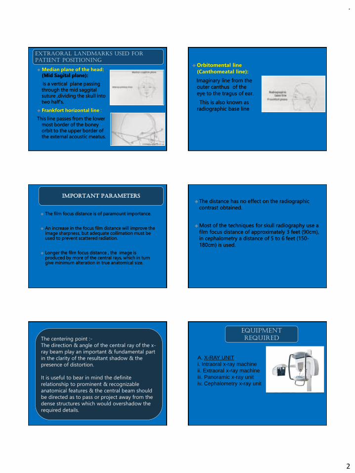
ExtraOral Landmarks used for
Patient Positioning
IMPORTANT PARAMETERS
The centering point :-
The direction & angle of the central ray of the x-
ray beam play an important & fundamental part
in the clarity of the resultant shadow & the
presence of distortion.
It is useful to bear in mind the definite
relationship to prominent & recognizable
anatomical features & the central beam should
be directed as to pass or project away from the
dense structures which would overshadow the
required details.
Equipment
Required
A. X-RAY UNITi. Intraoral x-ray machineii. Extraoral x-ray machineiii. Panoramic x-ray unitiv. Cephalometry x-ray unit

.
3
• High speed rare-earth screen combination
• Lateral oblique views of the mandible use a 5 ×7 inch film and cassette.
• Skull radiography requires atleast an 8×10 inch film.
• Place “R” or “L” on the appropriate corner of the
cassette
• Grids
Procedure
A. Equipment Preparation
1. Load the extraoral cassette in the dark room
under safelight conditions. Place one extraoral film
between two intensifying screens & securely close
the cassette.
2. Set the exposure factors ( kilovoltage ,
milliamperage , time) according to the
manufacturer’s recommendations.
3.Load the cassette into the the cassette carrier.
4.Print in the date, patient’s name, age, sex & the
case number.
B. Patient Preparation
1. Explain to the patient the radiographic procedure
about to be performed.
2. Place a lead apron without a lead collar over the
patient & secure it.
•The apron must be placed low around the back of
the neck so that it does not block the x-ray beam.
•A thyroid collar is not recommended for extra oral
radiography because it blocks part of the beam &
obscures important diagnostic information
•Remove all objects from head & neck that may
interfere with the film exposure.
•The patient must remove earrings, eyeglasses,
necklaces, napkin chains, hearing aids, hairpins &
complete or partial removable dentures or any
other removable appliance in the oral cavity.
Extra Oral Radiographic
Projections
A. Radiography Of Paranasal Sinuses
1. Posteroanterior projection (also known as
occipitofrontal projection of Nasal sinuses)
There are 2 methods for obtaining this projection.
a. Posterior Anterior ( Granger projection )
b. Modified method, Inclined Posterior Anterior (
Caldwell Projection )

.
4
B. Radiography Of The Maxillary Sinuses
1. Standard Occipitomental Projection
(0˚OM)
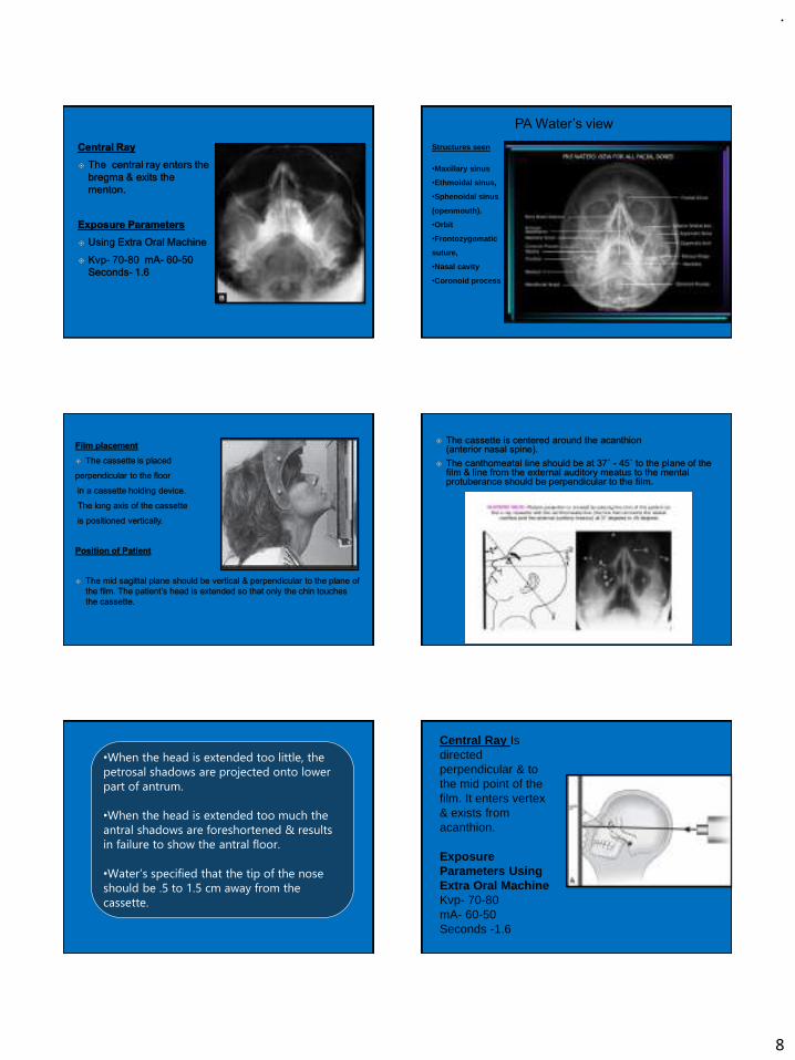
2. Modified method (30˚ OM)
3. Bregma Menton view
4. PA Water’s view
C .Radiography Of The Mandible
1. PA Mandible
2. Rotated PA Mandible
3. Lateral oblique
A. Anterior body of mandible
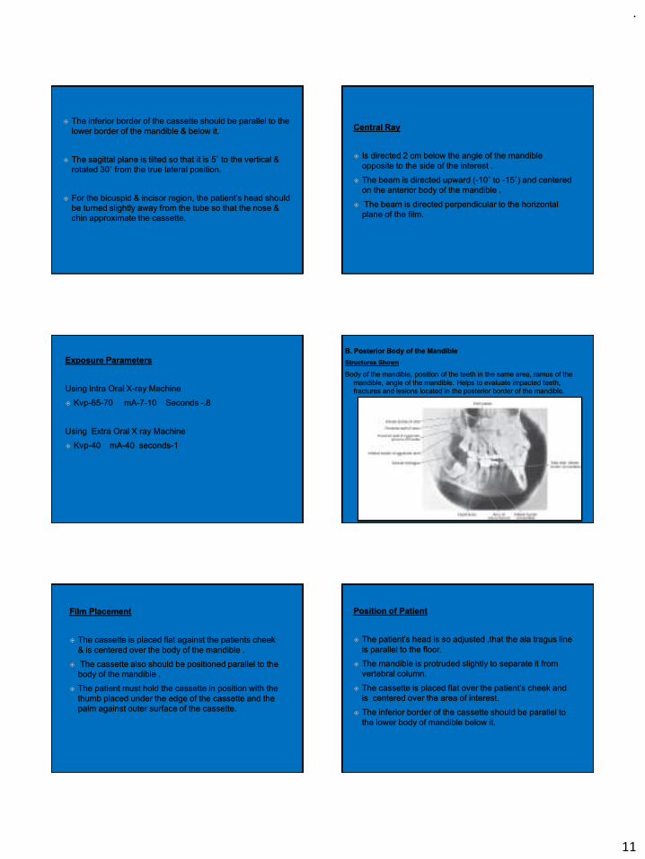
B. Posterior body of mandible
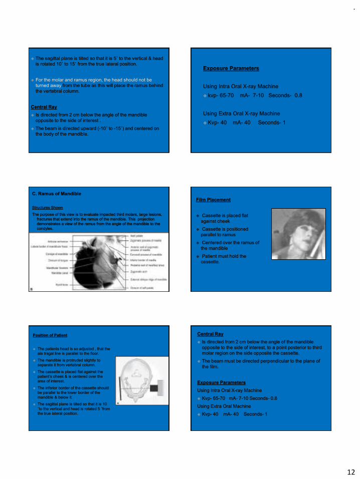
C. Ramus of mandible
Radiography of the Paranasal Sinuses
•This is used to study the relationship of the
sinuses to each other & to the surrounding
structures.
•Lateral/anteroposterior view may be taken.
•Routinely the paranasal sinuses are radiographed
with the patient in the erect position, so as to
demonstrate the presence or absence of fluid & in
order to differentiate between the shadows caused
by the fluids & those caused by other pathology.
.
5
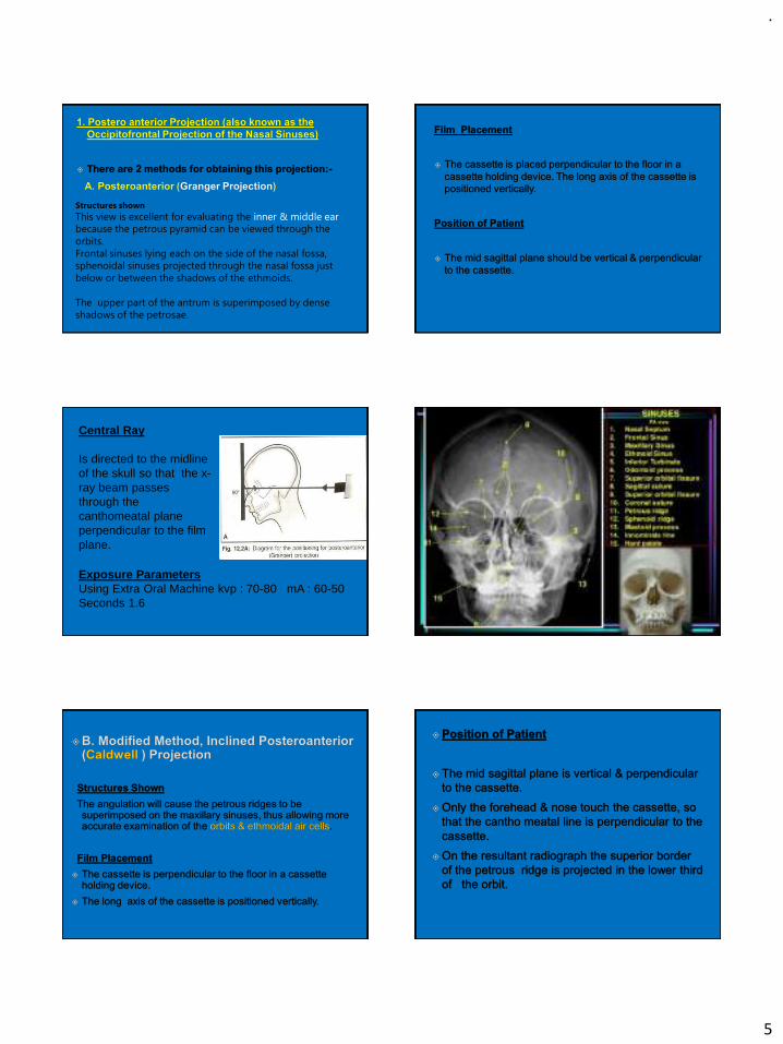
Structures shown
This view is excellent for evaluating the inner & middle ear
because the petrous pyramid can be viewed through the
orbits.
Frontal sinuses lying each on the side of the nasal fossa,
sphenoidal sinuses projected through the nasal fossa just
below or between the shadows of the ethmoids.
The upper part of the antrum is superimposed by dense
shadows of the petrosae.
Central Ray
Is directed to the midline of the skull so that the x-ray beam passes through the canthomeatal plane perpendicular to the film plane.
Exposure ParametersUsing Extra Oral Machine kvp : 70-80 mA : 60-50 Seconds 1.6
.
6
Radiography of the
Maxillary Sinuses
Indications
1. Investigation of the maxillary antra
2. Fracture of middle third of the face
a) Le fort i
b) Le fort ii
c) Le fort iii
d) Zygomatic complex
e) Nasoethmoidal complex
f) Orbital blow out fractures
g) Coronoid process fracture
3. Investigation of Frontal and Ethmoidal sinuses
4. Sphenoidal sinuses
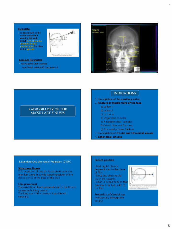
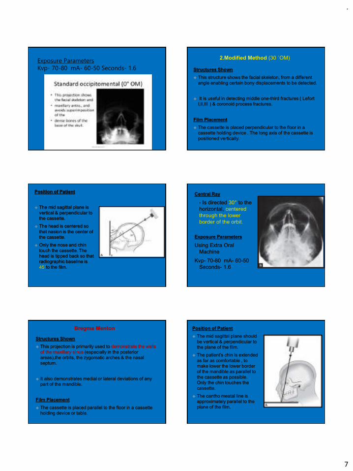
1.Standard Occipitomental Projection (0˚OM)
Structures Shown
This projection shows the facial skeleton & the
maxillary antra & avoids superimposition of the
dense bones of the base of the skull.
Film placement
The cassette is placed perpendicular to the floor in
a cassette holding device.
The long axis of the cassette is positioned
vertically.
Patient position:
• Mid sagital plane is
perpendicular to the plane
of film.
• Nose and chin should
touch the cassette.
• Head is tipped back so that
canthomental line is 45o to
the film.
Projection of Central ray
•Horizontally through the
occiput
.
7
Exposure Parameters
Kvp- 70-80 mA- 60-50 Seconds- 1.6
.
8
Structures seen
•Maxillary sinus
•Ethmoidal sinus,
•Sphenoidal sinus
(openmouth).
•Orbit
•Frontozygomatic
suture,
•Nasal cavity
•Coronoid process
PA Water’s view
•When the head is extended too little, the
petrosal shadows are projected onto lower
part of antrum.
•When the head is extended too much the
antral shadows are foreshortened & results
in failure to show the antral floor.
•Water’s specified that the tip of the nose
should be .5 to 1.5 cm away from the
cassette.
Central Ray Is directed perpendicular & to the mid point of the film. It enters vertex & exists from acanthion.
Exposure Parameters Using Extra Oral MachineKvp- 70-80 mA- 60-50 Seconds -1.6
.
9
RADIOGRAPHY OF THE
MANDIBLE
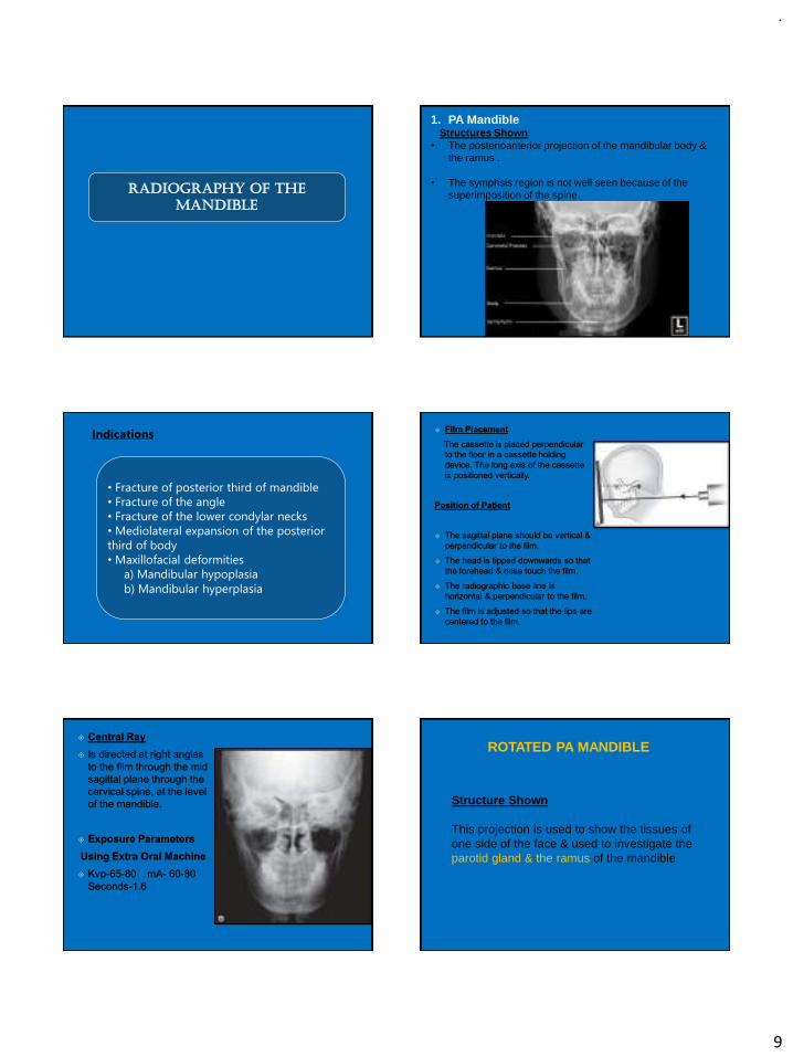
1. PA MandibleStructures Shown
• The posterioanterior projection of the mandibular body & the ramus .
• The symphsis region is not well seen because of the superimposition of the spine.
Indications
• Fracture of posterior third of mandible
• Fracture of the angle
• Fracture of the lower condylar necks
• Mediolateral expansion of the posterior
third of body
• Maxillofacial deformities
a) Mandibular hypoplasia
b) Mandibular hyperplasia
Structure Shown
This projection is used to show the tissues of one side of the face & used to investigate the parotid gland & the ramus of the mandible
ROTATED PA MANDIBLE
.
10
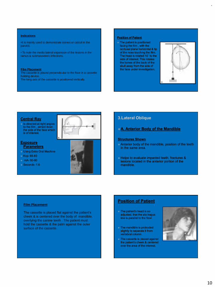
Indications
•It is mainly used to demonstrate stones or calculi in the parotid,
• To note the medio lateral expansion of the lesions in the ramus & submasseteric infections.
Film PlacementThe cassette is placed perpendicular to the floor in a cassette holding device.The long axis of the cassette is positioned vertically.
Film Placement
The cassette is placed flat against the patient’s
cheek & is centered over the body of mandible, overlying the canine teeth . The patient must hold the cassette & the palm against the outer surface of the cassette.
.
11
.
12
.
13
EXTRA ORAL RADIOGRAPHIC
TECHNIQUES
Dr.Philip Cyriac PG,
Department of OMDR
EXTRA ORAL RADIOGRAPHIC TECHNIQUES
Part II
Part I
PART II
RADIOGRAPHY OF
BASE OF THE
SKULL
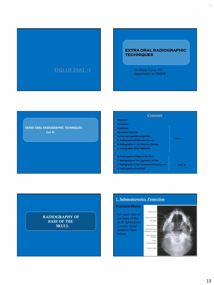
1. Submentovertex Projection
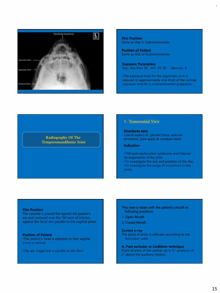
Structures Shown
Full axial view of
the base of the
skull, Sphenoidal
Sinuses, facial
skeleton from
below.
.
14
Indications
•Destructive or expansive lesions affecting palate,
pterygoid region, base of the skull, Sphenoidal
sinus.
•Warning : Rule out cervical spine fracture or
subluxation on trauma patient before attempting this
projection.
Film Placement
The cassette is placed perpendicular to the floor in a
cassette holding device. The long axis of the cassette
is placed vertically.
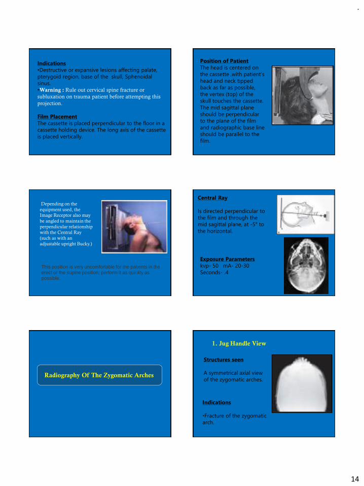
Position of Patient
The head is centered on
the cassette ,with patient’s
head and neck tipped
back as far as possible,
the vertex (top) of the
skull touches the cassette.
The mid sagittal plane
should be perpendicular
to the plane of the film
and radiographic base line
should be parallel to the
film.
Depending on the
equipment used, the
Image Receptor also may
be angled to maintain the
perpendicular relationship
with the Central Ray
(such as with an
adjustable upright Bucky.)
This position is very uncomfortable for the patients in the erect or the supine position; perform it as quickly as possible.
Central Ray
Is directed perpendicular to
the film and through the
mid sagittal plane, at -5° to
the horizontal.
Exposure Parameters
kvp- 50 mA- 20-30
Seconds- .4
Radiography Of The Zygomatic Arches
Structures seen
A symmetrical axial view
of the zygomatic arches.
1. Jug Handle View
Indications
•Fracture of the zygomatic
arch.
.
15
Film Position
Same as that in Submentovertex.
Position of Patient
Same as that of Submentovertex
Exposure Parameters
Kvp- less than 50 mA -20-30 Seconds .4
•The exposure time for the zygomatic arch is
reduced to approximately one-third of the normal
exposure time for a submentovertex projection.
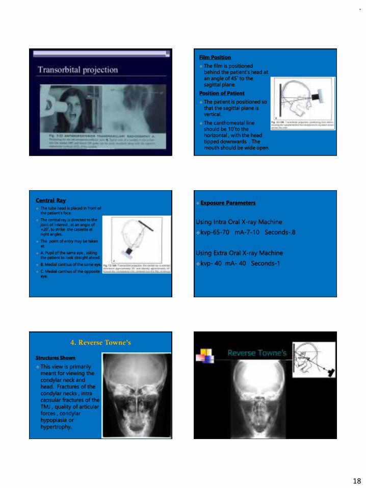
Radiography Of The
Temporomandibular Joint
Structures seen
Lateral aspect of: glenoid fossa, articular
eminence, joint space & condylar head
1. Transcranial View
Indication
•TMJ pain dysfunction syndrome and internal
derangements of the joint.
•To investigate the size and position of the disc.
•To investigate the range of movement in the
joints.
Film Position
The cassette is placed flat against the patient’s
ear and centered over the TM Joint of interest ,
against the facial skin parallel to the sagittal plane
Position of Patient
•The patient’s head is adjusted so that sagittal
plane is vertical.
•The ala- tragal line is parallel to the floor
Central x-ray
The point of entry is different according to the
technique used.
A. Post auricular or Lindblom technique
Point of entry of the central ray is ½” posterior &
2” above the auditory meatus.
.
16
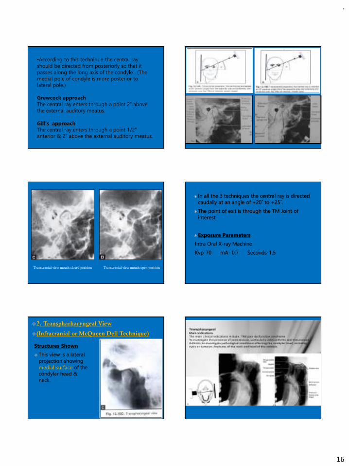
•According to this technique the central ray
should be directed from posteriorly so that it
passes along the long axis of the condyle . (The
medial pole of condyle is more posterior to
lateral pole.)
Grewcock approach
The central ray enters through a point 2” above
the external auditory meatus.
Gill’s approach
The central ray enters through a point 1/2”
anterior & 2” above the external auditory meatus.
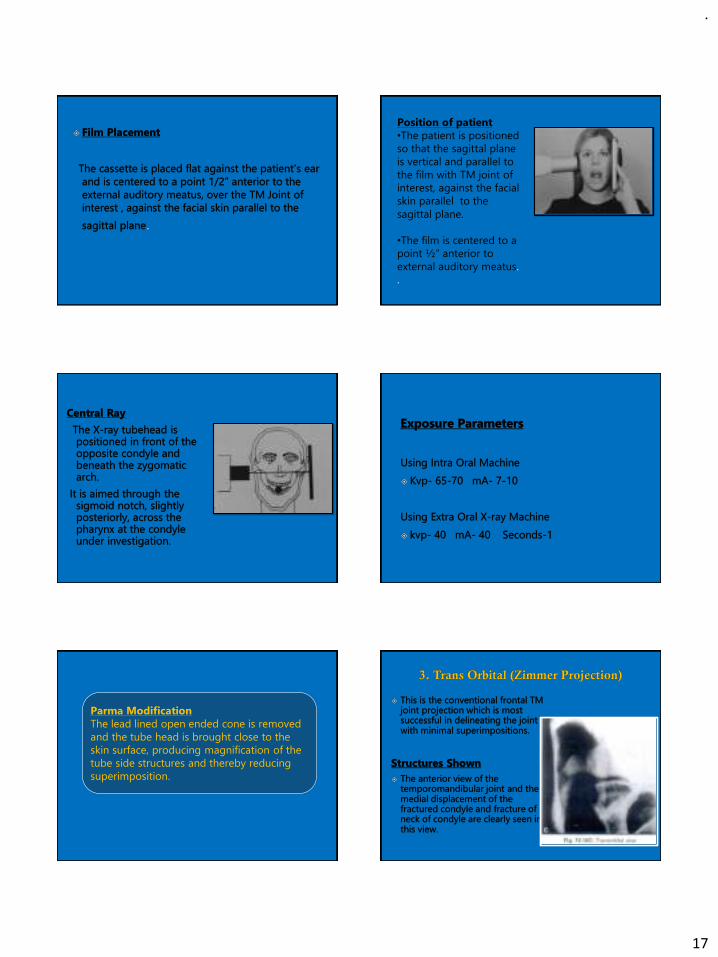
Transcranial view mouth closed position Transcranial view mouth open position
.
17
Position of patient
•The patient is positioned
so that the sagittal plane
is vertical and parallel to
the film with TM joint of
interest, against the facial
skin parallel to the
sagittal plane.
•The film is centered to a
point ½” anterior to
external auditory meatus.
.
Parma Modification
The lead lined open ended cone is removed
and the tube head is brought close to the
skin surface, producing magnification of the
tube side structures and thereby reducing
superimposition.
.
18
.
19
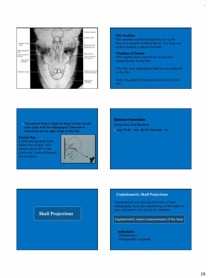
Film Position
The cassette is placed perpendicular to the
floor in a cassette holding device. The long axis
of the cassette is placed vertically.
•Position of Patient
•The sagittal plane should be vertical and
perpendicular to the film.
•The film is so adjusted so that lips are centered
to the film.
•Only the patient’s forehead should touch the
film.
Central Ray
Is directed upwards from
below the occiput, with
central ray at 30° to the
horizontal, centred through
the condyles.
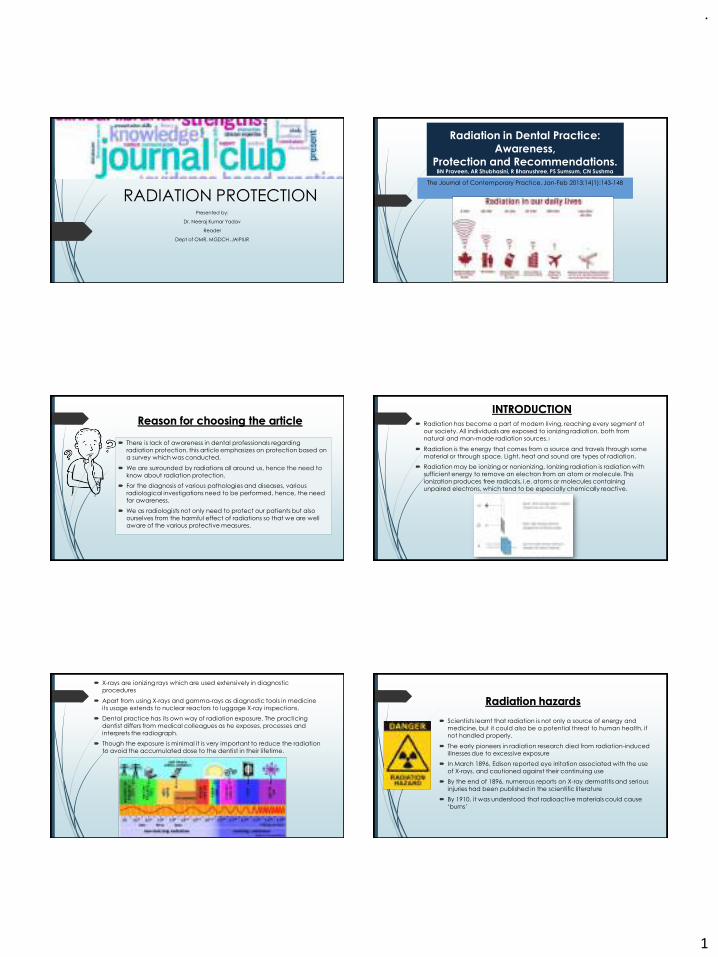
Skull Projections
Cephalometric Skull Projections
Standardized and reproducible form of skull
radiography, to access relationship of the teeth to
jaws and jaws to the rest of the skeleton.
Indications
Orthodontics
Orthognathic surgeries
Cephalometric means measurements of the head
.
20
A) Orthodontics
B) Orthognathic surgery
1. Pre operative evaluation
2. Assist in treatment planning
3. Post operative appraisal of the results of surgery
4. Follow up studies
1. Initial diagnosis
2. Treatment planning
3. Monitoring treatment progress
4. Appraisal of the treatment
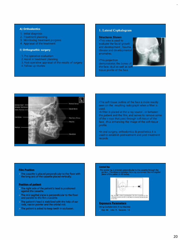
1. Lateral Cephalogram
Structures Shown
•This view is used to
evaluate the facial growth
and development , trauma ,
disease and developmental
anomalies.
•This projection
demonstrates the bones of
the face, skull as well as soft
tissue profile of the face.
•The soft tissue outline of the face is more readily
seen on the resulting radiograph when a filter is
used.
•A filter is placed at the x-ray source , or between
the patient and the film, and serves to remove some
of the x-rays that pass through soft tissue of the
face , thus enhancing the image of the soft tissue
profile.
•In oral surgery, orthodontics & prosthetics it is
used to establish pretreatment and post treatment
records.
.
21
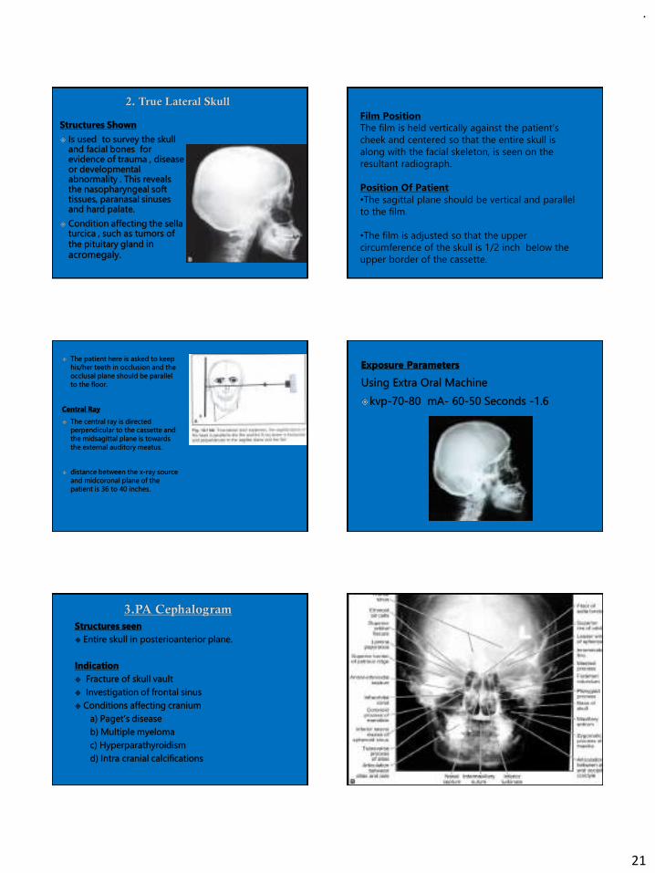
Film Position
The film is held vertically against the patient’s
cheek and centered so that the entire skull is
along with the facial skeleton, is seen on the
resultant radiograph.
Position Of Patient
•The sagittal plane should be vertical and parallel
to the film.
•The film is adjusted so that the upper
circumference of the skull is 1/2 inch below the
upper border of the cassette.
.
22
Film Position
The film is held vertically against the patient’s cheek
and centered so that the entire skull is along with
the facial skeleton, is seen on the resultant
radiograph.
Position of Patient
The sagittal plane should be vertical & perpendicular
to the film.
The head is tipped downwards so that only the nose
touches the film. The radiographic base line is 10˚
with the film.
Central Ray
Is directed at right angles
to the film through the mid
sagittal plane, centered at
the level of the bridge of
the nose.
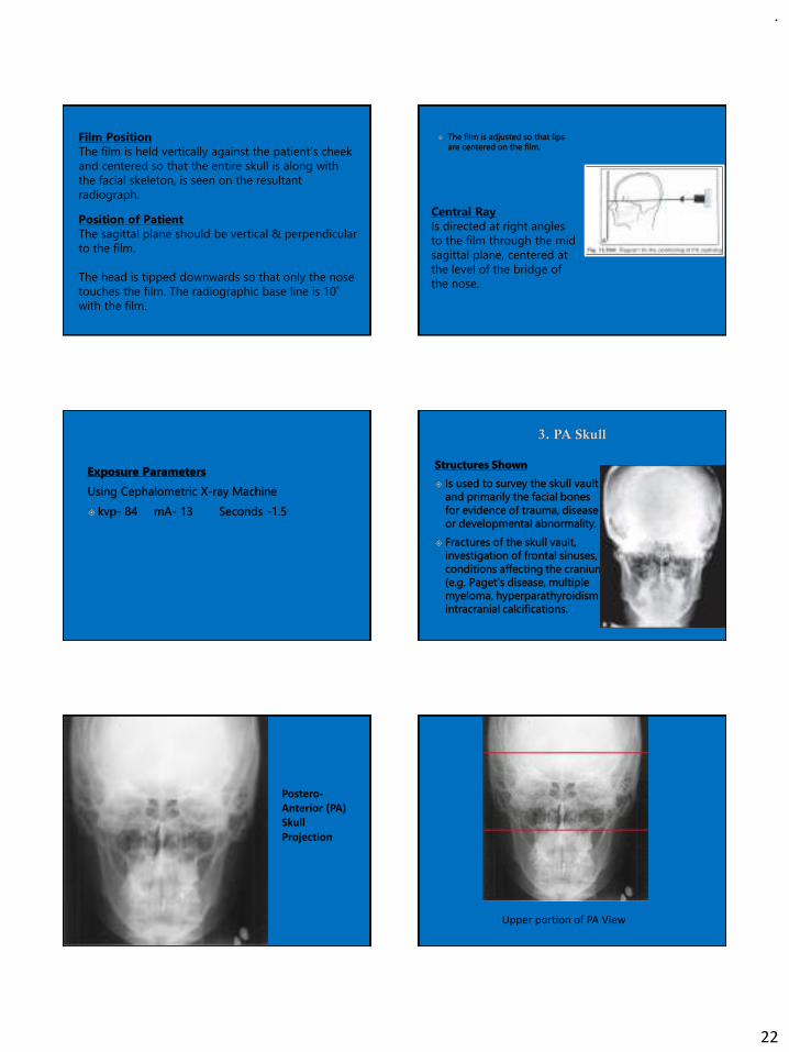
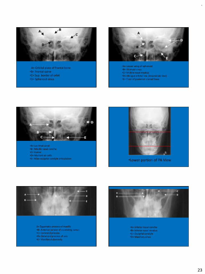
Postero-Anterior (PA) Skull Projection
Upper portion of PA View
.
23
A= Orbital plate of frontal bone•B= Frontal spine•C= Sup. border of orbit•D= Sphenoid sinus
•A= Lesser wing of sphenoid•B= Ethmoid sinus•C= Middle nasal meatus•D= Oblique orbital line (innominate line)•E= Floor of posterior cranial fossa
•A= Lacrimal canal•B= Middle nasal concha•C= Vomer•D= Mastoid air cells•E= Atlas-occipital condyle articulation •Lower portion of PA View
A= Zygomatic process of maxilla•B= Anterior border of ascending ramus•C= Coronoid process•D= Odontoid process of axis•E= Maxillary tuberosity
•A= Inferior nasal concha•B= Inferior nasal meatus•C= Occipital condyle•D= Maxillary sinus
.
24
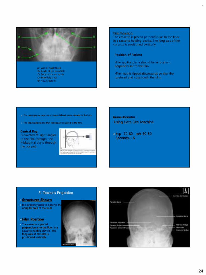
A= Wall of nasal fossa•B= Angle of the mandible•C= Body of the mandible•D= Maxillary sinus•E= Nasal septum
Film Position
The cassette is placed perpendicular to the floor
in a cassette holding device. The long axis of the
cassette is positioned vertically.
Position of Patient
•The sagittal plane should be vertical and
perpendicular to the film.
•The head is tipped downwards so that the
forehead and nose touch the film.
Central Ray
Is directed at right angles
to the film through the
midsagittal plane through
the occiput.
.
25
Thank you
.
1
RADIATION PROTECTIONPresented by:
Dr. Neeraj Kumar Yadav
Reader
Dept of OMR, MGDCH ,JAIPIUR
Radiation in Dental Practice:
Awareness,Protection and Recommendations.
BN Praveen, AR Shubhasini, R Bhanushree, PS Sumsum, CN Sushma
The Journal of Contemporary Practice, Jan-Feb 2013;14(1):143-148
Reason for choosing the article
There is lack of awareness in dental professionals regarding
radiation protection, this article emphasizes on protection based on
a survey which was conducted.
We are surrounded by radiations all around us, hence the need to
know about radiation protection.
For the diagnosis of various pathologies and diseases, various
radiological investigations need to be performed, hence, the need
for awareness.
We as radiologists not only need to protect our patients but also
ourselves from the harmful effect of radiations so that we are well
aware of the various protective measures.
INTRODUCTION Radiation has become a part of modern living, reaching every segment of
our society. All individuals are exposed to ionizing radiation, both from
natural and man-made radiation sources.1
Radiation is the energy that comes from a source and travels through some
material or through space. Light, heat and sound are types of radiation.
Radiation may be ionizing or nonionizing. Ionizing radiation is radiation with
sufficient energy to remove an electron from an atom or molecule. This
ionization produces free radicals, i.e. atoms or molecules containing
unpaired electrons, which tend to be especially chemically reactive.
X-rays are ionizing rays which are used extensively in diagnostic
procedures
Apart from using X-rays and gamma-rays as diagnostic tools in medicine
its usage extends to nuclear reactors to luggage X-ray inspections.
Dental practice has its own way of radiation exposure. The practicing
dentist differs from medical colleagues as he exposes, processes and
interprets the radiograph.
Though the exposure is minimal it is very important to reduce the radiation
to avoid the accumulated dose to the dentist in their lifetime.
Radiation hazards
Scientists learnt that radiation is not only a source of energy and
medicine, but it could also be a potential threat to human health, if
not handled properly.
The early pioneers in radiation research died from radiation-induced
illnesses due to excessive exposure
In March 1896, Edison reported eye irritation associated with the use
of X-rays, and cautioned against their continuing use
By the end of 1896, numerous reports on X-ray dermatitis and serious
injuries had been published in the scientific literature
By 1910, it was understood that radioactive materials could cause
‘burns’
.
2
By the 1920s, sufficient direct evidence and indirect evidence had
been accumulated to persuade the scientific community that an official body should be established to make recommendations
concerning human protection against exposure to X-rays and
radium.2-4
The International Commission of Radiation Protection (ICRP) is the
international regulatory body, formed in 1928 to lay down norms
for protection against radiation and recommend dose limits for radiation workers and general public.
The Indian regulatory board for protection against radiation is
AERB, Atomic Energy Regulatory Board which was constituted on November 15, 1983. The mission of the boards is to ensure that the
use of ionizing radiation and nuclear energy in India does not cause undue risk to health and surroundings.5
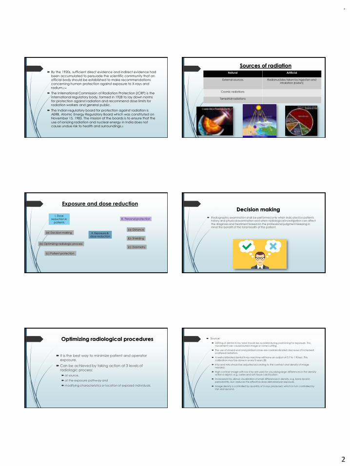
Sources of radiationNatural Artificial
External sources Radionuclides taken by ingestion and inhalation (radon)
Cosmic radiations
Terrestrial radiations
Exposure and dose reduction
I. Dose reduction in
patients
II. Exposure & dose reduction
III. Personal protection
(a) Decision making
(b) Optimizing radiologic process
(c) Patient protection
(a) Distance
(b) Shielding
(c) Dosimetry
Decision making
Radiographic examination shall be performed only when indicated by patients history and physical examination and when radiological investigation can affect
the diagnosis and treatment based on the professional judgment keeping in mind the benefit of the total health of the patient.
Optimizing radiological procedures
It is the best way to minimize patient and operator exposure.
Can be achieved by taking action at 3 levels of radiologic process:
at source,
at the exposure pathway and
modifying characteristics or location of exposed individuals.
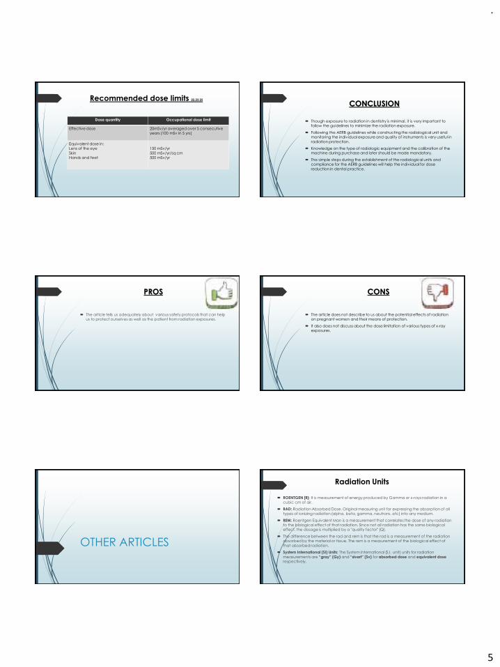
Source:
Drifting of dental X-ray tube should be avoided during positioning for exposure. This movement can cause blurred image or cone-cutting.
The use of closed end and pointed cones are contraindicated, because of increased scattered radiation.
A well-calibrated dental X-ray machine will have an output of 0.7 to 1 R/sec. This calibration must be done in every 3 years.(8)
kVp and mAs should be adjusted according to the contrast and density of image needed.
High contrast image with low kVp are used for visualizing large differences in the density within a object, e.g. caries and soft tissue calcification.
Increased kVp, allows visualization of small differences in density, e.g. bone level in periodontitis, but, reduces the effective dose delivered per exposure.
Image density is controlled by quantity of X-rays produced, which in turn controlled by mA and second.
.
3
Collimation:
use of rectangular open ended PID (3.5 ×4.4 cm) reduces the skin exposure by 60% than that of round (7 cm) PID.(13)
Focal spot film distance (FSFD)–when X-ray machine is operated above 50 kVp, source skin distance must be greater than 7 inches.(9)
Studies shows that 16 inch FSFD decreases 38% of thyroid dose, at 90 kVp and 45% decrease in 70 kVp, compared to 8 inch FSFD.(10)
This is because at the greater distance X-ray beam is less divergent and there will be 32% reduction in exposed tissue volume.(11)
The use of longer FSFD also results in a smaller apparent focal spot size and thereby increases the resolution of radiograph.(12)
Technique:
Paralleling technique gives more accurate image and lowers the exposure dose to thyroid gland and lens of eye.
In bisecting technique X-ray beam has steep vertical angulations that may put the thyroid gland and lens in the path primary as well as secondary radiation.(8)
Increasing FSFD and rectangular collimation may result in 70 to 80% decrease in exposure.(14)
Receptor selection:
It is advised to use maximum sensitive film (speed) consistent with image quality.
E (Ektaspeed) speed film is almost twice as fast as D speed films.(15)
In 1994 improved E speed film (Ektaspeed plus) was introduced, which was found to be faster, less sensitive to processing and less grainy than E speed film and have high contrast similar to D speed films.(16)
Dose reduction of 60% compared with E speed film can be achieved by using digital intraoral radiography. When compared with film, resolution was significantly lower in RVG whereas exposure reduction was to approximately half of Ektaspeed Plus.(24)
Similarly digital panoramic imaging has been reported to result in dose reduction of 70%.(1)
Processing and interpretation of images:
Thirty-percent of all retakes are because of the incorrect film density, directly related to processing variability.17
Radiographic images should be viewed under proper condition with illuminated viewer to attain maximum available information.
Quality radiographs reduces retaking and unnecessary second exposure.1,8
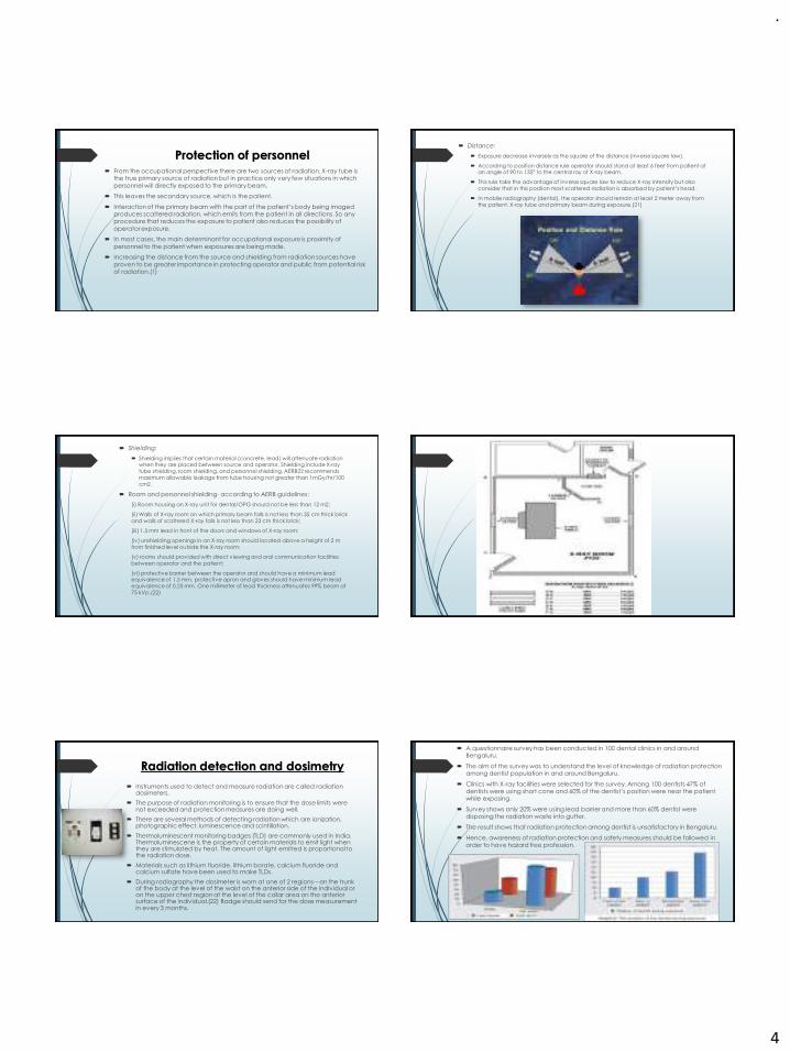
Patient protection
Stabilization of patient head before the exposure decrease blurring and cone-cutting of the image.
All radiation exposure must be based on the principle ALARA (as low as reasonably achievable).
Mean exposure at skin entrance for single periapical film is 217 mR and gonad dose will be 1/10,000 of total beam exposure (0.02 mR).(18)
Lead aprons reduce 98% of scattered radiation and attenuate dose to a 0.04 μR.(19) This quantity is 60 times less than the dose equivalent resulting from one airline flight.(1)
Thyroid collar attenuate 92% of scattered radiation.(20) So it should be made mandatory to use thyroid collar and lead aprons before any
exposure.
Film holders avoid unnecessary exposure to patient’s fingers. Patients exposure history must be maintained and updated after every exposure.(8)
The greatest risk to the fetus for chromosomal abnormalities and subsequent mental retardation is between 8 and 15 weeks of pregnancy. So the examination
involving radiation to the fetus should be avoided during this period. In second and third trimester, radiologic examination is advised, if it can alter the diagnosis and treatment planning and it is mandatory to use lead aprons and other dose
reduction procedures.(1)
.
4
Protection of personnel From the occupational perspective there are two sources of radiation, X-ray tube is
the true primary source of radiation but in practice only very few situations in which
personnel will directly exposed to the primary beam.
This leaves the secondary source, which is the patient.
Interaction of the primary beam with the part of the patient’s body being imaged produces scattered radiation, which emits from the patient in all directions. So any procedure that reduces the exposure to patient also reduces the possibility of
operator exposure.
In most cases, the main determinant for occupational exposure is proximity of
personnel to the patient when exposures are being made.
Increasing the distance from the source and shielding from radiation sources have
proven to be greater importance in protecting operator and public from potential risk of radiation.(1)
Distance:
Exposure decrease inversely as the square of the distance (inverse square law).
According to position distance rule operator should stand at least 6 feet from patient at an angle of 90 to 135° to the central ray of X-ray beam.
This rule take the advantage of inverse square law to reduce X-ray intensity but also consider that in this position most scattered radiation is absorbed by patient’s head.
In mobile radiography (dental), the operator should remain at least 2 meter away from the patient, X-ray tube and primary beam during exposure.(21)
Shielding:
Shielding implies that certain material (concrete, lead) will attenuate radiation when they are placed between source and operator. Shielding include X-ray tube shielding, room shielding, and personnel shielding. AERB22 recommends maximum allowable leakage from tube housing not greater than 1mGy/hr/100 cm2.
Room and personnel shielding- according to AERB guidelines:
(i) Room housing an X-ray unit for dental/OPG should not be less than 12 m2;
(ii) Walls of X-ray room on which primary beam falls is not less than 35 cm thick brick and walls of scattered X-ray falls is not less than 23 cm thick brick;
(iii) 1.5 mm lead in front of the doors and windows of X-ray room;
(iv) unshielding openings in an X-ray room should located above a height of 2 m from finished level outside the X-ray room;
(v) rooms should provided with direct viewing and oral communication facilities between operator and the patient;
(vi) protective barrier between the operator and should have a minimum lead equivalence of 1.5 mm, protective apron and gloves should have minimum lead equivalence of 0.25 mm. One millimeter of lead thickness attenuates 99% beam at 75 kVp.(22)
Radiation detection and dosimetry
Instruments used to detect and measure radiation are called radiation dosimeters.
The purpose of radiation monitoring is to ensure that the dose limits were not exceeded and protection measures are doing well.
There are several methods of detecting radiation which are ionization, photographic effect, luminescence and scintillation.
Thermoluminescent monitoring badges (TLD) are commonly used in India. Thermoluminescene is the property of certain materials to emit light when they are stimulated by heat. The amount of light emitted is proportional to the radiation dose.
Materials such as lithium fluoride, lithium borate, calcium fluoride and calcium sulfate have been used to make TLDs.
During radiography the dosimeter is worn at one of 2 regions—on the trunk of the body at the level of the waist on the anterior side of the individual or on the upper chest region at the level of the collar area on the anterior surface of the individual.(22) Badge should send for the dose measurement in every 3 months.
A questionnaire survey has been conducted in 100 dental clinics in and around Bengaluru.
The aim of the survey was to understand the level of knowledge of radiation protection among dentist population in and around Bengaluru.
Clinics with X-ray facilities were selected for the survey. Among 100 dentists 47% of dentists were using short cone and 60% of the dentist’s position were near the patient while exposing.
Survey shows only 20% were using lead barrier and more than 60% dentist were disposing the radiation waste into gutter.
The result shows that radiation protection among dentist is unsatisfactory in Bengaluru.
Hence, awareness of radiation protection and safety measures should be followed in order to have hazard free profession.
.
5
Recommended dose limits 22,23,25
Dose quantity Occupational dose limit
Effective dose 20mSv/yr averaged over 5 consecutive years (100 mSv in 5 yrs)
Equivalent dose in:Lens of the eye
SkinHands and feet
150 mSv/yr
500 mSv/yr/sq cm500 mSv/yr
CONCLUSION
Though exposure to radiation in dentistry is minimal, it is very important to follow the guidelines to minimize the radiation exposure.
Following the AERB guidelines while constructing the radiological unit and monitoring the individual exposure and quality of instruments is very useful in
radiation protection.
Knowledge on the type of radiologic equipment and the calibration of the machine during purchase and later should be made mandatory.
The simple steps during the establishment of the radiological units and compliance for the AERB guidelines will help the individual for dose
reduction in dental practice.
PROS
The article tells us adequately about various safety protocols that can help us to protect ourselves as well as the patient from radiation exposures.
CONS
The article does not describe to us about the potential effects of radiation on pregnant women and their means of protection.
It also does not discuss about the dose limitation of various types of x-ray exposures.
OTHER ARTICLES
Radiation Units
ROENTGEN [R]: It is measurement of energy produced by Gamma or x-rays radiation in a cubic cm of air.
RAD: Radiation Absorbed Dose. Original measuring unit for expressing the absorption of all types of ionizing radiation (alpha, beta, gamma, neutrons, etc) into any medium.
REM: Roentgen Equivalent Man is a measurement that correlates the dose of any radiation to the biological effect of that radiation. Since not all radiation has the same biological effect, the dosage is multiplied by a "quality factor" (Q).
The difference between the rad and rem is that the rad is a measurement of the radiation absorbed by the material or tissue. The rem is a measurement of the biological effect of
that absorbed radiation.
System International (SI) Units: The System International (S.I. unit) units for radiation
measurements are “gray” (Gy) and “sivert” (Sv) for absorbed dose and equivalent dose respectively.
.
6
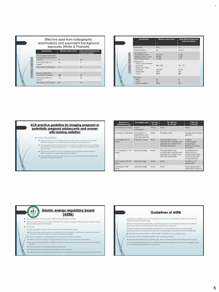
Effective dose from radiographic
examinations and equivalent background exposures (White & Pharoah)
Examination Effective dose (mSv) Equivalent background exposure (days)
Rectangular collimationPosterior bitewing: PSP or
F speed 5 0.6
Full mouth: PSP or F speed
35 4
Full mouth: CCD sensor 17 2
Round collimationFull mouth: D speed 388 46
Full mouth: PSP or F speed
171 20
Full mouth: CCD sensor 85 10
IN
TRA
O
RAL
EX
TRA
O
RAL
Examination Effective dose (mSv) Equivalent background exposure (days)
Panaromic 9-24 1-3
Cephalometric 2-6 0.3-0.7
Cone-beam CT- Long field of view
- Medium field of view- Small field of view
68-1073
45-86019-652
8-126
5-1012-77
Multislice CT- Head: Conventional
protocol- Head: Low dose
protocol
- Abdomen- Chest
860-1500
180-534
53005800
101-177
21-63
624682
Plain films- Skull
- Chest- Barium enema
70
207200
8
2847
ACR practice guideline for imaging pregnant or
potentially pregnant adolescents and women with ionizing radiation
Goals of the guidelines:
Outline the body of knowledge about the risks to the conceptus from ionizing radiation, taking into account the gestation age at time of exposure.
Provide guidance as to when and how to screen for pregnancy prior to imaging examinations using ionizing radiation, including evaluation of the adolescent and adult woman
Recommend means to control, manage and practicably minimize radiation dose to pregnant or potentially pregnant patients
Manage dose assessment, risk assessment and communication issues following exposure of pregnant patients.
Menstrual or Gestational age
Conception age <50 mGy(5 rad)
50-100mGy(5-10 rad)
>100 mGy(>10 rad)
0-2 weeks (0-14 days) Prior to conception
None None None
3-4 week (15-28 days) Conception to implantation and
preorganogenesis
None Probably none Possible spontaneous abortion
5-10 weeks (29-70 days)
Embryonic stage None Potential effects are scientifically uncertain and
probably too subtle to be clinically detected
Possible malformations
increasing in likelihood as dose increases
11-17 weeks (71-119 days)
Early fetal stage None Potential effects are scientifically uncertain and
probably too subtle to be clinically detectable
Increased risk of deficit of IQ or
mental retardation that increase with increasing dose
18-27 week (120-189 days)
Mid fetal stage None None IQ deficits not detectable
>27 weeks (>189 days)
Late fetal stage None None None applicable to diagnostic medicine
Atomic energy regulatory board
(AERB) Constituted on 14th November 1983 by the president of India.
Mission: To ensure use of ionizing radiation & nuclear energy in India does not cause undue
risk to health and environment.
Functions:
Develop safety policies in both radiation and industrial safety areas
Develop safety codes, guides and standards for sitting, design, construction, operation and grants consents of different types of nuclear and radiation facilities.
Review emergency preparedness plans for nuclear & radiation facilities
Safety reviews for transport of large radioactive sources and irradiated fuel material
Review of training program, syllabus for training, qualification and license for personnel of radiation facilities
Public informed on radiological safety significance
Promote research ad development effort in areas of safety
Maintain liaison with statutory bodies in the own country as well as abroad regarding safety matters.
Guidelines of AERB
1) Decide a suitable room for housing an X-ray unit to facilitate the easy movement of staff and patient positioning.
2) Room should have preferably one entrance door and window if present, should be above 2m from the finished floor level outside the x-ray room.
3) Door should have a hydraulic mechanism to ensure that door is closed during procedure and should be provided with overlapping at the joints to avoid streaming.
4) Identify the walls as Wall A, Wall B, Wall C & Wall D (in any sequence)
5) Decide on the material and thickness of walls and door
6) Measure the distances of all the walls, doors, windows from the centre of the couch
7) The required shielding of any material shall be provided at least up to the height of 2m
from external finished floor of x-ray room

.
7
8) Position the location of the equipment for each modality as follows:
a) Radiography and Fluoroscopy equipment:
• Couch, Control console and chest stand placed in such a way that chest stand is on the opposite wall of the entrance door and the control console.
• Mobile protective barrier with lead equivalent glass viewing window should be positioned in such a manner that the operator is completely shielded during the
exposure.
• Control console should be positioned as far away as possible from the x-ray tube.
b) Computed Tomography and Interventional radiology equipment: Gantry/ C-Arm, Couch, Separate control console room, viewing window, -Position the gantry and couch such
that the patient is completely visible from the control console, during the scanning - The entrance door to the gantry room from the control console shall have similar requirements as the patient entrance door.
c) Mammography/ OPG/ CBCT: Control console, Equipment and Protective barrier Positioning of equipment should be as far as possible from the door and the control console.
PLANNING OF DIAGNOSTIC X-RAY
INSTALLATIONS
ROOM SIZE
• The room housing an X-ray unit shall be not less than 18 m2 for general purpose
radiography and conventional fluoroscopy equipment.
• The size of the room housing the gantry of the CT unit shall not be less than 25 m2.
• Also, not more than one unit of any type shall be installed in the same room, and no single dimension of these X-ray rooms shall be less than 4 m.
• In the case of mammography, the room size shall be not less than 10 m2, and no single dimension of the room shall be less than 3 m.
WALL THICKNESS
• If the X-ray installation is located in a residential complex, it shall be ensured that:
• 1. Walls of the X-ray rooms on which primary X-ray beam falls are not less than 35 cm or 14 inch thick brick or equivalent.
• 2. Walls of the X-ray room on which scattered X-ray fall are not less than 23 cm or 9 inch thick brick or equivalent.
• 3. There is a shielding equivalent to at least 23 cm or 9 inch thick brick or 2 mm lead in front of the door(s) and windows of the X-ray room to protect the adjacent areas,
either by general public or not under possession of the owner of the X-ray room. The density of the normal masonary brick is considered as 1.6 g/cc.
• 4. The ceiling must have a thickness of concrete (density 2.35 g/cc), not less than 6 inch or 13.5 cm.
OPTIONS IN SHIELDING MATERIALS
• X-ray equipment must be installed in adequately shielded rooms to ensure that public in the vicinity of the x-ray installations are not unduly exposed to x-ray radiation.
• The adequacy of shielding depends on the material and thickness used for this purpose. Different materials can be used for shielding.
• However, brick or concrete are considered the best materials, as they are easily available, economical, and have good structural strength, While lead is a suitable shielding option for energies encountered in diagnostic x-rays, it is a weak structural material with tendency to lose uniformity and needs periodic radiation survey to ensure its continued adequacy.
• Also, Lead poses a serious environmental hazard and the use of it is being discouraged the world over. Recently, many new materials are being used/ developed as potential shielding materials, as an alternate to Lead.
• AERB would like to promote use of these materials, on demonstration of shielding adequacy
CONTROL ROOM
• For equipment operating at 125 kV or above, should have a separate control room, and provided with appropriate shielding, direct viewing (1.5 mm lead equivalence) and oral communication facilities between the operator and the patient.
• The X-ray units operating below 125 kVp in diagnostic radiology are exempted from the above class.
• In such a case, the control should be behind a mobile protective barrier of adequate thickness.
DOORS
• Doors are lined with 2 mm thick lead sheet with proper overlapping at the joint and junction and wall of 9 inch thickness of brick and ceiling of 6 inch of concrete
• Viewing Window
• Lead glass of suitable dimensions are provided as viewing windows with 1.5 mm thick lead equivalent.
• Mobile Protective Barrier
• Control panel should be kept behind the mobile protective barrier (MBP) of thickness 2 mm lead equivalence
GENERAL RADIOGRAPHY INSTALLATION
• These X-ray units are operated in the range of 50–150 kVp.
• Walls that are irradiated directly by the X-ray beam are primary barriers.
• Hence, additional shielding must be provided for the wall behind the chest stand.
• Provisions are made to observe and communicate with the patient on the table.
• The mobile protective barrier with lead shield must be a permanent/mobile one with 2.1 m height.
• The viewing window at the mobile protective barrier must be 45 × 45 cm size and centered

.
8
AERB GUIDELINES FOR DOSE LIMITS
The limits on effective dose apply to the sum of effective doses from external as well as internal sources. The limits exclude the exposures due to natural background radiation and medical exposures.
Calendar year shall be used for all prescribed dose limits
Occupational exposures
1. An effective dose of 20 mSv/yr averaged over five consecutive years (calculated on a sliding scale of five years);
2. An effective dose of 30 mSv in any year;
3. An equivalent dose to the lens of the eye of 150 mSv in a year;
4. An equivalent dose to the extremities (hands and feet) of 500 mSv in a year and
5. An equivalent dose to the skin of 500 mSv in a year;
6. Limits given above apply to female workers also. However, once pregnancy is declared the equivalent dose limit to embryo/fetus shall be 1 mSv for the remainder of the pregnancy.
Apprentices and Trainees
The occupational exposure of apprentices and trainees between 16 and 18 years of
age shall be so controlled that the following limits are not exceeded:
1. An effective dose of 6 mSv in a year;
2. An equivalent dose to the lens of the eye of 50 mSv in a year;
3. An equivalent dose to the extremities (hands and feet) of 150 mSv in a year and
4. An equivalent dose to the skin of 150 mSv in a year.
Dose Limits for Members of the Public
1. An effective dose of 1 mSv in a year;
2. An equivalent dose to the lens of the eye of 15 mSv in a year; and
3. An equivalent dose to the skin of 50 mSv in a year
REFERENCES
1. White, Pharoah. Oral radiology principles and interpretation, (4th ed) Mosby 2000:42-61.
2. Radiation and risk—a hard look at data, a brief history of radiation. Los Alamos Science. 1995 Nov-23 available at http://www.fas.org/sgp/othergov/doe/lanl/00326631.pdf.
3. Kathren RK. Pathway to paradigm, the linear non threshold dose response model in historical context. Health Phys 1995; 70(5):621-35.
4. Edison TA. NOTE, Nature 1987;53:421.
5. Narayanan P. Radiological safety in health care: Guidelines, practice and outcome. Medical Physics Chronicle 2010 Jan;2(1).Ramachandra TV, Eappen KP, Nair RN. Environmental assessment division, atomic research center. Background exposure levels. Indian scenario. Available at http:// www.dae.gov.in/ni/nifeb05/PDF/03Indian_scenario.pdf
6. United Nations Scientific Committee on the effects of Atomic Radiation (UNSCEAR) 2008 report to general assembly. Available at http://www.unscear.org/unscear/en/publications/2008_1.html.
7. Frommer, Savage S. Radiology for the dental professional (8th ed) Elsevier Mosby 2005:78-104.
8. Code of federal regulations 21, sub chapter j: Radiological part 1000, Washington DC. 1994. Available at http:// www.glenbrooktech.com/radiation-safety.php
9. Gibb SJ, et al patient risk from intraoral dental radiograph, J Dento Maxillo Facial Radiology 1988;17:25.
10. Frederikser NL. The radiograph in the diagnosis of periodontal disease, a quality assurance programme, technology assessment. Forum in Dental Radiology 1982;82:107.
11. Platin, et al. Effects of focal spot size on caries diagnosis with D and E speed images. Oral Surg Oral Med Oral Pathol Oral Radiol Endo 1996;81:235.
12. White SC. Assessment of radiation risks from dental radiography J Dento Maxillo Facial Radiology 1992;21:118.
13. Lederberg RA. et al. Effect of geometry of the intraoral PID on effective dose. Oral Surg Oral Med Oral Pathol Oral Radiol Endo 1997;84:101.
14. Richards, et al. Reduction in dental X-ray exposures during past 60 years. J Am Dent Assoc1981;103:713.
15. Ludlow, et al. Densitometric comparison of ultraspeed. Ektaspeed and ektaspeed plus intraoral films for 2 processing conditions. Oral Surg Oral Med Oral Pathol Oral Radiol Endo 1995;79: 105.
16. Goldman, et al. Automatic processing and quality assurance programme: Impact on radiology department. Radiology 1977; 125:591.
17. Richards AG. Roentgen ray doses in dental radiography. J Am Dent Assoc 1958;56:351.
18. Bean LR, Devore WD. The effect of protective aprons in dental Roentgenography. Oral Surg1969;28:505.
19. Sikorski PA, Taylor KW. The effectiveness of thyroid shield in dental radiography. Oral Surg 1989;68:776.
20. Seeram, Travis EC. Radiation protection Philadelphia. New York, Lippincott 1997.
21. AERB safety code (Code No. AERB/SE/MED-2) Mumbai 2001 (1-20).
22. International commission in radiological protection. The 2007 Recommendation of the International Commission on Radiological Protection, ICRP Publication 103. Ann ICRP 2007;37:1-332.
23. John Ludlow, et al. Image-receptor performance: A comparison of Trophy RVG UI sensor and kodak ektaspeed plus film. Oral Surg Oral Med Oral Pathol Oral Radiol Endod 2001;91: 109-19.
24. Le Heron J, Padovani R, Smith I. Renate Czarwinski Radiation protection of medical staff. European J Radiol 2010;76:20-23.
25. Nan DU, et al. Developing a wireless sensing method for the measurement of gamma radiation dose based on the polymerization of acrylamide. Radiation Measurements 2012; 47(5):371-74.
.
1
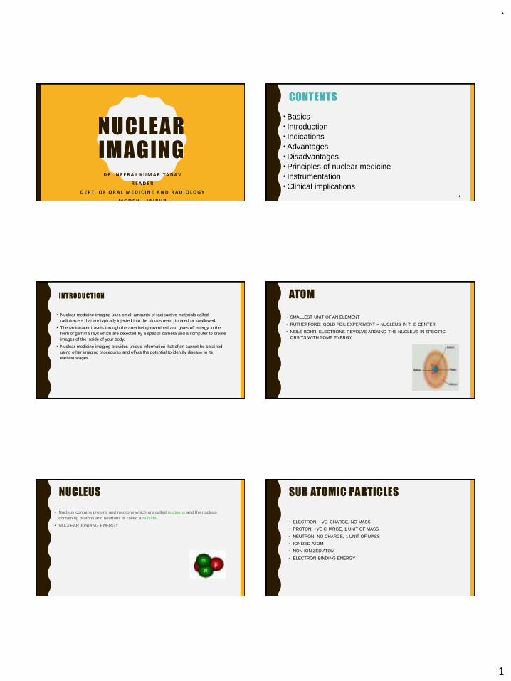
NUCLEAR IMAGING
D R . N E E R A J K U M A R YA D A V
R E A D E R
D E P T. O F O R A L M E D I C I N E A N D R A D I O L O G Y
M G D C H , J A I P U R
CONTENTS
• Basics• Introduction• Indications • Advantages• Disadvantages• Principles of nuclear medicine• Instrumentation• Clinical implications
2
INTRODUCTION
• Nuclear medicine imaging uses small amounts of radioactive materials called radiotracers that are typically injected into the bloodstream, inhaled or swallowed.
• The radiotracer travels through the area being examined and gives off energy in the form of gamma rays which are detected by a special camera and a computer to create images of the inside of your body.
• Nuclear medicine imaging provides unique information that often cannot be obtained using other imaging procedures and offers the potential to identify disease in its earliest stages.
ATOM
• SMALLEST UNIT OF AN ELEMENT
• RUTHERFORD: GOLD FOIL EXPERIMENT – NUCLEUS IN THE CENTER
• NEILS BOHR: ELECTRONS REVOLVE AROUND THE NUCLEUS IN SPECIFIC ORBITS WITH SOME ENERGY
NUCLEUS
• Nucleus contains protons and neutrons which are called nucleons and the nucleus containing protons and neutrons is called a nuclide.
• NUCLEAR BINDING ENERGY
SUB ATOMIC PARTICLES
• ELECTRON: –VE CHARGE, NO MASS
• PROTON: +VE CHARGE, 1 UNIT OF MASS
• NEUTRON: NO CHARGE, 1 UNIT OF MASS
• IONIZED ATOM
• NON-IONIZED ATOM
• ELECTRON BINDING ENERGY

.
2
7
• Isomer: Nucleus with different arrangements• Ground states: The most stable energy states• Excited states: Arrangements are so unstable that there is only a short
transient time (less than 10-12 sec) becoming ground states.• Metastable states: Arrangements are unstable, but relatively long-lived
(sometimes up to several hours) before becoming ground states.
8
• Elements containing atoms with same atomic number and different number of neutrons.
• A radionuclide is a radioactive form of an isotope that behaves chemically in a similarmanner to the nonradioactive counter part.
• The nuclear BE is not capable of holding the nucleus together and undergoesdisintegration releasing particulate or ionizing radiation.
Radioisotopes
9
RADIOACTIVE TRANSFORMATION
• Isobaric Transitions
– Beta Emission
– Positron Emission
– Electron Capture
• Isomeric transitions
– Gamma Emission
– Internal Conversion
– Excited state transitions
– Metastable state transitions
• Alpha transition
Neutron rich isotopes can decay by
Negative beta emission
Proton rich isotopes can decay by 2 modes
Electron capture
Positron emission
The result of the decay modes is a better balance betweenthe forces acting on the nucleus.
POSITRON EMISSION
• A positron is a particle similar to electron except that it has a positive electric charge.
• The behavior of positron in the tissue is very similar to β particles with one important difference – once the positron has been slowed down by the atomic collisions , it is annihilated by the interaction with an electron from a nearby atom.
• The combined mass of the proton & electron is converted into two annihilation photons – each with energy 511 KeV .
• The two photons are emitted at 180° to each other – this property is exploited by PET.

.
3
G A M MA E M IS S ION
• In most isomeric transitions, a nucleus will emit its excess energy in the form of a gamma photon.
• A gamma photon is a small unit of energy that travels with the speed of light and has no mass; its most significant characteristic is its energy.
• The photon energies useful for diagnostic procedures
are generally in the range of 100 keV to 500 keV.
ALPHA EMISSION
• An alpha particle consists of two neutrons and two protons.
• α particles interact strongly with matter – very short range of 1mm or less.
• Within this range α particles strongly collide with atoms – disrupting their chemistry – extremely damaging to the tissues.
• α particles have a potential to deliver a lethal radiation dose to small metastatic cell clusters, while mostly sparing the surrounding tissues.
SPONTANEOUS FISSION
• This is a very destructive process which occurs in some heavy nuclei which split into 2 or 3 fragments plus some neutrons.
• These fragments form new nuclei which are usually radioactive.
• Nuclear reactors exploit this phenomenon for the production of radioisotopes.
• It’s also used for nuclear power generation and in nuclear weaponry.
• Two protons and two neutrons are emitted together in a process called alpha-decay.
• A proton can release a positron in a process called beta-plus decay, and that a neutron can emit an electron in a process called beta-minus decay.
17
RADIOACTIVE DECAY is the process whereby the number of radioactive atoms of an element within a population is reduced through disintegration.
RADIOACTIVE PHYSICAL HALF LIFE is the time required for one half of the original number of atom in the radioactive sample to decay.
BIOLOGICAL HALF LIFE is the time required body to eliminate one half of an administered radio nuclide by a process of regular elimination.
EFFECTIVE HALF-LIFETime required for the radioactivity from an administered radio nuclide to be reduced to 50% of its initial value as a result of the combined effects of the physical and biologic half-lives.
18
DECAY RATE-• It is the number of nuclear disintegrations occurring per unit time –measuring
unit - curie
• The energy level of particulate or electromagnetic radiation is expressed in Kev or meV
• The radiation a patient receives is measured in “rads”.

.
4
Nuclear medicine
• Nuclear Medicine (e.g., PET,SPECT) is based on emission data from radioactive materials injected into the body
• Nuclear signals penetrated through the body are detected and reconstructed to form images
19
Pharmaceuticals that are labeled with radionuclides
Accumulate in organs of interest
Emit gamma radiation
Detection system sensitive to this obtain images
NUCLEAR MEDICINE VS DIAGNOSTIC RADIOLOGY
• Nuclear medicine and diagnostic radiology both utilizes ionizing radiation to obtain clinical information.
• Information with nuclear medicine is related to organ function while in diagnostic radiology information is related to anatomical structures.
21
Indications for nuclear imaging
• Tumor staging to assess metastasis
• Investigation of salivary gland function
• Evaluation of bone grafts
• Assessment of growth in condylar hyperplasia
• Investigation of thyroid and brain scans
22
• Bone scans to evaluate orthopedic injuries, fractures, tumors, or unexplained bone pain
• Heart scans to identify normal or abnormal blood flow to the heart muscle, measure heart function, or determine the existence or extent of damage to the heart tissues after a heart attack episode.
• Gallbladder or hepatobiliary scans to evaluate both liver as well as gallbladder function. This test can determine obstructions caused by the presence of gallstones.
• Lung scans to evaluate the fl ow of blood and movements into and out of the lungs, as well as the determination of the presence of blood clots.
INDICATION OF NUCLEAR MEDICINE IN DENTISTRY
• For the detection of active alveolar bone loss.
• For detection of viability of bone grafts
• For detection of osseointegration around dental Implants.
• For detection of salivary gland disorders
• For detection of function of lymph nodes

.
5
• For staging and grading of orofacial malignancies, colorectal cancer, non-small cell Lung cancer, Melanoma, Lymphomas, Head and Neck cancers.
• For determining the prognosis after treatment
• For evaluating the effect of Hyperbaric oxygen therapy in treatment of Bisphosphonate Induced osteoradionecrosis of the Jaws
Advantages
• NM imaging has an advantage of providing anatomical details and information thathelps in accurate diagnosis
• NM imaging is based on the principle of tracers and show functional images ofmetabolism, physiology or biochemistry by studying the dynamic behavior of tissuesand organs at various stages.
• It can help in early diagnosis of the disease and evaluates the outcome during theinitial posttreatment phase.
26
• By allowing easy demonstration of whole-body images and active display, NM helps indetecting metastatic activity.
• Different regions of the body can be examined at varied times using a single injectionof radioisotope in NM imaging without any increased radiation dose, which is unlikeother conventional techniques that need a number of exposures for examining differentanatomical locations
Disadvantages
• Poor image resolution• Radiation dose to the whole body can be high
• Images are not disease specific
• Difficult to localize exact anatomical site
• Some investigations take several hours
• Facilities are not widely available.
28
BRANCHES OF NUCLEAR MEDICINE
Imaging
Therapeutics Radioimmuno-assay
VARIOUS RADIOISOTOPES USED IN RADIONUCLIDE IMAGING
Radionuclides Half-life Uses
Technetium-99m 6 hrs Skeleton and heart muscle imaging, brain, thyroid, lungs (perfusion and ventilation), liver, spleen, kidney (structure and filtration rate), gall bladder, bone marrow, salivary and lacrimal glands, heart blood pool, infection
Xenon-133Krypton-81
5 days Used for pulmonary (lung) ventilation studies.
Ytterbium-169 32 days Used for cerebrospinal fluid studies in the brain.
Carbon-11 Nitrogen-13 Oxygen-15Fluorine-18
They are positron emitters used in PET for studying brain physiology and pathology, cardiology, detection of cancers and the monitoring of progress in their treatment.
Iodine-131 8 days Imaging of thyroid
Gallium-67 78 hrs Used for tumour imaging and localization of inflammatory lesions (infections).
Indium-111 2.8 days Used for brain studies, infection and colon transit studies
Rubidium-82 65 hrs PET agent in myocardial perfusion imaging
Thallium-201 73 hrs Used for diagnosis of coronary artery disease other heart conditions and for location of low-grade lymphomas.

.
6
Cyclotron Radioisotopes Uses
Carbon 11, nitrogen 13, oxygen 15, fluorine 18
Used in PET scans through use of F 18 in FDG which help to detect, diagnose and know the prognosis of a tumor
Cobalt 57 Can be used as a detector for hypertrophy of cells, which indirectly
determine the division and progression of cell growth to a stimulus
Copper 64 Is being used in PET scans and to check for the metabolism of copper
Copper 67 Used as an isotope for radiotherapy
Fluorine 18 as FLT
(fluorothymidine), F miso
(fluoromisonidazole), 18F choline
Used as traces, in radiotherapy
Gallium 67 For imaging of tumor size
Gallium 68 Used in PET scan to detect the metastatic activity
Germanium 68 Used to generate Ga
Rubidium 82 Used in PET scan to detect the cardiac myopathies
Strontium 82 Used to generate Rb 82
IDEAL PROPERTIES OF RADIONUCLIDES
• Detection : For external imaging of a radionuclide deposited within the body theenergy of gamma rays emitted should be high enough to be detected.
• High Energy : Energy should be somewhere within the range of 20 – 400 KeV.
• More energetic emission : For organs lying deeper within the body more energeticemissions are required.
• Half – life : The physical half – life should only be few hours and not much longerthan the time necessary to obtain the data.
• Easy availability : An ideal radionuclide should be readily available, at reasonablecost and in a sufficiently high specific gravity so that the administration of therequired dose of the radioactive substance does not produce a physiological, toxic orpharmacological response.
Radioisotopes used in conventional nuclear medicine
Technetium (99mTc) : The most commonly used isotope for the following reasons:
• Gamma emission : Single 141 KeV gamma emissions which are ideal for imaging purposes.
• Short half - life : A short half life of 6 ½ hours that ensures a minimal radiation dose.
• Readily attached to different substances : It can be readily attached to a variety of different substances that get concentrated in different organs . Eg: 99m Tc + MPD ( Methylene diphosphonate ) in bone , 99m Tc + RBC in blood , 99mTc + Sulphur colloid in the liver and spleen.
• Ionic form : It can be used on its own in its ionic form (pertechnetate 99m Tc O+) , since the thyroid and salivary glands take this up selectively.
• Easily produced : as and when required.
Technetium production
• FROM MOLYBDENUM by radioactive decay
RADIOPHARMACEUTICALS
Substances which tend to localize in the tissue of interest is tagged with gamma ray emitting radionuclide.
36
Technetium is administered
• Intravenously as Sodium pertechnetate -for salivary gland, brain, thyroid, gastric mucosal imaging.
• Chemical combination with sulphur colloid- for liver, spleen and bone marrow scanning
• Chelating agents -for brain and renal scanning
• Albumin macro particles- for perfusion lung scanning
• Phosphate complexes -for bone scanning

.
7
ROUTES OF ADMINISTRATION
• Injected into a vein• Swallowed • Inhaled as gas.
THE PROCEDURE
Pre-examination procedures:
Patient preparation:
• Explanation about the test
Pre-injection:
• Relevant history
• Current symptoms, physical findings.
• Results of previous radionuclide imaging
• Results of other imaging studies such as conventional radiographs, CT, MRI
• Relevant laboratory results
Radiopharmaceutical administration: The radiopharmaceutical should be administered by the intravenous route.
Post injection:
• Unless contraindicated, patients should be well-hydrated and instructed to drink one or more liters of water (4-8 glasses) between the time of injection and the time of imaging as well as during the 24 hours after administration.
• void frequently during the interval between injection and imaging.
Image acquisition:
• Between 2 and 5 hours after injection.
• Later (6-24 hour) delayed images (higher target-to-background ratio and may permit better evaluation)
Image Processing :
• No processing procedure is needed for planar images.
• In case of SPECT and PET one should consider the different types of gamma camera and software available: careful choice of imaging processing parameters should be adopted in order to optimize the imaging quality.
GAMMA IMAGING
The Device Components of a gamma camera
• Collimator
• Detector/ Scintillator
• Photomultiplier
COLLIMATOR• This is a device made of a highly absorbing material such as lead, which selects gamma
rays along a particular direction.
• They serve to suppress scatter and select a ray orientation.
• The simplest collimators contain parallel holes.

.
8
DETECTOR / SCINTILLATOR
• Made up of sodium iodide crystals.
• It produces multi-photon flashes of light when an impinging gamma ray, X-ray or charged particle interacts with the single sodium iodide crystal of which it is comprised.
• The scintillation counter not only detects the presence and type of particle or radiation but can
also measure their energy.
• The passage of gamma rays through the scintillator material excites electrons, which can
subsequently de-excite, emitting a photon.
Photomultiplier tube (PMT)
• This is an extremely sensitive photocell used to convert light signals of a few hundred photons into a usable current pulse
PULSE HEIGHT ANALYZER (PHA):
• It lets through only those pulses which lay within the window of ±10 % of the photopeak energy.
• The pulses so selected – ‘Counts’.
• The X Y Z pulses are next applied directly to a monitor for visual interpretation as in older machines or in newer systems via analogue-digital converters into a computer.
• This enables dynamic & gated studies to be undertaken as well as range of image processing.
.
9
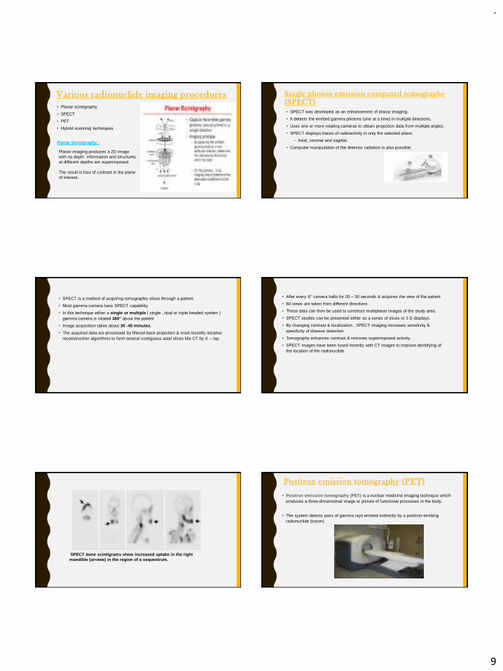
Various radionuclide imaging procedures
• Planar scintigraphy
• SPECT
• PET
• Hybrid scanning techniques
Planar Scintigraphy :
Planar imaging produces a 2D image with no depth information and structures at different depths are superimposed.
The result is loss of contrast in the plane of interest.
Single photon emission computed tomography (SPECT)
• SPECT was developed as an enhancement of planar imaging.
• It detects the emitted gamma photons (one at a time) in multiple directions.
• Uses one or more rotating cameras to obtain projection data from multiple angles.
• SPECT displays traces of radioactivity in only the selected plane.
– Axial, coronal and sagittal.
• Computer manipulation of the detector radiation is also possible.
• SPECT is a method of acquiring tomographic slices through a patient.
• Most gamma camera have SPECT capability.
• In this technique either a single or multiple ( single , dual or triple headed system ) gamma camera is rotated 360° about the patient
• Image acquisition takes about 30 -45 minutes.
• The acquired data are processed by filtered back projection & most recently iterative reconstruction algorithms to form several contiguous axial slices like CT by X – ray.
• After every 6° camera halts for 20 – 30 seconds & acquires the view of the patient
• 60 views are taken from different directions .
• These data can then be used to construct multiplanar images of the study area.
• SPECT studies can be presented either as a series of slices or 3 D displays.
• By changing contrast & localization , SPECT imaging increases sensitivity & specificity of disease detection.
• Tomography enhances contrast & removes superimposed activity.
• SPECT images have been fused recently with CT images to improve identifying of the location of the radionuclide.
SPECT bone scintigrams show increased uptake in the right mandible (arrows) in the region of a sequestrum.
Positron emission tomography (PET)
• Positron emission tomography (PET) is a nuclear medicine imaging technique which produces a three-dimensional image or picture of functional processes in the body.
• The system detects pairs of gamma rays emitted indirectly by a positron-emitting radionuclide (tracer).
.
10
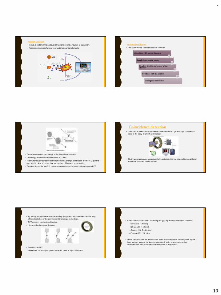
Positron Emission
• In this, a proton in the nucleus is transformed into a neutron & a positron.
• Positron emission is favored in low atomic number elements.
Positron Annihilation:
• The positron has short life in solids & liquids.
Interactions with atomic electrons
Rapidly loses kinetic energy
Reaches the thermal energy of the electron
Combines with the electron
Undergoes annihilation
• Their mass converts into energy in the form of gamma rays.
• The energy released in annihilation is 1022 KeV.
• To simultaneously conserve both momentum & energy, annihilation produces 2 gamma rays with 511 keV of energy that are emitted 180 degree to each other.
• The detection of the two 511 keV gamma rays forms the basis for imaging with PET.
Coincidence detection • Coincidence detection- simultaneous detection of the 2 gamma rays on opposite
sides of the body. (bismuth germinates )
• If both gamma rays can subsequently be detected, the line along which annihilation must have occurred can be defined.
• By having a ring of detectors surrounding the patient, it is possible to build a map of the distribution of the positron emitting isotope in the body.
• PET employs electronic collimation.
• 3 types of coincidence detection .
• Sensitivity in PET
- Measures capability of system to detect ‘trues’ & reject ‘randoms’
• Radionuclides used in PET scanning are typically isotopes with short half lives:
– Carbon-11 (~20 min),
– Nitrogen-13 (~10 min),
– Oxygen-15 (~2 min), and
– Fluorine-18 (~110 min).
• These radionuclides are incorporated either into compounds normally used by the body such as glucose (or glucose analogues), water or ammonia, or into molecules that bind to receptors or other sites of drug action.
.
11
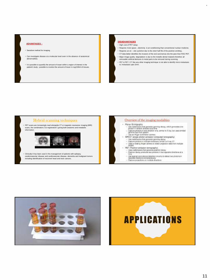
ADVANTAGES :
• Sensitive method for imaging.
• Can investigate disease at a molecular level even in the absence of anatomical abnormalities.
• It is possible to quantify the amount of tracer within a region of interest in the patient's body ; possible to monitor the amount of tracer in mg/100ml of tissues.
DISADVANTAGES• High cost of PET setup.
• Requires more space , electricity & air conditioning than conventional nuclear medicine.
• Requires an on – site cyclotron due to the short half life of the positron emitting .
• CT data better identifies the invasion of the oral carcinomas into the jaws than FDG PET.
• Major image quality degradation is due to the metallic dental implants therefore all removable artificial dentures & metal parts to be removed during scanning.
• PET & PET / CT like any other imaging technique is not able to identify micro metastasis ie; metastasis upto 2mm.
Hybrid scanning techniques
• PET scans are increasingly read alongside CT or magnetic resonance imaging (MRI) scans, the combination ("co-registration") giving both anatomic and metabolic information.
• Clinically it has been used in the management of patients with epilepsy, cerebrovascular disease and cardiovascular disease, dementia and malignant tumors including identification of recurrent head and neck cancers.
Overview of the imaging modalities
APPLICATIONS
.
12
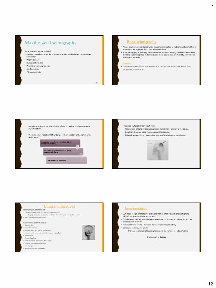
Maxillofacial scintigraphy
Bone scanning is used to detect
• metastatic neoplasia when the primary tumor originated in lungs,prostate,breast, head&neck
• Paget's disease
• Hyperparathyroidism
• Premature cranio synostosis
• Ameloblastoma
• Fibrous dysplasia.
67
Bone scintigraphy
• A bone scan or bone scintigraphy is a nuclear scanning test to find certain abnormalities in bone which are triggering the bone's attempts to heal.
• Bone scintigraphy is an highly sensitive method for demonstrating disease in bone, often providing earlier diagnosis or demonstrating more lesions than are found by conventional radiological methods.
Technique:
• The patient is injected with a small amount of radioactive material such as 600 MBq
of technetium-99m-MDP .
• Methylene Diphosphonate (MDP) has affinity for calcium rich hydroxyapatite crystals of bone.
• The technetium (Tc) 99m-MDP undergoes ‘chemisorption’ and gets bound to
bone matrix.
In exposed bone, bone remodeling (i.e. altered metabolism).
The hydroxyapatite crystal is most accessible to MDP
Increased radioactivity
Reduced radioactivity can result from:
• Replacement of bone by destructive lesion (lytic lesion) - primary or metastatic.
• Disruption of normal blood flow consequent to radiation.
• Reduced radioactivity is visualized as 'cold spot' or photopenic bone lesion.
Clinical indicationsThe oncological indications are:
• Primary tumors (e.g. Ewing’s sarcoma, osteosarcoma).
– Staging, evaluation of response to therapy and follow-up of primary bone tumors
• Secondary tumor's (metastases)
Non neoplastic diseases such as:
• Osteomyelitis
• Avascular necrosis
• Metabolic disorders (Paget, osteoporosis)
• Assessment of continued growth in condylar hyperplasia
• Arthropathies
• Fibrous Dysplasia
• Stress fractures, Shin splints, bone grafts
• Loose or infected joint prosthesis
• Low back pain
• Reflex sympathetic syndrome
Interpretation
• Symmetry of right and left sides of the skeleton and homogeneity of tracer uptake within bone structures - normal features.
• Both increase and decrease of tracer uptake have to be assessed; abnormalities can be either focal or diffuse.
• Increased tracer activity - indicates increased osteoblastic activity.
• Compared to a previous study:
Increase in intensity of tracer uptake and in the number of abnormalities
Progression of disease
.
13
• Focal decrease in radioactivity:
– Benign conditions
– Attenuation
– Artefact
– Absence of bone e.g. surgical resection.
• When compared to a previous study:
Decrease in intensity of tracer uptake and in number of abnormalities
Improvement or may be secondary to focal therapy (e.g. radiation therapy).
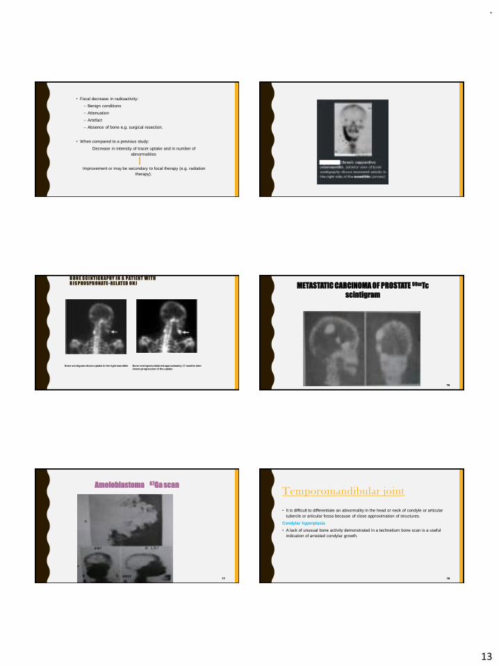
B O N E S C I N TI G R AP HY I N A P AT I EN T W I T H B I S P HO SP H O NATE -RE L AT ED O N J
Bone scintigram shows uptake in the right mandible Bone scintigram obtained approximately 17 months later shows progression of the uptake
76
METASTATIC CARCINOMA OF PROSTATE 99mTc
scintigram
77
Ameloblastoma 67Ga scanTemporomandibular joint
• It is difficult to differentiate an abnormality in the head or neck of condyle or articular tubercle or articular fossa because of close approximation of structures.
Condylar hyperplasia
• A lack of unusual bone activity demonstrated in a technetium bone scan is a useful indication of arrested condylar growth.
78

.
14
• In periapical abscess there will be increased uptake of radionuclide due to osteoblastic activity.
79 80
Anatomic localization of abnormalities of maxillofacial region detected by radionuclide imaging is done by multiple properly positioned views.
• Posteroanterior view
• Lateral view
• Oblique view
• Water’s view
Frontal and lateral (right and left) radionuclide images of the skull showing intense activity in the left temporomandibular joint in a 41-year-old woman with an MRI-confirmed temporomandibular joint meniscus dysfunction.
TUMOR DETECTION
• Most malignant neoplasm that spread to or arise in the bone evoke an osteoblastic response,
which accounts for localization of bone seeking radio pharmaceuticals in this areas. However a
purely destructive lesion may not have an associated reparative component
82
83
Some nonspecific physiological changes occurring with neoplasm that causes tumor seeking radio pharmaceuticals to accumulate are:
1.Localized increased blood volume
2.Abnormal tumor circulation with increased microvascular permeability
3.Increased affinity for radiotracer-labeled proteins in the metabolically active neoplastic site.
4.Prolonged extra vascular presence of radio nuclide that may be secondary to impaired lymphatic drainage from the tumor region
Salivary gland imaging
• Imaged following administration of 99m Tc pertechnetate or radioactive iodine.
• 99m Tc is preferred owing to its low radiation dose and the desirability of its photon emission (which is highly suitable for scintillation camera imaging).
84

.
15
85
• Salivary gland imaging is made possible by the glandular ability to concentrate radionuclide before excretion into the saliva.
• Salivary gland imaging is both functional and morphological test.
Technique
• 99MTc pertechnetate administered intravenously in dose range 0.5 to 10 mCi.
• A vascular blush outlining the parotid gland is seen immediately following pertechnetate administration.
86
87
• The concentration phase begins within first 10 minutes and represents the active accumulation of radionuclide by the ductal epithelium
• This phase may be enhanced by administration of pilocarpine nitrate 20 mgs subcutaneously.
88
• The excretory phase begins 10 to 40 minutes following administration and represents tracer being transported into the saliva and excreted into the oral cavity.
• This phase may be enhanced by oral administration of lemon juice or potassium perchlorate 1gm/70 kgs.
89
• Salivary excretion is blocked by intramuscular administration of 0.8 to 1mg atropine 30 minutes prior to scanning.
90
• Primary glandular malignancies as well as metastatic tumor of the gland ,abscess cyst fails to accumulate pertechnetate and appears as region devoid of radionuclide .this focus is called “cold focus”.
COLD FOCUS

.
16
HOT FOCUS
• Benign neoplasm like warthins tumor actively accumulate radionuclide to a greater depth than surrounding normal glandular tissue due to the ductal inclusion from which the tumor is thought to arise retaining their ability to concentrate pertechnitate.
91 92
WARM FOCUS
• Mixed tumor like pleomorphic adenoma accumulates radionuclide's nearly equal to surrounding normal gland
93 94
Regions of interest on dynamic scintigraphy. RP, right parotid; LP, left parotid; RSm, right submandibulargland; LSm, left submandibular gland; B, background
• Bilateral intraglandular lesions appears as cold defects on scintigraphy (arrows).
• (b) Dynamic images (1 min per frame) following intravenous injection of 99Tcm pertechnetate showed normal uptake and response to secretion stimulation in the upper poles of the parotid glands (arrows). Neither submandibulargland showed significant uptake (arrowheads). Note physiological uptake in thyroid gland

.
17
CLINICAL USES OF RADIONUCLIDE IN SALIVARY GLAND IMAGING
Abnormality Scintigraphic appearance
Tumors
Warthins tumor Hot Focus
Oxyphilic adenoma Hot focus or cold focus
Mixed tumors Cold or warm focus
Malignant tumors Cold focus
Cysts Cold focus
Abscesses Cold focus
97 98
• Sialadenitis• acute Increased uptake • Chronic Decreased uptake• Sjogren's syndrome uptake initially normal, later decreased
Conclusion
• Nuclear imaging had got its various applications not only in diagnosis but also in therapeutics of various disorders which also covers a wide spectrum of maxillofacial disorders.
• Several research and advances are going in increasing the specificity of these imaging modalities which may even widen the scope of nuclear imaging.
REFERENCES
• Oral radiology –White & Pharoh
• Essentials of dental radiography & radiology-Eric Whaites
• Goaz & White radiology
100
THANK YOU ☺
.
1
DR. NEERAJ KUMAR YADAV
READER
DEPT. OF ORAL MEDICINE AND RADIOLOGY
MGDCH , JAIPUR
ORAL CANCER AND TUMOR MARKERS
CONTENTS
▪ Introduction
▪ Epidemiology
▪ WHO classification of oral cancer
▪ Etiology and risk factors
▪ Carcinogenesis-theories
▪ Hall marks of oral cancer
▪ Clinical signs and symptoms
▪ TNM staging of oral cancer
▪ Diagnostic aids in the detection of oral cancer
▪ Imaging modalities in the detection of oral cancer
▪ Management of oral cancer
▪ References
INTRODUCTION
▪ Oral cancer is a broad term that includes various malignant diagnoses
that present in the oral tissues.
▪ It is traditionally defined as a squamous cell carcinoma (OSCC), because 90% of cancers are histologically originated in the squamouscells .
▪ It has different levels of differentiation and a propensity for lymph node metastasis
Burkit Textbook of oral medicine 12th edition
EPIDEMIOLOGY
▪ Cancer of the oral cavity and pharynx affects 10.8 of every 100,000 individuals in the United States, based on the National Cancer Institute data, and 7.2 of every 100,000 individuals will have cancer in oral cancer.
▪ Based on the data from Surveillance, Epidemiology, and End Results program for 2003 to 2009 the 5-year survival in the United States was 62.2%.
▪ In India,the incidence of oral cancer for males and females was highest in the central region of India.
▪ For males, it was 64.8% and for females it was 37.2% at 70 years of age.
▪ The next highest magnitude was observed in west and northeast regions (58.4%) at 60 years of age.
1) Burkit Textbook of oral medicine 12th edition2) Sharma S, Satyanarayana L, Asthana S, Shivalingesh KK, Goutham BS, Ramachandra S.
Oral cancer statistics in India on the basis of first report of 29 population-based cancer registries. J Oral Maxillofac Pathol. 2018;22(1):18–26.
ORAL CANCER CLASSIFICATION
WHO Classification of Oral Cancer
Epithelial cancerSquamous cell carcinoma
Verrucous carcinoma
Basaloid squamous cell carcinoma
Papillary squamous cell carcinoma
Spindle cell carcinoma (sarcomatoid SCC)
Acantholytic squamous cell carcinoma
Adenosquamous carcinoma
Carcinoma cuniculatum
Lymphoepithelial carcinoma
Hemato lymphoid cancerDiffuse large B-cell lymphoma
Mantle cell lymphoma
Follicular lymphoma
Extranodal marginal zone B-cell lymphoma of MALT type
Burkitt lymphoma
T-cell lymphoma
Extramedullary plasmacytoma
Langerhans cell hystiocytosis
Extramedullary myeloid sarcoma
Follicular dendritic cell sarcoma
Burkit Textbook of oral medicine 12th edition
Salivary gland cancer
Salivary gland carcinoma Salivary gland adenomas
Acinic cell carcinoma Pleomorphic adenoma
Mucoepidermoid carcinoma Myoepithelioma
Adenoid cystic carcinoma Basal cell adenoma
Polymorphous low-grade adenocarcinoma
Warthin’s tumor
Basal cell adenocarcinoma Canalicular adenoma
Epithelial-myoepithelial carcinoma Duct papilloma
Clear cell carcinoma Cystadenoma
Cystadenocarcinoma
Mucinous adenocarcinoma
Oncocytic carcinoma
Salivary duct carcinoma
Myoepithelial carcinoma
Carcinoma ex pleomorphic adenoma Burkit Textbook of oral medicine 12th
edition
.
2
Soft tissue cancer
Kaposi sarcoma
Malignant melanoma
Fibrosarcoma
Malignant fibrous histiocytoma
Liposarcoma
Malignant peripheral nerve sheath tumor
Angiosarcoma,
Leiomyosarcoma,
Rhabdomyosarcoma
Secondary tumours
Burkit Textbook of oral medicine 12th edition
WHO Classification of Odontogenic Cancer
Odontogenic carcinomas Odontogenic sarcomas
Metastasizing (malignant) ameloblastoma Ameloblastoma fibrosarcoma
Ameloblastic carcinoma—primary type Ameloblastic fibrodentino- and fibro-odontosarcoma
Ameloblastic carcinoma—secondary type (dedifferentiated),intraosseous
Ameloblastic carcinoma—secondary type (dedifferentiated),peripheral
Primary intraosseous squamous cell carcinoma—solid type
Other tumors
Primary intraosseous squamous cell carcinoma derived from KOT
Melanotic neuroectodermal tumor of infancy
Primary intraosseous squamous cell carcinoma derived from odontogenic cysts
Clear cell odontogenic carcinoma
Ghost cell odontogenic carcinomaBurkit Textbook of oral medicine 12th
edition
Etiology and Risk Factors
▪ The two main factors which influence the development of oral cancer are genetic and epigenetic factors.
▪ The main risk factors identified globally are tobacco and alcohol use.
▪ Other risk factors include:▪ Diet and nutrition▪ Viruses▪ Radiation▪ Ethnicity▪ Familial and genetic predisposition▪ Oral thrush▪ Immunosuppression▪ Use of mouthwash▪ Syphilis▪ Dental factors▪ Occupational risksKumar M, Nanavati R, Modi TG, Dobariya C. Oral cancer: Etiology and risk factors: A review. J Can Res Ther 2016;12:458-63.
Tobacco
Form of tobacco
Constituents Risk
Tobacco(Smokeless)
Nitrosamines, Polycyclic aromatic hydrocarbonsNitrosodiethanolamineNitrosoprolinePolonium
Short term-Benign hyperkeratosis and epithelial dysplasiaChronic use-risk of malignant lesions
Tobacco (smoke )
Carbon monoxideThiocyanateHydrogen cyanideNicotine
80%- primary oral cancerRisk of recurrent and second primary oral cancer
Kumar M, Nanavati R, Modi TG, Dobariya C. Oral cancer: Etiology and risk factors: A review. J Can Res Ther 2016;12:458-63.
ALCOHOL
▪ Beer and wine are associated with greater risk than hard liquor.
▪ The combined effects of tobacco and alcohol result in a synergistic effect on the development of oral cancer.
▪ Mechanism :Alcohol is shown to increase the permeability of oral mucosa producing an alteration in morphology characterized by epithelial atrophy, which in turn leads to easier penetration of carcinogens into the oral mucosa
▪ Smoking and alcohol interaction may influence central nervous system activity
Kumar M, Nanavati R, Modi TG, Dobariya C. Oral cancer: Etiology and risk factors: A review. J Can Res Ther 2016;12:458-63.
Betel (Areca) Nut
▪ People with a betel quid chewing habit, with or without added tobacco, are at a higher risk to develop oral cancer.
▪ In parts of Asia where the use of betel nut mixed with lime to form a quid is widespread (e.g., India, Taiwan), the incidence of oral cancer is high and more commonly involves the buccal mucosa.
▪ Substitutes for betel quid, such as gutkha and pan masala- are potential carcinogenic as well
Kumar M, Nanavati R, Modi TG, Dobariya C. Oral cancer: Etiology and risk factors: A review. J Can Res Ther 2016;12:458-63.
.
3
Nutritional Factors
▪ Consumption of fruits and vegetables is associated with a reduced risk for oral cancer.
▪ Diets high in eggs and butter and meats-Elevated but inconsistent risk of oral cancer
▪ vitamins A ,C and E;carotenoids (β-carotene); potassium, selenium-decrease the risk of oral cancer development
▪ Vitamin D deficiency - associated with oral cancer
Kumar M, Nanavati R, Modi TG, Dobariya C. Oral cancer: Etiology and risk factors: A review. J Can Res Ther 2016;12:458-63.
Maté
▪ Maté, which is a tea-like beverage consumed in South America and in parts of Europe has been shown to be an independent cause for development of oral and pharyngeal cancers.
▪ The exact pathogenesis of maté predisposing to oral cancer is still unknown.
▪ Many reasons that have proposed for maté’s carcinogenicity are
1) Thermal injury
2) Solvent for other chemical carcinogens
3) Presence of tannins and N-nitroso compounds.
Kumar M, Nanavati R, Modi TG, Dobariya C. Oral cancer: Etiology and risk factors: A review. J Can Res Ther 2016;12:458-63.
ENVIRONMENTAL FACTORS
Viral Infections
▪ Viruses have been strongly implicated in the development of malignant tumors of the squamous epithelia including the oral squamous epithelium.
▪ Viral infections of latent or chronic nature are usually responsible for inducing malignant transformation by interfering with the host’s cell cycle machinery.
▪ The viral genes and gene products may affect cell growth and proliferation.
▪ Certain viral genes such as proto-oncogenes which become oncogenes when inserted into the host’s DNA and ultimately resulting in malignant
transformation.
▪ Virus involved - human herpes virus (mainly Epstein– Barr virus (EBV)), human
papillomavirus (HPV), and herpes simplex virus.Kumar M, Nanavati R, Modi TG, Dobariya C. Oral cancer: Etiology and risk factors: A review. J Can Res Ther 2016;12:458-63.
▪ HPV are the most common viruses implicated in oral carcinogenesis.
▪ High-risk HPV types - 16, 18, 31,33, 35, and 39- associated with OSCC and oral premalignant lesions.
▪ HPV encodes two major oncoproteins - E6 and E7.
▪ The E6 and E7 proteins bind and destroy p53 and Rb tumor suppressor genes, respectively, thereby disrupting the cell cycle with loss of control on DNA replication, DNA repair, and apoptosis.
▪ HPV-16 and -18 are considered high-risk subtypes due to their association with malignant tumors.
▪ HPV-16 alone is associated with about 85% to 95% of HPV-positive OPCs
Kumar M, Nanavati R, Modi TG, Dobariya C. Oral cancer: Etiology and risk factors: A review. J Can Res Ther 2016;12:458-63.
▪ HSV has not been proven to be the direct cause of oral cancer
▪ Several studies show that oral cancer patients have high serum antibody titers to HSV.
▪ The available evidences are circumstantial and are rationalized that reactivation of HSV infection is due to immunosuppression, specifically of natural killer lymphocyte activity.
▪ Based on the evidence of in vitro studies, the possible role of HSV in carcinogenesis has been proposed as the enhancement of activation, amplification, and overexpression of preexisting oncogenes such as
c-myc and c-erb-B-1.Kumar M, Nanavati R, Modi TG, Dobariya C. Oral cancer: Etiology and risk factors: A review. J Can Res Ther 2016;12:458-63.
Fungal Infections
▪ Fungal infections caused by Candida species such as Candida albicans has been implicated in the pathogenesis of oral premalignant lesions.
▪ Superficial fungal hyphae of Candida albicans have been found superimposed on leukoplakia, especially nodular leukoplakia, many of which have undergone malignant transformation.
▪ Besides immunocompromised individuals,Candida infection can coexist or be associated with other risk factors like irondeficiency and in chronic smokers which may prove synergistic in the development of oral cancer.
▪ There is evidence that Candida possesses necessary enzymes from dietary substances to produce nitrosamines and chemicals that have been implicated in carcinogenesis.
Kumar M, Nanavati R, Modi TG, Dobariya C. Oral cancer: Etiology and risk factors: A review. J Can Res Ther 2016;12:458-63.
.
4
Syphilis
▪ Tertiary syphilis had been known to predispose to the development of oral cancer along with other risk factors such as tobacco and alcohol.
▪ However, nowadays, tertiary syphilis is rare in clinical practice as the infection is diagnosed and treated before the onset of tertiary stage
Kumar M, Nanavati R, Modi TG, Dobariya C. Oral cancer: Etiology and risk factors: A review. J Can Res Ther 2016;12:458-63.
IMMUNOSUPPRESSION
▪ Immunosuppressed individuals are more prone to develop oral cancers.
▪ Human immunodeficiency virus (HIV)-infected patients are predisposed to developing Kaposi’s sarcoma and lymphomas.
▪ Immunosuppressed organ transplant patients have been shown to develop lip cancers and the possible reason was attributed to increased exposure to solar radiation and other risk factors such as smoking.
▪ The direct role of immunosuppression with lip cancer development was not proven in the studies
Kumar M, Nanavati R, Modi TG, Dobariya C. Oral cancer: Etiology and risk factors: A review. J Can Res Ther 2016;12:458-63.
Occupational Risks
▪ Occupational risks, namely exposure to excessive solar radiation/ ultraviolet (UV) light is known to cause lip cancers.
▪ UV rays also causes actinic cheilitis which may transform to OSCCs.
▪ Sulfur dioxide, asbestos, pesticide exposures, and mists from strong inorganic acids and burning of fossil fuels have also been known to cause cancers of posterior mouth, pharynx, and larynx.
▪ Certain occupations have been reported to place people at increased risk for the development of salivary gland carcinomas; these include manufacturing of rubber products, plumbing (exposure of metals), and woodworking in an automobile industry
Kumar M, Nanavati R, Modi TG, Dobariya C. Oral cancer: Etiology and risk factors: A review. J Can Res Ther 2016;12:458-63.
Dental Factors
▪ Poor oral hygiene, poor dental status (sharp/fractured teeth due to caries/trauma), and chronic ulceration from an ill-fitting denture has been suggested to promote neoplasm in the presence of other risk factors.
▪ There has been difficulty in obtaining the evidence whether dental factors influence oral cancer development.
▪ This is due to the presence of coexisting risk factors like smoking and alcohol consumption.
▪ In a study, mechanical irritation by scratching with a pulp cleaner has been shown to significantly increase the incidence of a chemical carcinogen-induced tongue carcinoma.
▪ Therefore, it is prudent to closely monitor patients with known risk factors for signs and symptoms of irritation from teeth and appliances.
Kumar M, Nanavati R, Modi TG, Dobariya C. Oral cancer: Etiology and risk factors: A review. J Can Res Ther 2016;12:458-63.
Pathogenesis of cancer
▪ Oral carcinogenesis is a highly complex multifocal process that takes place
when squamous epithelium is affected by several genetic alterations.
Theories of carcinogenesis
➢ Molecular pathogenesis of cancer
➢ Chemical carcinogens and chemical carcinogenesis
➢ Physical carcinogens and radiation carcinogenesis
➢ Biologic carcinogens and viral carcinogenesis
Textbook of pathology by Harshamohan-6th edition
MOLECULAR PATHOGENESIS OF CANCER
▪ Genetic theory: This is the most popular theory, which suggests that cells become neoplastic because of alterations in the DNA.
▪ The mutated cells transmit their characters to the next progeny of cells.
▪ Evidence in support of genetic theory comes from all types of etiologic agents in carcinogenesis.
▪ Epigenetic theory: This theory is less well supported than the genetic theory.
▪ According to the epigenetic theory, the carcinogenic agents act on activators or suppressors of genes and not on the genes themselves and result in the abnormal expression of genes.
Textbook of pathology by Harshamohan-6th edition
.
5
Field theory of cancer
➢In an organ developing cancer, in the backdrop of normal cells, limited no. of cells
only grow in to cancer after undergoing sequence of changes under the influence of
etiological agents.
➢This is termed as field effect concept called field theory of cancer
Multistep process of cancer growth and progression
▪ Carcinogenesis is gradual multistep process involving many generation of cells.
▪ Ultimately so formed genetically and phenotypically transformed cells having
phenotypic features of malignancy.
➢ Shows excessive growth.
➢ Invasiveness.
➢ Distant metastasis Textbook of pathology by Harshamohan-6th edition
▪ Immune-surveillance theory-
▪ In 1909, Paul Ehrlich formulated the hypothesis that host defense may prevent neoplastic cells from developing into tumors
▪ Lewis Thomas suggested that the immune system recognize newly arising tumors through the expression of tumor specific neo-antigens on tumor cells and eliminate them.
▪ Sir Frank Mac Farlane Burnet hypothesize that tumor cell neo-antigens induce an immunological reaction against cancer and subsequently formulated the immune surveillance theory .
Ribatti D. The concept of immune surveillance against tumors: The first theories. Oncotarget. 2017;8(4).
▪ Monoclonal hypothesis
▪ It is proposed by Nowell in 1976
▪ Cancer cell develop strategies to survive a hostile host environment and use host resources to grow and proliferate
▪ According to this theory,cancer arise from single clone of transformed cells
Textbook of pathology by Harshamohan-6th edition
Genetic regulators of normal and abnormal mitosis
Normal cell growth Cancer cell growth
Proto oncogenes - growth promotingagents, they encode for cell proliferation pathway.
Proto oncogenes – activation of growth promoting oncogenes causing transformation of cells.
Anti oncogenes - growth inhibiting or growth inhibitors are present.
Inactivation of Anti oncogenes-suppressor genes- permitting the cellular proliferation of transformed cells.Active in receessive form
Apoptosis regulatory genes - controlthe programmed cell death.
Abnormal apoptosis regulatory genes - these act as oncogenes.Active in dominant or recessive form.
DNA repair genes - regulate the repair of DNA damage Occurred during mitosis and also control the damage to proto-oncogenes and antioncogenes.
Failure of DNA repair genes -inability to repair DNA damage resulting in mitosis.
Textbook of pathology by Harshamohan-6th edition
Chemical carcinogens and chemical carcinogenesis
• Chemicals which initiate carcinogenesis is called chemical carcinogens.
• Chemicals which increase the effectiveness of carcinogens is called co-
carcinogens.
• Stages in chemical carcinogenesis
• Initiation
• Promotion
• progression
Textbook of pathology by Harshamohan-6th edition Textbook of pathology by Harshamohan-6th edition
.
6
Tests for chemical carcinogenicity
Experimental induction
• The traditional method is to administer the chemical compound under test to a batch of experimental animals like mice.
• E.g. painting on skin, giving orally or parenterally,or by inhalation.
• Repeated administration, dose varied, after many months the animal is
autopsied and results obtained.
Textbook of pathology by Harshamohan-6th edition
Test for mutagenicity(Ames test)
• Ames test evaluate the ability of a chemical to induce mutation in the mutant strain of salmonella typhimurium that cannot synthesize histidine.
• Strains are incubated with the potential carcinogen to which liver homogenate is added to supply enzymes required to convert procarcinogen to ultimate carcinogen.
• If the chemical is mutagenic, induce
mutant strains of s.typhimurium
• S,typhimurium in the form of functional
histidine gene , which will be reflected
by the no. of bacterial colonies growing
on histidine free culture medium.
Physical carcinogens and radiation carcinogenesis
• UV light and ionizing radiation are two main forms of radiation carcinogenesis.
UV light
• Uv light penetrates in to the skin for few millimeters only so that its effect is limited to epidermis.
• The efficiency of UV light as carcinogen depends upon extent of light absorbing protective melanin pigmentation of skin.
• If exposure is more it will cause scc, bcc, malignant melanoma.
• Incidence is high in fair skinned Europeans.
Textbook of pathology by Harshamohan-6th edition
Mechanism
• Induction of mutation
• Inactivation of enzymes
• Some times causing cell death.
• Biochemical effects of uv radiation is formation of pyrimidine dimers in
DNA.
• Mutagenic effect of uv radiation associated with recessive hereditary
diseases characterized by defect in DNA repair and associated with high
incidence of cancers.
Textbook of pathology by Harshamohan-6th edition
Ionizing radiation
• X rays,α,β,γ rays and radioactive isotopes, photons, neutrons can cause cancers
in animals and man.
• Risk of cancer increases by dose with high LET such as neutrons, α rays.
• Low LET such as x rays and γ rays.
• High incidence of radiation dermatitis & malignant tumors of skin common in
x ray workers, radiotherapists.
• High incidence of osteosarcoma-young American watch working girls engaged
in painting the dials with luminous radium.
Textbook of pathology by Harshamohan-6th edition
Mechanism
• It may directly alter the cellular DNA.
• Formation of high reactive free radicles-damage.
Textbook of pathology by Harshamohan-6th edition
.
7
Biologic carcinogens and viral carcinogenesis
• Many DNA & RNA viruses are proved to be carcinogenic.
Oncogenic RNA viruses
• Oncogenic retrovirus, human T cell lymphotropic virus -1, (HTLV-1)has
demonstrated to cause cancer in humans.
Pathogenesis-
• Induced T cell leukemia lymphoma.
• HTLV -1 infects many T cells and initially caused polyclonal proliferation by
autocrine and paracrine pathways triggered by the TAX gene.
Textbook of pathology by Harshamohan-6th edition
• TAX neutralizes growth inhibitory signals by affecting TP53&CDKN2A/p16 genes.
• Ultimately a monoclonal T cell leukemia /lymphoma results when one proliferating T cell suffers additional mutations.
Oncogenic DNA viruses
• Four DNA viruses HPV,EBV, HBV associated with cancers.
HPV
• Oncogenic potential HPV related to products of 2 viral genes E6&E7.
• E7 protein binds to retinoblastoma proteins & release the E2F transcription factors that normally are sequestered by Rb, promoting progression through the cell cycle.
Textbook of pathology by Harshamohan-6th edition
• High risk stimulates loss of tumor suppressor genes, activate cyclins, inhibits apoptosis, and combats cellular senescence.
• Low risk HPV s give rise to benign warts.
HBV
• Contribute oncogenesis by B cell proliferation , leads to development of lymphoma and occurrence of additional mutations such leading to activation of the MYC gene.
• E.g.-lymphomas in immunocompromised patients, Hodgkin's lymphoma, nasopharyngeal carcinoma.
Textbook of pathology by Harshamohan-6th edition
HALLMARKS OF CANCER
▪ 1) Excessive and autonomous growth: Growth-promoting oncogenes.
▪ 2) Refractoriness to growth inhibition: Growth suppressing anti-oncogenes.
▪ 3) Escaping cell death by apoptosis: Genes regulating apoptosis and cancer
▪ 4) Avoiding cellular aging: Telomeres and telomerase in cancer
▪ 5) Continued perfusion of cancer: Cancer angiogenesis
▪ 6) Invasion and distant metastasis: Cancer dissemination.
▪ 7) DNA damage and repair system: Mutator genes and cancer.
▪ 8) Cancer progression and tumour heterogeneity: Clonal aggressiveness.
▪ 9) Cancer a sequential multistep molecular phenomenon: Multistep theory.
▪ 10)MicroRNAs in cancer: OncomiRs
Textbook of pathology by Harshamohan-6th edition
EXCESSIVE AND AUTONOMOUS GROWTH:
GROWTH PROMOTING ONCOGENES
▪ Mutated form of normal protooncogenes in cancer is called oncogenes.
▪
▪ Protooncogenes become activated oncogenes by mechanisms
➢ By mutation in the protooncogene which alters its structure and function.
➢ By retroviral insertion in the host cell.
➢ By damage to the DNA sequence that normally regulates growth-promoting signals of protooncogenes resulting in its abnormal activation.
➢ By erroneous formation of extra copies of protooncogene causing gene amplification and hence its overexpression or overproduction that promotes autonomous and excessive cellular proliferation
Textbook of pathology by Harshamohan-6th edition Textbook of pathology by Harshamohan-6th edition
.
8
REFRACTORINESS TO GROWTH INHIBITION:
GROWTH SUPPRESSING ANTI-ONCOGENES
▪ The mutation of normal growth suppressor anti-oncogenes results in removal of the brakes for growth; thus the inhibitory effect to cell growth is removed and the abnormal growth continues unchecked.
▪ Mutated antioncogenes behave like growth-promoting oncogenes.
ESCAPING CELL DEATH BY APOPTOSIS: GENES
REGULATING APOPTOSIS AND CANCER
▪ Besides the role of mutant forms of growth-promoting oncogenes and growth-suppressing anti-oncogenes, another mechanism of tumourgrowth is by escaping cell death by apoptosis.
▪ Apoptosis in normal cell is guided by cell death receptor, CD95, resulting in DNA damage.
▪ Besides, there is role of some other pro-apoptotic factors (BAD, BAX, BID and p53) and apoptosis-inhibitors (BCL2, BCL-X).
▪ In cancer cells, the function of apoptosis is interfered due to mutations in the above genes which regulate apoptosis in the normal cell
Textbook of pathology by Harshamohan-6th edition
AVOIDING CELLULAR AGING: TELOMERES AND
TELOMERASE IN CANCER
▪ After each mitosis (cell doubling) there is progressive shortening of telomeres which are the terminal tips of chromosomes.
▪ Telomerase is the RNA enzyme that helps in repair of such damage to DNA and maintains normal telomere length in successive cell divisions.
▪ However, it has been seen that after repetitive mitosis for a maximum of 60 to 70 times, telomeres are lost in normal cells and the cells cease to undergo mitosis.
▪ Telomerase is active in normal stem cells but not in normal somatic cells.
▪ Cancer cells in most malignancies have markedly upregulated telomerase enzyme, and hence telomere length is maintained.
▪ Thus, cancer cells avoid aging, mitosis does not slow down or cease, thereby immortalising the cancer cells
CONTINUED PERFUSION OF CANCER: TUMOUR
ANGIOGENESIS
▪ Cancers can only survive and thrive if the cancer cells are adequately nourished and perfused, as otherwise they cannot grow further.
▪ Neovascularisation in the cancers not only supplies the tumour with oxygen and nutrients, but the newly formed endothelial cells also elaborate a few growth factors for progression of primary as well as metastatic cancer.
Textbook of pathology by Harshamohan-6th edition
▪ The stimulus for angiogenesis is provided by the release of various factors:
▪ i) Promoters of tumour angiogenesis- vascular endothelial growth factor (VEGF) (released from genes in the parenchymal tumour cells) and basic fibroblast growth factor (bFGF).
▪ ii) Anti-angiogenesis factors inhibiting angiogenesis include thrombospondin-1 (also produced by tumour cells themselves), angiostatin, endostatin and vasculostatin.
▪ Mutated form of p53 gene in both alleles in various cancers results in removal of anti-angiogenic role of thrombospondin-1, thus favouringcontinued angiogenesis
Textbook of pathology by Harshamohan-6th edition
INVASION AND DISTANT METASTASIS: CANCER
DISSEMINATION
▪ One of the most important characteristic of cancers is invasiveness and metastasis.
▪ As the tumor progression advances, the cells lose their adherence property, detach from the tumor mass and invade the neighboring tissues.
▪ The detached cells also enter the circulating blood and lymph and are transported to other organs/tissues away from the site of the primary growth and develop into secondary tumors at the new sites.
▪ These form the distant metastases, resulting in widely spread cancers
Textbook of pathology by Harshamohan-6th edition
.
9
DNA DAMAGE AND REPAIR SYSTEM: MUTATOR
GENES AND CANCER
▪ Normal cells during complex mitosis suffer from minor damage to the DNA which is detected and repaired before mitosis is completed so that integrity of the genome is maintained.
▪ p53 gene is held responsible for detection and repair of DNA damage.
▪ If this system of DNA repair is defective as happens in some inherited mutations (mutator genes), the defect in unrepaired DNA is passed to the next progeny of cells and cancer results
Textbook of pathology by Harshamohan-6th edition
CANCER PROGRESSION AND HETEROGENEITY:
CLONAL AGGRESSIVENESS
▪ As time passes,cancers become more aggressive; this property is termed tumour progression.
▪ Clinical parameters of cancer progression are: increasing size of the tumour, higher histologic grade (as seen by poorer differentiation and greater anaplasia), areas of tumour necrosis (i.e. tumour outgrows its blood supply), invasiveness and distant metastasis.
▪ This means that though cancer cells remain monoclonal in origin, they acquire more and more mutations which, in turn, produce multiple-mutated subpopulations of more aggressive clones of cancer cells in the growth which have tendency to invade, metastasise and be refractory to hormonal influences.
▪ Some of these mutations in fact may kill the tumour cells as well
Textbook of pathology by Harshamohan-6th edition
CANCER A SEQUENTIAL MULTISTEP MOLECULAR
PHENOMENON: MULTISTEP THEORY
▪ Cancer occurs following several sequential steps of abnormalities in the target cell
▪ Initiation, promotion and progression in proper sequence.
▪ Multiple steps are involved at genetic level by which cell proliferation of cancer cells is activated: by activation of growth promoters, loss of growth suppressors, inactivation of intrinsic apoptotic mechanisms and escaping cellular aging.
Textbook of pathology by Harshamohan-6th edition
MICRORNAs IN CANCER: ONCOMIRS
▪ MicroRNAs (miRNAs) are evolutionally conserved, endogenous, noncoding single stranded RNA molecules with a length of 22 nucleotides only.
▪ Normally, miRNAs function as the posttranslational gene regulators of cell proliferation, differentiation and survival.
▪ Recent evidence indicates that miRNAs have an oncogenic role in initiation and progression of cancer and are termed as oncogenicmicroRNAs, abbreviated as oncomiRs.
▪ In combination with other tumour associated genes, oncomiRs can perform various functions: as tumour suppressor, as tumour promoter, and as pro-apoptotic.
Textbook of pathology by Harshamohan-6th edition
CLINICAL SIGNS AND SYMPTOMS
▪ Discomfort is the most common symptom that leads a patient to seek care and may be present at the time of diagnosis in up to 85% of patients.
▪ Individuals presents with mass in the mouth or neck.
▪ Dysphagia, odynophagia, otalgia, limited movement, oral bleeding, neck masses, and weight loss may occur with advanced disease.
▪ Loss of sensory function – unilateral - indicate neural involvement
▪ Loss of function involving the tongue can affect speech, swallowing, and diet.
Burkit Textbook of oral medicine 12th edition
▪ Tissue changes –
▪ 1) Red, white, or mixed red-and-white lesion
▪ 2) A change in the surface texture producing a smooth, granular, rough, or crusted lesion
▪ 3) Presence of a mass or ulceration
▪ 4) The lesion may be flat or elevated and may be minimally palpable or indurated
▪ High-risk sites - lower lip, the anterior floor of the mouth, and the lateral borders of the tongue.
Burkit Textbook of oral medicine 12th edition
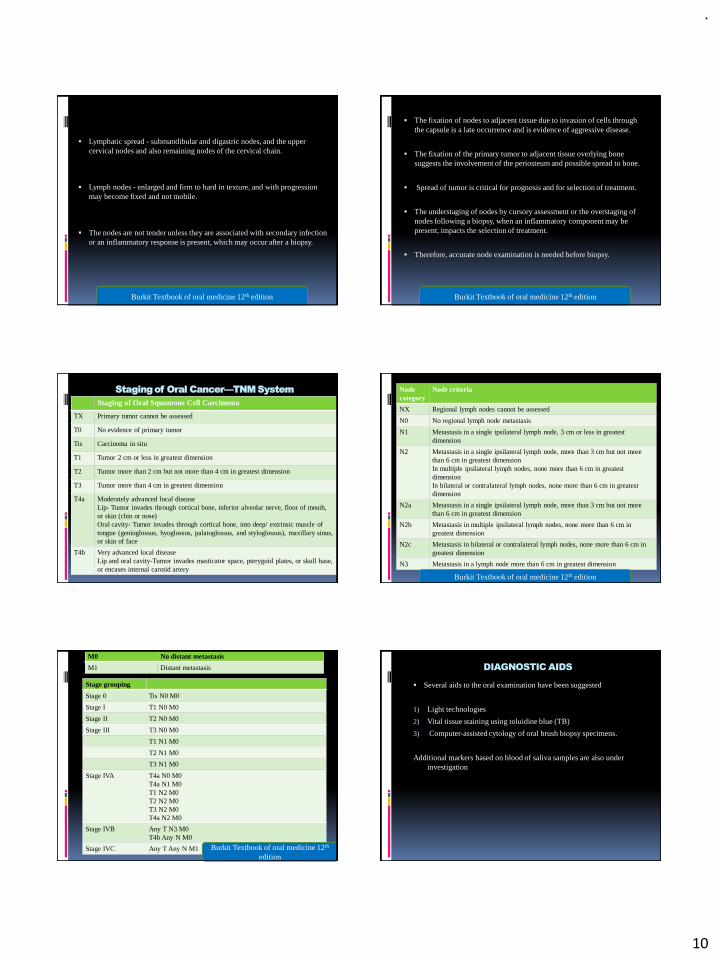
.
10
▪ Lymphatic spread - submandibular and digastric nodes, and the upper cervical nodes and also remaining nodes of the cervical chain.
▪ Lymph nodes - enlarged and firm to hard in texture, and with progression may become fixed and not mobile.
▪ The nodes are not tender unless they are associated with secondary infection or an inflammatory response is present, which may occur after a biopsy.
Burkit Textbook of oral medicine 12th edition
▪ The fixation of nodes to adjacent tissue due to invasion of cells through the capsule is a late occurrence and is evidence of aggressive disease.
▪ The fixation of the primary tumor to adjacent tissue overlying bone suggests the involvement of the periosteum and possible spread to bone.
▪ Spread of tumor is critical for prognosis and for selection of treatment.
▪ The understaging of nodes by cursory assessment or the overstaging of nodes following a biopsy, when an inflammatory component may be present, impacts the selection of treatment.
▪ Therefore, accurate node examination is needed before biopsy.
Burkit Textbook of oral medicine 12th edition
Staging of Oral Cancer—TNM System
Staging of Oral Squamous Cell Carcinoma
TX Primary tumor cannot be assessed
T0 No evidence of primary tumor
Tis Carcinoma in situ
T1 Tumor 2 cm or less in greatest dimension
T2 Tumor more than 2 cm but not more than 4 cm in greatest dimension
T3 Tumor more than 4 cm in greatest dimension
T4a Moderately advanced local disease Lip- Tumor invades through cortical bone, inferior alveolar nerve, floor of mouth, or skin (chin or nose)Oral cavity- Tumor invades through cortical bone, into deep/ extrinsic muscle of tongue (genioglossus, hyoglossus, palatoglossus, and styloglossus), maxillary sinus, or skin of face
T4b Very advanced local diseaseLip and oral cavity-Tumor invades masticator space, pterygoid plates, or skull base, or encases internal carotid artery
Node category
Node criteria
NX Regional lymph nodes cannot be assessed
N0 No regional lymph node metastasis
N1 Metastasis in a single ipsilateral lymph node, 3 cm or less in greatest dimension
N2 Metastasis in a single ipsilateral lymph node, more than 3 cm but not more than 6 cm in greatest dimensionIn multiple ipsilateral lymph nodes, none more than 6 cm in greatest dimensionIn bilateral or contralateral lymph nodes, none more than 6 cm in greatest dimension
N2a Metastasis in a single ipsilateral lymph node, more than 3 cm but not more than 6 cm in greatest dimension
N2b Metastasis in multiple ipsilateral lymph nodes, none more than 6 cm in greatest dimension
N2c Metastasis in bilateral or contralateral lymph nodes, none more than 6 cm in greatest dimension
N3 Metastasis in a lymph node more than 6 cm in greatest dimension
Burkit Textbook of oral medicine 12th edition
M0 No distant metastasis
M1 Distant metastasis
Stage grouping
Stage 0 Tis N0 M0
Stage I T1 N0 M0
Stage II T2 N0 M0
Stage III T3 N0 M0
T1 N1 M0
T2 N1 M0
T3 N1 M0
Stage IVA T4a N0 M0T4a N1 M0T1 N2 M0T2 N2 M0T3 N2 M0T4a N2 M0
Stage IVB Any T N3 M0T4b Any N M0
Stage IVC Any T Any N M1 Burkit Textbook of oral medicine 12th
edition
DIAGNOSTIC AIDS
▪ Several aids to the oral examination have been suggested
1) Light technologies
2) Vital tissue staining using toluidine blue (TB)
3) Computer-assisted cytology of oral brush biopsy specimens.
Additional markers based on blood of saliva samples are also under investigation
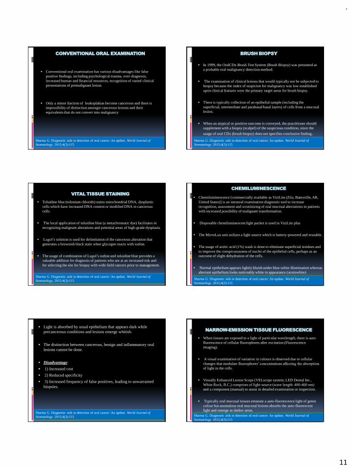
.
11
CONVENTIONAL ORAL EXAMINATION
▪ Conventional oral examination has various disadvantages like false positive findings, including psychological trauma, over-diagnosis, increased human and financial resources, recognition of varied clinical presentations of premalignant lesion.
▪ Only a minor fraction of leukoplakias become cancerous and there is impossibility of distinction amongst cancerous lesions and their equivalents that do not convert into malignancy
Sharma G. Diagnostic aids in detection of oral cancer: An update. World Journal of Stomatology. 2015;4(3):115
BRUSH BIOPSY
▪ In 1999, the OralCDx Brush Test System (Brush Biopsy) was presented as a probable oral malignancy detection method.
▪ The examination of clinical lesions that would typically not be subjected to biopsy because the index of suspicion for malignancy was low established upon clinical features were the primary target areas for brush biopsy.
▪ There is typically collection of an epithelial sample (including the superficial, intermediate and parabasal/basal layers) of cells from a mucosal lesion.
▪ When an atypical or positive outcome is conveyed, the practitioner should supplement with a biopsy (scalpel) of the suspicious condition, since the
usage of oral CDx (brush biopsy) does not specifies conclusive finding.Sharma G. Diagnostic aids in detection of oral cancer: An update. World Journal of Stomatology. 2015;4(3):115
VITAL TISSUE STAINING
▪ Toluidine blue (tolonium chloride) stains mitochondrial DNA, dysplastic cells which have increased DNA content or modified DNA in cancerous cells.
▪ The local application of toluidine blue (a metachromatic dye) facilitates in recognizing malignant alterations and potential areas of high-grade dysplasia.
▪ Lugol’s solution is used for delimitation of the cancerous alteration that generates a brownish black stain when glycogen reacts with iodine.
▪ The usage of combination of Lugol’s iodine and toluidine blue provides a valuable addition for diagnosis of patients who are at an increased risk and for selecting the site for biopsy with wide field cancers prior to management.
Sharma G. Diagnostic aids in detection of oral cancer: An update. World Journal of Stomatology. 2015;4(3):115
CHEMILUMINESCENCE
▪ Chemiluminescence [commercially available as ViziLite (Zila, Batesville, AR, United States)] is an intraoral examination diagnostic tool to increase recognition, assessment and scrutinizing of oral mucosal aberrations in patients with increased possibility of malignant transformation.
▪ Disposable chemiluminescent light packet is used in ViziLite plus
▪ The MicroLux unit utilizes a light source which is battery-powered and reusable.
▪ The usage of acetic acid (1%) wash is done to eliminate superficial residues and to improve the conspicuousness of nuclei of the epithelial cells, perhaps as an outcome of slight dehydration of the cells.
▪ Normal epithelium appears lightly bluish under blue-white illumination whereas aberrant epithelium looks noticeably white in appearance (acetowhite)
Sharma G. Diagnostic aids in detection of oral cancer: An update. World Journal of Stomatology. 2015;4(3):115
▪ Light is absorbed by usual epithelium that appears dark while precancerous conditions and lesions emerge whitish.
▪ The distinction between cancerous, benign and inflammatory oral lesions cannot be done.
▪ Disadvantage:
▪ 1) Increased cost
▪ 2) Reduced specificity
▪ 3) Increased frequency of false positives, leading to unwarranted biopsies.
Sharma G. Diagnostic aids in detection of oral cancer: An update. World Journal of Stomatology. 2015;4(3):115
NARROW-EMISSION TISSUE FLUORESCENCE
▪ When tissues are exposed to a light of particular wavelength, there is auto fluorescence of cellular fluorophores after excitation (Fluorescence imaging).
▪ A visual examination of variation in colours is observed due to cellular changes that modulate fluorophores’ concentrations affecting the absorption
of light in the cells.
▪ Visually Enhanced Lesion Scope (VELscope system; LED Dental Inc., White Rock, B.C.) comprises of light-source (wave length: 400-460 nm) and a component (manual) to assist in detailed examination or inspection.
▪ Typically oral mucosal tissues emanate a auto-fluorescence light of green colour but anomalous oral mucosal lesions absorbs the auto-fluorescent light and emerge as darker areas.
Sharma G. Diagnostic aids in detection of oral cancer: An update. World Journal of Stomatology. 2015;4(3):115
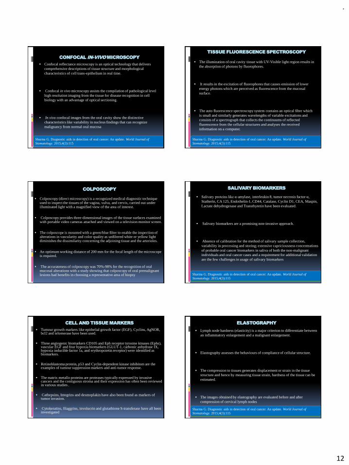
.
12
CONFOCAL IN-VIVO MICROSCOPY
▪ Confocal reflectance microscopy is an optical technology that delivers comprehensive descriptions of tissue structure and morphological characteristics of cell trans-epithelium in real time.
▪ Confocal in vivo microscopy assists the compilation of pathological level high resolution imaging from the tissue for disease recognition in cell biology with an advantage of optical sectioning.
▪ In vivo confocal images from the oral cavity show the distinctive characteristics like variability in nucleus findings that can recognize malignancy from normal oral mucosa
Sharma G. Diagnostic aids in detection of oral cancer: An update. World Journal of Stomatology. 2015;4(3):115
TISSUE FLUORESCENCE SPECTROSCOPY
▪ The illumination of oral cavity tissue with UV-Visible light region results in the absorption of photons by fluorophores.
▪ It results in the excitation of fluorophores that causes emission of lower energy photons which are perceived as fluorescence from the mucosal surface.
▪ The auto fluorescence spectroscopy system contains an optical fibre which is small and similarly generates wavelengths of variable excitations and consists of a spectrograph that collects the continuums of reflected fluorescence from the cellular structures and analyses the received information on a computer.
Sharma G. Diagnostic aids in detection of oral cancer: An update. World Journal of Stomatology. 2015;4(3):115
COLPOSCOPY
▪ Colposcopy (direct microscopy) is a recognized medical diagnostic technique used to inspect the tissues of the vagina, vulva, and cervix, carried out under illuminated light with a magnified view of the area of interest.
▪ Colposcopy provides three-dimensional images of the tissue surfaces examined with portable video cameras attached and viewed on a television monitor screen.
▪ The colposcope is mounted with a green/blue filter to enable the inspection of alterations in vascularity and color quality as unfiltered white or yellow light diminishes the dissimilarity concerning the adjoining tissue and the arterioles.
▪ An optimum working distance of 200 mm for the focal length of the microscope is required.
▪ The accurateness of colposcopy was 70%-98% for the recognition of oral mucosal alterations with a study showing that colposcopy of oral premalignant lesions had benefits in choosing a representative area of biopsy
SALIVARY BIOMARKERS
▪ Salivary proteins like α-amylase, interleukin 8, tumor necrosis factor-α,
Statherin, CA 125, Endothelin-1, CD44, Catalase, Cyclin D1, CEA, Maspin, Lactate dehydrogenase and Transthyretin have been evaluated.
▪ Salivary biomarkers are a promising non-invasive approach.
▪ Absence of calibration for the method of salivary sample collection, variability in processing and storing; extensive capriciousness concentrations of probable oral cancer biomarkers in saliva of both the non-malignant individuals and oral cancer cases and a requirement for additional validation are the few challenges in usage of salivary biomarkers
Sharma G. Diagnostic aids in detection of oral cancer: An update. World Journal of Stomatology. 2015;4(3):115
CELL AND TISSUE MARKERS
▪ Tumour growth markers like epithelial growth factor (EGF), Cyclins, AgNOR, bcl2 and telomerase have been used.
▪ Three angiogenic biomarkers CD105 and Eph receptor tyrosine kinases (Ephs), vascular EGF and four hypoxia biomarkers (GLUT-1, carbonic anhydrase IX, hypoxia inducible factor 1a, and erythropoietin receptor) were identified as biomarkers.
▪ Retinoblastoma protein, p53 and Cyclin-dependent kinase inhibitors are the examples of tumour suppression markers and anti-tumor response.
▪ The matrix metallo proteins are proteases typically expressed by invasive cancers and the contiguous stroma and their expression has often been reviewed in various studies .
▪ Cathepsins, Integrins and desmoplakin have also been found as markers of tumor invasion.
▪ Cytokeratins, filaggrins, involucrin and glutathione S-transferase have all been investigated
ELASTOGRAPHY
▪ Lymph node hardness (elasticity) is a major criterion to differentiate between an inflammatory enlargement and a malignant enlargement.
▪ Elastography assesses the behaviours of compliance of cellular structure.
▪ The compression to tissues generates displacement or strain in the tissue structure and hence by measuring tissue strain, hardness of the tissue can be estimated.
▪ The images obtained by elastography are evaluated before and after compression of cervical lymph nodes
Sharma G. Diagnostic aids in detection of oral cancer: An update. World Journal of Stomatology. 2015;4(3):115

.
13
SURFACE ENHANCED RAMAN SPECTROSCOPY
▪ Raman spectroscopy delivers a factual, great - exactitude and sensitive procurement of the molecular tissue structure due to the particular interaction of cellular molecules with photons.
▪ The spectral characters of lipids, nucleic acids and proteins functions as precise Raman biomarkers to differentiate between malignant and normal oral mucosal area.
▪ Disadvantages are that it is random and nonimaging, requires expensive equipment, extensive process, lack of spatial information and multifaceted algorithms to discern various categories of tissues.
Sharma G. Diagnostic aids in detection of oral cancer: An update. World Journal of Stomatology. 2015;4(3):115
OPTICAL COHERENCE TOMOGRAPHY
▪ The recording of subsurface images to develop an overall cross-sectional tissue structural representation is optical coherence tomography.
▪ The multimodal distribution of polyethylene glycol linked gold nanoparticlesthat are antibody-conjugated augments the distinction in in-vivo images of cancerous lesions in oral cavity in a hamster model.
▪ The practicality of managing optical coherence tomography to detect structural modifications in cancerous molecules was observed in a recent pilot research in 27 cancerous patients
Sharma G. Diagnostic aids in detection of oral cancer: An update. World Journal of Stomatology. 2015;4(3):115
ROSE BENGAL STAINING
▪ Rose Bengal stain (RB), the 4, 5, 6, 7-tetrachloro-2′, 4′, 5′, 7′-tetraiododerivative of fluorescein, can be utilized as a screening tool to detect oral precancerous lesions.
▪ Du et al concluded in a study that RB staining may be better than toluidine blue staining.
▪ Future research can ascertain the RB stain as an effective diagnostic tool in the recognition of oral precancerous lesions.
Sharma G. Diagnostic aids in detection of oral cancer: An update. World Journal of Stomatology. 2015;4(3):115
BIO-NANOCHIP
▪ A novel bio-nanochip (BNC) sensor which is a fast oral-cytology test thatamalgamates the power of cytological morphometric examination withquantification of neoplastic biomarkers was documented.
▪ Generally, microfluidics technology (lab-on-a-chip) is the adjustment,miniaturization, amalgamation, and automation of analytical laboratoryprocedures into a solitary chip.
▪ The conducted study on quantitative BNC method to oral cytologyeffectively revealed cancerous and pre-cancerous conditions in a short timeduration (< 45 min).
▪ The recognition of cancerous cells in the BNC sensor utilized membrane-related cell proteins that are especially present on the cellular membranestructure of neoplastic cells
Sharma G. Diagnostic aids in detection of oral cancer: An update. World Journal of Stomatology. 2015;4(3):115
DNA PLOIDY ANALYSIS
▪ Recent research has described the probable use of DNA ploidy analysis to predict the character of premalignant lesions in oral cavity.
▪ Aneuploidy (chromosomal imbalance) in dysplastic cells seen in premalignant lesions, as found by high resolution flow cytometry is suggestive of high possibility of oral malignancy.
▪ DNA ploidy analysis helps in compensating for intra-and inter-observer irregularity in the grading of dysplasia observed in premalignant lesions and might potentially aid in directing the management of the lesion, and probably suggest more aggressive treatment.
Sharma G. Diagnostic aids in detection of oral cancer: An update. World Journal of Stomatology. 2015;4(3):115
IMAGING MODALITIES
▪ The most commonly used modalities used for both diagnosis and the planning of treatment
▪ 1) Magnetic resonance imaging (MRI)
▪ 2)Computed tomography (CT)
▪ 3)Positron emission tomography (PET).
Pałasz P, Adamski Ł, Górska-Chrząstek M, Starzyńska A, Studniarek M. Contemporary Diagnostic Imaging of Oral Squamous Cell Carcinoma - A Review of Literature. Pol J Radiol. 2017;82:193–202.
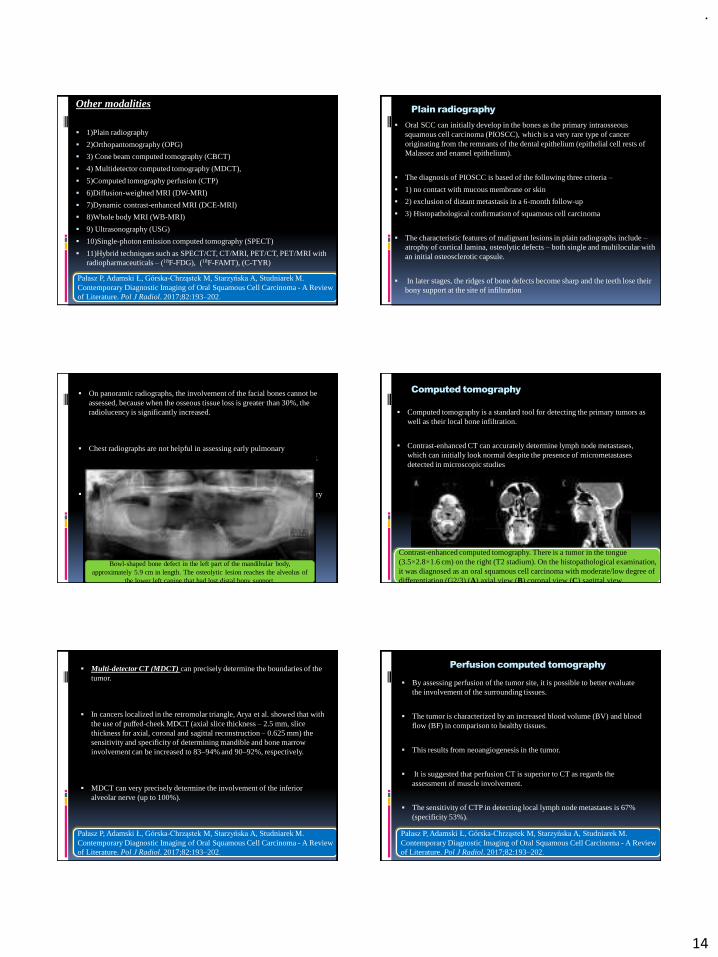
.
14
Other modalities
▪ 1)Plain radiography
▪ 2)Orthopantomography (OPG)
▪ 3) Cone beam computed tomography (CBCT)
▪ 4) Multidetector computed tomography (MDCT),
▪ 5)Computed tomography perfusion (CTP)
▪ 6)Diffusion-weighted MRI (DW-MRI)
▪ 7)Dynamic contrast-enhanced MRI (DCE-MRI)
▪ 8)Whole body MRI (WB-MRI)
▪ 9) Ultrasonography (USG)
▪ 10)Single-photon emission computed tomography (SPECT)
▪ 11)Hybrid techniques such as SPECT/CT, CT/MRI, PET/CT, PET/MRI with radiopharmaceuticals – (18F-FDG), (18F-FAMT), (C-TYR)
Pałasz P, Adamski Ł, Górska-Chrząstek M, Starzyńska A, Studniarek M. Contemporary Diagnostic Imaging of Oral Squamous Cell Carcinoma - A Review of Literature. Pol J Radiol. 2017;82:193–202.
Plain radiography
▪ Oral SCC can initially develop in the bones as the primary intraosseoussquamous cell carcinoma (PIOSCC), which is a very rare type of cancer originating from the remnants of the dental epithelium (epithelial cell rests of Malassez and enamel epithelium).
▪ The diagnosis of PIOSCC is based of the following three criteria –
▪ 1) no contact with mucous membrane or skin
▪ 2) exclusion of distant metastasis in a 6-month follow-up
▪ 3) Histopathological confirmation of squamous cell carcinoma
▪ The characteristic features of malignant lesions in plain radiographs include –atrophy of cortical lamina, osteolytic defects – both single and multilocular with an initial osteosclerotic capsule.
▪ In later stages, the ridges of bone defects become sharp and the teeth lose their bony support at the site of infiltration
▪ On panoramic radiographs, the involvement of the facial bones cannot be assessed, because when the osseous tissue loss is greater than 30%, the radiolucency is significantly increased.
▪ Chest radiographs are not helpful in assessing early pulmonary dissemination of oral cancers and low-dose CT (LDCT) is used routinely.
▪ It is a very accurate modality that can either confirm or exclude pulmonary metastases.
Bowl-shaped bone defect in the left part of the mandibular body, approximately 5.9 cm in length. The osteolytic lesion reaches the alveolus of
the lower left canine that had lost distal bony support.
Computed tomography
▪ Computed tomography is a standard tool for detecting the primary tumors as well as their local bone infiltration.
▪ Contrast-enhanced CT can accurately determine lymph node metastases, which can initially look normal despite the presence of micrometastasesdetected in microscopic studies
Contrast-enhanced computed tomography. There is a tumor in the tongue (3.5×2.8×1.6 cm) on the right (T2 stadium). On the histopathological examination, it was diagnosed as an oral squamous cell carcinoma with moderate/low degree of differentiation (G2/3) (A) axial view (B) coronal view (C) sagittal view.
▪ Multi-detector CT (MDCT) can precisely determine the boundaries of the tumor.
▪ In cancers localized in the retromolar triangle, Arya et al. showed that with the use of puffed-cheek MDCT (axial slice thickness – 2.5 mm, slice thickness for axial, coronal and sagittal reconstruction – 0.625 mm) the sensitivity and specificity of determining mandible and bone marrow involvement can be increased to 83–94% and 90–92%, respectively.
▪ MDCT can very precisely determine the involvement of the inferior alveolar nerve (up to 100%).
Pałasz P, Adamski Ł, Górska-Chrząstek M, Starzyńska A, Studniarek M. Contemporary Diagnostic Imaging of Oral Squamous Cell Carcinoma - A Review of Literature. Pol J Radiol. 2017;82:193–202.
Perfusion computed tomography
▪ By assessing perfusion of the tumor site, it is possible to better evaluate the involvement of the surrounding tissues.
▪ The tumor is characterized by an increased blood volume (BV) and blood flow (BF) in comparison to healthy tissues.
▪ This results from neoangiogenesis in the tumor.
▪ It is suggested that perfusion CT is superior to CT as regards the assessment of muscle involvement.
▪ The sensitivity of CTP in detecting local lymph node metastases is 67% (specificity 53%).
Pałasz P, Adamski Ł, Górska-Chrząstek M, Starzyńska A, Studniarek M. Contemporary Diagnostic Imaging of Oral Squamous Cell Carcinoma - A Review of Literature. Pol J Radiol. 2017;82:193–202.
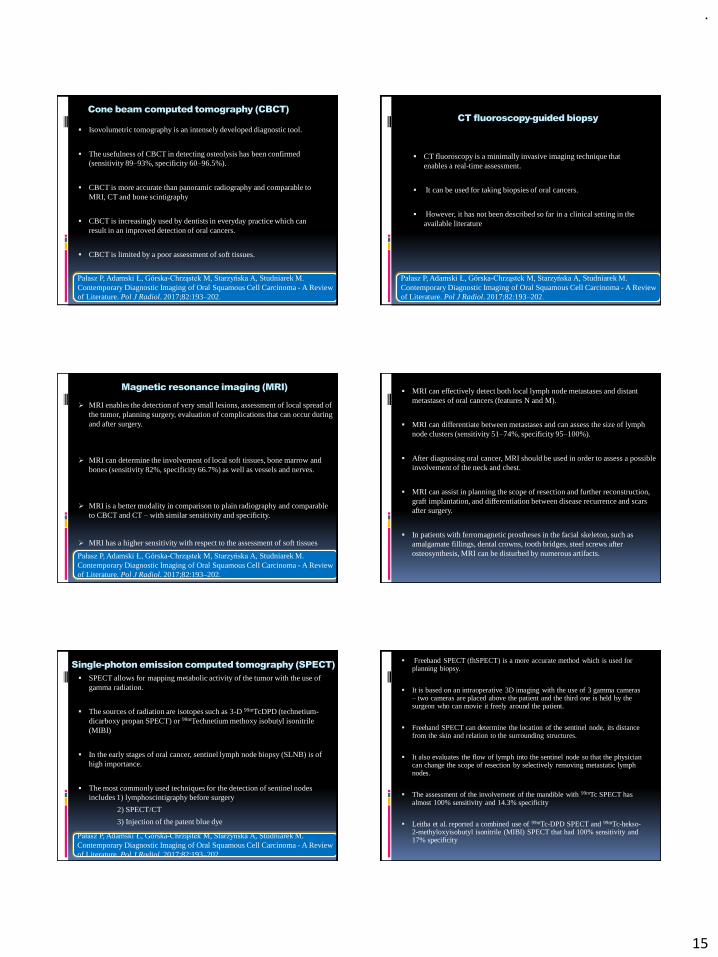
.
15
Cone beam computed tomography (CBCT)
▪ Isovolumetric tomography is an intensely developed diagnostic tool.
▪ The usefulness of CBCT in detecting osteolysis has been confirmed (sensitivity 89–93%, specificity 60–96.5%).
▪ CBCT is more accurate than panoramic radiography and comparable to MRI, CT and bone scintigraphy
▪ CBCT is increasingly used by dentists in everyday practice which can result in an improved detection of oral cancers.
▪ CBCT is limited by a poor assessment of soft tissues.
Pałasz P, Adamski Ł, Górska-Chrząstek M, Starzyńska A, Studniarek M. Contemporary Diagnostic Imaging of Oral Squamous Cell Carcinoma - A Review of Literature. Pol J Radiol. 2017;82:193–202.
CT fluoroscopy-guided biopsy
▪ CT fluoroscopy is a minimally invasive imaging technique that enables a real-time assessment.
▪ It can be used for taking biopsies of oral cancers.
▪ However, it has not been described so far in a clinical setting in the available literature
Pałasz P, Adamski Ł, Górska-Chrząstek M, Starzyńska A, Studniarek M. Contemporary Diagnostic Imaging of Oral Squamous Cell Carcinoma - A Review of Literature. Pol J Radiol. 2017;82:193–202.
Magnetic resonance imaging (MRI)
➢ MRI enables the detection of very small lesions, assessment of local spread of the tumor, planning surgery, evaluation of complications that can occur during and after surgery.
➢ MRI can determine the involvement of local soft tissues, bone marrow and bones (sensitivity 82%, specificity 66.7%) as well as vessels and nerves.
➢ MRI is a better modality in comparison to plain radiography and comparable to CBCT and CT – with similar sensitivity and specificity.
➢ MRI has a higher sensitivity with respect to the assessment of soft tissues
Pałasz P, Adamski Ł, Górska-Chrząstek M, Starzyńska A, Studniarek M. Contemporary Diagnostic Imaging of Oral Squamous Cell Carcinoma - A Review of Literature. Pol J Radiol. 2017;82:193–202.
▪ MRI can effectively detect both local lymph node metastases and distant metastases of oral cancers (features N and M).
▪ MRI can differentiate between metastases and can assess the size of lymph node clusters (sensitivity 51–74%, specificity 95–100%).
▪ After diagnosing oral cancer, MRI should be used in order to assess a possible involvement of the neck and chest.
▪ MRI can assist in planning the scope of resection and further reconstruction, graft implantation, and differentiation between disease recurrence and scars after surgery.
▪ In patients with ferromagnetic prostheses in the facial skeleton, such as amalgamate fillings, dental crowns, tooth bridges, steel screws after osteosynthesis, MRI can be disturbed by numerous artifacts.
Single-photon emission computed tomography (SPECT)
▪ SPECT allows for mapping metabolic activity of the tumor with the use of gamma radiation.
▪ The sources of radiation are isotopes such as 3-D 99mTcDPD (technetium-dicarboxy propan SPECT) or 99mTechnetium methoxy isobutyl isonitrile(MIBI)
▪ In the early stages of oral cancer, sentinel lymph node biopsy (SLNB) is of high importance.
▪ The most commonly used techniques for the detection of sentinel nodes includes 1) lymphoscintigraphy before surgery
2) SPECT/CT
3) Injection of the patent blue dye
Pałasz P, Adamski Ł, Górska-Chrząstek M, Starzyńska A, Studniarek M. Contemporary Diagnostic Imaging of Oral Squamous Cell Carcinoma - A Review of Literature. Pol J Radiol. 2017;82:193–202.
▪ Freehand SPECT (fhSPECT) is a more accurate method which is used for planning biopsy.
▪ It is based on an intraoperative 3D imaging with the use of 3 gamma cameras – two cameras are placed above the patient and the third one is held by the surgeon who can movie it freely around the patient.
▪ Freehand SPECT can determine the location of the sentinel node, its distance from the skin and relation to the surrounding structures.
▪ It also evaluates the flow of lymph into the sentinel node so that the physician can change the scope of resection by selectively removing metastatic lymph nodes.
▪ The assessment of the involvement of the mandible with 99mTc SPECT has almost 100% sensitivity and 14.3% specificity
▪ Leitha et al. reported a combined use of 99mTc-DPD SPECT and 99mTc-hekso-2-methyloxyisobutyl isonitrile (MIBI) SPECT that had 100% sensitivity and 17% specificity
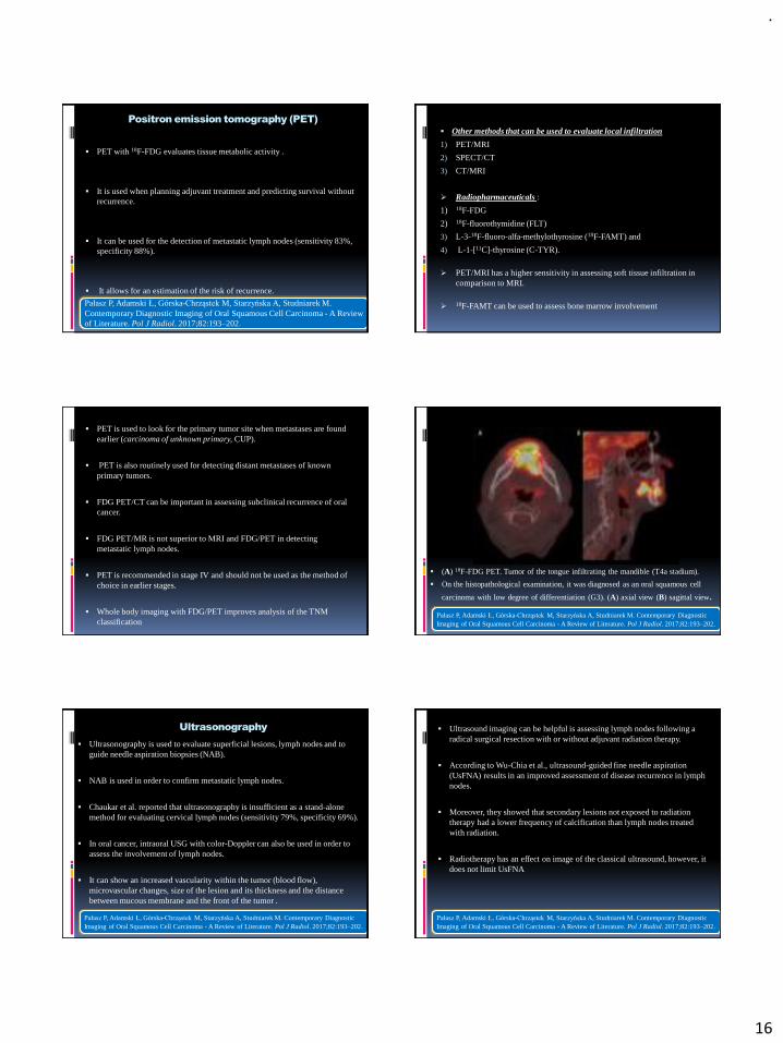
.
16
Positron emission tomography (PET)
▪ PET with 18F-FDG evaluates tissue metabolic activity .
▪ It is used when planning adjuvant treatment and predicting survival without recurrence.
▪ It can be used for the detection of metastatic lymph nodes (sensitivity 83%, specificity 88%).
▪ It allows for an estimation of the risk of recurrence.
Pałasz P, Adamski Ł, Górska-Chrząstek M, Starzyńska A, Studniarek M. Contemporary Diagnostic Imaging of Oral Squamous Cell Carcinoma - A Review of Literature. Pol J Radiol. 2017;82:193–202.
▪ Other methods that can be used to evaluate local infiltration
1) PET/MRI
2) SPECT/CT
3) CT/MRI
➢ Radiopharmaceuticals :
1) 18F-FDG
2) 18F-fluorothymidine (FLT)
3) L-3-18F-fluoro-alfa-methylothyrosine (18F-FAMT) and
4) L-1-[11C]-thyrosine (C-TYR).
➢ PET/MRI has a higher sensitivity in assessing soft tissue infiltration in comparison to MRI.
➢18F-FAMT can be used to assess bone marrow involvement
▪ PET is used to look for the primary tumor site when metastases are found earlier (carcinoma of unknown primary, CUP).
▪ PET is also routinely used for detecting distant metastases of known primary tumors.
▪ FDG PET/CT can be important in assessing subclinical recurrence of oral cancer.
▪ FDG PET/MR is not superior to MRI and FDG/PET in detecting metastatic lymph nodes.
▪ PET is recommended in stage IV and should not be used as the method of choice in earlier stages.
▪ Whole body imaging with FDG/PET improves analysis of the TNM classification
▪ (A) 18F-FDG PET. Tumor of the tongue infiltrating the mandible (T4a stadium).
▪ On the histopathological examination, it was diagnosed as an oral squamous cell
carcinoma with low degree of differentiation (G3). (A) axial view (B) sagittal view.
Pałasz P, Adamski Ł, Górska-Chrząstek M, Starzyńska A, Studniarek M. Contemporary Diagnostic Imaging of Oral Squamous Cell Carcinoma - A Review of Literature. Pol J Radiol. 2017;82:193–202.
Ultrasonography
▪ Ultrasonography is used to evaluate superficial lesions, lymph nodes and to guide needle aspiration biopsies (NAB).
▪ NAB is used in order to confirm metastatic lymph nodes.
▪ Chaukar et al. reported that ultrasonography is insufficient as a stand-alone method for evaluating cervical lymph nodes (sensitivity 79%, specificity 69%).
▪ In oral cancer, intraoral USG with color-Doppler can also be used in order to assess the involvement of lymph nodes.
▪ It can show an increased vascularity within the tumor (blood flow), microvascular changes, size of the lesion and its thickness and the distance between mucous membrane and the front of the tumor .
Pałasz P, Adamski Ł, Górska-Chrząstek M, Starzyńska A, Studniarek M. Contemporary Diagnostic Imaging of Oral Squamous Cell Carcinoma - A Review of Literature. Pol J Radiol. 2017;82:193–202.
▪ Ultrasound imaging can be helpful is assessing lymph nodes following a radical surgical resection with or without adjuvant radiation therapy.
▪ According to Wu-Chia et al., ultrasound-guided fine needle aspiration (UsFNA) results in an improved assessment of disease recurrence in lymph nodes.
▪ Moreover, they showed that secondary lesions not exposed to radiation therapy had a lower frequency of calcification than lymph nodes treated with radiation.
▪ Radiotherapy has an effect on image of the classical ultrasound, however, it does not limit UsFNA
Pałasz P, Adamski Ł, Górska-Chrząstek M, Starzyńska A, Studniarek M. Contemporary Diagnostic Imaging of Oral Squamous Cell Carcinoma - A Review of Literature. Pol J Radiol. 2017;82:193–202.

.
17
▪ Hypertrophy is an increase in the size of cells resulting in increase in the size of the organ.
▪ Hyperplasia is characterized by an increase in cell number because of proliferation of differentiated cells and replacement by tissue stem cells.
▪ Atrophy- Shrinkage in the size of the cell by the loss of cell substance
▪ Cancer is a genetic disorder caused by DNA mutations that are (for the most part) acquired spontaneously or induced by environmental insults.
▪ Malignant tumors are collectively referred to as cancers, derived from the Latin word for “crab”—that is, they adhere to any part that they seize in an obstinate manner, similar to a crab’s behavior.
▪ Malignant, as applied to a neoplasm, implies that the lesion can invade and destroy adjacent structures and spread to distant sites (metastasize) to cause death.
TUMOUR MARKERS
▪ Tumor markers are substances produced by tumor cells or by other cells within the body in response to cancer or certain benign conditions.
▪ It includes a variety of substances like cell surface antigens, cytoplasmic proteins, enzymes, hormones, oncofetal antigens, receptors, oncogens and their products.
Lavanya R, Mamatha B, Waghray S, Chaitanya N, Reddy M, Bau D. Role of Tumor Markers in Oral Cancer: An Overview. Br J Med Med Res. 2016;15(7):1-9.
Circulating body fluids Tissue
VEGF/VEGF-R(Vascular endothelialgrowth factor/receptor)
Epithelial markers➢Cell surface markers –Histocompatibility➢Intracellular markers – Cytokeratins➢Basement membrane markers – Type 4 collagen➢Matrix markers – Tenascin➢Membrane antigen – Blood group antigens
PD-ECGF (Platelet derived endothelial cell growth factor)
Connective tissue markers ➢Intermediate filament proteins –Desmin➢Other filament proteins – Laminin
FGF, BCL2 Cellular enzymes –Amylase, lysozyme,
Cathepsin –D,CD44, CD80, CD105, CEA, CA-19-9, CA-125, SCC-Ag
Cytoplasmic non-filamentous non-enzymatic proteins – Myoglobin, S100 protein
Endoglin, Cytokeritins, Calretinin, Cerb2,Cyclin, MIB and p53
Membrane antigen – Leukocyte specific antigen
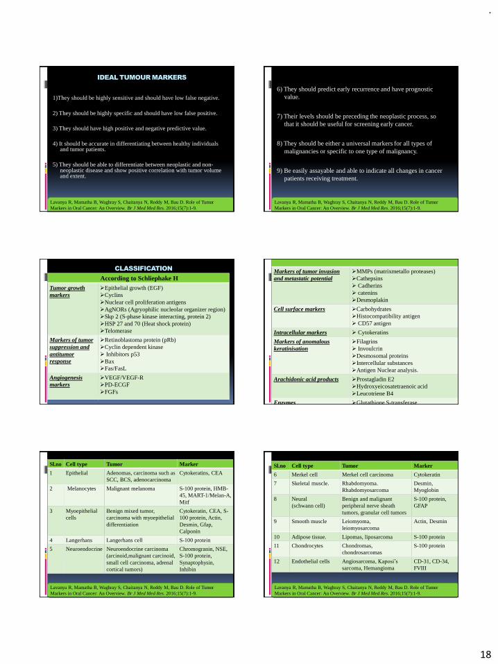
.
18
IDEAL TUMOUR MARKERS
1)They should be highly sensitive and should have low false negative.
2) They should be highly specific and should have low false positive.
3) They should have high positive and negative predictive value.
4) It should be accurate in differentiating between healthy individuals and tumor patients.
5) They should be able to differentiate between neoplastic and non-neoplastic disease and show positive correlation with tumor volume and extent.
Lavanya R, Mamatha B, Waghray S, Chaitanya N, Reddy M, Bau D. Role of Tumor Markers in Oral Cancer: An Overview. Br J Med Med Res. 2016;15(7):1-9.
6) They should predict early recurrence and have prognostic value.
7) Their levels should be preceding the neoplastic process, so that it should be useful for screening early cancer.
8) They should be either a universal markers for all types of malignancies or specific to one type of malignancy.
9) Be easily assayable and able to indicate all changes in cancer patients receiving treatment.
Lavanya R, Mamatha B, Waghray S, Chaitanya N, Reddy M, Bau D. Role of Tumor Markers in Oral Cancer: An Overview. Br J Med Med Res. 2016;15(7):1-9.
CLASSIFICATION
According to Schliephake HTumor growth markers
➢Epithelial growth (EGF)➢Cyclins➢Nuclear cell proliferation antigens➢AgNORs (Agryophilic nucleolar organizer region) ➢Skp 2 (S-phase kinase interacting, protein 2) ➢HSP 27 and 70 (Heat shock protein) ➢Telomerase
Markers of tumor suppression and antitumor response
➢Retinoblastoma protein (pRb)➢Cyclin dependent kinase➢ Inhibitors p53➢Bax➢Fas/FasL
Angiogenesis markers
➢VEGF/VEGF-R➢PD-ECGF➢FGFs
Markers of tumor invasion and metastatic potential
➢MMPs (matrixmetallo proteases)➢Cathepsins➢ Cadherins➢ catenins➢Desmoplakin
Cell surface markers ➢Carbohydrates ➢Histocompatibility antigen➢ CD57 antigen
Intracellular markers ➢ Cytokeratins
Markers of anomalous keratinisation
➢Filagrins➢ Invoulcrin➢Desmosomal proteins➢Intercellular substances➢Antigen Nuclear analysis.
Arachidonic acid products ➢Prostagladin E2➢Hydroxyeicosatetraenoic acid➢Leucotriene B4
Enzymes ➢Glutathione S-transferase
Sl.no Cell type Tumor Marker
1 Epithelial Adenomas, carcinoma such as SCC, BCS, adenocarcinoma
Cytokeratins, CEA
2 Melanocytes Malignant melanoma S-100 protein, HMB-45, MART-1/Melan-A, Mitf
3 Myoepithelialcells
Benign mixed tumor, carcinoma with myoepithelialdifferentiation
Cytokeratin, CEA, S-100 protein, Actin, Desmin, Gfap, Calponin
4 Langerhans Langerhans cell S-100 protein
5 Neuroendocrine Neuroendocrine carcinoma (arcinoid,malignant carcinoid, small cell carcinoma, adrenal cortical tumors)
Chromogranin, NSE, S-100 protein, Synaptophysin, Inhibin
Lavanya R, Mamatha B, Waghray S, Chaitanya N, Reddy M, Bau D. Role of Tumor Markers in Oral Cancer: An Overview. Br J Med Med Res. 2016;15(7):1-9.
Sl.no Cell type Tumor Marker
6 Merkel cell Merkel cell carcinoma Cytokeratin
7 Skeletal muscle. Rhabdomyoma. Rhabdomyosarcoma
Desmin, Myoglobin
8 Neural(schwann cell)
Benign and malignant peripheral nerve sheath tumors, granular cell tumors
S-100 protein, GFAP
9 Smooth muscle Leiomyoma, leiomyosarcoma
Actin, Desmin
10 Adipose tissue. Lipomas, liposarcoma S-100 protein
11 Chondrocytes Chondromas, chondrosarcomas
S-100 protein
12 Endothelial cells Angiosarcoma, Kaposi’s
sarcoma, HemangiomaCD-31, CD-34, FVIII
Lavanya R, Mamatha B, Waghray S, Chaitanya N, Reddy M, Bau D. Role of Tumor Markers in Oral Cancer: An Overview. Br J Med Med Res. 2016;15(7):1-9.
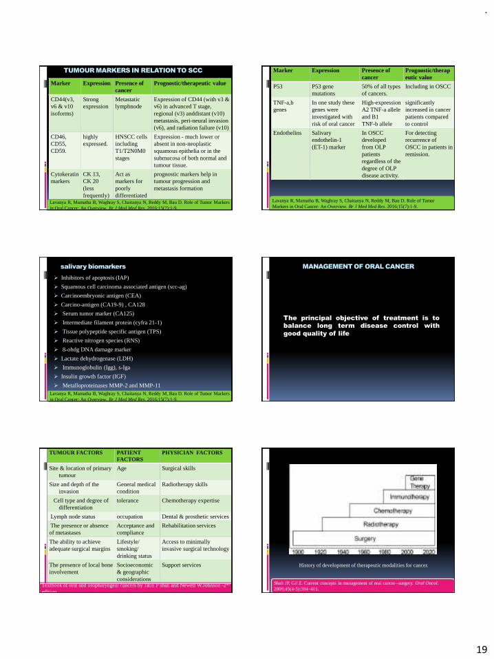
.
19
TUMOUR MARKERS IN RELATION TO SCC
Marker Expression Presence of cancer
Prognostic/therapeutic value
CD44(v3, v6 & v10isoforms)
Strong expression
Metastaticlymphnode
Expression of CD44 (with v3 & v6) in advanced T stage, regional (v3) anddistant (v10) metastasis, peri-neural invasion (v6), and radiation failure (v10)
CD46, CD55,CD59.
highly expressed.
HNSCC cellsincludingT1/T2N0M0 stages
Expression - much lower or absent in non-neoplasticsquamous epithelia or in the submucosa of both normal and tumour tissue.
Cytokeratinmarkers
CK 13,CK 20(less frequently)
Act as markers for poorly differentiatedsquamous cell
prognostic markers help in tumour progression and metastasis formation
Lavanya R, Mamatha B, Waghray S, Chaitanya N, Reddy M, Bau D. Role of Tumor Markers in Oral Cancer: An Overview. Br J Med Med Res. 2016;15(7):1-9.
Marker Expression Presence of cancer
Prognostic/therapeutic value
P53 P53 gene mutations
50% of all types of cancers.
Including in OSCC
TNF-a,bgenes
In one study these genes were investigated with risk of oral cancer
High-expression A2 TNF-a allele and B1TNF-b allele
significantly increased in cancer patients compared to control
Endothelins Salivary endothelin-1(ET-1) marker
In OSCC developedfrom OLP patientsregardless of thedegree of OLPdisease activity.
For detecting recurrence ofOSCC in patients in remission.
Lavanya R, Mamatha B, Waghray S, Chaitanya N, Reddy M, Bau D. Role of Tumor Markers in Oral Cancer: An Overview. Br J Med Med Res. 2016;15(7):1-9.
salivary biomarkers
➢ Inhibitors of apoptosis (IAP)
➢ Squamous cell carcinoma associated antigen (scc-ag)
➢ Carcinoembryonic antigen (CEA)
➢ Carcino-antigen (CA19-9) , CA128
➢ Serum tumor marker (CA125)
➢ Intermediate filament protein (cyfra 21-1)
➢ Tissue polypeptide specific antigen (TPS)
➢ Reactive nitrogen species (RNS)
➢ 8-ohdg DNA damage marker
➢ Lactate dehydrogenase (LDH)
➢ Immunoglobulin (Igg), s-Iga
➢ Insulin growth factor (IGF)
➢ Metalloproteinases MMP-2 and MMP-11Lavanya R, Mamatha B, Waghray S, Chaitanya N, Reddy M, Bau D. Role of Tumor Markers in Oral Cancer: An Overview. Br J Med Med Res. 2016;15(7):1-9.
MANAGEMENT OF ORAL CANCER
The principal objective of treatment is to
balance long term disease control with
good quality of life
TUMOUR FACTORS PATIENTFACTORS
PHYSICIAN FACTORS
Site & location of primary tumour
Age Surgical skills
Size and depth of the invasion
General medical condition
Radiotherapy skills
Cell type and degree ofdifferentiation
tolerance Chemotherapy expertise
Lymph node status occupation Dental & prosthetic services
The presence or absence of metastases
Acceptance and compliance
Rehabilitation services
The ability to achieve adequate surgical margins
Lifestyle/smoking/drinking status
Access to minimally invasive surgical technology
The presence of local bone involvement
Socioeconomic & geographic considerations
Support services
Textbook of oral and oropharyngeal cancers by Jatin P shah and Newell W.Johnson -2nd
edition
History of development of therapeutic modalities for cancer.
Shah JP, Gil Z. Current concepts in management of oral cancer--surgery. Oral Oncol. 2009;45(4-5):394–401.
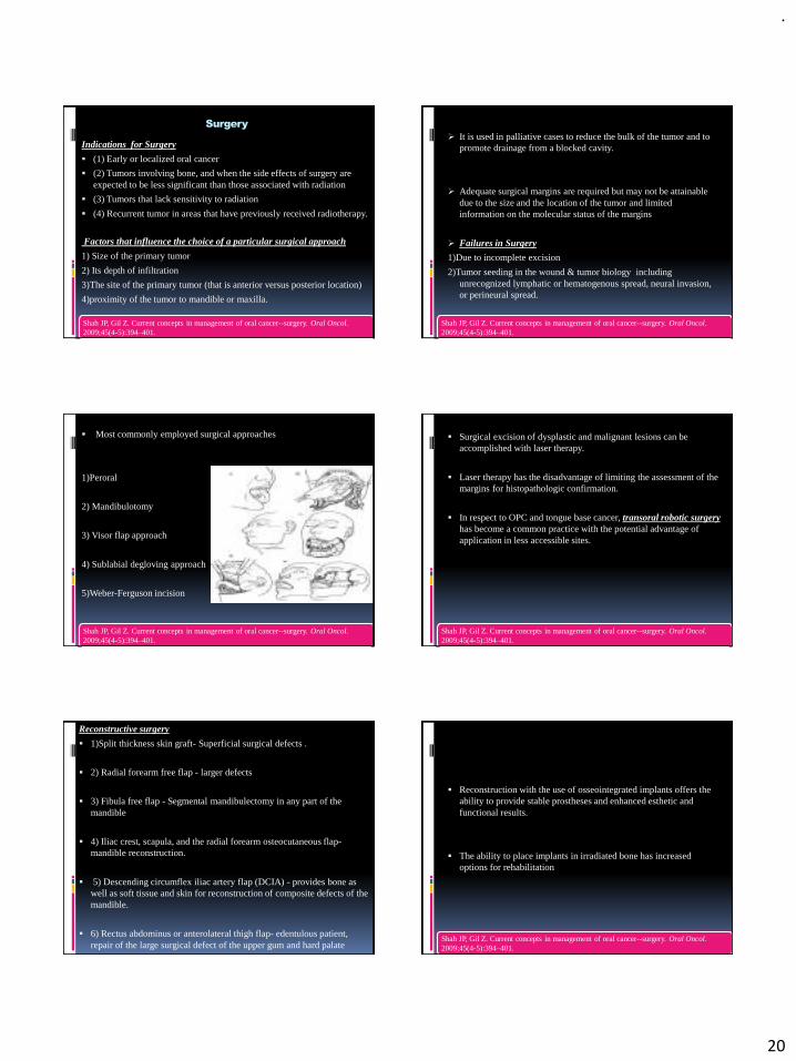
.
20
Surgery
Indications for Surgery
▪ (1) Early or localized oral cancer
▪ (2) Tumors involving bone, and when the side effects of surgery are expected to be less significant than those associated with radiation
▪ (3) Tumors that lack sensitivity to radiation
▪ (4) Recurrent tumor in areas that have previously received radiotherapy.
Factors that influence the choice of a particular surgical approach
1) Size of the primary tumor
2) Its depth of infiltration
3)The site of the primary tumor (that is anterior versus posterior location)
4)proximity of the tumor to mandible or maxilla.
Shah JP, Gil Z. Current concepts in management of oral cancer--surgery. Oral Oncol. 2009;45(4-5):394–401.
➢ It is used in palliative cases to reduce the bulk of the tumor and to promote drainage from a blocked cavity.
➢ Adequate surgical margins are required but may not be attainable due to the size and the location of the tumor and limited information on the molecular status of the margins
➢ Failures in Surgery
1)Due to incomplete excision
2)Tumor seeding in the wound & tumor biology including unrecognized lymphatic or hematogenous spread, neural invasion, or perineural spread.
Shah JP, Gil Z. Current concepts in management of oral cancer--surgery. Oral Oncol. 2009;45(4-5):394–401.
▪ Most commonly employed surgical approaches
1)Peroral
2) Mandibulotomy
3) Visor flap approach
4) Sublabial degloving approach
5)Weber-Ferguson incision
Shah JP, Gil Z. Current concepts in management of oral cancer--surgery. Oral Oncol. 2009;45(4-5):394–401.
▪ Surgical excision of dysplastic and malignant lesions can be accomplished with laser therapy.
▪ Laser therapy has the disadvantage of limiting the assessment of the margins for histopathologic confirmation.
▪ In respect to OPC and tongue base cancer, transoral robotic surgery has become a common practice with the potential advantage of application in less accessible sites.
Shah JP, Gil Z. Current concepts in management of oral cancer--surgery. Oral Oncol. 2009;45(4-5):394–401.
Reconstructive surgery
▪ 1)Split thickness skin graft- Superficial surgical defects .
▪ 2) Radial forearm free flap - larger defects
▪ 3) Fibula free flap - Segmental mandibulectomy in any part of the mandible
▪ 4) Iliac crest, scapula, and the radial forearm osteocutaneous flap-mandible reconstruction.
▪ 5) Descending circumflex iliac artery flap (DCIA) - provides bone as well as soft tissue and skin for reconstruction of composite defects of the mandible.
▪ 6) Rectus abdominus or anterolateral thigh flap- edentulous patient, repair of the large surgical defect of the upper gum and hard palate
▪ Reconstruction with the use of osseointegrated implants offers the ability to provide stable prostheses and enhanced esthetic and functional results.
▪ The ability to place implants in irradiated bone has increased options for rehabilitation
Shah JP, Gil Z. Current concepts in management of oral cancer--surgery. Oral Oncol. 2009;45(4-5):394–401.
.
21
Radiation Therapy
▪ Radiation therapy may be administered with intent to cure, as a single modality, as part of a combined radiation surgery and/or chemotherapy management, or for palliation.
▪ Radiation may provide symptomatic relief from pain, bleeding, ulceration, and oropharyngeal obstruction.
▪ Hyperfractionation of radiation is one of the strategies to increase intensity of treatment to increase tumoricidal effects, which results in more severe acute effects.
▪ High-dose re-irradiation is offered in some centers as salvage treatment and may be considered in case of recurrent or second primary head and neck cancer, particularly when salvage surgery is not feasible.
▪ Radiation kills cells by interaction with water molecules in the cells, producing charged molecules that interact with biochemical processes in the cells and by causing direct damage to DNA.
▪ The affected cells may die or remain incapable of division.
▪ Due to a greater potential for cell repair in normal tissue than in malignant cells and a greater susceptibility to radiation due to the higher growth fraction of cancer cells, a differential effect is achieved.
Three-dimensional (3D) conformal radiotherapy
▪ Three-dimensional (3D) conformal radiation therapy- uses reconstructed matched computed tomograms (CT) and Magnetic Resonance images (MRI) during treatment plan reduces the risk of geographic miss.
▪ The distribution of the beam can be conformed to the tumor size and shape using customized dense block or by multileaf collimators which has 40 pairs of tungsten measuring 1 cm in width.
▪ These can be adjusted to define the x ray beam thus reducing the dose of radiation up to 50% to normal tissues and this in turn reduces late damage
Amrit Kaur Mann, Indira AP, Maria Priscilla David. Recent advances in radiotherapy for head and neck cancer: a comprehensive review. International Journal of Contemporary Medical Research 2017;4(4):927-932
Intensity-modulated radiotherapy (IMRT)
▪ Intensity-modulated radiation therapy (IMRT) is an improved mode of high-precision radiotherapy that utilizes computer controlled linear accelerators to deliver precise radiation doses to a malignant tumor or to specific areas within the tumor.
▪ In this technique, the equipment can be rotated around the patient where the beam moves multiple times that may vary with intensity, resulting in three dimensionally sculpted radiation.
▪ It allows for the radiation dose to conform to the three-dimensional shape of the tumor, by modulating or regulating the intensity of the radiation beam in multiple trivial volumes.
▪ This permits increased radiation doses to be focused to regions within the tumor, while decreasing the dose to surrounding normal critical structures.
Amrit Kaur Mann, Indira AP, Maria Priscilla David. Recent advances in radiotherapy for head and neck cancer: a comprehensive review. International Journal of Contemporary Medical Research 2017;4(4):927-932
.
22
Volumated Intensity modulated Arc Therapy (VMAT)
▪ Volumated intensity modulated arc therapy (VMAT) is newer technique of delivering IMRT.
▪ VMAT delivers IMRT-like distributions in a single rotation of the gantry, varying the gantry speed and dose rate during delivery in contrast to standard IMRT, which uses fixed gantry beams.
▪ This technique has been implemented in the Eclipse treatment planning software under the name Rapid Arc(RA) Planning.
▪ Studies using RA demonstrate shorter planning and treatment time, lesser monitor units for treatment delivery, better dose homogeneity and normal tissue sparing.
Image Guided Radiotherapy (IGRT)
▪ It is often used in conjunction with intensity-modulated radiation therapy (IMRT), proton beam therapy, or stereotactic body radiotherapy (SBRT).
▪ The most basic and historical form of IGRT comprises of 2 dimensional portal images acquired in perpendicular/orthogonal planes to confirm the position of the isocenter, as well as the distinct fields.
▪ In most cases, these images are produced by the megavoltage (MV) beam of the treatment machine or less often by a devoted ancillary kilovoltage unit.
▪ These images are obtained prior to treatment.
▪ IGRT is used to treat Head and Neck cancers and tumors in areas of the body that are prone to movement, such as the lungs, as well as tumors located close to critical organs and tissues.
▪ Image guidance can be used for improved tumor delineation and to correct intra and inter fraction movement during radiotherapy.
▪ Computerized tomograms with image guidance provide three dimensional views of tumor and normal anatomy.
▪ This consists of a compact CT scanner that is combined into the linear accelerator unit.
▪ This scan is usually of lower resolution than a diagnostic CT scan but provides adequate bony and soft tissue resolution for anatomic alignment.
▪ IGRT can be a useful tool that can detect and correct the geographic miss that can occur in treatment delivery.
▪ This technique can be fused with Positron emission tomography, which aids in delineation of gross tumor volume by using radioactive tracer namely Fluorine 18 labelled fluoromisonidazolehave been shown to highlight the hypoxic areas of tumors.
Amrit Kaur Mann, Indira AP, Maria Priscilla David. Recent advances in radiotherapy for head and neck cancer: a comprehensive review. International Journal of Contemporary Medical Research 2017;4(4):927-932
Particle Radiation therapy
▪ Charged particle beams consisting of protons and carbon ions have the Bragg peak and allow highly localised deposition of energy that can be used for increasing radiation doses to target while minimising irradiation to adjacent normal tissues.
▪ Intensity modulated proton (IMPT) allows modulation of the fluence and position of Bragg peak, permitting three dimensional dose distributions.
▪ Combination of photon and proton therapy for advanced malignant sinonasal tumours demonstrated good results when used IMRT alone.
Amrit Kaur Mann, Indira AP, Maria Priscilla David. Recent advances in radiotherapy for head and neck cancer: a comprehensive review. International Journal of Contemporary Medical Research 2017;4(4):927-932
▪ The present role of proton therapy lies in the treatment of tumor close to the skull base or spinal cord and in pediatric patients.
▪ Proton therapy provides maximum benefit in terms of normal tissue sparing.
Amrit Kaur Mann, Indira AP, Maria Priscilla David. Recent advances in radiotherapy for head and neck cancer: a comprehensive review. International Journal of Contemporary Medical Research 2017;4(4):927-932

.
23
Thermo radiotherapy
▪ It involves the application of localized high temperature at the tumor site which is said to improve the radiation treatment outcome.
▪ Currently scientific research has been conducted on this treatment modality
Amrit Kaur Mann, Indira AP, Maria Priscilla David. Recent advances in radiotherapy for head and neck cancer: a comprehensive review. International Journal of Contemporary Medical Research 2017;4(4):927-932
Radioimmunotherapy(Targeted Radiotherapy)
▪ Radioimmunotherapy is a form of radiotherapy where cytotoxicradionuclides such as Yttrium 90, Iodine 131 are linked to antibodies in order to deliver toxins directly to the tumor targets.
▪ The efficiency of their radioisotopes is that it has longer path length and thus large tumors may receive a higher dose of radiation to a greater depth.
▪ Radioimmunotherapy has proved to be effective for treatment of lymphomas, whereas its application in oral cancer has been reported to be under clinical trials using monoclonal antibodies.
Amrit Kaur Mann, Indira AP, Maria Priscilla David. Recent advances in radiotherapy for head and neck cancer: a comprehensive review. International Journal of Contemporary Medical Research 2017;4(4):927-932
Stereotactic radiotherapy
▪ Stereotactic radiation uses a single high dose of radiation sent into cancerous tissue with very narrow beam of radiation.
▪ It is a radical type of surgery that uses highly targeted radiation to treat brain abnormalities which is proven safe and effective, with good outcomes.
Amrit Kaur Mann, Indira AP, Maria Priscilla David. Recent advances in radiotherapy for head and neck cancer: a comprehensive review. International Journal of Contemporary Medical Research 2017;4(4):927-932
Carbon ion Therapy
▪ Carbon ion radiotherapy can offer better dose conformitory to a target volume than other modalities.
▪ In addition high linear energy transfer (LET) radiation such as carbon ion beams, as greater biological effectiveness than low LET radiation, such as X rays and proton beams.
▪ Because of its better dose distribution and cell killing potency, carbon ion radiotherapy is a promising modality in the treatment of patient with malignancy
Amrit Kaur Mann, Indira AP, Maria Priscilla David. Recent advances in radiotherapy for head and neck cancer: a comprehensive review. International Journal of Contemporary Medical Research 2017;4(4):927-932
Boron Neutron Capture Therapy (BNCT)
▪ A new form of radiation therapy for cancer that is based upon the selective uptake of non-radioactive boron compounds.
▪ The boron compound is injected in the patient and when the required amount of boron reached the tumor site, the area could be irradiated with a neutron beam causing boron B10 to be transformed into B11, which would disintegrate releasing an α particle and 7Li, which have high
linear energy transfer (LET), thus killing the cancer cells with almost no damage to the surrounding normal tissues.
▪ The BNCT delivery agents currently used in clinical trials are sodium borocaptate (BSH) and boronophenylalanine or BPA.
▪ The merits of BNCT include distribution of high radiation dose only to the tumor cells while sparing the surrounding normal cells.
Sanctuary Therapy
▪ Sanctuary therapy is a form of treatment with radiation that offers promise of effectively treating a high risk site of metastasis which could escape the attention of the oncologists.
▪ The therapy consists of prophylactic treatment with radiation at a level of 2000 to 3000 rads to the area of site of interest.
▪ The term “sanctuaries” refers to the belief that these organs are
sanctuaries for tumour cells, since many chemotherapeutic agents do not cross the lipid barriers
Amrit Kaur Mann, Indira AP, Maria Priscilla David. Recent advances in radiotherapy for head and neck cancer: a comprehensive review. International Journal of Contemporary Medical Research 2017;4(4):927-932
.
24
Intraoperative Electron Radiation Therapy (IOERT)
▪ IOERT excludes the irradiation of normal tissues and the critical structures in and around the target volume, hence called precision radiotherapy as the clinician views the tumour directly.
▪ As the dose falls off rapidly below the target site sparing the underlying healthy tissue, electron radiation can be applied directly on the tumour, which is vulnerable for destruction during intraoperative procedures.
▪ IOERT has proven to be beneficial when used in conjunction with endovascular brachytherapy which in turn reduces integral dose and treatment time
Amrit Kaur Mann, Indira AP, Maria Priscilla David. Recent advances in radiotherapy for head and neck cancer: a comprehensive review. International Journal of Contemporary Medical Research 2017;4(4):927-932
Brachytherapy
▪ Interstitial and intracavitary implants are used to treat primary cancers in the head and neck.
▪ Brachytherapy is the primary treatment modality for localized tumors in the anterior two-thirds of the oral cavity, for boosted doses of radiation to a specific site, or for treatment following recurrence.
▪ Isotopes - cesium, iridium, and gold.
▪ The frequency of tissue necrosis is related to the treated volume, fraction size and total dose of radiation therapy, the proximity of the radioactive implant to bone, and the presence of comorbid risk factors including dental status.
Chemotherapy
Cytotoxic Chemotherapy
▪ Chemotherapy is largely palliative in patients with recurrent or metastatic disease.
▪ Chemotherapy is combined with radiotherapy &/or surgery to increase locoregional control,decrease distant metastasis and improve survival
▪ Chemotherapy may be used as induction therapy prior to local therapies, concurrent chemoradiotherapy (CCRT), and adjuvant chemotherapy after local treatment
Induction chemotherapy
▪ Induction, or neoadjuvant, therapy, refers to a modality used before definitive therapy with the goal of maximizing the success of the definitive therapy.
▪ The objective of induction chemotherapy is to promote initial tumor reduction and to provide early treatment of micrometastasesdue to the recognition that local control has improved with aggressive combined therapy, but distant failure due to metastatic disease has increased.
▪ In 1970,cisplatin & bleomycin -2 cycles prior to local treatment-objective response rate (71%) & complete response rate (20%)
▪ Cisplatin & 5FU-3 cycles prior to local treatment-objective response rate(88%) & complete response rate(54%)
CONCURRENT CHEMORADIATION THERAPY
▪ Chemotherapy, particularly in combination with radiation therapy, is associated with potential local toxicities, such as mucositis; nausea, vomiting, and bone marrow suppression.
▪ The initial tumor response to chemotherapy prior to radiotherapy may predict tumor responsiveness to radiation.
▪ CCRT protocols are now the standard of care for stage 3 and 4 as primary therapy and for disease with poor prognostic findings following surgery including close margin and vascular invasion by tumor.
▪ As CCRT has become more widely used, the morbidities associated with these therapies have become more pronounced
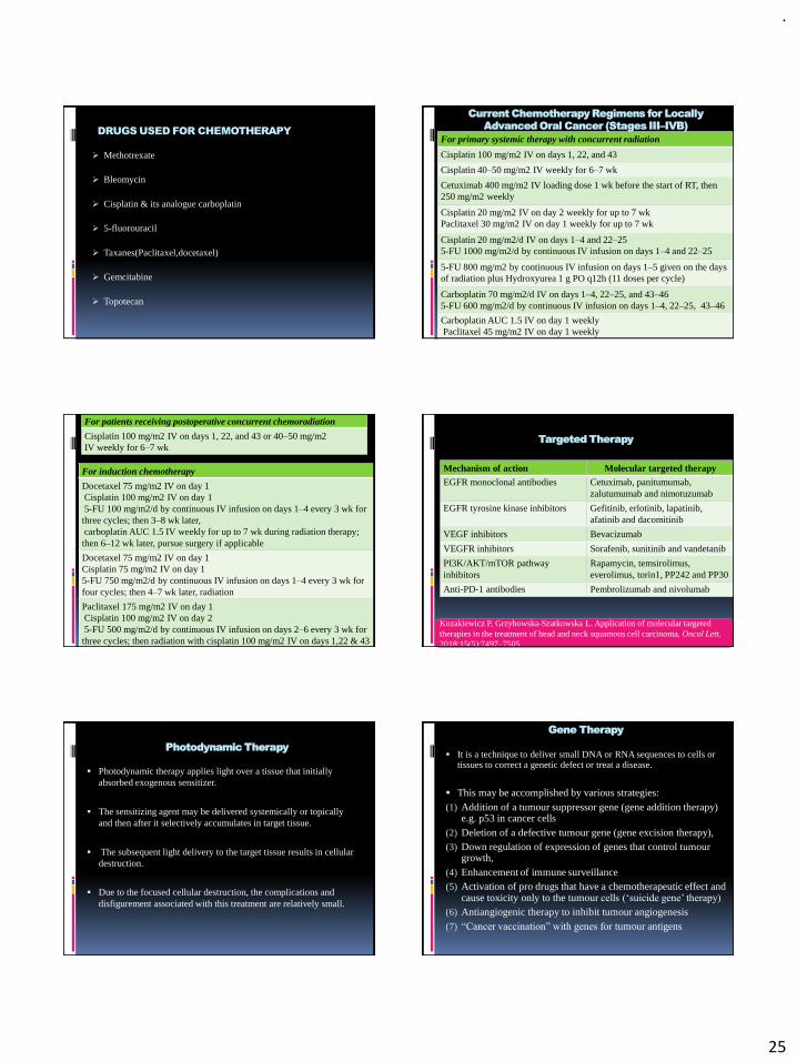
.
25
DRUGS USED FOR CHEMOTHERAPY
➢ Methotrexate
➢ Bleomycin
➢ Cisplatin & its analogue carboplatin
➢ 5-fluorouracil
➢ Taxanes(Paclitaxel,docetaxel)
➢ Gemcitabine
➢ Topotecan
Current Chemotherapy Regimens for Locally
Advanced Oral Cancer (Stages III–IVB)
For primary systemic therapy with concurrent radiation
Cisplatin 100 mg/m2 IV on days 1, 22, and 43
Cisplatin 40–50 mg/m2 IV weekly for 6–7 wk
Cetuximab 400 mg/m2 IV loading dose 1 wk before the start of RT, then 250 mg/m2 weekly
Cisplatin 20 mg/m2 IV on day 2 weekly for up to 7 wk Paclitaxel 30 mg/m2 IV on day 1 weekly for up to 7 wk
Cisplatin 20 mg/m2/d IV on days 1–4 and 22–25 5-FU 1000 mg/m2/d by continuous IV infusion on days 1–4 and 22–25
5-FU 800 mg/m2 by continuous IV infusion on days 1–5 given on the days of radiation plus Hydroxyurea 1 g PO q12h (11 doses per cycle)
Carboplatin 70 mg/m2/d IV on days 1–4, 22–25, and 43–46 5-FU 600 mg/m2/d by continuous IV infusion on days 1–4, 22–25, 43–46
Carboplatin AUC 1.5 IV on day 1 weeklyPaclitaxel 45 mg/m2 IV on day 1 weekly
For patients receiving postoperative concurrent chemoradiation
Cisplatin 100 mg/m2 IV on days 1, 22, and 43 or 40–50 mg/m2IV weekly for 6–7 wk
For induction chemotherapy
Docetaxel 75 mg/m2 IV on day 1Cisplatin 100 mg/m2 IV on day 1 5-FU 100 mg/m2/d by continuous IV infusion on days 1–4 every 3 wk for three cycles; then 3–8 wk later,carboplatin AUC 1.5 IV weekly for up to 7 wk during radiation therapy; then 6–12 wk later, pursue surgery if applicable
Docetaxel 75 mg/m2 IV on day 1 Cisplatin 75 mg/m2 IV on day 1 5-FU 750 mg/m2/d by continuous IV infusion on days 1–4 every 3 wk for four cycles; then 4–7 wk later, radiation
Paclitaxel 175 mg/m2 IV on day 1 Cisplatin 100 mg/m2 IV on day 2 5-FU 500 mg/m2/d by continuous IV infusion on days 2–6 every 3 wk for three cycles; then radiation with cisplatin 100 mg/m2 IV on days 1,22 & 43
Mechanism of action Molecular targeted therapy
EGFR monoclonal antibodies Cetuximab, panitumumab, zalutumumab and nimotuzumab
EGFR tyrosine kinase inhibitors Gefitinib, erlotinib, lapatinib, afatinib and dacomitinib
VEGF inhibitors Bevacizumab
VEGFR inhibitors Sorafenib, sunitinib and vandetanib
PI3K/AKT/mTOR pathway inhibitors
Rapamycin, temsirolimus, everolimus, torin1, PP242 and PP30
Anti-PD-1 antibodies Pembrolizumab and nivolumab
Targeted Therapy
Kozakiewicz P, Grzybowska-Szatkowska L. Application of molecular targeted therapies in the treatment of head and neck squamous cell carcinoma. Oncol Lett. 2018;15(5):7497–7505.
Photodynamic Therapy
▪ Photodynamic therapy applies light over a tissue that initially absorbed exogenous sensitizer.
▪ The sensitizing agent may be delivered systemically or topically and then after it selectively accumulates in target tissue.
▪ The subsequent light delivery to the target tissue results in cellular destruction.
▪ Due to the focused cellular destruction, the complications and disfigurement associated with this treatment are relatively small.
Gene Therapy
▪ It is a technique to deliver small DNA or RNA sequences to cells or tissues to correct a genetic defect or treat a disease.
▪ This may be accomplished by various strategies:(1) Addition of a tumour suppressor gene (gene addition therapy)
e.g. p53 in cancer cells(2) Deletion of a defective tumour gene (gene excision therapy),(3) Down regulation of expression of genes that control tumour
growth,(4) Enhancement of immune surveillance(5) Activation of pro drugs that have a chemotherapeutic effect and
cause toxicity only to the tumour cells (‘suicide gene’ therapy)
(6) Antiangiogenic therapy to inhibit tumour angiogenesis(7) “Cancer vaccination” with genes for tumour antigens
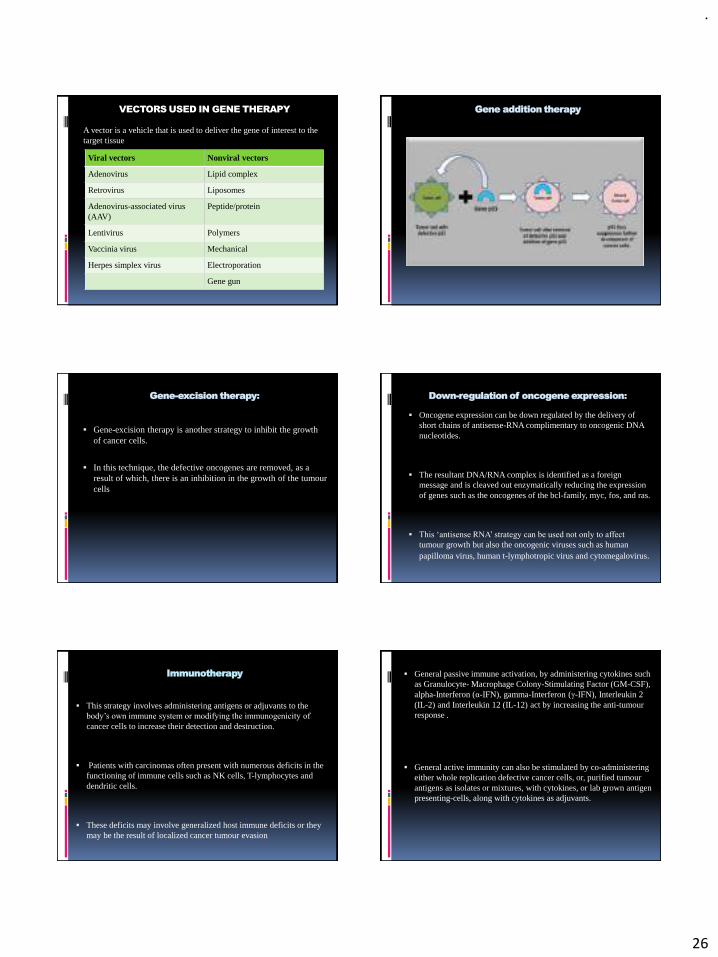
.
26
Viral vectors Nonviral vectors
Adenovirus Lipid complex
Retrovirus Liposomes
Adenovirus-associated virus (AAV)
Peptide/protein
Lentivirus Polymers
Vaccinia virus Mechanical
Herpes simplex virus Electroporation
Gene gun
VECTORS USED IN GENE THERAPY
A vector is a vehicle that is used to deliver the gene of interest to the target tissue
Gene addition therapy
Gene-excision therapy:
▪ Gene-excision therapy is another strategy to inhibit the growth of cancer cells.
▪ In this technique, the defective oncogenes are removed, as a result of which, there is an inhibition in the growth of the tumourcells
Down-regulation of oncogene expression:
▪ Oncogene expression can be down regulated by the delivery of short chains of antisense-RNA complimentary to oncogenic DNA nucleotides.
▪ The resultant DNA/RNA complex is identified as a foreign message and is cleaved out enzymatically reducing the expression of genes such as the oncogenes of the bcl-family, myc, fos, and ras.
▪ This ‘antisense RNA’ strategy can be used not only to affect
tumour growth but also the oncogenic viruses such as human papilloma virus, human t-lymphotropic virus and cytomegalovirus.
Immunotherapy
▪ This strategy involves administering antigens or adjuvants to the body’s own immune system or modifying the immunogenicity of
cancer cells to increase their detection and destruction.
▪ Patients with carcinomas often present with numerous deficits in the functioning of immune cells such as NK cells, T-lymphocytes and dendritic cells.
▪ These deficits may involve generalized host immune deficits or they may be the result of localized cancer tumour evasion
▪ General passive immune activation, by administering cytokines such as Granulocyte- Macrophage Colony-Stimulating Factor (GM-CSF), alpha-Interferon (α-IFN), gamma-Interferon (γ-IFN), Interleukin 2 (IL-2) and Interleukin 12 (IL-12) act by increasing the anti-tumourresponse .
▪ General active immunity can also be stimulated by co-administering either whole replication defective cancer cells, or, purified tumourantigens as isolates or mixtures, with cytokines, or lab grown antigen presenting-cells, along with cytokines as adjuvants.
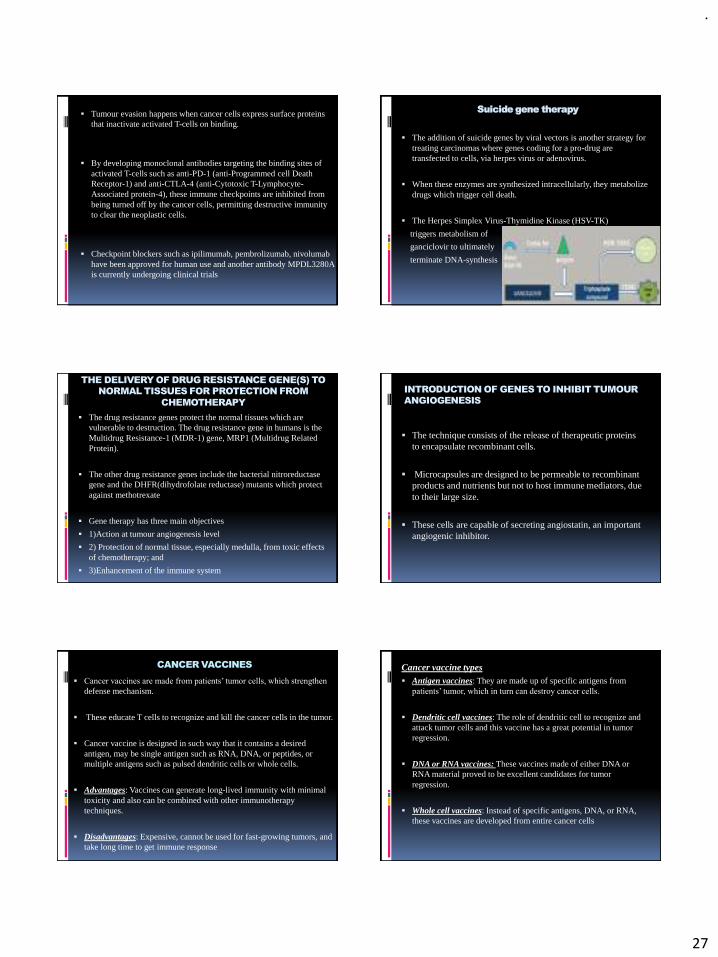
.
27
▪ Tumour evasion happens when cancer cells express surface proteins that inactivate activated T-cells on binding.
▪ By developing monoclonal antibodies targeting the binding sites of activated T-cells such as anti-PD-1 (anti-Programmed cell Death Receptor-1) and anti-CTLA-4 (anti-Cytotoxic T-Lymphocyte-Associated protein-4), these immune checkpoints are inhibited from being turned off by the cancer cells, permitting destructive immunity to clear the neoplastic cells.
▪ Checkpoint blockers such as ipilimumab, pembrolizumab, nivolumabhave been approved for human use and another antibody MPDL3280A is currently undergoing clinical trials
Suicide gene therapy
▪ The addition of suicide genes by viral vectors is another strategy for treating carcinomas where genes coding for a pro-drug are transfected to cells, via herpes virus or adenovirus.
▪ When these enzymes are synthesized intracellularly, they metabolize drugs which trigger cell death.
▪ The Herpes Simplex Virus-Thymidine Kinase (HSV-TK)
triggers metabolism of
ganciclovir to ultimately
terminate DNA-synthesis
THE DELIVERY OF DRUG RESISTANCE GENE(S) TO
NORMAL TISSUES FOR PROTECTION FROM
CHEMOTHERAPY
▪ The drug resistance genes protect the normal tissues which are vulnerable to destruction. The drug resistance gene in humans is the Multidrug Resistance-1 (MDR-1) gene, MRP1 (Multidrug Related Protein).
▪ The other drug resistance genes include the bacterial nitroreductasegene and the DHFR(dihydrofolate reductase) mutants which protect against methotrexate
▪ Gene therapy has three main objectives
▪ 1)Action at tumour angiogenesis level
▪ 2) Protection of normal tissue, especially medulla, from toxic effects of chemotherapy; and
▪ 3)Enhancement of the immune system
INTRODUCTION OF GENES TO INHIBIT TUMOUR
ANGIOGENESIS
▪ The technique consists of the release of therapeutic proteins to encapsulate recombinant cells.
▪ Microcapsules are designed to be permeable to recombinant products and nutrients but not to host immune mediators, due to their large size.
▪ These cells are capable of secreting angiostatin, an important angiogenic inhibitor.
CANCER VACCINES
▪ Cancer vaccines are made from patients’ tumor cells, which strengthen
defense mechanism.
▪ These educate T cells to recognize and kill the cancer cells in the tumor.
▪ Cancer vaccine is designed in such way that it contains a desired antigen, may be single antigen such as RNA, DNA, or peptides, or multiple antigens such as pulsed dendritic cells or whole cells.
▪ Advantages: Vaccines can generate long-lived immunity with minimal toxicity and also can be combined with other immunotherapy techniques.
▪ Disadvantages: Expensive, cannot be used for fast-growing tumors, and take long time to get immune response
Cancer vaccine types▪ Antigen vaccines: They are made up of specific antigens from
patients’ tumor, which in turn can destroy cancer cells.
▪ Dendritic cell vaccines: The role of dendritic cell to recognize and attack tumor cells and this vaccine has a great potential in tumor regression.
▪ DNA or RNA vaccines: These vaccines made of either DNA or RNA material proved to be excellent candidates for tumor regression.
▪ Whole cell vaccines: Instead of specific antigens, DNA, or RNA, these vaccines are developed from entire cancer cells
.
28
REFERENCES▪ Burkit Textbook of oral medicine 12th edition
▪ Kumar M, Nanavati R, Modi TG, Dobariya C. Oral cancer: Etiology and risk factors: A review. J Can Res Ther 2016;12:458-63.
▪ Ribatti D. The concept of immune surveillance against tumors: The first theories. Oncotarget. 2017;8(4).
▪ Sharma G. Diagnostic aids in detection of oral cancer: An update. World Journal of Stomatology. 2015;4(3):115
▪ Lavanya R, Mamatha B, Waghray S, Chaitanya N, Reddy M, Bau D. Role of Tumor Markers in Oral Cancer: An Overview. Br J Med Med Res. 2016;15(7):1-9.
▪ Pałasz P, Adamski Ł, Górska-Chrząstek M, Starzyńska A, Studniarek M. Contemporary Diagnostic Imaging of Oral Squamous Cell Carcinoma - A Review of Literature. Pol J Radiol. 2017;82:193–202.
▪ Shah JP, Gil Z. Current concepts in management of oral cancer--surgery. Oral Oncol. 2009;45(4-5):394–401.

.
1
C
PIGMENTED LESIONS OF ORAL MUCOSA
Dr. Neeraj Kumar Yadav
Reader
Dept. of Oral Medicine and Radiology
MGDCH , Jaipur
Contents
INTRODUCTION
ORAL MUCOUS MEMBRANE
ORAL PIGMENTATION- MELANIN
CLASSIFICATION OF PIGMENTED LESIONS
ENDOGENOUS PIGMENTATION
FOCAL MELANOCYTIC PIGMENTATION
MULTIFOCAL/DIFFUSE PIGMENTATION
MELANOSIS ASSOCIATED WITH SYSTEMIC OR GENETIC DISEASE
DEPIGMENTATION
HEMOGLOBIN AND IRON-ASSOCIATED PIGMENTATION
EXOGENOUS PIGMENTATION
SYNDROMES ASSOCIATED WITH PIGMENTATION
TREATMENT OF MUCOCUTANEOUS MELANOSIS
INTRODUCTION• Healthy oral soft tissues present a typical pink to red hue with
slight topographical variations of color.
• This chromatic range is due to the interaction of a number of tissues that compose the mucosal lining
1. Presence or absence of keratin on the surface epithelium
2. Quantity, superficial or deep location of blood vessels in the subjacent stroma
3. Existence of lobules of adipocytes, and
4. Absence of melanin pigmentation in the basal cell layer of the epithelium
The oral cavity is lined by a mucous membrane (the oral mucosa) consisting of a stratified squamous epithelium, which may or may not be keratinized, and an underlying connective tissue layer, the lamina propria.
Oral mucous membrane
Oral pigmentation
Pigmentation is defined as the process of deposition of pigments in tissues. Various diseases can lead to varied colorations in the mucosa. Pigmented lesions of oral cavity
are due to:
• Augmentation of melanin production
• Increased number of melanocytes (melanocytosis)
• Deposition of accidentally introduced exogenous materials
C. Sreeja, K. Ramakrishnan, D. Vijayalakshmi, M. Devi, I. Aesha, B. VijayabanuJ Pharm Bioallied Sci. 2015 Aug; 7(Suppl 2): S403–S408. doi: 10.4103/0975-7406.163471
There are four pigments which contribute to the normal color of the skin and mucosa.
Melanin
Carotenoids
Reduced HB
Oxygenated HB
Sreeja C, Ramakrishnan K, Vijayalakshmi D, Devi M, Aesha I, Vijayabanu B. Oral pigmentation: A review. J Pharm Bioallied Sci. 2015;7(Suppl 2):S403–S408. doi:10.4103/0975-7406.163471
.
2
Melanin • Melanin is found universally in nature, the pigment derivative of
tyrosine and is synthesized by melanocytes, which typically reside in the basal cell layer of the epithelium.
• Melanin is synthesized within specialized structures known as melanosomes.
• Becker in 1927 first identified melanocytes in the oral epithelium.
• During early stages of intrauterine life, precursors of melanocytes, i.e. melanoblasts differentiate into the dendritic cells and migrate to the epidermis from the neural crest
• keratinocytes actually control melanocytic growth.
• Their presence in the skin is thought to protect against the damaging effects of actinic irradiation.
• They also act as scavengers in protecting against various cytotoxic intermediates.
Types of melanin
Eumelanin- brown-black
Pheomelanin- red-yellow
Mixed type melanin
Neuromelanin
Oxymelanin
• Overproduction of melanin may be caused by a variety of mechanisms, the most common of which is related to increased sun exposure.
Intraorally hyperpigmentation is more commonly a consequence of
• Physiologic or idiopathic sources
• Neoplasia
• Medication or oral contraceptive use
• High serum concentrations of pituitary adrenocorticotropic hormone
• Post inflammatory changes
• Genetic factors
• Autoimmune disease
The function of oral melanocytes
• Melanin -determines the colour of skin, hair and eyes
• Protection from stressors such as UV radiation, reactive oxygen species (ROS) and free
radicals in the environment- melanin
• Sequester metal ions and to bind certain drugs and organic molecules- melanin
• Neutralise bacteria-derived enzymes and toxins
• Act as antigen presenting cells, can stimulate T-cell proliferation, and can phagocytose microorganism
Feller, L., Masilana, A., Khammissa, R.A. et al. Melanin: the biophysiology of oral melanocytes and physiological oral pigmentation. Head Face Med 10, 8 (2014). https://doi.org/10.1186/1746-160X-10-8
The keratinocyte-melanocyte unit
• Mature melanocytes contain all the proteins required for melanin biosynthesis and for the
structural maturation of melanosomes, including tryosinase (TYR), tyrosinase-related proteins-1 (TRP-1) and TRP-2, gp 100, and melanoma antigen recognizable by T lymphocytes.
Melanosomes are packed in globules enclosed by the melanocyte plasma membrane,
released into the extracellular space from various areas of the melanocyte dendrites, phagocytosed by keratinocytes, and are then
dispersed around the perinuclear area.
Production of melanin by melanosomes
.
3
CCLASSIFICATION
Classification Dr. Reshna Roy. Classification of oral pigmented lesions: A review .International Journal of Applied Dental Sciences 2019; 5(2): 397-402
MISCELLANOUS LESIONS THAT MAY BE ASSOCIATED WITH ORAL MUCOSAL DISCOLORATION
Investigations
• Biopsy
• Diascopy
• Radiography
• Blood tests
• Dermascopy /epiluminiscence microscopy
• Binocular stereomicroscopes
Primary lesions

.
4
C
ENDOGENOUS PIGMENTATION
• Hemoglobin, hemosiderin, and melanin represent the most common endogenous sources of mucosal color change.
• A submucosal collection of hemoglobin or hemosiderin, produced by extravasation and/or lysis of red blood cells, may impart a red, blue, or brown ephemeral
appearance to the oral mucosa.
• Melanin, which is synthesized by melanocytes and nevus cells, may appear brown,
blue, or black, depending on the amount of melanin and its spatial location within the tissue (i.e., superficial vs. deep).
Common Causes of Endogenous Oral and Perioral Discoloration
C
FOCAL MELANOCYTIC PIGMENTATION
Freckle/ Ephelis
Melanotic macule
Oral Melanoacanthoma
Melanocytic nevus
Malignant melanoma
Freckle/Ephelis
• common, asymptomatic, small (1–3 mm), well-circumscribed, tan- or brown-colored macule
• seen on the sun-exposed regions of the facial and perioral skin
• commonly observed in light-skinned individuals and are quite prevalent in red- or light blond–haired individuals.
• developmental in origin
• Polymorphisms in the MC1R gene are strongly associated with the development of childhood freckles.
• Another putative freckles-predisposition gene has also been mapped to chromosome 4q32–q34
• More abundant in number and darker in intensity during childhood and
adolescence.
• become darker during periods of
prolonged sun exposure (spring, summer) and less intense during the autumn and winter months.
• With increasing age, the number of ephelides and color intensity tends to
diminish.
• no therapeutic intervention is required.
.
5
Oral/Labial Melanotic Macule
• The melanotic macule is a unique, benign, pigmented lesion that has no known dermal counterpart.
• Melanotic macules are the most common oral lesions of melanocytic origin.
• Although the etiology remains elusive, trauma has been postulated to play a role.
• Sun exposure is not a precipitating factor
Clinical Features
• more frequently in females, usually in the lower lip (labial melanotic macule) and gingiva
• any mucosal site may be affected
• Congenital melanotic macules -occur primarily in the tongue.
• small (<1 cm), well circumscribed, oval or irregular in outline, and often uniformly pigmented.
• Unlike an ephelis, a melanotic macule does not become darker with continued sun
exposure
• Differential diagnosis – melanocytic nevus,malignant melanoma, amalgum
tattoo,focal ecchymosis
Oral Melanoacanthoma
• Unusual, benign, melanocytic lesion that is unique to the mucosal tissues.
• Innocuous melanocytic lesion that may spontaneously resolve, with or without
surgical intervention.
• Rapid onset; and acute trauma or a history of chronic irritation usually
precedes the development of the lesion.
• The biopsy procedure itself may lead to spontaneous regression of the lesion.
• The underlying source of the irritation should be eliminated to minimize
recurrence.
Clinical Features Oral melanoacanthoma usually presents as a rapidly enlarging, ill-defined, darkly pigmented macular or plaque-like lesion, and mostly develop in black females.
Occur between the third and fourth decades of life.
Typically, a solitary lesion , may be bilateral and multifocal lesions
Generally asymptomatic; may be associated with pain
Although any mucosal surface may be involved, close to 50% of melanoacanthomas arise on the buccal mucosa.
The size of the lesion is variable, ranging from small and localized to large, diffuse areas of involvement, measuring several centimeters in diameter.
The borders are typically irregular in appearance, and the pigmentation may or may not be uniform
Melanoacanthoma
Alawi F. Pigmented lesions of the oral cavity: an update. Dent Clin North Am. 2013;57(4):699–710. doi:10.1016/j.cden.2013.07.006
Diagnosis
• Because oral melanoacanthoma may resemble other melanocytic lesions, such as pigmented nevus, melanotic macule, and melanoma, a biopsy is warranted to obtain
a definitive diagnosis.
.
6
Oral Nevi (Oral melanocytic nevus, nevocellular nevus, mole, mucosal melanocytic nevi)• Categorized as hamartomas, developmental malformations, the nevi are benign
proliferations of nevus cells in either epithelium or connective tissue.
• In 1943, Ackermann and Field have reported the first case of an oral nevus.
• King et al, adopted the less anatomically specific term, intramucosal nevus.
• Adult whites harbor this lesion rather commonly but intraoral lesions are much less
common
• Clinically, a pigmented nevus is an asymptomatic, well-circumscribed, round
or oval, flat or slightly elevated spot or plaque, and of size usually ranging
between 0.1 cm and 3 cm. The color varies from brown to blue, bluish gray to
black.
• The hard palate, buccal mucosa, and gingiva are the most commonly
affected intraoral sites.
• They can be seen in persons of all ages with the mean age group affected
being 3 rd -4 th decade. Women are affected more commonly than men
Nevus cells
• Nevus cells are a variant of melanocytes
• They are larger than typical melanocytes, do not have dendrites, and have more abundant cytoplasm with coarse granules. Multinucleated cell variants are also seen.
• They are usually located at the dermoepidermaljunction or in the dermis of the skin.
• most nevic cells tend to be round, ovoid, or spindle shaped
• nevus cells have tendency to closely approximate one another, if not aggregate in clusters, and have ability to migrate into and/or within the submucosal tissues
NEVUS CLASSIFICATION• Nevi may also be classified as congenital or acquired (Buchner and Hansen).
• On the basis of the histologic location of the nevus cells, cutaneous acquired nevi can be
classified into three categories:
1. Junctional nevus—when nevus cells are limited to the basal cell layer of the epithelium.
2. Compound nevus—nevus cells are in the epidermis and dermis
3. Intradermal nevus (common mole)—nests of nevus cells are entirely in the dermis.
• Junctional nevi that are first noted in infants, children and young adults typically mature into compound nevi. Then, during later adulthood, the lesions mature into
intramucosal nevi.
• As the nevus cells penetrate into the dermis, their pigmentation diminishes;
approximately 15% of intramucosal nevi are nonpigmented.
• The most common mucosal type is the intramucosal nevus, which accounts for more
than one half of all reported oral nevi.
Etiology and Pathogenesis • Unlike ephelides and melanotic macules, which result from an increase in melanin
pigment synthesis, nevi arise as a consequence of melanocytic growth and
proliferation.
• Genetic and environmental factors,sun exposure -play a role in nevogenesis
• Familial atypical multiple mole melanoma syndrome
• Epithelioid blue nevus may be associated with the Carney complex
• Turner’s syndrome and Noonan’s syndrome; and congenital nevi are typical of
neurocutaneous melanosis
• A recent study by Pollock et al. demonstrated that up to 90% of dermal melanocytic
nevi exhibit somatic, activating mutations in the BRAF oncogene. Mutations in the HRAS and NRAS oncogenes have also been identified. This lends further credence to the notion that melanocytic nevi are neoplastic
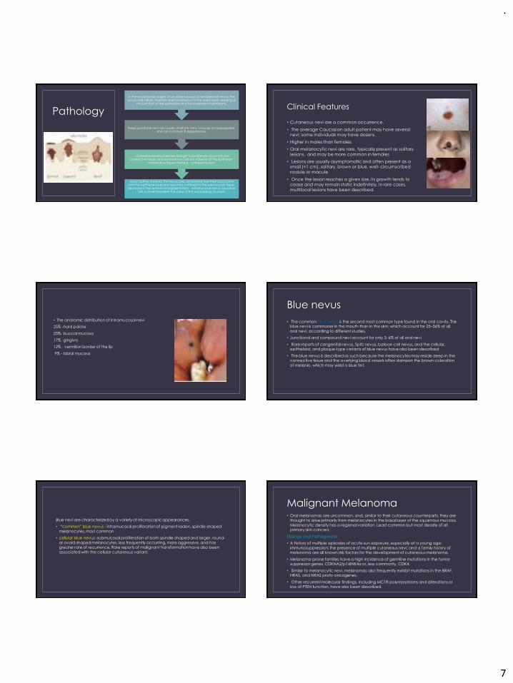
.
7
Pathology
lesion further matures, the nevus cells completely lose their association with the epithelial layer and become confined to the submucosal tissue, decrease in the amount of pigmentation - intramucosal nevus- brown or
tan or even resemble the color of the surrounding mucosa.
clustered melanocytes are thought to proliferate down into the connective tissue, and some nevus cells are still seen at the epithelial–
connective tissue interface. -compound nevi
These junctional nevi are usually small (<5 mm), macular or nonpalpable, and tan to brown in appearance.
In the evolutionary stages of an intramucosal (or intradermal) nevus, the nevus cells initially maintain their localization to the basal layer, residing at
the junction of the epithelium and the basement membrane
Clinical Features
• Cutaneous nevi are a common occurrence.
• The average Caucasian adult patient may have several
nevi; some individuals may have dozens.
• Higher in males than females.
• Oral melanocytic nevi are rare, typically present as solitary
lesions, and may be more common in females
• Lesions are usually asymptomatic and often present as a
small (<1 cm), solitary, brown or blue, well- circumscribed
nodule or macule
• Once the lesion reaches a given size, its growth tends to
cease and may remain static indefinitely. In rare cases,
multifocal lesions have been described.
• The anatomic distribution of Intramucosal nevi
25% -hard palate
25%- buccal mucosa
17% -gingiva
12% - vermilion border of the lip
9% - labial mucosa
Blue nevus
• The common blue nevus is the second most common type found in the oral cavity. The blue nevi is commoner in the mouth than in the skin; which account for 25–36% of all
oral nevi, according to different studies.
• Junctional and compound nevi account for only 3–6% of all oral nevi
• Rare reports of congenital nevus, Spitz nevus, balloon cell nevus, and the cellular, epithelioid, and plaque-type variants of blue nevus have also been described
• The blue nevus is described as such because the melanocytes may reside deep in the connective tissue and the overlying blood vessels often dampen the brown coloration
of melanin, which may yield a blue tint.
Blue nevi are characterized by a variety of microscopic appearances.
• “common” blue nevus - intramucosal proliferation of pigment-laden, spindle-shaped
melanocytes, most common
• cellular blue nevus -submucosal proliferation of both spindle-shaped and larger, round-
or ovoid-shaped melanocytes, less frequently occurring, more aggressive, and has greater rate of recurrence. Rare reports of malignant transformation have also been
associated with the cellular cutaneous variant.
Malignant Melanoma • Oral melanomas are uncommon, and, similar to their cutaneous counterparts, they are
thought to arise primarily from melanocytes in the basal layer of the squamous mucosa.
Melanocytic density has a regional variation. Least common but most deadly of all primary skin cancers.
Etiology and Pathogenesis
• A history of multiple episodes of acute sun exposure, especially at a young age;
immunosuppression; the presence of multiple cutaneous nevi; and a family history of melanoma are all known risk factors for the development of cutaneous melanoma.
• Melanoma-prone families have a high incidence of germline mutations in the tumor suppressor genes, CDKNA2/p16INK4a or, less commonly, CDK4.
• Similar to melanocytic nevi, melanomas also frequently exhibit mutations in the BRAF, HRAS, and NRAS proto-oncogenes.
• Other recurrent molecular findings, including MC1R polymorphisms and alterations or loss of PTEN function, have also been described.
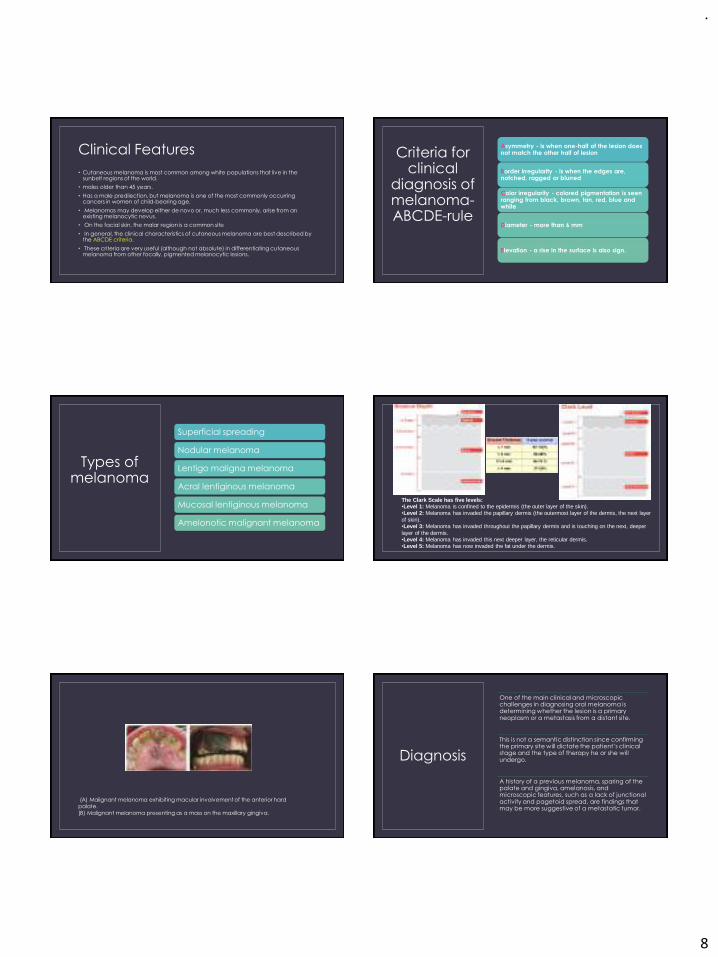
.
8
Clinical Features
• Cutaneous melanoma is most common among white populations that live in the sunbelt regions of the world.
• males older than 45 years.
• Has a male predilection, but melanoma is one of the most commonly occurring cancers in women of child-bearing age.
• Melanomas may develop either de novo or, much less commonly, arise from an existing melanocytic nevus.
• On the facial skin, the malar region is a common site
• In general, the clinical characteristics of cutaneous melanoma are best described by the ABCDE criteria.
• These criteria are very useful (although not absolute) in differentiating cutaneous melanoma from other focally, pigmented melanocytic lesions.
Criteria for clinical
diagnosis of melanoma-ABCDE-rule
Asymmetry - is when one-half of the lesion does not match the other half of lesion
Border irregularity - is when the edges are, notched, ragged or blurred
Color irregularity - colored pigmentation is seen ranging from black, brown, tan, red, blue and white
Diameter - more than 6 mm
Elevation - a rise in the surface is also sign.
Types of melanoma
Superficial spreading
Nodular melanoma
Lentigo maligna melanoma
Acral lentiginous melanoma
Mucosal lentiginous melanoma
Amelonotic malignant melanoma
The Clark Scale has five levels:•Level 1: Melanoma is confined to the epidermis (the outer layer of the skin).•Level 2: Melanoma has invaded the papillary dermis (the outermost layer of the dermis, the next layer of skin).•Level 3: Melanoma has invaded throughout the papillary dermis and is touching on the next, deeper layer of the dermis.•Level 4: Melanoma has invaded this next deeper layer, the reticular dermis.•Level 5: Melanoma has now invaded the fat under the dermis.
(A) Malignant melanoma exhibiting macular involvement of the anterior hard palate.
(B) Malignant melanoma presenting as a mass on the maxillary gingiva.
Diagnosis
One of the main clinical and microscopic challenges in diagnosing oral melanoma is determining whether the lesion is a primary neoplasm or a metastasis from a distant site.
This is not a semantic distinction since confirming the primary site will dictate the patient’s clinical stage and the type of therapy he or she will undergo.
A history of a previous melanoma, sparing of the palate and gingiva, amelanosis, and microscopic features, such as a lack of junctional activity and pagetoid spread, are findings that may be more suggestive of a metastatic tumor.
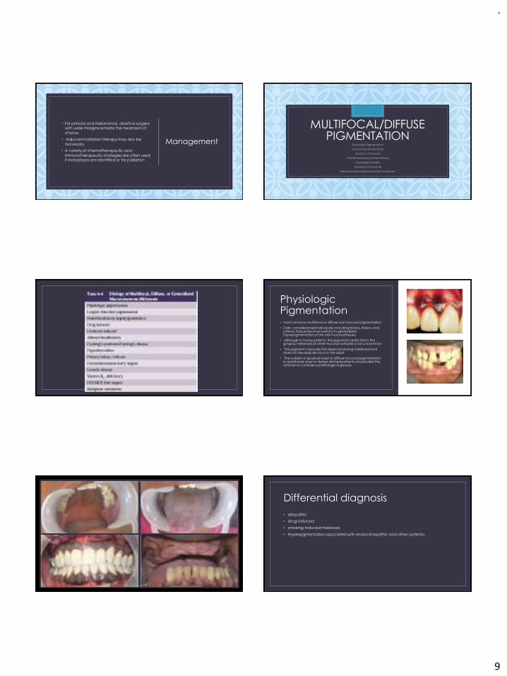
.
9
Management
• For primary oral melanomas, ablative surgery with wide margins remains the treatment of
choice.
• Adjuvant radiation therapy may also be
necessary.
• A variety of chemotherapeutic and
immunotherapeutic strategies are often used if metastases are identified or for palliation
C
MULTIFOCAL/DIFFUSE PIGMENTATION
Physiologic Pigmentation
Drug-Induced Melanosis
Smoker’s Melanosis
Postinflammatory (Inflammatory)
Hyperpigmentation
Melasma (Chloasma)
Melanosis associated with systemic diseases
Physiologic Pigmentation • most common multifocal or diffuse oral mucosal pigmentation
• Dark- complexioned individuals, including blacks, Asians, and Latinos, frequently show patchy to generalized hyperpigmentation of the oral mucosal tissues.
• Although in many patients, the pigment is restricted to the gingiva, melanosis of other mucosal surfaces is not uncommon
• The pigment is typically first observed during childhood and does not develop de novo in the adult.
• The sudden or gradual onset of diffuse mucosal pigmentation in adulthood, even in darker-skinned patients, should alert the clinician to consider a pathological genesis
Differential diagnosis
• idiopathic
• drug-induced
• smoking-induced melanosis
• Hyperpigmentation associated with endocrinopathic and other systemic.

.
10
• Microscopically, physiologic pigmentation is characterized by the presence of increased amounts of melanin pigment within the basal cell layer.
• This pigmentation is considered a variation of normal.
• Gingivectomy and laser therapy have been used to remove pigmented oral mucosa.
• Cryosurgery has also been reported to effectively remove oral pigmentation.
Drug-Induced
Melanosis
Etiology and Pathogenesis
Medications may induce a variety of different forms of mucocutaneous pigmentation, including melanosis
The chief drugs implicated in drug-induced melanosis are the antimalarials, including chloroquine, hydroxychloroquine, and quinacrine. In the Western world, these medications are typically used for the treatment of autoimmune disease.
Other common classes of medications that induce melanosis include the phenothiazines, such as chlorpromazine, oral contraceptives, and cytotoxic medications such as cyclophosphamide and busulfan
Clinical Features
It has been estimated that 10%–20% of all cases of acquired melanocytic pigmentation may be drug induced.
Intraorally, the pigment can be diffuse yet localized to one mucosal surface, often the hard palate, or it can be multifocal and involve multiple surfaces.
Some drugs may even be associated with a specific pattern of pigmentation.
Much like other forms of diffuse pigmentation, the lesions are flat and without any evidence of nodularity or swelling.
Sun exposure may exacerbate cutaneous drug-induced pigmentation
Drugs causing oral perioral pigmentation• Amiodarone
• Amodioquine
• Aziodothymidine
• Bleomycin
• Chloroquine
• Chlorpromazine
• Clofazamine
• Gold
• Hydroxychloroquine
• Hydroxyurea
• Imipramine
• Ketoconazole
• Mepacrin
Bhateja S, Bohra A, Arora G (2015) Drug Induced Oral Mucosal Pigmentation- A Review. Pigmentary Disorders 2: 198. doi:10.4172/2376-0427.100019
Pathology
Microscopically, there is usually evidence of basilar hyperpigmentation and melanin incontinence without a concomitant increase in the number of melanocytes.
Although the mechanisms by which melanin synthesis is increased remain unknown, one theory is that the drugs or drug metabolites stimulate melanogenesis.
Alternatively, some drugs, including chloroquine and chlorpromazine, have been shown to physically bind melanin. This complexation of melanin and drugs within melanocytes may contribute to the adverse mucocutaneous effects.
Drug-induced pigmentation of the palate in a patient who was taking quinacrine for the treatment of discoid lupus erythematosus
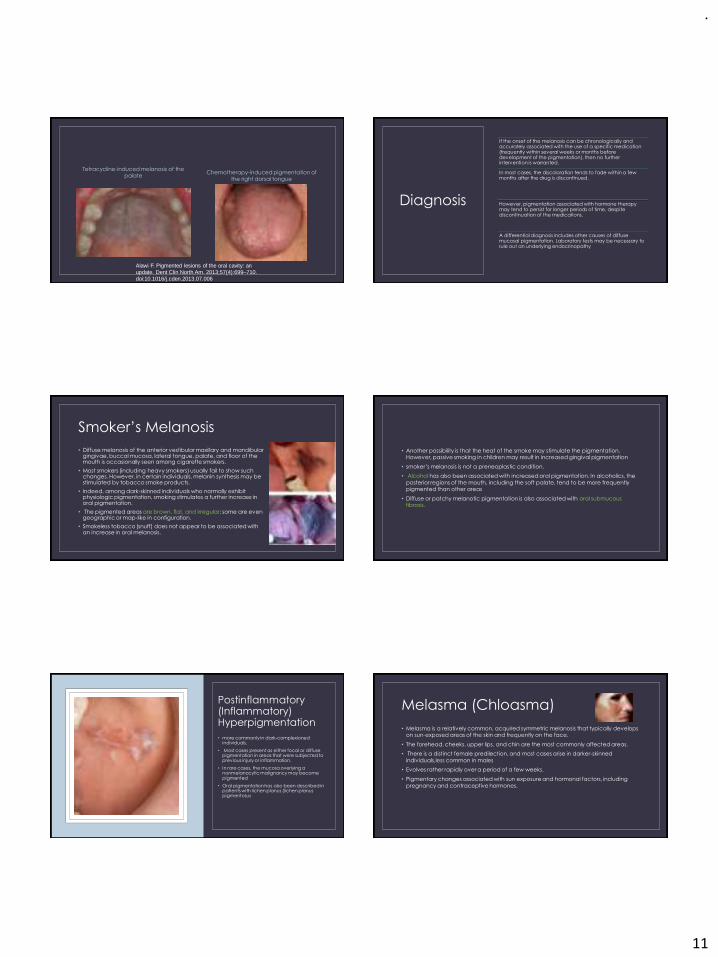
.
11
Tetracycline-induced melanosis of the palate
Chemotherapy-induced pigmentation of the right dorsal tongue
Alawi F. Pigmented lesions of the oral cavity: an update. Dent Clin North Am. 2013;57(4):699–710. doi:10.1016/j.cden.2013.07.006
Diagnosis
If the onset of the melanosis can be chronologically and accurately associated with the use of a specific medication (frequently within several weeks or months before development of the pigmentation), then no further intervention is warranted.
In most cases, the discoloration tends to fade within a few months after the drug is discontinued.
However, pigmentation associated with hormone therapy may tend to persist for longer periods of time, despite discontinuation of the medications.
A differential diagnosis includes other causes of diffuse mucosal pigmentation. Laboratory tests may be necessary to rule out an underlying endocrinopathy
Smoker’s Melanosis
• Diffuse melanosis of the anterior vestibular maxillary and mandibular gingivae, buccal mucosa, lateral tongue, palate, and floor of the mouth is occasionally seen among cigarette smokers.
• Most smokers (including heavy smokers) usually fail to show such changes. However, in certain individuals, melanin synthesis may be stimulated by tobacco smoke products.
• Indeed, among dark-skinned individuals who normally exhibit physiologic pigmentation, smoking stimulates a further increase in oral pigmentation.
• The pigmented areas are brown, flat, and irregular; some are even geographic or map-like in configuration.
• Smokeless tobacco (snuff) does not appear to be associated with an increase in oral melanosis.
• Another possibility is that the heat of the smoke may stimulate the pigmentation. However, passive smoking in children may result in increased gingival pigmentation
• smoker’s melanosis is not a preneoplastic condition.
• Alcohol has also been associated with increased oral pigmentation. In alcoholics, the
posterior regions of the mouth, including the soft palate, tend to be more frequently pigmented than other areas
• Diffuse or patchy melanotic pigmentation is also associated with oral submucousfibrosis.
Postinflammatory(Inflammatory) Hyperpigmentation
• more commonly in dark-complexioned individuals.
• Most cases present as either focal or diffuse pigmentation in areas that were subjected to previous injury or inflammation.
• In rare cases, the mucosa overlying a nonmelanocytic malignancy may become pigmented
• Oral pigmentation has also been described in patients with lichen planus (lichen planus pigmentosus
Melasma (Chloasma)
• Melasma is a relatively common, acquired symmetric melanosis that typically develops on sun-exposed areas of the skin and frequently on the face.
• The forehead, cheeks, upper lips, and chin are the most commonly affected areas.
• There is a distinct female predilection, and most cases arise in darker-skinned
individuals,less common in males
• Evolves rather rapidly over a period of a few weeks.
• Pigmentary changes associated with sun exposure and hormonal factors, including
pregnancy and contraceptive hormones.

.
12
• In Various thyroid abnormalities, including hypothyroidism, may also play a role in the pathogenesis of pregnancy- and nonpregnancy-associated melisma
• A biopsy typically reveals basilar melanosis with no increase in the number of melanocytes. However, the melanocytes that are present may be larger than those in
the adjacent normally pigmented areas.
• spontaneously resolve after parturition, cessation of the exogenous hormones, or regulation of endogenous sex hormone levels.
• A successful therapeutic approach for the treatment of melasma consists in the topical administration of a triple combination product (4% hydroquinone, 0.05% tretinoin, and 0.01% fluocinolone
C
MELANOSIS ASSOCIATED WITH
SYSTEMIC OR GENETIC DISEASE
Hypoadrenocorticism (Adrenal Insufficiency or Addison’s Disease) Etiology and Pathogenesis
• In adults, autoimmune disease represents one of the most common causes where the
majority of patients show the presence of circulating autoantibodies to steroidogenic enzyme 21-hydroxylase.
• However, infectious agents, neoplasia, trauma, certain medications, and iatrogenic causes may lead to adrenal destruction or an impairment of endogenous steroid
production. In rare cases, adrenal insufficiency may also be a consequence of genetic disease.
• As a result decrease in endogenous corticosteroid levels occurs.
• As steroid levels decrease, there is a compensatory activation of ACTH secretion from
the anterior pituitary gland. ACTH then acts on the adrenal cortex to stimulate steroid production and ACTH secretion stops
• If low steroid levels persist, there is a loss of feedback inhibition, resulting in persistent secretion of ACTH into the serum.
• Concurrently, the serum levels of α-melanocyte- stimulating hormone (α-MSH) also increase.
• At the molecular level, this is explained by the fact that the precursor proopiomelanocortin gene contains the sequences of both the ACTH and α-MSH genes.
• During processing of the progenitor hormone, the ACTH and α-MSH genes may be cleaved independently of one another, thus creating two distinct hormones.
• Apart from the wide array of tissues and organs that these hormones act upon, both α-MSH and ACTH are also thought to have stimulatory effects on melanocytes. However, the exact mechanism by which melanin synthesis increases remains unclear.
Clinical Features
• Weakness, poorly defined fatigue, and depression are some of the typical presenting signs of the illness.
• The first sign of disease may be mucocutaneoushyperpigmentation
• Generalized bronzing of the skin and diffuse but patchy melanosis of the oral mucosa are hallmarks of hypoadrenocorticism.
• Any oral surface may be affected. In some patients, oral melanosis may be the first manifestation of their adrenal disease
• Diffuse hyperpigmentation is more commonly associated with chronic rather than acute-onset disease.
Diagnosis
• Endocrinopathic disease should be suspected whenever oral melanosis is accompanied by cutaneous bronzing.
• An oral biopsy typically shows increased melanin in the basal cell layer with melanin incontinence.
• Differential diagnosis - physiologic and drug-induced pigmentation.
• Laboratory tests- serum cortisol and electrolyte levels.Serum cortisol levels of less than
100 nmol/L at 9:00 a.m. is a diagnostic of deficiency.Hyponatremia and hyperkalemia are frequently associated with adrenal insufficiency.
• Treatment -exogenous steroid replacement therapy with glucocorticoids and mineralocorticoids. There is evidence supporting the use of adrenal androgens such as
dehydroepiandrosterone to improve the quality of life of patients with Addison’s disease.
.
13
Cushing’s Syndrome/Cushing’s
Disease
Etiology and Pathogenesis
• Cushing’s syndrome develops as a consequence of prolonged exposure to relatively high concentrations of endogenous or exogenous corticosteroids.
• Most cases are iatrogenic in origin and associated with poorly controlled or unmonitored use of topical or systemic steroids.
• Pituitary tumor
• Hyperadrenocorticism
• Ectopic secretion of corticosteroids, ACTH, or corticotropin-releasing hormone by various neoplasms, including small cell carcinoma of the lung.
• Germ line mutations in ACTH receptor
Clinical Features
• more prevalent in female patients.
• prepubertal onset is more commonly seen in boys.
• Apart from the wide array of systemic complications, including weight gain and the characteristic “moon
facies,” diffuse mucocutaneous pigmentation may be seen in a subset of patients, specifically those
whose pathology is associated with increased ACTH secretion.
Diagnosis and
management
Three main tests are used for the diagnosis of Cushing’s syndrome:
low-dose dexamethasone suppression test,
midnight plasma cortisol
24-hour urinary free cortisol.
The pigmentation often resolves following appropriate surgical, radiation, or drug therapy for the specific source of the endocrinopathy.
Pasireotide (a somatostatin analog) has been approved for the treatment of Cushing’s syndrome
Hyperthyroidism (Graves’ Disease)
Melanosis is a common consequence of hyperthyroidism (Graves’ disease), especially in dark-skinned individuals.
Studies suggest that at least 40% of black patients with thyrotoxicosis may present with mucocutaneoushyperpigmentation.
In contrast, melanosis is very rarely observed in Caucasian patients with the disease. The pigmentation tends to resolve following treatment of the thyroid abnormality.
The mechanism by which excessive thyroid activity stimulates melanin synthesis remains unclear
Primary Biliary Cirrhosis
Diffuse mucocutaneous hyperpigmentation may be one of the earliest manifestations of primary biliary cirrhosis.
Up to 47% of patients with this condition develop diffuse melanosis.
This uncommon disease is of unknown etiology, although it is thought to be autoimmune in nature as evidenced by the presence of antimitochondrial antibodies.
Primary biliary cirrhosis develops mainly in middle-aged women. The disease results from damage to small intrahepatic bile ducts. The mechanism by which melanosis develops is unknown.
• Primary biliary cirrhosis may also be a source of generalized nonmelanocytic discoloration.
• Hyperbilirubinemia often induces a yellowish discoloration of the skin, eyes, and mucous membranes.
• Treatment of the underlying disease will lead to resolution of jaundice.
• Differential diagnosis - carotenemia (excessive β-carotene levels) and
lycopenemia. However, the oral mucosal tissues are not affected in either of these latter conditions.
Clinical appearance of the upper and lowerteeth presenting green discoloration in patient
with persistent conjugated hyperbilirubinemia
Green Teeth in the Primary and Permanent Dentition.J Pediatr 2017;191:275
.
14
Vitamin B12 (Cobalamin) Deficiency
• Diffuse mucocutaneous hyperpigmentation is a rare, and poorly recognized, complication of vitamin B12 deficiency.
• This hyperpigmentation is reminiscent of Addison’s disease.
• The pigmentation resolves following restoration of vitamin B12 levels.
• The predominant mechanism of hyper pigmentation in vitamin B12 is hypothesised as 1) Deficiency of vitminB12 decreases the level of reduced glutathione, which activate
tyrosinase and thus leads to transfer to melanosomes.
• 2) Defect in the melanin transfer between melanocytes and keratinocytes, resulting in pigmentary incontinence. The dominant mechanism of hyper pigmentation is not a defect in melanin transport but is rather an increase in melanin synthesis.
Agrawala RK, ETAL . Pigmentation in vitamin B12 deficiency masquerading Addison's pigmentation: A rare presentation. Indian J Endocrinol Metab. 2013;17(Suppl 1):S254–S256. doi:10.4103/2230-8210.119591
Peutz–Jeghers Syndrome • An autosomal dominant disease that is associated with mutations in
the STK11/LKB1 tumor suppressor gene.
• Clinical manifestations include intestinal polyposis, cancer
susceptibility, and multiple, small, pigmented macules of the lips,
perioral skin, hands, and feet.
• The macules may resemble ephelides, usually measuring <0.5 cm in
diameter. However, the intensity of the macular pigment is not
influenced by sun exposure.
• Although uncommon, similar-appearing lesions may also develop on
the anterior tongue and buccal and labial mucosae.
Typical pattern of pigmentation representative of Peutz Jeghers syndrome
Alawi F. Pigmented lesions of the oral cavity: an update. Dent Clin North Am. 2013;57(4):699–710. doi:10.1016/j.cden.2013.07.006
Multiple pigmented macules on the fingertips in in an 11-year-old male with Peutz–Jeghers syndrome
Multiple pigmented macules in the perioral region
• Histologically, these lesions show increased basilar melanin without an increase
in the number of melanocytes.
• The medical management for Peutz–Jeghers syndrome consists in surveillance
and treatment of hamartomatous polyps.
• Other genetic diseases associated with a triad of gastrointestinal disease,
cancer susceptibility, and mucocutaneous pigmented macules among other
findings include Cowden syndrome (and the allelic Bannayan–Riley–
Ruvalcaba and Lhermitte–Duclos syndromes) and Cronkhite–Canada
syndrome.
Café au Lait Pigmentation • Solitary, idiopathic café au lait (“coffee
with milk”) spots are occasionally observed in the general population, but multiple café au lait spots are often indicative of an underlying genetic disorder.
• Tan- or brown-colored, irregularly shaped macules of variable size.
• They may occur anywhere on the skin
• Café au lait pigmentation may be identified in a number of different genetic diseases, including neurofibromatosis type I, McCune–Albright syndrome, and Noonan’s syndrome

.
15
Diseases Commonly Associated With Café au Lait Pigmentation
Neurofibromatosis type I
• Neurofibromatosis type I is an autosomal dominant disease caused by a mutation or a deletion of the NF1 gene localized in
chromosome
• Neurofibromatosis type I is associated with the development of
multiple neurofibromas of various histologic subtypes.
• In addition, the size, number, and age at onset of the cutaneous
café au lait spots are of diagnostic importance for this disease.
• Axillary and/ or inguinal freckling (Crowe’s sign) and pigmented
lesions of the iris (Lisch nodules) are also highly characteristic of neurofibromatosis type I.
McCune–Albright syndrome
• It is an extremely rare disorder that classically affects the bones, skin, and endocrinesystem
• fibrous dysplasia of bone ,patches of abnormal skin and multiple endocrine dysfunction
• McCune–Albright syndrome and the genetically and phenotypically similar Mazabrauddisease are sporadically occurring diseases that are characterized by polyostoticfibrous dysplasia, various endocrinopathies (McCune– Albright), and soft tissuemyxomas (Mazabraud disease).
• In some patients, Addison’s disease or Cushing’s syndrome may be a potentialconsequence of McCune–Albright syndrome.
• The café au lait spots in McCune–Albright syndrome appear distinct from thoseassociated with neurofibromatosis.
• The borders of the pigmented macules are irregularly outlined, whereas inneurofibromatosis, the borders are typically smooth
• Noonan’s syndrome and the allelic LEOPARD syndrome (multiple lentigines,
electrocardiographic-conduction abnormalities, ocular hypertelorism,
pulmonary stenosis, abnormal genitalia, retardation of growth, and
sensorineural deafness) are autosomal dominant disorders that, among other
findings, are also associated with pigmented mucocutaneous macules and
multiple melanocytic nevi.
• The classic-appearing café au lait spots are more characteristically seen in
patients with the Noonan’s phenotype.
• The LEOPARD phenotype is typically associated with numerous, small, freckle-
like macules often involving the facial skin.
HIV/AIDS-Associated Melanosis
• Diffuse or multifocal mucocutaneous pigmentation has been frequently described in HIV-seropositive patients.
• The pigmentation may be related to intake of various medications, including antifungal and antiretroviral drugs, or as a result of adrenocortical destruction by
virulent infectious organisms.
• Recent studies suggest that melanosis may be an actual, potentially late-stage, clinical
manifestation of HIV/ AIDS.
• Goldstein et al. demonstrated a significant correlation between mucocutaneous
pigment and CD4 counts cells.
• Studies have also shown that the immune dysregulation associated with HIV/AIDS
leads to increased secretion of α-MSH from the anterior pituitary gland, which may also stimulate increased melanin synthesis
• Progressive hyperpigmentation of the skin, nails, and mucous membranes
• The buccal mucosa is the most frequently affected site, but the gingiva, palate, and
tongue may also be involved.
• Like all diffuse melanoses, HIV- associated pigmentation is microscopically
characterized by basilar melanin pigment, with incontinence into the underlying submucosa
.
16
C
IDIOPATHIC PIGMENTATION
Laugier–Hunziker Pigmentation
Etiology and Pathogenesis
• Laugier–Hunziker pigmentation (also known as Laugier– Hunziker syndrome) was initially described as an acquired, idiopathic, macular hyperpigmentation of the oral mucosal tissues, specifically involving the lips and buccal mucosae.
• Pigmentation of the esophageal, genital, and conjunctival mucosae and the acralsurfaces is seen
• Up to 60% of affected patients also may have nail involvement, usually in the form of longitudinal melanotic streaks and without any evidence of dystrophic change. The fingernails are more commonly affected than the toenails
• Rare condition,in adult patients with equal sex predilection
• Caucasian or light skinned individuals
• Possible genetic predisposition
Clinical and Microscopic Features
• Patients typically present with multiple, discrete, irregularly shaped brown or
dark brown oral macules.
• Individual macules are usually no more than 5 mm in diameter.
• In rare instances, the lesions may coalesce to produce a diffuse area of
involvement.
• Increased melanin pigmentation in the basal cell layer without an increase in
the number of melanocytes and melanin incontinentia in the superficial
lamina propria are characteristic of this syndrome
• A relatively limited number of cases have been reported in the literature. This suggests that either this form of pigmentation is exceedingly rare or it is poorly recognized and thus, underreported.
• Laugier–Hunziker pigmentation is typically identified in adult patients, with relatively equal sex predilection. This condition more commonly develops in Caucasian or light-skinned individuals.
: (a) Hyperpigmentation of all nails of hand with
longitudinal melanonychia. (b) Hyperpigmentation involving all toe nails
(a) Pigmented macules over tongue and lower lip, upper lip
is spared. (b) Pigmented macules over the volar aspect of fingertips
Verma B, Behra A, Ajmal AK, Sen S. Laugier–Hunziker syndrome in a young female. Indian
Dermatol Online J 2017;8:148-50.
Differential Diagnosis
• Physiologic pigmentation
• Drug- or heavy metal–induced
pigmentation
• Endocrinopathic disease
• Peutz–Jeghers syndrome.
Management
• generally not indicated
• laser and cryotherapy CDEPIGMENTATION
.
17
Vitiligo• Vitiligo is a relatively common, acquired,
autoimmune disease that is associated with hypomelanosis.
• Vitiligo affects 0.5%–2.0% of the world population with no gender or racial preference.
Etiology and Pathogenesis
• Autoimmunity, cytotoxicity, genetics, and alterations from metabolic or oxidative stress have been implicated in this condition where the end result is a destruction of the melanocytes.
• The pathogenesis of vitiligo is multifactorial, with both genetic and environmental factors playing a role in the development of this disease.
Clinical Features
• classification - non segmental vitiligo, segmental vitiligo, and unclassified/undetermined vitiligo.
• Multiple achromic patches with remitting-relapsing course are seen in nonsegmental vitiligo.
• Segmental vitiligo shows a characteristic dermatomeric distribution of the achromicpatches with a rapid onset that is usually not progressive.
• The onset of vitiligo –any age but mostly second and third decade of life.
• The depigmentation is more apparent in patients who have a darker skin tone. Yet the
disease actually occurs in all races.
• Vitiligo may also arise in patients undergoing immunotherapy for the treatment of malignant melanoma.
• Vitiligo rarely affects the intraoral mucosal tissues. However, hypomelanosis of the inner and outer surfaces of the lips and perioral skin may be seen in up to 20% of patients
Management
• Objective is to stimulate repigmentation
• Topical corticosteroids, topical calcineurin inhibitors, ultraviolet B narrow band, and psoralen and ultraviolet A exposure
• From the standpoint of therapy, labial vitiligo is more resistant to the typical treatments used for cutaneous vitiligo. Due to a lack of hair follicles, the lips do not have a reservoir of melanocytes that can be stimulated to produce pigment.Thus, surgical intervention may be the only option to achieve an esthetic result. Autologous epithelial grafts have been used successfully, with patients often reporting a more acceptable cosmetic appearance.
• Split-thickness skin grafts have been reported as having the highest repigmentation success rate.
• Punch grafting and micropigmentation (whereby an exogenous brown pigment is injected into the lip, much like a tattoo)
• In rare instances, surgical intervention may stimulate spontaneous repigmentation of vitiligenous lesions elsewhere on the body.
C
HEMOGLOBIN AND IRONASSOCIATED
PIGMENTATION
Ecchymosis
Traumatic ecchymosis is common on the lips and face yet uncommon in the oral mucosa, except in cases related to blunt-force trauma and oral intubation.
Immediately following the traumatic event, erythrocyte extravasation into the connective tissue will appear as a bright red macule or as a swelling if a hematoma forms. The lesion will assume a brown discoloration within a few days, after the hemoglobin is degraded to hemosiderin.
Patients taking anticoagulants may present with oral ecchymosis, particularly on the buccal mucosa or tongue, either of which can be traumatized while chewing.
Ecchymoses of the oral mucosa may also be encountered in patients with liver cirrhosis, leukemia, end-stage renal disease who are undergoing dialysis treatment.
Purpura/Petechiae
• Capillary hemorrhages will appear red initially and turn brown in a few days once the extravasated red cells have lysed and have been degraded to hemosiderin.
• The distinction between purpura and petechiae is essentially semantic and based solely on the size of the focal hemorrhages.
• Petechiae are typically characterized as being pinpoint or slightly larger than pinpoint and purpura as multiple, small 2–4 mm collections of extravasated blood. The same precipitating events can elicit either clinical presentation.
• Oral purpura/petechiae may develop as a consequence of trauma, viral, or systemic disease

.
18
• Viral disease is more commonly associated with oral rather than cutaneous petechiae. In most cases, the petechiae are identified on the soft palate, although any mucosal
site may be affected.
• Within two weeks, the lesions should resolve. Failure to do so should arouse suspicion of
a hemorrhagic diathesis, a persistent infectious disease, or other systemic disease, and appropriate laboratory investigations must be undertaken.
Causes of Oral Purpura/Petechiae
Amyloidosis
Aplastic anemia Bulimia
Chronic renal failureForceful coughing
HemophiliaHenoch–Schönlein purpura
HIV/AIDSInfectious mononucleosis
LeukemiaLiver cirrhosis
Nonspecific trauma Oral intubation
Oral submucous fibrosis Overexertion
Papular-purpuric “gloves and socks” syndromeStreptococcal infection
Systemic lupus erythematosusThrombocytopenia
von Willebrand’s disease
Hemochromatosis
• Hemochromatosis is a chronic, progressive disease that is characterized by
excessive iron deposition (usually in the form of hemosiderin) in the liver and
other organs and tissues.
• Idiopathic, neonatal, blood transfusion, and heritable forms of this disease are
recognized.
• Complications of hemochromatosis may include liver cirrhosis, diabetes,
anemia, heart failure, hypertension, and bronzing of the skin.
• Studies also suggest an increased incidence of cancer in patients with
hemochromatosis. The cutaneous pigmentation is seen in over 90% of affected
patients, regardless of the etiology of the disease.
• The primary oral manifestation of hemochromatosis is a blue-gray to brown pigmentation affecting mainly the palate and gingiva.
• Early on in the course of disease, the pigmentation may be more commonly a result of basilar melanosis rather than iron-associated pigment.
• Iron deposition within the adrenal cortex may lead to hypoadrenocorticism and ACTH hypersecretion, with the associated addisonian-type changes. In the later stages of
hemochromatosis, the pigmentation is usually a result of hemosiderosis and melanosis.
• A lower labial gland biopsy has been shown to be an easy and effective method for
the diagnosis of hemochromatosis.
C
EXOGENOUS PIGMENTATION
Sources of Exogenous Oral and Perioral Pigmentation
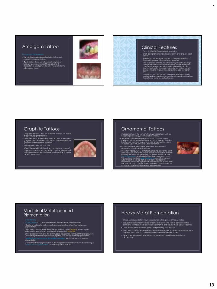
.
19
Amalgam Tattoo
Etiology and Pathogenesis
• The most common pigmented lesion in the oral
mucosa is amalgam tattoo.
• By definition, these are iatrogenic in origin and
typically a consequence of the inadvertent deposition of amalgam restorative material into the
submucosal tissue.
Clinical Features • Found in 1%–3% of the general population.
• small, asymptomatic, macular, and bluish gray or even black lesions
• The gingiva, alveolar mucosa, buccal mucosa, and floor of the mouth represent the most common sites.
• The lesions are often found in the vicinity of teeth with large amalgam restorations or crowned teeth that probably had amalgams, around the apical region of endodontically treated teeth with retrograde restorations or obturated with silver points, and in areas in and around healed extraction sites
• Amalgam tattoo of the head and neck skin may occur in dentists and represents an occupational hazard resulting from failure to use facial protective barriers
Graphite Tattoos • Graphite tattoos are an unusual source of focal
exogenous pigmentation.
• They are most commonly seen on the palate andgingiva and represent traumatic implantation ofgraphite particles from a pencil
• solitary gray or black macule.
• When the graphite tattoo involves areas of cosmeticconcern, removal of the lesion and a subsequentautogenous connective tissue graft provide a highlyesthetic outcome
Ornamental Tattoos • Mucosal tattoos in the form of lettering or intricate artwork are
becoming increasingly common phenomena.
• Amateur tattoo inks are permanent and consist of simple, carbon particles originating from a variety of sources, including burnt wood, plastic, or paper, and from a variety of inks, such as India ink, pen ink, and plant-derived matter.
• Q-Switched laser therapy has been used successfully to remove tattoos of the oral mucosa.
• In certain tribal cultures, -esthetically pleasing, pigment is plant derived.An unusual South African female tribal custom includes brushing the teeth and gums with a chewed root of the tree Euclea natalensis, with the belief that it promotes oral health. This plant root contains naphthoquinones and other organic substances that have putative antibacterial properties. Naphthoquinones are pigmented, and the mouths of root users are typically bright orange. Unlike ornamental tattoos, this form of pigmentation is usually transient and reversible.
Medicinal Metal-Induced Pigmentation • Gold therapy
• Colloidal silver -“complementary and alternative medicine therapies.
• Gold and colloidal silver have both been associated with diffuse cutaneous pigmentation.
• Silver may cause a generalized blue-gray discoloration (argyria), whereas gold-induced pigment may appear blue-gray or purple (chrysiasis).
• Chrysiasis does not involve the oral mucosal tissues since it is thought that exposure to ultraviolet light or other high- intensity light sources precipitate the pigmentation.
• metal salts-silver nitrate and zinc oxide-associated with focal mucocutaneous pigmentation
• Generalized black pigmentation of the tongue has been attributed to the chewing of bismuth subsalicylate tablets, a commonly used antacid
Heavy Metal Pigmentation
• Diffuse oral pigmentation may be associated with ingestion of heavy metals.
• occupational and health hazard for some individuals who work in certain industrial
plants and for those who live in the environment in and around these types of facilities.
• Other environmental sources - paints, old plumbing, and seafood.
• Lead, mercury, bismuth, and arsenic have all been shown to be deposited in oral tissue if ingested in sufficient quantities or over an extended period of time.
• These ingested metal salts tend to extravasate from vessels in areas of chronic
inflammation.
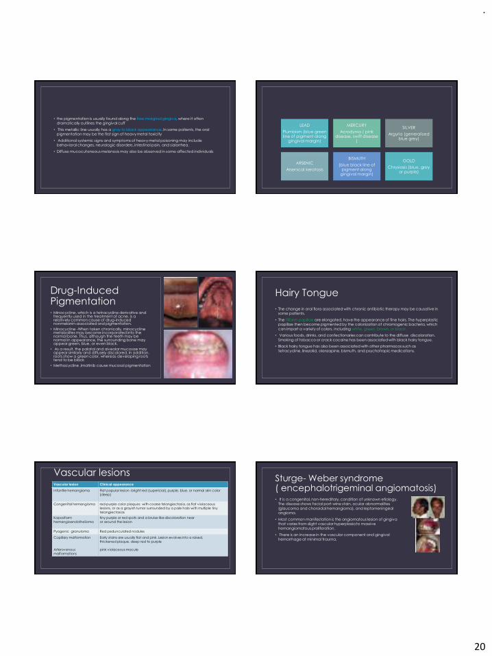
.
20
• the pigmentation is usually found along the free marginal gingiva, where it often dramatically outlines the gingival cuff
• This metallic line usually has a gray to black appearance. In some patients, the oral pigmentation may be the first sign of heavy metal toxicity
• Additional systemic signs and symptoms of heavy metal poisoning may include behavioral changes, neurologic disorders, intestinal pain, and sialorrhea.
• Diffuse mucocutaneous melanosis may also be observed in some affected individuals
LEAD
Plumbism (blue green line of pigment along
gingival margin)
MERCURY
Acrodynia ( pink disease, swift disease
)
SILVER
Argyria (generalised blue grey)
ARSENIC
Arsenical keratosis
BISMUTH
(blue black line of pigment along
gingival margin)
GOLD
Chrysiasis (blue, grey or purple)
Drug-Induced Pigmentation • Minocycline, which is a tetracycline derivative and
frequently used in the treatment of acne, is a relatively common cause of drug-induced nonmelanin-associated oral pigmentation.
• Minocycline -When taken chronically, minocycline metabolites may become incorporated into the normal bone. Thus, although the teeth may be normal in appearance, the surrounding bone may appear green, blue, or even black.
• As a result, the palatal and alveolar mucosae may appear similarly and diffusely discolored. In addition, roots show a green color, whereas developing roots tend to be black
• Methacycline ,Imatinib cause mucosal pigmentation
Hairy Tongue
• The change in oral flora associated with chronic antibiotic therapy may be causative in some patients.
• The filiform papillae are elongated, have the appearance of fine hairs. The hyperplastic papillae then become pigmented by the colonization of chromogenic bacteria, which
can impart a variety of colors, including white, green, brown, or black.
• Various foods, drinks, and confectionaries can contribute to the diffuse discoloration.
Smoking of tobacco or crack cocaine has been associated with black hairy tongue.
• Black hairy tongue has also been associated with other pharmacos such as
tetracycline, linezolid, olanzapine, bismuth, and psychotropic medications.
Vascular lesions Vascular lesion Clinical appearance
Infantile hemangioma Flat popular lesion -bright red (supericial), purple, blue, or normal skin color(deep)
Congenital hemangioma red-purple color plaques with coarse telangiectasia, as flat violaceous lesions, or as a grayish tumor surrounded by a pale halo with multiple tiny telangiectasias
Kaposiformhemangioendothelioma
tiny purple or red spots and a bruise-like discoloration near or around the lesion
Pyogenic granuloma Red pedunculated nodules
Capillary malformation
Arterovenousmalformations
Early stains are usually flat and pink. Lesion evolves into a raised, thickened plaque, deep red to purple
pink violaceous macule
Sturge- Weber syndrome ( encephalotrigeminal angiomatosis)• It is a congenital, non-hereditary, condition of unknown etiology.
The disease shows facial port-wine stain, ocular abnormalities
(glaucoma and choroidal hemangioma), and leptomeningeal angioma.
• Most common manifestation is the angiomatous lesion of gingiva that varies from slight vascular hyperplasia to massive hemangiomatous proliferation.
• There is an increase in the vascular component and gingival hemorrhage at minimal trauma.

.
21
OTHER SYNDROMES ASSOCIATED WITH HYPERPIGMENTATION• Cowden syndrome
• Cronkhite–Canada syndrome
• Dowling–Degos syndrome
• Naegeli–Franceschetti– Jadassohn syndrome
• LEOPARD syndrome
• Nelson’s syndrome
C
TREATMENT OF MUCOCUTANEOUS
MELANOSIS
• Perioral and facial pigmentation are more challenging to treat since the skin type may dictate the occurrence of postoperative complications ,including post inflammatory
hyperpigmentation.
• Experimental forms of phototherapy have also been employed, including intense
pulsed light and fractional photothermolysis.
• First-line therapy remains the application of topical medications- bleaching creams.
• Although single agents such as azelaic acid or hydroquinone have been used, more commonly, dual- or triple-combination therapy is recommended.
• A combination of 4% hydroquinone (0.05%) retinoic acid (0.01%) fluocinolone acetonide has proven to be effective in greater than 90% of patients.
• However, the majority of patients undergoing such therapy may experience immunologic sensitivity or other treatment-related adverse events, including the
development of exogenous ochronosis
Desai SR. Hyperpigmentation therapy: a review. J ClinAesthet Dermatol. 2014;7(8):13–17.
• Different thickness flap, gingivectomy, cryotherapy, electrosurgery, bur abrasion,
and scraping with a scalpel have been successfully used to treat gingival pigmentation.
• Laser therapy has also proven to be an effective modality for use in the treatment of bothersome oral pigmentation.
• Various types of lasers have been used, including super pulsed CO2, Q-switched Nd-YAG, and Q-switched alexandrite lasers
Conclusion
• Pigmented lesions within the oral cavity may present a diagnostic dilemma for the
clinician.
• A differential diagnosis for a pigmented lesion may include traumatic, reactive,
neoplastic and systemic pathologies.
• A clinico-pathologic correlation is often required to ensure accurate diagnosis of
systemic causes of diffuse pigmentation

.
22
Refernces
• Textbook of Oral Medicine Burkets 12th edition
• Textbook of oral pathology shafers 7th edition
• Bhateja S, Bohra A, Arora G (2015) Drug Induced Oral Mucosal Pigmentation- A Review. Pigmentary Disorders 2: 198. doi:10.4172/23760427.1000198
• Reshna Roy.Classification of oral pigmented lesions: A review . International Journal of Applied Dental Sciences 2019; 5(2): 397-402
• Faizan Alaw. Pigmented lesions of the oral cavity: An Update. Dent Clin North Am . 2013 October ; 57(4): 699–710. doi:10.1016/j.cden.2013.07.006
• Sreeja C, Ramakrishnan K, Vijayalakshmi D, Devi M, Aesha I, Vijayabanu B. Oral pigmentation: A review. J Pharm Bioallied Sci. 2015;7(Suppl 2):S403–S408. doi:10.4103/0975-7406.163471
• Verma B, Behra A, Ajmal AK, Sen S. Laugier– Hunziker syndrome in a young female. Indian Dermatol Online J 2017;8:148-50.
• Kamal Uddin Zaidi,1,2 Ayesha S. Ali,2 Sharique A. Ali,2 and Ishrat Naaz.Microbial Tyrosinases: Promising Enzymes for Pharmaceutical, Food Bioprocessing, and Environmental Industry Biochemistry Research International · May 2014
• HideyaAndo et al .Melanosomes Are Transferred from Melanocytes to Keratinocytes through the Processes of Packaging, Release, Uptake, and Dispersion Journal of Investigative Dermatology:Volume 132, Issue 4, April 2012, Pages 1222-1229
• Feller, L., Masilana, A., Khammissa, R.A. et al. Melanin: the biophysiology of oral melanocytes and physiological oral pigmentation. Head Face Med 10, 8 (2014). https://doi.org/10.1186/1746-160X-10-8
• Bashamalla R, Rao GV, Ramulu S, Sravya T. Pigmented intramucosal nevus of gingiva with a special insight on its pathophysiology: Report of a rare entity. J NTR Univ Health Sci 2017;6:181-4
• Dutta D, Kamath VV, Rajkumar K. Oral melanocytic nevi: Report of two cases with immunohistochemical elaboration of their probable origin and maturation. Indian J Dermatopathol Diagn Dermatol 2015;2:29-33
• Agrawala RK, Sahoo SK, Choudhury AK, Mohanty BK, Baliarsinha AK. Pigmentation in vitamin B12 deficiency masquerading Addison's pigmentation: A rare presentation. Indian J EndocrinolMetab. 2013;17(Suppl 1):S254–S256. doi:10.4103/2230-8210.119591
• Gulati N, Dutt P, Gupta N, Tyagi P. Gingival Pigmentation: Revisited. J Adv Med Dent
Scie Res 2016;4(1):48-57.
• Desai SR. Hyperpigmentation therapy: a review. J Clin Aesthet Dermatol. 2014;7(8):13–17.
CTHANKYOU

.
1
PSYCHOSOMATIC DISORDERS
OF ORAL CAVITY
Dr. Neeraj Kumar Yadav
Reader
Dept. of Oral Medicine and Radiology
MGDCH , Jaipur
CONTENTS
• Introduction
• Classification
• Details of each disorder
• Management
• Conclusion
• References
INTRODUCTION
• Mouth is directly or symbolically related to major human instincts and passion.
• The oral mucosa is highly reactive to psychological influences.
• Psychosomatic disorders are defined as disorders characterized by physiological changes that originate partially from emotional factors.
• A psychosomatic disorder involves both body and mind.
• These diseases have physical symptoms originating from mental or emotional
causes. Most common ones are stress, anxiety and depression.
• A wide spectrum of psychiatric disorders affects oral and para oral structures
which have a definite psychosomatic cause, but unfortunately they remain
unrecognized because of the common and limited nature of their presenting
features.
CLASSIFICATION
Pain Related Disorders
1. Myofascial pain dysfunction
syndrome(MPDS)
2. Atypical facial pain
3. Atypical odontogenic pain
4. Phantom bite syndrome
Disorders Related to Altered
Oral Sensation
1. Burning mouth syndrome
2. Idiopathic xerostomia
3. Idiopathic dysgeusia
4. Glossodynia
5. Glossopyrosis
Disorders Induced
by Neurotic Habits
1. Dental and
periodontal diseases
caused by bruxism
2. Biting of oral
mucosa (self
mutilation)
Autoimmune
Disorders
1. Oral lichen planus
2. Recurrent aphthous
stomatitis
3. Psoriasis
4. Mucous membrane
pemphigoid
5. Erythema multiforme
Miscellaneous
Disorders
1. Recurrent herpes
labialis
2. Necrotising ulcerative
gingivostomatitis
3. Chronic periodontal
diseases
4. Cancerophobia

.
2
MPDS
• Myofascial Pain Dysfunction Syndrome is a heterogeneous group of signs and symptoms
that affect the jaw joint and/or the chewing musculature.
• Myofascial pain syndrome (MPS) is defined as pain that originates from myofascial trigger
points in skeletal muscle.
• IN 1940, TRAVELL reported that skeletal muscle in spasm could be source of pain
• In 1959, SCHWARTZ was first to implicate the physiologic make up of the patient as a
predisposing factor in pain dysfunction and hypothesized that the stress was the significant
cause of clenching and grinding habits which results in spasm.
• In 1969 MPDS term was given by LASKIN, he proposed PSYCHOPHYSIOLOGIC
THEORY that MPDS is a result of emotional rather than occlusal and mechanical factors.
• The pain pathway that culminates in muscle spasm and pain in MPDS according to theory
begins with stress.
• Theory suggests that stress could cause clenching and grinding which in turn lead to muscle
fatigue and finally spasm
EPIDEMIOLOGY
• Very common
• Exact incidence unknown
• May occur as primary disease
• May occur in conjugation with other painful condition
• Psychological and behavioral co-morbidities also present
• The diagnosis was made based on the signs and symptoms of MPDS, as outlined by Laskin
• that is, Unilateral, dull pain in the ear or preauricular region that is commonly worse on
awakening. Tenderness of one or more muscles' mastication on palpation. Limited or
deviation of mandible on opening.
Treatment
• A number of successful treatment outcomes have been reported, including occlusal splints,
physiotherapy, muscle-relaxing appliances, and pharmacological interventions.
• Based on Wall & Melzack’s Gate Control Theory, TENS has been used very commonly for
pain relief in the last 30 years.
• The tricyclic antidepressants such as amitriptyline and nortryptyline and cognitive behavioral
therapy are often generally helpful
Atypical Facial Pain (AFP)
• According to the International Association for the Study of Pain (IASP),
chronic facial pain refers to symptoms which have been present for at least 6
months.
• ‘Atypical’ pain is a diagnosis of exclusion after other conditions have been
considered and eliminated (i.e. it is idiopathic) and is characterized by
chronic, constant pain in the absence of any apparent cause in the face or
brain.

.
3
• Many information sources suggest that all ‘unexplained’ facial pains are
termed Atypical Facial Pain but this is not the case.
• Categories of idiopathic facial pain conditions include Neuropathic Pain due
to sensory nerve damage, Chronic Regional Pain Syndrome (CRPS) from
sympathetic nerve damage and Atypical Facial Pain.
• Facial pain, often described as burning, aching or cramping, occurs on one
side of the face, often in the region of the trigeminal nerve and can extend
into the upper neck or back of the scalp.
• The pain is called “atypical” because it is a different type of pain than that of a typical toothache.
• The pain is poorly localized and does not conform to the anatomical boundaries of sensory nerve supply.
• The areas affected may be one or more of those supplied by the fifth or ninth cranial nerves, or the second and third cervical nerves.
• Sometimes the pain is bilateral
Treatment
• The patients with chronic pain including facial pain need to be screened for depression.
• Pharmacological treatment with antidepressants, antiepileptic or other drugs can also be tried.
• Cognitive–behavioural therapy may be indicated.
• Patients with AFP may be helped by a technique termed ‘reattribution’ which involves demonstrating and understanding of the complaints by taking a history of related physical, mood and social factors.
• It may help explain that depression/tiredness lowers the pain threshold and that muscle over activity and spasm (being ‘uptight’) causes pain
Atypical Odontalgia
• Atypical odontogenic (AO) pain implies toothache of unknown origin.
• Exact etiology of this condition is unknown.
• It is considered to be deafferentation neuralgia (causalgia) arising when a
dental extraction or pulp extirpation produces either an amputation neuroma
or a central degenerative change in the trigeminal nucleus.
• Some consider AO as vascular/neurovascular in origin.
• Recently, psychogenic etiology was considered.
• AO affects 10% of adults and 50% of elderly population. It is more common in women than in men in the age of fourth to fifth decade of life.
• Trauma and psychological factors are implicated factors.
Treatment
• The tricyclic antidepressants such as amitriptyline and nortryptyline are often generally helpful
BURNING MOUTH SYNDROME
• BMS is any form of burning or stinging sensation in the mouth in
association with a normal mucosa in the absence of local or systemic disease.
• It is a multifactorial disorder associated with psychological components such
as anxiety, depression and cancerophobia.
• The term “syndrome” is justified because of the simultaneous presence of
several subjective symptoms, including feeling of dryness (subjective
xerostomia), altered taste, and burning sensation of the oral tissues and
tingling, or numb feeling in the oral cavity.

.
4
• Burning is almost always bilateral and symmetrical and does not follow anatomical distribution of
a peripheral sensory nerve.
• The onset of pain is spontaneous, bilateral with no identifiable precipitating factors. Pain may be
felt deep within the mucosa, continuous for at least 4-6 months, with moderate to severe
intensity that may vary during the day
• BMS is most commonly found in adults over the age of 60. It is estimated to be about five times
more frequent in women than in men.
• There is no specific test for BMS, which makes it hard to diagnose. No specific treatment works
for everyone. However, one can prescribe medications to help in managing the pain, dry mouth,
or other symptoms
Treatment
• Cognitive-behavioural therapy or a specialist referral may be indicated.
• ‘Reattribution’ helps manage these patients.
• Topical application of capsaicin (0.025% cream) has been used.
• Topical application of 0.5 ml Aloe vera gel at 70%, 3 times a day combined with tongue
protector is found to be effective.
• The topical application of clonazepam (by sucking a tablet of 1 mg), 3 times a day for 14
days found some success in some.
• Gabapentin, an anticonvulsant drug, is advised 300-1,600 mg/day; 100 mg at bedtime.
RECURRENT APTHOUS STOMATITIS
• Recurrent apthous stomatitis (RAS) is the most common type of ulcerative disease of the
oral mucosa, and it affects approximately 20% of the general population.
• The classic presentation of RAS is recurrent, self-limiting ulcers that mainly affect non-
keratinized oral mucosa.
• Previous studies have suggested that psychological disturbances such as stress and anxiety
could play a role in the onset and recurrence of RAS lesions
• Factor potentially related with RAS exacerbations is stress
• Psychological stresses induces immunoregulatory activity by increasing the number of
leukocytes at sites of inflammation; this is a characteristic often observed during the
pathogenesis of RAU
• Onset of RAS usually is during childhood, with a tendency for ulcers to diminish in
frequency and intensity with age. There are 3 variants described-minor, major, herpetic
form.
IDIOPATHIC XEROSTOMIA
• Xerostomia is a subjective sensation of dry mouth; hyposalivation is defined as an objective assessment
of reduced salivary flow rate.
• Depressive symptoms are usually evident in individuals with idiopathic subjective dry mouth.
• Mason and Glen (1967) have stated that as the secretion of saliva is regulated by ANS and is subjected
to reflex stimulation from physical and psychic causes, then xerostomia may result from 4 basic causes
in which factor affecting salivary centre are primary cause which include:
• 1. Emotions, fear, excitement, stress
• 2. Depression
• 3. Organic diseases e.g. brain tumor, Parkinson’s disease
• 4. Drugs: A number of reports have shown that salivary cortisol is associated
with depression and anxiety, and hence, salivary cortisol can be used as an
important non invasive biological indicator of stress.

.
5
Treatment
• Salivary substitutes and lubricants with moistening properties are designed to provide
prolonged mucosal wetting.
• Products include “artificial” saliva, rinses, gels, and sprays, which may contain carboxymethyl
cellulose (CMC), a mucopolysaccharide, glycerate polymer gel base, or natural mucins, singly
or in combination.
• Pilocarpine increases salivary flow and affect subjective dryness as well.
• The selective serotonin reuptake inhibitors like sertaline are often generally helpful.
DYSGEUSIA
• Dysgeusia is defined as a distorted gustatory perception or persistent gustatory sensation in the absence of gustatory stimulants.
• These are often perceived as bitter, sour, or metallic.
• Association between stress and taste might have a possible central mechanism;
• enhanced activation of multiple neurobiological pathways is involved in stress and appetite regulation.
Treatment
• Zinc supplementation is believed to aid in treating taste disorders by promoting proliferation
of normal taste bud cells, even in patients without zinc deficiency.
• The tricyclic antidepressants such as amitriptyline and nortryptyline are often generally
helpful.
CHRONIC BITING OF THE ORAL
MUCOSA
• It is a form of factitial/unintentional injury that is observed commonly on the buccal and
labial mucosa and lateral surface of tongue.
• Habitual lip or cheek biting usually occurs as an unconscious psychogenic habit caused by a
wide range of emotions.
• This mild form of self mutilation may sometimes emerge as a response to oral stimuli or as
an attempt to gain attention from family members or caretakers.
Treatment
• Counseling, biofeedback, relaxation techniques and hypnosis or psychiatric
treatment have been suggested along with the dental management of the
effects of habit.
RECURRENT HERPES LABIALIS
• RHL occurs due to either physical trauma or emotional stress leading to lesions of skin and labial mucosa.
• Emotional stress apparently serves to prevent the antibodies from acting at the local mucosal site.
• Triggers may include sun exposure, psychological stress, onset of menses, illness and physical trauma.
• Infection with herpes simplex virus 1 (HSV1), which manifests as primary gingivostomatitis, usually occurs in preschool or kindergarten children, adolescents and young adults, and does not recur in the same form.

.
6
Treatment
• Antiviral compounds for the treatment of HSL infection have been advocated.
• The tricyclic antidepressants such as amitriptyline and nortryptyline are often generally
helpful
CANCEROPHOBIA
• Fear is an unpleasant emotion and the pervasiveness of cancer fear in the population may
have implications for quality of life.
• In addition, cancer fear has been shown to be associated with screening uptake and
presentation of suspicious symptoms, although both motivating and deterrent associations
have been found.
• This disorder falls under hypochondriasis (It is a persistent fear in the patient’s mind that
they have contracted cancer).
• Cancerophobia has been noted to be associated with depression but exact pathogenesis is
unclear.
Treatment
• A better understanding of the nature of cancer fear may help identify those who suffer from
maladaptive and undue fears, and help explain why the behavioral responses seem to vary.
• The tricyclic antidepressants such as amitriptyline and nortryptyline are often generally helpful.
Periodontal Diseases
• The etiology of inflammatory periodontal disease is complex. The etiological significance of biological and behavioral risk factors, including systemic conditions, smoking, oral hygiene, and age has been demonstrated.
• However, a significant proportion of the variation in disease severity cannot be explained by taking only these factors into consideration. A psychosomatic disorder affects periodontium by two ways:
• 1) Self-inflicted injuries seen in these patients
• 2) Via disturbance in autonomic nervous system altering tissue response
Necrotizing Ulcerative Gingivitis
• Necrotizing ulcerative gingivitis (NUG) is a fusospirochetal infection caused
by local and systemic predisposing factors.
• Among these, emotional stress appears to be the most common, although
debilitating diseases, nutritional deficiencies, and neurologic diseases also play
important roles.
• Emotional stress may lead to NUG indirectly by an expression of cortisol
and catecholamine levels
Management of Psychosomatic Disorders
Various treatment modalities tried out are:
1. Psychotherapy or the remedial influence of mind
• a. Cognitive–behavioral therapy
• b. Self-observation
• c. Relaxation training
• d. Hypnotherapy
• e. Biofeedback

.
7
• 2. Pharmacotherapy
• a. Antidepressants
• b. Antianxiety drugs
• c. Antipsychotic drugs.
CONCLUSION
To conclude, we can say that many diseases manifesting in the oral cavity have a psychological
component in their etiology or have some effect of psychologic factors. Further, many
psychiatric disorders have an influence upon health of oral tissue. Because stress is increasing
in everyday life due to cut-throat competition in every field, there are more chances of dental
practitioners encountering patients with such disorders. Hence, one should be familiar with
such manifestations, and if accounted, should try to manage them with psychiatrists, whenever
needed.
REFERENCES
• Tripathi RM, Seth P, Awasthi P, Bhattacharya A, Bajpai AK and ThahrianiA Psychosomatic disorders affecting the oral cavity: A review article The Pharma Innovation Journal 2018; 7(8): 327-332
• Ankit Dhimole1*, Neha Bhasin1,Psychosomatic Disorders Affecting the Mouth: A Critical Review British Journal of Medicine & Medical Research 14(5): 1-9, 2016, Article no.BJMMR.24743 ISSN: 2231-0614, NLM ID: 101570965
• Dangore-Khasbage S, Khairkar PH, Degwekar SS, Bhowate RR, Bhake AS, Singh A, et al. Prevalence of oral mucosal disorders in institutionalized and non-institutionalized psychiatric patients: a study from AVBR Hospital in central India. J Oral Sci 2012; 54:85-91
• Psychosomatic problems in dentistry Toyofuku BioPsychoSocial Medicine (2016) 10:14
Thank you
.
1
ORAL RED AND WHITE LESIONS
Dr. Neeraj Kumar Yadav
Reader
Dept. of Oral Medicine and Radiology
MGDCH , Jaipur
WHITE LESIONS
• A white appearance of oral mucosa may be caused by a variety of factors.
• Oral epithelium may be
✓Stimulated to an increased production of keratin: Hyperkeratosis
✓An abnormal but benign thickening of stratum spinosum: Acanthosis
✓Intra and extracellular accumulation of fluid in the epithelium
✓Microbes particularly fungi, can produce whitish pseudomembranesconsisting of sloughed epithelial cells, fungal mycelium and neutrophils which are loosely attached to oral mucosa
✓Juxtaepithelial hyalinization
✓Hypopigmentation
RED LESIONS
• May develop as a result of
✓Atrophic epithelium
✓Reduction in number of epithelial cells
✓Increased vascularization that is dilatation of vessels and/or proliferation of vessels
• Red lesions if persistent can be:
✓FOCAL
✓MULTIFOCAL
✓DISCOID
✓DIFFUSE
✓LINEAR
CLASSIFICATION
• INFECTIOUS DISEASES:Oral candidiasis, hairy leukoplakia
• PREMALIGNANT LESIONS: Oral leukoplakia and erythroplakia, OSMF
• IMMUNOPATHOLOGIC DISEASES:OLP,Drug induced LRs,GVHD, SLE
• ALLERGIC REACTIONS:LCR,Reactions to CHX and dentrifice
• TOXIC REACTIONS: Reactions to smokeless tobacco, Smokers palate
• REACTIONS TO MECHANICAL TRAUMA: Morsaicatio
• OTHER RED AND WHITE LESIONS:BMG, Leukoodema,White sponge nevus, Hairy tongue
CANDIDIASIS
• Most prevalant opportunistic infection
• Vast majority of cases caused by candida albicans
• Affects very young, very sick and very old
• Divided into primary and secondary infections
ETIOLOGY AND PATHOGENESIS
• C.albicans, C.tropicalis, C.glabrata comprise 80% of species isolated from candidal infections
• Factors causing its infection:
✓Adhesion
✓Lipase production
• Varoius predisposing factors:
✓Local factors
✓General factors
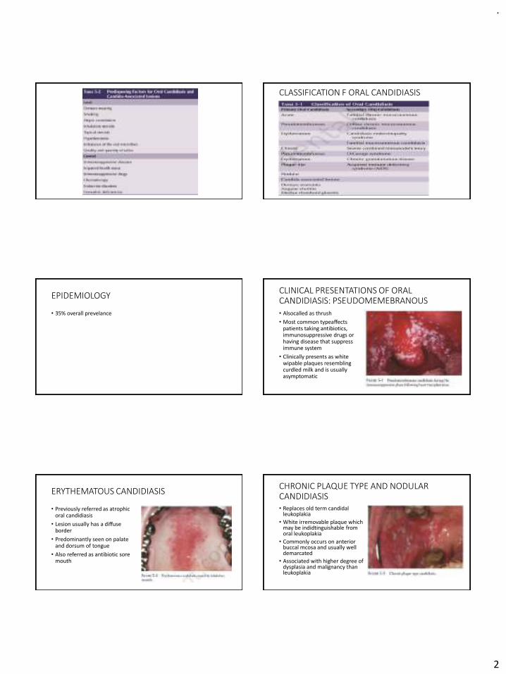
.
2
CLASSIFICATION F ORAL CANDIDIASIS
EPIDEMIOLOGY
• 35% overall prevelance
CLINICAL PRESENTATIONS OF ORAL CANDIDIASIS: PSEUDOMEMEBRANOUS• Alsocalled as thrush
• Most common typeaffectspatients taking antibiotics, immunosuppressive drugs or having disease that suppress immune system
• Clinically presents as white wipable plaques resembling curdled milk and is usually asymptomatic
ERYTHEMATOUS CANDIDIASIS
• Previously referred as atrophic oral candidiasis
• Lesion usually has a diffuse border
• Predominantly seen on palate and dorsum of tongue
• Also referred as antibiotic sore mouth
CHRONIC PLAQUE TYPE AND NODULAR CANDIDIASIS• Replaces old term candidal
leukoplakia
• White irremovable plaque which may be indidtinguishable from oral leukoplakia
• Commonly occurs on anterior buccal mcosa and usually well demarcated
• Associated with higher degree of dysplasia and malignancy than leukoplakia
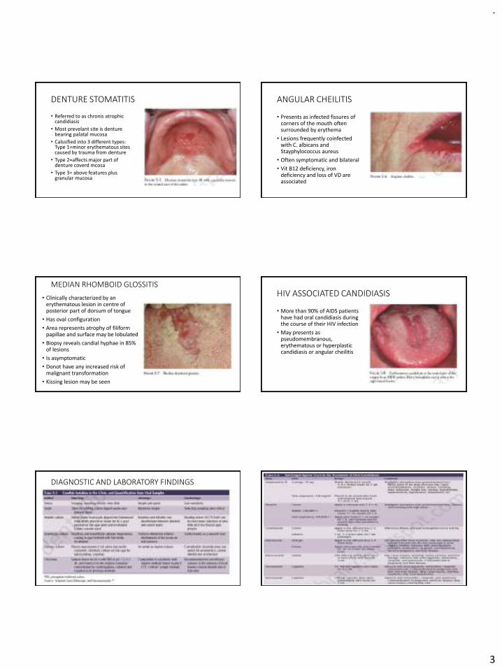
.
3
DENTURE STOMATITIS
• Referred to as chronis atrophic candidiasis
• Most prevelant site is denture bearing palatal mucosa
• Calssified into 3 different types: Type 1=minor erythematous sites caused by trauma from denture
• Type 2=affects major part of denture coverd mcosa
• Type 3= above features plus granular mucosa
ANGULAR CHEILITIS
• Presents as infected fissures of corners of the mouth often surrounded by erythema
• Lesions frequently coinfectedwith C. albicans and Stayphylococcus aureus
• Often symptomatic and bilateral
• Vit B12 deficiency, iron deficiency and loss of VD are associated
MEDIAN RHOMBOID GLOSSITIS
• Clinically characterized by an erythematous lesion in centre of posterior part of dorsum of tongue
• Has oval configuration
• Area represents atrophy of filiform papillae and surface may be lobulated
• Biopsy reveals candial hyphae in 85% of lesions
• Is asymptomatic
• Donot have any increased risk of malignant transformation
• Kissing lesion may be seen
HIV ASSOCIATED CANDIDIASIS
• More than 90% of AIDS patients have had oral candidiasis during the course of their HIV infection
• May presents as pseudomembranous, erythematous or hyperplastic candidiasis or angular cheilitis
DIAGNOSTIC AND LABORATORY FINDINGS
.
1
Respiratory diseasesDr. Neeraj Kumar Yadav
Reader
Dept. of Oral Medicine
and Radiology
MGDCH , Jaipur
CONTENTS
1) Introduction
2) Upper airway diseases
3) Lower airway diseases
4) Pathophysiology
5) Clinical and Laboratory findings
6) Classification
7) Diagnosis
8) Management
9) Prognosis
10) Oral health considerations
11) References.
INTRODUCTION
• Respiration : It is the process by which oxygen is taken in and carbon dioxide isgiven out.
• The first breath takes place only after birth.
• Fetal lungs are non-functional. So, during intrauterine life the exchange of gasesbetween fetal blood and mother’s blood occurs through placenta.
• Normal Respiratory Rate:• New born : 30 to 60/minute
• Early childhood : 20 to 40/minute
• Late childhood : 15 to 25/minute
• Adult : 12 to 16/minute.
• Types of respiration:
1. External respiration
2. Internal respiration
• Phases of respiration:
1. Inspiration
2. Expiration
• FUNCTIONAL ANATOMY OF RESPIRATORY TRACT:
• Respiratory tract is the anatomical structure through which air moves in and out. Itincludes Nose, Pharynx, Larynx, Trachea, Bronchi and Lungs.
i) Pleura
ii) Tracheobronchial Tree:
• Trachea and bronchi are together called tracheobronchial tree. It forms a part of airpassage.
• Components :1. Trachea
2. Secondary bronchi
3. Tertiary bronchi.
4. Bronchioles
5.Terminal bronchiole
6. Respiratory bronchioles
• Upper and lower respiratory tracts:
• „Respiratory unit:• Parenchyma of lungs is formed by respiratory unit that forms the terminal
portion of respiratory tract.
• Respiratory unit is defined as the structural and functional unit of lung. Exchangeof gases occurs only in this part of the respiratory tract.
• Structure of respiratory unit:
• 1. Respiratory bronchioles
• 2. Alveolar ducts
• 3. Alveolar sacs
• 4. Antrum
• 5. Alveoli.
.
2
• Alveolar Cells or Pneumocytes:
• Type I alveolar cells:
• Type I alveolar cells are the squamous epithelial cells forming about 95% of thetotal number of cells. These cells form the site of gaseous exchange between thealveolus and blood.
• Type II alveolar cells:
• Type II alveolar cells are cuboidal in nature and form about 5% of alveolar cells.These cells are also called Granular pneumocytes. Type II alveolar cells secretealveolar fluid and surfactant.
• Non-respiratory functions of respiratory tract:
1. Olfaction
2. Vocalization
3. Prevention of dust particles
4. Defense mechanism
These cells are leukocytes, macrophages, mast cells, natural killer cells anddendritic cells.
5. Maintenance of water balance
6. Regulation of body temperature
7. Regulation of acid-base balance
8. Anticoagulant function
9. Secretion of angiotensin - converting enzyme
10. Synthesis of hormonal substances
• Respiratory protective reflexes:
i) Cough reflex
ii) Sneezing reflex
iii) Swallowing (deglutition) reflex
Diseases of the respiratory tract
Upper airway Diseases
1) Viral upper respiratory tract infections2) Allergic Rhinitis and Conjunctivitis3) Otitis media4) Sinusitis5)Laryngitis and Laryngotracheobronchitis6) Pharyngitis and Tonsillitis.
Lower airway Diseases
1)Acute Bronchitis2) Pneumonia3) Bronchiolitis4) Asthma5) Tuberculosis6) COPD7) Cystic fibrosis8) Pulmonary embolism9) Pulmonary Neoplasms
Viral upper respiratory tract infections
• Cause: Virus (Rhinovirus)
• Age: Children
• These are the RNA viruses which infect the respiratory tree.
• It is closely transmitted by close person to person contact and by respiratorydroplets.
• Shedding can occur from nasopharyngeal secretions for upto 3 wks but seven daysor less is typical.

.
3
• Pathophysiology:
• Viral particles can lodge in either the upper or lower respiratory tract.
Invade the respiratory epithelium
Viral replication.
• Incubation period for rhinovirus - 2 to 5 days.
• During this time, active and specific immune responses are triggered, andmechanisms for viral clearance are enhanced.
• Clinical and laboratory findings:
• Symptoms: i) Rhinorrhea
ii) Nasal congestion
iii) Oroparyngeal irritation
• Other symptoms include Cough, Fever, Malaise, Fatigue, Headache and myalgia.
• Complete Blood count (CBC) with differential may demonstrate an increase in :
i) Mononuclear cells
ii) Lymphocytes
iii) Monocytes. (Right shift).
• Diagnosis:
• Basis of medical history as well as confirmatory physical findings.
• Diagnoses that should be excluded include:
i) Acute bacterial rhinosinusitis,
ii) Allergic rhinitis and
iii) Group A streptococcal pharyngitis.
• Management:• Analgesics - sore throat and myalgias.
• Antipyretics - febrile patients.
• Anticholinergic agents - Reducing rhinorrhea.
• Oral or topical decongestants such as the sympathomimetic amines, are an effective means of decreasing nasal congestion
• Prognosis:
• Excellent.
• Oral health considerations:• Presence of small round erythematous macular lesions on the soft palate.
• These lesions may be caused directly by the viral infection, or they may represent aresponse of lymphoid tissue.
• Individuals with excessive lingual tonsillar tissue - enlargement of foci of lymphoidtissue, particularly at the lateral borders at the base of the tongue.
• Oral dryness.
Allergic Rhinitis and Conjunctivitis
• Allergic rhinitis is a chronic recurrent inflammatory disorder of the nasal mucosa.
• Allergic conjunctivitis is an inflammatory disorder involving the conjunctiva.
• When both conditions occur, the term Allergic Rhinoconjunctivitis is used.
• The basis of the inflammation is an allergic hypersensitivity (type Ihypersensitivity) to environmental triggers.
• Typical seasonal triggers include grass, tree, and weed pollens.
• Common perennial triggers include dust mites, animal dander, and mold spores.
• Allergic rhinitis is the most prevalent chronic medical disorder.
Pathophysiology:

.
4
• Clinical and Laboratory Findings:
• Conjunctival symptoms : i) Pruritus
ii) Lacrimation,
iii) Crusting, and burning.
• Nasal symptoms may include sneezing, pruritus, clear rhinorrhea, and nasalcongestion.
• Other symptoms such as postnasal drainage with throat irritation, pruritus of thepalate and ear canals, and fatigue.
• Direct examination of the nasal mucosa reveals significant edema and a pale bluecoloration of the turbinates.
• The clinical signs of allergic Rhinoconjunctivitis include:
• Postnasal drainage or oropharyngeal cobblestoning might be identified uponexamination of the oropharynx.
• A High-arched palate, protrusion of the tongue and overbite may be seen.
i) Injection of the conjunctiva with or without cobblestoning.ii) Prominent infraorbital creases (Dennie-Morgan folds/pleats)iii) Swelling, and darkening (allergic shiners)iv) A transverse nasal crease; and frequent upward rubbing of the tip ofthe nose (allergic salute).
• Patients with allergic rhinitis might have elevated levels of serum IgE and totaleosinophil count.
• Microscopic examination of nasal secretions often demonstrates significantnumbers of eosinophils.
• The Radioallergosorbent test (RAST) is method of testing for specific allergicsensitivities that is based on circulating levels of specific IgE.
• Specific IgE levels are determined by using serum samples and are quantified byusing radioactive markers.
• Classification:
• Diagnosis:• Patients present with a history suggestive of allergic sensitivity and recurrent
symptoms with specific exposures.
• Symptoms that have recurred for 2 or more years during the same season aresuggestive of seasonal allergic disease.
i) Perennial - Indoor allergens (eg, house dust mites, cockroaches,pets)ii) Seasonal illness – Outdoor allergens (eg, trees, grasses, weeds).
• The Preferred method of testing for allergic sensitivities is skin testing, which is
performed with epicutaneous (prick/ scratch) tests, often followed by intradermal
testing.
• Prick skin testing is the type most widely used.
• With prick testing, a small amount of purified allergen is inoculated through the
epidermis only (ie, epicutaneously) with a pricking device.
• Positive (histamine) and negative (albumin-saline) controls are used for
comparison.
• Reactions are measured at 15 minutes, and positive reactions indicate prior allergen
sensitization.
• Tests that yield negative results may be repeated intradermally to increase the
sensitivity of the testing.
• Management:
• General treatment modalities are used in the treatment of allergicRhinoconjunctivitis:
i) Allergen avoidance
ii) Pharmacotherapy (medication) and
iii) Immunotherapy (allergy injections).
• The best treatment is avoidance of the offending allergen.
• Immunotherapy is an effective means of treatment for patients with allergicRhinoconjunctivitis.
• Immunotherapy is available for a variety of airborne allergens, including grass, tree,and weed pollens, dust mites, animal dander, and mold spores.
.
5
• Prominent sneezing, pruritus, or rhinorrhea – antihistamines.
• Second-generation Nonsedating antihistamines – Cetrizine, Loratadine andfexofenadine.
• Oral decongestants can be added to oral antihistamines to relieve nasal congestionand obstruction.
• Therapy with a cysLT1-receptor antagonist plus antihistamine may have a greatereffect.
• Topical anti-inflammatory agents - Cromolyn sodium and topical corticosteroidsprays.
• Prognosis:
• Excellent.
• Oral Health Considerations:• Oral dryness.
• Oral candidiasis.
• Allergic respiratory hypersensitivity from exposure to dental materials such asmethacrylates and natural rubber latex.
Otitis Media
• Otitis media is inflammation of the middle-ear space and tissues.
• Age: children under 8 years of age or younger.
• Otitis media can be subdivided into:
i) Acute otitis media,
ii) Recurrent otitis media,
iii) Otitis media with effusion and
iv) Chronic suppurative otitis media.
• The underlying problem in all types of otitis media is dysfunction of the eustachiantube.
• The most common infectious causes are viruses, Streptococcus pneumoniae,Haemophilus influenzae, and Moraxella catarrhalis
• Pathophysiology:
Aspiration of Nasopharyngeal pathogens
Impairs normal eustachain tube function
Negative pressure in the middle ear space
Otitis Media
• Clinical and Laboratory Findings:
• Acute otitis media - Fever and otalgia.
• Other symptoms - Irritability, anorexia, and vomiting.
• On physical examination, the tympanic membrane may appear erythematous andbulging, suggesting inflammation of the middle ear.
• Otoscopic findings include: i) A loss of landmarks and
ii) Decreased mobility of the tympanic membrane as
seen by pneumatic otoscopy.
• Otitis media with effusion - Clogged ears and popping.
• Otoscopic examination – Presence of Serous middle-ear fluid and air-fluid levels
• Chronic suppurative otitis media - Otorrhea
• Investigations : i) Tympanometry and
ii) Myringotomy with aspiration
• Tympanometry is a technique that measures the compliance of the tympanicmembrane by using an electroacoustic impedance bridge.
• Myringotomy with aspiration can be useful in situations when culture of themiddle ear fluid is needed, such as with immunocompromised hosts or withpatients who have persistent effusions despite medical management.

.
6
• Classification:
Acute otitis media is defined as middle-ear inflammation with an infectiousetiology and a rapid onset of signs and symptoms.
Otitis media with effusion is defined as a middle ear effusion (oftenasymptomatic) that can be either residual (3 to 16 weeks following acuteotitis media) or persistent (lasting more than 16 weeks).
Recurrent otitis media is defined as three new episodes of acute otitis mediain 6 months time or four new episodes in a 12-month period.
Chronic suppurative otitis media is defined as persistent otorrhea lastinglonger than 6 weeks.
• Diagnosis:
• Pneumatic otoscopy - Clinician not only to visualize the tympanic membrane butalso to assess its mobility.
• Management:
• Antibiotics
• Common choices include amoxicillin, azithromycin, and trimethoprim-sulfamethoxazole.
• The duration of therapy varies from 3 to 14 days.
• Surgical modalities: i) Myringotomy with or without tympanostomy tube insertion,
ii) Tympanocentesis, and
iii) Adenoidectomy.
• Chronic suppurative otitis media - Parenteral antibiotics to cover infection byPseudomonas species and anaerobic bacteria.
• Prognosis:
• Acute otitis media - Excellent. The most common complication is conductivehearing loss related to persistent effusions.
• Serious complications - Mastoiditis, cholesteatoma and labyrinthitis.
• Oral Health Considerations:• Many children with recurrent otitis media are treated frequently with various
antibiotics.
• It has been demonstrated that antibiotic regimens used for the treatment of otitismedia promote the emergence of antibiotic-resistant bacteria.
• Oral candidiasis.
Sinusitis
• Sinusitis is defined as an inflammation of the epithelial lining of the paranasalsinuses.
• The inflammation of these tissues causes mucosal edema and an increase inmucosal secretions.
• Common trigger Factor - Acute upper respiratory infection.
• If blockage of sinus drainage occurs, retained secretions can promote bacterialgrowth and Subsequent acute bacterial sinusitis.
• Types: i) Acute sinusitis
ii) Chronic sinusitis
• Rhythmic ciliary movement and clearance of secretions
Several factors
Foreign bodies and deviated nasal septum
Cause obstruction
Blockage of the sinus ostia
Stasis of sinus secretions
Facilitates bacterial growth.
Pathophysiology• Clinical and Laboratory Findings:
• Acute sinusitis - Facial pain, tenderness, and headache.
• Sinusitis affecting the sphenoid sinuses or posterior ethmoid sinuses can causeheadache or pain in the occipital region.
• Other symptoms - Purulent nasal discharge, fever, malaise, and postnasal drainagewith fetid breath, toothache or pain with mastication.
• Common complaints - Chronic rhinorrhea, postnasal drainage, nasal congestion,sore throat, and facial fullness.
• Physical examination: Sinus tenderness and purulent nasal drainage, erythema andswelling of the overlying skin.
• Computed tomography (CT) - Chronic sinusitis with underlying disease of theosteomeatal complex.
• Magnetic resonance imaging (MRI) - Bony abnormalities.

.
7
• Classification:
• Sinusitis is classified as : i) Acute
ii) Subacute
iii) Chronic.
• Acute sinusitis is defined as inflammation of less than 4 weeks, subacute as 4 to 12weeks, and chronic as longer than 12 weeks in duration.
• Diagnosis:• Other predisposing factors: Tobacco smoke exposure, immunodeficiency and septal
deviation.
• Evaluation of the osteomeatal complex is crucial in the management of thesepatients.
• Rhinoscopy - Direct visualization of sinus ostia.
• Management:
• First-line antibiotics - Amoxicillin, second-generation cephalosporins,azithromycin, and amoxicillin-clavulanate.
• Comprehensive treatment - Adequate hydration, steam inhalation, andpharmacologic measures to restore ostial patency.
• Nasal glucocorticosteroids.
• Acute frontal or sphenoid sinusitis is very serious because of the potential forintracranial complications.
• Topical corticosteroids - Reduce the swelling and obstruction of the osteomeatalcomplex.
• Intravenous antibiotics and surgical intervention.
• Functional endoscopic sinus surgery involves the removal of the osteomeatalobstruction through an intranasal approach.
• Prognosis:
• Ethmoid and maxillary sinusitis - Periorbital or orbital cellulitis.
• Periorbital - Broad-spectrum antibiotics .
• Orbital cellulitis - Hospital admission with broad-spectrum intravenous antibiotics.
• Frontal sinusitis can extend through the anterior wall and present as Pott’s puffytumor.
• Sinusitis can also spread intracranially and result in abscess or meningitis.
• Chronic medication - Rhinitis medicamentosa from prolonged use of topicaldecongestants.
• Surgical intervention will often reverse the chronic process.
• Oral Health Considerations :
• Patients with sinus infections – Toothache.
• The oral health care professional must be able to differentiate between anodontogenic infection and sinus pain.
• Sinus infections usually present with pain involving more than one tooth in thesame maxillary quadrant, whereas a toothache usually involves only a single tooth.
• Chronic sinus infections - Mouth breathing.
• Oral dryness and gingivitis.
Laryngitis and Laryngotracheobronchitis
• Laryngitis is defined as an inflammation of the larynx, usually because of a viralinfection.
• Laryngotracheobronchitis is an inflammation involving the larynx, trachea, andlarge bronchi.
• Laryngitis - adult population.
• Laryngotracheobronchitis – young children and second and third years of life.
• The viruses most commonly implicated in laryngitis are parainfluenza virus,coxsackieviruses, adenoviruses, and herpes simplex virus.
• The viruses most commonly associated with laryngotracheobronchitis areparainfluenza virus, RSV, influenza virus, and adenovirus.
• Pathophysiology:
• Infection of the respiratory epithelium
Inflammatory response
Mononuclear cells and polymorphonuclear leukocytes
Vascular congestion and edema.
Denudation of areas of respiratory epithelium canresult.
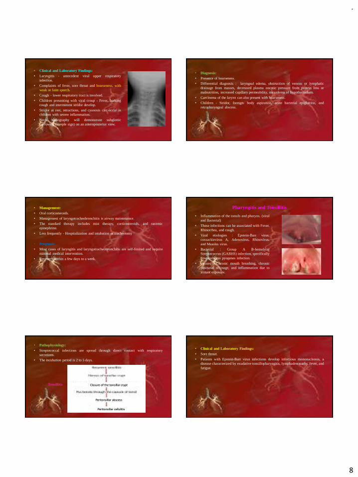
.
8
• Clinical and Laboratory Findings:
• Laryngitis - antecedent viral upper respiratoryinfection.
• Complaints of fever, sore throat and hoarseness, withweak or faint speech.
• Cough – lower respiratory tract is involved.
• Children presenting with viral croup - Fever, barkingcough and intermittent stridor develop.
• Stridor at rest, retractions, and cyanosis can occur inchildren with severe inflammation.
• Neck radiography will demonstrate subglotticnarrowing (steeple sign) on an anteroposterior view.
• Diagnosis:
• Presence of hoarseness.
• Differential diagnosis : laryngeal edema, obstruction of venous or lymphaticdrainage from masses, decreased plasma oncotic pressure from protein loss ormalnutrition, increased capillary permeability, myxedema of hypothyroidism.
• Carcinoma of the larynx can also present with hoarseness.
• Children - Stridor, foreign- body aspiration, acute bacterial epiglottitis, andretropharyngeal abscess.
• Management:
• Oral corticosteroids.
• Management of laryngotracheobronchitis is airway maintenance.
• The standard therapy includes mist therapy, corticosteroids, and racemicepinephrine.
• Less frequently - Hospitalization and intubation or tracheotomy
• Prognosis:• Most cases of laryngitis and laryngotracheobronchitis are self-limited and require
minimal medical intervention.
• Recovery within a few days to a week.
Pharyngitis and Tonsillitis
• Inflammation of the tonsils and pharynx. (viraland Bacterial)
• These infections can be associated with Fever,Rhinorrhea, and cough.
• Viral etiologies : Epstein-Barr virus,coxsackievirus A, Adenovirus, Rhinovirus,and Measles virus.
• Bacterial : Group A B-hemolyticStreptococcus (GABHS) infection, specificallyStreptococcus pyogenes infection.
• Causes : Chronic mouth breathing, chronicpostnasal drainage, and inflammation due toirritant exposure.
• Pathophysiology:
• Streptococcal infections are spread through direct contact with respiratorysecretions.
• The incubation period is 2 to 5 days.
Tonsillitis Closure of the tonsillar crypt
Peritonsillar abscess
Peritonsillar cellulitis
• Clinical and Laboratory Findings:
• Sore throat.
• Patients with Epstein-Barr virus infections develop infectious mononucleosis, adisease characterized by exudative tonsillopharyngitis, lymphadenopathy, fever, andfatigue.
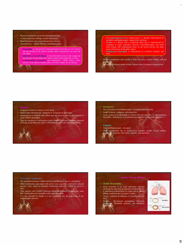
.
9
• Physical examination can reveal hepatosplenomegaly.
• Common laboratory findings include leukocytosis.
• Blood chemistries may reveal elevated liver enzymes.
• Coxsackievirus - distinct illnesses, tonsillopharyngitis.
• Herpangina is a disease that is characterized by ulcers that are 2 to 3 mm insize and located on the anterior tonsillar pillars and possibly the uvula andsoft palate.
• Hand-foot-and-mouth disease is characterized by ulcers on the tongue andoral mucosa, in association with vesicles found on the palms and or soles.
• Lymphonodular pharyngitis can characterize Small yellow- whitenodules on the anterior tonsillar pillars and these nodules do not ulcerate.
• Physical examination often reveals a beefy red uvula, cervical adenitis, and oralpetechiae.
• Laboratory evaluation should include a throat culture for group A Streptococcus.
• Pharyngoconjunctival fever (adenovirus) is a disorder characterized byexudative tonsillopharyngitis, conjunctivitis and fever.
• Measles is a disease with a prodromal phase that is characterized bysymptoms of upper respiratory infection, tonsillopharyngitis and smallwhite lesions with erythematous bases on the buccal mucosa and inneraspect of the lower lip (Koplik’s spots).
• Streptococcal pharyngitis is characterized by exudative tonsillitis andfever.
• Diagnosis:
• Diagnosis is based on a history of sore throat .
• Rapid antigen detection test - Diagnosis of streptococcal pharyngitis.
• Antistreptococcal antibody titers reflect past and are of no value in the diagnosis ofacute GABHS pharyngitis.
• They are valuable for confirmation of prior GABHS infections in patients suspectedof having Acute Rheumatic fever or Poststreptococcal Acute Glomerulonephritis.
• Management:
• The viral causes of tonsillopharyngitis are treated symptomatically.
• Gargle solutions, analgesics, and antipyretics.
• Acute streptococcal pharyngitis is treated with oral penicillin V, cephalosporins,macrolides, clindamycin, or an intramuscular injection of benzathine penicillin G.
• Prognosis:• Viral tonsillopharyngitis - very good.
• Other complications due to streptococcal tonsillitis include cervical adenitis,peritonsillar abscesses, otitis media, cellulitis, and septicemia.
• Oral Health Considerations:
• The association between GABHS infection and Rheumatic fever is well known.
• When toothbrushes were rinsed with sterile water, organisms could not be culturedbeyond 3 days, where as nonrinsed toothbrushes harbored GABHS for up to 15days.
• Thus, patients with GABHS infections should be instructed to thoroughly cleantheir toothbrushes and removable acrylic appliances daily.
• It is also advisable to change to a new toothbrush after the acute stage of anyoropharyngeal infection.
Lower Airway diseases
• Acute Bronchitis:• Acute bronchitis is an acute respiratory infection
involving the large airways (Trachea and Bronchi) thatis manifested predominantly by cough with or withoutphlegm production that lasts up to 3 weeks.
• Viruses : Influenza B, Influenza A, Parainfluenza, andRSV.
• Bacteria : Mycoplasma pneumoniae, Chlamydiapneumoniae, Bordetella pertussis, and Bordetellaparapertussis.

.
10
• Pathophysiology:
• Infection of the mucosal cells and congestion of the respiratory mucosa
Inflammation causes an increase in secretory activity.
Increased sputum production.
Polymorphonuclear leukocytes infiltrate the bronchial walls and lumen.
Desquamation of the ciliated epithelium.
Spasm of bronchial smooth muscle.
• Clinical and Laboratory Findings:
• Acute viral bronchitis - Sudden onset of cough, with or without sputumexpectoration, common cold, acute asthma, or an acute exacerbation of chronicbronchitis.
• Chest discomfort.
• Other symptoms: Dyspnea and respiratory distress.
• Physical examination may reveal wheezing.
• Symptoms gradually resolve over a period of 1 to 2 weeks.
• Diagnosis:
• Diagnosis of acute bronchitis is based on a suggestive history and a physicalexamination.
• Chest radiography may be helpful in distinguishing bacterial bronchitis frompneumonia.
• Patients with recurrent bouts of acute bronchitis should be evaluated for possibleasthma.
• This evaluation would include pulmonary function testing.
• Sputum cultures.
• Management:
• Viral bronchitis - Supportive care.
• Airway obstruction or hyperreactivity - Inhaled bronchodilators, such as albuterol.
• Cough suppressants – Codeine.
• Bacterial bronchitis - Amoxicillin, amoxicillin-clavulanate, macrolides, andcephalosporins.
• Pertussis infection - Macrolide or trimethoprim-sulfamethoxazole is appropriate.
• Inhaled B2-agonist bronchodilators.
• Prognosis:
• Acute bronchitis - Excellent prognosis .
• Chronic lung disease and respiratory compromise - Hospitalization and respiratoryfailure.
• High-risk individuals such as Human immunodeficiency virus (HIV) infectionacute bronchitis may lead to the development of bronchiectasis.
• Oral Health Considerations:• Resistance to antibiotics may develop rapidly and last for 10 to 14 days.
• Amoxicillin for acute bronchitis should be prescribed with another type ofantibiotic, (such as clindamycin or a cephalosporin) when an antibiotic is neededfor an odontogenic infection.
Pneumonia
• Pneumonia is defined pathologically as an infection and a subsequent inflammationinvolving the lung parenchyma.
• Pneumonias can be broadly classified as either community acquired or nosocomial.
• Nosocomial infections are infections that are acquired in a hospital or health carefacility and often affect debilitated or chronically ill individuals.
• Community-acquired infections can affect all persons but are more commonly seenin healthy individuals.
• The most common bacterial cause of community acquired pneumonia is S.pneumoniae, followed by H. influenzae.

.
11
• Nosocomial pneumonia : Aerobic gram-negative bacilli, such as P. aeruginosa,Escherichia coli, Klebsiella pneumoniae.
• Aspiration pneumonia - Dysphagia, depressed consciousness, aspiration of oralcontents into the lung.
• Atypical organisms commonly associated with pneumonia include M. pneumoniae,Legionella, and Chlamydia.
• Pneumonia can also be caused by viruses (such as influenza, pararinfluenza,adenovirus); fungi such as Candida, Histoplasma, Cryptococcus, and Aspergillus;and by protozoa such as Pneumocystis carinii, Nocardia, and Mycobacteriumtuberculosis.
• Pathophysiology:
• Bacteria first enter the alveolar spaces after inhalation.
Edema develops.
Cause a vigorous inflammatory response
Influx of polymorphonuclear leukocytes and capillary leakage.
Deposition of fibrin ensues and the inflammatory response resolves.
• Atypical infections of the lung (ie, viral, mycoplasmal).
Inhaled into the alveolar spaces.
Type I pneumocytes
Type II pneumocytes
Interstitial inflammatory response
Mononuclear leukocytes.
Progress to interstitial fibrosis.
• Clinical and Laboratory Findings:
• Community-acquired bacterial infection : Common symptoms include fever,pleuritic chest pain, and coughing that produces purulent sputum, Chills and rigors.
• Nosocomial pneumonia with Staphylococcus secondary to aspiration - Fever,dyspnea, cough, and purulent sputum.
• Physical examination demonstrates crackles (rales) in the affected lung fields.
• Decreased breath sounds and dullness to percussion and signs of respiratory distress
• Symptoms usually develop over 3 to 4 days and initially consist of lowgrade fever,malaise, a nonproductive cough, and headache.
• Diagnosis:
• When a patient with probable pneumonia is being evaluated, the possible causativeorganism will be suggested by:
(1) The clinical presentation and course of the illness,
(2) The degree of immunocompetency of the patient,
(3) The presence or absence of underlying lung disease, and
(4) The place of acquisition (hospital or community).
• The goal is rapid diagnosis to initiate an antimicrobial therapy.
• Sputum analysis.
• Chest radiography.
• A pattern of lobar consolidation and air bronchograms - pneumococcal pneumonia.The lower lobes and right middle lobe are most commonly involved.
• A pattern of patchy nonhomogeneous infiltrates, pleural effusion, and cavitarylesions - staphylococcal pneumonia.
• Interstitial infiltrative pattern or patchy segmental infiltrates - Viral or atypicalorganisms.
• Rapid accumulation of pleural fluid or empyema - bacterial infection.
• The Presence of cold agglutinins - Mycoplasma infection.
• Cold agglutinins are antibodies (produced in response to Mycoplasma infection) thatagglutinate red blood cells upon cold exposure.

.
12
• Management:
• Empiric treatment.
• Community-acquired pneumonia include B-lactams (eg: Amoxicillin-clavulanate),macrolides, and fluoroquinolones.
• Nosocomial pneumonia - Empiric therapy such as a third-generation cephalosporinor fluoroquinolone.
• Nonspecific treatment - Aggressive hydration to aid in sputum clearance.
• Chest physiotherapy.
• Active immunization - Pneumococcal vaccine
• Prognosis:
• Mortality due to community-acquired pneumonia is low.
• Mortality - Higher for older patients, patients with underlying pulmonary disease,patients with immunodeficiency (ie, asplenia), and patients with positive bloodcultures.
• Most deaths occur within 5 days of the onset of disease.
• Mortality due to staphylococcal pneumonia - high.
• Mortality due to atypical pneumonia – low.
• Oral Health Considerations:
• Provision of effective oral care is an important strategy in reducing nosocomialpneumonia.
• Systematic clinical assessment of the oral cavity using standardised methods.
• The use of soft bristled brush can remove debris and subsequent plaque.
• Mouth swabs should be used where there is a contraindication to brushing (eg:Bleeding gums associated with thrombocytopenia.)
• The use of oral rinse over another was considered Questionable (with exception ofchlorehexidine gluconate 0.12% in individuals undergoing cardiac surgery.)
• Tap water should not be used for oral hygiene in the critically ill.
• Subglottic suctioning in mechanically ventilated patients to limit aspiration ofcontaminated secretions.
• Although the optimal frequency for oral hygiene has never been evaluated,brushing atleast twice a day was suggested.
• Although the optimal duration for oral hygiene has never been evaluated brushing,oral cleansing for 3-4 min using a brush that allows access to all areas of the mouthwas suggested.
• While there is no evidence available to support the use of individual, clean storagedevices for oral hygiene tools, guideline committee recommends the use ofdesignated containers.
• Other measures: Intubated patients may include removal of all dental appliancesupon admissions of the critical care unit, periodic repositioning of the tube ordeflation of the cuff.
Bronchiolitis
• Bronchiolitis is a disease that is characterized by infection of the lower respiratorytract, with the bronchioles that affects children under the age of 2 years.
• The inflammatory response can be caused by various pathogens, including RSV,human metapneumovirus, parainfluenza virus, influenza virus, adenovirus, and M.pneumoniae.
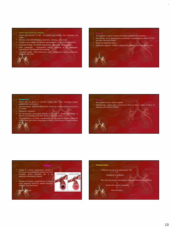
.
13
• Clinical and Laboratory Findings:
• Infants with infection of URT : Low-grade fever, profuse clear rhinorrhea, andcough.
• Infection in the LRT: Tachypnea, retractions, wheezing, and cyanosis.
• Crackles can be audible, and thoracic hyperresonance can be noted on percussion.
• Associated findings can include conjunctivitis, otitis media, and pharyngitis.
• Chest radiography : Peribronchial cuffing, flattening of the diaphragms,hyperinflation, and increased lung markings.
• Laboratory studies : Mild leukocytosis with a prominence of polymorphonuclearleukocytes (left shift).
• Diagnosis:
• The diagnosis is clinical, based on the history and physical examination.
• The etiology can be determined by performing a nasopharyngeal culture for RSVand other respiratory viruses.
• Rapid viral diagnostic assays.
• Differential diagnosis : Asthma, congenital heart disease, and cystic fibrosis (CF).
• Management:
• Infants may be placed in cool-mist oxygen tents, where continuous oxygenadministration can be given.
• Antiviral therapy : Ribavirin (delivered by aerosol on a semicontinuous basis for upto 1 week).
• Mechanical ventilation .
• An intramuscular monoclonal antibody to the RSV F protein, palivizumab, iseffective in preventing severe RSV disease in high-risk infants .
• This prophylaxis is currently recommended only for high-risk patient populationssuch as those with chronic lung disease, a history of prematurity, or congenital heartdisease.
• Prognosis:
• Most patients recover without sequelae.
• Epidemiologic studies with a several-year follow-up show a higher incidence ofwheezing and asthma in children.
Asthma
• Asthma is a chronic inflammatory disorder ofthe airways characterized by recurrent and oftenreversible airflow limitation due to anunderlying inflammatory process.
• Multiple risk factors : Family history of asthma,atopy, respiratory infections, inhaled pollutants,allergens, food sensitivities.
• Onset : childhood
• Pathophysiology:
• Infiltration of airway by inflammatory cells
Denudation of epithelium
Mast cells and Alveolar macrophages secrete proinflammatory mediators
Increase the capillary permiaility
Mucosal edema.

.
14
• Clinical and Laboratory Findings:
• Recurrent reversible airflow limitation and airway hyper-responsiveness.
• Signs and symptoms : Intermittent wheezing, coughing, dyspnea, and chesttightness.
• Triggers include allergens, exercise, cold air, respiratory irritants and viralinfections.
• Severe disease : Increased anteroposterior chest diameter, a prolonged expiratoryphase, wheezing, and diminished breath sounds.
• Allergic rhinitis.
• Signs of respiratory distress, with tachypnea, intercostal retractions, nasal flaring,and cyanosis.
• Pulmonary function testing or spirometry.
• The technique involves a maximal forced expiration following a maximalinspiration.
• The key measurements are:
i) The forced vital capacity (FVC) - which is the amount of air expired during theforced expiration.
ii) The forced expiratory volume in 1 second (FEV1) - which is the volume of airexpired during the first second of expiration and FEV1 is a measure of the rate atwhich air can be exhaled.
• Reversibility can be demonstrated after administration of a short-actingbronchodilator (such as albuterol) and a repeat spirometric measurement.
• Subsequent measurements of peak expiratory flow rate (PEFR) can be helpful forthe diagnosis and management of asthma.
• Allergen skin testing:
• This testing allows the accurate identification of allergic triggers, which cantranslate into more specific therapies, such as allergen avoidance andimmunotherapy.
• Chest radiography.
• Classification:
• National Asthma Education and Prevention Program (NAEPP) are the most widelyused in the United States.
• Asthma patients are classified as :
i) Mild-intermittent
ii) Mild-persistent
iii) Moderate-persistent or
iv) Severe-persistent disease.
• The categories are defined by both subjective (historical) and objective(spirometric) points.
• Diagnosis:
• Spirometry or PEFR determinations
• Differential diagnosis : Chronic coughing andwheezing, chronic rhinitis or sinusitis, CF,gastroesophageal reflux disease and COPD(chronic bronchitis).
• Factors : Intermittent symptoms withasymptomatic periods, complete or nearlycomplete reversibility with bronchodilators, theabsence of digital clubbing, and a history ofatopy.
• Management:
• Regular monitoring; spirometry, PEFR measurement, exhaled nitric oxide levels,and questionnaires may be useful tools for this purpose.
• Pharmacotherapy of asthma is based on the severity of disease.
• Mild-intermittent disease - Short-acting bronchodilators (such as albuterol).
• Mild-persistent asthma - Inhaled corticosteroids.
• Alternative agents - Non steroidal anti-inflammatory agents (Nedocromil andCromolyn).
• Leukotriene receptor antagonists – Montelukast and Zafirllukast.
• Moderate and Severe persistent disease – Inhaled corticosteroid therapy and longterm corticosteroids such as salmeterol.

.
15
• Prognosis:
• Early diagnosis and a comprehensive management plan, patients with asthma canexperience a normal life expectancy with good quality of life.
• Oral Health Considerations:
1. Fluoride supplements should be instituted for all asthmatic patients, particular thosetaking R2-agonists.
2. The patient should be instructed to rinse his or her mouth with water after usinginhalers.
3. Oral hygiene should be reinforced to reduce the incidence of gingivitis andperiodontitis.
4. Antifungal medications should be administered particularly in patients who aretaking inhaled corticosteroids.
5. Steroid prophylaxis needs to be used with patients who are taking long-termsystemic corticosteroids.
6. Use stress-reducing techniques.
• Conscious sedation - Hydroxyzine.
• Barbiturates and narcotics should be avoided .
• Nitrous oxide can be used for all but patients with severe asthma as it may irritatethe airways.
7. Avoid dental materials that may precipitate an attack. Dental materials withoutmethylmethacrylate should be considered.
8. Schedule these patients’ appointments for late morning or later in the day tominimize the risk of an asthmatic attack.
9. Have oxygen and bronchodilators available in case of an exacerbation of asthma.
10. There are no contraindications to the use of local anesthetics containingepinephrine, but preservatives such as sodium metabisulfite may contribute to asthmaexacerbation in susceptible patients.
11. Judicious use of rubber dams will prevent reduced breathing capability.
12. Care should be used in the positioning of suction tips as they may elicit a coughreflex.
13. Up to 10% of adult asthmatic patients have an allergy to aspirin and othernonsteroidal anti-inflammatory agents.
14. Macrolide antibiotics may increase the level of theophylline, whereasphenobarbitals may reduce the level.
• Drugs such as tetracycline have been associated with more accentuated side effectswhen given together with theophylline.
15. During an acute asthmatic attack, discontinue the dental procedure, remove allintraoral devices, place the patient in a comfortable position, make sure the airway isopened, and administer a R2-agonist and oxygen.
• If no improvement is noted, administer epinephrine subcutaneously (1:1,000concentration, 0.01 mg/kg of body weight, up to a maximum of 0.3 mg) and alertemergency medical assistance.
Tuberculosis
• TB is an airborne bacterial infection caused by M.Tuberculosis which affects any part of the body and mostcommonly the lungs.
• M. Tuberculosis is exposed to the air as droplet nuclei fromcoughing, sneezing, shouting with pulmonary or laryngealTB.
• Transmission occurs through inhalation of these dropletnuclei which passes through the mouth or nasal cavities, theupper respiratory tract, bronchi and finally reaches thealveoli of the lungs
• Pathogenesis:

.
16
• Spread of TB:
1. Local spread
2. Lymphatic spread
3. Haematogenous spread
4. By the natural passages :
- Lung lesions into pleura
- Transbronchial spread into adjacent lung segments.
- Infected sputum into larynx
- Swallowing of infected sputum
- Renal lesions into ureter & down to bladder
• Classification:
• i) Pulmonary TB
a) Primary
b) Secondary
c) Miliary
ii) Extra pulmonary TB
• Clinical findings:
• Primary pulmonary TB: febrile illness and cough, which may be dry or productive.
• Post-primary TB or secondary TB - fever, cough, chest pain, and hemoptysis.
• Miliary TB - acute febrile illness (children) and it is more insidious with gradualdevelopment of ill health, anorexia, loss of weight, and fever (adults).
• Bilateral crackles on auscultation, hepatosplenomegaly and lymphadenopathy maybe present.
• Choroid tubercles – children.
• Tuberculous infection of submandibular and cervical lymph nodes – Scrofula.
• Primary tuberculosis of the skin - Lupus vulgaris.
• Oral manifestations:
• Primary oral tuberculosis – Gingiva and is present as diffuse, hyperemic, nodular,or papillary proliferation of the gingival tissues.
• Secondary tuberculosis - Tongue is most commonly affected, followed by thepalate, lips, buccal mucosa, gingiva, and frenula.
• The usual presentation is an irregular, superficial or deep, painful ulcer which tendsto increase slowly in size.
• It is frequently found in areas of trauma.
• Occasional mucosal lesions show swelling, granular, nodular or fissured lesions,but no obvious clinical ulceration.
• Diagnosis:
• 1) Skin or blood tests
• Mantoux tuberculin test : Injecting a standard dose oftuberculin fluid into the skin of the lower portion of thearm subcutaeneously.
• The results depend on the diameter in millimetres of askin reaction characterised by an induration (a palpableraised hardened area free from erythema) after 48 to 72hours of testing.
• A diameter of 0 to 4 mm represents a negative skin testand 5 to 9 mm is a doubtful result while 10 mm or moreis positive.
• Interferon-Gamma Release Array (IGRA): It measures the extent to which theimmune system reacts to the tubercle bacilli.
• The United States Food and Drug Administration (FDA) has approved the use oftwo IGRAs:
i) QuantiFERON-TB Gold In-Tube test (QFT-GIT) and
ii) T-SPOT TB test.
• Positive response - Presence of tubercle bacilli.
• Negative response - Absence of TB infection.
• Drug susceptibility testing (DST): It is performed on the isolated tubercle bacillispecimen to test for resistance to any of the first-line anti-tuberculosis drugs.
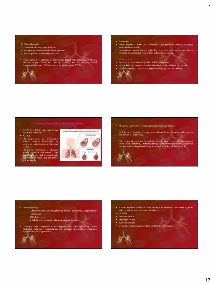
.
17
2) Chest radiography
3) Computerized tomography (CT) scan
4) Bacteriologic examination of clinical specimens.
5) Specific purified protein derivative (PPD)
• Newer methods of diagnosis of tuberculosis include radioimmunoassays (RIAs),soluble antigen fluorescent antibody (SAFA) test and enzyme linkedimmunosorbent assay (ELISA), polymerase chain reaction (PCR)
• Prevention:
• Bacilli calmette –Guerin (BCG vaccine) - Used preventive measure to controltuberculosis worldwide.
• Administered to newborns in a single dose, it prevents severe disease and reducesmortality among children from miliary and meningeal disease.
• Management:• Isoniazid is corner stone drug and include in all regimens.
• DOTS: Directly Observed treatment short course is also followed in TB cases.
• INR +R- 9 months/ INH+R+P- 2 months followed by INH+R- 4 months.
Chronic Obstructive Pulmonary Disease
• COPD is a disease state characterizedby airflow limitation.
• COPD:
• A) Emphysema: an anatomicallydefined condition characterized bydestruction and enlargement of the lungalveoli
• B) Chronic bronchitis: a clinicallydefined condition with chronic coughand phlegm, and small airways disease,a condition in which small bronchiolesare narrowed.
• Diagnosis : Symptoms of cough, sputum production or dyspnea.
• Risk factors : Environmental exposures and host factors (hereditary deficiency inthe enzyme A1-antitrypsin).
• This enzyme is responsible for inhibiting the activity of trypsin and other proteasesin the serum and tissues. The characteristic panlobular emphysematous changes thatare seen in a1- antitrypsin deficiency are related to the loss of alveolar walls.
• Pathophysiology:
i) Chronic inflammation throughout the airways, parenchyma, and pulmonary
vasculature
ii) Oxidative stress
iii) Imbalance of proteases and antiproteases in the lung.
• These pathologic changes lead to mucus hypersecretion, ciliary dysfunction, airflowlimitation, pulmonary hyperinflation, gas exchange abnormalities, pulmonaryhypertension and cor pulmonale.
• Chronic exposure to tobacco smoke results in an increase in the number of gobletcells because of hyperplasia and metaplasia.
• Acrolein.
• Nitrogen dioxide.
• Hydrogen cyanide.
• Carbon monoxide.
• Polycyclic hydrocarbons have been implicated as carcinogens.
.
18
• Clinical and Laboratory Findings:
• Symptoms : Dyspnea, cough, and sputum production. An increase in the productionof purulent sputum is a sign of exacerbation due to respiratory infection.
• Physical findings : Diffuse wheezing, respiratory distress, including the use ofaccessory muscles of respiration (retractions) and tachypnea.
• Liver enlargement due to congestion, ascites, and peripheral edema can develop asthe disease progresses to pulmonary hypertension and cor pulmonale. This leads tothe characteristic clinical patient presentation termed the Blue bloater.
• Patients can be adequately oxygenated in the early stages of the disease and thuscan have fewer signs of hypoxia (pink puffer) .
• Classification:
• COPD is classified into five stages: at risk, mild, moderate, severe, and verysevere.
• The at-risk stage is defined by normal spirometry, but patients have chronicsymptoms of cough and sputum production.
• Mild, moderate, and severe COPD has evidence of increasing airway obstructionon spirometry in each progressive stage.
• Very severe COPD is defined by severe airway obstruction with chronicrespiratory failure.
• At this stage, quality of life is significantly impaired and exacerbations may belife-threatening.
• Diagnosis:
• Patients often have cough, dyspnea, and sputum production.
• Complete pulmonary function tests are a valuable means of assessing airflowlimitation and the reversibility of airway obstruction.
• Pulse oximetry.
• Chest radiography.
• A determination of arterial blood gases is important for the management ofhospitalized patients.
• Management:
• Maintenance therapy : Trials of inhaled bronchodilators such as Albuterol and ipratropium bromide.
• Long-acting bronchodilators : Formoterol or salmeterol (Global Initiative for Obstructive Lung Disease).
• A partial pressure of arterial oxygen of 55-60 mg of Hg - Reduce hypoxemia while maintaining respiratory drive.
• Typical treatment includes 7-10 days of second generation cephalosporin.
• Oral Health Considerations:
• The association between oral disease and lung disease was analyzed by the NationalHealth and Nutrition Examination Survey I (NHANES I).
• Individuals with a confirmed chronic respiratory disease had a significantly greateroral hygiene index than subjects without a respiratory disease.
Cystic Fibrosis
• CF is a multisystem genetic disorder that ischaracterized chiefly by chronic airwaysobstruction and infection and by exocrinepancreatic insufficiency, with its effects ongastrointestinal function, nutrition, growth, andmaturation.
• Disorder - Ttransmembrane conductanceregulator (CFTR).
• The disease is characterized by hyperviscoussecretions in multiple organ systems.
Pathophysiology:
.
19
• Clinical and Laboratory Findings:
• Infancy - extrapulmonary manifestations such as meconium ileus or failure tothrive.
• Pulmonary manifestations : cough, recurrent infections of the lower respiratorytract, refractory lung infiltrates, and bronchospasm.
• Physical examination : Tachypnea and crackles.
• As the disease progresses, digital clubbing and bronchiectasis may becomeapparent.
• Spirometry and pulmonary function testing.
• CT analysis of the remarkable lung structural changes may be another potentialoutcome measure to monitor disease progression.
• Diagnosis:
• A sweat test can be performed to confirm the diagnosis.
• The procedure involves the collection of sweat after stimulation with pilocarpine.
• Samples containing > 60 mEq/L chloride are considered positive.
• Management:• Conventional treatment : Antibiotics, bronchodilators, anti-inflammatory agents,
chest physiotherapy with postural drainage, and mucolytic agents.
• Inhaled antibiotics – Tobramycin.
• Long-term macrolide antibiotics - Diffuse panbronchiolitis.
• Recombinant enzyme deoxyribonuclease therapy - Purulent airway secretions.
• Exercise.
• Oral Health Considerations:
• The tongue, buccal mucosa, dental plaque, and saliva serve as a reservoir ofcolonization by both mucoid and nonmucoid strains of P. aeruginosa, an importantbacterial pathogen for CF patients.
• This suggests that oral hygiene strategies may help reduce the level of thesepathogens in the mouth and thus reduce potential lung infection.
Pulmonary Embolism
• Pulmonary embolism (PE) is a result of anexogenous or endogenous material travelling tothe lung and causing blockage of a pulmonaryarterial vessel.
• Substances : Neoplastic cells, air bubbles, carbondioxide, intravenous catheters and fat droplets.
• Risk factors : Prolonged immobilization, lowerextremity trauma, a history of deep venousthromboses, and the use of estrogen-containingoral contraceptives.
• Pathophysiology:
• Occlusion of pulmonary arterial vessels
Ventilation-perfusion mismatch.
Right-sided heart failure.
Local bronchoconstriction
Platelets and mast cells.
Pulmonary hypertension and arterial vasospasm
• Clinical and Laboratory Findings:
• Dyspnea.
• Other features : Chest pain, fever, diaphoresis, cough, hemoptysis, and syncope.
• Physical findings : Evidence of a lower extremity deep venous thrombosis,tachypnea, crackles or rub on lung auscultation, and heart murmur.
• Hypoxemia.
• Measurements of arterial blood gases are helpful as patients may demonstrate adecrease in partial pressure of arterial oxygen (PaO2) and partial pressure of arterialcarbon dioxide (PaCO2) with an increase in hydrogen ion concentration (pH).

.
20
• Classification:
(1) Massive PE
(2) PE with pulmonary infarction
(3) PE without infarction or cor pulmonale
(4) Organized emboli in central arteries.
• Diagnosis:
• Negative d-dimer - venous thromboembolism is absent.
• Chest radiography - Elevated hemi-diaphragm, pleural effusions, and pulmonaryartery dilatation.
• Troponin levels may be elevated, the echocardiogram may be abnormal withincreased right ventricular volume.
• Ventilation-perfusion scan - PE is suspected.
• Spiral (helical) CT scanning.
• Pulmonary arteriography is the gold standard study.
• Pulmonary angiography.
• Management:
• Heparin (both unfractionated and low molecular weight).
• Systemic thrombolytic therapy (such as streptokinase, urokinase, and tissue-typeplasminogen activator).
• Pulmonary embolectomy - patients who are unable to receive thrombolytic therapy.
• Oxygen administration.
• Mechanical ventilation.
• Oral Health Considerations:
• Provision of dental care - Managed with oral anticoagulants.
• Dental care (including simple extractions) can safely be provided for patients withprothrombin times of up to 20 seconds or an international normalized ratio of 2.5.
Pulmonary Neoplasm
• Lung cancer is the leading cause of cancer deaths in both men and women.
• Squamous cell carcinomas account for one-third of all lung cancers. The neoplasmderives from bronchial epithelial cells that have undergone squamous metaplasia.This is a slow-growing neoplasm that invades the bronchi and leads to airwayobstruction.
• Small cell carcinomas account for approximately one-fourth of all lung cancers.This type of lung cancer has the highest association with smoking. These derivefrom neuroendocrine cells in the airways and metastasize rapidly.
• Adenocarcinomas account for approximately one-third of all lung cancers. Theseneoplasms are of glandular origin and develop in a peripheral distribution. Theygrow more rapidly than squamous cell carcinomas and tend to invade the pleura.The bronchoalveolar tumor is derived from bronchiolar or alveolar epithelium. Thiscancer is not associated with exposure to tobacco smoke.
• Large cell carcinomas include anaplastic and giant cell tumors. They are poorlydifferentiated tumors that resemble neither squamous cell carcinomas noradenocarcinomas.
• Pathophysiology:
• Metaplasia of the respiratory epithelium occurs in response to injury, such as thatinduced by tobacco smoking.
• With continued injury, the cells become dysplastic, with the loss of differentiatingfeatures.
• Neoplastic change first occurs locally; invasive carcinoma usually follows shortly.
.
21
• Clinical and Laboratory Findings:
• Chronic nonproductive cough.
• Sputum production, Hemoptysis, Dyspnea.
• Superior vena cava syndrome - Facial edema, cyanosis, and orthopnea caused bycompression of the superior vena cava by tumor. The acute onset of hoarseness maysignal tumor compression of the recurrent laryngeal nerve.
• Shoulder and forearm pain might suggest the presence of Pancoast’s tumor, whichis found in the apical region of the lungs below the pleura.
• The bones, the brain, and the liver are common sites of metastasis.
• Paraneoplastic effects : Endocrine abnormalities that are due to tumors that secretehormones such as antidiuretic hormone, adrenocorticotropic hormone, andparathyroid hormone–related peptides.
• Classification:
• The World Health Organization hasdifferentiated pulmonary neoplasms into 12distinct histologic types.
• The four major pathologic categories are:
i) Squamous cell carcinoma
ii) Small cell carcinoma
iii)Adenocarcinoma
iv) Large cell carcinoma.
• Diagnosis:
• CT scanning.
• Other diagnostic modalities:
i) Sputum or pleural fluid cytology,
ii) Excisional biopsy,
iii) Transthoracic needle aspiration, and
iv) Bronchoscopy.
• Management:
• Complete surgical resection.
• Treatment is based on the stage of the disease and the patient’s clinical status.
• Early-stage disease – surgical.
• Locally advanced disease - chemotherapy and radiotherapy.
• Advanced disease – chemotherapy with supportive care.
• Radiation therapy is an important palliative measure, especially for patients withsuperior vena cava syndrome, brain metastases, or bone lesions.
• Prognosis:• Patients with lung cancer has remained poor.
• Most pulmonary cancers are found too late for a cure only about 20% of patientsundergo a radical surgical procedure, which is the only curative treatment.
References
• 1) Michael Glick – Burkets oral medicine – 10th edition.
• 2) Guidelines for the diagnosis and treatment of COPD – 3rd edition.
• 3) K.Sembulingam – Essentials of Medical Physiology – 6th edition.
• 4) Akosua Adom Agyeman, Richard Ofori-Asenso. Tuberculosis—anoverview.Journal of Public Health and Emergency;2017;1(7).
• 5) Shafers Textbook of Oral Pathology – 7th edition.

.
1
CORONAVIRUS (COVID-19): A PANDEMIC DISEASE
1
Dr. Neeraj Kumar Yadav
Reader
Dept. of Oral Medicine and Radiology
MGDCH , Jaipur
CONTENT1. Introduction
2. Types of corona virus
3. Structure
4. Pathogenesis
5. Epidemiologic Characteristic
6. Clinical Manifestation
7. Diagnosis
8.Treatment
9.Complication
10.Infection control in dental setting
11.Impact of corona virus infectious disease on oral health
12.Saliva as potential diagnosis
2
INTRODUCTION•The emerging coronavirus disease (COVID-19) swept across the world, affecting
more than 200 countries and territories. Genomic analysis suggests that the
COVID-19 virus originated in bats and transmitted to humans through
unknown intermediate hosts in the Wuhan seafood market, China, in December
of 2019.
•This virus belongs to the Betacoronavirus group, the same group of the 2003
severe acute respiratory syndrome coronavirus (SARS-CoV), and for the
similarity, it was named SARS-CoV-2.
1st case in world: In December 2019 ,55 yr old individual from Hubei Huanan
seafood market, China.
1st case in India: In January-27-2020, 20 yr old female presented to the
emergency department in general hospital Thrissur, Kerela.
3
EPIDEMIOLOGY1. 31-DECEMBER-2019, WHO alerted by Chinese authorities of a string of pneumonia
cases in Wuhan.
2. 1-JANUARY-2020, Huanan seafood market identified as source of pneumonia.
3. 9-JANUARY-2020, 1st death from COVID-19 occurs in Wuhan.
4. 10-JANUARY-2020, Gene sequencing data of COVID-19 established and shared globally.
5. 13-JANUARY-2020, 1st infection occur in Thailand outside of china.
6. 16-JANUARY-2020, 1st infection occur in Japan.
7. 19-JANUARY-2020, sudden spike in reporting of another 136 cases in Wuhan.
8. 20-JANUARY-2020, 1st infection occurs in South Korea.
9. 21-JANUARY-2020, Taiwan and the United States reports there 1st infection.
10.27-JANUARY-2020,first infection occurs in India.
11. 22-JANUARY-2020, 1st infection occurs in Macau and Hongkong.
12. 23-JANUARY-2020, 1st infection occurs in Singapore and Vietnam.Additional cities in
quarantined.
4
12. February/26/2020: COVID 19 has been recognized in 34 countries ,with a total of
80,239 laboratory confirmed cases and 2,700 death.
13. July/22/2020: Over 14.9 million confirmed cases and more than 610,000 deaths
related to covid 19 has been reported worldwide.
14. 2/JAN/2021: In Asia, Africa, Europe, North America, South America,
83,963,941Confirmed cases are found and 1,827,544 Deaths occur.
5
CORONA DIVERSITY??•CoVs members belong to the subfamily Coronovirinae within the family Coronaviridae and the order
Nidovirales. Based on their protein sequences and phylogenetic relationships, members of the
Coronavirinae subfamily can be classified into four groups, Alphacoronaviruses, Betacoronaviruses,
Gammacoronaviruses, and Deltacoronaviruses. Gammacoronaviruses and Deltacoronaviruses infect
birds and might infect mammals, but never reported to cause any illnesses in humans .
•On the other hand, Alphacoronaviruses and Betacoronaviruses are capable of causing respiratory
illnesses in humans and gastrointestinal illnesses in animals.
• SARS-CoV and MERS-CoV belong to Betacoronaviruses, SARS-CoV-2 belongs to the Betacoronavirus group.
•SEVEN STRAINS OF CORONA VIRUS:
• Dangerous strains are:
1.SARS-CoV was 1st reported in Asia in Feb 2003 and quickly spread to 26 countries in 4 Months
more than 8000 people fall ill and 774 died.
2.MERS-CoV is a middle east respiratory syndrome. Corona virus first reported in Sep 2012 in Saudi
Arabia and 2,519 people fall ill and 866 died.
3.SARS-CoV-2(severe acute respiratory syndrome) is zoonotic, with Chinese horseshoe bats and
pangolins are intermediate host.
• Harmless strains are:
1. serotype 229e
2. serotype oc43
3. serotype nl63
4. serotype huk1 6
.
2
CORONA VIRUS FAMILY
STRUCTURE OF CORONA VIRUS
8
PATHOGENESIS•Following viral transmission, SARS-CoV-2 attaches to the surface of the epithelial
membrane of the oral cavity, the mucosal membranes of the conjunctiva or the
otic canal.
•ACE 2 protein, which is highly expressed on multiple human cells including type
II alveolar cells (AT2), oral, esophageal, ileal epithelial cells, myocardial cells,
proximal tubule cells of the kidneys as well as urothelial cells of the bladder is
believed to mediate the internalization of SARS-CoV2.
•The spike (S) protein of SARS-CoV2 is cleaved by a cellular enzyme named furin at
the S1/S2 site. This cleavage is essential for viral entry to the lung cells .
•The activated S protein is primed by the TMPRSS2 and finally attaches ACE 2
receptors to enter the host cells.
• SARS-CoV-2 binds ACE 2 receptors with tenfold higher affinity.
•Severe COVID-19 patients demonstrates a remarkable reduction of lymphocytic T
Cells (CD4+ and CD8+) and natural killer (NK) cells.
9
PATHOPHYSIOLOGY OF CORONA VIRUS
LIFE CYCLE OF CORONA VIRUS IN HUMAN PATHOPHYSIOLOGY OF CORONA VIRUS IN HUMAN BODY
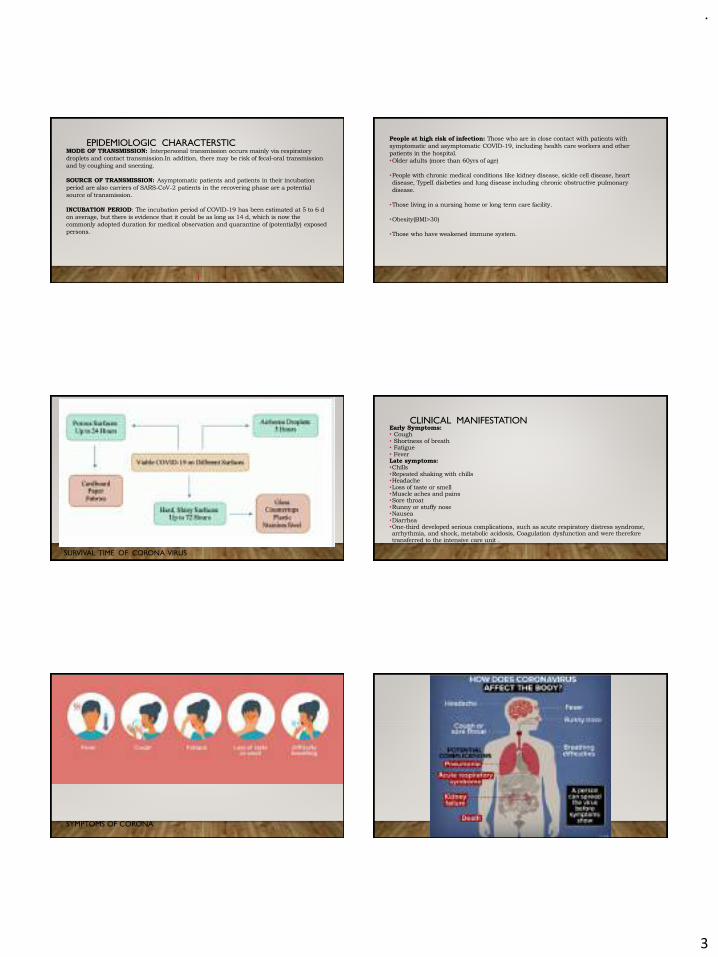
.
3
EPIDEMIOLOGIC CHARACTERSTICMODE OF TRANSMISSION: Interpersonal transmission occurs mainly via respiratory
droplets and contact transmission.In addition, there may be risk of fecal-oral transmission
and by coughing and sneezing.
SOURCE OF TRANSMISSION: Asymptomatic patients and patients in their incubation
period are also carriers of SARS-CoV-2 patients in the recovering phase are a potential
source of transmission.
INCUBATION PERIOD: The incubation period of COVID-19 has been estimated at 5 to 6 d
on average, but there is evidence that it could be as long as 14 d, which is now the
commonly adopted duration for medical observation and quarantine of (potentially) exposed
persons.
1
3
People at high risk of infection: Those who are in close contact with patients with
symptomatic and asymptomatic COVID-19, including health care workers and other
patients in the hospital.
•Older adults (more than 60yrs of age)
•People with chronic medical conditions like kidney disease, sickle cell disease, heart
disease, TypeII diabeties and lung disease including chronic obstructive pulmonary
disease.
•Those living in a nursing home or long term care facility.
•Obesity(BMI>30)
•Those who have weakened immune system.
1
4
SURVIVAL TIME OF CORONA VIRUS
CLINICAL MANIFESTATIONEarly Symptoms: • Cough • Shortness of breath• Fatigue• FeverLate symptoms:•Chills•Repeated shaking with chills•Headache•Loss of taste or smell•Muscle aches and pains•Sore throat•Runny or stuffy nose•Nausea •Diarrhea•One-third developed serious complications, such as acute respiratory distress syndrome, arrhythmia, and shock, metabolic acidosis, Coagulation dysfunction and were therefore transferred to the intensive care unit .
1
6
SYMPTOMS OF CORONA
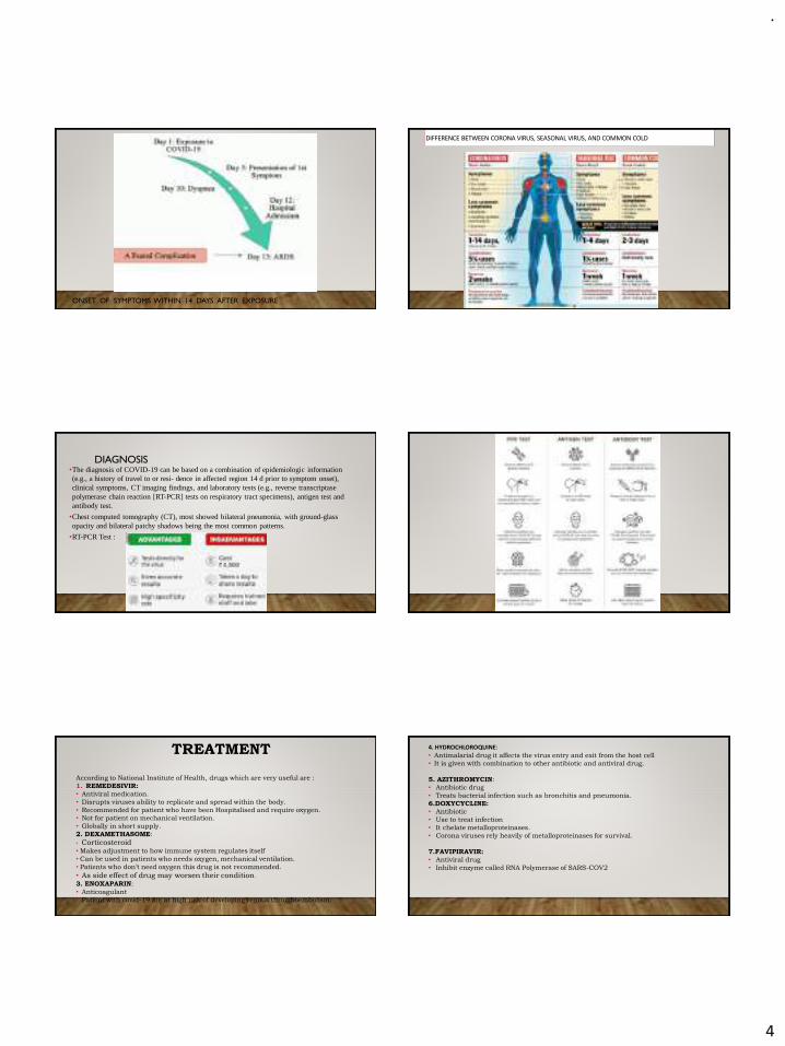
.
4
ONSET OF SYMPTOMS WITHIN 14 DAYS AFTER EXPOSURE
DIFFERENCE BETWEEN CORONA VIRUS, SEASONAL VIRUS, AND COMMON COLD
Title
DIAGNOSIS •The diagnosis of COVID-19 can be based on a combination of epidemiologic information (e.g., a history of travel to or resi- dence in affected region 14 d prior to symptom onset), clinical symptoms, CT imaging findings, and laboratory tests (e.g., reverse transcriptase polymerase chain reaction [RT-PCR] tests on respiratory tract specimens), antigen test and antibody test.
•Chest computed tomography (CT), most showed bilateral pneumonia, with ground-glass opacity and bilateral patchy shadows being the most common patterns.
•RT-PCR Test :
2
1
2
2
TREATMENT
According to National Institute of Health, drugs which are very useful are :
1. REMEDESIVIR:
• Antiviral medication.
• Disrupts viruses ability to replicate and spread within the body.
• Recommended for patient who have been Hospitalised and require oxygen.
• Not for patient on mechanical ventilation.
• Globally in short supply.
2. DEXAMETHASOME:
• Corticosteroid • Makes adjustment to how immune system regulates itself
• Can be used in patients who needs oxygen, mechanical ventilation.
• Patients who don’t need oxygen this drug is not recommended.
• As side effect of drug may worsen their condition.
3. ENOXAPARIN:
• Anticoagulant
• Patient with covid-19 are at high risk of developing venous thromboembolism.
4. HYDROCHLOROQUINE:• Antimalarial drug it affects the virus entry and exit from the host cell
• It is given with combination to other antibiotic and antiviral drug.
5. AZITHROMYCIN:
• Antibiotic drug
• Treats bacterial infection such as bronchitis and pneumonia.
6.DOXYCYCLINE:
• Antibiotic
• Use to treat infection
• It chelate metalloproteinases.
• Corona viruses rely heavily of metalloproteinases for survival.
7.FAVIPIRAVIR:
• Antiviral drug
• Inhibit enzyme called RNA Polymerase of SARS-COV2

.
5
8.FAMCICLOVIR:• Antiviral drug• Preventing viruses from multiplying • Reduces the severity of infection.
9.zinc:• Keeps immune system healthy.• Maintain ability to taste and smell.
10.f/b FORMOTEROL/BUDESONIDE:• Relieves symptoms of asthma and copd .• Makes breathing easier.• Inhibits release of certain chemical ,• That causes inflammation and swelling.• Relaxes the muscle in airway.11.IVERMECTIN:• Anti parasite drug.• Inhibit SARSCOV-2 replication in vitro.
TREATMENT(for Home isolated Asymptomatic patient )
TREATMENT IN MILD CASES
SUPPORTIVE TREATMENT OF CORONA VIRUS
OXYGEN THERAPY IN MODERATE AND SEVERE CASES
1. People who develop severe symptoms are need to be hospitalized.2. Treatment focused on supporting individual so that their body fights against infection.3. And to Reduce patient symptoms.4. People who have troubled in breathing may be given supplemental oxygen.

.
6
5. If oxygen alone is insufficient to help the patient condition then mechanical ventilation is given. 6. And in some cases intubation and oxygenate the patient by conventional ventilators.
5. CORONA VACCINE :• COVAX IN,India indigenous covid 19 vaccine by Bharat biotech is developed in
collaboration with Indian council of medical research.
• National regulatory authorities approved six vaccine:
• Two RNA vaccine TOZINAMERAN and PFIZER BIONTECH MRNA-1273 from moderna.
• 2 conventional inactivated vaccine BBIBP CORV from sinopharm and CORONA VAC
from sinovac and 2viral vector vaccine GAM COVID -VAC from gamaliya research
institute and AZD1222 from university of Oxford .
COMPLICATION•Respiratory system involvement:
The predominant manifestation of COVID-19 is the interstitial and alveolar
pneumonia.
The CT scan of COVID-19 patients demonstrates a various pattern, ranging from
single ground-glass opacity (GGO) to bilateral diffuse heterogeneous consolidation
with air bronchogram and bronchiectasis, the ‘white lung.
•Cardiovascular involvement :
Hypertension, heart failure and coronary artery
•Kidney involvement :
Acute kidney injury (AKI)
•Ocular involvement:
SARS-CoV has been detected from the tear samples in the previous reports. Evidence
suggests possible conjunctivitis related to SARS-CoV via inoculation of droplets to
the eyes, lacrimal infection and virus migration from the nasolacrimal duct.
3
4
•Hematologic involvement:
Thrombocytopenia ,damage to the spleen and lymph nodes (congestion,
hemorrhage, atrophy, depletion of lymphoid follicles) has also been
observed. And venous thromboembolism .
•Coagulopathy:
thrombosis in pulmonary vessels in severe COVID-19.
•Electrolyte imbalance: urinary potassium loss .severe diarrhea and/or
vomiting, extrarenal hypokalemia.
•Liver involvement:
elevation of liver enzymes is frequently transient in mild cases of the
disease, significant elevation of liver enzymes is observed in severe COVID-
19.
•Endocrine involvement:
diabetic ketoacidosis ,hyperglycemia.
3
5
•Obstetric & gynecologic complications:
Limited evidence suggests that vertical transmission of COVID-19 during late pregnancy
is possible. Increased oxygen demand and physiologic anemia during pregnancy are the
potential factors that could exacerbate the severity of COVID-19.
COVID-19 probably increases the risk of miscarriage and intrauterine growth restriction.
•GI- Tract involvement:
diarrhea, abdominal pain and vomiting.
•Neuromuscular Involvement:
dizziness, headache, impaired consciousness, cerebrovascular disease, ataxia and
epilepsy. Guillain Barre syndrome is another neurologic complication proceeded by
COVID-19. Progressive weakness of extremities, paresthesia, facial diplegia, ataxia, and
bulbar weak- ness occurred 5–14 days after the infection.
•Central nervous system involvement:
acute cerebrovascular diseases and encephalopathy.
•Skin involvement:
Several skin conditions, including erythema, papules, maceration and scaling
accompanied with symptoms of burning, itching and stinging
3
6
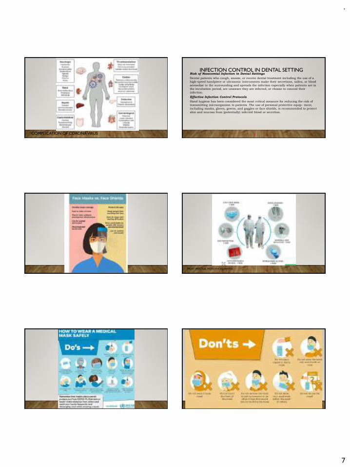
.
7
COMPLICATION OF CORONA VIRUS
INFECTION CONTROL IN DENTAL SETTINGRisk of Nosocomial Infection in Dental Settings
Dental patients who cough, sneeze, or receive dental treatment including the use of a
high-speed handpiece or ultrasonic instruments make their secretions, saliva, or blood
aerosolize to the surrounding and spreads the infection especially when patients are in
the incubation period, are unaware they are infected, or choose to conceal their
infection.
Effective Infection Control Protocols
Hand hygiene has been considered the most critical measure for reducing the risk of
transmitting microorganism to patients .The use of personal protective equip- ment,
including masks, gloves, gowns, and goggles or face shields, is recommended to protect
skin and mucosa from (potentially) infected blood or secretion.
3
8
PPE KIT (PERSONAL PROTECTIVE EQUIPMENT) (

.
8
HOW DOES SANITISERS WORK?? COMPOSITION OF HAND SANITISERS
RECOMMENDED MEASURES DURING THE COVID-19 OUTBREAK
Recommendations for Dental Practice:
Dentists should take strict personal protection measures and avoid or minimize operations that can
produce droplets or aerosols. The use of saliva ejectors with low or high volume can reduce the
production of droplets and aerosols.
Evaluation of Patients:
During the outbreak of COVID-19, dental clinics are recommended to establish precheck triages to measure and record the temperature of every staff and patient as a routine procedure. Precheck staff
should ask patients ques- tions about the health status and history of contact or travel.
Oral Examination.
Preoperative antimicrobial mouth rinse could reduce the number of microbes in the oral cavity.
Procedures that are likely to induce coughing should be avoided (if possible) or performed cautiously.
Aerosol-generating procedures, such as the use of a 3-way syringe, should be minimized. Intraoral x-ray
examination can stimulate saliva secretion and coughing extraoral dental radiographies, such as pan-
oramic radiography and cone beam CT, are appropriate alter- natives during the outbreak of COVID-19.
Treatment of Emergency Cases.
Rubber dams and high-volume saliva ejectors can help minimize aerosol or spatter in dental procedures.
Further- more, face shields and goggles are essential with use of high- or low-speed drilling with water
spray.
4
5
THE IMPACT OF CORONA VIRUS INFECTIOUS DISEASE 19(COVID-19) ON ORAL HEALTH
•Anti-viral drugs may be responsible for side effects (<2%) affecting oral cavity among
the other parts of gastrointestinal track, such as stomatitis, mouth ulcers and dry
mouth.
•Interferon's more common side effects related to oral medicine comprise dry mouth,
which can result in oral thrush.
•Vast proportion of COVID-19 symptomatic and intensely treated patients must
develop some sort of oral problems and pathological.
•As systemic and topical steroids are deemed not to be suitable in COVID-19 infection ,
some of immune-related long-term oral medicine conditions (pemphigus, lichen
planus, pemphigoid) may potentially exacerbate in SARS-CoV- 2-positive patients who
were advised to discontinue such therapy.
4
6
•As reported, SARS-CoV-2 may potentially affect the functioning of salivary
glands, taste/smell sensations and oral mucosa integrity, interfering with
dynamic oral environment also by exerting influence on microbiota
balance.
•Severe COVID-19 acute infection, could potentially leading to various
opportunistic fungal infections, xerostomia ,ulcerations and gingivitis as a
result of impaired immune system.
• Persons recovering from COVID-19 need additional postacute care ,close
monitoring of their oral health.
• oral health had been found to deteriorate, especially in case of those
staying in intensive care units.
•Intubation, tracheostomy, external ventilation, as well as mouth breathing,
hyposalivation can lead to rapid oral health deterioration and affecting
lower respiratory track.
4
7
VULNERABLE SITE FOR CORONA VIRUS INFECTION
.
9
COVID-19 PATIENT PRESENTING A WHITE PLAQUE ON TONGUE DORSUM, A NODULE LOCATED IN LOWER LIP WAS OBSEVED SUGGESTING A REACTIVE LESION(FIBROMA)
PATIENT PRESENTING ATROPHIC AREAS SUGGESTING GEOGRAPHIC TONGUE ASSOCIATED WITH FISSURED TONGUE
PATIENT SHOWING ATROPHIC AREAS MODERATE GEOGRAPHIC TONGUE , SLIGHTLY ERYTHEMATOUS AREA IN RIGHT PALATINE TONSIL REGION
SALIVA AS POTENTIAL DIAGNOSIS IN CORONA VIRUS- RECENT STUDY •Some virus strains have been detected in saliva as long as 29 days after infection ,
indicating that a non-invasive platform to rapidly differentiate the biomarkers using
saliva could enhance disease detection.
•there is a minimum of three different pathways for COVID-19 to present in saliva:
firstly, from COVID-19 in the lower and upper respiratory tract that enters the oral
cavity together with the liquid droplets frequently exchanged by these organs.
•Secondly, COVID-19 present in the blood can access the mouth via crevicular fluid,
an oral cavity-specific exudate that contains local proteins derived from extracellular
matrix and serum-derived proteins .
•Finally, another way for COVID- 19 to occur in the oral cavity is by major- and minor-
salivary gland infection, with subsequent release of particles in saliva via salivary
duct.
•Further studies are needed to investigate the potential diagnostic of COVID-19 in
saliva.
5
2
THANK YOU
Related Documents