3 Extra Hepatic Portal Venous Obstruction in Children Narendra K. Arora and Manoja K. Das The INCLEN Trust International, New Delhi, India 1. Introduction Portal hypertension is the commonest cause of upper gastrointestinal bleeding in children and up to 30% of cases with upper gastrointestinal hemorrhage can be fatal. Extrahepatic portal venous obstruction (EHPVO) is the commonest cause of portal hypertension in children and also one of the common causes in adults in India and other tropical countries (Arora, 1998; Poddar, 2008; Poddar 2000). In India, EHPVO is responsible for portal hypertension in about one third cases of adults and more than half of the cases in children (Sarin 2002; Dilawari 1992). EHPVO is characteristically refers to obstruction in the trunk of portal vein and it can extend to its branches and even splanchnic veins. Unlike cirrhosis, in EHPVO, the liver function is normal. The causes of portal venous obstruction and risk factors for upper gastrointestinal hemorrhage in children with EHPVO are not clearly understood. Most of the bleeds are spontaneous and some may be preceded by febrile illness, ingestion of drugs. Management of EHPVO involves acute management of the bleeding, secondary prophylaxis and shunting to reduce the portal pressure. This chapter attempts to compile the available evidences on EHPVO in children with special reference to experiences from India. 2. Definitions and terminology EHPVO indicates portal hypertension due to blockage in the portal vein before the blood reaches the liver. EHPVO is a distinct disease entity of primarily vascular in nature and not classically associated with association of a primary liver disease. Portal vein thrombosis, a known complication of liver cirrhosis, other systemic diseases and malignancy, is not generally included in the disease entity EHPVO. Over time, EHPVO terminology has replaced the use of portal vein thrombosis (PVT) as it does not exclude the intrahepatic portal vein thrombosis and does not include formation of portal cavernoma associated with portal hypertension. Baveno V workshop consensus statement defined EHPVO as (De Franchis, 2010): EHPVO is defined by obstruction of the extra-hepatic portal vein with or without involvement of the intra-hepatic portal veins and does not include isolated thrombosis of splenic vein or superior mesenteric vein (SMV). EHPVO is characterized by features of recent thrombosis or of portal hypertension with portal cavernoma as a sequel of portal vein obstruction Presence of cirrhosis and/or malignancy should be stated. www.intechopen.com

Welcome message from author
This document is posted to help you gain knowledge. Please leave a comment to let me know what you think about it! Share it to your friends and learn new things together.
Transcript
3
Extra Hepatic Portal Venous Obstruction in Children
Narendra K. Arora and Manoja K. Das The INCLEN Trust International, New Delhi,
India
1. Introduction
Portal hypertension is the commonest cause of upper gastrointestinal bleeding in children and up to 30% of cases with upper gastrointestinal hemorrhage can be fatal. Extrahepatic portal venous obstruction (EHPVO) is the commonest cause of portal hypertension in children and also one of the common causes in adults in India and other tropical countries (Arora, 1998; Poddar, 2008; Poddar 2000). In India, EHPVO is responsible for portal hypertension in about one third cases of adults and more than half of the cases in children (Sarin 2002; Dilawari 1992). EHPVO is characteristically refers to obstruction in the trunk of portal vein and it can extend to its branches and even splanchnic veins. Unlike cirrhosis, in EHPVO, the liver function is normal. The causes of portal venous obstruction and risk factors for upper gastrointestinal hemorrhage in children with EHPVO are not clearly understood. Most of the bleeds are spontaneous and some may be preceded by febrile illness, ingestion of drugs. Management of EHPVO involves acute management of the bleeding, secondary prophylaxis and shunting to reduce the portal pressure. This chapter attempts to compile the available evidences on EHPVO in children with special reference to experiences from India.
2. Definitions and terminology
EHPVO indicates portal hypertension due to blockage in the portal vein before the blood reaches the liver. EHPVO is a distinct disease entity of primarily vascular in nature and not classically associated with association of a primary liver disease. Portal vein thrombosis, a known complication of liver cirrhosis, other systemic diseases and malignancy, is not generally included in the disease entity EHPVO. Over time, EHPVO terminology has replaced the use of portal vein thrombosis (PVT) as it does not exclude the intrahepatic portal vein thrombosis and does not include formation of portal cavernoma associated with portal hypertension.
Baveno V workshop consensus statement defined EHPVO as (De Franchis, 2010):
EHPVO is defined by obstruction of the extra-hepatic portal vein with or without involvement of the intra-hepatic portal veins and does not include isolated thrombosis of splenic vein or superior mesenteric vein (SMV).
EHPVO is characterized by features of recent thrombosis or of portal hypertension with portal cavernoma as a sequel of portal vein obstruction
Presence of cirrhosis and/or malignancy should be stated.
www.intechopen.com
Portal Hypertension – Causes and Complications
42
3. Epidemiology
EHPVO is a common cause of portal hypertension in the developing countries (30-55% of all variceal bleeders) and is second to cirrhosis (up to 5-13%) (Arora, 1998; Valla, 2002). EHPVO is also the most common cause of upper gastrointestinal bleeding in children. It accounts for almost 70% of pediatric patients with portal hypertension (Yachha, 1996; Arora, 1998). Literature from different parts of India indicated that in children EHPVO is responsible for 54% of portal hypertension (Poddar, 2008). Most (85-92%) of the upper gastrointestinal bleeding in Indian children was result of portal hypertension due to EHPVO (Poddar, 2008; Yachha, 1996).
Investigators across India have consistently observed that EHPVO occurs commonly in children belonging to low and lower middle socio-economic strata attending public sector hospitals. Pediatric gastroenterologists practicing in private sector hospitals see such patients infrequently. During last 10-15 years, the number of EHPVO patients gradually appears to be dwindling even in public sector hospitals. Thus it appears that the disease may have something to do with the living condition of the families and with improving economic state of the families, disease may become less frequent.
4. Etiology and pathogenesis
4.1 Etiology
The etiology of EHPVO in children has not been well documented. There are many etiologies for EHPVO namely infections, surgical procedures, vascular interventions, abdominal trauma, dehydration, congenital anomalies have been postulated. EHPVO is considered to be heterogenous with regard to etiology and pathogenesis, and vary with respect to age and geographical location. The causes can broadly be of five types; infection and inflammation, portal vein injury, developmental anomaly, prothrombotic causes and idiopathic.
4.1.1 Infection
Omphalitis, neonatal umbilical sepsis, intraabdominal infection, post umbilical catheterization have been linked to development of EHPVO in children. Although it is documented that portal vein thrombosis may occurs in neonates having umbilical vein catheterization, but most of these thrombi resolve within a short period (Larroche, 1970; Thompson, 1964). It is also proposed that phlebosclerosis is primary as a result of infection and/or and inflammation followed by thrombosis as a secondary event (Stringer, 1994). But evidence on this linkage is not very strong in children with EHPVO.
4.1.2 Trauma and surgery
Umbilical vein cannulation, abdominal surgery, and abdominal trauma in childhood could also lead to EHPVO (Yadav, 1993).
4.1.3 Congenital anomaly
Congenital defects like web, valves in the portal vein, portal vein stenosis, atresia or agenesis have also been reported (Odievre, 1977). There may be presence of other cardiovascular system congenital defects.
www.intechopen.com
Extra Hepatic Portal Venous Obstruction in Children
43
4.1.4 Prothrombotic state
Hypercoagulable states like deficiencies of antithrombotic factors (protein-C, protein-S, anti-thrombin III) have been reported in children with EHPVO (Bhattacharya, 2004; Pinto, 2004; Koshy, 1984; Dubuisson, 1997). Absence of genetic mutations in EHPVO patients and normal levels of antithrombotic factors in their parents suggested that the deficiency of antithrombotic factors may not be primarily of genetic origin, but may be secondary to the low hepatic blood flow due to portal vein thrombosis (low synthesis) and portosystemic shunt (increased clearance or consumption) (Dubuisson, 1997; Sharma, 2006). Other conditions like polycythemia vera, dehydration have also been proposed as the causes of EHPVO.
4.1.5 Idiopathic
Despite all efforts, the etiology of blocked portal vein remains obscure in a large proportion of patients. In India the majority of cases (up to 90%) are categorized as idiopathic (Poddar, 2003). Probably this is due to want of detailed etiological work-up.
4.2 Pathogenesis
4.2.1 Portal vein changes
In EHPVO, usually the entire length of the portal vein is occluded with extension into the
splenic vein and sometimes into the superior mesenteric vein. In small proportions only the
terminus of the portal vein at the hilum is occluded. Mostly the site of blockage is at the
portal vein formation and in small proportions the total splenoportal axis is blocked. In a
report by our group among Indian children with EHPVO (n=88), blockage were observed at
portal vein formation, entire portal vein, spleninc vein and entire splenoportal axis in 39%,
34%, 16% and 11% respectively (Arora, 2002). On gross examination, the original portal vein
is difficult to identify as it is usually replaced by a cluster of variable-sized vessels arranged
haphazardly within a connective tissue support, called as the portal cavernoma. In cases of
shorter duration, cavernoma may not be there. The porto-systemic collaterals develop over
time leading to development of verices at different places in the gastrointestinal tract, and at
other places depending on the portal venous pressure and duration.
In EHPVO the blockage being presinusoidal, intrahepatic venous pressure is normal and intrasplenic pressure is increased. The hepatic blood flow is relatively normal as the portoportal collateral vessels enable bypassing the blocked area. The hepatic blood flow may be reduced if the collaterals are not enough. EHPVO is a hyperkinetic circulatory state with increased cardiac output as a result of extensive porto-systemic venous collaterals.
4.2.2 Liver changes
Classically, in EHPVO cases liver is normal and the architectural pattern is preserved. Histopathology may reveal concentric condensation of reticulin fibers around portal tracts, sometimes extending into the parenchyma. Altered hepatic storage capacity and transport maximum for bromsulphalin and liodocaine (MEGX) excretion has been reported in EHPVO. Usually liver biopsy is not required in EHPVO cases, unless liver functions are deranged.
www.intechopen.com
Portal Hypertension – Causes and Complications
44
5. Clinical presentations
In children, EHPVO may manifest at any age beyond the neonatal period. EHPVO can
present in two clinical forms: recent or acute and chronic. The clinical presentation also
differs in children and adults.
5.1 Recent or acute EHPVO
This can present with abdominal pain, ascites or fever. This may, however, be asymptomatic
and go unnoticed. In recent EHPVO, there is no evidence of porto-systemic collaterals and
portal cavernoma. The extent of obstruction in portal vein and speed of evolution of
thrombosis predicts the clinical manifestation. If associated with infection, features of sepsis
may be there. But many a times these may be passed as episodes of acute abdomen or
sepsis.
5.2 Chronic EHPVO
Most of the EHPVO present as chronic cases with the typical presenting symptoms like
episodes of variceal or gastrointestinal bleeding, lump in the abdomen and hypersplenism.
EHPVO in childhood is most often chronic and presents with features of variceal bleeding
and splenomegaly, whereas in adults it could be either acute or chronic.
5.2.1 Variceal bleeding
Esophageal varices are the most common site of variceal bleeding. The majority of children (85% to 90%) with EHPVO present with variceal bleeding (Arora, 1998; Poddar, 2008; Valla, 2002). The usual presentation for children with EHPVO is sudden, unexpected and, often, massive hemetemesis. Bleeding usually occurs in first or second decade of life. Some of the children also present with recurrent, well-tolerated bleeds with variable degree of anemia. There is no significant hepatocellular failure in these patients. Although many clinicians think that the frequency of variceal bleeding reduces after puberty, but this has not been confirmed.
Gastic varices are common in EHPVO. According to reports, about 70% children at
diagnosis have gastric varices. Most of these are gastroesophageal varices while a few are
isolated gastric varices (Sarin, 1992; Arora, 1998; Poddar, 2004). Following eradication of
esophageal varices, gastric varices become more evident with increase in risk of bleeding
(Poddar, 2004; Goncalves, 2000).
Rectal varices are documented in 80-90% of adult EHPVO cases (Misra, 2005). The scanty data in children indicate presence of rectal varices in 36-64% (Heaton, 1993; Yachha, 1996). It also probably related to the severity and duration of portal hypertension or site of obstruction (at the junction of splenic and superior mesenteric veins leading to redistribution of portal pressure along the inferior mesenteric vein) (Misra, 2005; Chawla, 1991; Ganguly, 1995). Appearance of these varices may increase after obliteration of the esophageal varices.
5.2.1.1 Risk factors for bleeding and rebleeding after initial sclerotherapy
Bleeding from varices occurs usually due to erosion or rupture of variceal vessel wall. Bleeding due to erosion of vericeal wall is precipitated by reflux esophagitis, drug ingestion
www.intechopen.com
Extra Hepatic Portal Venous Obstruction in Children
45
(NSAIDs), and febrile illness (Yadav, 1993; Arora, 2002; Lilly, 1982). Size of varix, vessel wall thickness and tissue support also determine the chance of variceal bleeding. Considerable agreement exists regarding the relationship of variceal size to the risk of bleeding. Large variceal size appears to predict bleeding (Rector, 1985). Incomplete variceal obliteration has also been suggested as risk factor for rebleeding (Kokawa, 1993). Vessel wall thickness and tissue support are difficult to assess by endoscopy, but reddish-blue tense varices that are not obliterated on insufflations and congestive mucosa on and around the varices (cherry red spots) are likely to bleed (Alvarez, 1983). Evidence relating to risk of variceal hemorrhage secondary to raised portal pressure is inconclusive.
Our group at AIIMS, New Delhi followed 58 children with EHPVO (age group: 5 months-158 months) who underwent endoscopic sclerotherapy (EST) for a median period of 20.5 months (17-31 months). About half of the patients (28 children) had one or more bleeding episodes (total 88 bleeding episodes) during the follow up period, out of which 21 patients had minor and 7 patients had major bleeding episodes. Congestive gastropathy was observed in all of these patients. The median time interval of occurrence of the any rebleed after completion of EST was 3.5 months [95% CI = 2-7]. Kaplan Meier Survival Analysis indicated that 50 % of patients [95% CI: 34.9 - 63.5%] had first rebleeding episode by 27 months and most of these patients had recurrence of bleeding within 12 months. The relative risks of rebleeding due to risk factors were 2.37 (95% CI = 1.36 - 3.85), 8.9 (95% CI = 2.4 - 23) and 6.1 (95% CI = 3.3 - 10.4) for fever alone, NSAID drug (Ibuprofen and Paracetamol) intake alone and when both fever and drug ingestion respectively. There was no significant difference in the nutritional status of these patients at the end of this follow up period (Arora, 2000).
5.2.2 Mass abdomen
About 10% of the children present with isolated abdominal mass due to splenomegaly (Arora, 1998; Mittal, 1994). EHPVO is associated with splenomagaly and size of spleen usually increases with age and it may even reach the right iliac fossa. Massive splenomegaly may result in anemia and thrombocytopenia due to hypersplenism in about 1/3rd of cases. In cases of acute variceal bleeding, the spleen size may reduce temporarily and reappear on hemodynamic stabilization.
5.2.3 Pain abdomen
Dull abdominal pain may be there due to the splenmegaly. Presence of continuous abdominal pain may indicate splenic infarction or extension of venous thrombosis (Yachha, 1996).
5.2.4 Ascites and edema
Ascites may develop in a proportion of children following hemorrhage or surgery, and are
often transient. Ascites have been reported in up to one fifth of children with EHPVO
(Webb, 1979; Rangari, 2003). Development of ascites signifies and circulatory changes. Some
patients may also have pedal edema. Some children with EHPVO develop intractable ascites
with no evidence of hepatic dysfunction. These patients are likely to benefit from large
peritoneal drainage or may require shunt surgery. In all cases of ascites, hepatic dysfunction
must be carefully looked for.
www.intechopen.com
Portal Hypertension – Causes and Complications
46
5.2.5 Ectopic varices
These have been reported in 27-40% of patients with EHPVO, and are commonly seen in the duodenum, anorectal region and gallbladder bed (Sarin, 2006). Bleeding from the varices in duodenum and anorectal regions are not uncommon.
5.2.6 Jaundice
Jaundice is rarely a presenting feature of EHPVO and is generally due to portal biliopathy or stricture of bile duct in longstanding cases.
5.3 Complications and sequelae of EHPVO
EHPVO is associated with several complications and sequalae those need mention from the clinical manifestation and management point of views. The documented sequelae observed in long course of EHPVO are as follows.
5.3.1 Portal biliopathy
Portal biliopathy refers to abnormalities of the extrahepatic and intrahepatic bile ducts in patients with portal hypertension. These include compression by paracholedochal collaterals on bile ducts resulting in displacement, narrowing, strictures, angulation, dilatations and irregularity of bile ducts. Extrinsic compression, ischemia, and a combination of both have been proposed as the possible mechanisms for this. Although portal biliopathy features have been reported in 80-100% of adult patients with EHPVO on endoscopic retrograde cholangio-pancreatography (ERCP), up to one fifth are symptomatic (Dilawari, 1992; Khuroo, 1993; Malkan, 1999; Nagi, 2000; Poddar, 2001). These changes may become severe enough to cause obstructive jaundice and choledocholithiasis and cholelithiasis due to bile stasis. Cholangiographic changes are also evident in children with EHPVO, but mostly symptomatic in adulthood (Poddar, 2011). Although, biliary abnormalities are common in portal hypertension, only a few patients present with jaundice, pain and cholangitis. Choleodocholithiasis has been reported to occur in 17% of patients with portal biliopathy (Bhatia, 1995). ERCP is the definitive method for diagnosis of biliopathy. MRCP is a good non-invasive alternative to ERCP. However, as the majority of patients with biliopathy are asymptomatic, ERCP is recommended only if a therapeutic intervention is contemplated.
5.3.2 Ectopic varices
Gall bladder varices are observed in 34% of adult patients with EHPVO (Chawla, 1995). Reports in children with EHPVO are scanty. Presence of portal vein thrombosis is an important determinant for the development of gall bladder varices. Rectal varices may be seen in up to 80% of patients with EHPVO and the development is related to the duration. These varices may also bleed profusely (Sarin, 1992). Gall bladder varices are reported in 12-30% of adults with EHPVO (Chawla, 1995; West, 1991).
5.3.3 Gastropathy and colopathy
Gastropathy and colopathy represent congested mucosa at different parts of gastrointestinal tract which may present as occult gastrointestinal bleeding, but sometimes also as overt
www.intechopen.com
Extra Hepatic Portal Venous Obstruction in Children
47
bleeding. About one fourth of the EHPVO children have gastropathy at presentation, but the prevalence increases significantly (2 times or more) after variceal eradication (Hyams, 1993; Poddar, 2004). Portal colopathy may be seen in up to half of the patients with EHPVO (Sarin, 1992; Arora, 1999).
5.3.4 Hyersplenism
Massive splenomegaly may cause pooling and excessive destruction of the pooled blood cell components leading to thrombocytopenia, leucopenia and anemia. It can also predispose to infection and or bleeding. The hypersplenism does not correlate with splenic size. Splenic sequestration crisis may occur leading to hypovolemic shock and death.
6. Specific issues in children
6.1 Growth retardation
EHPVO in children often reported to be associated with growth retardation.
Reports from India indicated that the majority of the Indian children with EHPVO were stunted compared to the national control or to the National Center for Health Statistics (USA) reference (Mehrotra, 1997; Sarin, 2002; Arora, 2000). It also appears that the lean mass building is more likely to be affected than the fat storage (Mehrotra, 1997). Although the growth velocity have not been documented in any of these studies but the Z scores for height and weight at diagnosis and during follow-up after sclerotherapy were not statistically different. One study from India reported lower growth velocity in majority of children with EHPVO compared to the controls despite adequate nutrition (Sarin, 1992). A report from Brazil (n= 24; median age 5.9 years) reported that children with EHPVO had adequate growth for the age at diagnosis and over follow up period of 3.8±2.5 years compared to the NCHS reference (Bellomo-Brandao, 2003). Although the exact mechanism of the growth failure is not known, the following possible mechanisms have been proposed.
- Poor substrate utilization or/and malabsorption due to portal hypertensive enteropathy is the proposed mechanism. This is supported by improved growth in many children with EHPVO after shunt surgery (Menon, 2005).
- Impaired synthesis of growth factors due to shunting of blood away from the liver is another hypothesis . Significantly increased levels of growth hormone and decreased levels of insulin-like growth factor-1 (IGF-1) and insulin-like growth factor binding protein-3 (IGFBP-3) have been noted in EHPVO patients, suggesting growth hormone resistance (Mehrotra, 1997; Nihal, 2009). It is proposed that reduced portal blood flow results in decreased insulin reaching the liver and thereby decreased production of IGF-I and IGFBP-3. Shunt surgery, mesenterico-left portal vein bypass (MLPVB) with improvement in growth parameters in children with EHPVO supports this hypothesis (Lautz, 2009; Stringer, 2007).
- Frequency of bleeding has been linked to the growth in these children. It is observed that children with frequent bleeding have severe growth retardation while children with asymptomatic course are normal or near normal (Mowat, 1987).
While growth parameters in children with EHPVO have shown improvement after shunt surgery, no change in growth trend were documented in children after sclerotherapy (Arora, 2000; Sarin, 2002), even with adequate caloric intake (Sarin, 1992). This also support
www.intechopen.com
Portal Hypertension – Causes and Complications
48
the proposed substrate metabolism, insulin and growth factor mediated mechanisms as the cause of growth failure. The overall nutritional status and underlying etiology and the associated complications of recurrent bleeds may influence the growth in children with EHPVO.
6.2 Mental function and encephalopathy
Mild cognitive and psychomotor deficit has been reported in patients with EHPVO in
absence of liver disease (Sarin, 2006). Hyperammonia as a result of porto-systemic shunting
in EHPVO may lead to generalized low grade cerebral edema. Abnormal cognitive
functions may be evident on neuropsychological tests in some of these patients with
apparently normal neurological status, which is described as having minimal hepatic
encephalopathy. Normal liver functions probably reduce the severity of hepatic
encephalopathy. A term type B hepatic encephalopathy has been proposed for this
condition (Ferenci, 1998). In a recent Indian series of children, about one-third of children
with EHPVO had abnormal neuropsychological test findings (visual-motor coordination
and spatial orientation), raised blood ammonia, abnormal MRI signal, and abnormal brain
metabolites on MR spectroscopy compared to the normal children (Yadav, 2010).
7. Diagnosis and investigation findings
A child with upper gastrointestinal bleeding (manifesting as either hematemesis and/or
malena) and splenomegaly but no features of chronic liver disease is likely to be suffering
from EHPVO. Normal liver function test and absence of virologic markers in a child with
portal hypertension further suggest towards EHPVO. But the other causes of non-cirrhotic
portal hypertension and compensated childhood cirrhosis are to be excluded. Imaging of
spleno-portal axis is the mainstay for the diagnosis of EHPVO. According to Baveno V
consensus statement, EHPVO is diagnosed by Doppler US, CT, or MRI, which demonstrate
portal vein obstruction, presence of intraluminal material or portal vein cavernoma. Recent
EHPVO is characterized by demonstrated portal venous obstruction without cavernoma (De
Franchis, 2010).
7.1 Ultrasonography
Ultrasound (US) is a reliable non-invasive diagnostic tool with a high degree of accuracy for
the detection of blockage in splenoportal axis and portal portal cavernoma, and is the
investigation of choice (Sharma, 1997). Usually the normal portal vein and branches are
invisible and are replaced by multiple tortuous collateral veins with hepatopetal flow, the
cavernomatous transformation of the portal vein. Large collateral vein may simulate to be
portal vein but tortuosity and presence of surrounding small channels help in
differentiation. Portal vein thrombus may be observed as an echogenic lesion within the
vessel, although a recently formed thrombus may be anechoic. Colour Doppler imaging is
very helpful in detecting portal vein flow and diagnosing portal vein obstruction, spleno-
portal collaterals and shunts with sensitivity of 70 - 90% and specificity of 99% (Nyandak,
2011). Ultrasound may also indicate about the liver pathology (size and echogenicity) and
portal biliopathy which may have intrahepatic bile duct dilatation due to compression of the
www.intechopen.com
Extra Hepatic Portal Venous Obstruction in Children
49
common bile duct by the cavernoma. Endoscopic ultrasound (EUS) may further advance the
diagnostic accuracy in patients with portal vein thrombosis.
7.2 CECT
In contrast-enhanced CT scan, thrombus may be seen as a nonenhanced intraluminal-filling
defect. CECT has the advantage of displaying varices and parenchymal hepatic
abnormalities also. The combination of CT scan and Doppler ultrasound is common in the
evaluation of portal vein obstruction.
7.3 MRI
MRI and magnetic resonance angiography (MRA) are helpful for evaluating hepatic
parenchymal details, to quantitate portal and hepatic vessel flow for planning of
interventions, such as shunt surgery. MRI can help detecting and differentiating acute and
old clots and blockage due to tumor thrombi. MRI and MRA are highly sensitive and
specific for detecting both the thrombi and submucosal, serosal, paraoesophageal collaterals.
CT and MRA may be more useful for detecting the recent EHPVO.
7.4 Endoscopy
Upper gastrointestinal endoscopy is must for all EHPVO patients to detect the status of
varices, any site of bleeding and associated congestive gastropathy. For detecting the lower
gastrointestinal bleeding due to rectal or colonic varices, lower gastrointestinal endoscopy
may be needed.
7.5 Biochemical tests
Biochemical liver function test parameters are usually within normal range, unless associated with underlying liver disease. But with longstanding disease liver function may deteriorate. Viral markers for hepatotropic viruses are to be screened as these children are at high risk for blood borne transmission due to repeated blood transfusions. Liver biopsy is not indicated in EHPVO unless underlying liver pathology is suspected. Screening for prothrombotic disorders may help in identifying underlying coagulopathy.
7.6 Radionuclide scan
In cases of gastrointestinal bleeding not visualized by endoscopy may need radionuclide scanning (99m Technetium sulfur colloid and 99m technetium pertechnate labeled RBCs) to detect the site of bleeding if not evident on endoscopy. 99m Tc scan can even detect bleeding rate of 0.1–0.5 mL/min. Radionuclide scan is only rarely required.
7.7 Additional tests
Additional tests like ERCP and MRCP may be indicated to detect the presence of portal biliopathy, if suspected. Endoscopic retrograde cholangiopancreatography (ERCP) is indicated for confirming portal biliopathy, particularly if there are features of cholangitis or obstructive jaundice.
www.intechopen.com
Portal Hypertension – Causes and Complications
50
8. Management
8.1 Concepts and principles of management
Management of EHPVO depends on the age of the patient, the site of obstruction and the clinical presentation. Liver involvement necessitates modification in approach to management. Management in children also needs to address the nutrition to ensure optimal growth.
8.2 Prophylaxis
Prophylaxis is very important in cases with portal hypertension to reduce the risk of initial bleeding (primary prophylaxis) or rebleeding (secondary prophylaxis) through reducing the vascular pressure in splenoportal axis.
8.2.1 Primary prophylaxis
Evidence on the role of primary prophylaxis in the management of portal hypertension and portal hypertension has not been adequately studied in children. Use of Propranolol (1-2mg/kg body weight/day) as primary prophylaxis has been shown to be effective in reducing bleeding compared to secondary prophylaxis (15.6% vs 53.3% respectively) (Ozsoylu, 2000). So, children with varices on endoscopy are to be initiated on beta blocker, but follow up endoscopy is to be done to check the status every 6-12 monthly interval.
Prophylactic endoscopic sclerotherapy and variceal obliteration is also in practice. Use of prophylactic EST children with portal hypertension had lower bleeding episodes (24%) compared to controls (42%) over a median follow-up of 4.5 years (Goncalves, 2000). But the evidence and experience in children is limited to support recommendation of prophylactic EST or EVL.
8.2.2 Secondary prophylaxis
Evidences on secondary prophylaxis are better than that of primary prophylaxis. Beta
blockers are in use for reducing portal pressure and thereby reducing risk or variceal
bleeding. Propranolol is commonly used at dosage 2mg/kg body weight/day in 2 divided
doses. Heart rate is monitored and dosage is adjusted to prevent fall of heart rate more than
20% of baseline or absolute heart rate below 60/minute. Metaanalysis indicated the benefit
of beta blocker in reducing variceal bleeding (Sarin, 1996). About 20% of the patients do not
respond to the beta blockers and some cannot be given due to contraindications like
congestive heart failures, peripheral vascular diseases, chronic obstructive pulmonary
diseases, asthma, and insulin dependent diabetes mellitus.
8.2.3 Management of gastrointestinal bleeding
Gastrointestinal bleeding related to portal hypertension and EHPVO may be of variceal or
mucosal in nature. Variceal bleeding may be of acute nature presenting as life threatening
episode or of chronic with recurrent episodes or malena with or without anemia. Usually
acute variceal bleedings are life threatening with sudden onset and may be associated with
loss of large volumes of blood. It can be fatal in 30-40% of cases.
www.intechopen.com
Extra Hepatic Portal Venous Obstruction in Children
51
Quick history and clinical examination is to be done to rule out non-GI bleeding, assess the
severity of bleeding and hemodynamic status. There is no controversy about the
management of acute variceal bleeding and involves the following five steps.
i. Emergency resuscitation and hemodynamic stabilisation
ii. Identification of source and site of bleeding
iii. Stopping bleeding source
iv. Monitoring
v. Variceal obliteration
While planning the management of acute bleeding, along with hemodynamic status, liver
function status is also to be assessed as soon as possible to plan the management strategy.
Urgent upper gastrointestinal endoscopy should be undertaken at the earliest after full
resuscitation, to identify source and to intervene to stop bleeding.
8.3 Endoscopic management of variceal bleeding
8.3.1 Management of esophageal variceal bleeding
Endoscopic variceal sclerotherapy (EST) and endoscopic variceal ligation (EVL) are two
effective methods of controlling acute bleeding from esophageal varices. EST involved
injection of sclerosant (sclerotherapy) into the varix and/or surrounding submucosa.
Commonly used sclerosants ate sodium morruhate (1.5%), tetradecyl sulfate (0.5-1%),
phenol (3%) and polidocanol (1%). EST is effective in eradicating esophageal varices in 88-
100% cases. However, complications like retrosternal pain (30-60%), fever (39%), ulcer (8%
to 30%) and stricture (6% to 20%) are often encountered with EST (Poddar, 2011; Arora,
2002). Meta-analysis of sclerotherapy in adults has demonstrated its advantage over
vasopressin; and comparable efficacy with somatostatin or octreotide in controlling acute
variceal bleeding (De Franchis, 1999). On the other hand endoscopic variceal ligation (EVL)
involves ligation of the varix using bands. Although, EVL has the advantages of rapid
eradication of varices requiring fewer sessions and portending fewer complications, use in
younger children (< 2 years) may be an issue. Over the years, EVL has become the preferred
mode of treatment of variceal bleeding in adults. However, the additional cost of banding is
a constraint, especially in developing countries. The limited experience of EVL in children
suggests its superiority over EST (Zargar, 2002).
Newer endoscopic treatment modalities like thermal coagulation, electrocoagulation and
laser photocoagulation are also being tried in adults. But the experience in children is not
available.
After initial control of bleeding, eradication of varices is necessary to prevent subsequent
bleeding. Either EST or EVL in isolation or EST followed by EVL can be adopted for this.
EVL alone although appears to be simpler and effective for initial eradication of varices,
recurrence of varices is high, as obliteration of smaller varices and perforators is difficult.
EST/EVL sessions are repeated at 1-2 weekly interval and usually 6-10 sessions are required
to completely obliterate the esophageal varices. Variceal recurrence has been documented in
40% of children after 2 years of EVL (McKiernan, 2002). So a combination of EVL and EST,
initial EVL followed by low-dose EST appears to be more effective in children. It reduces the
www.intechopen.com
Portal Hypertension – Causes and Complications
52
sessions required, complications associated with large-dose EST for initial variceal
eradication and the risk of variceal recurrence. Two studies from India supported this
combination intervention (Poddar, 2005; Poddar, 2011).
8.3.2 Management of gastric varices
The experience in children is limited. Gastric varices bleed less frequently but when they do,
they bleed profusely. Endoscopic injection of tissue adhesive agent (n-butyl 2 cyanoacrylate)
has been effective in controlling such bleeding, as EST or EVL may not be suitable. Large
gastric varices with or without bleeding are considered as an indication for shunt surgery.
8.3.3 Management of colorectal varices
Management of bleeding rectal varices include sclerotherapy or band ligation. For presence
of larger rectal varices and colopathy with or without bleeding, shunt surgery may be
considered.
8.4 Medical management of variceal bleeding
Several drugs are also used to reduce the portal venous pressure and thereby variceal
bleeding before undertaking endoscopic therapy. These agents enable stabilization of the
patient till the definite interventions are done or if not possible.
8.4.1 Vasopressin
Vasopressin is the most potent splanchnic vasoconstrictor. It reduces blood flow to all
splanchnic organs, decreasing portal venous inflow and thereby decreasing portal pressure.
Vasopressin is administered as continuous infusion and increased till maximum dosage is
reached in titration with the bleeding. The infusion is to be continued for at least 24-48 hours
after stoppage of bleeding. In about 60% of the patients bleeding is controlled using this
agent (Tuggle, 1988). Although this drug in adults has been shown to control bleeding yet
the mortality does not decrease and dose at highest level may be associated with
complications. The usage is limited by adverse effects related to vasoconstriction in
splanchnic bed leading to bowel ischemia and systemic circulation resulting in hypertension
and myocardial ischemia. To minimize the systemic adverse effects, vasopressin should
always be accompanied by intravenous nitroglycerin.
8.4.2 Terlipressin
Terlipressin is a synthetic analogue of vasopressin with longer biological activity and
significantly fewer adverse effects. Terlipressin may have more sustained hemodynamic
effects in patients with bleeding varices.
8.4.3 Somatostatin
Natural hormone somatostatin decreases portal pressure by causing splanchnic
vasoconstriction. It is associated with lesser side effects compared to vasopressin. There is
www.intechopen.com
Extra Hepatic Portal Venous Obstruction in Children
53
limited experience in children. In adult patients somatostatin is effective in 64-92% cases
(Burroughs, 1990). Blood sugar is to be monitored every 6 hourly to detect hypoglycemia.
8.4.4 Octreotide
It is a synthetic analogue of somatostatin and effective in controlling acute variceal bleeding in most (84-95%) of adult patients. It can be given as infusion or intermittent subcutaneous injections. Monitoring is similar to Somatostatin.
There are limited data on the use of pharmacological agents for the control of acute bleeding in children with EHPVO. However, if EHPVO is due to acute thrombophilia, there is a theoretical risk of a decrease in splanchnic blood flow induced by bleeding and by therapeutic vasconstrictive agents that may cause extension of thrombus in the portal venous system resulting in intestinal ischemia.
8.5 Other interventions to manage acute variceal bleeding
8.5.1 Balloon tamponade
Direct compression of varices with balloon (Sengstaken-Blackemore tube) has been used to stop bleeding. The smaller tube is also available for pediatric use. It has two balloons, one each for esophageal and gastric compression and one lumen for aspiration of gastric contents. The balloons are inflated using air or saline to compresses the varices at gastroesophageal junction and in esophagus. Intermittent deflation of the balloon is important to prevent mucosal ischemia. Aspiration is another complication that can potentially be fatal. This balloon device can control acute variceal bleeding up to 80% of cases (Pitcher, 1971; Brodoff, 1980). Positioning of the tube and is to be confirmed with plain X-ray abdomen. Selection of appropriate tube size, experience and following simple precautions are necessary to prevent tissue necrosis and aspiration.
8.5.2 Transjugular Intrahepatic Portosystemic Shunt (TIPS)
This procedure involves creation of an artificial shunt between hepatic vein and portal vein within liver via a catheter passed through internal jugular vein. The tract maintained by a self expandable metal stent enables decompression of the portal system. Mostly it has been used in patients of cirrhosis awaiting liver transplantation. This is a technically difficult procedure, costly and needs expertise. The experience in children is small and success rates variable between 75-90% (Heyman, 1999).
8.5.3 Management of mucosal bleeding
Bleeding or oozing may occur from mucosal lesions associated with congestive gastropathy or colopathy due to portal hypertension. Management of gastropathy involves neutralization of the gastric acid and stoppage of bleeding from the sites/ lesions. The medications used are:
- Antacids for neutralizing the gastric acid.
- H2 receptor blockers or proton pump blockers to reduce the gastric acid secretion.
Duration of use is to be decided based on the nature of mucosal lesion.
www.intechopen.com
Portal Hypertension – Causes and Complications
54
- Sucralfate forms a protective barrier on the mucosal lesions and prevent contact of
gastric acid.
Management of colopathy is difficult and the endoscopic photocoagulation or laser
coagulation like measures may be helpful. Additionally the beta-blockers are recommended
for these patients. Recurrent bleeding from portal hypertensive gastropathy and/or
colopathy usually indicates shunt surgery.
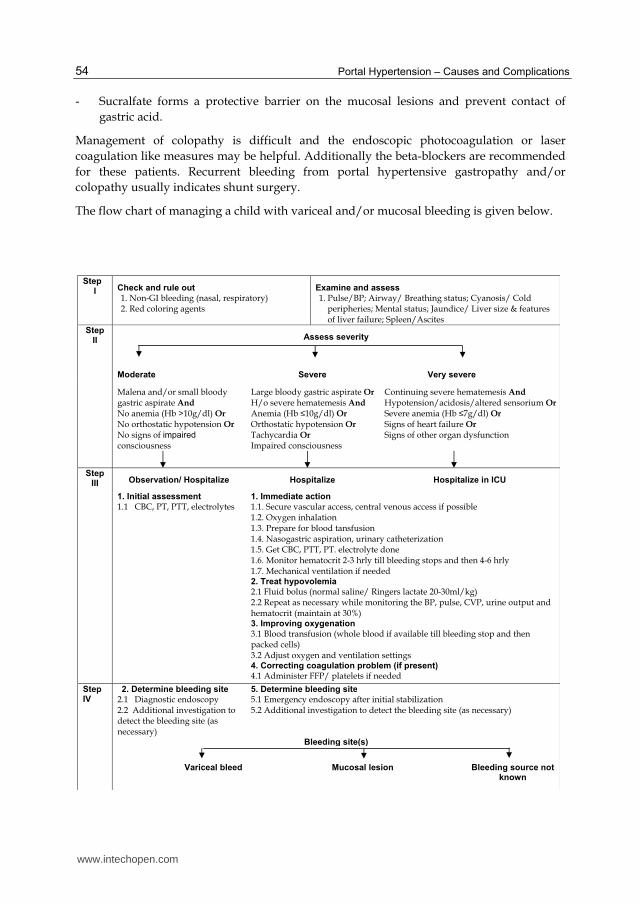
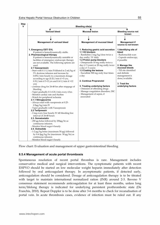
The flow chart of managing a child with variceal and/or mucosal bleeding is given below.
www.intechopen.com
Extra Hepatic Portal Venous Obstruction in Children
55
Flow chart: Evaluation and management of upper gastrointestinal bleeding
8.5.4 Management of acute portal thrombosis
Spontaneous resolution of recent portal thrombus is rare. Management includes
conservative medical and surgical interventions. The symptomatic patients with recent
EHPVO should be started on low molecular weight heparin immediately after detection
followed by oral anticoagulant therapy. In asymptomatic patients, if detected early,
anticoagulation should be considered. Dosage of anticoagulation therapy is to be titrated
with target to maintain international normalized ration (INR) around 2-3. Baveno V
consensus statement recommends anticoagulation for at least three months, unless long
term/lifelong therapy is indicated for underlying persistent prothrombotic state (De
Franchis, 2010). Repeat Doppler is to be done after 3-6 months to check for recanalisation of
portal vein. In acute thrombosis cases, evidence of infection must be ruled out. If any
www.intechopen.com
Portal Hypertension – Causes and Complications
56
evidence of infection is there, antibiotic therapy should be given. Advanced minimally
invasive intervention techniques like direct thrombolysis of portal vein, thrombolysis
through TIPS are being used. But experience in children is limited.
8.5.5 Surgical management of EHPVO
Surgical management of EHPVO includes shunt and non-shunt procedures. The shunt procedures aim at reducing portal pressure by diverting the blood from the high-pressure portal venous system to the systemic circuit. Shunt surgeries are recommended for EHPVO cases not responding to medical and endoscopic management and if associated with complications. The indications for shunt surgery include failure of endotherapy to control bleeding, presence of gastric or ectopic varices (not amenable to endoscopic management), and associated complications like portal biliopathy and large rectal varices. Emergency shunt surgery is undertaken now only rarely due to wide availability of endoscopic management. The additional indications for shunt surgery include portal biliopathy, severe growth failure, massive splenomegaly, hypersplenism, rare blood group, isolated splenic vein thrombosis, non- compliance of elective EST/EVL and poor chance of follow up (living in remote area). Individualization of each child with EHPVO for shunt surgery is to be done considering all the aspects and in consultation with the surgical team.
Shunts may be non-selective shunts or selective shunts, partial shunts and the more recently
introduced "Rex shunt"(mesenterico-left portal bypass).
The non-selective shunts enable complete decompression of the entire portal venous system by
diverting total portal blood flow away from the liver. These shunts are end to side and side-to-
side portacaval shunts, central lienorenal shunt, end to side mesocaval shunt and large
diameter interposition portacaval or mesocaval shunts. Although these shunts are effective in
controlling variceal bleeding, encephalopathy is a concern in patients with poor liver
functions. Follow up studies have shown shunt patency in 85-98% of children with long term
survival in >95% cases (Lautz, 2009; Bismuth, 1980; Alvarez, 1983; Gauthier, 1989; Mitra, 1993;
Prasad, 1994; Orloff, 1994; Superina, 2006; Shariff, 2010). Risk of variceal recurrence and re-
bleed is there in case of shunt blockage. But there is no report of post shunt encephalopathy in
EHPVO cases. Splenectomy usually done with central lienorenal shunt has not been found to
be associated with an increased risk of post-splenectomy sepsis (Arora, 2002).
The selective shunts divert the blood of gastroesophageal-splenic segment and maintain the blood flow in mesenteric segment based on the functional classification of portal venous system into the splenogastric and mesenteric segments. This enables portal decompression with maintenance of hepatopetal portal perfusion, mesenteric venuos pressure and hepatotrophic factors. Distal splenorenal shunt, a selective shunt has technical advantage of lower chance of post-shunt encephalopathy. The mesoportal bypass (MPB) is a recent advancement in shunt restoring mesenteric blood flow to the liver through the Rex venous recessus (interposition of a jugular venous allograft between the superior mesenteric vein and the intrahepatic left portal vein). This "Rex shunt" restores the physiological hepatopetal flow and thereby corrects the liver dysfunction, coagulation parameters and also improves the growth potential (Superina, 2006; Mack, 2003; Lautz, 2009). It appears that doing the Rex shunt early after diagnosis rather than waiting till complications will be beneficial. But in want of clear evidence and experience, there is no consensus on this issue.
www.intechopen.com

Extra Hepatic Portal Venous Obstruction in Children
57
A subgroup of EHPVO patients may not be suitable for shunt surgery in view of
complicated vascular anatomy due to extensive thrombosis of the splenoportal axis, prior
splenectomy or a previously performed but failed shunt procedure. These cases merit non-
surgical management and non-shunt surgical procedures, if required.
8.5.6 Portosystemic shunt vs endoscopic sclerotherapy
In a randomized clinical trial by our group in India, we assessed the complications, technical problems and rebleeding rates after EST and shunt surgery. Out of the 297 children with EHPVO screened, 156 children were randomized into surgical and EST groups (78 children in each group). Out of the 78 children randomized into in surgical group, 8 opted out before intervention and 28 could not undergo surgery due to technical reason (blockage of splenic vein) and hence received EST. Out of the 78 children in EST group, 75 children undergone EST (2 opted out before intervention and 1 died). The children were followed up (median follow up period 24 months; range: 21-29 months). The pooled incidence of bleeds decreased from 0.11/month in pre-intervention period to 0.014/month in post-intervention phase, when both the interventions were considered together. The fall in the incidence rate for major bleeds was higher (0.05 to 0.002) than that for minor bleeds (0.06 to 0.013). Survival analysis showed that among the children who underwent EST, 25% had rebleeding by 16.9 months after intervention while 50% had rebleeding by 38.4 months. Children who underwent surgery had rebleeding by 18.3 months in 25% subjects and less than half of these subjects ever rebled. Survivor function at various 6 monthly time intervals during follow up was similar between EST and surgery groups. This was true for any kind of re-bleed; major bleeds and minor bleeds. Relative risk of rebleeding after controlling for period of follow up was similar for all types of bleeds. Recurrence of esophageal varices was higher in surgery group compared to the EST group. By 6 months, half of the patients who underwent surgical intervention, had recurrence of varices, while the same was observed after 24 months in patients who underwent EST. But the risk of rebleeding was not influenced by recurrence of varices. Risk of re-bleeding was associated with the age of intervention, lesser rebleeding was noticed among children aged >10 years. Hypersplenism was observed in 17 children in the EST group compared to 3 children in the surgery group. Eight patients in the EST group died compared to 6 patients in the surgery group. It appeared from this data that both EST and surgery appear to perform equally well to control rebleeding in EHPVO patients. (Arora, 2004).
8.6 Management of special situations related to EHPVO
8.6.1 Portal biliopathy
Symptomatic portal biliopathy is a definite indication for intervention. Primary biliary tract
surgery is associated with significant morbidity and mortality due to presence of extensive
collaterals around the bile ducts. Some experts suggest shunt surgery first and if it fails to
resolve biliary obstruction, then to undertake staged biliary surgery (Chaudhary 1998).
While others recommend endoscopic management of biliary obstruction first and if
obstruction persists, then to do the shunt surgery (Khare, 2005). Presence of
choledocholithiasis without biliary stricture can be managed by endoscopic sphincterotomy
and stone removal. Presence of stricture may need balloon dilatation and stent placement. In
cases of endoscopic failure, portosystemic shunt followed by biliary surgery should be
www.intechopen.com
Portal Hypertension – Causes and Complications
58
undertaken (Khare 2005). There is no clear consensus regarding timing of biliary surgery
after shunt surgery. The experience in children in this context is limited. Children with
portal biliopathy undergoing shunt surgery may have normalization of serum
aminotransferases and gamma glutamyl transpeptidase (GGT) levels within 1 to 6 weeks
after surgery (Gauthier, 2005). Shunt surgery is indicated in children with symptomatic
portal biliopathy or even asymptomatic portal biliopathy in presence of growth failure,
symptomatic hypersplenism or ectopic varices.
8.6.2 Hypersplenism
Traditionally, hypersplenism has been treated by splenectomy, open or laparoscopic. In view of the risk of overwhelming sepsis in children who are asplenic, the risks and benefits must be carefully weighed when splenectomy is being considered. Age of removal of spleen is an important consideration; risk of overwhelming sepsis may increase if child is less than 5 years. Vaccines against pneumococcus, Haemophilus influenza and meningococcus should be administered to all children particularly these below 5 years, who are about to undergo splenectomy, at least 10 days before splenectomy. Additionally, antibiotic prophylaxis (penicillin) is recommended to asplenic children who have not received vaccines. In recent times, alternative technique of partial splenic embolisation (PSE) is in use (Dwivedi, 2002). It causes ischemic necrosis of a part of spleen and thereby decreases in splenic size and hypersplenism. It allows the preservation of adequate splenic tissue and thereby prevents the immunological function and protection against overwhelming infection.
8.6.3 Coagulopathy
The management of coagulopathy depends on the cause. There is no consensus or clear guideline about anticoagulant therapy for prothrombotic conditions in chronic EHPVO. In a patient with established chronic EHPVO, anticoagulants are recommended only if there is a history of recurrent thrombotic episodes and after shunt surgery (Valla, 2002). Anticoagulation therapy is contraindicated in cases with active variceal bleeding and can be initiated after variceal obliteration and patient is on beta blocker drug.
9. Natural history
The natural course of EHPVO is mainly determined by the age at presentation, presence or absence of underlying liver disease, associated systemic diseases. Most patients with EHPVO in the absence of liver disease and systemic disease have a relatively benign course. Morbidity is mainly related to variceal bleeding, recurrent thrombosis (if associated with prothrombotic disorder), symptomatic portal biliopathy, and hypersplenism. In case of children with EHPVO, especially in developing countries, nutritional status and growth retardation also influence the outcome.
9.1 Quality of life with EHPVO
Children with EHPVO suffering in view of repeated episodes of bleeding, undergoing
diagnostic and therapeutic procedures, associated complications and morbidities, delayed
growth, may have psychological issues that may impeded their ability and potential in
physical, emotional, social, academic and other extracurricular segments. A study among
www.intechopen.com
Extra Hepatic Portal Venous Obstruction in Children
59
Indian children with EHPVO compared the quality of life (QOL) scores in children before
variceal eradication, after variceal eradication and after surgery with controls. Overall
children with EHPVO had lower median QOL scores in physical, emotional, social, and
school functioning health domains compared to controls. Esophageal variceal eradication
had no significant effect on QOL, but QOL improved after surgery. Most likely surgery
provided a feeling of cure and reduced further risk and morbidity for both patient and the
parents and thus resulted improvement in psychological status. Spleen size significantly
influenced quality of life (QOL) scores and children with massive splenomegaly had poor
QOL scores as compared with patients with mild and moderate splenomegaly. Total,
physical, and psychosocial QOL scores were significantly lower in children with growth
retardation. Both spleen size and growth retardation were independent predictors that affect
the QOL (Radha Krishna, 2010).
10. Prognosis
Unlike patients with chronic liver disease, patients with EHPVO have a good prognosis as their liver functions are preserved. Mortality is mainly due to variceal bleeding and it is to the tune of 5% (Mowat, 1986). Long term studies after endotherapy have shown almost no mortality. Many of these bleeding episodes occurred within the first 4-10 years of variceal eradication (Zargar, 2004; Thomas, 2009; Stringer, 1994). The risk of rebleeding reduces with increasing age, especially after 10 years of age. With increasing age of survival, children with EHPVO are likely to face the longer term consequences in nutrition, mental function and portal biliopathy. Long term follow up studies shall be able to answer some of the unanswered issues in EHPVO.
11. Conclusion
Extrahepatic portal venous obstruction is the commonest cause of portal hypertension and variceal bleeding in children. With the availability of diagnostic facilities and effective endotherapy, the mortality due to EHPVO related emergencies has become rare. But with increasing survival, the sequelae and related morbidities are rising. The evolving surgical interventions have raised the hope of averting the long term complications of EHPVO. Further generation of evidence on etiology and course of disease including the effect of management in children with EHPVO is required, especially in developing countries. At the same time availability of trained manpower and facilities for management of acute emergencies and advanced surgical interventions will further improve the outcome.
12. References
[1] Alvarez F, Bernard O, Brunell F, Hadchouel P, Odièvre M, Alagille D. Portal obstruction in children. II. Results of surgical portosystemic shunts. J Pediatr. 1983;103:703–7.
[2] Alvarez F, Bernard O, Brunnelle F et al. portal hypertension in children: Clinical investigations and hemorrhage risk. J Pediatr 1983; 103: 696-702.
[3] Arora NK, Ganguly S, Mathur P, Ahuja A, Patwari A. Upper Gastrointestinal bleeding: Etiology and Management. Indian J Pediatrics. 2002; 69:155-168.
[4] Arora NK, Lodha R, Gulati S, Gupta AK, Mathur P, Joshi MS, et al. Portal hypertension in north Indian children. Indian J Pediatr. 1998;65:585–91.
www.intechopen.com
Portal Hypertension – Causes and Complications
60
[5] Arora NK. (Principal Investigator), Mathur P, Das MK.. Project titled “Risk factors for re-bleeding after endoscopic sclerotherapy in children with Extrahepatic Portal Hypertension” . Funded by Indian Council of Medical Research (ICMR). India, Report 2000. Conducted at All India Institute of Medical Sciences, New Delhi.
[6] Arora NK. (Principal Investigator), Jain S, Mathur P, Das MK. Project titled “Management of Extra-hepatic Portal Hypertension in Childhood: Efficacy and Efficiency of Endoscopic Sclerotherapy versus Porto-systemic Shunt Surgery”. Funded by Indian Council of Medical Research (ICMR). India, Report 2004. Conducted at All India Institute of Medical Sciences, New Delhi.
[7] Bellomo-Brandão MA, Morcillo AM, Hessel G, Cardoso SR, Servidoni MFPC, da-Costa-Pinto EA. Growth assessment in children with extra-hepatic portal vein obstruction and portal hypertension. Gastroenterologia Pediatrica.2003; 40:247-250.
[8] Bhatia V, Jain AK, Sarin SK. Choledocholithiasis associated with portal bililopathy in patients with extrahepatic portal vein obstruction and management with endoscopic sphincterotomy. Gastrointest Endosc 1995;42:178-181.
[9] Bhattacharya M, Makhani G, Kannan M, Ahmed R P, Gupta P K, Saxena R. Inherited prothrombotic defects in Budd-Chiari syndrome and portal vein thrombosis: a study from North India. Am J Clin Pathol 2004; 121: 844-7.
[10] Bismuth H, Franco D, Alagille D. Portal diversion for portal hypertension in children. The first ninety patients. Ann Surg. 1980; 192:18–24.
[11] Brodoff M, Conn HO. Esophageal tamponade in the management of bleeding esophageal varices. Dig Dis Sc. 1980; 25: 267-272.
[12] Burroughs AK, McCormick PA, Hughes MD et al. Randomized double blind placebo controlled trial of somatostatin for variceal bleeding. Emergency control and prevention of early variceal bleeding. Gastroenterology 1990; 99: 1388-1395.
[13] Chaudhary A, Dhar P, Sarin SK, Sachdev A, Agarwal AK, Vij JC, et al. Bile duct obstruction due to portal biliopathy in extrahepatic portal hypertension: surgical management. Br J Surg. 1998; 85: 326–9.
[14] Chawla A, Dewan R, Sarin SK. The fequency and influence of gall bladder varices on gall bladder function in patients with portal hyper tention . Am j Gastroenterol 1995;90:2011-14
[15] Chawla Y, Dilawari JB. Anorectal varices—their frequency in cirrhotic and non-cirrhotic portal hypertension. Gut.1991; 32: 309–11.
[16] De Franchis R, On behalf of the Baveno V Faculty. Revising consensus in portal hypertension: Report of the Baveno V consensus workshop on methodology of diagnosis and therapy in portal hypertension. Journal of Hepatology 2010; 53: 762–768.
[17] De Franchis R, Primignani M. Endoscopic treatment for portal hypertension. Seminars in Liver Disease 1999; 19: 499-455.
[18] Dilawari JB, Chawla YK. Pseudosclerosing cholangitis in extrahepatic portal venous obstruction. Gut. 1992; 33: 272–6.
[19] Dilawari JB, Ganguly S, Chawla Y. Non-cirrhotic portal fibrosis. Ind J Gastroenterol 1992; 11: 31-36.
[20] Dubuisson C, Boyer-Neumann C, Wolf M, Meyer D, Bernard O. Protein C, protein S and antithrombin III in children with portal vein obstruction. J Hepatol. 1997; 27: 132–5.
[21] Dwivedi MK, Pal RK, Dewanga L, Nag P. Efficacy of partial splenic embolisation in the management of hypersplenism. Indian J Radiol Imaging. 2002;12:371-4
www.intechopen.com
Extra Hepatic Portal Venous Obstruction in Children
61
[22] Ferenci P, Lockwood A, Mullen K, et al. Hepatic Encephalopathy: Definition, Nomenclature, Diagnosis, and Quantification: Final Report of the Working Party at the 11th World Congress of Gastroenterology, Vienna, 1998. Hepatology 2002; 35: 716-21.
[23] Ganguly S, Sarin SK, Bhatia V, Lahoti D. The prevalence and spectrum of colonic lesions in patients with cirrhosis and noncirrhotic portal hypertension. Hepatology. 1995; 21: 1226–31.
[24] Gauthier F, De Dreuzy O, Valayer J, Montupet P. H-type shunt with an autologous venous graft for treatment of portal hypertension in children. J Pediatr Surg. 1989; 24: 1041–3.
[25] Gauthier-Villars M, Franchi S, Gauthier F, Fabre M, Pariente D, Bernard O. Cholestasis in children with portal vein obstruction. J Pediatr. 2005; 146: 568–73.
[26] Goncalves ME, Cardoso SR, Maksoud JG. Prophylactic sclerotherapy in children with esophageal varices: long-term results of a controlled prospective randomized trial. J Pediatr Surg. 2000; 35: 401–5.
[27] Heaton ND, Davenport M, Howard ER. Incidence of haemorrhoids and anorectal varices in children with portal hypertension. Br J Surg. 1993;80:616–8.
[28] Heyman'MB, LaBerge JM. Role of transjugular intrahepatic portosystemic shunt in the treatment of portal hypertension in pediatric patients. J Pediatr Gastroenterol Nutr 1999; 29 : 209-249.
[29] Hyams JS, Treem WR. Portal hypertensive gastropathy in children. J Pediatr Gastroenterol Nutr. 1993;17:13–8.
[30] Khare R, Sikora SS, Srikanth G, Choudhuri G, Saraswat VA, Kumar A, et al. Extrahepatic portal venous obstruction and obstructive jaundice: approach to management. J Gastroenterol Hepatol. 2005;20:56–61.
[31] Khuroo MS, Yattoo GN, Zargar SA, Javid G, Dar MY, Khan BA, et al. Biliary abnormalities associated with extrahepatic portal venous obstruction. Hepatology. 1993;17:807–13.
[32] Kokawa H, Shijo H, Kubara K, et al. Long term risk factors for bleeding after first course of endoscopic injection sclerotherapy : A univariate and multivariate analysis. Am J Gastroenterol 1993; 88 : 1206 - 1211.
[33] Koshy A, Bhasin DK, Kapoor KK. Bleeding in extrahepatic portal vein obstruction. Indian J Gastroenterol. 1984;3:13–4.
[34] Larroche J. Umbilical catheterization: its complications. Biol.Neonate 1970;16:101-103. [35] Lautz TB, Sundaram SS, Whitington PF, Keys L, Superina RA. Growth impairment in
children with extrahepatic portal vein obstruction is improved by mesenterico-left portal vein bypass. J Pediatr Surg. 2009;44:2067–70.
[36] Lilly JR, Stiegmann GV, Stellin G. Esophageal endosclerosis in children with portal vein thrombosis. J Pediatr Surg 1982;17:571-575.31.
[37] Mack CL, Superina RA, Whitington PF. Surgical restoration of portal flow corrects procoagulant and anticoagulant deficiencies associated with extrahepatic portal vein thrombosis. J Pediatr. 2003;142:197–9.
[38] Malkan GH, Bhatia SJ, Bashir K, Khemani R, Abraham P, Gandhi MS, et al. Cholangiopathy associated with portal hypertension: diagnostic evaluation and clinical implications. Gastrointest Endosc. 1999;49:344–8.
[39] McKiernan PJ, Beath SV, Davison SM. A prospective study of endoscopic esophageal variceal ligation using multiband ligator. J Pediatr Gastroenterol Nutr. 2002;34:207–11.
www.intechopen.com
Portal Hypertension – Causes and Complications
62
[40] Mehrotra RN, Batia V, Dabadghao P, Yachha SK. Extrahepatic portal vein obstruction in children: anthropometry, growth hormone, and insulin-like growth factor I. J Pediatr Gastroenterol Nutr 1997;25:520-3.
[41] Menon P, Rao KL, Bhattacharya A, Thapa BR, Chowdhary SK, Mahajan JK, et al. Extrahepatic portal hypertension: quality of life and somatic growth after surgery. Eur J Pediatr Surg. 2005;15:82–7.
[42] Misra SP, Dwivedi M, Misra V, Dharmani S, Kunwar BK, Arora JS. Colonic changes in patients with cirrhosis and in patients with extrahepatic portal vein obstruction. Endoscopy. 2005;37:454–9.
[43] Mitra SK, Rao KLN, Narasimhan KL, Dilawari JB, Batra YK, Chawla Y, et al. Side-to-side lienorenal shunt without splenectomy in noncirrhotic portal hypertension in children. J Pediatr Surg. 1993;28:398–401; discussion 401–2.
[44] Mittal SK, Kalra KK, Aggarwal V. Diagnostic upper GI endoscopy for hematemesis in children: experience from a pediatric gastroenterology centre in north India. Indian J Pediatr.1994;61:651–4.
[45] Mowat AP. Disorders of portal and hepatic venous systems. In: Mowat AP, editor. Liver disorders in childhood. 2nd ed. London: Butterworths; 1987.p.298-323.
[46] Mowat AP. Prevention of variceal bleeding. J Pediatr Gastroenterol Nutr. 1986;5:679–87. [47] Nagi B, Kochhar R, Bhasin D, Singh K. Cholangiopathy in extrahepatic portal venous
obstruction. Radiological appearances. Acta Radiol. 2000;41:612–5. [48] Nihal N, Bapat MR, Rathi P, Shah NS, Karvat A, Abraham P, et al. Relation of insulin-
like growth factor-1 and insulin-like growth factor binding protein-3 levels to growth retardation in extrahepatic portal vein obstruction. Hepatol Int. 2009;3:305–9.
[49] Nyandak T, Prakash P, Das U, Yadav P, Sharma SC, Srivastava D, Rewari BB. Portal Vein Thrombosis – Clinical Profile. Journal of Indian Academy of Clinical Medicine. 2011; 12:134-40.
[50] Odievre M, Pige G, Alagille D. Congenital abnormalities associated with extraheptic portal hypertension. Arch Dis Child 1977; 52: 383-5.
[51] Orloff MJ, Orloff MS, Rambotti M. Treatment of bleeding esophagogastric varices due to extrahepatic portal hypertension: results of portal-systemic shunts during 35 years. J Pediatr Surg. 1994;29:142–51; discussion 151–4.
[52] Ozsoylu S, Kocak N, Demir H, Yuce A, Gurakan F, Ozen H. Propranolol for primary and secondary prophylaxis of variceal bleeding in children with cirrhosis. Turk J Peidtr 2000; 42 : 31-33.
[53] Pinto R B, Silveira T R, Bandenilli E, Rohsig L. Portal vein thrombosis in children and adolescents: the low prevalence of hereditary thrombophilic disorders. J Pediatr Surg 2004; 39: 1356-61.
[54] Pitcher JL. Safety and effectiveness of the modified Sangstaken-Blackemore tube. A prospective study. Gastroenterology 1971; 61 : 291-298.
[55] Poddar U, Bhatnagar S, Yachha SK. Endoscopic band ligation followed by sclerotherapy: Is it superior to sclerotherapy in children with extrahepatic portal venous obstruction? J Gastroenterol Hepatol. 2011;26:255–9.
[56] Poddar U, Borkar V. Management of extra hepatic portal venous obstruction (EHPVO): current strategies. Tropical Gastroenterology 2011;32(2):94–102.
[57] Poddar U, Thapa BR, Bhasin DK, Prasad A, Nagi B, Singh K. Endoscopic retrograde cholangiopancreatography in the management of pancreaticobiliary disorders in children. J Gastroenterol Hepatol. 2001;16:927–31.
www.intechopen.com
Extra Hepatic Portal Venous Obstruction in Children
63
[58] Poddar U, Thapa BR, Puri P, Girish CS, Vaiphei K, Vasishta RK, et al. Non-cirrhotic portal fibrosis in children. Indian J Gastroenterol. 2000;19:12–3.
[59] Poddar U, Thapa BR, Rao KL, Singh K. Etiological spectrum of esphageal varices due to portal hypertension in Indian children: is it different from the West? J Gastroenterol Hepatol. 2008;23:1354–7.
[60] Poddar U, Thapa BR, Singh K. Band ligation plus sclerotherapy versus sclerotherapy alone in children with extrahepatic portal vein obstruction. J Clin Gastroenterol. 2005;39:626–9.
[61] Poddar U, Thapa BR, Singh K. Endoscopic sclerotherapy in children: experience with 257 cases of extrahepatic portal venous obstruction. Gastrointest Endosc. 2003;57:683–6.
[62] Poddar U, Thapa BR, Singh K. Frequency of gastropathy and gastric varices in children with extrahepatic portal venous obstruction treated with sclerotherapy. J Gastroenterol Hepatol. 2004;19:1253–6.
[63] Prasad AS, Gupta S, Kohli V, Pande GK, Sahni P, Nundy S. Proximal splenorenal shunts for extrahepatic portal venous obstruction in children. Ann Surg. 1994;219:193–6.
[64] Radha Krishna Y, Yachha SK, Srivastava A, Negi D, Lal R, Poddar U. Quality of Life in Children Managed for Extrahepatic Portal Venous Obstruction. J Pediatr Gastroenterol Nutr. 2010;50: 531–536
[65] Rangari M, Gupta R, Jain M, Malhotra V, Sarin S K. Hepatic dysfunction in patients with extrahepatic portal venous obstruction. Liver Int 2003 Dec; 23: 434-9.
[66] Rector WG, Reynolds TB. Risk factors for hemorrhage from esophageal varices and acute gastric erosions. Clin Gastroenterol. 1985;14:139-153.
[67] Sarin SK, Agarwal SR. Extrahepatic portal vein obstruction. Semin liver disease. 2002; 22:43-58.
[68] Sarin SK, Bansal A,Sasan S ,et al. Portal vein obstruction in children leads to growth retardation. Hepatology 1992;15:229-233.
[69] Sarin SK, Guptan RC, Jain A, Sundaram KR. A randomized controlled trial of endoscopic variceal band ligation for primary prophylaxis of variceal bleeding. Eur J Gastroenterol Hepatol. 1996; 8 : 337-342.
[70] Sarin SK, Lahoti D, Saxena SP, Murthy NS, Makwana UK. Prevalence, classification and natural history of gastric varices: a long-term follow up study in 568 portal hypertension patients. Hepatology. 1992;16:1343–9.
[71] Sarin SK, Sollano JD, Chawla YK, et al on behalf of Members of the APASL Working Party on Portal Hypertension. Consensus on Extra-hepatic Portal Vein Obstruction. Liver International. 2006; 26:512-519.
[72] Sharif K, Mckiernan P, de Ville de Goyet J. Mesoportal bypass for extrahepatic portal vein obstruction in children: close to cure for most! J Pediatr Surg. 2010;45:272–6.
[73] Sharma MP. Sonographic signs in portal hypertension: a multivariate analysis.Ultrasound in Medicine & Biology. 1997; 23: S17-S17.
[74] Sharma S, Kumar SI, Poddar U, Yachha SK, Aggarwal R. Factor V Leiden and prothrombin gene G20210A mutations are uncommon in portal vein thrombosis in India. Indian J Gastroenterol. 2006;25:236–9.
[75] Stringer M D, Heaton N D, Karani J., et al. Patterns of portal vein occlusion and their etiological significance. Br J Surg 1994; 81: 1328-31.
www.intechopen.com
Portal Hypertension – Causes and Complications
64
[76] Stringer MD, Howard ER. Long term outcome after injection sclerotherapy for esophageal varices in children with extra hepatic portal hypertension. Gut. 1994;35;257–9.
[77] Stringer MD. Improved body mass index after mesenterico-portal bypass. Pediatr Surg Int. 2007;23:539–43.
[78] Superina R, Bambini DA, Lokar J, Rigsby C, Whittington PF. Correction of extrahepatic portal vein thrombosis by the meserteric to left portal vein bypass. Ann Surg. 2006;243:515–21.
[79] Thomas V, Jose T, Kumar S. Natural history of bleeding after esophageal variceal eradication in patients with extrahepatic portal venous obstruction; a 20-year follow-up. Indian J Gastroenterol.2009;28:206–11.
[80] Thompson EN, Sherlock S.The etiology of portal vein thrombosis with particular reference to the role of infection and exchange transfusion. Q J Med 1964;33:465-480.
[81] Tuggle DW, Bennett KG, Scott J, Tunell WP. Intravenous vasopressin and gastrointestinal hemorrhage in children. J Pediatr Surg 1988; 23 : 627-629. Bottom of Form
[82] Valla D C, Condat B, Lebrec D. Spectrum of portal vein thrombosis in the West. J Gastroenterol Hepatol 2002; 17 (Suppl. 3): S224-7.
[83] Webb L J, Sherlock S. The etiology, presentation and natural history of extra-hepatic portal venous obstruction. Q J Med 1979; 192: 627-39.
[84] West MS, Garra BS, Horti SC, et al. Gallbladder varices: Imaging findings in patients with portal hypertension. Radiology 1991;179:179-82
[85] Yachha S K, Khandur A, Sharma B C, Kumar M. Gastrointestinal bleeding in children. J Gastroenterol Hepatol 1996; 11: 903-7.
[86] Yachha SK, Dhiman RK, Gupta R, Ghoshal UC. Endosonographic evaluation of the rectum in children with extrahepatic portal venous obstruction. J Pediatr Gastroenterol Nutr. 1996; 23:438–41.
[87] Yadav S, Dutta A K, Sarin S K. Do umbilical vein catheterization and sepsis lead to portal vein thrombosis? A prospective, clinical and sonographic evaluation. J Pediatr Gastroenterol Nutr 1993; 17: 392-6.
[88] Yadav S, Srivastava A, Srivastava A, et al. Encephalopathy assessment in children with extra-hepatic portal vein obstruction with MR, psychometry and critical flicker frequency. J Hepatology. 2010; 52 (3): 348-354.)
[89] Zargar SA, Javid G, Khan BA, Yattoo GN, Shah AH, Gulzar GM, et al. Endoscopic ligation compared with sclerotherapy for bleeding esophageal varices in children with extrahepatic portal venous obstruction. Hepatology. 2002; 36:666–72.
[90] Zargar SA, Yattoo GN, Javid G, Khan BA, Shah AH, Shah NA, et al. Fifteen-year follow up of endoscopic injection sclerotherapy in children with extrahepatic portal venous obstruction. J Gastroenterol Hepatol. 2004; 19:139–45.
www.intechopen.com
Portal Hypertension - Causes and ComplicationsEdited by Prof. Dmitry Garbuzenko
ISBN 978-953-51-0251-9Hard cover, 156 pagesPublisher InTechPublished online 14, March, 2012Published in print edition March, 2012
InTech EuropeUniversity Campus STeP Ri Slavka Krautzeka 83/A 51000 Rijeka, Croatia Phone: +385 (51) 770 447 Fax: +385 (51) 686 166www.intechopen.com
InTech ChinaUnit 405, Office Block, Hotel Equatorial Shanghai No.65, Yan An Road (West), Shanghai, 200040, China
Phone: +86-21-62489820 Fax: +86-21-62489821
Portal hypertension is a clinical syndrome defined by a portal venous pressure gradient, exceeding 5 mm Hg.In this book the causes of its development and complications are described. Authors have presented personalexperiences on conducting patients with various displays of portal hypertension. Moreover, the book presentsmodern data about molecular mechanisms of pathogenesis of portal hypertension in liver cirrhosis, theinformation about the original predictor of risk of bleeding from gastro-esophageal varices and new methodsfor their conservative treatment.
How to referenceIn order to correctly reference this scholarly work, feel free to copy and paste the following:
Narendra K. Arora and Manoja K. Das (2012). Extra Hepatic Portal Venous Obstruction in Children, PortalHypertension - Causes and Complications, Prof. Dmitry Garbuzenko (Ed.), ISBN: 978-953-51-0251-9, InTech,Available from: http://www.intechopen.com/books/portal-hypertension-causes-and-complications/extra-hepatic-portal-venous-obstruction-in-children-

© 2012 The Author(s). Licensee IntechOpen. This is an open access articledistributed under the terms of the Creative Commons Attribution 3.0License, which permits unrestricted use, distribution, and reproduction inany medium, provided the original work is properly cited.
Related Documents


