Expression, purification and structural studies of a short antimicrobial peptide Mateja Zorko, Boštjan Japelj, Iva Hafner-Bratkovič, Roman Jerala ⁎ , 1 Department of Biotechnology, National Institute of Chemistry, Hajdrihova 19, POB 660,1000 Ljubljana, Slovenia abstract article info Article history: Received 11 April 2008 Received in revised form 17 September 2008 Accepted 21 October 2008 Available online 5 November 2008 Keywords: Antimicrobial peptide Solution structure Recombinant peptide NMR spectroscopy Fluorescence Lipid membrane We have produced a small antimicrobial peptide PFWRIRIRR in bacteria utilizing production in the form of insoluble fusion protein with ketosteroid isomerase. The recombinant peptide was rapidly and efficiently isolated by acidic cleavage of the fusion protein based on the acid labile Asp–Pro bond at the N-terminus of the peptide. The peptide has antibacterial activity and neutralizes macrophage activation by LPS. The selectivity of the peptide against bacteria correlates with preferential binding to acidic phospholipid vesicles. Solution structure of the peptide in SDS and DPC micelles was determined by NMR. The peptide adopts a well-defined structure, comprising a short helical segment. Cationic and hydrophobic clusters are segregated along the molecular axis of the short helix, which is positioned perpendicular to the membrane plane. The position of the helix is shifted in two micellar types and more nonpolar surface is exposed in anionic micelles. Overall structure explains the advantageous role of the N-terminal proline residue, which forms an integral part of the hydrophobic cluster. © 2008 Elsevier B.V. All rights reserved. 1. Introduction Treatment of bacterial infection with antibiotics is one of the mainstays of medicine [1]. Increasing resistance of virtually all microbes toward common antibiotics is a major health concern. Antimicrobial peptides are present in a variety of organisms where they protect the host from microbial infection. They have been identified in and isolated from a variety of sources including mammals, insects, amphibians, fish, plants, prokaryotes [2,3]. These peptides are less susceptible to the development of bacterial resistance because they act on the bacterial membrane causing multiple stress through several targets [4] but have minimal toxic and allergic effects to the host [5]. Thus, antimicrobial peptides may potentially be applied in medicine as safe antimicrobial agents due to their activities against bacteria and fungi [6]. Recent studies have shown that many cationic antimicrobial peptides are able to neutralize LPS [7], block cytokine induction in macrophages and prevent sepsis in animal models. Sepsis due to a Gram-negative bacteria is usually caused by the release of a bacterial outer membrane component, endotoxin (lipopolysaccharide, LPS) [7]. In order to provide large quantities of these peptides for physiological investigation and clinical trials, efficient production methods are necessary. Short antimicrobial peptides can be efficiently prepared by chemical synthesis, but may still be prohibitively expensive for therapy [8]. As an alternative, recombinant methods permit the production of peptides and proteins in microorganisms. Production of antimicrobial peptides in expression systems, such as bacterial, yeast or insect cells has been reported. Escherichia coli has been used for production of many antimicrobial peptides, e.g. lactoferricin [9], dermicin [10], moricidin [11], defensins [12–14] and buforin [15]. This biological expression system is also suitable to obtain uniformly or partially isotopically enriched peptides, which are required for structural investigation of the ligand–receptor interaction by NMR spectroscopy and provides additional information on molecular dynamics, improvement of the precision of the determined structures and filtered experiments in the complex systems [16,17]. However, difficulties have been encountered in expression of antimicrobial peptides because of their cytotoxicity to host cells, sensitivity to proteolytic degradation and low expression level [18]. The expression system with an antimicrobial peptide fused to a partner protein is most effective because of the reduced toxicity against host cells, enhanced product stability and facilitated product recovery [19]. Such fusion proteins generally lack antimicrobial activity if they form insoluble products or interact with carrier protein [20–24]. The peptide is released from the fusion by chemical or enzymatic cleavage [25]. For this study we selected the peptide based on the structure– activity development of the peptide LF11 originating from human lactoferrin We have already reported solution structure of LF11 in complex with anionic and zwitterionic amphiphiles [16,26–28], where LF11 adopted a defined and unique fold in the micellar environment. We deleted most of the noncharged polar residues that do not contribute to the favorable interactions with anionic lipids or LPS and exchanged several residues, which increased its antimicrobial activity and neutralization of LPS. One of the key properties of antimicrobial Biochimica et Biophysica Acta 1788 (2009) 314–323 ⁎ Corresponding author. Tel.: +386 1 476 0335; fax: +386 1 476 0300. E-mail address: [email protected] (R. Jerala). 1 Faculty of chemistry and chemical technology, University of Ljubljana, 1000 Ljubljana, Slovenia. 0005-2736/$ – see front matter © 2008 Elsevier B.V. All rights reserved. doi:10.1016/j.bbamem.2008.10.015 Contents lists available at ScienceDirect Biochimica et Biophysica Acta journal homepage: www.elsevier.com/locate/bbamem

Welcome message from author
This document is posted to help you gain knowledge. Please leave a comment to let me know what you think about it! Share it to your friends and learn new things together.
Transcript
Biochimica et Biophysica Acta 1788 (2009) 314–323
Contents lists available at ScienceDirect
Biochimica et Biophysica Acta
j ourna l homepage: www.e lsev ie r.com/ locate /bbamem
Expression, purification and structural studies of a short antimicrobial peptide
Mateja Zorko, Boštjan Japelj, Iva Hafner-Bratkovič, Roman Jerala ⁎,1
Department of Biotechnology, National Institute of Chemistry, Hajdrihova 19, POB 660, 1000 Ljubljana, Slovenia
⁎ Corresponding author. Tel.: +386 1 476 0335; fax: +E-mail address: [email protected] (R. Jerala).
1 Faculty of chemistry and chemical technology,Ljubljana, Slovenia.
0005-2736/$ – see front matter © 2008 Elsevier B.V. Adoi:10.1016/j.bbamem.2008.10.015
a b s t r a c t
a r t i c l e i n f oArticle history:
We have produced a small Received 11 April 2008Received in revised form 17 September 2008Accepted 21 October 2008Available online 5 November 2008Keywords:Antimicrobial peptideSolution structureRecombinant peptideNMR spectroscopyFluorescenceLipid membrane
antimicrobial peptide PFWRIRIRR in bacteria utilizing production in the form ofinsoluble fusion protein with ketosteroid isomerase. The recombinant peptide was rapidly and efficientlyisolated by acidic cleavage of the fusion protein based on the acid labile Asp–Pro bond at the N-terminus ofthe peptide. The peptide has antibacterial activity and neutralizes macrophage activation by LPS. Theselectivity of the peptide against bacteria correlates with preferential binding to acidic phospholipid vesicles.Solution structure of the peptide in SDS and DPC micelles was determined by NMR. The peptide adopts awell-defined structure, comprising a short helical segment. Cationic and hydrophobic clusters are segregatedalong the molecular axis of the short helix, which is positioned perpendicular to the membrane plane. Theposition of the helix is shifted in twomicellar types and more nonpolar surface is exposed in anionic micelles.Overall structure explains the advantageous role of the N-terminal proline residue, which forms an integralpart of the hydrophobic cluster.
© 2008 Elsevier B.V. All rights reserved.
1. Introduction
Treatment of bacterial infection with antibiotics is one of themainstays of medicine [1]. Increasing resistance of virtually allmicrobes toward common antibiotics is a major health concern.Antimicrobial peptides are present in a variety of organisms wherethey protect the host from microbial infection. They have beenidentified in and isolated from a variety of sources includingmammals, insects, amphibians, fish, plants, prokaryotes [2,3]. Thesepeptides are less susceptible to the development of bacterialresistance because they act on the bacterial membrane causingmultiple stress through several targets [4] but have minimal toxicand allergic effects to the host [5]. Thus, antimicrobial peptides maypotentially be applied in medicine as safe antimicrobial agents due totheir activities against bacteria and fungi [6]. Recent studies haveshown thatmany cationic antimicrobial peptides are able to neutralizeLPS [7], block cytokine induction in macrophages and prevent sepsisin animal models. Sepsis due to a Gram-negative bacteria is usuallycaused by the release of a bacterial outer membrane component,endotoxin (lipopolysaccharide, LPS) [7].
In order to provide large quantities of these peptides forphysiological investigation and clinical trials, efficient productionmethods are necessary. Short antimicrobial peptides can be efficientlyprepared by chemical synthesis, but may still be prohibitivelyexpensive for therapy [8]. As an alternative, recombinant methods
386 1 476 0300.
University of Ljubljana, 1000
ll rights reserved.
permit the production of peptides and proteins in microorganisms.Production of antimicrobial peptides in expression systems, such asbacterial, yeast or insect cells has been reported. Escherichia coli hasbeen used for production of many antimicrobial peptides, e.g.lactoferricin [9], dermicin [10], moricidin [11], defensins [12–14] andbuforin [15]. This biological expression system is also suitable toobtain uniformly or partially isotopically enriched peptides, which arerequired for structural investigation of the ligand–receptor interactionby NMR spectroscopy and provides additional information onmolecular dynamics, improvement of the precision of the determinedstructures and filtered experiments in the complex systems [16,17].However, difficulties have been encountered in expression ofantimicrobial peptides because of their cytotoxicity to host cells,sensitivity to proteolytic degradation and low expression level [18].The expression system with an antimicrobial peptide fused to apartner protein is most effective because of the reduced toxicityagainst host cells, enhanced product stability and facilitated productrecovery [19]. Such fusion proteins generally lack antimicrobialactivity if they form insoluble products or interact with carrier protein[20–24]. The peptide is released from the fusion by chemical orenzymatic cleavage [25].
For this study we selected the peptide based on the structure–activity development of the peptide LF11 originating from humanlactoferrin We have already reported solution structure of LF11 incomplexwith anionic and zwitterionic amphiphiles [16,26–28], whereLF11 adopted a defined and unique fold in the micellar environment.We deleted most of the noncharged polar residues that do notcontribute to the favorable interactions with anionic lipids or LPS andexchanged several residues, which increased its antimicrobial activityand neutralization of LPS. One of the key properties of antimicrobial
315M. Zorko et al. / Biochimica et Biophysica Acta 1788 (2009) 314–323
peptides is their ability to differentiate between foreign and host cells,which is defined but by the peptide structure and target membraneproperties [29]. Importantly, eukaryotic and bacterial membraneshave very different lipid compositions. The membrane of host cellscomprises mainly phosphatidylcholine, sphingomyelin and choles-terol, whereas bacterial cells expose negatively charged phospholipids,phosphatidylglycerol, cardiolipin and lipopolysaccharides [30]. Asdemonstrated before, a high content of arginine and hydrophobicresidues is essential for activity against bacterial membranes [31].Short peptides are devoid of defined tertiary structure but can adopt adefined conformation in the lipid environment.
We have produced and purified a recombinant antimicrobialpeptide PFWRIRIRR (“PFR peptide”) using ketosteroid isomerase (KSI)as a fusion partner in E. coli. The fusion protein formed insolubleinclusion bodies in bacteria. We engineered a single Asp–Pro acidlabile dipeptide between KSI and the PFR peptide, which allowedefficient production and isolation of the recombinant peptide inbacteria. We found that the peptide binds to the anionic but not tozwitterionic lipid vesicles and that the type of lipid mimeticssignificantly affects the peptide structure and may account for itsantibacterial activity.
2. Materials and methods
2.1. Gene construct preparation
Oligonucleotide (5 ′-phosphate-GATCCGTTCTGGCGTATTCGCATCCGTCGCTGAATG-3′), the encoding peptide sequence,annealed with the corresponding complementary oligonucleotide,was ligated into pET31b (+) expression vector (Novagen) 3′ to theketosteroid isomerase (KSI) gene using restriction site AlwN I [32].
2.2. Media and culture conditions
For protein expression, the E. coli BL21 (DE3) pLysS (Invitrogen)transformed with pET31b(+) encoding KSI–PFR gene and was grownin TB medium. Fermentation was performed in shake flasks with100 μg/ml ampicillin at 37 °C and 220 rpm. Measuring the opticaldensity at 600 nm monitored cell growth. When cultures reached anOD600 of 0.8, IPTG at 0.4 mM final concentration was added to theculture broth for induction of the production of recombinant KSI–PFRprotein. Four hours after induction, bacteria were harvested bycentrifugation at 700 rpm for10 min.
2.3. Purification of the expressed proteins
To purify the recombinant proteins, the cell pellet was suspendedin the lysis buffer (10mMTris pH 8.0,1 mM EDTA, 0.1% DOC) and lysedby sonication. The bacterial lysate was centrifuged at 12000 rpm for15 min at 4 °C to separate the soluble supernatant and insoluble pelletfraction containing inclusion bodies. The insoluble inclusion bodyfraction containing KSI–PFR fusion proteins was washed with a seriesof wash buffers (10 mM Tris pH=8.0, 1 mM EDTA, 0.1% DOC, twice;10 mM Tris pH=8.0, 1 mM EDTA, 0.1% DOC, 2 M urea, twice and with20 mM Tris pH=8.0 three times). After every wash the mixture wascentrifuged at 12,000 rpm for 10 min at 4 °C. The insoluble inclusionbodies were dissolved in 6 M guanidine–HCl and centrifuged. Solublesupernatant dialyzed against deionized water that caused precipita-tion of KSI–PFR. Fusion proteins were resuspended in 90 mM HCl, thesuspension was mixed for 2 h at 85 °C to cleave the aspartyl–prolylbond between the KSI segment and recombinant peptide. The peptidewas purified by HPLC on a C5 RP-HPLC column (Supelco) using agradient of 5% acetonitrile, 5 mM HCl and 95% acetonitrile and 5 mMHCl and freeze dried using repeated washes with water. The peptidepeak was detected by UV absorbance at 280 nm. The identity of thepeptide peak was determined by mass spectrometry.
2.4. Peptide synthesis
The PFR peptide was chemically synthesized by KECK center (YaleUniversity, New Haven, CT, USA) using Fmoc chemistry. The purity ofthe synthetic peptide was determined by HPLC and MS analysis.
2.5. Assay of antimicrobial activity
The antibacterial activity of the purified recombinant PFR wascompared to that of the synthetic peptide, using E. coli (NCTC 8007,serotype 0111 K58 H2) that was provided by Prof. Ignacio Moriyon,University of Navarra Pamplona, Spain. Bacterial cultures were storedat −80 °C and grown on minimal medium at 37 °C. Antimicrobialactivity was determined using standard microbroth dilution assay inminimal medium.
2.6. Inhibition of NO production in LPS-stimulated RAW264.7macrophages
The RAW264.7 macrophage cell line was cultured in RPMIsupplemented with antibiotic (penicillin 100 IU/ml and streptomycin100 μg/ml; Gibco) and 12% fetal bovine serum (Perbio) at 37 °C under5% CO2. The cell suspension of 1 million cells/well and 100 μMcarboxy-PTIO (Sigma) was treated with either LPS (100 ng/ml) aloneor with various concentrations of PFR (10–100 μg/ml) for 24 h. Nitricoxide was measured as its end product using Griess reagent (Sigma).The 100 μl culture supernatant from the culture of RAW264.7 cells wasmixedwith an equal volume of Griess reagent and the absorbancewasmeasured at 570 nm [33].
2.7. Preparation of unilamellar vesicles
Large unilamellar lipid vesicles (LUVs) used for ITC and fluores-cence experiments were prepared by extrusion method using a Mini-Extruder (Avanti Polar lipids Inc.). The multilamellar vesicle suspen-sion was freeze–thawed for 5 cycles and then extruded 10 timesthrough polycarbonate filters (0.1 μm).
2.8. Fluorescence spectroscopy
Tryptophan fluorescencewasmeasured using a Perkin-Elmer LS 55luminescence spectrometer. Either 1 mM POPG or 1 mM POPC wasintroduced to 12 μM peptide in PBS. The excitation wavelength usedwas 295 nm and the emission scanned from 300–450 nmwith a scanspeed of 300 nm/s. Fluorescence quenching experiments wereperformed by stepwise addition of acrylamide from stock solution of1 M into peptide containing solutions in the absence or presence ofvesicles at peptide/lipid molar ratio 1:140. Emission spectra, aspreviously described, were collected after each addition of quencherup to the final acrylamide concentration of 0.5 M. The data wereanalyzed according to the Stern–Volmer equation, F0/F=1+KSV [Q],where F0 and F are the fluorescence intensities in the absence and thepresence of the quencher (Q) and KSV is the Stern–Volmer quenchingconstant, providing ameasure for the accessibility of Trp to acrylamide.
2.9. Determination of haemolytic activity
Heparin (4 μl at 5000 IU/ml)was added to 100 μl of fresh peripheralblood froma healthy volunteer and centrifuged at 2000 rpm for 10minat room temperature. We washed the pellet of red blood cells withphosphate-buffered saline (PBS) and prepared a 2% (vol/vol) suspen-sion of erythrocytes in PBS. Fifty microliters of the peptide in theconcentration range from10−6 to 10−4M in PBS and50 μl of erythrocytesuspension were incubated at 37 °C for 1 h. For a positive control, weused 2% (vol/vol) Triton X-100 in PBS, which caused 100% hemolysis.After the incubation, samples were centrifuged for 5 min at 2200 rpmat room temperature, and absorbance at 405 nmwas measured.
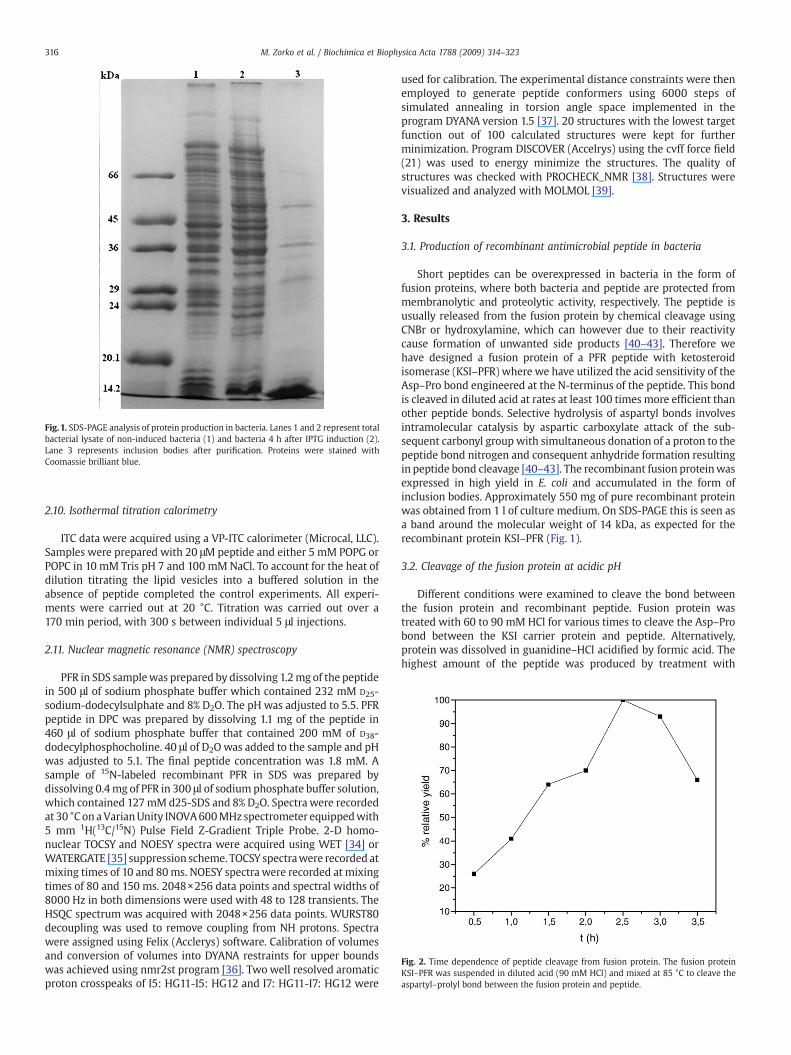
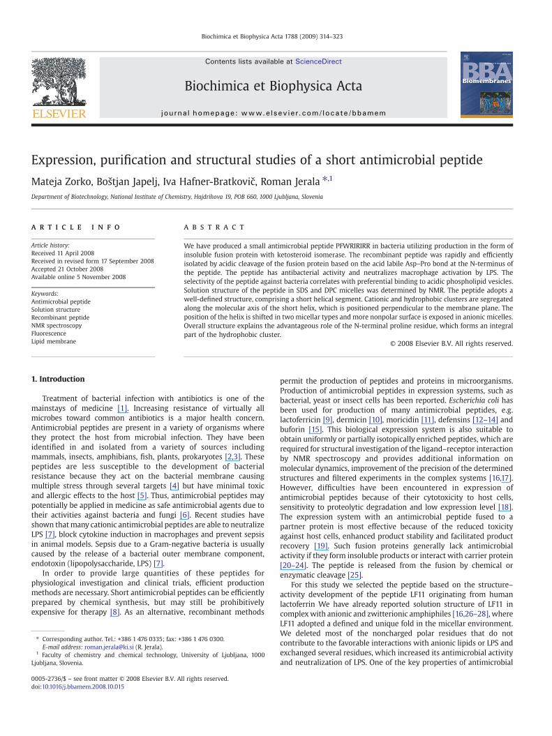
Fig. 1. SDS-PAGE analysis of protein production in bacteria. Lanes 1 and 2 represent totalbacterial lysate of non-induced bacteria (1) and bacteria 4 h after IPTG induction (2).Lane 3 represents inclusion bodies after purification. Proteins were stained withCoomassie brilliant blue.
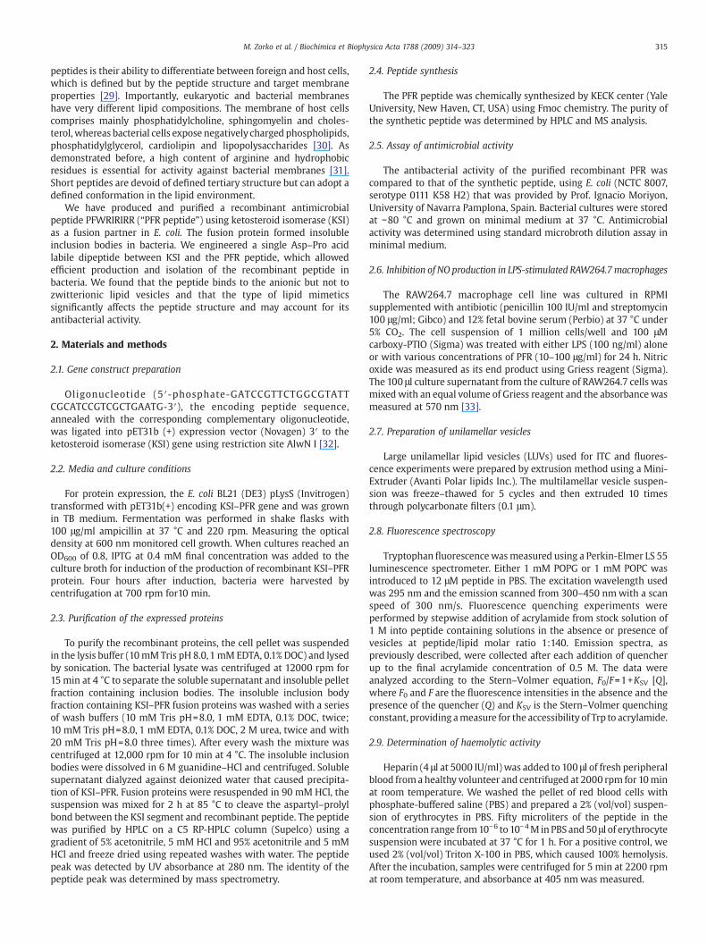
Fig. 2. Time dependence of peptide cleavage from fusion protein. The fusion proteinKSI–PFR was suspended in diluted acid (90 mM HCl) and mixed at 85 °C to cleave theaspartyl–prolyl bond between the fusion protein and peptide.
316 M. Zorko et al. / Biochimica et Biophysica Acta 1788 (2009) 314–323
2.10. Isothermal titration calorimetry
ITC data were acquired using a VP-ITC calorimeter (Microcal, LLC).Samples were prepared with 20 μM peptide and either 5 mM POPG orPOPC in 10 mM Tris pH 7 and 100 mMNaCl. To account for the heat ofdilution titrating the lipid vesicles into a buffered solution in theabsence of peptide completed the control experiments. All experi-ments were carried out at 20 °C. Titration was carried out over a170 min period, with 300 s between individual 5 μl injections.
2.11. Nuclear magnetic resonance (NMR) spectroscopy
PFR in SDS samplewas prepared by dissolving 1.2mg of the peptidein 500 μl of sodium phosphate buffer which contained 232 mM D25-sodium-dodecylsulphate and 8% D2O. The pH was adjusted to 5.5. PFRpeptide in DPC was prepared by dissolving 1.1 mg of the peptide in460 μl of sodium phosphate buffer that contained 200 mM of D38-dodecylphosphocholine. 40 μl of D2O was added to the sample and pHwas adjusted to 5.1. The final peptide concentration was 1.8 mM. Asample of 15N-labeled recombinant PFR in SDS was prepared bydissolving 0.4mg of PFR in 300 μl of sodium phosphate buffer solution,which contained 127mMd25-SDS and 8% D2O. Spectra were recordedat 30 °C on aVarianUnity INOVA600MHz spectrometer equippedwith5 mm 1H(13C/15N) Pulse Field Z-Gradient Triple Probe. 2-D homo-nuclear TOCSY and NOESY spectra were acquired using WET [34] orWATERGATE [35] suppression scheme. TOCSY spectrawere recorded atmixing times of 10 and 80ms. NOESY spectra were recorded at mixingtimes of 80 and 150 ms. 2048×256 data points and spectral widths of8000 Hz in both dimensions were used with 48 to 128 transients. TheHSQC spectrum was acquired with 2048×256 data points. WURST80decoupling was used to remove coupling from NH protons. Spectrawere assigned using Felix (Acclerys) software. Calibration of volumesand conversion of volumes into DYANA restraints for upper boundswas achieved using nmr2st program [36]. Two well resolved aromaticproton crosspeaks of I5: HG11-I5: HG12 and I7: HG11-I7: HG12 were
used for calibration. The experimental distance constraints were thenemployed to generate peptide conformers using 6000 steps ofsimulated annealing in torsion angle space implemented in theprogram DYANA version 1.5 [37]. 20 structures with the lowest targetfunction out of 100 calculated structures were kept for furtherminimization. Program DISCOVER (Accelrys) using the cvff force field(21) was used to energy minimize the structures. The quality ofstructures was checked with PROCHECK_NMR [38]. Structures werevisualized and analyzed with MOLMOL [39].
3. Results
3.1. Production of recombinant antimicrobial peptide in bacteria
Short peptides can be overexpressed in bacteria in the form offusion proteins, where both bacteria and peptide are protected frommembranolytic and proteolytic activity, respectively. The peptide isusually released from the fusion protein by chemical cleavage usingCNBr or hydroxylamine, which can however due to their reactivitycause formation of unwanted side products [40–43]. Therefore wehave designed a fusion protein of a PFR peptide with ketosteroidisomerase (KSI–PFR) where we have utilized the acid sensitivity of theAsp–Pro bond engineered at the N-terminus of the peptide. This bondis cleaved in diluted acid at rates at least 100 times more efficient thanother peptide bonds. Selective hydrolysis of aspartyl bonds involvesintramolecular catalysis by aspartic carboxylate attack of the sub-sequent carbonyl group with simultaneous donation of a proton to thepeptide bond nitrogen and consequent anhydride formation resultingin peptide bond cleavage [40–43]. The recombinant fusion proteinwasexpressed in high yield in E. coli and accumulated in the form ofinclusion bodies. Approximately 550 mg of pure recombinant proteinwas obtained from 1 l of culture medium. On SDS-PAGE this is seen asa band around the molecular weight of 14 kDa, as expected for therecombinant protein KSI–PFR (Fig. 1).
3.2. Cleavage of the fusion protein at acidic pH
Different conditions were examined to cleave the bond betweenthe fusion protein and recombinant peptide. Fusion protein wastreated with 60 to 90 mM HCl for various times to cleave the Asp–Probond between the KSI carrier protein and peptide. Alternatively,protein was dissolved in guanidine–HCl acidified by formic acid. Thehighest amount of the peptide was produced by treatment with
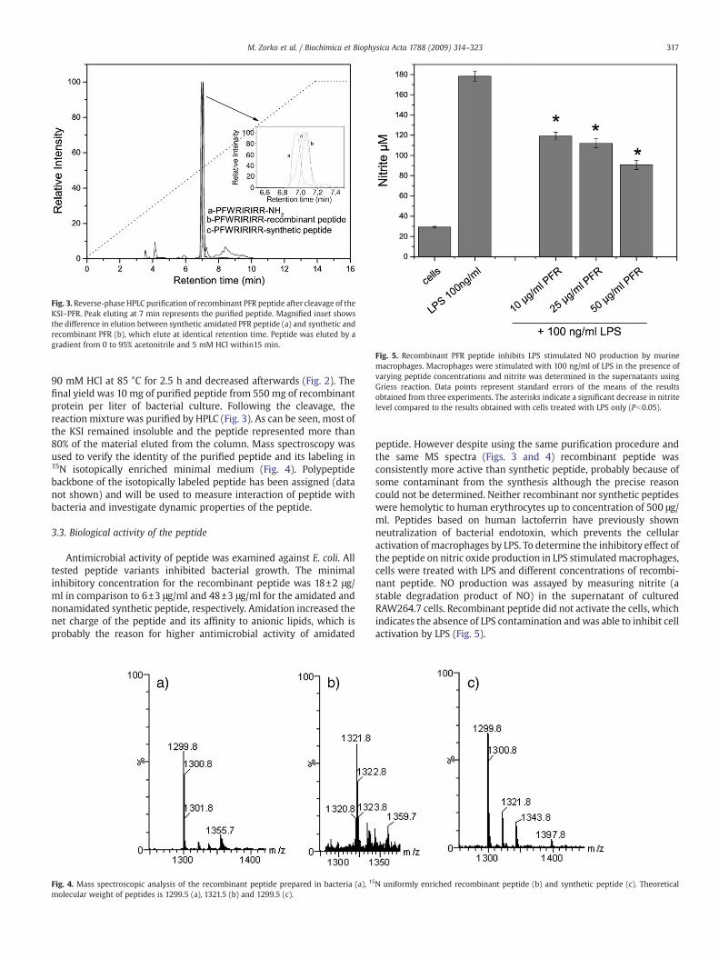
Fig. 3. Reverse-phase HPLC purification of recombinant PFR peptide after cleavage of theKSI–PFR. Peak eluting at 7 min represents the purified peptide. Magnified inset showsthe difference in elution between synthetic amidated PFR peptide (a) and synthetic andrecombinant PFR (b), which elute at identical retention time. Peptide was eluted by agradient from 0 to 95% acetonitrile and 5 mM HCl within15 min.
Fig. 5. Recombinant PFR peptide inhibits LPS stimulated NO production by murinemacrophages. Macrophages were stimulated with 100 ng/ml of LPS in the presence ofvarying peptide concentrations and nitrite was determined in the supernatants usingGriess reaction. Data points represent standard errors of the means of the resultsobtained from three experiments. The asterisks indicate a significant decrease in nitritelevel compared to the results obtained with cells treated with LPS only (Pb0.05).
317M. Zorko et al. / Biochimica et Biophysica Acta 1788 (2009) 314–323
90 mM HCl at 85 °C for 2.5 h and decreased afterwards (Fig. 2). Thefinal yield was 10 mg of purified peptide from 550 mg of recombinantprotein per liter of bacterial culture. Following the cleavage, thereaction mixture was purified by HPLC (Fig. 3). As can be seen, most ofthe KSI remained insoluble and the peptide represented more than80% of the material eluted from the column. Mass spectroscopy wasused to verify the identity of the purified peptide and its labeling in15N isotopically enriched minimal medium (Fig. 4). Polypeptidebackbone of the isotopically labeled peptide has been assigned (datanot shown) and will be used to measure interaction of peptide withbacteria and investigate dynamic properties of the peptide.
3.3. Biological activity of the peptide
Antimicrobial activity of peptide was examined against E. coli. Alltested peptide variants inhibited bacterial growth. The minimalinhibitory concentration for the recombinant peptide was 18±2 μg/ml in comparison to 6±3 μg/ml and 48±3 μg/ml for the amidated andnonamidated synthetic peptide, respectively. Amidation increased thenet charge of the peptide and its affinity to anionic lipids, which isprobably the reason for higher antimicrobial activity of amidated
Fig. 4. Mass spectroscopic analysis of the recombinant peptide prepared in bacteria (a), 15
molecular weight of peptides is 1299.5 (a), 1321.5 (b) and 1299.5 (c).
peptide. However despite using the same purification procedure andthe same MS spectra (Figs. 3 and 4) recombinant peptide wasconsistently more active than synthetic peptide, probably because ofsome contaminant from the synthesis although the precise reasoncould not be determined. Neither recombinant nor synthetic peptideswere hemolytic to human erythrocytes up to concentration of 500 μg/ml. Peptides based on human lactoferrin have previously shownneutralization of bacterial endotoxin, which prevents the cellularactivation of macrophages by LPS. To determine the inhibitory effect ofthe peptide on nitric oxide production in LPS stimulatedmacrophages,cells were treated with LPS and different concentrations of recombi-nant peptide. NO production was assayed by measuring nitrite (astable degradation product of NO) in the supernatant of culturedRAW264.7 cells. Recombinant peptide did not activate the cells, whichindicates the absence of LPS contamination andwas able to inhibit cellactivation by LPS (Fig. 5).
N uniformly enriched recombinant peptide (b) and synthetic peptide (c). Theoretical
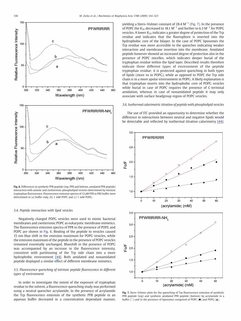
Fig. 6. Differences in synthetic PFR peptide (top, PFR and bottom, amidated PFR peptide)interactionwith anionic and zwitterionic phospholipid vesicles determined by intrinsictryptophan fluorescence. Fluorescence emission spectra of 12 μMPFR in PBS buffer weredetermined in (a) buffer only, (b) 1 mM POPC and (c) 1 mM POPG.
Fig. 7. Stern–Volmer plots for the quenching of Trp fluorescence emission of syntheticPFR peptide (top) and synthetic amidated PFR peptide (bottom) by acrylamide in abuffer (□) and in the presence of liposomes composed of POPC (●) and POPG (▲).
318 M. Zorko et al. / Biochimica et Biophysica Acta 1788 (2009) 314–323
3.4. Peptide interaction with lipid vesicles
Negatively charged POPG vesicles were used to mimic bacterialmembranes and zwitterionic POPC as eukaryotic membranemimetics.The fluorescence emission spectra of PFR in the presence of POPG andPOPC are shown in Fig. 6. Binding of the peptide to vesicles caused15 nm blue shift in the emission maximum for POPG vesicles, whilethe emissionmaximumof the peptide in the presence of POPC vesiclesremained essentially unchanged. Blueshift in the presence of POPGwas accompanied by an increase in the fluorescence intensity,consistent with partitioning of the Trp side chain into a morehydrophobic environment [44]. Both amidated and nonamidatedpeptide displayed a similar effect of different membrane mimetics.
3.5. Fluorescence quenching of intrinsic peptide fluorescence in differenttypes of environment
In order to investigate the extent of the exposure of tryptophanresidue to the solvent, a fluorescence-quenching study was performedusing a neutral quencher acrylamide. In the presence of acrylamidethe Trp fluorescence emission of the synthetic PFR peptide in anaqueous buffer decreased in a concentration dependant manner,
yielding a Stern–Volmer constant of 28.4 M−1 (Fig. 7). In the presenceof POPC the KSV decreased to 18.1 M−1 and further to 6.3 M−1 for POPGvesicles. A lower KSV indicates a greater degree of protection of the Trpresidue and indicates that the fluorophore is inserted into thehydrophobic core of the bilayer. In the case of POPC liposomes theTrp residue was more accessible to the quencher indicating weakerinteraction and membrane insertion into the membrane. Amidatedpeptide however showed an increased degree of protection also in thepresence of POPC micelles, which indicates deeper burial of thetryptophan residue within the lipid layer. Described results thereforeindicate three different types of environment of the peptidetryptophan residue: it is protected against quenching in both typesof lipids (more so in POPG), while as opposed to POPC the Trp sidechain is in a more apolar environment in POPG. A likely explanation isthat tryptophan inserts into the hydrophobic core of POPG vesicleswhile burial in case of POPC requires the presence of C-terminalamidation, whereas in case of nonamidated peptide it may onlyassociate with surface headgroup region of POPC vesicles.
3.6. Isothermal calorimetric titration of peptidewith phospholipid vesicles
The use of ITC provided an opportunity to determine whether thedifference in interactions between neutral and negative lipids wouldbe detectable and reflected by isothermal titration calorimetry [44].
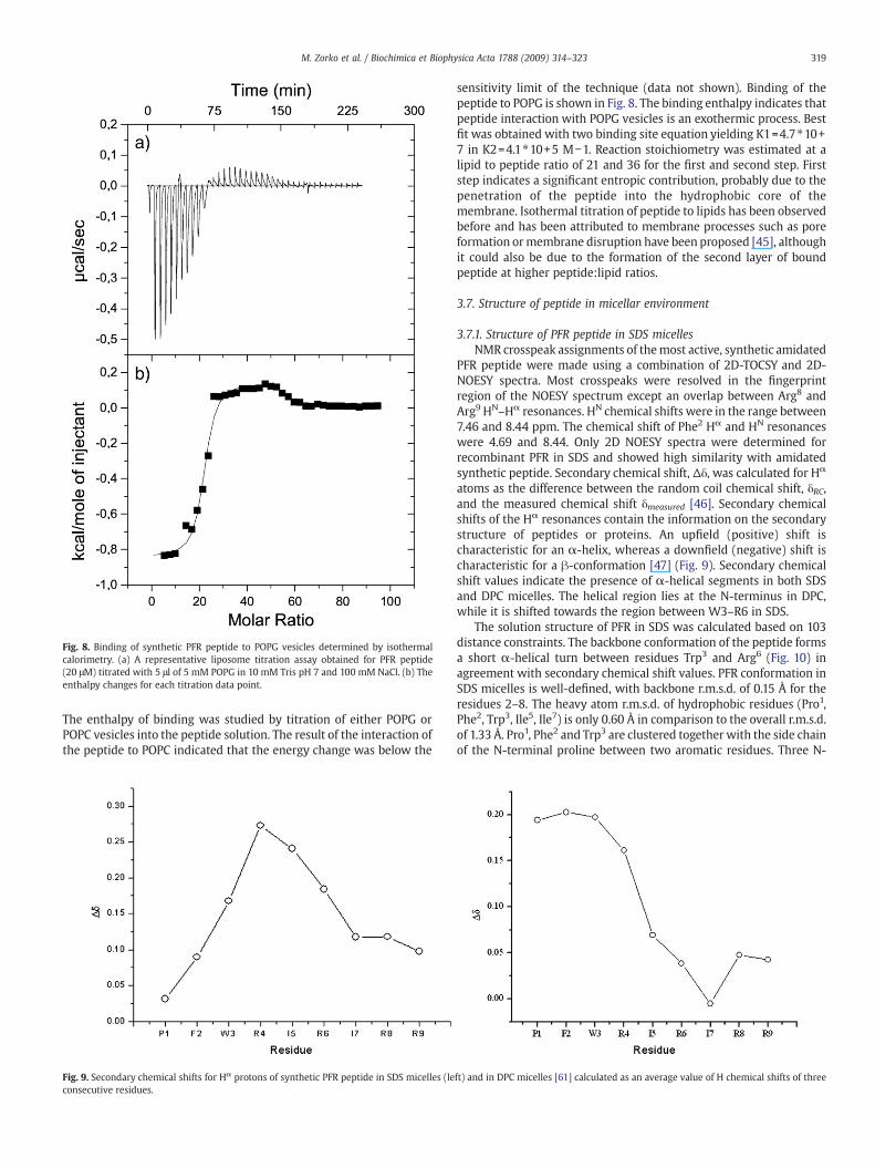
Fig. 8. Binding of synthetic PFR peptide to POPG vesicles determined by isothermalcalorimetry. (a) A representative liposome titration assay obtained for PFR peptide(20 μM) titrated with 5 μl of 5 mM POPG in 10 mM Tris pH 7 and 100 mM NaCl. (b) Theenthalpy changes for each titration data point.
319M. Zorko et al. / Biochimica et Biophysica Acta 1788 (2009) 314–323
The enthalpy of binding was studied by titration of either POPG orPOPC vesicles into the peptide solution. The result of the interaction ofthe peptide to POPC indicated that the energy change was below the
Fig. 9. Secondary chemical shifts for Hα protons of synthetic PFR peptide in SDS micelles (leconsecutive residues.
sensitivity limit of the technique (data not shown). Binding of thepeptide to POPG is shown in Fig. 8. The binding enthalpy indicates thatpeptide interaction with POPG vesicles is an exothermic process. Bestfit was obtained with two binding site equation yielding K1=4.7⁎10+7 in K2=4.1⁎10+5 M−1. Reaction stoichiometry was estimated at alipid to peptide ratio of 21 and 36 for the first and second step. Firststep indicates a significant entropic contribution, probably due to thepenetration of the peptide into the hydrophobic core of themembrane. Isothermal titration of peptide to lipids has been observedbefore and has been attributed to membrane processes such as poreformation ormembrane disruption have been proposed [45], althoughit could also be due to the formation of the second layer of boundpeptide at higher peptide:lipid ratios.
3.7. Structure of peptide in micellar environment
3.7.1. Structure of PFR peptide in SDS micellesNMR crosspeak assignments of themost active, synthetic amidated
PFR peptide were made using a combination of 2D-TOCSY and 2D-NOESY spectra. Most crosspeaks were resolved in the fingerprintregion of the NOESY spectrum except an overlap between Arg8 andArg9 HN–Hα resonances. HN chemical shifts were in the range between7.46 and 8.44 ppm. The chemical shift of Phe2 Hα and HN resonanceswere 4.69 and 8.44. Only 2D NOESY spectra were determined forrecombinant PFR in SDS and showed high similarity with amidatedsynthetic peptide. Secondary chemical shift, Δδ, was calculated for Hα
atoms as the difference between the random coil chemical shift, δRC,and the measured chemical shift δmeasured [46]. Secondary chemicalshifts of the Hα resonances contain the information on the secondarystructure of peptides or proteins. An upfield (positive) shift ischaracteristic for an α-helix, whereas a downfield (negative) shift ischaracteristic for a β-conformation [47] (Fig. 9). Secondary chemicalshift values indicate the presence of α-helical segments in both SDSand DPC micelles. The helical region lies at the N-terminus in DPC,while it is shifted towards the region between W3–R6 in SDS.
The solution structure of PFR in SDS was calculated based on 103distance constraints. The backbone conformation of the peptide formsa short α-helical turn between residues Trp3 and Arg6 (Fig. 10) inagreement with secondary chemical shift values. PFR conformation inSDS micelles is well-defined, with backbone r.m.s.d. of 0.15 Å for theresidues 2–8. The heavy atom r.m.s.d. of hydrophobic residues (Pro1,Phe2, Trp3, Ile5, Ile7) is only 0.60 Å in comparison to the overall r.m.s.d.of 1.33 Å. Pro1, Phe2 and Trp3 are clustered together with the side chainof the N-terminal proline between two aromatic residues. Three N-
ft) and in DPC micelles [61] calculated as an average value of H chemical shifts of three
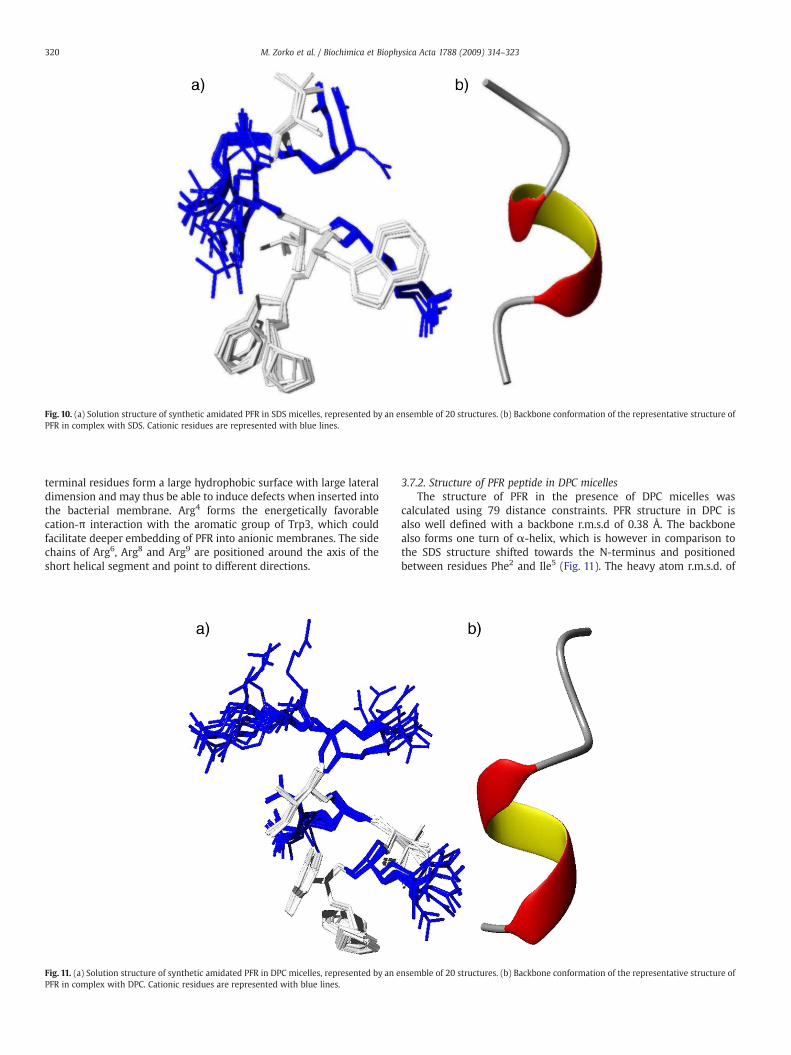
Fig. 10. (a) Solution structure of synthetic amidated PFR in SDS micelles, represented by an ensemble of 20 structures. (b) Backbone conformation of the representative structure ofPFR in complex with SDS. Cationic residues are represented with blue lines.
320 M. Zorko et al. / Biochimica et Biophysica Acta 1788 (2009) 314–323
terminal residues form a large hydrophobic surface with large lateraldimension and may thus be able to induce defects when inserted intothe bacterial membrane. Arg4 forms the energetically favorablecation-π interaction with the aromatic group of Trp3, which couldfacilitate deeper embedding of PFR into anionic membranes. The sidechains of Arg6, Arg8 and Arg9 are positioned around the axis of theshort helical segment and point to different directions.
Fig. 11. (a) Solution structure of synthetic amidated PFR in DPC micelles, represented by an ePFR in complex with DPC. Cationic residues are represented with blue lines.
3.7.2. Structure of PFR peptide in DPC micellesThe structure of PFR in the presence of DPC micelles was
calculated using 79 distance constraints. PFR structure in DPC isalso well defined with a backbone r.m.s.d of 0.38 Å. The backbonealso forms one turn of α-helix, which is however in comparison tothe SDS structure shifted towards the N-terminus and positionedbetween residues Phe2 and Ile5 (Fig. 11). The heavy atom r.m.s.d. of
nsemble of 20 structures. (b) Backbone conformation of the representative structure of
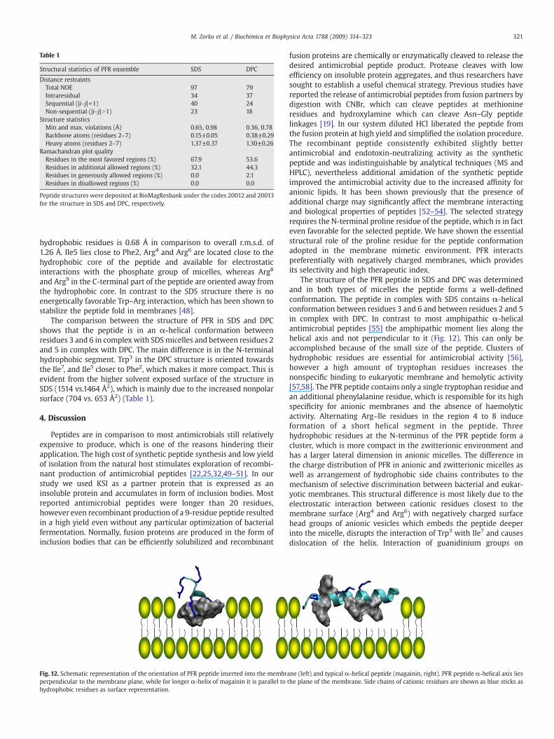
Table 1
Structural statistics of PFR ensemble SDS DPC
Distance restraintsTotal NOE 97 79Intraresidual 34 37Sequential (|i–j|=1) 40 24Non-sequential (|i–j|N1) 23 18
Structure statisticsMin and max. violations (Å) 0.65, 0.98 0.36, 0.78Backbone atoms (residues 2–7) 0.15±0.05 0.38±0.29Heavy atoms (residues 2–7) 1.37±0.37 1.30±0.26
Ramachandran plot qualityResidues in the most favored regions (%) 67.9 53.6Residues in additional allowed regions (%) 32.1 44.3Residues in generously allowed regions (%) 0.0 2.1Residues in disallowed regions (%) 0.0 0.0
Peptide structures were deposited at BioMagResbank under the codes 20012 and 20013for the structure in SDS and DPC, respectively.
321M. Zorko et al. / Biochimica et Biophysica Acta 1788 (2009) 314–323
hydrophobic residues is 0.68 Å in comparison to overall r.m.s.d. of1.26 Å. Ile5 lies close to Phe2, Arg4 and Arg6 are located close to thehydrophobic core of the peptide and available for electrostaticinteractions with the phosphate group of micelles, whereas Arg8
and Arg9 in the C-terminal part of the peptide are oriented away fromthe hydrophobic core. In contrast to the SDS structure there is noenergetically favorable Trp–Arg interaction, which has been shown tostabilize the peptide fold in membranes [48].
The comparison between the structure of PFR in SDS and DPCshows that the peptide is in an α-helical conformation betweenresidues 3 and 6 in complex with SDSmicelles and between residues 2and 5 in complex with DPC. The main difference is in the N-terminalhydrophobic segment. Trp3 in the DPC structure is oriented towardsthe Ile7, and Ile5 closer to Phe2, which makes it more compact. This isevident from the higher solvent exposed surface of the structure inSDS (1514 vs.1464 Å2), which is mainly due to the increased nonpolarsurface (704 vs. 653 Å2) (Table 1).
4. Discussion
Peptides are in comparison to most antimicrobials still relativelyexpensive to produce, which is one of the reasons hindering theirapplication. The high cost of synthetic peptide synthesis and low yieldof isolation from the natural host stimulates exploration of recombi-nant production of antimicrobial peptides [22,25,32,49–51]. In ourstudy we used KSI as a partner protein that is expressed as aninsoluble protein and accumulates in form of inclusion bodies. Mostreported antimicrobial peptides were longer than 20 residues,however even recombinant production of a 9-residue peptide resultedin a high yield even without any particular optimization of bacterialfermentation. Normally, fusion proteins are produced in the form ofinclusion bodies that can be efficiently solubilized and recombinant
Fig. 12. Schematic representation of the orientation of PFR peptide inserted into the membrperpendicular to the membrane plane, while for longer α-helix of magainin it is parallel tohydrophobic residues as surface representation.
fusion proteins are chemically or enzymatically cleaved to release thedesired antimicrobial peptide product. Protease cleaves with lowefficiency on insoluble protein aggregates, and thus researchers havesought to establish a useful chemical strategy. Previous studies havereported the release of antimicrobial peptides from fusion partners bydigestion with CNBr, which can cleave peptides at methionineresidues and hydroxylamine which can cleave Asn–Gly peptidelinkages [19]. In our system diluted HCl liberated the peptide fromthe fusion protein at high yield and simplified the isolation procedure.The recombinant peptide consistently exhibited slightly betterantimicrobial and endotoxin-neutralizing activity as the syntheticpeptide and was indistinguishable by analytical techniques (MS andHPLC), nevertheless additional amidation of the synthetic peptideimproved the antimicrobial activity due to the increased affinity foranionic lipids. It has been shown previously that the presence ofadditional charge may significantly affect the membrane interactingand biological properties of peptides [52–54]. The selected strategyrequires the N-terminal proline residue of the peptide, which is in facteven favorable for the selected peptide. We have shown the essentialstructural role of the proline residue for the peptide conformationadopted in the membrane mimetic environment. PFR interactspreferentially with negatively charged membranes, which providesits selectivity and high therapeutic index.
The structure of the PFR peptide in SDS and DPC was determinedand in both types of micelles the peptide forms a well-definedconformation. The peptide in complex with SDS contains α-helicalconformation between residues 3 and 6 and between residues 2 and 5in complex with DPC. In contrast to most amphipathic α-helicalantimicrobial peptides [55] the amphipathic moment lies along thehelical axis and not perpendicular to it (Fig. 12). This can only beaccomplished because of the small size of the peptide. Clusters ofhydrophobic residues are essential for antimicrobial activity [56],however a high amount of tryptophan residues increases thenonspecific binding to eukaryotic membrane and hemolytic activity[57,58]. The PFR peptide contains only a single tryptophan residue andan additional phenylalanine residue, which is responsible for its highspecificity for anionic membranes and the absence of haemolyticactivity. Alternating Arg–Ile residues in the region 4 to 8 induceformation of a short helical segment in the peptide. Threehydrophobic residues at the N-terminus of the PFR peptide form acluster, which is more compact in the zwitterionic environment andhas a larger lateral dimension in anionic micelles. The difference inthe charge distribution of PFR in anionic and zwitterionic micelles aswell as arrangement of hydrophobic side chains contributes to themechanism of selective discrimination between bacterial and eukar-yotic membranes. This structural difference is most likely due to theelectrostatic interaction between cationic residues closest to themembrane surface (Arg4 and Arg6) with negatively charged surfacehead groups of anionic vesicles which embeds the peptide deeperinto the micelle, disrupts the interaction of Trp3 with Ile7 and causesdislocation of the helix. Interaction of guanidinium groups on
ane (left) and typical α-helical peptide (magainin, right). PFR peptide α-helical axis liesthe plane of the membrane. Side chains of cationic residues are shown as blue sticks as
322 M. Zorko et al. / Biochimica et Biophysica Acta 1788 (2009) 314–323
arginine residues with phosphate groups of phospholipids has beendetermined before for PG-1 by solid state NMR and proposed tocontribute to the formation of membrane defects [59]. N-terminalproline, which was introduced because of the recombinant peptideproduction approach was essential for the integrity of the structureand improved the antimicrobial activity. Proline forms an essentialpart of the hydrophobic cluster, with many side chain contacts withside chains of Phe2 or Trp3. The N-terminal hydrophobic cluster of thePFR peptide has similar function as the short acyl chain of thelipopeptide [60]. The structures in this report can be used to improvethe design of more selective peptides by enhancing the differences inlocal conformational changes caused in zwitterionic and anionicmembranes.
Acknowledgements
Wewould like to thank Dr. Karl Lohner, Dr. Sylvie Blondelle, Dr. JörgAndrä, Dr. Klaus Brandenburg, Dr. Guillermo Martinez de Tejada, andDr. Ignacio Moriyon for their collaboration and discussions within theANEPID project developing the more effective antimicrobial peptidesequences. Project was funded by the Slovenian Research Agency. Wewould like to thank Mireille Treeby Premuš for careful reading of themanuscript.
References
[1] K.A. Brogden, Antimicrobial peptides: pore formers or metabolic inhibitors inbacteria? Nat. Rev., Microbiol. 3 (2005) 238–250.
[2] H.G. Boman, Peptide antibiotics and their role in innate immunity, Annu. Rev.Immunol. 13 (1995) 61–92.
[3] R.E.W. Hancock, R. Lehrer, Cationic peptides: a new source of antibiotics, TrendsBiotechnol. 16 (1998) 82–88.
[4] U. Pag, M. Oedenkoven, V. Sass, Y. Shai, O. Shamova, N. Antcheva, A. Tossi, H.G.Sahl, Analysis of in vitro activities and modes of action of synthetic antimicrobialpeptides derived from an alpha-helical ‘sequence template’, J. Antimicrob.Chemother. 61 (2008) 341–352.
[5] R.I. Lehrer, A.K. Lichtenstein, T. Ganz, Defensins — antimicrobial and cytotoxicpeptides of mammalian-cells, Annu. Rev. Immunol. 11 (1993) 105–128.
[6] K. Hilpert, R. Volkmer-Engert, T. Walter, R.E.W. Hancock, High-throughputgeneration of small antibacterial peptides with improved activity, Nat. Biotechnol.23 (2005) 1008–1012.
[7] Y. Rosenfeld, N. Papo, Y. Shai, Endotoxin (lipopolysaccharide) neutralization byinnate immunity host-defense peptides— peptide properties and plausiblemodesof action, J. Biol. Chem. 281 (2006) 1636–1643.
[8] C.B. Park, H.S. Kim, S.C. Kim, Mechanism of action of the antimicrobial peptidebuforin II: buforin II kills microorganisms by penetrating the cell membrane andinhibiting cellular functions, Biochem. Biophys. Res. Commun. 244 (1998)253–257.
[9] H.K. Kim, D.S. Chun, J.S. Kim, C.H. Yun, J.H. Lee, S.K. Hong, D.K. Kang, Expression ofthe cationic antimicrobial peptide lactoferricin fused with the anionic peptide inEscherichia coli, Appl. Microbiol. Biotechnol. 72 (2006) 330–338.
[10] I. Cipakova, J. Gasperik, E. Hostinova, Expression and purification of humanantimicrobial peptide, dermcidin, in Escherichia coli, Protein Expression Purif. 45(2006) 269–274.
[11] S. Hara, M. Yamakawa, Production in Escherichia coli of moricin, a novel typeantibacterial peptide from the silkworm, Bombyx mori (vol 220, pg 664, 1996),Biochem. Biophys. Res. Commun. 224 (1996) 877–878.
[12] I. Cipakova, E. Hostinova, J.G. Gasperik, V. Velebny, High-level expression andpurification of a recombinant hBD-1 fused to LMM protein in Escherichia coli,Protein Expression Purif. 37 (2004) 207–212.
[13] Z.N. Xu, L. Peng, Z.X. Zhong, X.M. Fang, P.L. Cen, High-level expression of a solublefunctional antimicrobial peptide, human beta-defensin 2, in Escherichia coli,Biotechnol. Prog. 22 (2006) 382–386.
[14] Z. Xu, Z.X. Zhong, L. Huang, L. Peng, F. Wang, P. Cen, High-level production ofbioactive human beta-defensin-4 in Escherichia coli by soluble fusion expression,Appl. Microbiol. Biotechnol. 72 (2006) 471–479.
[15] J.H. Lee, I. Minn, C.B. Park, S.C. Kim, Acidic peptide-mediated expression of theantimicrobial peptide buforin II as tandem repeats in Escherichia coli, ProteinExpression Purif. 12 (1998) 53–60.
[16] A. Majerle, J. Kidric, R. Jerala, Production of stable isotope enriched antimicrobialpeptides in Escherichia coli: an application to the production of a N-15-enrichedfragment of lactoferrin, J. Biomol. NMR 18 (2000) 145–151.
[17] T.T. Mac, M. Beyermann, J.R. Pires, P. Schmieder, H. Oschkinat, High yieldexpression and purification of isotopically labelled human endothelin-1 for usein NMR studies, Protein Expression Purif. 48 (2006) 253–260.
[18] F.L. Jin, X.X. Xu,W.Q. Zhang, D.X. Gu, Expression and characterization of a houseflycecropin gene in the methylotrophic yeast, Pichia pastoris, Protein ExpressionPurif. 49 (2006) 39–46.
[19] Q.D. Wei, Y.S. Kim, J.H. Seo, W.S. Jang, I.H. Lee, H.J. Cha, Facilitation of expressionand purification of an antimicrobial peptide by fusionwith baculoviral polyhedrinin Escherichia coli, Appl. Environ. Microbiol. 71 (2005) 5038–5043.
[20] J.Y. Moon, K.A. Henzler-Wildman, A. Ramamoorthy, Expression and purification ofa recombinant LL-37 from Escherichia coli, Biochim. Biophys. Acta-Biomembr. 1758(2006) 1351–1358.
[21] X.X. Xu, F.L. Jin, X.Q. Yu, S.X. Ren, J. Hu, W.Q. Zhang, High-level expression of therecombinant hybrid peptide cecropinA(1–8)–magainin2(1–12) with an ubiquitinfusion partner in Escherichia coli, Protein Expression Purif. 55 (2007) 175–182.
[22] X.X. Xu, F.L. Jin, X.Q. Yu, S.X. Ji, J. Wang, H.X. Cheng, C. Wang, W.Q. Zhang,Expression and purification of a recombinant antibacterial peptide, cecropin, fromEscherichia coli, Protein Expression Purif. 53 (2007) 293–301.
[23] S.H. Pyo, J.H. Lee, H.B. Park, J.S. Cho, H.R. Kim, B.H. Han, Y.S. Park, Expression andpurification of a recombinant buforin derivative from Escherichia coli, ProcessBiochem. 39 (2004) 1731–1736.
[24] Y. Shen, X.G. Lao, Y. Chen, H.Z. Zhang, X.X. Xu, High-level expression of cecropin Xin Escherichia coli, Int. J. Mol. Sci. 8 (2007) 478–491.
[25] A.B. Ingham, R.J. Moore, Recombinant production of antimicrobial peptides inheterologous microbial systems, Biotechnol. Appl. Biochem. 47 (2007) 1–9.
[26] B. Japelj, P. Pristovsek, A. Majerle, R. Jerala, Structural origin of endotoxinneutralization and antimicrobial activity of a lactoferrin-based peptide, J. Biol.Chem. 280 (2005) 16955–16961.
[27] R. Jerala, B. Japelj, P. Pristovsek, A. Majerle, Tertiary structure of lactoferrin peptidein complex with LPS for design of novel endotoxin-neutralizing peptides, Shock 21(2004) 62.
[28] A. Majerle, J. Kidric, R. Jerala, Enhancement of antibacterial and lipopolysaccharidebinding activities of a human lactoferrin peptide fragment by the addition of acylchain, J. Antimicrob. Chemother. 51 (2003) 1159–1165.
[29] N. Papo, Y. Shai, Canwepredict biological activity of antimicrobial peptides from theirinteractions with model phospholipid membranes? Peptides 24 (2003) 1693–1703.
[30] R. Sood, Y. Domanov, P.K.J. Kinnunen, Fluorescent temporin B derivative and itsbinding to liposomes, J. Fluoresc. 17 (2007) 223–234.
[31] D.I. Chan, E.J. Prenner, H.J. Vogel, Tryptophan- and arginine-rich antimicrobialpeptides: structures and mechanisms of action, Biochim. Biophys. Acta 1758(2006) 1184–1202.
[32] A. Kuliopulos, C.T. Walsh, Production, purification, and cleavage of tandem repeatsof recombinant peptides, J. Am. Chem. Soc. 116 (1994) 4599–4607.
[33] P.N. Yadav, Z.H. Liu, M.M. Rafi, A diarylheptanoid from lesser galangal (Alpiniaofficinarum) inhibits proinflammatory mediators via inhibition of mitogen-activated protein kinase, p44/42, and transcription factor nuclear factor-kappaB, J. Pharmacol. Exp. Ther. 305 (2003) 925–931.
[34] S.H. Smallcombe, S.L. Patt, P.A. Keifer, WET solvent suppression and itsapplications to LC NMR and high-resolution NMR spectroscopy. J. Magn. Reson.,Ser. A 117 (2) (1995) 295–303.
[35] M. Piotto, V. Saudek, V. Sklenar, Gradient-tailored excitation for single-quantumNMR spectroscopy of aqueous solutions, 2 (1992) 661.
[36] P. Pristovsek, H. Ruterjans, R. Jerala, Semiautomatic sequence-specific assignmentof proteins based on the tertiary structure–the program st2nmr, 23 (2002) 335.
[37] P. Guntert, W. Braun, K. Wuthrich, Efficient computation of three-dimensionalprotein structures in solution from nuclear magnetic resonance data using theprogram DIANA and the supporting programs CALIBA, HABAS and GLOMSA, 217(1991) 517.
[38] R.A. Laskowski, J.A. Rullmannn, M.W. MacArthur, R. Kaptein, J.M. Thornton, AQUAand PROCHECK-NMR: programs for checking the quality of protein structuressolved by NMR, 8 (1996) 477.
[39] R. Koradi, M. Billeter, K. Wuthrich, MOLMOL: a program for display and analysis ofmacromolecular structures, 14 (1996) 51.
[40] D. Piszkiew, M. Landon, E.L. Smith, Anomalous cleavage of aspartyl–prolinepeptide bonds during amino acid sequence determinations, Biochem. Biophys.Res. Commun. 40 (1970) 1173–1178.
[41] I. Segalas, R. Thai, R. Menez, C. Vita, A particularly labile Asp–Pro bond in the greenmamba muscarinic toxin Mtx2 — effect of protein conformation on the rate ofcleavage, FEBS Lett. 371 (1995) 171–175.
[42] M.E. Lidell, M.E.V. Johansson, G.C. Hansson, An autocatalytic cleavage in the cterminus of the human MUC2 mucin occurs at the low pH of the late secretorypathway, J. Biol. Chem. 278 (2003) 13944–13951.
[43] E.L. Smith, M. Landon, D. Piszkiew, W.J. Brattin, T.J. Langley, M.D. Melamed, Bovineliver glutamate dehydrogenase — tentative amino acid sequence — Identificationof a reactive lysine— nitration of a specific tyrosine and loss of allosteric inhibitionby guanosine triphosphate, Proc. Natl. Acad. Sci. U. S. A. 67 (1970) 724–730.
[44] H.N. Hunter, W.G. Jing, D.J. Schibli, T. Trinh, I.Y. Park, S.C. Kim, H.J. Vogel, Theinteractions of antimicrobial peptides derived from lysozyme with modelmembrane systems, Biochim. Biophys. Acta-Biomembr. 1668 (2005) 175–189.
[45] R.F. Epand, R.I. Lehrer, A.Waring,W.Wang, R. Maget-Dana, D. Lelievre, R.M. Epand,Direct comparison of membrane interactions of model peptides composed of onlyLeu and Lys residues, Biopolymers 71 (2003) 2–16.
[46] K. Wüthrich, NMR of Proteins and Nucleic Acids, Wiley & Sons, 1986.[47] D.S. Wishart, B.D. Sykes, F.M. Richards, Relationship between nuclear magnetic
resonance chemical shift and protein secondary structure, J. Mol. Biol. 222 (1991)311–333.
[48] W. Jing, J.S. Svendsen, H.J. Vogel, Comparison of NMR structures and model-membrane interactions of 15-residue antimicrobial peptides derived from bovinelactoferricin, Biochem. Cell Biol. 84 (2006) 312–326.
[49] C. Haught, G.D. Davis, R. Subramanian, K.W. Jackson, R.G. Harrison, Recombinantproduction and purification of novel antisense antimicrobial peptide in Escheri-chia coli, Biotechnol. Bioeng. 57 (1998) 55–61.
323M. Zorko et al. / Biochimica et Biophysica Acta 1788 (2009) 314–323
[50] J.C. Pierce, W.L. Maloy, L. Salvador, C.F. Dungan, Recombinant expression of theantimicrobial peptide polyphemusin and its activity against the protozoan oysterpathogen Perkinsus marinus, Mol. Mar. Biol. Biotechnol. 6 (1997) 248–259.
[51] A. Majerle, J. Kidric, R. Jerala, Production of stable isotope enriched antimicrobialpeptides in Escherichia coli: an application to the production of a 15N-enrichedfragment of lactoferrin, J. Biomol. NMR 18 (2000) 145–151.
[52] Y.L. Pan, J.T. Cheng, J. Hale, J. Pan, R.E. Hancock, S.K. Straus, Characterization ofthe structure and membrane interaction of the antimicrobial peptides aurein2.2 and 2.3 from Australian southern bell frogs, Biophys. J. 92 (2007)2854–2864.
[53] L.E. Yandek, A. Pokorny, P.F. Almeida, Small changes in the primary structure oftransportan 10 alter the thermodynamics and kinetics of its interaction withphospholipid vesicles, Biochemistry 47 (2008) 3051–3060.
[54] D.J. Schibli, L.T. Nguyen, S.D. Kernaghan, O. Rekdal, H.J. Vogel, Structure–functionanalysis of tritrpticin analogs: potential relationships between antimicrobialactivities, model membrane interactions, and their micelle-bound NMR struc-tures, Biophys. J. 91 (2006) 4413–4426.
[55] A. Tossi, L. Sandri, A. Giangaspero, Amphipathic, alpha-helical antimicrobialpeptides, Biopolymers 55 (2000) 4–30.
[56] A. Wessolowski, M. Bienert, M. Dathe, Antimicrobial activity of arginine- andtryptophan-rich hexapeptides: the effects of aromatic clusters, D-amino acidsubstitution and cyclization, J. Pept. Res. 64 (2004) 159–169.
[57] Z. Liu, A. Brady, A. Young, B. Rasimick, K. Chen, C. Zhou, N.R. Kallenbach, Lengtheffects in antimicrobial peptides of the (RW)n series, Antimicrob. AgentsChemother. 51 (2007) 597–603.
[58] M.B. Strom, B.E. Haug, O. Rekdal, M.L. Skar, W. Stensen, J.S. Svendsen, Importantstructural features of 15-residue lactoferricin derivatives and methods forimprovement of antimicrobial activity, Biochem. Cell Biol. 80 (2002) 65–74.
[59] M. Tang, A.J. Waring, M. Hong, Phosphate-mediated arginine insertion into lipidmembranes and pore formation by a cationic membrane peptide from solid-stateNMR, J. Am. Chem. Soc. 129 (2007) 11438–11446.
[60] B. Japelj, M. Zorko, A. Majerle, P. Pristovsek, S. Sanchez-Gomez, G. Martinez deTejada, I. Moriyon, S.E. Blondelle, K. Brandenburg, J. Andra, K. Lohner, R. Jerala, Theacyl group as the central element of the structural organization of antimicrobiallipopeptide, J. Am. Chem. Soc. 129 (2007) 1022–1023.
[61] J.A. Mitchell, M.J. Paul-Clark, G.W. Clarke, S.K. McMaster, N. Cartwright, Criticalrole of toll-like receptors and nucleotide oligomerisation domain in the regulationof health and disease, J. Endocrinol. 193 (2007) 323–330.
Related Documents


