Expression patterns of endothelial and inducible nitric oxide synthase isoforms in corpora lutea of pseudopregnant rabbits at different luteal stages C Boiti, D Zampini, G Guelfi, F Paolocci 1 , M Zerani 2 and A Gobbetti 2 Dipartimento di Scienze Biopatologiche Veterinarie, Universita` di Perugia, Via S Costanzo 4, I-06126 Perugia, Italy 1 Istituto Ricerche Miglioramento Piante Foraggere, CNR, Via Madonna Alta 130, Perugia, Italy 2 Dipartimento di Biologia Molecolare, Cellulare e Animale, Universita` di Camerino, Camerino, Italy (Requests for offprints should be addressed to C Boiti; Email: cboiti@unipg.it) Abstract Total activity of nitric oxide (NO) synthase (NOS) and expression of both endothelial (eNOS) and inducible (iNOS) isoforms were examined in corpora lutea (CL) of rabbits across pseudopregnancy by quantitative RT-PCR analysis, Western blot and immunohistochemistry. CL were collected at early- (day 4), mid- (day 9) and late- (day 13) luteal phases of pseudopregnancy. The PCR product of rabbit luteal eNOS was cloned and its direct sequence exhibited 90% homology with those of other species. The steady-state mRNA levels encoding eNOS remained fairly constant throughout both early- and mid-luteal stages of pseudopregnancy but dropped almost to half (P0·05) by day 13. By contrast, luteal eNOS proteins increased 2-fold (P0·05) from the early- to late-luteal phase. Independently of CL age, iNOS mRNA was very poorly expressed while protein levels gradually declined from the early- to late-luteal stage. Intense eNOS-like immunoreactivity was detected in large luteal cells, while iNOS staining was targeted to a few, isolated cells, probably macrophages. Basal NOS activity was greater in day 4 CL than in both day 9 and day 13 CL. These data are the first to characterize in rabbit CL the temporal expression patterns of NOS isoforms across different luteal stages of pseudopregnancy and, collec- tively, suggest the existence of an expressional control for this constitutive isoform, which might have a physiological role in regulating CL function during development. Journal of Endocrinology (2002) 173, 285–296 Introduction Nitric oxide (NO) has emerged as a relevant effector and messenger molecule mediating multiple biological actions in many systems, including vasodilatation, neurotrans- mission and cytotoxicity, under both normal and patho- physiological conditions (Moncada et al. 1991, Snyder & Bredt 1992, Schmidt & Walter 1994). In a variety of mammalian cell types, NO is produced from -arginine by the action of one of three different NO synthase (NOS) isoforms (Förstermann et al. 1995), two being constitutive, the endothelial (eNOS) and the neuronal, and one inducible (iNOS). Following the first demonstration that both eNOS and iNOS isoforms are expressed in the ovary (Van Voorhis et al. 1995, Chatterjee et al. 1996, Zackrisson et al. 1996), an increasing amount of experimental works supports the hypothesis that NO may also have a significant role in the physiology of this organ by controlling several key func- tions such as ovarian steroidogenesis and luteolysis (Van Voorhis et al. 1994, Olson et al. 1996, Motta & Gimeno 1997, Vega et al. 1998, Fridén et al. 2000). The NO/NOS system has also been directly implicated in the ovulatory process as inhibitors of NO reduce the ovulation rate in vivo and in vitro in both rats and rabbits (Shukovski & Tsafriri 1995, Bonello et al. 1996, Hesla et al. 1997, Yamauchi et al. 1997, Faletti et al. 1999, Nakamura et al. 1999). Recent work from our laboratory determined the pres- ence of NOS activity in corpora lutea (CL) of pseudopreg- nant rabbits, where NO was shown to exert a regulatory role on progesterone release in response to both luteolytic and luteotropic factors in vitro (Gobbetti et al. 1999, Boiti et al. 2000). However, the impact of NO on CL function still remains to be better elucidated and several key points on the mechanisms regulating NO production are still unclear (Van Voorhis et al. 1995, Jablonka-Shariff & Olson 1997, Srivastava et al. 1997). Moreover, the specific cell types expressing both eNOS and iNOS isoenzymes have not been detailed in the rabbit CL. 285 Journal of Endocrinology (2002) 173, 285–296 0022–0795/02/0173–285 2002 Society for Endocrinology Printed in Great Britain Online version via http://www.endocrinology.org

Welcome message from author
This document is posted to help you gain knowledge. Please leave a comment to let me know what you think about it! Share it to your friends and learn new things together.
Transcript
Expression patterns of endothelial and inducible nitric oxidesynthase isoforms in corpora lutea of pseudopregnant rabbits atdifferent luteal stages
C Boiti, D Zampini, G Guelfi, F Paolocci1, M Zerani2 andA Gobbetti2
Dipartimento di Scienze Biopatologiche Veterinarie, Universita di Perugia, Via S Costanzo 4, I-06126 Perugia, Italy1Istituto Ricerche Miglioramento Piante Foraggere, CNR, Via Madonna Alta 130, Perugia, Italy2Dipartimento di Biologia Molecolare, Cellulare e Animale, Universita di Camerino, Camerino, Italy
(Requests for offprints should be addressed to C Boiti; Email: [email protected])
Abstract
Total activity of nitric oxide (NO) synthase (NOS) andexpression of both endothelial (eNOS) and inducible(iNOS) isoforms were examined in corpora lutea (CL) ofrabbits across pseudopregnancy by quantitative RT-PCRanalysis, Western blot and immunohistochemistry. CLwere collected at early- (day 4), mid- (day 9) and late- (day13) luteal phases of pseudopregnancy. The PCR productof rabbit luteal eNOS was cloned and its direct sequenceexhibited 90% homology with those of other species. Thesteady-state mRNA levels encoding eNOS remainedfairly constant throughout both early- and mid-lutealstages of pseudopregnancy but dropped almost to half(P�0·05) by day 13. By contrast, luteal eNOS proteinsincreased 2-fold (P�0·05) from the early- to late-luteal
phase. Independently of CL age, iNOS mRNA was verypoorly expressed while protein levels gradually declinedfrom the early- to late-luteal stage. Intense eNOS-likeimmunoreactivity was detected in large luteal cells, whileiNOS staining was targeted to a few, isolated cells,probably macrophages. Basal NOS activity was greater inday 4 CL than in both day 9 and day 13 CL.
These data are the first to characterize in rabbit CL thetemporal expression patterns of NOS isoforms acrossdifferent luteal stages of pseudopregnancy and, collec-tively, suggest the existence of an expressional control forthis constitutive isoform, which might have a physiologicalrole in regulating CL function during development.Journal of Endocrinology (2002) 173, 285–296
Introduction
Nitric oxide (NO) has emerged as a relevant effector andmessenger molecule mediating multiple biological actionsin many systems, including vasodilatation, neurotrans-mission and cytotoxicity, under both normal and patho-physiological conditions (Moncada et al. 1991, Snyder &Bredt 1992, Schmidt & Walter 1994). In a variety ofmammalian cell types, NO is produced from -arginine bythe action of one of three different NO synthase (NOS)isoforms (Förstermann et al. 1995), two being constitutive,the endothelial (eNOS) and the neuronal, and oneinducible (iNOS).
Following the first demonstration that both eNOS andiNOS isoforms are expressed in the ovary (Van Voorhiset al. 1995, Chatterjee et al. 1996, Zackrisson et al. 1996),an increasing amount of experimental works supports thehypothesis that NO may also have a significant role in thephysiology of this organ by controlling several key func-tions such as ovarian steroidogenesis and luteolysis (Van
Voorhis et al. 1994, Olson et al. 1996, Motta & Gimeno1997, Vega et al. 1998, Fridén et al. 2000). The NO/NOSsystem has also been directly implicated in the ovulatoryprocess as inhibitors of NO reduce the ovulation rate invivo and in vitro in both rats and rabbits (Shukovski &Tsafriri 1995, Bonello et al. 1996, Hesla et al. 1997,Yamauchi et al. 1997, Faletti et al. 1999, Nakamura et al.1999).
Recent work from our laboratory determined the pres-ence of NOS activity in corpora lutea (CL) of pseudopreg-nant rabbits, where NO was shown to exert a regulatoryrole on progesterone release in response to both luteolyticand luteotropic factors in vitro (Gobbetti et al. 1999, Boitiet al. 2000). However, the impact of NO on CL functionstill remains to be better elucidated and several key pointson the mechanisms regulating NO production are stillunclear (Van Voorhis et al. 1995, Jablonka-Shariff & Olson1997, Srivastava et al. 1997). Moreover, the specific celltypes expressing both eNOS and iNOS isoenzymes havenot been detailed in the rabbit CL.
285
Journal of Endocrinology (2002) 173, 285–2960022–0795/02/0173–285 � 2002 Society for Endocrinology Printed in Great Britain
Online version via http://www.endocrinology.org
With this in view, the main objective was to charac-terize the expression patterns of both eNOS and iNOS inCL of rabbits in vivo across pseudopregnancy at bothcellular, gene transcript and protein level by means ofimmunohistochemistry, RT-PCR analysis and immuno-blotting. For this work, CL were collected in early-,mid- and late-luteal phases at days 4, 9 and 13 ofpseudopregnancy respectively. These stages were pur-posely selected because the corresponding CL have beenextensively characterized in our laboratory by in vitromodels (Gobbetti et al. 1999, Boiti et al. 2000, 2001) andbecause they show selective age-dependent responsivenessto many luteolytic and luteotropic factors (Miller & Keyes1978, Boiti et al. 1998, Niswender et al. 2000).
Materials and Methods
Animals, hormonal regimen and luteal tissue collectionLuteal tissue was obtained from unmated New ZealandWhite (HY/CR strain) rabbits (Charles River Italia,Lecco, Italy) of 5 months of age, weighing 3·5–3·8 kg.The animals were maintained at the University of PerugiaCentral Animal Facility under controlled conditions oflight (14 h light:10 h darkness) and temperature (18 �C),with freely fed water and commercial pellets. All rabbitsreceived an i.m. injection of 20 IU of pregnant mareserum gonadotropin (Folligon; Intervet, Milan, Italy) fol-lowed 3 days later by an i.m. injection of 0·8 µg ofgonadotropin-releasing hormone (GnRH) (Receptal;Hoechst-Roussel Vet, Milan, Italy) to induce pseudopreg-nancy. This ovulation procedure was effective in generat-ing an average of 14 or 15 CL per rabbit. The day ofGnRH injection was designated day 0. Rabbits werekilled on days 4, 9 and 13 of pseudopregnancy (fiveanimals/day) by cervical dislocation. On the day of killinga blood sample was collected from each rabbit by venouspuncture of the marginal ear vein for plasma progesteroneassay. CL were promptly excised from the ovaries of eachrabbit, rinsed with RNAse-free PBS and non-luteal tissuecarefully dissected away with fine forceps under stereo-scopic magnification. The CL employed for molecularbiology, immunoblot analysis, and enzymatic studies ofNOS activity were immediately frozen in liquid nitrogenand stored at �196 �C until processed. The CL used forimmunohistochemistry, immediately upon collection,were fixed in 100% ethanol for 48 h at 4 �C, cleared inxylene, then infiltrated with xylene/paraffin at 53 �C for30 min and, finally, embedded in paraffin, followingroutine tissue preparation procedures.
RNA extraction and cDNA synthesisFor each animal, an average of eight to ten CL were usedfor total RNA extraction. Luteal tissues were homogen-ized in 1 ml of the solution provided with TRIzol (Life
Technologies Italia, Giuliano Milanese, Milan, Italy) usingthe procedure described by the manufacturer. RNApellets were air dried and resuspended in distilled RNAse-free water and stored at �80 �C. Concentration of totalRNA was determined spectrophotometrically (OD260),and RNA integrity of each sample was assessed byelectrophoresis of an aliquot of 3 µg RNA in agaroseformaldehyde gel using ethidium bromide staining.Genomic DNA contamination was prevented by treat-ment with deoxyribunoclease I (DNAase I Amp. Grade;Life Technologies) according to the manufacturer’sinstructions.
cDNA was synthesized in 20 µl of final reaction mix-ture, using samples of 5 µg total RNA in the presence ofSuperscript II reverse transcriptase (Life Technologies) assuggested by the manufacturer. The reaction was carriedout in the RT buffer supplied with the enzyme. Thereverse transcription mixture consisted of 1 mM dNTPsand 100 ng random hexamer primers (Life Technologies),and was carried out for 50 min at 45 �C followed by aninactivation step for 10 min at 94 �C. Genomic DNAcontamination was checked by carrying samples throughthe PCR procedure without adding reverse transcriptase.The reverse transcription products were stored at�20 �C.
Multiplex RT-PCR amplification of eNOS
An aliquot (1·0 µl) of the resulting single-stranded cDNAwas used as a template for the subsequent PCR amplifi-cation reaction containing two primer sets, one for theeNOS gene and the other for 18S rRNA (QuantumRNA18S Internal Standards; Ambion, Austin, TX, USA), usedas a housekeeping gene. As the gene sequence for rabbiteNOS was not available, the primer sets (Table 1) werebased on those published for human eNOS (Van Voorhiset al. 1994) and were custom obtained from LifeTechnologies.
The PCR reaction was carried out with 0·5 µl TaqDNA polymerase (5 U/µl, Platinum; Life Technologies),1·0 µl dNTPs (10 mM), 5·0 µl Taq buffer 10� , 1·0 µl(10 µM) of both forward and reverse eNOS primers,2·0 µl of mixed 18S primers with competimers (2:8respectively), and RNAse-free water to 50 µl finalvolume. Amplification was performed on a thermal cycler
Table 1 Oligonucleotide sequences used as primers to amplifyeNOS and iNOS by PCR in rabbit corpora lutea
Direction Primer sequence
NOS geneEndothelial Forward CAGTGTCCAACATGCTGCTGGAAATTG
Reverse TAAAGGTCTTCTTCCTGGTGATGCC
Inducible Forward CAGGACCACACCCCCTCGGAReverse AGCCACATCCCGAGCCATGC
C BOITI and others · NO synthases in rabbit CL286
www.endocrinology.orgJournal of Endocrinology (2002) 173, 285–296
(GeneAmp, PCR System; Perkin Elmer Biosystems,Foster City, CA, USA). After an initial denaturation step(94 �C, 150 s), the amplification profile consisted of 35denaturation (94 �C, 15 s), annealing (62 �C, 30 s), andextension (72 �C, 45 s) reaction cycles before a finalextension step at 72 �C for 10 min. To minimize errors,within each experiment, the complete set of samples wasprocessed in parallel in a single PCR using aliquots of thesame PCR master mix. Each set of determinations wasperformed in triplicate.
Preliminary experiments were carried out to establishthe optimal ratio between 18S primers and their com-petimers, blocked primers which are mixed to attenuatethe amplification rate of highly abundant 18S rRNA.
Multiplex RT-PCR amplification of iNOS
The PCR amplification reactions were run on the sameRT products as above by using primer sets for iNOS,based on known partial cDNA sequences (GenBankAccession No. U85094), and 18S rRNA (Table 1). Thevolume of the cDNA template, however, was doubled(2·0 µl aliquot) and the ratio between 18S primers andtheir competimers was 1:9. The amplification profile wasalso modified for the number of cycles that were increasedto 40 and for the annealing temperature, raised to 68 �C.
Analysis of amplification products
The amplified products (20 µl of 50 µl total reactionvolume) were analyzed by electrophoresis on 2% agarosegel using ethidium bromide staining. One product of eachluteal stage was electrophoresed on a single gel togetherwith a negative control that contained no RNA and astandard DNA ladder. The gels were photographed usinga digital camera and the background-corrected bandintensities (absolute optical densities subtracted by thebackground levels taken from corresponding lanes) foreach PCR product were quantified using Quantity Onesoftware (Bio-Rad Laboratories, Hercules, CA, USA). Toevaluate the temporal changes in relative levels of NOSmRNAs, the band intensities for the target sequenceof interest obtained from each aliquot of PCR productswere normalized against those of the housekeeping 18SrRNA co-amplified product in that aliquot. Values wereexpressed as arbitrary units of relative abundance of thespecific target genes.
DNA sequencing of eNOS
The RT-PCR products of eNOS fragment were clonedin pGEM T-Easy vector (Promega, Madison, WI, USA)according to the supplier’s instructions. Double-strandDNA sequencing of this clone was carried out using aBig Dye Terminator Kit (Perkin Elmer Biosystems) and
reaction products were run on ABIPRISM 310 GeneticAnalyzer automated sequencer (Perkin Elmer Biosystems).
NOS activity determination
NOS activity was determined by monitoring the conver-sion of [3H]-arginine (Sigma, St Louis, MO, USA) into[3H]-citrulline with a commercial NOS assay kit(NOS detectTM Assay Kit; Alexis Corp. Läufelfingen,Switzerland), according to the experimental protocol pre-viously described (Boiti et al. 2000). Following thawing atroom temperature of luteal tissue, total NOS luteal activityof each rabbit was independently assayed in four CLrandomly selected from the corresponding pool.
Immunohistochemistry
For each luteal stage, serial 3 µm thick sections cut fromparaffin-embedded CL were mounted on poly--lysine-coated glass slides and processed at the same time andunder the same conditions. Briefly, the sections weredewaxed, re-hydrated in descending grades of alcohol andin water, and rinsed twice with PBS (0·01 M, pH 7·5).The sections were then incubated for 30 min with 0·3%hydrogen peroxide in methanol to block endogenousperoxidase activity. All subsequent steps were carried outin moist chambers at room temperature. To preventnon-specific binding of primary antibody, the sectionswere pre-incubated for 30 min with 10% (v/v) normalhorse serum. The primary monoclonal antibodies, mouseanti-human eNOS and anti-mouse iNOS (TransductionLaboratories, Lexington, KY, USA), were both diluted1:25 in PBS and added for 2 h to sections. After incubationwith primary antibody, the sections were washed twice inPBS and then exposed for 2 h to purified biotinylatedhorse anti-mouse IgG (Vectastain Elite ABC Kit; VectorLaboratories, Burlingame, CA, USA) used as secondary
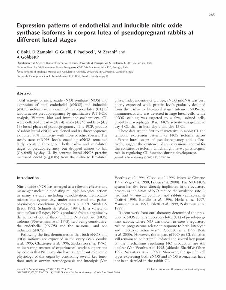
Figure 1 Immunolocalization of eNOS in a section of rabbitmyocardium which served as positive control for eNOS (�400).
NO synthases in rabbit CL · C BOITI and others 287
www.endocrinology.org Journal of Endocrinology (2002) 173, 285–296
(a) (b)
(c) (d )
(e) (f )
(h)(g)
C BOITI and others · NO synthases in rabbit CL288
www.endocrinology.orgJournal of Endocrinology (2002) 173, 285–296
antibody. After PBS rinsing, the avidin-biotinylated com-plex peroxidase (ABC-P reagent; Vector Laboratories),prepared according to the manufacturer’s instructions, wasapplied to the sections for 30 min. The slides were thenwashed in PBS, and antigen–antibody binding sites werestained for 15 min in 0·05% 3,3�-diaminobenzidine chro-mogen solution (DAB substrate Kit for peroxidase; VectorLaboratories) and 0·02% hydrogen peroxide. After wash-ing in tap water, the slides were dehydrated sequentially inascending grades of alcohol and cleared in xylene. Finally,the sections were counter-stained with Harris’s hematoxy-lin, washed in tap water and cover-slipped using aqueousmounting medium. The intensity of the staining wasdetermined under a light microscope. Positive reactionswere recognized as reddish brown precipitates. Sections inwhich the primary antibodies were omitted or substitutedby non-immune mouse gamma globulin were used for thenegative control of non-specific staining. These tissuesections were devoid of any significant staining. Sections ofrabbit myocardium served as a positive control for eNOS(Fig. 1).
Western blot analysis of eNOS and iNOS isoforms
For the Western blotting analysis of eNOS and iNOS,total luteal proteins were extracted from CL of each rabbitin extraction buffer (Tris–HCl 10 mM pH 7·6, 1% SDS,1·0 mM sodium ortho-vanadate). The homogenates werecentrifuged at 14 000 g for 15 min at 4 �C. The proteinconcentrations in the resulting supernatant samples weredetermined using the Bio-Rad protein assay kit (Bio-RadLaboratories). After heating at 95 �C for 5 min, equiva-lent amounts of proteins (10 µg) were separated usingan electrophoresis system (Mini-Protean; Bio-RadLaboratories) by discontinuous 10% SDS-PAGE with a4% stacking gel for 60 min at 200 V, constant current.The separated proteins were then electrophoreticallytransferred onto a nitrocellulose membrane (Protran;Schleicher & Schuell, Dassel, Germany) for 1 h at 100 Vconstant current using a Mini Trans Blot (Bio-Rad)apparatus. The membranes were washed and blocked with1% casein in Tris-buffered saline (TBS-C; Tris–HCl,20 mM, pH 7·4, NaCl 137 mM) and incubated over-night at 4 �C with anti-eNOS antibody diluted 1:2000or anti-iNOS antibody diluted 1:5000, both fromTransduction Laboratories. Membranes were then incu-bated with biotinylated anti-mouse IgG secondaryantibody (Vector Laboratories) for 30 min at 37 �Cunder gentle agitation. All antibody incubations and wash-ings were performed in TBS-C. The avidin-biotinylated
complex (ABC reagent; Vector Elite Kit) was thenadded. The DAB substrate for peroxidase (VectorLaboratories) was used for detection of immunoreactivebands. Mouse macrophage iNOS and human eNOSlysates (Transduction Laboratories) were used as negativeand positive controls. The membranes were scanned usinga flatbed scanner and the optical densities of the bandswere quantified using Quantity One Software (Bio-Rad).Each specific band signal was corrected by subtracting thebackground levels taken within the same lane and reportedas arbitrary units.
Progesterone assay
Progesterone concentrations in plasma samples weredetermined by RIA, using specific antibody according tothe procedure reported elsewhere (Gobbetti et al. 1992).The compounds that cross-reacted with the antibody morethan 0·2% were: progesterone (100%), pregnenolone(6·2%) and 5�-pregnan-3�-2-one (1·7%). Progesteronewas extracted from plasma with ethyl ether. The assaysensitivity was 10 pg/ml; intra-assay and inter-assaycoefficients of variations were 6 and 11% respectively.
Statistical analysis
All experiments were repeated at least three times. Theratios of each PCR product for the target gene normalizedagainst 18S co-amplified product were analyzed by two-factor ANOVA (luteal stage and gel as the two sources ofvariability) and a Newman–Keuls multi-comparison post-test. All statistical analyses were performed using Prism 3·0(GraphPad Software, Inc., San Diego, CA, USA).
Results of mRNA and protein levels for both eNOSand iNOS, as well as total NOS activity, were expressed asmeans�S.E.M. of arbitrary units for the number of separateexperiments as reported in Figure legends. Data relative todifferent stages of pseudopregnancy were analyzed byANOVA (Sokal & Rohlf 1981).
Results
Immunolocalization of eNOS and iNOS in CL
Using a monoclonal antibody, positive eNOS staining wasdetected in all examined CL obtained from rabbits atdifferent luteal phases of pseudopregnancy. Independentlyof CL age, eNOS-like immunoreactivity was localizedprimarily in the large luteal cells as indicated by the
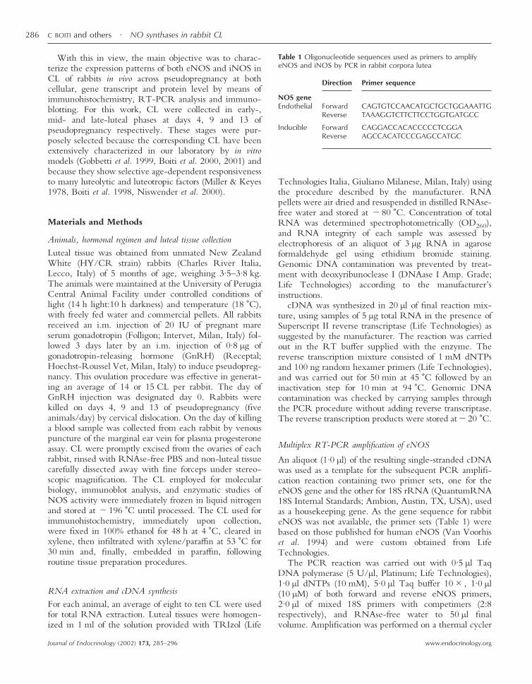
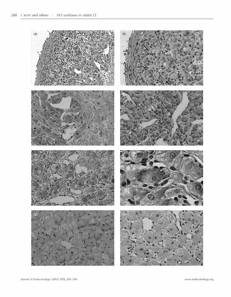
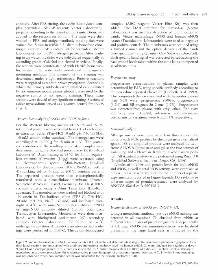
Figure 2 Immunolocalization of eNOS in corpora lutea (CL) of rabbits at different luteal stages. Representative photomicrographs of 3 µmthick luteal sections immunostained with a primary monoclonal antibody (1:25) to human eNOS. CL were obtained from rabbits at days 4,9 and 13 of pseudopregnancy (a–d, e and g respectively). (f) A higher magnification (�1000) of a day 4 CL section. Positive staining isrecognized as a brown precipitate. (h) A representative photomicrograph of a section prepared from day 4 CL in which immunostainingwas not observed when non-immune serum was substituted for the primary antibody (�400).
NO synthases in rabbit CL · C BOITI and others 289
www.endocrinology.org Journal of Endocrinology (2002) 173, 285–296
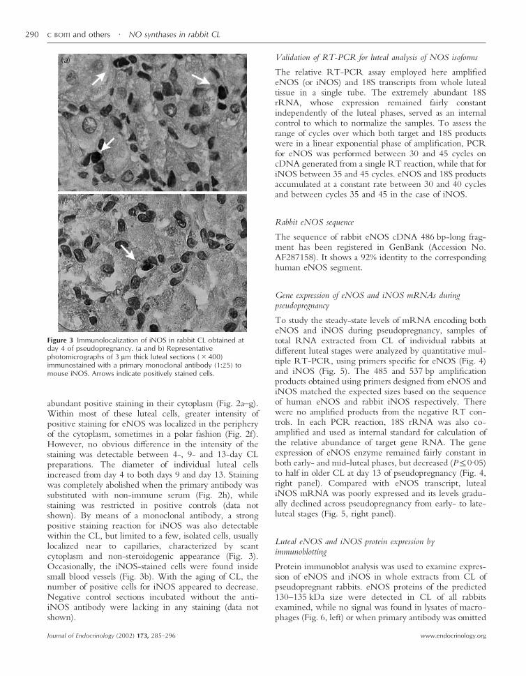
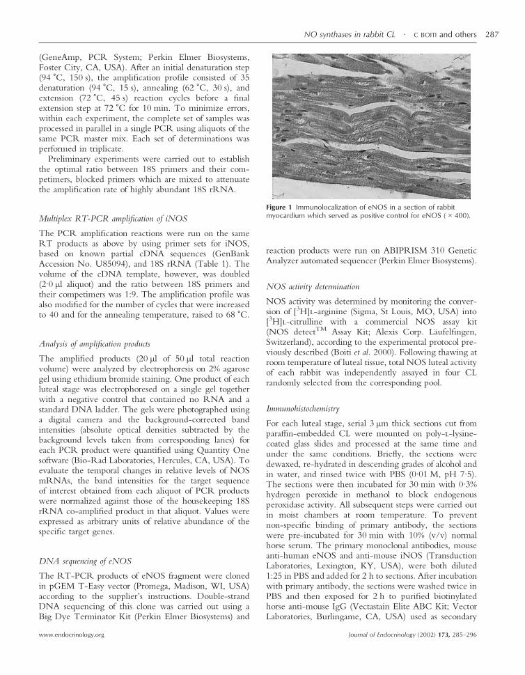
abundant positive staining in their cytoplasm (Fig. 2a–g).Within most of these luteal cells, greater intensity ofpositive staining for eNOS was localized in the peripheryof the cytoplasm, sometimes in a polar fashion (Fig. 2f).However, no obvious difference in the intensity of thestaining was detectable between 4-, 9- and 13-day CLpreparations. The diameter of individual luteal cellsincreased from day 4 to both days 9 and day 13. Stainingwas completely abolished when the primary antibody wassubstituted with non-immune serum (Fig. 2h), whilestaining was restricted in positive controls (data notshown). By means of a monoclonal antibody, a strongpositive staining reaction for iNOS was also detectablewithin the CL, but limited to a few, isolated cells, usuallylocalized near to capillaries, characterized by scantcytoplasm and non-steroidogenic appearance (Fig. 3).Occasionally, the iNOS-stained cells were found insidesmall blood vessels (Fig. 3b). With the aging of CL, thenumber of positive cells for iNOS appeared to decrease.Negative control sections incubated without the anti-iNOS antibody were lacking in any staining (data notshown).
Validation of RT-PCR for luteal analysis of NOS isoforms
The relative RT-PCR assay employed here amplifiedeNOS (or iNOS) and 18S transcripts from whole lutealtissue in a single tube. The extremely abundant 18SrRNA, whose expression remained fairly constantindependently of the luteal phases, served as an internalcontrol to which to normalize the samples. To assess therange of cycles over which both target and 18S productswere in a linear exponential phase of amplification, PCRfor eNOS was performed between 30 and 45 cycles oncDNA generated from a single RT reaction, while that foriNOS between 35 and 45 cycles. eNOS and 18S productsaccumulated at a constant rate between 30 and 40 cyclesand between cycles 35 and 45 in the case of iNOS.
Rabbit eNOS sequence
The sequence of rabbit eNOS cDNA 486 bp-long frag-ment has been registered in GenBank (Accession No.AF287158). It shows a 92% identity to the correspondinghuman eNOS segment.
Gene expression of eNOS and iNOS mRNAs duringpseudopregnancy
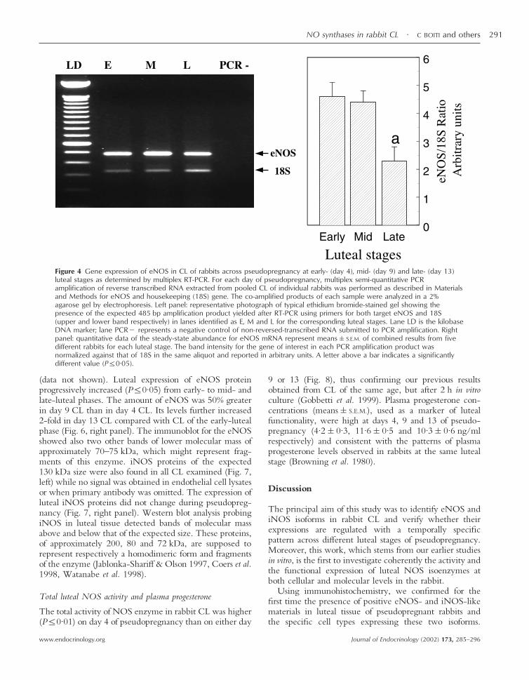
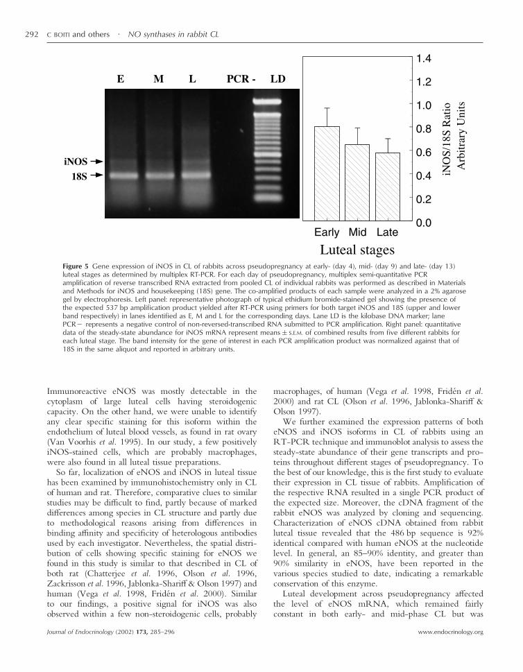
To study the steady-state levels of mRNA encoding botheNOS and iNOS during pseudopregnancy, samples oftotal RNA extracted from CL of individual rabbits atdifferent luteal stages were analyzed by quantitative mul-tiple RT-PCR, using primers specific for eNOS (Fig. 4)and iNOS (Fig. 5). The 485 and 537 bp amplificationproducts obtained using primers designed from eNOS andiNOS matched the expected sizes based on the sequenceof human eNOS and rabbit iNOS respectively. Therewere no amplified products from the negative RT con-trols. In each PCR reaction, 18S rRNA was also co-amplified and used as internal standard for calculation ofthe relative abundance of target gene RNA. The geneexpression of eNOS enzyme remained fairly constant inboth early- and mid-luteal phases, but decreased (P�0·05)to half in older CL at day 13 of pseudopregnancy (Fig. 4,right panel). Compared with eNOS transcript, lutealiNOS mRNA was poorly expressed and its levels gradu-ally declined across pseudopregnancy from early- to late-luteal stages (Fig. 5, right panel).
Luteal eNOS and iNOS protein expression byimmunoblotting
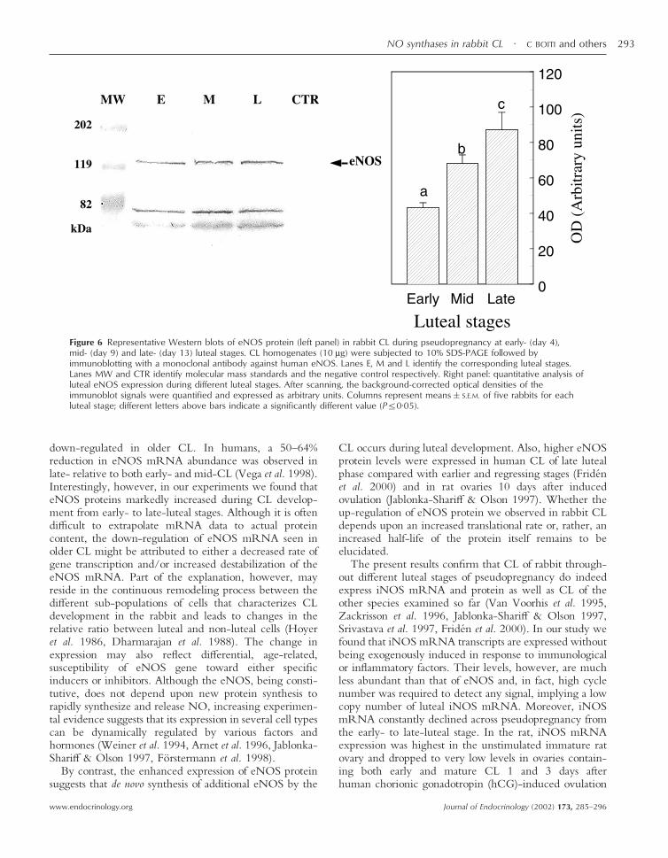
Protein immunoblot analysis was used to examine expres-sion of eNOS and iNOS in whole extracts from CL ofpseudopregnant rabbits. eNOS proteins of the predicted130–135 kDa size were detected in CL of all rabbitsexamined, while no signal was found in lysates of macro-phages (Fig. 6, left) or when primary antibody was omitted
Figure 3 Immunolocalization of iNOS in rabbit CL obtained atday 4 of pseudopregnancy. (a and b) Representativephotomicrographs of 3 µm thick luteal sections (�400)immunostained with a primary monoclonal antibody (1:25) tomouse iNOS. Arrows indicate positively stained cells.
C BOITI and others · NO synthases in rabbit CL290
www.endocrinology.orgJournal of Endocrinology (2002) 173, 285–296
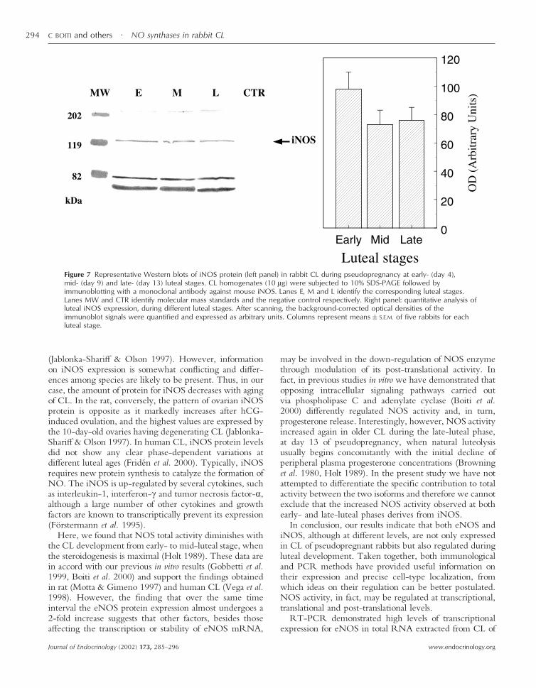
(data not shown). Luteal expression of eNOS proteinprogressively increased (P�0·05) from early- to mid- andlate-luteal phases. The amount of eNOS was 50% greaterin day 9 CL than in day 4 CL. Its levels further increased2-fold in day 13 CL compared with CL of the early-lutealphase (Fig. 6, right panel). The immunoblot for the eNOSshowed also two other bands of lower molecular mass ofapproximately 70–75 kDa, which might represent frag-ments of this enzyme. iNOS proteins of the expected130 kDa size were also found in all CL examined (Fig. 7,left) while no signal was obtained in endothelial cell lysatesor when primary antibody was omitted. The expression ofluteal iNOS proteins did not change during pseudopreg-nancy (Fig. 7, right panel). Western blot analysis probingiNOS in luteal tissue detected bands of molecular massabove and below that of the expected size. These proteins,of approximately 200, 80 and 72 kDa, are supposed torepresent respectively a homodimeric form and fragmentsof the enzyme (Jablonka-Shariff & Olson 1997, Coers et al.1998, Watanabe et al. 1998).
Total luteal NOS activity and plasma progesterone
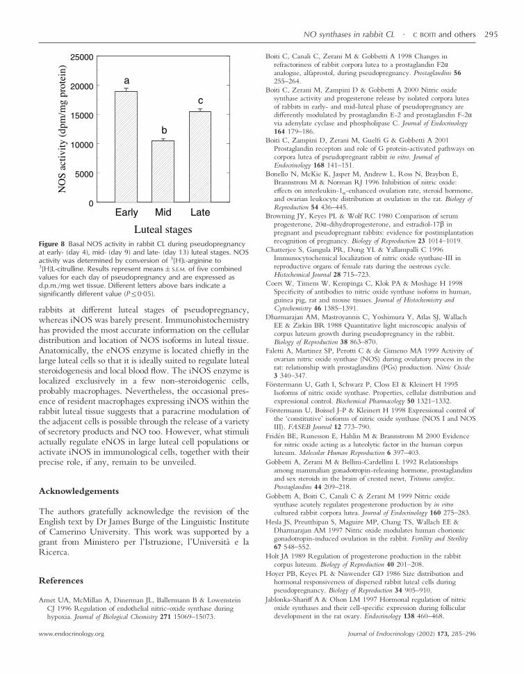
The total activity of NOS enzyme in rabbit CL was higher(P�0·01) on day 4 of pseudopregnancy than on either day
9 or 13 (Fig. 8), thus confirming our previous resultsobtained from CL of the same age, but after 2 h in vitroculture (Gobbetti et al. 1999). Plasma progesterone con-centrations (means�S.E.M.), used as a marker of lutealfunctionality, were high at days 4, 9 and 13 of pseudo-pregnancy (4·2�0·3, 11·6�0·5 and 10·3�0·6 ng/mlrespectively) and consistent with the patterns of plasmaprogesterone levels observed in rabbits at the same lutealstage (Browning et al. 1980).
Discussion
The principal aim of this study was to identify eNOS andiNOS isoforms in rabbit CL and verify whether theirexpressions are regulated with a temporally specificpattern across different luteal stages of pseudopregnancy.Moreover, this work, which stems from our earlier studiesin vitro, is the first to investigate coherently the activity andthe functional expression of luteal NOS isoenzymes atboth cellular and molecular levels in the rabbit.
Using immunohistochemistry, we confirmed for thefirst time the presence of positive eNOS- and iNOS-likematerials in luteal tissue of pseudopregnant rabbits andthe specific cell types expressing these two isoforms.
Figure 4 Gene expression of eNOS in CL of rabbits across pseudopregnancy at early- (day 4), mid- (day 9) and late- (day 13)luteal stages as determined by multiplex RT-PCR. For each day of pseudopregnancy, multiplex semi-quantitative PCRamplification of reverse transcribed RNA extracted from pooled CL of individual rabbits was performed as described in Materialsand Methods for eNOS and housekeeping (18S) gene. The co-amplified products of each sample were analyzed in a 2%agarose gel by electrophoresis. Left panel: representative photograph of typical ethidium bromide-stained gel showing thepresence of the expected 485 bp amplification product yielded after RT-PCR using primers for both target eNOS and 18S(upper and lower band respectively) in lanes identified as E, M and L for the corresponding luteal stages. Lane LD is the kilobaseDNA marker; lane PCR� represents a negative control of non-reversed-transcribed RNA submitted to PCR amplification. Rightpanel: quantitative data of the steady-state abundance for eNOS mRNA represent means�S.E.M. of combined results from fivedifferent rabbits for each luteal stage. The band intensity for the gene of interest in each PCR amplification product wasnormalized against that of 18S in the same aliquot and reported in arbitrary units. A letter above a bar indicates a significantlydifferent value (P�0·05).
NO synthases in rabbit CL · C BOITI and others 291
www.endocrinology.org Journal of Endocrinology (2002) 173, 285–296
Immunoreactive eNOS was mostly detectable in thecytoplasm of large luteal cells having steroidogeniccapacity. On the other hand, we were unable to identifyany clear specific staining for this isoform within theendothelium of luteal blood vessels, as found in rat ovary(Van Voorhis et al. 1995). In our study, a few positivelyiNOS-stained cells, which are probably macrophages,were also found in all luteal tissue preparations.
So far, localization of eNOS and iNOS in luteal tissuehas been examined by immunohistochemistry only in CLof human and rat. Therefore, comparative clues to similarstudies may be difficult to find, partly because of markeddifferences among species in CL structure and partly dueto methodological reasons arising from differences inbinding affinity and specificity of heterologous antibodiesused by each investigator. Nevertheless, the spatial distri-bution of cells showing specific staining for eNOS wefound in this study is similar to that described in CL ofboth rat (Chatterjee et al. 1996, Olson et al. 1996,Zackrisson et al. 1996, Jablonka-Shariff & Olson 1997) andhuman (Vega et al. 1998, Fridén et al. 2000). Similarto our findings, a positive signal for iNOS was alsoobserved within a few non-steroidogenic cells, probably
macrophages, of human (Vega et al. 1998, Fridén et al.2000) and rat CL (Olson et al. 1996, Jablonka-Shariff &Olson 1997).
We further examined the expression patterns of botheNOS and iNOS isoforms in CL of rabbits using anRT-PCR technique and immunoblot analysis to assess thesteady-state abundance of their gene transcripts and pro-teins throughout different stages of pseudopregnancy. Tothe best of our knowledge, this is the first study to evaluatetheir expression in CL tissue of rabbits. Amplification ofthe respective RNA resulted in a single PCR product ofthe expected size. Moreover, the cDNA fragment of therabbit eNOS was analyzed by cloning and sequencing.Characterization of eNOS cDNA obtained from rabbitluteal tissue revealed that the 486 bp sequence is 92%identical compared with human eNOS at the nucleotidelevel. In general, an 85–90% identity, and greater than90% similarity in eNOS, have been reported in thevarious species studied to date, indicating a remarkableconservation of this enzyme.
Luteal development across pseudopregnancy affectedthe level of eNOS mRNA, which remained fairlyconstant in both early- and mid-phase CL but was
Figure 5 Gene expression of iNOS in CL of rabbits across pseudopregnancy at early- (day 4), mid- (day 9) and late- (day 13)luteal stages as determined by multiplex RT-PCR. For each day of pseudopregnancy, multiplex semi-quantitative PCRamplification of reverse transcribed RNA extracted from pooled CL of individual rabbits was performed as described in Materialsand Methods for iNOS and housekeeping (18S) gene. The co-amplified products of each sample were analyzed in a 2% agarosegel by electrophoresis. Left panel: representative photograph of typical ethidium bromide-stained gel showing the presence ofthe expected 537 bp amplification product yielded after RT-PCR using primers for both target iNOS and 18S (upper and lowerband respectively) in lanes identified as E, M and L for the corresponding days. Lane LD is the kilobase DNA marker; lanePCR� represents a negative control of non-reversed-transcribed RNA submitted to PCR amplification. Right panel: quantitativedata of the steady-state abundance for iNOS mRNA represent means�S.E.M. of combined results from five different rabbits foreach luteal stage. The band intensity for the gene of interest in each PCR amplification product was normalized against that of18S in the same aliquot and reported in arbitrary units.
C BOITI and others · NO synthases in rabbit CL292
www.endocrinology.orgJournal of Endocrinology (2002) 173, 285–296
down-regulated in older CL. In humans, a 50–64%reduction in eNOS mRNA abundance was observed inlate- relative to both early- and mid-CL (Vega et al. 1998).Interestingly, however, in our experiments we found thateNOS proteins markedly increased during CL develop-ment from early- to late-luteal stages. Although it is oftendifficult to extrapolate mRNA data to actual proteincontent, the down-regulation of eNOS mRNA seen inolder CL might be attributed to either a decreased rate ofgene transcription and/or increased destabilization of theeNOS mRNA. Part of the explanation, however, mayreside in the continuous remodeling process between thedifferent sub-populations of cells that characterizes CLdevelopment in the rabbit and leads to changes in therelative ratio between luteal and non-luteal cells (Hoyeret al. 1986, Dharmarajan et al. 1988). The change inexpression may also reflect differential, age-related,susceptibility of eNOS gene toward either specificinducers or inhibitors. Although the eNOS, being consti-tutive, does not depend upon new protein synthesis torapidly synthesize and release NO, increasing experimen-tal evidence suggests that its expression in several cell typescan be dynamically regulated by various factors andhormones (Weiner et al. 1994, Arnet et al. 1996, Jablonka-Shariff & Olson 1997, Förstermann et al. 1998).
By contrast, the enhanced expression of eNOS proteinsuggests that de novo synthesis of additional eNOS by the
CL occurs during luteal development. Also, higher eNOSprotein levels were expressed in human CL of late lutealphase compared with earlier and regressing stages (Fridénet al. 2000) and in rat ovaries 10 days after inducedovulation (Jablonka-Shariff & Olson 1997). Whether theup-regulation of eNOS protein we observed in rabbit CLdepends upon an increased translational rate or, rather, anincreased half-life of the protein itself remains to beelucidated.
The present results confirm that CL of rabbit through-out different luteal stages of pseudopregnancy do indeedexpress iNOS mRNA and protein as well as CL of theother species examined so far (Van Voorhis et al. 1995,Zackrisson et al. 1996, Jablonka-Shariff & Olson 1997,Srivastava et al. 1997, Fridén et al. 2000). In our study wefound that iNOS mRNA transcripts are expressed withoutbeing exogenously induced in response to immunologicalor inflammatory factors. Their levels, however, are muchless abundant than that of eNOS and, in fact, high cyclenumber was required to detect any signal, implying a lowcopy number of luteal iNOS mRNA. Moreover, iNOSmRNA constantly declined across pseudopregnancy fromthe early- to late-luteal stage. In the rat, iNOS mRNAexpression was highest in the unstimulated immature ratovary and dropped to very low levels in ovaries contain-ing both early and mature CL 1 and 3 days afterhuman chorionic gonadotropin (hCG)-induced ovulation
Figure 6 Representative Western blots of eNOS protein (left panel) in rabbit CL during pseudopregnancy at early- (day 4),mid- (day 9) and late- (day 13) luteal stages. CL homogenates (10 µg) were subjected to 10% SDS-PAGE followed byimmunoblotting with a monoclonal antibody against human eNOS. Lanes E, M and L identify the corresponding luteal stages.Lanes MW and CTR identify molecular mass standards and the negative control respectively. Right panel: quantitative analysis ofluteal eNOS expression during different luteal stages. After scanning, the background-corrected optical densities of theimmunoblot signals were quantified and expressed as arbitrary units. Columns represent means�S.E.M. of five rabbits for eachluteal stage; different letters above bars indicate a significantly different value (P�0·05).
NO synthases in rabbit CL · C BOITI and others 293
www.endocrinology.org Journal of Endocrinology (2002) 173, 285–296
(Jablonka-Shariff & Olson 1997). However, informationon iNOS expression is somewhat conflicting and differ-ences among species are likely to be present. Thus, in ourcase, the amount of protein for iNOS decreases with agingof CL. In the rat, conversely, the pattern of ovarian iNOSprotein is opposite as it markedly increases after hCG-induced ovulation, and the highest values are expressed bythe 10-day-old ovaries having degenerating CL (Jablonka-Shariff & Olson 1997). In human CL, iNOS protein levelsdid not show any clear phase-dependent variations atdifferent luteal ages (Fridén et al. 2000). Typically, iNOSrequires new protein synthesis to catalyze the formation ofNO. The iNOS is up-regulated by several cytokines, suchas interleukin-1, interferon-� and tumor necrosis factor-�,although a large number of other cytokines and growthfactors are known to transcriptically prevent its expression(Förstermann et al. 1995).
Here, we found that NOS total activity diminishes withthe CL development from early- to mid-luteal stage, whenthe steroidogenesis is maximal (Holt 1989). These data arein accord with our previous in vitro results (Gobbetti et al.1999, Boiti et al. 2000) and support the findings obtainedin rat (Motta & Gimeno 1997) and human CL (Vega et al.1998). However, the finding that over the same timeinterval the eNOS protein expression almost undergoes a2-fold increase suggests that other factors, besides thoseaffecting the transcription or stability of eNOS mRNA,
may be involved in the down-regulation of NOS enzymethrough modulation of its post-translational activity. Infact, in previous studies in vitro we have demonstrated thatopposing intracellular signaling pathways carried outvia phospholipase C and adenylate cyclase (Boiti et al.2000) differently regulated NOS activity and, in turn,progesterone release. Interestingly, however, NOS activityincreased again in older CL during the late-luteal phase,at day 13 of pseudopregnancy, when natural luteolysisusually begins concomitantly with the initial decline ofperipheral plasma progesterone concentrations (Browninget al. 1980, Holt 1989). In the present study we have notattempted to differentiate the specific contribution to totalactivity between the two isoforms and therefore we cannotexclude that the increased NOS activity observed at bothearly- and late-luteal phases derives from iNOS.
In conclusion, our results indicate that both eNOS andiNOS, although at different levels, are not only expressedin CL of pseudopregnant rabbits but also regulated duringluteal development. Taken together, both immunologicaland PCR methods have provided useful information ontheir expression and precise cell-type localization, fromwhich ideas on their regulation can be better postulated.NOS activity, in fact, may be regulated at transcriptional,translational and post-translational levels.
RT-PCR demonstrated high levels of transcriptionalexpression for eNOS in total RNA extracted from CL of
Figure 7 Representative Western blots of iNOS protein (left panel) in rabbit CL during pseudopregnancy at early- (day 4),mid- (day 9) and late- (day 13) luteal stages. CL homogenates (10 µg) were subjected to 10% SDS-PAGE followed byimmunoblotting with a monoclonal antibody against mouse iNOS. Lanes E, M and L identify the corresponding luteal stages.Lanes MW and CTR identify molecular mass standards and the negative control respectively. Right panel: quantitative analysis ofluteal iNOS expression, during different luteal stages. After scanning, the background-corrected optical densities of theimmunoblot signals were quantified and expressed as arbitrary units. Columns represent means�S.E.M. of five rabbits for eachluteal stage.
C BOITI and others · NO synthases in rabbit CL294
www.endocrinology.orgJournal of Endocrinology (2002) 173, 285–296
rabbits at different luteal stages of pseudopregnancy,whereas iNOS was barely present. Immunohistochemistryhas provided the most accurate information on the cellulardistribution and location of NOS isoforms in luteal tissue.Anatomically, the eNOS enzyme is located chiefly in thelarge luteal cells so that it is ideally suited to regulate lutealsteroidogenesis and local blood flow. The iNOS enzyme islocalized exclusively in a few non-steroidogenic cells,probably macrophages. Nevertheless, the occasional pres-ence of resident macrophages expressing iNOS within therabbit luteal tissue suggests that a paracrine modulation ofthe adjacent cells is possible through the release of a varietyof secretory products and NO too. However, what stimuliactually regulate eNOS in large luteal cell populations oractivate iNOS in immunological cells, together with theirprecise role, if any, remain to be unveiled.
Acknowledgements
The authors gratefully acknowledge the revision of theEnglish text by Dr James Burge of the Linguistic Instituteof Camerino University. This work was supported by agrant from Ministero per l’Istruzione, l’Universita e laRicerca.
References
Arnet UA, McMillan A, Dinerman JL, Ballermann B & LowensteinCJ 1996 Regulation of endothelial nitric-oxide synthase duringhypoxia. Journal of Biological Chemistry 271 15069–15073.
Boiti C, Canali C, Zerani M & Gobbetti A 1998 Changes inrefractoriness of rabbit corpora lutea to a prostaglandin F2�analogue, alfaprostol, during pseudopregnancy. Prostaglandins 56255–264.
Boiti C, Zerani M, Zampini D & Gobbetti A 2000 Nitric oxidesynthase activity and progesterone release by isolated corpora luteaof rabbits in early- and mid-luteal phase of pseudopregnancy aredifferently modulated by prostaglandin E-2 and prostaglandin F-2�via adenylate cyclase and phospholipase C. Journal of Endocrinology164 179–186.
Boiti C, Zampini D, Zerani M, Guelfi G & Gobbetti A 2001Prostaglandin receptors and role of G protein-activated pathways oncorpora lutea of pseudopregnant rabbit in vitro. Journal ofEndocrinology 168 141–151.
Bonello N, McKie K, Jasper M, Andrew L, Ross N, Braybon E,Brannstrom M & Norman RJ 1996 Inhibition of nitric oxide:effects on interleukin-1�-enhanced ovulation rate, steroid hormone,and ovarian leukocyte distribution at ovulation in the rat. Biology ofReproduction 54 436–445.
Browning JY, Keyes PL & Wolf RC 1980 Comparison of serumprogesterone, 20�-dihydroprogesterone, and estradiol-17� inpregnant and pseudopregnant rabbits: evidence for postimplantationrecognition of pregnancy. Biology of Reproduction 23 1014–1019.
Chatterjee S, Gangula PR, Dong YL & Yallampalli C 1996Immunocytochemical localization of nitric oxide synthase-III inreproductive organs of female rats during the oestrous cycle.Histochemical Journal 28 715–723.
Coers W, Timens W, Kempinga C, Klok PA & Moshage H 1998Specificity of antibodies to nitric oxide synthase isofoms in human,guinea pig, rat and mouse tissues. Journal of Histochemistry andCytochemistry 46 1385–1391.
Dharmarajan AM, Mastroyannis C, Yoshimura Y, Atlas SJ, WallachEE & Zirkin BR 1988 Quantitative light microscopic analysis ofcorpus luteum growth during pseudopregnancy in the rabbit.Biology of Reproduction 38 863–870.
Faletti A, Martinez SP, Perotti C & de Gimeno MA 1999 Activity ofovarian nitric oxide synthase (NOS) during ovulatory process in therat: relationship with prostaglandins (PGs) production. Nitric Oxide3 340–347.
Förstermann U, Gath I, Schwarz P, Closs EI & Kleinert H 1995Isoforms of nitric oxide synthase. Properties, cellular distribution andexpressional control. Biochemical Pharmacology 50 1321–1332.
Förstermann U, Boissel J-P & Kleinert H 1998 Expressional control ofthe ‘constitutive’ isoforms of nitric oxide synthase (NOS I and NOSIII). FASEB Journal 12 773–790.
Fridén BE, Runesson E, Hahlin M & Brannstrom M 2000 Evidencefor nitric oxide acting as a luteolytic factor in the human corpusluteum. Molecular Human Reproduction 6 397–403.
Gobbetti A, Zerani M & Bellini-Cardellini L 1992 Relationshipsamong mammalian gonadotropin-releasing hormone, prostaglandinsand sex steroids in the brain of crested newt, Triturus carnifex.Prostaglandins 44 209–218.
Gobbetti A, Boiti C, Canali C & Zerani M 1999 Nitric oxidesynthase acutely regulates progesterone production by in vitrocultured rabbit corpora lutea. Journal of Endocrinology 160 275–283.
Hesla JS, Preutthipan S, Maguire MP, Chang TS, Wallach EE &Dharmarajan AM 1997 Nitric oxide modulates human chorionicgonadotropin-induced ovulation in the rabbit. Fertility and Sterility67 548–552.
Holt JA 1989 Regulation of progesterone production in the rabbitcorpus luteum. Biology of Reproduction 40 201–208.
Hoyer PB, Keyes PL & Niswender GD 1986 Size distribution andhormonal responsiveness of dispersed rabbit luteal cells duringpseudopregnancy. Biology of Reproduction 34 905–910.
Jablonka-Shariff A & Olson LM 1997 Hormonal regulation of nitricoxide synthases and their cell-specific expression during folliculardevelopment in the rat ovary. Endocrinology 138 460–468.
Figure 8 Basal NOS activity in rabbit CL during pseudopregnancyat early- (day 4), mid- (day 9) and late- (day 13) luteal stages. NOSactivity was determined by conversion of 3[H]L-arginine to3[H]L-citrulline. Results represent means�S.E.M. of five combinedvalues for each day of pseudopregnancy and are expressed asd.p.m./mg wet tissue. Different letters above bars indicate asignificantly different value (P�0·05).
NO synthases in rabbit CL · C BOITI and others 295
www.endocrinology.org Journal of Endocrinology (2002) 173, 285–296
Miller JB & Keyes PL 1978 Transition of the rabbit corpus luteum toestrogen dependence during early luteal development. Endocrinology102 31–38.
Moncada S, Palmer RMJ & Higgs EA 1991 Nitric oxide: physiology,pathophysiology, and pharmacology. Pharmacological Reviews 42109–142.
Motta AB & Gimeno MAF 1997 Nitric oxide participates in thecorpus luteum regression in ovaries isolated from pseudopregnantrats. Canadian Journal of Physiology and Pharmacology 75 1335–1339.
Nakamura Y, Kashida S, Nakata M, Takiguchi S, Yamagata Y,Takayama H, Sugino N & Kato H 1999 Changes in nitric oxidesynthase activity in the ovary of gonadotropin treated rats: the roleof nitric oxide during ovulation. Endocrine Journal 46 529–538.
Niswender GD, Juengel JL, Silva PJ, Rollyson MK & McIntush EW2000 Mechanisms controlling the function and life span of thecorpus luteum. Physiological Reviews 80 1–29.
Olson LM, Jones-Burton CM & Jablonka-Shariff A 1996 Nitric oxidedecreases estradiol synthesis of rat luteinized ovarian cells: possiblerole for nitric oxide in functional luteal regression. Endocrinology 1373531–3539.
Schmidt HHHW & Walter U 1994 NO at work. Cell 78 919–925.Shukovski L & Tsafriri T 1995 The involvement of nitric oxide in the
ovulatory process in the rat. Endocrinology 135 2287–2290.Snyder SH & Bredt DS 1992 Biological roles of nitric oxide. Scientific
American 5 28–35.Sokal RR & Rohlf FJ 1981 Biometry, pp 253–261. New York: WH
Freeman and Co.Srivastava V, Jones BJ, Dookwah H, Hiney JK & Dees WL 1997
Ovarian nitric oxide synthase (NOS) gene expression duringperipubertal development. Life Sciences 61 1507–1516.
Van Voorhis BJ, Dunn MS, Snyder GD & Weiner CP 1994 Nitricoxide: an autocrine regulator of human granulosa-luteal cellsteroidogenesis. Endocrinology 135 1799–1806.
Van Voorhis BJ, Moore K, Strijbos PJ, Nelson S, Baylis SA, GrzybickiD & Weiner CP 1995 Expression and localization of inducible andendothelial nitric oxide synthase in the rat ovary. Effects ofgonadotropin stimulation in vivo. Journal of Clinical Investigation 962719–2726.
Vega M, Johnson MC, Diaz HA, Urrutia LR, Troncoso JL & DevotoL 1998 Regulation of human luteal steroidogenesis in vitro by nitricoxide. Endocrine 8 185–191.
Watanabe Y, Nishio M, Hamaji S & Hidaka H 1998 Inter-isoformalregulation of nitric oxide synthase through heteromericdimerization. Biochimica et Biophysica Acta 1388 199–208.
Weiner CP, Lizasoain I, Baylis SA, Knowles RG, Charles IG &Moncada S 1994 Induction of calcium-dependent nitric oxidesynthesis by sex hormones. PNAS 91 5212–5216.
Yamauchi J, Miyazaki T, Iwasaki S, Kishi I, Kuroshima M, Tei C &Yoshimura Y 1997 Effects of nitric oxide on ovulation and ovariansteroidogenesis and prostaglandin production in the rabbit.Endocrinology 138 3630–3637.
Zackrisson U, Mikuni M, Wallin A, Delbro D, Hedin L &Brannstrom M 1996 Cell-specific localization of nitric oxidesynthases (NOS) in the rat ovary during follicular development,ovulation and luteal formation. Human Reproduction 11 2667–2673.
Received in final form 31 December 2001Accepted 21 January 2002
C BOITI and others · NO synthases in rabbit CL296
www.endocrinology.orgJournal of Endocrinology (2002) 173, 285–296
Related Documents


