Expression of the Rice Yellow Mottle Virus P1 Protein in Vitro and in Vivo and Its Involvement in Virus Spread Caroline Bonneau,* Christophe Brugidou,* Lili Chen,* Roger N. Beachy,* , ² and Claude Fauquet* ,1 *International Laboratory for Tropical Agricultural Biotechnology (ILTAB/ORSTOM/TSRI) and ²Division of Plant Biology, The Scripps Research Institute BCC 206, 10550 North Torrey Pines Road, La Jolla, California 92037 Received November 3, 1997; returned to author for revision December 1, 1997; accepted February 16, 1998 Rice yellow mottle sobemovirus (RYMV) is responsible for the yellow mottle disease on rice in Africa. The expression and function of the protein P1 (17.8 kDa) encoded by the first open reading frame (ORF) of RYMV was investigated. Using an antibody raised against purified P1, two proteins with apparent molecular masses of 18 and 19 kDa were identified in in vitro translation reactions of transcripts of the full-length cDNA of RYMV. Likewise, gene products with similar molecular mass were detected in inoculated and systemically infected rice leaves and in infected rice protoplasts. A mutant from which ORF1 nucleotides 88 to 547 were deleted and a frameshift mutant that resulted in truncation of 83 amino acids from the C terminus of P1 were incapable of replicating in protoplasts. In contrast, a mutant that does not express P1 due to a mutation at the initiation codon replicated efficiently in protoplasts but at a reduced level (about 0.5- to 2-fold less) compared to replication of wild-type RNA. None of these mutants caused systemic infection in rice plants. Transgenic rice plants that express P1 complemented the initiation codon mutant, but not the deletion mutants, and produced systemic infection. These experiments demonstrate that P1 of RYMV is dispensible for virus replication, although nucleotide deletions or additions in ORF1 are apparently lethal for virus replication. Furthermore, P1 of RYMV is required for the infection of plants and is important for virus spread. © 1998 Academic Press Key Words: sobemovirus; rice; replication; infection; complementation; movement. INTRODUCTION Rice yellow mottle virus (RYMV) is a member of the genus Sobemovirus, for which the southern bean mosaic virus (SBMV) is the type species (Murphy et al., 1995). This rice pathogen is transmitted mechanically and by chrysolemid beetles and causes a severe disease in Africa (Bakker, 1974; Fauquet and Thouvenel, 1977). RYMV symptoms consist of yellow mottling of the leaves and stunting of the plant (Bakker, 1974). The virus parti- cles are isometric and around 26 nm in diameter (Bakker, 1974). The RYMV genome is composed of a positive single-stranded RNA of 4450 nucleotides (nt) and con- tains four putative open reading frames (ORFs) (Ngon A Yassi et al., 1994). ORF1 encodes the protein P1 (17.8 kDa) of unknown function. ORF2 encodes a polyprotein of 110 kDa that is processed by a virus-encoded pro- tease to yield the VPg, the protease, the helicase, and the RNA polymerase (protein functions are predicted by sequence homology to SBMV and other viruses). ORF3 is nested in ORF2 in a different reading frame and encodes the putative protein P3 (13.7 kDa) of undefined function. ORF4 overlaps ORF2 at its 39 end and encodes the coat protein (CP) of 26 kDa. It has been demonstrated that the CP of sobemoviruses is translated from a subgenomic RNA (for review see Seghal, 1995; Hacker and Sivaku- maran, 1997). There have been limited numbers of stud- ies related to the specific functions of sobemovirus pro- teins. It was demonstrated by inserting point mutations in ORF4 in an infectious full-length cDNA of RYMV (FL5) that RYMV CP is not required for RNA replication but is necessary for virus spread in rice plants (Brugidou et al., 1995). The protein P3 of SBMV was demonstrated to be required for infectivity in plants (Hacker and Sivakuma- ran, 1996). The involvement of P1, encoded by ORF1, in RYMV infection is unknown, and no significant homologies have been found between P1 and other protein se- quences. We report in this paper a functional analysis of the ORF1 of RYMV. Expression of P1 was studied during RYMV infection using a specific antibody raised against purified P1. The analysis of mutations intro- duced into ORF1 indicates that P1 is not essential for the replication of RYMV in protoplasts but is required for infection on rice plants. However, mutants resulting from nucleotide deletions or additions in ORF1 were lethal for virus replication. A mutant of RYMV that does not produce P1 caused a systemic infection in trans- genic plants that express wild-type P1. Our data sug- gest that in addition to a possible effect of ORF1 sequence on viral replication, P1 plays a significant role in virus spread. 1 To whom reprint requests should be addressed. VIROLOGY 244, 79±86 (1998) ARTICLE NO. VY989100 0042-6822/98 $25.00 Copyright © 1998 by Academic Press All rights of reproduction in any form reserved. 79

Welcome message from author
This document is posted to help you gain knowledge. Please leave a comment to let me know what you think about it! Share it to your friends and learn new things together.
Transcript
Expression of the Rice Yellow Mottle Virus P1 Protein in Vitro and in Vivoand Its Involvement in Virus Spread
Caroline Bonneau,* Christophe Brugidou,* Lili Chen,* Roger N. Beachy,*,† and Claude Fauquet*,1
*International Laboratory for Tropical Agricultural Biotechnology (ILTAB/ORSTOM/TSRI) and †Division of Plant Biology,The Scripps Research Institute BCC 206, 10550 North Torrey Pines Road, La Jolla, California 92037
Received November 3, 1997; returned to author for revision December 1, 1997; accepted February 16, 1998
Rice yellow mottle sobemovirus (RYMV) is responsible for the yellow mottle disease on rice in Africa. The expression andfunction of the protein P1 (17.8 kDa) encoded by the first open reading frame (ORF) of RYMV was investigated. Using anantibody raised against purified P1, two proteins with apparent molecular masses of 18 and 19 kDa were identified in in vitrotranslation reactions of transcripts of the full-length cDNA of RYMV. Likewise, gene products with similar molecular masswere detected in inoculated and systemically infected rice leaves and in infected rice protoplasts. A mutant from which ORF1nucleotides 88 to 547 were deleted and a frameshift mutant that resulted in truncation of 83 amino acids from the C terminusof P1 were incapable of replicating in protoplasts. In contrast, a mutant that does not express P1 due to a mutation at theinitiation codon replicated efficiently in protoplasts but at a reduced level (about 0.5- to 2-fold less) compared to replicationof wild-type RNA. None of these mutants caused systemic infection in rice plants. Transgenic rice plants that express P1complemented the initiation codon mutant, but not the deletion mutants, and produced systemic infection. These experimentsdemonstrate that P1 of RYMV is dispensible for virus replication, although nucleotide deletions or additions in ORF1 areapparently lethal for virus replication. Furthermore, P1 of RYMV is required for the infection of plants and is important forvirus spread. © 1998 Academic Press
Key Words: sobemovirus; rice; replication; infection; complementation; movement.
INTRODUCTION
Rice yellow mottle virus (RYMV) is a member of thegenus Sobemovirus, for which the southern bean mosaicvirus (SBMV) is the type species (Murphy et al., 1995).This rice pathogen is transmitted mechanically and bychrysolemid beetles and causes a severe disease inAfrica (Bakker, 1974; Fauquet and Thouvenel, 1977).RYMV symptoms consist of yellow mottling of the leavesand stunting of the plant (Bakker, 1974). The virus parti-cles are isometric and around 26 nm in diameter (Bakker,1974). The RYMV genome is composed of a positivesingle-stranded RNA of 4450 nucleotides (nt) and con-tains four putative open reading frames (ORFs) (Ngon AYassi et al., 1994). ORF1 encodes the protein P1 (17.8kDa) of unknown function. ORF2 encodes a polyproteinof 110 kDa that is processed by a virus-encoded pro-tease to yield the VPg, the protease, the helicase, andthe RNA polymerase (protein functions are predicted bysequence homology to SBMV and other viruses). ORF3 isnested in ORF2 in a different reading frame and encodesthe putative protein P3 (13.7 kDa) of undefined function.ORF4 overlaps ORF2 at its 39 end and encodes the coatprotein (CP) of 26 kDa. It has been demonstrated that theCP of sobemoviruses is translated from a subgenomic
RNA (for review see Seghal, 1995; Hacker and Sivaku-maran, 1997). There have been limited numbers of stud-ies related to the specific functions of sobemovirus pro-teins. It was demonstrated by inserting point mutationsin ORF4 in an infectious full-length cDNA of RYMV (FL5)that RYMV CP is not required for RNA replication but isnecessary for virus spread in rice plants (Brugidou et al.,1995). The protein P3 of SBMV was demonstrated to berequired for infectivity in plants (Hacker and Sivakuma-ran, 1996).
The involvement of P1, encoded by ORF1, in RYMVinfection is unknown, and no significant homologieshave been found between P1 and other protein se-quences. We report in this paper a functional analysisof the ORF1 of RYMV. Expression of P1 was studiedduring RYMV infection using a specific antibody raisedagainst purified P1. The analysis of mutations intro-duced into ORF1 indicates that P1 is not essential forthe replication of RYMV in protoplasts but is requiredfor infection on rice plants. However, mutants resultingfrom nucleotide deletions or additions in ORF1 werelethal for virus replication. A mutant of RYMV that doesnot produce P1 caused a systemic infection in trans-genic plants that express wild-type P1. Our data sug-gest that in addition to a possible effect of ORF1sequence on viral replication, P1 plays a significantrole in virus spread.1 To whom reprint requests should be addressed.
VIROLOGY 244, 79–86 (1998)ARTICLE NO. VY989100
0042-6822/98 $25.00Copyright © 1998 by Academic PressAll rights of reproduction in any form reserved.
79
RESULTS
Identification of P1 in in vitro translation productsfrom FL5 transcripts
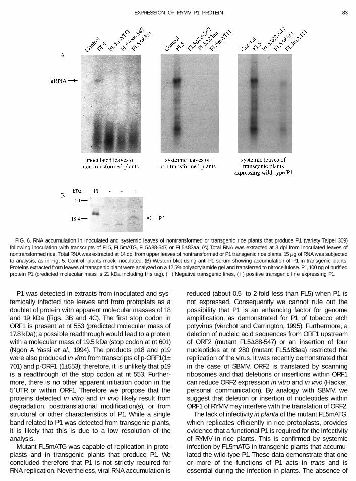
Translation of ORF1 from transcripts derived from thefull-length cloned cDNA of RYMV, FL5, was carried out invitro in wheat germ extracts in the presence of [35S]cys-teine. Two major products with apparent molecular massof 18 and 19 kDa were detected following SDS–PAGE(Fig. 2A). Following Western immunoblot reactions usingP1 antiserum, the 18- and 19-kDa were shown to reactspecifically with P1 antiserum (Fig. 2B). Preimmune se-rum showed no reaction (data not shown). To determinewhether the 18- and 19-kDa polypeptides are producedby in-frame readthrough of the UGA stop codon (nt 553),in vitro translation products of transcripts derived fromp-ORF1(1–701) and p-ORF1(1–553) (see Fig. 1) were an-alyzed; these reactions resulted in two proteins withapparent molecular masses of 18 and 19 kDa (Fig. 2C).
In vitro translation patterns of transcripts derived frommutants FL5mATG, FL5D88-547 and FL5D83aa (Fig. 1)did not result in the production of the 18- and 19-kDapolypeptides, but other products that did not react withthe P1 antiserum were observed (Fig. 2A). An 8-kDapolypeptide was translated from mutant FL5D83aa andcorresponds in size to the truncated P1 (predicted mo-
lecular mass of 8 kDa) (Fig. 2A); however, this truncatedprotein was not detected in Western blots using P1antiserum (Fig. 2B). These results indicate that two pro-teins with apparent molecular masses of 18 and 19 kDaare translated in vitro in wheat germ extracts from ORF1sequence and immunoreact with P1 specific antibody.
Detection of products of ORF1 in RYMV-infected riceplants and rice protoplasts
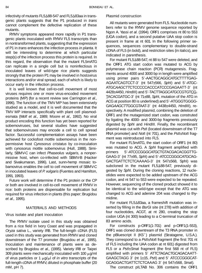
Infection of rice plants inoculated with RYMV wasmonitored by Northern blot analyses (data not shown)and the accumulation of P1 was analyzed by Westernblot analyses using P1 antiserum. A doublet of proteinswith apparent molecular masses (based on electro-phoretic mobility) of 18 and 19 kDa was detected ininoculated leaves at 5 days postinoculation (dpi) andaccumulated thereafter in the systemically infectedleaves (Fig. 3). P1 purified from Escherichia coli and usedas a positive control in Western blot analyses has acalculated molecular mass of 21 kDa due to the additionof the six histidine residues, while the RYMV P1 has acalculated molecular mass of 17.8 kDa. The difference insize appears in Fig. 3.
The replication of RYMV was also analyzed in riceprotoplasts infected with virus. Total RNA was extracted
FIG. 1. Schematic representation of RYMV ORF1 constructs. Mutations and deletions were introduced in the full-length cDNA of RYMV (FL5).FL5mATG contains a point mutation of the ORF1 initiation codon ATG to ACG. FL5D88-547 contains a deletion from nt 88 to 547 and a mutation atthe initiation start codon ATG to ACG. FL5D83aa contains four additional nucleotides at an BsrGI site (nt 278) leading to a frameshift and therecognition of a UGA stop codon at nt 300. Constructs pORF1 (1–553) and pORF1 (1–701) contain the P1 coding sequence, ending at UGA stop codon(nt 553) and putative UAA stop codon (nt 601), respectively, that were cloned downstream of the T3 RNA promoter. The white boxes represent theoriginal ORF1 and dashed boxes nontranslated regions. The dotted box represents a region where the amino acid sequence differs from the wild-typeP1. Dashed lines correspond to a deleted sequence.
80 BONNEAU ET AL.
from protoplasts at various time points until 72 h post-inoculation (hpi) and the accumulation of newly synthe-sized viral RNA was followed by Northern blot analysesusing a probe corresponding to RYMV ORF4 (CP). A verysmall amount of genomic RNA (gRNA) was detected at16 hpi and accumulated until 72 hpi (Fig. 4A). A sub-genomic RNA (sgRNA, around 1 kb) was detected with atime course of appearance similar to that of gRNA; this
RNA did not hybridize with a probe corresponding toORF1 (data not shown).
RYMV CP was detected at 24 hpi and accumulatedthrough 72 hpi (Fig. 4B). Similarly the accumulation of P1was followed by Western blot analyses with P1 anti-serum and showed two bands of 18 and 19 kDa thataccumulated from 24 to 72 hpi (Fig. 4C). These resultsindicate that P1 of RYMV is expressed from early to latestages of the infection in rice.
Effects of mutations in RYMV ORF1 on viralreplication and infectivity in rice
To determine whether P1 is required for replication,rice protoplasts were inoculated with the mutants of FL5described above. Transcripts of the mutants FL5D88-547
FIG. 2. In vitro translation products in wheat germs extracts ofwild-type (FL5) or mutated transcripts (FL5mATG, FL5D88-547,FL5D83aa) and of ORF1 gene (p-ORF1 (1–553) and p-ORF1 (1–701)). NoRNA, no RNA was added in the extracts. (A) Detection of translatedproducts labeled with [35S]cysteine and analyzed on a 18% polyacryl-amide gel after transfer of protein to membrane support. (B) Translatedproducts obtained in (A) were blotted onto a membrane after electro-phoresis on a 18% polyacrylamide gel and subjected to Western blotanalysis using P1 antiserum. (C) Translation products were labeledwith [35S]cysteine and analyzed on a 12.5% polyacrylamide gel.
FIG. 3. Detection of P1 in RYMV infected rice plants (variety IR8).Proteins were extracted from inoculated leaves from 1 to 5 dayspostinoculation (dpi) and from systematically infected leaves from 6 to28 dpi. After 5 dpi, inoculated leaves were yellow and dried and couldnot be sampled. Thirty micrograms of protein was separated on a 15%polyacrylamide gel and analyzed by Western blot analysis using anti-P1serum. P1, 370 ng of purified protein P1 (predicted molecular mass is 21kDa including the His tag). C, mock-inoculated plants. M, molecularweight standards.
FIG. 4. Detection of viral RNA, CP, and P1 in RYMV-infected riceprotoplasts. Total RNA and protein were extracted at various hourspostinoculation (hpi). C, mock-inoculated protoplasts. M, molecularweight standards. (A) Northern blot showing viral RNA accumulation.RNA was extracted from rice protoplasts, analyzed on a 1.2% agarosegel, blotted to nitrocellulose, and hybridized with a probe correspond-ing to ORF4. (B) Western blot using anti-CP serum. Proteins wereanalyzed on a 12.5% polyacrylamide gel. VP, 50 ng of purified virus. (C)Western blot using anti-P1 serum. Proteins were analyzed on a 15%polyacrylamide gel. P1, 370 ng of purified protein P1 (predicted molec-ular mass is 21 kDa including His tag).
81EXPRESSION OF RYMV P1 PROTEIN
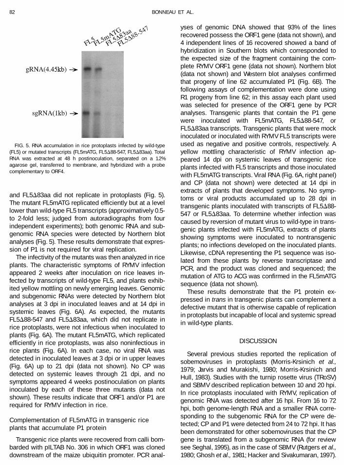
and FL5D83aa did not replicate in protoplasts (Fig. 5).The mutant FL5mATG replicated efficiently but at a levellower than wild-type FL5 transcripts (approximatively 0.5-to 2-fold less; judged from autoradiographs from fourindependent experiments); both genomic RNA and sub-genomic RNA species were detected by Northern blotanalyses (Fig. 5). These results demonstrate that expres-sion of P1 is not required for viral replication.
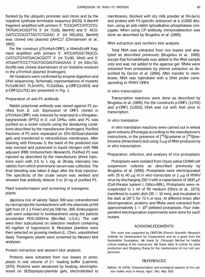
The infectivity of the mutants was then analyzed in riceplants. The characteristic symptoms of RYMV infectionappeared 2 weeks after inoculation on rice leaves in-fected by transcripts of wild-type FL5, and plants exhib-ited yellow mottling on newly emerging leaves. Genomicand subgenomic RNAs were detected by Northern blotanalyses at 3 dpi in inoculated leaves and at 14 dpi insystemic leaves (Fig. 6A). As expected, the mutantsFL5D88-547 and FL5D83aa, which did not replicate inrice protoplasts, were not infectious when inoculated toplants (Fig. 6A). The mutant FL5mATG, which replicatedefficiently in rice protoplasts, was also noninfectious inrice plants (Fig. 6A). In each case, no viral RNA wasdetected in inoculated leaves at 3 dpi or in upper leaves(Fig. 6A) up to 21 dpi (data not shown). No CP wasdetected on systemic leaves through 21 dpi, and nosymptoms appeared 4 weeks postinoculation on plantsinoculated by each of these three mutants (data notshown). These results indicate that ORF1 and/or P1 arerequired for RYMV infection in rice.
Complementation of FL5mATG in transgenic riceplants that accumulate P1 protein
Transgenic rice plants were recovered from calli bom-barded with pILTAB No. 306 in which ORF1 was cloneddownstream of the maize ubiquitin promoter. PCR anal-
yses of genomic DNA showed that 93% of the linesrecovered possess the ORF1 gene (data not shown), and4 independent lines of 16 recovered showed a band ofhybridization in Southern blots which corresponded tothe expected size of the fragment containing the com-plete RYMV ORF1 gene (data not shown). Northern blot(data not shown) and Western blot analyses confirmedthat progeny of line 62 accumulated P1 (Fig. 6B). Thefollowing assays of complementation were done usingR1 progeny from line 62; in this assay each plant usedwas selected for presence of the ORF1 gene by PCRanalyses. Transgenic plants that contain the P1 genewere inoculated with FL5mATG, FL5D88-547, orFL5D83aa transcripts. Transgenic plants that were mockinoculated or inoculated with RYMV FL5 transcripts wereused as negative and positive controls, respectively. Ayellow mottling characteristic of RYMV infection ap-peared 14 dpi on systemic leaves of transgenic riceplants infected with FL5 transcripts and those inoculatedwith FL5mATG transcripts. Viral RNA (Fig. 6A, right panel)and CP (data not shown) were detected at 14 dpi inextracts of plants that developed symptoms. No symp-toms or viral products accumulated up to 28 dpi intransgenic plants inoculated with transcripts of FL5D88-547 or FL5D83aa. To determine whether infection wascaused by reversion of mutant virus to wild-type in trans-genic plants infected with FL5mATG, extracts of plantsshowing symptoms were inoculated to nontransgenicplants; no infections developed on the inoculated plants.Likewise, cDNA representing the P1 sequence was iso-lated from these plants by reverse transcriptase andPCR, and the product was cloned and sequenced; themutation of ATG to ACG was confirmed in the FL5mATGsequence (data not shown).
These results demonstrate that the P1 protein ex-pressed in trans in transgenic plants can complement adefective mutant that is otherwise capable of replicationin protoplasts but incapable of local and systemic spreadin wild-type plants.
DISCUSSION
Several previous studies reported the replication ofsobemoviruses in protoplasts (Morris-Krsinich et al.,1979; Jarvis and Murakishi, 1980; Morris-Krsinich andHull, 1983). Studies with the turnip rosette virus (TRoSV)and SBMV described replication between 10 and 20 hpi.In rice protoplasts inoculated with RYMV, replication ofgenomic RNA was detected after 16 hpi. From 16 to 72hpi, both genome-length RNA and a smaller RNA corre-sponding to the subgenomic RNA for the CP were de-tected; CP and P1 were detected from 24 to 72 hpi. It hasbeen demonstrated for other sobemoviruses that the CPgene is translated from a subgenomic RNA (for reviewsee Seghal, 1995), as in the case of SBMV (Rutgers et al.,1980; Ghosh et al., 1981; Hacker and Sivakumaran, 1997).
FIG. 5. RNA accumulation in rice protoplasts infected by wild-type(FL5) or mutated transcripts (FL5mATG, FL5D88-547, FL5D83aa). TotalRNA was extracted at 48 h postinoculation, separated on a 1.2%agarose gel, transferred to membrane, and hybridized with a probecomplementary to ORF4.
82 BONNEAU ET AL.
P1 was detected in extracts from inoculated and sys-temically infected rice leaves and from protoplats as adoublet of protein with apparent molecular masses of 18and 19 kDa (Figs. 3B and 4C). The first stop codon inORF1 is present at nt 553 (predicted molecular mass of17.8 kDa); a possible readthrough would lead to a proteinwith a molecular mass of 19.5 kDa (stop codon at nt 601)(Ngon A Yassi et al., 1994). The products p18 and p19were also produced in vitro from transcripts of p-ORF1(1–701) and p-ORF1 (1–553); therefore, it is unlikely that p19is a readthrough of the stop codon at nt 553. Further-more, there is no other apparent initiation codon in the59UTR or within ORF1. Therefore we propose that theproteins detected in vitro and in vivo likely result fromdegradation, posttranslational modification(s), or fromstructural or other characteristics of P1. While a singleband related to P1 was detected from transgenic plants,it is likely that this is due to a low resolution of theanalysis.
Mutant FL5mATG was capable of replication in proto-plasts and in transgenic plants that produce P1. Weconcluded therefore that P1 is not strictly required forRNA replication. Nevertheless, viral RNA accumulation is
reduced (about 0.5- to 2-fold less than FL5) when P1 isnot expressed. Consequently we cannot rule out thepossibility that P1 is an enhancing factor for genomeamplification, as demonstrated for P1 of tobacco etchpotyvirus (Verchot and Carrington, 1995). Furthermore, adeletion of nucleic acid sequences from ORF1 upstreamof ORF2 (mutant FL5D88-547) or an insertion of fournucleotides at nt 280 (mutant FL5D83aa) restricted thereplication of the virus. It was recently demonstrated thatin the case of SBMV, ORF2 is translated by scanningribosomes and that deletions or insertions within ORF1can reduce ORF2 expression in vitro and in vivo (Hacker,personal communication). By analogy with SBMV, wesuggest that deletion or insertion of nucleotides withinORF1 of RYMV may interfere with the translation of ORF2.
The lack of infectivity in planta of the mutant FL5mATG,which replicates efficiently in rice protoplasts, providesevidence that a functional P1 is required for the infectivityof RYMV in rice plants. This is confirmed by systemicinfection by FL5mATG in transgenic plants that accumu-lated the wild-type P1. These data demonstrate that oneor more of the functions of P1 acts in trans and isessential during the infection in plants. The absence of
FIG. 6. RNA accumulation in inoculated and systemic leaves of nontransformed or transgenic rice plants that produce P1 (variety Taipei 309)following inoculation with transcripts of FL5, FL5mATG, FL5D88-547, or FL5D83aa. (A) Total RNA was extracted at 3 dpi from inoculated leaves ofnontransformed rice. Total RNA was extracted at 14 dpi from upper leaves of nontransformed or P1 transgenic rice plants. 15 mg of RNA was subjectedto analysis, as in Fig. 5. Control, plants mock inoculated. (B) Western blot using anti-P1 serum showing accumulation of P1 in transgenic plants.Proteins extracted from leaves of transgenic plant were analyzed on a 12.5% polyacrylamide gel and transferred to nitrocellulose. P1, 100 ng of purifiedprotein P1 (predicted molecular mass is 21 kDa including His tag). (2) Negative transgenic lines, (1) positive transgenic line expressing P1.
83EXPRESSION OF RYMV P1 PROTEIN
infectivity of mutants FL5D88-547 and FL5D83aa in trans-genic plants suggests that the P1 produced in transcannot complement the defective replication of thesemutants.
RYMV symptoms appeared more rapidly in P1 trans-genic plants inoculated with RYMV FL5 transcripts thanin nontransformed plants, suggesting that the expressionof P1 in trans enhances the infection process in planta. Itwill be interesting to determine at which particularstep(s) of the infection process this protein is required. Inthis regard, the observation that the mutant FL5mATGcan replicate in a single cell but is noninfectious ininoculated leaves of wild-type rice plants suggestsstrongly that the protein P1 may be involved in host–virusinteractions and/or viral spread, each of which is likely tooccur early in the infection process.
It is well known that cell-to-cell movement of mostviruses requires one or more virus-encoded movementproteins (MP) (for a recent review see Carrington et al.,1996). The function of the TMV MP has been extensivelystudied as a model, and it is well documented that theTMV MP interacts specifically with plant cell plasmod-esmata (Wolf et al., 1989; Moore et al., 1992). No viralproduct encoding this function has yet been reported forsobemoviruses, but several studies have suggestedthat sobemoviruses may encode a cell to cell spreadfactor. Successful complementation assays have beenachieved for cocksfoot mottle sobemovirus in the non-permissive host Cynosorus cristatus by co-inoculationwith cynosorus mottle sobemovirus (Hull, 1988). Simi-larly, SBMV-C can infect Phaseolus vulgaris, a nonper-missive host, when co-infected with SBMV-B (Hackerand Sivakumaran, 1996). Last, sunn-hemp mosaic to-bamovirus enables the cell to cell movement of SBMV-Cin inoculated leaves of P. vulgaris (Fuentes and Hamilton,1991, 1993).
Future work will determine if the P1 protein or the CPor both are involved in cell-to-cell movement of RYMV inrice: both proteins are dispensable for replication butrequired for virus infection in plants (this paper; Brugidouet al., 1995).
MATERIALS AND METHODS
Virus isolate and plant inoculation
The RYMV isolate used in this study was obtainedfrom a rice field in Ivory Coast and was propagated inOryza sativa L., variety IR8. The full-length cDNA (FL5)used in this study was generated from RYMV and cloneddownstream of the T7 promoter (Brugidou et al., 1995).Inoculation and maintenance of plants were as de-scribed by Brugidou et al., (1995). Variety IR8 or Taipei309 plants were mechanically inoculated with 100 mg/mlof virus particles or 1 mg/ml of in vitro transcripts of thefull-length cDNA of RYMV, diluted in phosphate buffer (20mM, pH 7).
Plasmid construction
All mutants were generated from FL5. Nucleotide num-bers refer to the RYMV genome sequence reported byNgon A. Yassi et al. (1994). ORF1 comprises nt 80 to 553(UGA codon), and a second putative UAA stop codon ispresent in frame at nt 601. In the following primer se-quences, sequences complementary to double-strandcDNA of FL5 (in bold), and restriction sites (in italics), areindicated in parenthesis.
For mutant FL5D88-547, nt 88 to 547 were deleted, andthe ORF1 ATG start codon was mutated to ACG bypolymerase chain reaction (PCR) mutagenesis. Frag-ments around 4000 and 3000 bp in length were amplifiedusing primer pairs 59-AACTGCAGGCATGCTTTTGAG-AGAATCAGTCCT-39 (nt 547–566, SphI) and 59-ATGC-ATGCAAGCTTCTCCCCCACCCATCCCGAGAATT-39 (nt4428–4450, HindIII) and 59-TACTTAGGCATGCCGTGTCG-TACACGATGC-39 (nt 71–88, SphI, mutation of the ATG inACG at position 80 is underlined) and 59-ATGGGTGGGG-GAGAAGCTTGGCGTAAT-39 (nt 4438–4450, HindIII), re-spectively. A modified plasmid, combining the deletion ofORF1 and the mutagenized start codon, was construtedby ligating the 4000- and 3000-bp fragments previouslydigested by SphI and HindIII. Finally, the recombinantplasmid was cut with PstI (located downstream of the T7RNA promoter) and NotI (nt 701), and the PstI–NotI frag-ment was reintroduced into FL5.
For mutant FL5mATG, the start codon of ORF1 (nt 80)was mutated to ACG. A SphI fragment amplified withprimers 59-ATCCCGGGCATGCTGTACGACACGGTTG-GAAG-39 (nt 77–95, SphI) and 59-ATCCCGGGCATGCAG-GACTGATTCTCTCAAAAG-39 (nt 547–568, SphI) wassubcloned in the mutant FL5D88-547, previously di-gested by SphI. During the cloning reactions, 12 nucle-otides were expected to be added upstream of the ACGcodon, and nt 547 to 553 were expected to be repeated.However, sequencing of the cloned product showed it tobe identical to the wild-type except that the ATG waschanged to ACG and adenine 204 was changed to thy-midine.
For mutant FL5D83aa, a frameshift mutation was in-serted by filling in the BsrGI site (nt 278) with addition offour nucleotides, ACGT, at nt 280, creating the stopcodon UGA (nt 300) leading to a C-terminal truncation of83 amino acids.
For constructs p-ORF1(1-701) and p-ORF1(1-553),ORF1 was cloned downtream of the T3 RNA promoter inthe pBluescript II (KS1) plasmid (Stratagene, U.S.A.).They correspond to a PstI–NotI fragment (the first 701 ntof FL5 including the UAA codon at nt 601) digested fromFL5 or a PstI–SmaI fragment (the first 568 nt of FL5)amplified with primers 59 ATTCTAGACTGCAGACAATT-GAAGCTAGG 39 (nt 1–15, PstI) and 59 ATCCCGGGCAT-GCAGGACTGATTCTCTCAAAG 39 (nt 547–568, SmaI).
The construct pILTAB No. 306 contains the ORF1
84 BONNEAU ET AL.
flanked by the ubiquitin promoter and intron and by thenopaline synthase terminator sequence (NOS). A BamHIfragment amplified with primers 59 TCGGATCCATCGTG-TATGACACGGTTG 39 (nt 72–91, BamHI) and 59 ACG-GATCCCGCGTTAGTCTGAAC 39 (nt 591–610, BamHI)was cloned into plasmid pAHC17 (Christensen et al.,1992).
For the construct pTrcHisA-ORF1, a NheI–EcoRI frag-ment amplified with primers 59 ATCCATGGCTAGCG-CATCGTGTATGACACGGTT 39 (nt 71–90, NheI) and 59ATGAATTCCCTTGGTGCGAGTGAGAAA 39 (nt 631–710,EcoRI) was cloned downstream of six histidine residuesin the pTrcHisA plasmid (Invitrogen).
All mutations were confirmed by enzyme digestion andDNA sequencing. Schematic representations of mutantsFL5D88-547, FL5mATG, FL5D83aa, p-ORF1(1–553) andp-ORF1(1–701) are presented in Fig. 1.
Preparation of anti-P1 antibody
Rabbit polyclonal antibody was raised against P1 pu-rified from E. coli. Expression of ORF1 cloned inpTrcHisA-ORF1 was induced by isopropyl-b-D-thiogalac-topyranoside (IPTG) in E. coli DH5a cells and P1 waspurified on a nickel column using the denaturing condi-tions described by the manufacturer (Invitrogen). Purifiedfractions of P1 were separated on 15% SDS–acrylamidegels and transferred to nitrocellulose membranes. Afterstaining with Ponceau S, the band of the predicted sizewas excised and pulverized in liquid nitrogen with RIBIadjuvant (RIBI Immunochem Research). Purified P1 wasinjected as described by the manufacturer (three injec-tions each with 0.5 to 1 mg, at 28-day intervals) intorabbits from which preimmune serum was collected. Thefinal bleeding was taken 8 days after the final injection.The specificity of the crude serum was verified andshowed that antibody detects 5 to 10 ng of purified P1.
Plant transformation and screening of transgenicplants
Japonica rice of variety Taipei 309 was cotransformedby microprojectile bombardment with the plasmids pHX4(gift from Dr J. J. Finer) and pILTAB No. 306. Embryogeniccalli were subjected to bombardment using the particleaccelerator PDS-1000/He (Bio-Rad, U.S.A.). The calliwere then subcultured on selection medium containing50 mg/liter of hygromycin B. Resistant plantlets werethen selected on growing medium (L. Chen, unpublisheddata). Transgenic plants were screened by Western blotanalyses.
Protein extraction and western blot analysis
Proteins were extracted from rice leaves or proto-plasts in one volume of 23 loading buffer (Laemmli,1970). Proteins were denatured by heating, electropho-resed on SDS–polyacrylamide gels, electroblotted to
membranes, blocked with dry milk powder at 5% (w/v),and probed with P1-specific antiserum at a 1/1000 dilu-tion, using an anti-rabbit IgG–alkaline phosphatase con-jugate. When using CP antibody, immunodetection wasdone as described by Brugidou et al. (1995).
RNA extraction and northern blot analysis
Total RNA was extracted from rice leaves and ana-lyzed as described previously (Brugidou et al., 1995),except that formaldehyde was added to the RNA sampleonly and was not added to the agarose gel. RNAs wereextracted from protoplasts by phenol extraction as de-scribed by Ga-lon et al. (1994). After transfer to mem-brane, RNA was hybridized with a DNA probe corre-sponding to RYMV ORF4.
In vitro transcription
Transcription reactions were done as described byBrugidou et al. (1995). For the constructs p-ORF1 (1–701)and p-ORF1 (1–553), DNA was cut with NotI prior totranscription.
In vitro translation
In vitro translation reactions were carried out in wheatgerm extracts (Promega) according to the manufacturer’sinstructions, in the presence of [35S]cysteine or [35S]me-thionine (Amersham) and using 3 mg of RNA produced byin vitro transcription.
Preparation, infection, and analysis of rice protoplasts
Protoplasts were isolated from Oryza sativa O2468 cellsuspension cultures as described previously byBrugidou et al. (1995). Protoplasts were electroporatedwith 25 to 40 mg of in vitro transcripts or 1 mg of RYMVvirus by discharging 250 V with a capacitance of 1600 mF(Cell-Porator system I, Gibco-BRL). Protoplasts were re-suspended in 1 ml of R2 medium (Ohira et al., 1973),transfered to a petri dish (35 3 10 mm), and incubated inthe dark at 28°C for 72 h or less. At different times afterelectroporation, proteins and RNAs were extracted fromapproximatively 1 3 106 pelleted protoplasts. Four inde-pendent electroporation experiments were done for eachmutant.
ACKNOWLEDGMENTS
This work was supported by ORSTOM (French Scientific ResearchInstitute for Development by Cooperation) and by a grant from theRockefeller Foundation. We thank Dr. Christoph Reichel for helpfulcritical reading of the manuscript. We thank Sally Jo Leitner for plantproduction and Shipping Zhang for the maintenance of rice cell sus-pensions.
REFERENCES
Bakker, W. (1974). Characterization and ecological aspects of rice yel-low mottle virus in Kenya. Agric. Res. Rep. 829.
85EXPRESSION OF RYMV P1 PROTEIN
Brugidou, C., Holt, C., Ngon A Yassi, M., Zhang, S., Beachy, R., andFauquet, C. (1995). Synthesis of an infectious full-length cDNA cloneof rice yellow mottle virus and mutagenesis of the coat protein.Virology 206, 108–115.
Carrington, J. C., Kasschau, K. D., Mahajan, S. K., and Schaad, M. C.(1996). Cell-to-cell and long distance transport of viruses in plants.Plant Cell 8, 1669–1681.
Christensen, A. H., Sharrock, R. A., and Quail, P. H. (1992). Maizepolyubiquitin genes: Structure, thermal perturbation of expressionand transcript splicing, and promoter activity following transfer toprotoplasts by electroporation. Plant Mol. Biol. 18, 675–689.
Fauquet, C., and Thouvenel, J. C. (1977). Isolation of the rice yellowmottle virus in Ivory Coast. Plant Dis. Rep. 61, 443–446.
Fuentes, A. L., and Hamilton, R. I. (1991). Sunn-hemp mosaic virusfacilitates cell to cell spread of southern bean mosaic virus in a nonpermissive host. Phytopathology 81, 1302–1305.
Fuentes, A. L., and Hamilton, R. I. (1993). Failure of long distancemovement of southern bean mosaic virus in a resistant host iscorrelated with lack of normal virion formation. J. Gen. Virol. 74,1903–1910.
Ga-lon, A., Kaplan, I., Roossinck, M. J., and Palukaitis, P. (1994). Thekinetics of infection of zucchini squash by cucumber mosaic virusindicate a function for RNA1 in virus movement. Virology 205, 280–289.
Ghosh, A., Rutgers, T., Ke-Qiang, M., and Kaesberg, P. (1981). Charac-terization of the coat protein mRNA of southern bean mosaic virusand its relationship to the genomic RNA. J. Virol. 39, 87–92.
Hacker, D. L., and Sivakumaran, K. (1996). Virus-host interactions insouthern bean mosaic virus gene expression and assembly. In ‘‘Bi-ology of Plant–Microbe Interactions’’ (G. Stacey, B. Mullin, and P. M.Gresshoff, Eds.), pp. 277–280. Int. Soc. for Molecular Plant–MicrobeInteractions, St. Paul, Minnesota.
Hacker, D. L., and Sivakumaran, K. (1997). Mapping and expression ofsouthern bean mosaic virus genomic and subgenomic RNAs. Virol-ogy 234, 317–327.
Hull, R. (1988). The sobemovirus group. In ‘‘The Plant Viruses,’’ Vol. 3.(Renate Koenig, Ed.), pp. 113–146. Plenum, New York.
Jarvis, N. P., and Murakishi, H. H. (1980). Infection of protoplasts fromsoybean cell culture with southern bean mosaic and cowpea mosaicviruses. J. Gen. Virol. 48, 365–376.
Laemmli, U. K. (1970). Cleavage of structural proteins during the as-sembly of the head of bacteriophage T4. Nature 227, 680–685.
Moore, P. J., Fenczik, C. A., Deom, C. M., and Beachy, R. N. (1992).Developmental changes in plasmodesmata in transgenic tobaccoexpressing the movement protein of tobacco mosaic virus. Proto-plasma 170, 115–127.
Morris-Krsinich, B., and Hull, R. (1983). Replication of turnip rosettevirus RNA in inoculated turnip protoplasts. J. Gen. Virol. 64, 2661–2668.
Morris-Krsinich, B. A. M., Hull, R., and Russo, M. (1979). Infection ofturnip leaf protoplasts with turnip rosette virus. J. Gen. Virol. 43,339–347.
Murphy F. A., Fauquet, C. A., Bishop, D. H. L., Ghabrial, S. A., Jarvis, A. W.et al. (1995). ‘‘Virus Taxonomy.’’ Springer-Verlag, Vienna.
Ngon A Yassi, M., Ritzenthaler, C., Brugidou, C., Fauquet, C., andBeachy, R. N. (1994). Nucleotide sequence and genome character-ization of rice yellow mottle virus RNA. J. Gen. Virol. 75, 249–257.
Ohira, K., Ojima, K., and Fujiwara, A. (1973). Studies on the nutrition ofrice cell culture: A simple defined medium for rapid growth in sus-pension culture. Plant Cell Physiol. 14, 1113–1121.
Rutgers, T., Salerno-Rife, T., and Kaesberg, P. (1980). Messenger RNAfor the coat protein of southern bean mosaic virus. Virology 104,506–509.
Seghal, O. P. (1995). Sobemoviruses. In ‘‘Pathogenesis and Host Spec-ificity in Plant Diseases: Histological, Biochemical Genetic and Mo-lecular Bases’’ (R. P. Singh, U. S. Singh, and K. Kohomota, Eds.), pp.115–128. Pergamon Press, U.K.
Verchot, J., and Carrington, J. C. (1995). Evidence that the potyvirusproteinase functions in trans as an accessory factor for genomeamplification. J. Gen. Virol. 69, 3668–3674.
Wolf, S., Deom, C. M., Beachy, R. N., and Lucas, W. J. (1989). Movementprotein of tobacco mosaic virus modifies plasmodesmatal size ex-clusion limit. Science 246, 377–379.
86 BONNEAU ET AL.
Related Documents