Expression of Proinflammatory Cytokines by Human Mesenchymal Stem Cells in Response to Cyclic Tensile Strain RUWAN D. SUMANASINGHE, 1 T. WAYNE PFEILER, 1 NANCY A. MONTEIRO-RIVIERE, 1,2 AND ELIZABETH G. LOBOA 1 * 1 Joint Department of Biomedical Engineering, University of North Carolina at Chapel Hill and North Carolina State University, Raleigh, North Carolina 2 Center for Chemical and Toxicology Research and Pharmacokinetics, North Carolina State University, Raleigh, North Carolina Mesenchymal stem cells produce proinflammatory cytokines during their normal growth. Direct or indirect regulation of bone resorption by these cytokines has been reported. However, the effects of osteogenic conditions—chemical and/or mechanical—utilized during in vitro bone tissue engineering on expression of cytokines by hMSCs have not been studied. In this study, we investigated the effects of cyclic tensile strain, culture medium (with and without dexamethasone), and culture duration on the expression of tumor necrosis factor-a (TNF-a), interleukin-1b (IL-1b), interleukin-6 (IL-6), and interleukin-8 (IL-8) by bone marrow derived human mesenchymal stem cells (hMSCs). Human MSCs seeded in three-dimensional Type I collagen matrices were subjected to 0%, 10%, and 12% uniaxial cyclic tensile strains at 1 Hz for 4 h/day for 7 and 14 days in complete growth or dexamethasone-containing osteogenic medium. Viability of hMSCs was maintained irrespective of strain level and media conditions. Expression of either TNF-a or IL-1b was not observed in hMSCs under any of the conditions investigated in this study. Expression of IL-6 was dependent on culture medium. An increase in IL-6 expression was caused by both 10% and 12% strain levels. Both 10% and 12% strain levels caused an increase in IL-8 production by hMSCs that was dependent on the presence of dexamethasone. IL-6 and IL-8 expressions by hMSCs were induced by cyclic tensile strain and osteogenic differentiating media, indicating that IL-6 and IL-8 may be functioning as autocrine signals during osteogenic differentiation of hMSCs. J. Cell. Physiol. 219: 77–83, 2009. ß 2008 Wiley-Liss, Inc. Stimulation of human mesenchymal stem cells (hMSCs) by tensile strain and osteogenic media conditions to induce osteogenic differentiation has been widely investigated (Simmons et al., 2003; Jagodzinski et al., 2004; Koike et al., 2005; Sumanasinghe et al., 2006). Many investigations have focused on determining appropriate strain levels (Simmons et al., 2003; Jagodzinski et al., 2004; Loboa et al., 2005; Koike et al., 2005; Sumanasinghe et al., 2006) and media supplements (Bruder et al., 1997; Jaiswal et al., 1997; Halvorsen et al., 2001) required to achieve osteogenesis of hMSCs. Tensile strains of 10–12% have been shown to initiate intramembranous bone formation in rats (Loboa et al., 2004, 2005). A typical investigation of hMSC osteogenesis includes analyses of bone markers and their corresponding proteins or calcium deposition in the extracellular matrix (Jaiswal et al., 1997; Halvorsen et al., 2001; Frank et al., 2002; Sumanasinghe et al., 2006). Previous studies have indicated production of proinflammatory cytokines such as tumor necrosis factor-a (TNF-a), interleukin-1b (IL-1b), interleukin-6 (IL-6), and interleukin-8 (IL-8) by hMSCs (Filipak et al., 1988; Haynesworth et al., 1996; Majumdar et al., 2000; Kim et al., 2005) and their involvement in either osteogenic differentiation (Gimble et al., 1994; Erices et al., 2002) or bone resorption (Thomson et al., 1987; Fuller et al., 1995; Collin et al., 1996). Interleukin-1b is a major mediator of host inflammatory response and generates systemic responses including fever and the release of acute phase proteins from liver (Carty and Laliberte, 1989). TNF-a is also a proinflammatory cytokine secreted by many non- immune cells and immune cells such as macrophages/ monocytes during acute inflammation (Idriss and Naismith, 2000) and after toxicity (Allen et al., 2000). Its numerous signaling events within cells can cause necrosis and apoptosis (Idriss and Naismith, 2000). Both TNF-a and IL-1b are involved in bone metabolism and regulation of osteoblast and osteoclast function (Gowen et al., 1983; Pacifici et al., 1987; Thomson et al., 1987). IL-6 is a multifunctional cytokine with a major role in inflammatory responses, cell proliferation, and induction of acute phase proteins (Kishimoto, 1989). Increased concentration of IL-6 in the bone microenvironment causes enhanced bone resorption via indirect modulation of osteoclast differentiation (Suda et al., 1999). IL-8, formerly known as neutrophil attractant/activating protein 1 (NAP-1), is a chemotactic cytokine (Wuyts et al., 1998; Dovio et al., 2004) and functions as a proinflammatory substance that involves recruitment and trafficking of neutrophils during an immunologic or inflammatory response (Wuyts et al., 1998). IL-8 is produced and released by many cells, including osteoblasts derived from mesenchymal progenitors (Bilbe et al., 1996; Lisignoli et al., 2002). Previous investigations have shown cytokine involvement during osteogenic differentiation and bone resorption by both Contract grant sponsor: Ralph E. Powe Junior Faculty Enhancement Award. Contract grant sponsor: North Carolina Biotechnology Center Institutional Development. *Correspondence to: Elizabeth G. Loboa, Joint Department of Biomedical Engineering at UNC-Chapel Hill and NC State University, 2147 Burlington Laboratories, Campus Box 7115, Raleigh, NC 27695-7115. E-mail: [email protected] Received 23 January 2007; Accepted 30 October 2008 Published online in Wiley InterScience (www.interscience.wiley.com.), 16 December 2008. DOI: 10.1002/jcp.21653 ORIGINAL ARTICLE 77 Journal of Journal of Cellular Physiology Cellular Physiology ß 2008 WILEY-LISS, INC.

Welcome message from author
This document is posted to help you gain knowledge. Please leave a comment to let me know what you think about it! Share it to your friends and learn new things together.
Transcript

ORIGINAL ARTICLE 77J o u r n a l o fJ o u r n a l o f
CellularPhysiologyCellularPhysiology
Expression of ProinflammatoryCytokines by HumanMesenchymal Stem Cells inResponse to Cyclic Tensile Strain
RUWAN D. SUMANASINGHE,1 T. WAYNE PFEILER,1 NANCY A. MONTEIRO-RIVIERE,1,2AND ELIZABETH G. LOBOA1*1Joint Department of Biomedical Engineering, University of North Carolina at Chapel Hill and North Carolina State University,
Raleigh, North Carolina2Center for Chemical and Toxicology Research and Pharmacokinetics, North Carolina State University, Raleigh, North Carolina
Mesenchymal stem cells produce proinflammatory cytokines during their normal growth. Direct or indirect regulation of bone resorptionby these cytokines has been reported. However, the effects of osteogenic conditions—chemical and/or mechanical—utilized during invitro bone tissue engineering on expression of cytokines by hMSCs have not been studied. In this study, we investigated the effects of cyclictensile strain, culture medium (with and without dexamethasone), and culture duration on the expression of tumor necrosis factor-a(TNF-a), interleukin-1b (IL-1b), interleukin-6 (IL-6), and interleukin-8 (IL-8) by bone marrow derived human mesenchymal stem cells(hMSCs). Human MSCs seeded in three-dimensional Type I collagen matrices were subjected to 0%, 10%, and 12% uniaxial cyclic tensilestrains at 1 Hz for 4 h/day for 7 and 14 days in complete growth or dexamethasone-containing osteogenic medium. Viability of hMSCs wasmaintained irrespective of strain level and media conditions. Expression of either TNF-aor IL-1bwas not observed in hMSCs under any ofthe conditions investigated in this study. Expression of IL-6 was dependent on culture medium. An increase in IL-6 expression was causedby both 10% and 12% strain levels. Both 10% and 12% strain levels caused an increase in IL-8 production by hMSCs that was dependent onthe presence of dexamethasone. IL-6 and IL-8 expressions by hMSCs were induced by cyclic tensile strain and osteogenic differentiatingmedia, indicating that IL-6 and IL-8 may be functioning as autocrine signals during osteogenic differentiation of hMSCs.
J. Cell. Physiol. 219: 77–83, 2009. � 2008 Wiley-Liss, Inc.
Contract grant sponsor: Ralph E. Powe Junior Faculty EnhancementAward.Contract grant sponsor: North Carolina Biotechnology CenterInstitutional Development.
*Correspondence to: Elizabeth G. Loboa, Joint Department ofBiomedical Engineering at UNC-Chapel Hill and NC StateUniversity, 2147 Burlington Laboratories, Campus Box 7115,Raleigh, NC 27695-7115. E-mail: [email protected]
Received 23 January 2007; Accepted 30 October 2008
Published online in Wiley InterScience(www.interscience.wiley.com.), 16 December 2008.DOI: 10.1002/jcp.21653
Stimulation of human mesenchymal stem cells (hMSCs) bytensile strain and osteogenic media conditions to induceosteogenic differentiation has been widely investigated(Simmons et al., 2003; Jagodzinski et al., 2004; Koike et al., 2005;Sumanasinghe et al., 2006). Many investigations have focused ondetermining appropriate strain levels (Simmons et al., 2003;Jagodzinski et al., 2004; Loboa et al., 2005; Koike et al., 2005;Sumanasinghe et al., 2006) and media supplements (Bruderet al., 1997; Jaiswal et al., 1997; Halvorsen et al., 2001) requiredto achieve osteogenesis of hMSCs. Tensile strains of 10–12%have been shown to initiate intramembranous bone formationin rats (Loboa et al., 2004, 2005). A typical investigation of hMSCosteogenesis includes analyses of bone markers and theircorresponding proteins or calcium deposition in theextracellular matrix (Jaiswal et al., 1997; Halvorsen et al., 2001;Frank et al., 2002; Sumanasinghe et al., 2006).
Previous studies have indicated production ofproinflammatory cytokines such as tumor necrosis factor-a(TNF-a), interleukin-1b (IL-1b), interleukin-6 (IL-6), andinterleukin-8 (IL-8) by hMSCs (Filipak et al., 1988; Haynesworthet al., 1996; Majumdar et al., 2000; Kim et al., 2005) and theirinvolvement in either osteogenic differentiation (Gimble et al.,1994; Erices et al., 2002) or bone resorption (Thomson et al.,1987; Fuller et al., 1995; Collin et al., 1996). Interleukin-1b is amajor mediator of host inflammatory response and generatessystemic responses including fever and the release of acutephase proteins from liver (Carty and Laliberte, 1989). TNF-a isalso a proinflammatory cytokine secreted by many non-immune cells and immune cells such as macrophages/monocytes during acute inflammation (Idriss and Naismith,2000) and after toxicity (Allen et al., 2000). Its numeroussignaling events within cells can cause necrosis and apoptosis(Idriss and Naismith, 2000). Both TNF-a and IL-1b are involved
� 2 0 0 8 W I L E Y - L I S S , I N C .
in bone metabolism and regulation of osteoblast and osteoclastfunction (Gowen et al., 1983; Pacifici et al., 1987; Thomsonet al., 1987). IL-6 is a multifunctional cytokine with a major rolein inflammatory responses, cell proliferation, and induction ofacute phase proteins (Kishimoto, 1989). Increasedconcentration of IL-6 in the bone microenvironment causesenhanced bone resorption via indirect modulation of osteoclastdifferentiation (Suda et al., 1999). IL-8, formerly known asneutrophil attractant/activating protein 1 (NAP-1), is achemotactic cytokine (Wuyts et al., 1998; Dovio et al., 2004)and functions as a proinflammatory substance that involvesrecruitment and trafficking of neutrophils during animmunologic or inflammatory response (Wuyts et al., 1998).IL-8 is produced and released by many cells, includingosteoblasts derived from mesenchymal progenitors (Bilbe et al.,1996; Lisignoli et al., 2002).
Previous investigations have shown cytokine involvementduring osteogenic differentiation and bone resorption by both
78 S U M A N A S I N G H E E T A L .
autocrine and paracrine mechanisms in hMSCs. Analysis ofcytokines would provide valuable information on the signalingmechanisms or pathways involved during osteogenicdifferentiation of hMSCs. However, the effects of variousosteogenic differentiation conditions on the expression ofthese cytokines have not been investigated. The purpose of thisstudy was to investigate the effects of cyclic tensile strain,culture medium, and culture duration on the expression ofTNF-a, IL-1b, IL-6, and IL-8 cytokines by hMSCs in three-dimensional (3D) collagen matrices.
Materials and MethodsCell culture
Bone marrow derived hMSCs (24-year-old Caucasian male donor;Tulane University, New Orleans, LA) were passaged three times ingrowth medium containing fetal bovine serum (FBS), 4 mML-glutamine, 0.05 U/ml penicillin, and 0.05 mg/ml streptomycin(Cambrex, Walkersville, MD). All studies were performed usingpassage 3 cells.
Fabrication of hMSC-seeded 3D collagen matrices
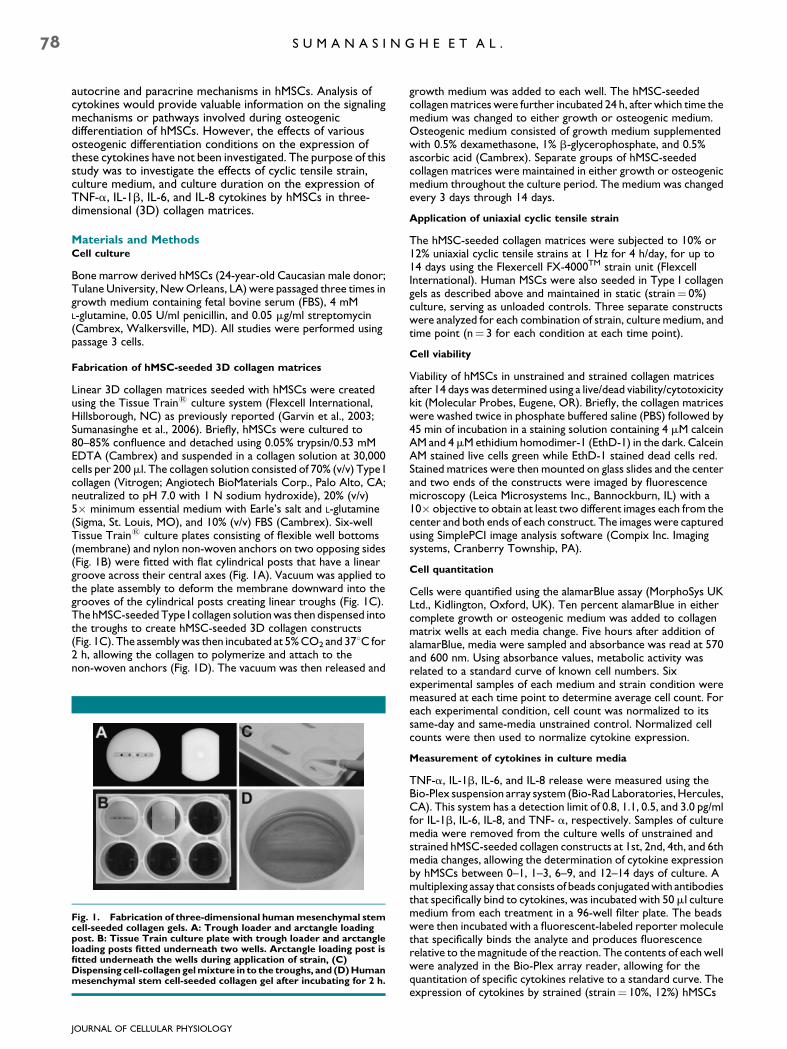
Linear 3D collagen matrices seeded with hMSCs were createdusing the Tissue Train1 culture system (Flexcell International,Hillsborough, NC) as previously reported (Garvin et al., 2003;Sumanasinghe et al., 2006). Briefly, hMSCs were cultured to80–85% confluence and detached using 0.05% trypsin/0.53 mMEDTA (Cambrex) and suspended in a collagen solution at 30,000cells per 200ml. The collagen solution consisted of 70% (v/v) Type Icollagen (Vitrogen; Angiotech BioMaterials Corp., Palo Alto, CA;neutralized to pH 7.0 with 1 N sodium hydroxide), 20% (v/v)5� minimum essential medium with Earle’s salt and L-glutamine(Sigma, St. Louis, MO), and 10% (v/v) FBS (Cambrex). Six-wellTissue Train1 culture plates consisting of flexible well bottoms(membrane) and nylon non-woven anchors on two opposing sides(Fig. 1B) were fitted with flat cylindrical posts that have a lineargroove across their central axes (Fig. 1A). Vacuum was applied tothe plate assembly to deform the membrane downward into thegrooves of the cylindrical posts creating linear troughs (Fig. 1C).The hMSC-seeded Type I collagen solution was then dispensed intothe troughs to create hMSC-seeded 3D collagen constructs(Fig. 1C). The assembly was then incubated at 5% CO2 and 378C for2 h, allowing the collagen to polymerize and attach to thenon-woven anchors (Fig. 1D). The vacuum was then released and
Fig. 1. Fabrication of three-dimensional human mesenchymal stemcell-seeded collagen gels. A: Trough loader and arctangle loadingpost. B: Tissue Train culture plate with trough loader and arctangleloading posts fitted underneath two wells. Arctangle loading post isfitted underneath the wells during application of strain, (C)Dispensing cell-collagen gel mixture in to the troughs, and (D) Humanmesenchymal stem cell-seeded collagen gel after incubating for 2 h.
JOURNAL OF CELLULAR PHYSIOLOGY
growth medium was added to each well. The hMSC-seededcollagen matrices were further incubated 24 h, after which time themedium was changed to either growth or osteogenic medium.Osteogenic medium consisted of growth medium supplementedwith 0.5% dexamethasone, 1% b-glycerophosphate, and 0.5%ascorbic acid (Cambrex). Separate groups of hMSC-seededcollagen matrices were maintained in either growth or osteogenicmedium throughout the culture period. The medium was changedevery 3 days through 14 days.
Application of uniaxial cyclic tensile strain
The hMSC-seeded collagen matrices were subjected to 10% or12% uniaxial cyclic tensile strains at 1 Hz for 4 h/day, for up to14 days using the Flexercell FX-4000TM strain unit (FlexcellInternational). Human MSCs were also seeded in Type I collagengels as described above and maintained in static (strain¼ 0%)culture, serving as unloaded controls. Three separate constructswere analyzed for each combination of strain, culture medium, andtime point (n¼ 3 for each condition at each time point).
Cell viability
Viability of hMSCs in unstrained and strained collagen matricesafter 14 days was determined using a live/dead viability/cytotoxicitykit (Molecular Probes, Eugene, OR). Briefly, the collagen matriceswere washed twice in phosphate buffered saline (PBS) followed by45 min of incubation in a staining solution containing 4 mM calceinAM and 4mM ethidium homodimer-1 (EthD-1) in the dark. CalceinAM stained live cells green while EthD-1 stained dead cells red.Stained matrices were then mounted on glass slides and the centerand two ends of the constructs were imaged by fluorescencemicroscopy (Leica Microsystems Inc., Bannockburn, IL) with a10� objective to obtain at least two different images each from thecenter and both ends of each construct. The images were capturedusing SimplePCI image analysis software (Compix Inc. Imagingsystems, Cranberry Township, PA).
Cell quantitation
Cells were quantified using the alamarBlue assay (MorphoSys UKLtd., Kidlington, Oxford, UK). Ten percent alamarBlue in eithercomplete growth or osteogenic medium was added to collagenmatrix wells at each media change. Five hours after addition ofalamarBlue, media were sampled and absorbance was read at 570and 600 nm. Using absorbance values, metabolic activity wasrelated to a standard curve of known cell numbers. Sixexperimental samples of each medium and strain condition weremeasured at each time point to determine average cell count. Foreach experimental condition, cell count was normalized to itssame-day and same-media unstrained control. Normalized cellcounts were then used to normalize cytokine expression.
Measurement of cytokines in culture media
TNF-a, IL-1b, IL-6, and IL-8 release were measured using theBio-Plex suspension array system (Bio-Rad Laboratories, Hercules,CA). This system has a detection limit of 0.8, 1.1, 0.5, and 3.0 pg/mlfor IL-1b, IL-6, IL-8, and TNF- a, respectively. Samples of culturemedia were removed from the culture wells of unstrained andstrained hMSC-seeded collagen constructs at 1st, 2nd, 4th, and 6thmedia changes, allowing the determination of cytokine expressionby hMSCs between 0–1, 1–3, 6–9, and 12–14 days of culture. Amultiplexing assay that consists of beads conjugated with antibodiesthat specifically bind to cytokines, was incubated with 50ml culturemedium from each treatment in a 96-well filter plate. The beadswere then incubated with a fluorescent-labeled reporter moleculethat specifically binds the analyte and produces fluorescencerelative to the magnitude of the reaction. The contents of each wellwere analyzed in the Bio-Plex array reader, allowing for thequantitation of specific cytokines relative to a standard curve. Theexpression of cytokines by strained (strain¼ 10%, 12%) hMSCs

C Y T O K I N E E X P R E S S I O N B Y M E S E N C H Y M A L S T E M C E L L S 79
was first normalized against that of same-day unstrained(strain¼ 0%) hMSCs and then normalized against the normalizedcell counts. Three hMSC-seeded matrices were used for eachtreatment combination and the assay was carried out in triplicate.Standard curves were generated for each cytokine using 8concentration data points from 3,200 to 0.2 pg/ml created throughserial dilutions from a stock concentration of 50,000 pg/ml.
Statistical analysis
Factorial effects of culture medium, cyclic tensile strain, and cultureduration on normalized cytokine expressions were investigatedusing F-tests from an analysis of variance (ANOVA) appropriate tothe complete, crossed, 2� 3� 4 experimental design. Thesignificance level was defined as P< 0.05.
ResultsCell viability
Fluorescence microscopy images of hMSC-seeded collagenmatrices cultured in both growth and osteogenic media showedthat the majority of cells in the matrices were viable after14 days, irrespective of the strain level applied (Fig. 2).
Cell quantitation
Cell counts varied from unstrained controls in at least oneexperimental condition for each time point (Fig. 3). At days 0–1,all strained hMSCs showed lower cell numbers than unstrainedcontrols. By days 1–3, only 10% strained hMSCs showed lowercell counts than control. The hMSCs strained at 10% showed asignificantly higher cell count than those strained at 12% at days6–9 and 12–14 for both medium conditions.
Expression of TNF-a and IL-1b
Unstrained and strained hMSCs cultured in either growth orosteogenic medium had very low expression of TNF-a at alltime points analyzed (data not shown). The mean concentrationof TNF-a in the hMSCs ranged from 0 to 0.1 pg/ml. Similarly, themean expression of IL-1b in both strained and unstrainedhMSCs ranged from 0 to 0.04 pg/ml. There was no significanteffect from strain, culture medium, or culture duration oneither TNF-a or IL-1b expressions.
Expression of IL-6
Concentration levels of IL-6 in hMSCs cultured in growthmedium significantly increased during days 6–9 and continued
Fig. 2. Strained and unstrained hMSC-seeded type 1 collagenmatrices cultured in growth (A–C) and osteogenic medium (D–F) for2 weeks and stained with calcein AM and EthD-1 for live (green) anddead (red) cells, respectively. Arrows indicate dead cells. A,D:unstrained; (B,E) strained at 10%; (C,F) strained at 12%. [Color figurecan be viewed in the online issue, which is available atwww.interscience.wiley.com.]
JOURNAL OF CELLULAR PHYSIOLOGY
to increase through day 14 (Fig. 4A). The concentrations of IL-6significantly reduced in the presence of dexamethasone in theculture media after 1–3 days (Fig. 4B). Cyclically strained hMSCscultured in growth medium showed statistically significantlower levels of IL-6 expression compared to their unstrainedcontrols (0% strained hMSCs) at all time points with theexception of 10% strain during days 1–3 (Fig. 5A). Compared tounstrained controls and 12% strained hMSCs, there was asignificant increase in IL-6 expression in 10% strained hMSCscultured in growth medium between 1 and 3 days (Fig. 5A).However, the expression levels in strained hMSCs (10% and12%) significantly reduced during culture days 6–9 andremained unchanged through day 14. In contrast to hMSCscultured in growth medium, those cultured in osteogenicmedium (i.e., with dexamethasone) and cyclically strained ateither 10% or 12% usually showed significantly greater IL-6expression (Fig. 5B).
Expression of IL-8
The greatest expression of IL-8 in strained hMSCs cultured ingrowth medium was observed during days 1–3 (Fig. 6A) whilethe highest IL-8 expression overall was observed in osteogenicmedium cultures during days 12–14 (Fig. 6B). Theconcentration of IL-8 in media of hMSCs cultured withdexamethasone (osteogenic medium) continuously increasedfrom days 1 to 14 with the exception of 10% strained hMSCsbetween days 6 and 9 (Fig. 6B). These expression levels weresignificantly greater than those observed withoutdexamethasone in culture media after days 6–9, and days 12–14(Fig. 6A). Both 10% and 12% strained hMSCs cultured in growthmedium expressed significantly greater levels of IL-8 duringdays 1–3 and 6–9 relative to unstrained controls (Fig. 7A). Inosteogenic medium, the hMSCs strained at 10% showed thegreatest IL-8 levels relative to unstrained controls during days1–3, with levels then significantly decreased during days 6–14(Fig. 7B).
Discussion
The mitotic, metabolic, and developmental activities ofmesenchymal stem cells are regulated by components in theextracellular environment including autocrine and paracrinefactors synthesized by the hMSCs themselves (Bruder et al.,1997; Oreffo et al., 1999; Erices et al., 2002). Signals thatmodulate the growth and differentiation capacity ofmesenchymal stem cells provide valuable information on signalcascades involved in their terminal differentiation. Theexistence of, and changes in, these signals during in vitroosteogenic differentiation need to be investigated in order toidentify key factors and to understand the mechanisms ofosteogenesis of hMSCs. The proinflammatory cytokinesTNF-a, IL-1b, IL-6, and IL-8 have been reported to beexpressed in hMSCs under normal growth conditions(Haynesworth et al., 1996; Majumdar et al., 2000; Kim et al.,2005) and have also been found to be indirectly or directlyconnected with modulating bone resorption (Thomson et al.,1987; Fuller et al., 1995). However, the effects of different invitro osteogenic differentiating conditions on the expression ofthese cytokines in hMSCs have not been investigated.
Cell viability studies indicated that the hMSCs remainedviable in the 3D collagen matrices throughout the experimentalperiod irrespective of the strain levels and media conditions.Cell quantitation studies showed significant differences in cellgrowth during the experimental period.
Previous studies indicate that both TNF-a and IL-1b play arole in bone metabolism and regulate the function ofosteoblasts and osteoclasts (Gowen et al., 1983; Pacifici et al.,1987; Thomson et al., 1987). Investigations by Thomson et al.showed that TNF-a stimulated bone resorption by osteoclasts

Fig. 3. Number of human mesenchymal stem cells in collagen gels subjected to 0% (control), 10%, and 12% cyclic tensile strain cultured in; (A)Growthmedium,and(B)Osteogenicmedium,normalizedtosame-dayunstrainedcontrols (0%strainedhMSCs).Unless indicatedotherwisewitha horizontal bracket, statistical significance symbols represent significance with reference to same-day unstrained control (0% strained). Datarepresented as mean W SEM, n U 6.
80 S U M A N A S I N G H E E T A L .
in the presence of osteoblasts or when supernatants ofosteoblasts were added to the culture media. This indicatedthat activation of bone resorption by TNF-a requires signalmolecules from the osteoblasts. Filipak et al. (1988) reportedthat terminal differentiation of MSCs down the adipogenicpathways was inhibited by TNF-a. However, his study did notinvestigate the effect of TNF-a on osteogenic differentiation ofMSCs. The maximum release of TNF-a by human epidermalkeratinocytes (HEK) exposed to different types of jet fuels hasbeen observed at 4 h, which decreased in a time-dependentmanner (Allen et al., 2000). Release of TNF-a by HEKs has alsobeen shown to induce IL-8 release from HEKs exposed to theinsecticide permethrin and the insect repellent N,N-diethyl-m-toluamide (DEET) (Luger and Schwarz, 1990; Allen et al., 2000;Monteiro-Riviere et al., 2003). The expression of TNF-a andIL-1b observed in both unstrained and strained hMSCs weresignificantly lower than seen in other cell types exposed tochemical as opposed to mechanical stresses (Luger andSchwarz, 1990; Allen et al., 2000; Monteiro-Riviere et al., 2003).
Fig. 4. Expressionof IL-6byhMSCsseededin3Dcollagenmatricesandsubmedium,and(B)Osteogenicmedium.Unless indicatedotherwisewithahowith reference to same-day unstrained control (0% strained). Data repres
JOURNAL OF CELLULAR PHYSIOLOGY
It is possible that TNF-a and IL-1b might have expressed atearlier time points in our studies (i.e., within hours as opposedto days). Alternatively, according to observations made byThomson et al. (1987), the non-expression of TNF-a might bedue to the absence of signal molecules from differentiatedosteoblasts. Expression of TNF-a by human osteoblasts (Walshet al., 2000; Bu et al., 2003), human osteoblast-like MG-63 cells(Sohrabi et al., 2000), and human bone biopsies (Ralston, 1994)has been reported. IL-8 expression has also been detected inhuman osteoblasts (Birch et al., 1993; Walsh et al., 2000)derived from bone explants. Application of low magnitudetensile strain in the range of 5–10% to chondrocytes has beenshown to inhibit inflammation via suppression of IL-1b andTNF-a while high magnitude strains in the range of 20% causedproinflammatory reactions (Deschner et al., 2003). Exposure offibrochondrocytes to dynamic tensile strains from 5% to 20%has been shown to suppress expression of proinflammatorygenes including IL-1b and TNF-a (Ferretti et al., 2006).Combining these findings with those of the present study, it is
jectedto0%(control),10%,and12%cyclictensilestrains in; (A)Growthrizontalbracket, statistical significancesymbolsrepresent significanceented as mean W SEM, n U 3.

Fig. 5. Foldchange inexpressionof IL-6byhMSCsseeded in3Dcollagenmatricesandsubjectedto0%(control),10%,and12%cyclic tensilestrainsin; (A)Growthmedium,and(B)Osteogenicmedium.Foldchange inexpressioncalculatedbynormalizingexpressionagainstsame-dayunstrainedcontrol(0%strained).Unless indicatedotherwisewithahorizontalbracket,statisticalsignificancesymbolsrepresentsignificancewithreferencetosame-day unstrained control (0% strained). Data represented as mean W SEM, n U 3.
C Y T O K I N E E X P R E S S I O N B Y M E S E N C H Y M A L S T E M C E L L S 81
possible to conclude that certain magnitudes of cyclic tensilestrain might function as an anti-inflammatory signal in hMSCs,inhibiting expression of both IL-1b and TNF-a.
Bone marrow derived hMSCs have been reported tocontinuously express IL-6 under normal growth conditions(Haynesworth et al., 1996; Majumdar et al., 1998, 2000; Kimet al., 2005). Haynesworth et al. (1996) and Majumdar et al.(2000) reported that the expression of IL-6 by human bonemarrow derived MSCs was significantly inhibited by thepresence of dexamethasone in the culture medium. Kim et al.(2005) also reported that the initial expression of IL-6 in bonemarrow derived MSCs was decreased when MSCs weresubjected to osteogenic differentiation conditions (in thepresence of dexamethasone) for 2 weeks. IL-6 has been foundto mediate signals through receptors that use gp130 in itssignaling pathway and to regulate bone marrow stromal celldifferentiation (Gimble et al., 1994). IL-6 has also been reportedto increase cell proliferation and expression of alkaline
Fig. 6. ExpressionofIL-8byhMSCsseededin3Dcollagenmatricesandsubmedium,and(B)Osteogenicmedium.Unless indicatedotherwisewithahowith reference to same-day unstrained control (0% strained). Data repres
JOURNAL OF CELLULAR PHYSIOLOGY
phosphatase, and to decrease mRNA expression of osteocalcin(Haynesworth et al., 1996). In the present study, observationson initial IL-6 concentration data indicated a significant downregulation of IL-6 expression in all hMSCs after day 1 in thepresence of dexamethasone, irrespective of strain. This couldbe due to the decrease in cell proliferation of hMSCs in thepresence of osteogenic differentiating media (Jaiswal et al.,1997) as confirmed by the normalized cell count results in thisstudy (Fig. 3) or perhaps to the anti-inflammatory action ofdexamethasone itself. These results correlate well withprevious investigations where an inhibition of IL-6 expressionwas observed in the presence of dexamethasone (Haynesworthet al., 1996; Majumdar et al., 2000; Kim et al., 2005).
The initial concentration data in the present study werenormalized against same-day unstrained controls (0% strainedhMSCs) and normalized cell counts to determine the effect ofstrain on IL-6 expression. The effect of strain on IL-6 expressionwas dependent on culture medium and time. The IL-6
jectedto0%(control),10%,and12%cyclictensilestrains in; (A)Growthrizontalbracket, statistical significancesymbolsrepresent significanceented as mean W SEM, n U 3.

Fig. 7. Foldchange inexpressionof IL-8byhMSCsseeded in3Dcollagenmatricesandsubjectedto0%(control),10%,and12%cyclictensilestrainsin; (A)Growthmedium,and(B)Osteogenicmedium.Foldchange inexpressioncalculatedbynormalizingexpressionagainstsame-dayunstrainedcontrol(0%strained).Unless indicatedotherwisewithahorizontalbracket,statisticalsignificancesymbolsrepresentsignificancewithreferencetosame-day unstrained control (0% strained). Data represented as mean W SEM, n U 3.
82 S U M A N A S I N G H E E T A L .
expression in hMSCs was upregulated briefly (during 1–3 daysof culture) by 10% strain stimuli regardless of the mediaconditions, while the temporary (during days 1–3 and 6–9) IL-6upregulation by 12% strain stimuli occurred only in thepresence of dexamethasone in culture media. However,increased IL-6 expression levels due to both 10% and 12% strainstimuli in the presence of dexamethasone remained elevatedlonger than those observed without dexamethasone. Theseresults indicate that strain stimuli might diminish the inhibitionof IL-6 expression by dexamethasone. Since IL-6 is releasedduring irritation (Barker et al., 1991; Grone, 2002), it is possiblethat irritation of hMSCs caused by cyclic strain could triggerrelease of IL-6 even in the presence of dexamethasone.
Previous studies have reported that IL-8 can function as aninhibitor to osteoclast bone resorptive activity (Fuller et al.,1995; Collin et al., 1996). Investigations have shown that boneresorption was modulated in avian marrow derived osteoclastlike cells through nitric oxide (NO) production that wasupregulated by IL-8 action (Sunyer et al., 1996). Fuller et al.reported that IL-8, while inhibiting the proportion ofosteoclasts that were resorbing bone, also stimulatedosteoclast motility. The expression of IL-8 in IL-1b stimulatedhuman bone marrow stromal cells has been reported to bedown regulated in the presence of dexamethasone (Chaudharyand Avioli, 1994, 1996), but increased when dexamethasonewas not present. Kim et al. reported that expression of IL-8 wasslightly upregulated when bone marrow derived MSCs weresubjected to osteogenic differentiation culture medium for14 days. In the present study, we observed a continuousupregulation of IL-8 in both unstrained and strained hMSCs inthe presence of dexamethasone. Our results with hMSCs thatwere unstrained and cultured in growth medium correlate withobservations made by Kim et al. The difference between ourresults and results of Chaudhary et al. could be due to the factthat hMSCs in the present study were not stimulated by IL-1b inthe presence of dexamethasone. In the present study, analysisof strain, medium, and culture duration on IL-8 expression usingexpression data normalized to both cytokine expression andcell counts of same-day unstrained controls (0% strainedhMSCs) revealed that strain briefly increased IL-8 expression inhMSCs. The significant effects of strain stimuli were observed toremain longer (during days 1–3 and 6–9) in hMSCs cultured
JOURNAL OF CELLULAR PHYSIOLOGY
without dexamethasone, while in the presence ofdexamethasone, IL-8 expression was increased during the first3 days. Since IL-8 has been shown to be released duringirritation (Barker et al., 1991; Grone, 2002), the initial increasein IL-8 even in the presence of dexamethasone could be due tosimilar conditions caused by cyclic strain. The elevation of IL-8by hMSCs due to cyclic strain irrespective of the presence ofdexamethasone suggests that stimulation of hMSCs by strainnot only induces expression of genes indicative of osteogenicdifferentiation (Sumanasinghe et al., 2006) but also producesfactors such as IL-8 to inhibit bone resorption.
A limitation of this study is that it was performed usinghMSCs isolated from a single donor. Donor-to-donorvariability is common and it is possible other donors might havedisparate results from those found here. This should be furtherinvestigated.
In summary, we have investigated the effects of cyclic tensilestrain (10% and 12%), culture duration (through 14 days), andculture medium (both with and without dexamethasone) on theexpression of TNF-a, IL-1b, IL-6, and IL-8 cytokines by bonemarrow derived hMSCs cultured in 3D collagen matrices.Results of this study showed that TNF-a and IL-1b wereminimally expressed in hMSCs under all conditions investigatedin this study. The presence of dexamethasone caused adecrease in IL-6 expression in all hMSCs irrespective of the levelof strain, but strain further affected the inhibition of IL-6expression by dexamethasone. The expression of IL-8 instrained hMSCs suggested that cyclic tensile straintemporarily induced hMSCs to produce IL-8, which couldlead to inhibition of bone resorption during osteogenesis. Thisstudy provides useful information on the activity of cytokinesinvolved in regulating bone resorption and remodeling duringosteogenic induction of hMSCs cultured in a 3D environmentunder cyclic tensile strain and in the presence ofdexamethasone.
Acknowledgments
Financial support for this work was provided by the Ralph E.Powe Junior Faculty Enhancement Award and the NorthCarolina Biotechnology Center Institutional DevelopmentGrant (E.G.L.). The authors wish to thank Alfred Inman for his

C Y T O K I N E E X P R E S S I O N B Y M E S E N C H Y M A L S T E M C E L L S 83
technical assistance and Dr. Susan Bernacki for her advice andguidance.
Literature Cited
Allen DG, Riviere JE, Monteiro-Riviere NA. 2000. Identification of early biomarkers ofinflammation produced by keratinocytes exposed to jet fuels jet A, JP-8, and JP-8(100).J Biochem Mol Toxicol 14:231–237.
Barker J, Mitra R, Griffiths C, Dixit V, Nickoloff B. 1991. Keratinocytes as initiators ofinflammation. Lancet 337:211–214.
Bilbe G, Roberts E, Birch M, Evans DB. 1996. PCR phenotyping of cytokines, growth factorsand their receptors and bone matrix proteins in human osteoblast-like cell lines. Bone19:437–445.
Birch MA, Ginty AF, Walsh CA, Fraser WD, Gallagher JA, Bilbe G. 1993. PCR detection ofcytokines in normal human and pagetic osteoblast-like cells. J Bone Miner Res 8:1155–1162.
Bruder SP, Jaiswal N, Haynesworth SE. 1997. Growth kinetics, self-renewal, and theosteogenic potential of purified human mesenchymal stem cells during extensivesubcultivation and following cryopreservation. J Cell Biochem 64:278–294.
Bu R, Borysenko CW, Li Y, Cao L, Sabokbar A, Blair HC. 2003. Expression and function ofTNF-family proteins and receptors in human osteoblasts. Bone 33:760–770.
Carty TJ, Laliberte RE. 1989. Meeting report. The biochemistry and pharmacology ofinterleukins-1 and -6. Agents Actions 26:391–393.
Chaudhary LR, Avioli LV. 1994. Dexamethasone regulates IL-1 beta and TNF-alpha-inducedinterleukin-8 production in human bone marrow stromal and osteoblast-like cells. CalcifTissue Int 55:16–20.
Chaudhary LR, Avioli LV. 1996. Regulation of interleukin-8 gene expression by interleukin-1beta, osteotropic hormones, and protein kinase inhibitors in normal human bone marrowstromal cells. J Biol Chem 271:16591–16596.
Collin OP, Kirsch D, Anderson F, Joost O, Dean A, Osdoby P. 1996. The chemokine IL-8 as anautocrine inhibitor of osteoclast bone resorptive activity via IL-8 receptors expressed byavain osteoclasts and human osteoclasts like cells. J Bone Miner Res 11:S357.
Deschner J, Hofman CR, Piesco NP, Agarwal S. 2003. Signal transduction by mechanical strainin chondrocytes. Curr Opin Clin Metab Care 6:289–293.
Dovio A, Sartori ML, Masera RG, Peretti L, Perotti L, Angeli A. 2004. Effects of physiologicalconcentrations of steroid hormones and interleukin-11 on basal and stimulatedproduction of interleukin-8 by human osteoblast-like cells with different functionalprofiles. Clin Exp Rheumatol 22:79–84.
Erices A, Conget P, Rojas C, Minguell JJ. 2002. Gp130 activation by soluble interleukin-6receptor/interleukin-6 enhances osteoblastic differentiation of human bone marrow-derived mesenchymal stem cells. Exp Cell Res 280:24–32.
Ferretti M, Madhavan S, Deschner J, Rath-Deshner B, Wypasek E, Agarwal S. 2006. Dynamicbiophysical strain modulates proinflammatory gene induction in meniscalfibrochondrocytes. Am J Physiol Cell Physiol 290:C1610–C1615.
Filipak M, Sparks RL, Tzen CY, Scott RE. 1988. Tumor necrosis factor inhibits the terminalevent in mesenchymal stem cell differentiation. J Cell Physiol 137:367–373.
Frank O, Heim M, Jakob M, Barbero A, Schafer D, Bendik I, Dick W, Heberer M, Martin I.2002. Real-time quantitative RT-PCR analysis of human bone marrow stromal cells duringosteogenic differentiation in vitro. J Cell Biochem 85:737–746.
Fuller K, Owens JM, Chambers TJ. 1995. Macrophage inflammatory protein-1 alpha and IL-8stimulate the motility but suppress the resorption of isolated rat osteoclasts. J Immunol154:6065–6072.
Garvin J, Qi J, Maloney M, Banes AJ. 2003. Novel system for engineering bioartificial tendonsand application of mechanical load. Tissue Eng 9:967–979.
Gimble JM, Wanker F, Wang CS, Bass H, Wu X, Kelly K, Yancopoulos GD, Hill MR. 1994.Regulation of bone marrow stromal cell differentiation by cytokines whose receptorsshare the gp130 protein. J Cell Biochem 54:122–133.
Gowen M, Wood DD, Ihrie EJ, McGuire MK, Russell RG. 1983. An interleukin 1 like factorstimulates bone resorption in vitro. Nature 306:378–380.
Grone A. 2002. Keratinocytes and cytokines. Vet Immunol Immunopathol 88:1–12.Halvorsen YD, Franklin D, Bond AL, Hitt DC, Auchter C, Boskey AL, Paschalis EP, Wilkison
WO, Gimble JM. 2001. Extracellular matrix mineralization and osteoblast gene expressionby human adipose tissue-derived stromal cells. Tissue Eng 7:729–741.
Haynesworth SE, Baber MA, Caplan AI. 1996. Cytokine expression by human marrow-derived mesenchymal progenitor cells in vitro: Effects of dexamethasone and IL-1 alpha.J Cell Physiol 166:585–592.
JOURNAL OF CELLULAR PHYSIOLOGY
Idriss HT, Naismith JH. 2000. TNF alpha and the TNF receptor superfamily: Structure-function relationship(s). Microsc Res Tech 50:184–195.
Jagodzinski M, Drescher M, Zeichen J, Hankemeier S, Krettek C, Bosch U, van Griensven M.2004. Effects of cyclic longitudinal mechanical strain and dexamethasone on osteogenicdifferentiation of human bone marrow stromal cells. Eur Cell Mater 7:35–41 discussion 41.
Jaiswal N, Haynesworth SE, Caplan AI, Bruder SP. 1997. Osteogenic differentiation ofpurified, culture-expanded human mesenchymal stem cells in vitro. J Cell Biochem 64:295–312.
Kim DH, Yoo KH, Choi KS, Choi J, Choi SY, Yang SE, Yang YS, Im HJ, Kim KH, Jung HL,Sung KW, Koo HH. 2005. Gene expression profile of cytokine and growth factorduring differentiation of bone marrow-derived mesenchymal stem cell. Cytokine 31:119–126.
Kishimoto T. 1989. The biology of interleukin-6. Blood 74:1–10.Koike M, Shimokawa H, Kanno Z, Ohya K, Soma K. 2005. Effects of mechanical strain on
proliferation and differentiation of bone marrow stromal cell line ST2. J Bone Miner Metab23:219–225.
Lisignoli G, Toneguzzi S, Grassi F, Piacentini A, Tschon M, Cristino S, Gualtieri G, Facchini A.2002. Different chemokines are expressed in human arthritic bone biopsies: IFN-gammaand IL-6 differently modulate IL-8, MCP-1 and rantes production by arthritic osteoblasts.Cytokine 20:231–238.
Loboa EG, Fang TD, Parker DW, Warren SM, Fong KD, Longaker MT, Carter DR. 2005.Mechanobiology of mandibular distraction osteogenesis: Finite element analyses with a ratmodel. J Orthop Res 23:663–670.
Loboa EG, Fang TD, Warren SM, Lindsey DP, Fong KD, Longaker MT, Carter DR. 2004.Mechanobiology of mandibular distraction osteogenesis: Experimental analyses with a ratmodel. Bone 34:336–343.
Luger TA, Schwarz T. 1990. Evidence for an epidermal cytokine network. J Invest Dermatol95:100S–104S.
Majumdar MK, Thiede MA, Haynesworth SE, Bruder SP, Gerson SL. 2000. Human marrow-derived mesenchymal stem cells (MSCs) express hematopoietic cytokines and supportlong-term hematopoiesis when differentiated toward stromal and osteogenic lineages.J Hematother Stem Cell Res 9:841–848.
Majumdar MK, Thiede MA, Mosca JD, Moorman M, Gerson SL. 1998. Phenotypic andfunctional comparison of cultures of marrow-derived mesenchymal stem cells (MSCs) andstromal cells. J Cell Physiol 176:57–66.
Monteiro-Riviere NA, Baynes RE, Riviere JE. 2003. Pyridostigmine bromide modulates topicalirritant-induced cytokine release from human epidermal keratinocytes and isolatedperfused porcine skin. Toxicology 183:15–28.
Oreffo RO, Kusec V, Romberg S, Triffitt JT. 1999. Human bone marrow osteoprogenitorsexpress estrogen receptor-alpha and bone morphogenetic proteins 2 and 4 mRNA duringosteoblastic differentiation. J Cell Biochem 75:382–392.
Pacifici R, Rifas L, Teitelbaum S, Slatopolsky E, McCracken R, Bergfeld M, Lee W, Avioli LV,Peck WA. 1987. Spontaneous release of interleukin 1 from human blood monocytesreflects bone formation in idiopathic osteoporosis. Proc Natl Acad Sci USA 84:4616–4620.
Ralston SH. 1994. Analysis of gene expression in human bone biopsies by polymerase chainreaction: Evidence for enhanced cytokine expression in postmenopausal osteoporosis.J Bone Miner Res 9:883–900.
Simmons CA, Matlis S, Thornton AJ, Chen S, Wang CY, Mooney DJ. 2003. Cyclic strainenhances matrix mineralization by adult human mesenchymal stem cells via theextracellular signal-regulated kinase (ERK1/2) signaling pathway. J Biomech 36:1087–1096.
Sohrabi A, Holland C, Kue R, Nagle D, Hungerford DS, Frondoza CG. 2000. Proinflammatorycytokine expression of IL-1beta and TNF-alpha by human osteoblast-like MG-63 cells uponexposure to silicon nitride in vitro. J Biomed Mater Res 50:43–49.
Suda T, Takahashi N, Udagawa N, Jimi E, Gillespie MT, Martin TJ. 1999. Modulation ofosteoclast differentiation and function by the new members of the tumor necrosis factorreceptor and ligand families. Endocr Rev 20:345–357.
Sumanasinghe RD, Bernacki SH, Loboa EG. 2006. Osteogenic differentiation of humanmesenchymal stem cells in collagen matrices: Effect of uniaxial cyclic tensile strain on bonemorphogenetic protein (BMP-2) mRNA expression. Tissue Eng 12:3459–3465.
Sunyer T, Rothe L, Jiang X, Osdoby P, Collin-Osdoby P. 1996. Proinflammatory agents, IL-8and IL-10, upregulate inducible nitric oxide synthase expression and nitric oxideproduction in avian osteoclast-like cells. J Cell Biochem 60:469–483.
Thomson BM, Mundy GR, Chambers TJ. 1987. Tumor necrosis factors alpha and beta induceosteoblastic cells to stimulate osteoclastic bone resorption. J Immunol 138:775–779.
Walsh CA, Birch MA, Fraser WD, Ginty AF, Gallagher JA. 2000. Cytokine expression bycultured osteoblasts from patients with osteoporotic fractures. Int J Exp Path 81:159–163.
Wuyts A, Proost P, Van Damme J. 1998. In: Thompson A, editor. Interleukin-8 and otherCXC chemokines. London: Academic Press. pp. 271–311.
Related Documents