ORIGINAL ARTICLE Korean J Intern Med 2013;28:573-578 http://dx.doi.org/10.3904/kjim.2013.28.5.573 Copyright © 2013 The Korean Association of Internal Medicine This is an Open Access article distributed under the terms of the Creative Commons Attribution Non-Commercial License (http://creativecommons.org/licenses/ by-nc/3.0/) which permits unrestricted noncommercial use, distribution, and reproduction in any medium, provided the original work is properly cited. pISSN 1226-3303 eISSN 2005-6648 http://www.kjim.org 1 Department of Biotechnology, Invertis University, Bareilly; 2 Department of Surgical Oncology, Institute of Medical Sciences, Banaras Hindu University, Varanasi; 3 Department of Biochemistry, Allahabad University, Allahabad, India Received : December 28, 2012 Revised : February 14, 2013 Accepted: May 27, 2013 Correspondence to Sanjeev Kumar Maurya, M.D. Department of Biotechnology, Invertis University, Invertis Village, Lucknow National Highway 24, Bareilly 243123, India Tel: +91-945-436-5225 Fax: +91-581-246-0454 E-mail: [email protected] Background/Aims: The higher incidence of gallbladder cancer (GBC) in females has been accredited to the involvement of hormones. The clinical implications of sex hormone receptors in GBC are well established. Cysteine proteases (such as caspase-3-9, etc.) are known to play a central role in the apoptotic pathway. Of these, the downstream enzyme caspase-3 is often activated in the apoptotic path- way. The aim of this work was to examine the status of apoptosis (which directly correlated with the level of active caspase-3) in hormone-responsive GBC. Methods: We used 10 androgen receptor (AR)-positive, 14 estrogen receptor (ER)-positive, 12 HER/neu-positive, eight triple positive, and 10 triple negative malignant GBC human tissue samples. We isolated the total cellular protein from tumor tissues and carried out Western blotting using antipro-caspase-3 and anti-activated caspase-3 antibodies. Results: ER and HER/neu-positive GBC exhibited high caspase-3 activity and low procaspase-3 activity, whereas AR-positive GBC showed no significant level of apoptosis. We also evaluated the apoptosis status of triple positive GBC and triple negative GBC, and found significant apoptosis in triple positive GBC. Conclusions: The results indicate that ER and HER/neu-positive GBCs had active apoptosis, whereas AR-positive GBC was highly resistant to apoptosis. Keywords: Gallbladder neoplasms; Antiapoptotic molecules; Apoptosis; Signal transduction; Up-regulation INTRODUCTION Gallbladder carcinoma (GBC) is a gastrointestinal and often fatal malignancy, ranked fifth overall and the third most common biliary tract malignancy [1,2]. Correct diagnosis at a time which would enable a cure for GBC remains problematic due to its asymptom- atic nature during the initial stages [3,4]. In general, it is one of the most aggressive biliary cancers with a very short survival duration [5]. The higher incidence of GBC in females has been attributed in part to hor- monal factors, including estrogen and progesterone receptor expression and their effects on GBC prog- nosis [6,7]. Many other reports have also referred to the effects of hormone receptor status in GBC [8-14]. More recently, a significant correlation between hor- mone receptor polymorphism and gallbladder (GB) carcinogenesis has been reported [15]. Androgen re- ceptor (AR) CAG repeats have been directly correlated with gallstone formation and GB carcinogenesis [16]. These studies have shown that hormone receptors are important for GB carcinogenesis, with whole GB re- Expression of procaspase 3 and activated caspase 3 and its relevance in hormone-responsive gallbladder carcinoma chemotherapy Sanjeev Kumar Maurya 1 , Mallika Tewari 2 , Bechan Sharma 3 , and Hari Shanker Shukla 2

Welcome message from author
This document is posted to help you gain knowledge. Please leave a comment to let me know what you think about it! Share it to your friends and learn new things together.
Transcript
ORIGINAL ARTICLE
Korean J Intern Med 2013;28:573-578http://dx.doi.org/10.3904/kjim.2013.28.5.573
Copyright © 2013 The Korean Association of Internal MedicineThis is an Open Access article distributed under the terms of the Creative Commons Attribution Non-Commercial License (http://creativecommons.org/licenses/by-nc/3.0/) which permits unrestricted noncommercial use, distribution, and reproduction in any medium, provided the original work is properly cited.
pISSN 1226-3303eISSN 2005-6648
http://www.kjim.org
1Department of Biotechnology, Invertis University, Bareilly; 2Department of Surgical Oncology, Institute of Medical Sciences, Banaras Hindu University, Varanasi; 3Department of Biochemistry, Allahabad University, Allahabad, India
Received : December 28, 2012Revised : February 14, 2013 Accepted : May 27, 2013
Correspondence to Sanjeev Kumar Maurya, M.D.Department of Biotechnology, Invertis University, Invertis Village, Lucknow National Highway 24, Bareilly 243123, IndiaTel: +91-945-436-5225Fax: +91-581-246-0454E-mail: [email protected]
Background/Aims: The higher incidence of gallbladder cancer (GBC) in females has been accredited to the involvement of hormones. The clinical implications of sex hormone receptors in GBC are well established. Cysteine proteases (such as caspase-3-9, etc.) are known to play a central role in the apoptotic pathway. Of these, the downstream enzyme caspase-3 is often activated in the apoptotic path-way. The aim of this work was to examine the status of apoptosis (which directly correlated with the level of active caspase-3) in hormone-responsive GBC.Methods: We used 10 androgen receptor (AR)-positive, 14 estrogen receptor (ER)-positive, 12 HER/neu-positive, eight triple positive, and 10 triple negative malignant GBC human tissue samples. We isolated the total cellular protein from tumor tissues and carried out Western blotting using antipro-caspase-3 and anti-activated caspase-3 antibodies. Results: ER and HER/neu-positive GBC exhibited high caspase-3 activity and low procaspase-3 activity, whereas AR-positive GBC showed no significant level of apoptosis. We also evaluated the apoptosis status of triple positive GBC and triple negative GBC, and found significant apoptosis in triple positive GBC. Conclusions: The results indicate that ER and HER/neu-positive GBCs had active apoptosis, whereas AR-positive GBC was highly resistant to apoptosis.
Keywords: Gallbladder neoplasms; Antiapoptotic molecules; Apoptosis; Signal transduction; Up-regulation
INTRODUCTION
Gallbladder carcinoma (GBC) is a gastrointestinal and often fatal malignancy, ranked fifth overall and the third most common biliary tract malignancy [1,2]. Correct diagnosis at a time which would enable a cure for GBC remains problematic due to its asymptom-atic nature during the initial stages [3,4]. In general, it is one of the most aggressive biliary cancers with a very short survival duration [5]. The higher incidence of GBC in females has been attributed in part to hor-
monal factors, including estrogen and progesterone receptor expression and their effects on GBC prog-nosis [6,7]. Many other reports have also referred to the effects of hormone receptor status in GBC [8-14]. More recently, a significant correlation between hor-mone receptor polymorphism and gallbladder (GB) carcinogenesis has been reported [15]. Androgen re-ceptor (AR) CAG repeats have been directly correlated with gallstone formation and GB carcinogenesis [16]. These studies have shown that hormone receptors are important for GB carcinogenesis, with whole GB re-
Expression of procaspase 3 and activated caspase 3 and its relevance in hormone-responsive gallbladder carcinoma chemotherapySanjeev Kumar Maurya1, Mallika Tewari2, Bechan Sharma3, and Hari Shanker Shukla2
574
The Korean Journal of Internal Medicine Vol. 28, No. 5, September 2013
www.kjim.org http://dx.doi.org/10.3904/kjim.2013.28.5.573
section being the preferred curative treatment [17]. Ad-juvant chemotherapy and molecular-targeted therapy are potential therapeutic options for advanced-stage cancer. In GBC, due to its typically late diagnosis and presentation, chemotherapy is administered before surgical removal of the GB tumor. It is therefore vital to develop new drugs for GBC, with a view to combat-ing the cancer at the molecular level.
There are 14 caspases in mammals [18,19]. The initi-ator caspases (e.g., caspases 8, 9, and 10) are activated by apoptotic stimulation, which activates further effec-tor caspases [20]. Additionally, many studies have shown that active caspase-3 is needed to induce apop-tosis in response to chemotherapeutic treatments us-ing regimens such as taxanes, 5-f luorouracil, and doxorubicin [21-24]. Caspase-3 is synthesized as a 32-kDa proenzyme, which is cleaved into 12- and 17-kDa subunits. Two 12-kDa and two 17-kDa subunits are re-associated to form the functionally active caspase-3 enzyme [25]. After activation, the effector caspases, which include caspase-3, -6, and -7, initiate the cleav-age of many key cellular proteins, including poly (ADP-ribose) polymerase, inhibitors of caspase-activat-ed DNase, gelsolin, 4-GDI, α- and β-fodrin, and epi-dermal growth factor receptors [19,26]. These cellular proteins cause blebbing of the membrane, condensa-tion of chromatin and DNA fragmentation. Based on the central role played by caspase-3 in apoptosis, we assessed the expression levels of procaspase-3 and acti-vated caspase-3 in advanced GB tumor tissue samples to determine the apoptotic status of estrogen receptor (ER)-, AR-, and HER/neu-positive GBC tissues.
METHODS
Sample collectionGB tumor tissue was collected from the surgical on-cology operation theater at the Institute of Medical Sciences, Banaras Hindu University, Varanasi, In-dia by H.S.S. and M.T. Tumor samples were frozen in liquid nitrogen and stored at -80°C. Parts of the tumors were sent to the Department of Pathology at the Institute of Medical Sciences, Banaras Hindu University for histopathology and hormone receptor profiling. Well-characterized and histopathological-
ly proven malignant tumors were selected. A total of 10 AR-positive, 14 ER-positive, 12 HER/neu-positive, eight triple positive, and 10 triple negative malignant tissue samples were selected for this study. None of the patients had received preoperative radiotherapy or ad-juvant chemotherapy. This study was approved by the Human Ethics Committee of the Institute of Medical Sciences, Banaras Hindu University, Varanasi, India.
Protein isolation and Western blottingProtein isolation and quantization were carried out according to methods described previously [27]. Equal amounts of GB tissue cellular proteins were separat-ed by 10% sodium dodecyl sulfate polyacrylamide gel electrophoresis. The proteins were electrotransferred to a nitrocellulose membrane. Antisera against pro-caspase-3 (ab13586, Abcam, Cambridge, MA, USA), active caspase-3 (Cell Signaling, Boston, MA, USA), and β-actin (Sigma, St. Louis, MO, USA) were used, with horseradish peroxidase-conjugated secondary antibody (Santa Cruz Biotechnology, Santa Cruz, CA, USA) depending on the primary antibody. The specific bands were visualized by enhanced chemi-luminescence (Abcam). Autoradiograms were quanti-fied by densitometry using Alpha Innotech software (Alpha Innotech, San Leandro, CA, USA). The same membrane was reprobed with a β-actin-specific anti-body as a loading control. Relative protein levels were calculated compared to the β-actin standard.
Statistical analysisStudent t test was used to determine significance levels (SPSS version 11, SPSS Inc., Chicago, IL, USA) in paired data. For multiple comparison, a two-way analysis of variance was used. Differences with value of p < 0.05 were considered significant.
RESULTS
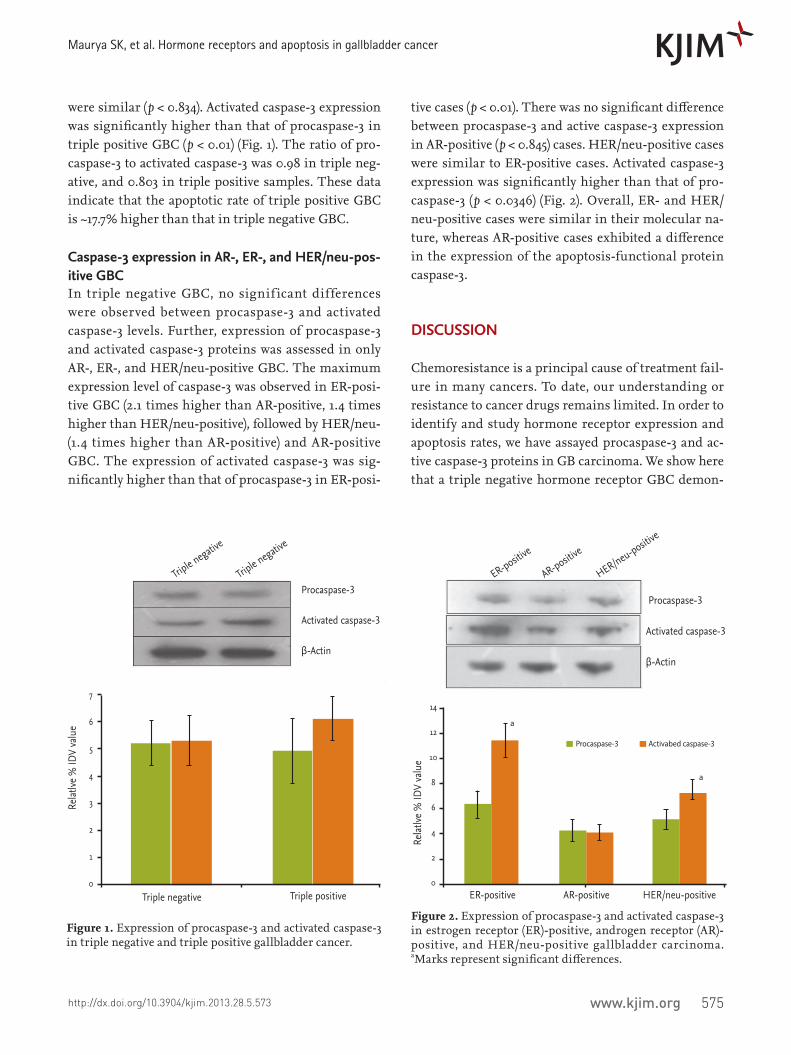
Caspase-3 expression in triple positive and negative hormone receptors of GBCProcaspase-3 and activated caspase-3 expression was evaluated in AR-, ER-, and HER-positive and negative GBC samples. In triple negative GBC, the mean levels of procaspase-3 and activated caspase-3 expression
575
Maurya SK, et al. Hormone receptors and apoptosis in gallbladder cancer
www.kjim.orghttp://dx.doi.org/10.3904/kjim.2013.28.5.573
were similar (p < 0.834). Activated caspase-3 expression was significantly higher than that of procaspase-3 in triple positive GBC (p < 0.01) (Fig. 1). The ratio of pro-caspase-3 to activated caspase-3 was 0.98 in triple neg-ative, and 0.803 in triple positive samples. These data indicate that the apoptotic rate of triple positive GBC is ~17.7% higher than that in triple negative GBC.
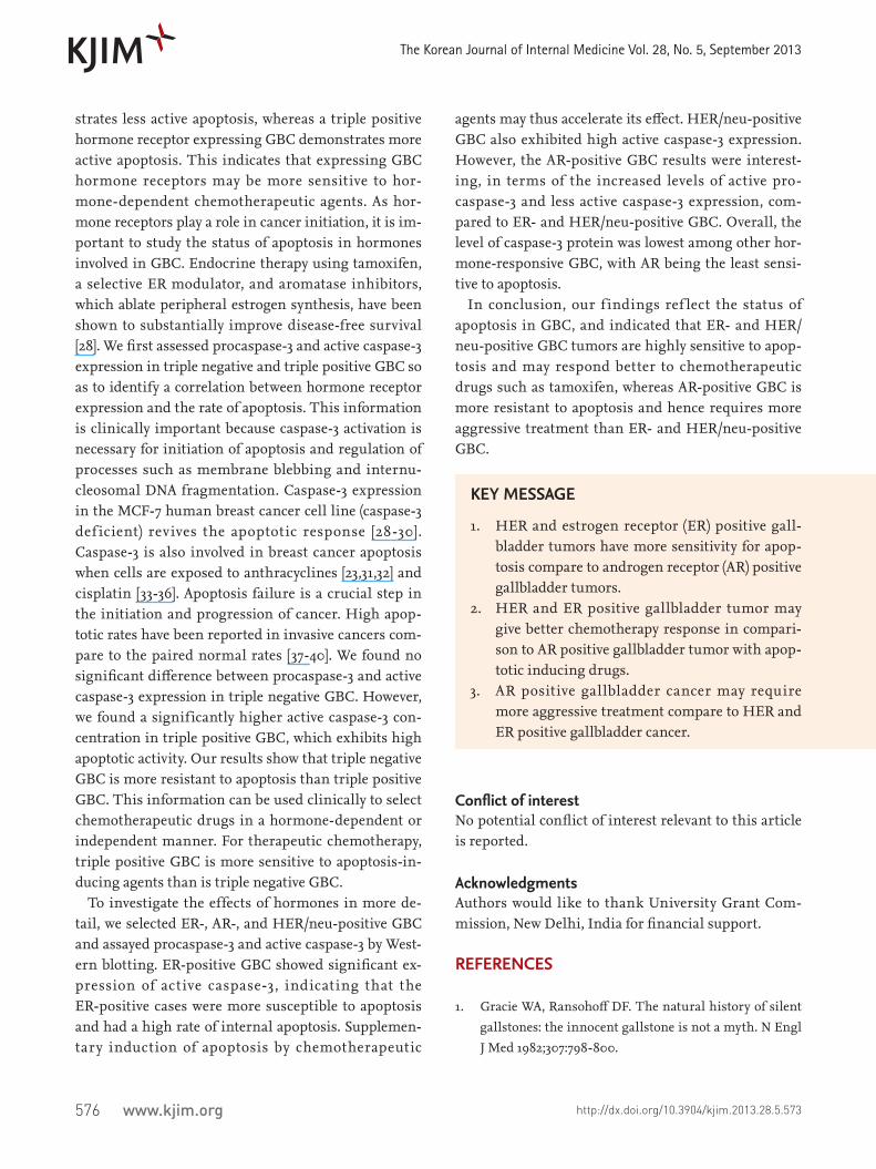
Caspase-3 expression in AR-, ER-, and HER/neu-pos-itive GBCIn triple negative GBC, no signif icant differences were observed between procaspase-3 and activated caspase-3 levels. Further, expression of procaspase-3 and activated caspase-3 proteins was assessed in only AR-, ER-, and HER/neu-positive GBC. The maximum expression level of caspase-3 was observed in ER-posi-tive GBC (2.1 times higher than AR-positive, 1.4 times higher than HER/neu-positive), followed by HER/neu- (1.4 times higher than AR-positive) and AR-positive GBC. The expression of activated caspase-3 was sig-nificantly higher than that of procaspase-3 in ER-posi-
tive cases (p < 0.01). There was no significant difference between procaspase-3 and active caspase-3 expression in AR-positive (p < 0.845) cases. HER/neu-positive cases were similar to ER-positive cases. Activated caspase-3 expression was significantly higher than that of pro-caspase-3 (p < 0.0346) (Fig. 2). Overall, ER- and HER/neu-positive cases were similar in their molecular na-ture, whereas AR-positive cases exhibited a difference in the expression of the apoptosis-functional protein caspase-3.
DISCUSSION
Chemoresistance is a principal cause of treatment fail-ure in many cancers. To date, our understanding or resistance to cancer drugs remains limited. In order to identify and study hormone receptor expression and apoptosis rates, we have assayed procaspase-3 and ac-tive caspase-3 proteins in GB carcinoma. We show here that a triple negative hormone receptor GBC demon-
Triple negative
Triple negativ
e
Triple negativ
e
Relat
lve %
IDV
valu
e
Triple positive
Procaspase-3
Activated caspase-3
β-Actin
7
6
5
4
3
2
1
0ER-positive AR-positive HER/neu-positive
ER-positive
AR-positive
HER/neu-positive
Relat
lve %
IDV
valu
e
Procaspase-3
Activated caspase-3
β-Actin
14
12
10
8
6
4
2
0
a
a
Procaspase-3 Activabed caspase-3
Figure 1. Expression of procaspase-3 and activated caspase-3 in triple negative and triple positive gallbladder cancer.
Figure 2. Expression of procaspase-3 and activated caspase-3 in estrogen receptor (ER)-positive, androgen receptor (AR)-positive, and HER/neu-positive gallbladder carcinoma. aMarks represent significant differences.
576
The Korean Journal of Internal Medicine Vol. 28, No. 5, September 2013
www.kjim.org http://dx.doi.org/10.3904/kjim.2013.28.5.573
strates less active apoptosis, whereas a triple positive hormone receptor expressing GBC demonstrates more active apoptosis. This indicates that expressing GBC hormone receptors may be more sensitive to hor-mone-dependent chemotherapeutic agents. As hor-mone receptors play a role in cancer initiation, it is im-portant to study the status of apoptosis in hormones involved in GBC. Endocrine therapy using tamoxifen, a selective ER modulator, and aromatase inhibitors, which ablate peripheral estrogen synthesis, have been shown to substantially improve disease-free survival [28]. We first assessed procaspase-3 and active caspase-3 expression in triple negative and triple positive GBC so as to identify a correlation between hormone receptor expression and the rate of apoptosis. This information is clinically important because caspase-3 activation is necessary for initiation of apoptosis and regulation of processes such as membrane blebbing and internu-cleosomal DNA fragmentation. Caspase-3 expression in the MCF-7 human breast cancer cell line (caspase-3 def icient) revives the apoptotic response [28-30]. Caspase-3 is also involved in breast cancer apoptosis when cells are exposed to anthracyclines [23,31,32] and cisplatin [33-36]. Apoptosis failure is a crucial step in the initiation and progression of cancer. High apop-totic rates have been reported in invasive cancers com-pare to the paired normal rates [37-40]. We found no significant difference between procaspase-3 and active caspase-3 expression in triple negative GBC. However, we found a significantly higher active caspase-3 con-centration in triple positive GBC, which exhibits high apoptotic activity. Our results show that triple negative GBC is more resistant to apoptosis than triple positive GBC. This information can be used clinically to select chemotherapeutic drugs in a hormone-dependent or independent manner. For therapeutic chemotherapy, triple positive GBC is more sensitive to apoptosis-in-ducing agents than is triple negative GBC.
To investigate the effects of hormones in more de-tail, we selected ER-, AR-, and HER/neu-positive GBC and assayed procaspase-3 and active caspase-3 by West-ern blotting. ER-positive GBC showed significant ex-pression of active caspase-3, indicating that the ER-positive cases were more susceptible to apoptosis and had a high rate of internal apoptosis. Supplemen-tary induction of apoptosis by chemotherapeutic
agents may thus accelerate its effect. HER/neu-positive GBC also exhibited high active caspase-3 expression. However, the AR-positive GBC results were interest-ing, in terms of the increased levels of active pro-caspase-3 and less active caspase-3 expression, com-pared to ER- and HER/neu-positive GBC. Overall, the level of caspase-3 protein was lowest among other hor-mone-responsive GBC, with AR being the least sensi-tive to apoptosis.
In conclusion, our f indings ref lect the status of apoptosis in GBC, and indicated that ER- and HER/neu-positive GBC tumors are highly sensitive to apop-tosis and may respond better to chemotherapeutic drugs such as tamoxifen, whereas AR-positive GBC is more resistant to apoptosis and hence requires more aggressive treatment than ER- and HER/neu-positive GBC.
Conflict of interestNo potential conflict of interest relevant to this article is reported.
Acknowledgments Authors would like to thank University Grant Com-mission, New Delhi, India for financial support.
REFERENCES
1. Gracie WA, Ransohoff DF. The natural history of silent gallstones: the innocent gallstone is not a myth. N Engl J Med 1982;307:798-800.
KEY MESSAGE
1. HER and estrogen receptor (ER) positive gall-bladder tumors have more sensitivity for apop-tosis compare to androgen re ceptor (AR) positive gallbladder tumors.
2. HER and ER positive gallbladder tumor may give better chemotherapy response in compari-son to AR positive gallbladder tumor with apop-totic inducing drugs.
3. AR positive gallbladder cancer may require more aggressive treatment compare to HER and ER positive gallbladder cancer.
577
Maurya SK, et al. Hormone receptors and apoptosis in gallbladder cancer
www.kjim.orghttp://dx.doi.org/10.3904/kjim.2013.28.5.573
2. Shaffer EA. Epidemiology and risk factors for gallstone disease: has the paradigm changed in the 21st century? Curr Gastroenterol Rep 2005;7:132-140.
3. Tewari M, Kumar V, Mishra RR, Shukla HS. Gallblad-der carcinoid masquerading as gallbladder carcinoma. Hepatobiliary Pancreat Dis Int 2009;8:326-328.
4. Dhulkotia A, Kumar S, Kabra V, Shukla HS. Aberrant gallbladder situated beneath the left lobe of liver. HPB (Oxford) 2002;4:39-42.
5. Shukla HS, Awasthi K, Naithani YP, Gupta SC. A clini-co-pathological study of carcinoma of the gall bladder. Indian J Cancer 1981;18:198-201.
6. Ohnami S, Nakata H, Nagafuchi Y, Zeze F, Eto S. Es-trogen receptors in human gastric, hepatocellular, and gallbladder carcinomas and normal liver tissues. Gan To Kagaku Ryoho 1988;15:2923-2928.
7. Nakamura S, Muro H, Suzuki S. Estrogen and pro-gesterone receptors in gallbladder cancer. Jpn J Surg 1989;19:189-194.
8. Barreto SG, Haga H, Shukla PJ. Hormones and gall-bladder cancer in women. Indian J Gastroenterol 2009;28:126-130.
9. Egawa N, Tu Y, Kamisawa T. Why is gallbladder cancer more frequent in women than in men? Nihon Rinsho 2006;64 Suppl 1:344-347.
10. Sumi K, Matsuyama S, Kitajima Y, Miyazaki K. Loss of estrogen receptor beta expression at cancer front cor-relates with tumor progression and poor prognosis of gallbladder cancer. Oncol Rep 2004;12:979-984.
11. Ko CY, Schmit P, Cheng L, Thompson JE. Estrogen re-ceptors in gallbladder cancer: detection by an improved immunohistochemical assay. Am Surg 1995;61:930-933.
12. Chen A, Huminer D. The role of estrogen receptors in the development of gallstones and gallbladder cancer. Med Hypotheses 1991;36:259-260.
13. Yamamoto M, Nakajo S, Tahara E. Immunohistochemi-cal analysis of estrogen receptors in human gallbladder. Acta Pathol Jpn 1990;40:14-21.
14. Gupta P, Agarwal A, Gupta V, Singh PK, Pantola C, Amit S. Expression and clinicopathological significance of estrogen and progesterone receptors in gallbladder can-cer. Gastrointest Cancer Res 2012;5:41-47.
15. Park SK, Andreotti G, Rashid A, et al. Polymorphisms of estrogen receptors and risk of biliary tract cancers and gallstones: a population-based study in Shanghai, China. Carcinogenesis 2010;31:842-846.
16. Srivastava A, Sharma KL, Srivastava N, Misra S, Mittal B. Significant role of estrogen and progesterone receptor sequence variants in gallbladder cancer predisposition: a multi-analytical strategy. PLoS One 2012;7:e40162.
17. Meyer TE, O’Brien TG, Andreotti G, et al. Androgen receptor CAG repeat length and risk of biliary tract cancer and stones. Cancer Epidemiol Biomarkers Prev 2010;19:787-793.
18. Zacks SL, Sandler RS, Rutledge R, Brown RS Jr. A pop-ulation-based cohort study comparing laparoscopic cholecystectomy and open cholecystectomy. Am J Gas-troenterol 2002;97:334-340.
19. Evan GI, Vousden KH. Proliferation, cell cycle and apoptosis in cancer. Nature 2001;411:342-348.
20. Johnstone RW, Ruef li AA, Lowe SW. Apoptosis: a link between cancer genetics and chemotherapy. Cell 2002;108:153-164.
21. Slee EA, Harte MT, Kluck RM, et al. Ordering the cytochrome c-initiated caspase cascade: hierarchi-cal activation of caspases-2, -3, -6, -7, -8, and -10 in a caspase-9-dependent manner. J Cell Biol 1999;144:281-292.
22. Keane MM, Ettenberg SA, Nau MM, Russell EK, Lip-kowitz S. Chemotherapy augments TRAIL-induced apoptosis in breast cell lines. Cancer Res 1999;59:734-741.
23. Bellarosa D, Ciucci A, Bullo A, et al. Apoptotic events in a human ovarian cancer cell line exposed to anthracy-clines. J Pharmacol Exp Ther 2001;296:276-283.
24. Kottke TJ, Blajeski AL, Martins LM, et al. Comparison of paclitaxel-, 5-fluoro-2’-deoxyuridine-, and epidermal growth factor (EGF)-induced apoptosis: evidence for EGF-induced anoikis. J Biol Chem 1999;274:15927-15936.
25. Suzuki A, Kawabata T, Kato M. Necessity of interleu-kin-1beta converting enzyme cascade in taxotere-initi-ated death signaling. Eur J Pharmacol 1998;343:87-92.
26. Thornberry NA, Lazebnik Y. Caspases: enemies within. Science 1998;281:1312-1316.
27. Cryns V, Yuan J. The cutting edge: caspases in apop-tosis and disease. In: Lockshin RA, Zakeri Z, Tilly JL, eds. When Cells Die: A Comprehensive Evaluation of Apoptosis and Programmed Cell Death. New York: Wi-ley-Liss, 1998:177-210.
28. Maurya SK, Tewari M, Kumar M, Thakur MK, Shukla HS. Expression pattern of tumor endothelial marker 8 protein in gallbladder carcinomas. Asian Pac J Cancer
578
The Korean Journal of Internal Medicine Vol. 28, No. 5, September 2013
www.kjim.org http://dx.doi.org/10.3904/kjim.2013.28.5.573
Prev 2011;12:507-512.29. Horvitz HR, Sternberg PW, Greenwald IS, Fixsen W,
Ellis HM. Mutations that affect neural cell lineages and cell fates during the development of the nematode Caenorhabditis elegans. Cold Spring Harb Symp Quant Biol 1983;48 Pt 2:453-463.
30. Janicke RU, Ng P, Sprengart ML, Porter AG. Caspase-3 is required for alpha-fodrin cleavage but dispensable for cleavage of other death substrates in apoptosis. J Biol Chem 1998;273:15540-15545.
31. Yang XH, Sladek TL, Liu X, Butler BR, Froelich CJ, Thor AD. Reconstitution of caspase 3 sensitizes MCF-7 breast cancer cells to doxorubicin- and etoposide-in-duced apoptosis. Cancer Res 2001;61:348-354.
32. Kwon KB, Park EK, Ryu DG, Park BH. D4-GDI is cleaved by caspase-3 during daunorubicin-induced apoptosis in HL-60 cells. Exp Mol Med 2002;34:32-37.
33. Henkels KM, Turchi JJ. Cisplatin-induced apoptosis proceeds by caspase-3-dependent and -independent pathways in cisplatin-resistant and -sensitive human ovarian cancer cell lines. Cancer Res 1999;59:3077-3083.
34. Blanc C, Deveraux QL, Krajewski S, et al. Caspase-3 is essential for procaspase-9 processing and cisplatin-in-duced apoptosis of MCF-7 breast cancer cells. Cancer
Res 2000;60:4386-4390.35. Cummings BS, Schnellmann RG. Cisplatin-induced
renal cell apoptosis: caspase 3-dependent and -indepen-dent pathways. J Pharmacol Exp Ther 2002;302:8-17.
36. Kishimoto S, Kawazoe Y, Ikeno M, Fukushima S, Takeuchi Y. Continuous exposure to low-dose cisplatin and apoptosis. Biol Pharm Bull 2005;28:1954-1957.
37. Wong SC, Chan JK, Lee KC, Hsiao WL. Differential ex-pression of p16/p21/p27 and cyclin D1/D3, and their re-lationships to cell proliferation, apoptosis, and tumour progression in invasive ductal carcinoma of the breast. J Pathol 2001;194:35-42.
38. Mommers EC, van Diest PJ, Leonhart AM, Meijer CJ, Baak JP. Balance of cell proliferation and apopto-sis in breast carcinogenesis. Breast Cancer Res Treat 1999;58:163-169.
39. Zhao H, Morimoto T, Sasa M, Tanaka T, Izumi K. Im-munohistochemical expression of uPA, PAI-1, cathepsin D and apoptotic cells in ductal carcinoma in situ of the breast. Breast Cancer 2002;9:118-126.
40. Parton M, Krajewski S, Smith I, et al. Coordinate ex-pression of apoptosis-associated proteins in human breast cancer before and during chemotherapy. Clin Cancer Res 2002;8:2100-2108.
Related Documents