Increased NF- κ B expression in Preeclampsia 195 195 Tohoku J. Exp. Med., 2004, 204, 195-202 Received June 8, 2004; revision accepted for publication September 8, 2004. Address for reprints: Dr. Meral Aban, Fatih Mah. Alden Sitesi D-Blok No:18 Mezitli Mersin, Turkey. e-mail: [email protected] Expression of Nuclear Factor-Kappa B and Placental Apoptosis in Pregnancies Complicated with Intrauterine Growth Restriction and Preeclampsia: An Immunohistochemical Study MERAL ABAN, L EYLA C INEL , 1 MURAT ARSLAN, UMUT DILEK, MUSTAFA KAPLANOGLU, RABIA ARPACI 1 and SAFFET DILEK Department of Obstetrics and Gynecology, 1 Department of Pathology, University of Mersin, Mersin, Turkey ABAN, M., CINEL, L., ARSLAN, M., DILEK, U., KAPLANOGLU, M., ARPACI, R. and DILEK, S. Expression of Nuclear Factor-Kappa B and Placental Apoptosis in Pregnancies Complicated with Intrauterine Growth Restriction and Preeclampsia: An Immunohistochemical Study. Tohoku J. Exp. Med., 2004, 204 (3), 195-202 ── Preeclampsia affects 7-10% of all pregnancies, and is a major cause of maternal and fetal morbidity and mortality. Although enhanced apoptosis is well known in placen- tas with preeclampsia, the role of transcription factor nuclear factor-kappa B (NF- κ B) in the process is still being debated. In this work, we investigate the relationship be- tween NF- κ B expression and trophoblastic cell apoptosis in pregnancies complicated with preeclampsia or intrauterine growth restriction (IUGR) by immunohistochemi- cal analysis of NF- κ B and three apoptosis related markers: bcl-2, caspase-3, and M30 CytoDeath antibody that identifies early apoptotic changes in the cytoskeleton related to action of caspase. The study was conducted on placental samples from 19 pre- eclamptic, 5 IUGR-complicated and 10 normal pregnant women. The three conclu- sions from the statistical analysis of the data are obtained; (i) Significantly higher ex- pression of NF- κ B in IUGR-complicated (p = 0.003) and preeclamptic placentas (p = 0.004) than the control placentas, (ii) significantly higher M30 index and caspase 3 expression in IUGR and preeclampsia placentas (p = 0.003), and (iii) decreased ex- pression of bcl-2 in IUGR and preeclampsia placentas (p = 0.001). Based on these observations, we suggest that increased trophoblastic apoptosis is at least partially in- duced by NF- κ B and reduced bcl-2 expression. ──── preeclampsia; IUGR; apop- tosis; NF- κ B; bcl-2 © 2004 Tohoku University Medical Press

Welcome message from author
This document is posted to help you gain knowledge. Please leave a comment to let me know what you think about it! Share it to your friends and learn new things together.
Transcript
Increased NF-κB expression in Preeclampsia 195
195
Tohoku J. Exp. Med., 2004, 204, 195-202
Received June 8, 2004; revision accepted for publication September 8, 2004.Address for reprints: Dr. Meral Aban, Fatih Mah. Alden Sitesi D-Blok No:18 Mezitli Mersin, Turkey.e-mail: [email protected]
Expression of Nuclear Factor-Kappa B and Placental Apoptosis in Pregnancies Complicated with Intrauterine Growth Restriction and Preeclampsia: An Immunohistochemical Study
MERAL ABAN, LEYLA CINEL,1 MURAT ARSLAN, UMUT DILEK, MUSTAFA KAPLANOGLU, RABIA ARPACI
1 and SAFFET DILEK
Department of Obstetrics and Gynecology, 1 Department of Pathology, University of Mersin, Mersin, Turkey
ABAN, M., CINEL, L., ARSLAN, M., DILEK, U., KAPLANOGLU, M., ARPACI, R. and DILEK, S. Expression of Nuclear Factor-Kappa B and Placental Apoptosis in Pregnancies Complicated with Intrauterine Growth Restriction and Preeclampsia: An Immunohistochemical Study. Tohoku J. Exp. Med., 2004, 204 (3), 195-202 ── Preeclampsia affects 7-10% of all pregnancies, and is a major cause of maternal and fetal morbidity and mortality. Although enhanced apoptosis is well known in placen-tas with preeclampsia, the role of transcription factor nuclear factor-kappa B (NF-κB) in the process is still being debated. In this work, we investigate the relationship be-tween NF-κB expression and trophoblastic cell apoptosis in pregnancies complicated with preeclampsia or intrauterine growth restriction (IUGR) by immunohistochemi-cal analysis of NF-κB and three apoptosis related markers: bcl-2, caspase-3, and M30 CytoDeath antibody that identifies early apoptotic changes in the cytoskeleton related to action of caspase. The study was conducted on placental samples from 19 pre-eclamptic, 5 IUGR-complicated and 10 normal pregnant women. The three conclu-sions from the statistical analysis of the data are obtained; (i) Significantly higher ex-pression of NF-κB in IUGR-complicated (p = 0.003) and preeclamptic placentas (p = 0.004) than the control placentas, (ii) significantly higher M30 index and caspase 3 expression in IUGR and preeclampsia placentas (p = 0.003), and (iii) decreased ex-pression of bcl-2 in IUGR and preeclampsia placentas (p = 0.001). Based on these observations, we suggest that increased trophoblastic apoptosis is at least partially in-duced by NF-κB and reduced bcl-2 expression. ──── preeclampsia; IUGR; apop-tosis; NF-κB; bcl-2© 2004 Tohoku University Medical Press
M. Aban et al.196 Increased NF-κB expression in Preeclampsia 197
stages of apoptosis (Leers et al. 1999). M30 CytoDeath® antibody recognizes a caspase-medi-ated fragment of cytokeratin 18 and therefore is most likely found only in caspase-dependent apoptosis, but not in nonapoptotic cells (Kadyrov et al. 2001). On the other hand, one of the major genes responsible for regulating apoptotic cell death is the protooncogene, bcl-2, which can pre-vent apoptosis through regulation of cellular anti-oxidant defense mechanisms (Hockenbery et al. 1993).
NF-κB is a redox-sensitive transcription fac-tor regulating a battery of inflammatory genes, and has a variety of different effects in numerous pathological states. Although the role of NF-κB in programmed cell death is still controversial, some researchers have claimed a direct action of NF-κB in the process of apoptosis (Kitajima et al. 1996; Qin et al. 1998; Matsushita et al. 2000). It has also been shown that the activation of NF-κB binding and increased caspase-3 both affect the endothelial cells under hypoxic conditions (Roebuck et al. 1999). However, little is known about the relationship between the expression of NF-κ B and apoptotic changes in trophoblastic cells of IUGR and preeclampsia in humans. In this study, we investigate NF-κB expression and placental trophoblastic apoptosis and its relation with bcl-2 in placental specimens that have been obtained from pregrant women complicated with preeclampsia or IUGR.
MATERIALS AND METHODS
Placental materialsThis study was conducted on 34 pregnant
women between January 2001 and December 2003 in the Department of Obstetrics and Gynecology at the University of Mersin. Written informed consent was obtained from all subjects, and the study protocol was approved by Institutional Ethics Committee. Ten of the placentas were considered between 36 and 38 weeks of singleton pregrancy and were from normal patients who had cesarean delivery for various obstetric indica-tions such as previous cesarean section and pre-
Preeclampsia affects 7-10% of all pregnan-cies, and is a major cause of maternal and fetal morbidity and mortality. Despite recent advances in medicine, its etiology still remains unknown (Roberts et al. 1990). Maintenance of trophoblast structure and specialized function is essential for the provision of adequate oxygen and nutrients to the fetus. Fetal complications of preeclampsia and intrauterine growth restriction (IUGR) are commonly attributed to clinical conditions that lead to underperfusion and hypoxia of the placen-ta (Levy et al. 2002). The pathogenic mecha-nisms of preeclampsia converge on the vascular endothelium and numerous stimuli, including oxi-dative stress. Furthermore, TNF-α and high glu-cose are well known to induce apoptosis in endo-thelial cells (Muschel et al. 1995; Haimovitz-Friedman 1997).
Apoptosis is a form of programmed cell death and has been observed in placentas of nor-mal human pregnancies (Smith et al. 1997a, b; Allaire et al. 2000). There are some studies that have focused on the role of apoptosis in placenta in relation to the pathophysiology of preeclampsia and IUGR (DiFederico et al. 1999; Levy et al. 2002). It is well known that placentas from wom-en complicated with preeclampsia or IUGR show enhanced apoptosis as compared to placentas of normal pregnant women (Smith et al. 1997b; Allaire et al. 2000; Ishihara et al. 2002). However, the molecular mechanisms leading to apoptosis are complex, and include an ever-ex-panding list of signaling molecules such as the bcl-2 family of genes, the caspase-3 and NF-κB (Hetts 1998; Adams and Cory 1999).
The balance between pro- and anti-apoptotic proteins determines whether apoptosis will be triggered or not. Caspase-3 is a cysteine protease protein that exists as inactive zymogene in almost all cells, and is involved in the development of apoptotic cell death. Activation of caspase-3 oc-curs in response to a variety of apoptotic inducers (Mallat et al. 1997). It has been shown that cyto-keratins, in particular cytokeratin 18, which is cleaved by caspase 3 or 7, are affected in the early
M. Aban et al.196 Increased NF-κB expression in Preeclampsia 197
sentation anomalies. All normal term infants had birth weights ≧10th percentile of the individual-ized birth weight ratio. Term placentas compli-cated by either severe preeclampsia or IUGR from a singleton pregnancy between 36 and 38 weeks of gestation were obtained from 24 patients who had cesarean delivery. Severe preeclampsia was defined by the criteria established by the American Collage of Obstetric and Gynecology (ACOG 1996 Technical bulletin no: 219).
The IUGR was based on 3 criteria: ultraso-nographic evidence of deviation from an appro-priate growth percentile, clinical evidence of sub-optimal growth, and individualized birth weight ratios of < 10th percentile. None of the women had smoking or drinking habits, or had a concur-rent medical illness. The gestational age of the placenta was first determined by an approximate computation of the duration of pregnancy from patient’s last menstrual period and was further confirmed with ultrasonography.
Histopathological materialVillous tissues from 3-5 cotyledons were
dissected, rinsed in saline solution, and fixed with %10 formalin for 24 hours at 4°C. Hematoxylin and eosin stained sections from each specimen were examined under light mi-croscope and representative sections from each tissue sample were selected for immunohisto-chemistry.
Immunohistochemistry for NF-κ B, bcl-2, M30 (CytoDeath monoclonal antibody, 1 :50, Roche®) and Caspase-3 was performed using a combination of the streptavidin-biotin-peroxidase method and microwave antigen retrieval on for-maline-fixed paraffin-embedded tissues. After deparaffinization, sections were treated with 10% hydrogen peroxidase in filtered water to block en-dogenous peroxidase activity. To retrieve antigen, slides were treated with 10 mmol/liter citrate buf-fer (pH = 6.8) for 10 min. After preincubation with Ultra V block (LabVision®) for 20 minutes, sections were incubated with primary antibody for an hour at room temperature for bcl-2 (mono-
clonal mouse anti-Human bcl-2 oncoprotein, 1 :50, Dako®), NF-κ B (Rabbit polyclonal anti-body, p50 Ab-2, 1 :75, Neomarkers®), M30 or caspase-3 (Rabbit polyclonal antibody, CPP32, 1 :50, Neomarkers®) followed sequentially by bi-otinylated goat anti-polyvalent (Lab Vision®) for 20 minutes, and streptavidin peroxidase complex (Lab Vision®) for 30 minutes. 3-Amino-9 Ethylcarbazole/AEC (Lab Vision®) and hematox-ylin were used as chromagen and for nuclear counterstain, respectively. Negative control was achieved by omitting the primary antibody. The positive controls were tonsil for bcl-2, testis for NF-κB, infiltrative ductal carcinoma of breast for caspase-3 and M30.
Scoring and statistical analysisImmunohistochemical evaluation was car-
ried out only in the epithelial component of the trophoblastic cells. All specimens were examined by the pathologist (CL) who was blinded to clini-cal conditions. The staining score for bcl-2, cas-pase-3, or NF-κ B was labeled as “0” for no immunostaining, “1+” for weak and focal immu-nostaining, “2+” for weak and diffuse immunos-taining, “3+” for strong and diffuse immunostain-ing according to the overall staining feature. Previously defined (Chiu et al. 2001) M30 index was used, whith is the percentage of positively stained area to the total area of villous trophoblast in each section. The sections were examined at high power (× 400). Kruskal-Wallis test was used to analyse the relationship between scores from each experimental groups.
RESULTS
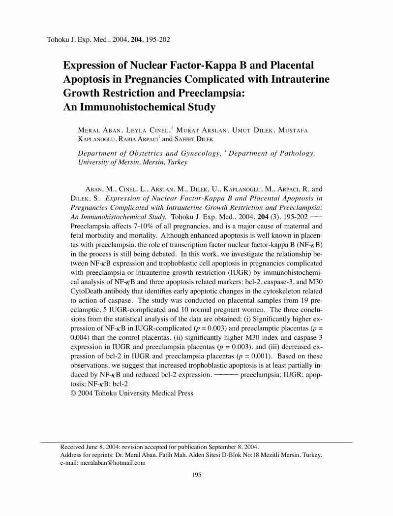
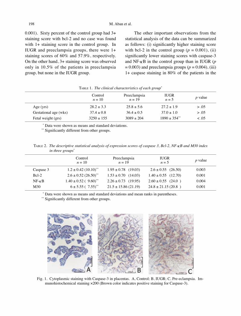
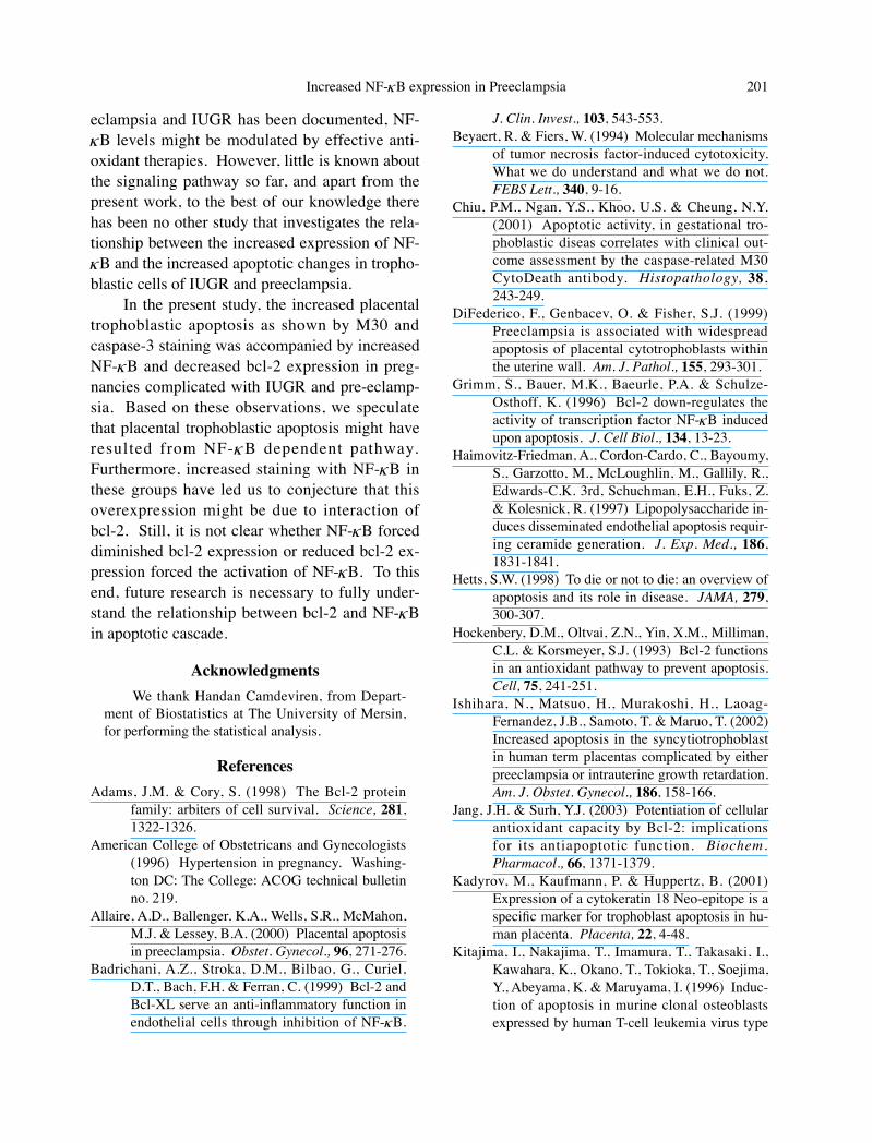
Table 1 summarises the clinical characteris-tics for each patient. Immunohistochemical anal-ysis of the NF-κB, bcl-2, or caspase-3 expressions and the M30 index are shown in Table 2. All cas-es showed positive staining immunolocalized to the cytoplasm of trophoblastic cells (Figs. 1-4). In IUGR and preeclampsia groups, apoptosis as-sessed by M30 CytoDeath® was observed to be more significant than in the control group (p =
M. Aban et al.198 Increased NF-κB expression in Preeclampsia 199
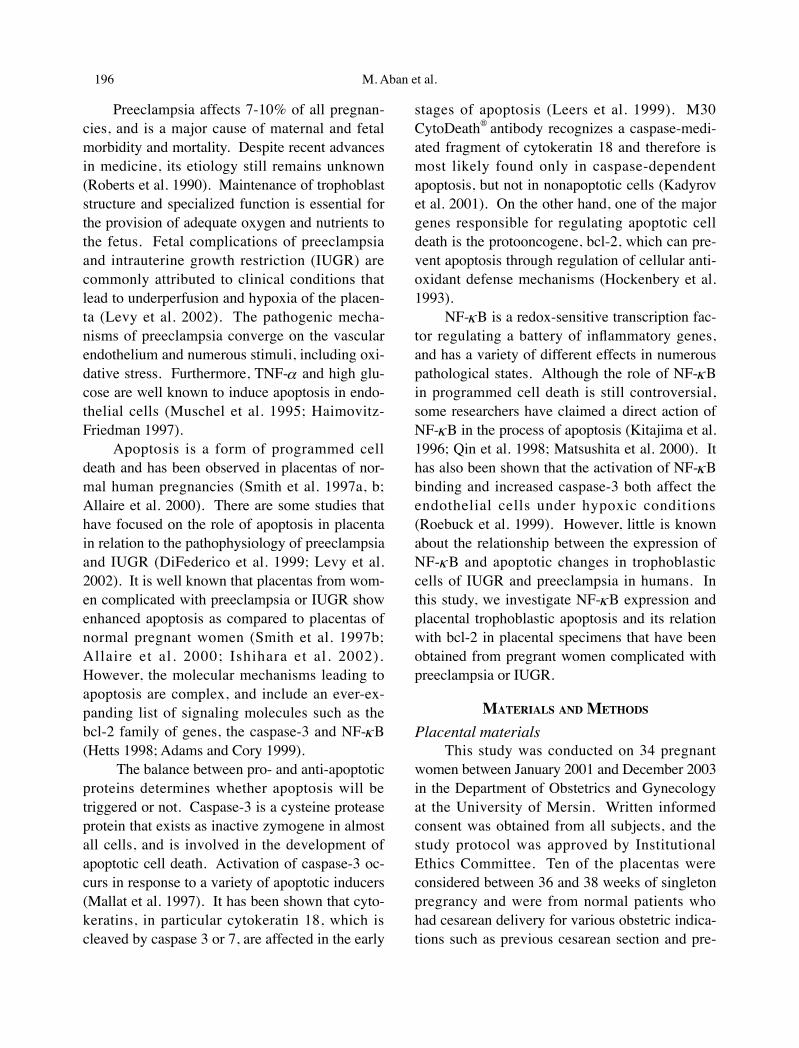
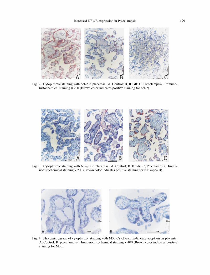
0.001). Sixty percent of the control group had 3+ staining score with bcl-2 and no case was found with 1+ staining score in the control group. In IUGR and preeclampsia groups, there were 1+ staining scores of 60% and 57.9%, respectively. On the other hand, 3+ staining score was observed only in 10.5% of the patients in preeclampsia group, but none in the IUGR group.
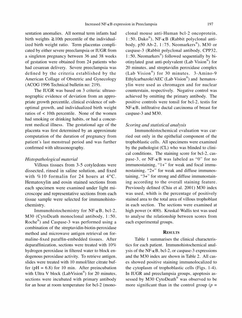
The other important observations from the statistical analysis of the data can be summarized as follows: (i) significantly higher staining score with bcl-2 in the control group (p = 0.001), (ii) significantly lower staining scores with caspase-3 and NF-κB in the control group than in IUGR (p = 0.003) and preeclampsia groups (p = 0.004), (iii) 1+ caspase staining in 80% of the patients in the
TABLE 1. The clinical characteristics of each group*
Controln = 10
Preeclampsian = 19
IUGRn = 5 p value
Age (yrs) 28.2 ± 3.3 25.8 ± 5.6 27.2 ± 1.9 > .05Gestational age (wks) 37.4 ± 0.8 36.4 ± 0.5 37.0 ± 1.0 > .05Fetal weight (grs) 3250 ± 155 3089 ± 204 1890 ± 354** < .05
* Data were shown as means and standard deviations.** Significantly different from other groups.
TABLE 2. The descriptive statistical analysis of expression scores of caspase 3, Bcl-2, NF-κB and M30 index in three groups*
Controln = 10
Preeclampsian = 19
IUGRn = 5 p value
Caspase 3 1.2 ± 0.42 (10.10)** 1.95 ± 0.78 (19.03) 2.6 ± 0.55 (26.50) 0.003Bcl-2 2.6 ± 0.52 (26.50)** 1.53 ± 0.70 (14.03) 1.40 ± 0.55 (12.70) 0.001NF-κB 1.40 ± 0.52 ( 9.60)** 2.26 ± 0.73 (19.95) 2.60 ± 0.55 (24.0 ) 0.004M30 6 ± 5.55 ( 7.55)** 21.5 ± 15.86 (21.19) 24.8 ± 21.15 (20.8 ) 0.001
* Data were shown as means and standard deviations and mean ranks in parentheses.** Significantly different from other groups.
Fig. 1. Cytoplasmic staining with Caspase-3 in placentas. A, Control; B, IUGR; C, Pre-eclampsia. Im-munohistochemical staining ×200 (Brown color indicates positive staining for Caspase-3).
M. Aban et al.198 Increased NF-κB expression in Preeclampsia 199
Fig. 2. Cytoplasmic staining with bcl-2 in placentas. A, Control; B, IUGR; C, Preeclampsia. Immuno-histochemical staining × 200 (Brown color indicates positive staining for bcl-2).
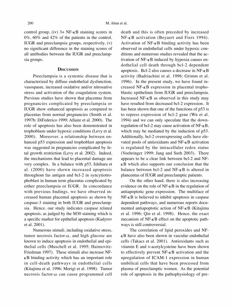
Fig. 3. Cytoplasmic staining with NF-κB in placentas. A, Control; B, IUGR; C, Preeclampsia. Immu-nohistochemical staining × 200 (Brown color indicates positive staining for NF kappa B).
Fig. 4. Photomicrograph of cytoplasmic staining with M30 CytoDeath indicating apoptosis in placenta. A, Control; B, preeclampsia. Immunohistochemical staining × 400 (Brown color indicates positive staining for M30).
M. Aban et al.200 Increased NF-κB expression in Preeclampsia 201
control group, (iv) 3+ NF-κB staining scores in 0%, 60% and 42% of the patients in the control, IUGR and preeclampsia groups, respectively, (v) no significant difference in the staining scores of all antibodies between the IUGR and preeclamp-sia groups.
DISCUSSION
Preeclampsia is a systemic disease that is characterized by diffuse endothelial dysfunction, vasospasm, increased oxidative and/or nitrosative stress and activation of the coagulation system. Previous studies have shown that placentas from pregnancies complicated by preeclampsia or IUGR show enhanced apoptosis as compared to placentas from normal pregnancies (Smith et al. 1997b; DiFederico 1999; Allaire et al. 2000). The role of apoptosis has also been demonstrated in trophoblasts under hypoxic conditions (Levy et al. 2000). Moreover, a relationship between en-hanced p53 expression and trophoblast apoptosis was suggested in pregnancies complicated by fe-tal growth restriction (Levy et al. 2002). Indeed, the mechanisms that lead to placental damage are very complex. In a balance with p53, Ishihara et al. (2000) have shown increased apoptosis throughout fas antigen and bcl-2 in syncytiotro-phoblast in human term placentas complicated by either preeclampsia or IUGR. In concordance with previous findings, we have observed in-creased human placental apoptosis as shown by caspase-3 staining in both IUGR and preeclamp-sia. Hence, our study indicates caspase related apoptosis, as judged by the M30 staining which is a specific marker for epithelial apoptosis (Kadyrov et al. 2001).
Numerous stimuli, including oxidative stress, tumor necrosis factor-α , and high glucose are known to induce apoptosis in endothelial and epi-thelial cells (Muschell et al. 1995; Haimovitz-Friedman 1997). These stimuli also increase NF-κB binding activity which has an important role in cell-death pathways in endothelial cells (Kitajima et al. 1996; Morigi et al. 1998). Tumor necrosis factor-α can cause programmed cell
death and this is often preceded by increased NF-κ B activation (Beyaert and Fiers 1994). Activation of NF-κ B binding activity has been observed in endothelial cells under hypoxic con-ditions and numerous studies revealed that the ac-tivation of NF-κB induced by hypoxia causes en-dothelial cell death through bcl-2 dependent apoptosis. Bcl-2 also causes a decrease in NF-κB activity (Badriachini et al. 1996; Grimm et al. 1996). In the present study, we have found in-creased NF-κ B expression in placental tropho-blastic epithelium from IUGR and preeclampsia. Increased NF-κB as observed in this study may have resulted from decreased bcl-2 expression. It has been shown that one of the functions of p53 is to repress expression of bcl-2 gene (Wu et al. 1994) and we can only speculate that the down-regulation of bcl-2 may cause activation of NF-κB, which may be mediated by the induction of p53. Additionally, bcl-2 overexpressing cells have ele-vated pools of antioxidants and NF-κB activation is regulated by the intracelluler redox status (Voehringer 1999; Jang and Surh 2003). There appears to be a clear link between bcl-2 and NF-κB which also supports our conclusion that the balance between bcl-2 and NF-κ B is altered in plancentas of IUGR and preeclamptic patients.
On the other hand, there is also increasing evidence on the role of NF-κB in the regulation of antiapoptotic gene expression. The multiface of NF-κB is believed to inhibit apoptosis in caspase dependent pathways, and numerous reports docu-mented antiapoptotic action of NF-κB (Kitajima et al. 1996; Qin et al. 1998). Hence, the exact mecanism of NF-κB effect on the apoptotic path-ways is still controversial.
The correlation of lipid peroxides and NF-κB have also been shown in vascular endothelial cells (Takacs et al. 2001). Antioxidants such as vitamin E and N-acetylcysteine have been shown to effectively prevent NF-κB activation and the upregulation of ICAM-1 expression in human umbilical cells that have been processed from plasma of preeclamptic women. As the potential role of apoptosis in the pathophysiology of pre-
M. Aban et al.200 Increased NF-κB expression in Preeclampsia 201
eclampsia and IUGR has been documented, NF-κB levels might be modulated by effective anti-oxidant therapies. However, little is known about the signaling pathway so far, and apart from the present work, to the best of our knowledge there has been no other study that investigates the rela-tionship between the increased expression of NF-κB and the increased apoptotic changes in tropho-blastic cells of IUGR and preeclampsia.
In the present study, the increased placental trophoblastic apoptosis as shown by M30 and caspase-3 staining was accompanied by increased NF-κB and decreased bcl-2 expression in preg-nancies complicated with IUGR and pre-eclamp-sia. Based on these observations, we speculate that placental trophoblastic apoptosis might have resulted from NF-κ B dependent pathway. Furthermore, increased staining with NF-κ B in these groups have led us to conjecture that this overexpression might be due to interaction of bcl-2. Still, it is not clear whether NF-κB forced diminished bcl-2 expression or reduced bcl-2 ex-pression forced the activation of NF-κB. To this end, future research is necessary to fully under-stand the relationship between bcl-2 and NF-κB in apoptotic cascade.
AcknowledgmentsWe thank Handan Camdeviren, from Depart-
ment of Biostatistics at The University of Mersin, for performing the statistical analysis.
ReferencesAdams, J.M. & Cory, S. (1998) The Bcl-2 protein
family: arbiters of cell survival. Science, 281, 1322-1326.
American College of Obstetricans and Gynecologists (1996) Hypertension in pregnancy. Washing-ton DC: The College: ACOG technical bulletin no. 219.
Allaire, A.D., Ballenger, K.A., Wells, S.R., McMahon, M.J. & Lessey, B.A. (2000) Placental apoptosis in preeclampsia. Obstet. Gynecol., 96, 271-276.
Badrichani, A.Z., Stroka, D.M., Bilbao, G., Curiel, D.T., Bach, F.H. & Ferran, C. (1999) Bcl-2 and Bcl-XL serve an anti-inflammatory function in endothelial cells through inhibition of NF-κB.
J. Clin. Invest., 103, 543-553.Beyaert, R. & Fiers, W. (1994) Molecular mechanisms
of tumor necrosis factor-induced cytotoxicity. What we do understand and what we do not. FEBS Lett., 340, 9-16.
Chiu, P.M., Ngan, Y.S., Khoo, U.S. & Cheung, N.Y. (2001) Apoptotic activity, in gestational tro-phoblastic diseas correlates with clinical out-come assessment by the caspase-related M30 CytoDeath antibody. Histopathology, 38, 243-249.
DiFederico, F., Genbacev, O. & Fisher, S.J. (1999) Preeclampsia is associated with widespread apoptosis of placental cytotrophoblasts within the uterine wall. Am. J. Pathol., 155, 293-301.
Grimm, S., Bauer, M.K., Baeurle, P.A. & Schulze-Osthoff, K. (1996) Bcl-2 down-regulates the activity of transcription factor NF-κB induced upon apoptosis. J. Cell Biol., 134, 13-23.
Haimovitz-Friedman, A., Cordon-Cardo, C., Bayoumy, S., Garzotto, M., McLoughlin, M., Gallily, R., Edwards-C.K. 3rd, Schuchman, E.H., Fuks, Z. & Kolesnick, R. (1997) Lipopolysaccharide in-duces disseminated endothelial apoptosis requir-ing ceramide generation. J. Exp. Med., 186, 1831-1841.
Hetts, S.W. (1998) To die or not to die: an overview of apoptosis and its role in disease. JAMA, 279, 300-307.
Hockenbery, D.M., Oltvai, Z.N., Yin, X.M., Milliman, C.L. & Korsmeyer, S.J. (1993) Bcl-2 functions in an antioxidant pathway to prevent apoptosis. Cell, 75, 241-251.
Ishihara, N., Matsuo, H., Murakoshi, H., Laoag-Fernandez, J.B., Samoto, T. & Maruo, T. (2002) Increased apoptosis in the syncytiotrophoblast in human term placentas complicated by either preeclampsia or intrauterine growth retardation. Am. J. Obstet. Gynecol., 186, 158-166.
Jang, J.H. & Surh, Y.J. (2003) Potentiation of cellular antioxidant capacity by Bcl-2: implications for its antiapoptotic function. Biochem. Pharmacol., 66, 1371-1379.
Kadyrov, M., Kaufmann, P. & Huppertz, B. (2001) Expression of a cytokeratin 18 Neo-epitope is a specific marker for trophoblast apoptosis in hu-man placenta. Placenta, 22, 4-48.
Kitajima, I., Nakajima, T., Imamura, T., Takasaki, I., Kawahara, K., Okano, T., Tokioka, T., Soejima, Y., Abeyama, K. & Maruyama, I. (1996) Induc-tion of apoptosis in murine clonal osteoblasts expressed by human T-cell leukemia virus type
M. Aban et al.202
I tax by NF-κ B and TNF-α . J. Bone Miner. Res., 11, 200-210.
Leers, M.P., Kolgen, W., Bjorklund, V., Bergman, T., Tribbick, G., Persson, B., Bjorklund, P., Ramaekers, F.C., Bjorklund, B., Nap, M., Jornvall, H. & Schutte, B. (1999) Immunocyto-chemical detection and mapping of a cytokera-tin 18 neo-epitope exposed during early apopto-sis. J. Pathol., 187, 567-572.
Levy, R., Smith, S.D., Chandler, K., Sadovsky, Y. & Nelson, D.M. (2000) Apoptosis in human cul-tured trophoblasts is enhanced by hypoxia and diminished by epidermal growth factor. Am. J. Physiol. Cell Physiol., 278, 982-988.
Levy, R., Smith, S.D., Yusuf, K., Huettner, P.C., Kraus, F.T., Sadovsky, Y. & Nelson, D.M. (2002) Tro-phoblast apoptosis from pregnancies complicat-ed by fetal growth restriction is associated with enhanced p53 expression. Am. J. Obstet. Gynecol., 186, 1056-1061.
Mallat, Z., Ohan, J., Leseche, G. & Tedgui, A. (1997) Colocalinization of CPP-32 with apoptotic cells in human atherosclerotic plagues. Circulation, 96, 424-428.
Maulik, N., Goswami, S., Galang, N. & Das, D.K. (1999) Differential regulation of Bcl-2, AP-1, and NF-κB on cardiomyocyte apoptosis during myocardial ischemic stress adoption. FEBS Lett., 443, 331-336.
Matsushita, H., Morishita, R., Nata, T., Aoki, M., Nakagami, H., Taniyama, Y., Yamamoto, K., Higaki, J., Yasufumi, K. & Ogihara, T. (2000) Hypoxia-induced endothelial apoptosis through nuclear factor-kappaB (NF-κB)-mediated bcl-2 suppression: invivo evidence of the importance of NF-κB in endothelial cell regulation. Circ. Res., 86, 974-981.
Morigi, M., Angioletti, S., Imberti, B., Donadelli, R., Micheletti, G., Figliuzzi, M., Remuzzi, A., Zoja, C. & Remuzzy, G. (1998) Leukocyte-endothe-lial interaction is augmented by high glucose concentrations and hyperglycemia in a NF-κ B-dependent fashion. J. Clin. Invest., 101, 1905-1915.
Muschel, R.J., Bernhard, E.J., Garza, L., McKenna,
W.G. & Koch, C.J. (1995) Induction of apopto-sis at different oxygen tensions: evidence that oxygen radicals do not mediate apoptotic sig-naling. Cancer Res., 55, 995-998.
Roberts, J.M., Taylor, R.N., Friedman, S.A. & Goldfien, A. (1990) New Developments In Pre-eclampsia. Fetal Medical Review, edited by W. Dunlap, Edward Arnold Publishers London, 125-141.
Roebuck, K.A. (1999) Oxidant stress regulation of IL-8 and ICAM-1 gene expression: differential activation and binding of the transcription fac-tors AP-1 and NF-κB. Int. J. Mol. Med., 44, 223-230.
Qin, Z.H., Wang, Y., Nakai, M. & Chase, T.N. (1998) Nuclear factor-kappa B contributes to excito-toxin-induced apoptosis in rat striatum. Mol. Pharmacol., 53, 33-42.
Smith, S.C., Baker, P.N. & Symonds, E.M. (1997a) Placental apoptosis in normal human pregnan-cy. Am. J. Obstet. Gynecol., 177, 57-65.
Smith, S.C., Baker, P.N. & Symonds, E.M. (1997b) Increased placental apoptosis in intrauterine growth restriction. Am. J. Obstet. Gynecol., 177, 1395-401.
Sugiyama, S., Kugiyama, K., Ogata, N., Doi, H., Ota, Y., Ohgushi, M., Matsumura, T., Oka, H. & Yasue, H. (1998) Biphasic regulation of tran-scription factor nuclear factor-kappaB activity in human endothelial cells by lysophosphatidyl-choline through protein kinase C-mediated pathway. Arterioscler. Thromb. Vasc. Biol., 18, 568-576.
Takacs, P., Kauma, S.W., Sholley, M.M., Walsh, S.W. & Dinsmoor, M.J. (2001) Increased circulating lipid peroxides in severe preeclampsia activate NF-κB and upregulate ICAM-1 in vascular en-dothelial cells. FASEB. J., 15, 279-281.
Voehringer, D.W. (1999) BCL-2 and glutathione: al-terations in cellular redox state that regulate apoptosis sensitivity. Free Radic. Biol. Med., 27, 945-950.
Wu, H. & Lozano, G. (1994) NF-κ B activation of p53. J. Biol. Chem., 269, 20067-20074.
Related Documents


