1 EXPRESSION OF MCSP AND PRAME IN CONJUNCTIVAL MELANOMA. Dr. Henrike Westekemper 1 , Sara Karimi 1 , Dr. Daniela Süsskind 2 , PD Dr. Gerasimos Anastassiou 1 , Dr. Michael Freistühler 1 , PD Dr. Daniel Meller 1 , Dr. Michael Zeschnigk 3 , Prof. Dr. Klaus-Peter Steuhl 1 , Prof. Dr. Norbert Bornfeld 1 , Prof. Dr. Kurt- Werner Schmid 4 , Dr. Florian Grabellus 4 . 1 Department of Ophthalmology, University of Duisburg-Essen, Hufelandstr.55, 45122 Essen, Germany 2 Department of Ophthalmology, Eberhard-Karls University Tübingen, Schleichstr. 12-16, 72076 Tübingen, Germany 3 Institute for Humangenetics, University of Duisburg-Essen, Germany 4 Institute of Pathology and Neuropathology, University of Duisburg-Essen, Germany Short title: MCSP and PRAME in conjunctival melanoma. Key words: MCSP, PRAME, Conjunctiva, melanoma, immunohistochemistry Corresponding Author: Dr. Henrike Westekemper Department of Ophthalmology University Hospital of Essen Hufelandstr. 55 D-45147 Essen Germany Phone: +49 201 723 84380 Fax: +49 201 723 5979 peer-00572624, version 1 -

Welcome message from author
This document is posted to help you gain knowledge. Please leave a comment to let me know what you think about it! Share it to your friends and learn new things together.
Transcript
1
EXPRESSION OF MCSP AND PRAME IN CONJUNCTIVAL MELANOMA.
Dr. Henrike Westekemper1, Sara Karimi1, Dr. Daniela Süsskind2, PD Dr. Gerasimos
Anastassiou1, Dr. Michael Freistühler1, PD Dr. Daniel Meller1, Dr. Michael
Zeschnigk3, Prof. Dr. Klaus-Peter Steuhl1, Prof. Dr. Norbert Bornfeld1, Prof. Dr. Kurt-
Werner Schmid4, Dr. Florian Grabellus4.
1 Department of Ophthalmology, University of Duisburg-Essen, Hufelandstr.55,
45122 Essen, Germany
2 Department of Ophthalmology, Eberhard-Karls University Tübingen,
Schleichstr. 12-16, 72076 Tübingen, Germany
3 Institute for Humangenetics, University of Duisburg-Essen, Germany
4 Institute of Pathology and Neuropathology, University of Duisburg-Essen,
Germany
Short title: MCSP and PRAME in conjunctival melanoma.
Key words: MCSP, PRAME, Conjunctiva, melanoma, immunohistochemistry
Corresponding Author:
Dr. Henrike Westekemper
Department of Ophthalmology
University Hospital of Essen
Hufelandstr. 55
D-45147 Essen
Germany
Phone: +49 201 723 84380
Fax: +49 201 723 5979
peer
-005
7262
4, v
ersi
on 1
-

2
Mail: [email protected]
Total word count: 3161 words (including text).
peer
-005
7262
4, v
ersi
on 1
-
3
ABSTRACT:
Background: To analyse the expression of melanoma chondroitin sulphate
proteoglycan (MCSP) and the preferentially expressed antigen of melanoma
(PRAME) in conjunctival melanoma (CoM), lymph-node metastases of cutaneous
melanoma (LN-met of CM) and conjunctival nevi (CoN) by immunohistology.
Methods: Immunohistology was performed in 70 samples of CoM, 25 of LN-met. of
CM and 12 of CoN. and assessed by an immuno-reactive-score (0-12 points).
Statistical analysis was performed to disclose relevant differences in the expression-
pattern. The diagnostic value of the markers was tested by receiver operating
characteristics (ROC-analysis).
Results: MCSP and PRAME were expressed significantly higher in CoM and LN-
metastases of CM than in CoN (p=<0.0001). Within CoM, a MCSP-expression <9.0
points meant higher risk for recurrences (Cox HR=3.1) and a shorter recurrence-free
survival (p=0.002) than a MCSP-expression >9.0 points. ROC-analysis showed an
area under the curve of 91.3% for MCSP (p=0.0002) and 93.8% for PRAME
(p<0.0001).
Conclusions: MCSP and PRAME are differentially expressed in conjunctival
melanomas and nevi. MCSP might have an impact on the risk for recurrence in being
inversely correlated to the event. Both markers have high potential to discriminate
CoM from CoN. Our results indicate that immunohistological characteristics gain
relevance in the assessment of conjunctival melanoma.
peer
-005
7262
4, v
ersi
on 1
-
4
INTRODUCTION:
In conjunctival melanoma (CoM) most predictable factors are based on clinical or
histopathological characteristics [1-6] but little is known about the tumour’s biology
and protein expression profile.[7] Concerning growth pattern and metastatic
pathways, CoM resembles its cutaneous counterpart rather than other ocular
melanoma (i.e. uveal).[8-11] The data about the origin of conjunctival melanomas
and their rates for local recurrence and metastatic disease show that the tumour’s
biology needs to be further illuminated in order to understand the underlying
mechanisms. New therapeutic strategies are required to reduce recurrence rates and
improve survival.[12-14]
Melanoma chondroitin sulphate proteoglycan (MCSP) has become a target in
melanoma research when immunotherapy became relevant in cutaneous melanoma.
It was identified in the late 1970s as a transmembrane proteoglycan with two
components.[15] As a cell surface proteoglycan, MCSP serves as an adhesion
receptor influencing cell adhesion and migration.[16] Additionally, it influences cell
invasion [17] and proliferation.[18, 19] It has been found that the MCSP-expression
correlates to the metastatic potential of a soft tissue sarcoma and melanoma.[20-23]
A limited expression of MCSP is shown in normal tissues but is expressed in a high
percentage of melanoma lesions.[24]; reviewed by Campoli et al. [25]
The tumour antigen “preferentially expressed antigen of melanoma” (PRAME) is a
non-mutated cancer-testis antigen that is expressed in fetal tissue, testis and in
cancer, mainly in cutaneous melanoma. PRAME is expressed in 88% of primary
cutaneous melanomas and 95% of melanoma metastases.[26, 27] As it is neither
expressed in benign cutaneous nevi nor normal skin, it is likely that PRAME
expression occurs under melanocyte transformation.[28] It supports cell survival and
peer
-005
7262
4, v
ersi
on 1
-
5
suppresses differentiation by interfering with retinoic acid signalling.[29] No data exist
about PRAME expression in conjunctival melanoma.
In this study we analysed the expression of MCSP and PRAME in 70 cases of
conjunctival melanoma (CoM) by immunohistochemistry and compared the results
with lymph node (LN) metastases of cutaneous melanoma (CM) and conjunctival
nevi (CoN). The differential diagnosis between conjunctival melanoma and nevi often
gives rise to controversy between pathologists or pathologists and clinicians and has
an important impact on the therapeutic approach. The established antigens in
melanoma diagnostics cannot clarify the diagnosis in all cases. So we examined the
potential of MCSP and PRAME to differentiate between these two conjunctival
entities. Both antigens have an impact on melanoma progression and may represent
therapeutic targets. While MCSP is of special interest as a melanoma specific
antigen, PRAME is connected to a very important pathway being a target for modern
therapies (RA-signalling pathway). We chose these two antigens to cover these
issues for conjunctival melanoma. There does not exist any information about the two
antigens in conjunctival melanoma research so far. peer
-005
7262
4, v
ersi
on 1
-

6
MATERIAL AND METHODS:
The study was funded by the Medical Faculty of the University Duisburg-Essen.
Demographic data:
In this study we analysed the clinical data of 70 patients with histologically confirmed
malignant melanoma of the conjunctiva (CoM) and performed immunohistology of
their tumour samples. The patients were treated between 1974 and 2006 in the
Department of Ophthalmology of the University Hospital Essen or Tübingen. The
samples were acquired from these departments and the Department of Pathology
and Neuropathology of the University Hospital Essen. The mean age was 63.7 ± 16.0
years at the time of diagnosis. The mean follow-up was 69.6 months (min. 0.33, max
317.16 months, median 38.83). Forty cases (57.1%) were presented at the time of
first diagnosis and then treated primarily in one of the participating centres. Twenty-
two cases (31.4%) had already had a local recurrence at the time of presentation; in
8 cases (11.5%) we had no information about the earlier course of the disease. The
tumours were staged following the 6th edition of the TNM-staging-system for
conjunctival melanoma (American Joint Committee on Cancer 2002). For
demographic- and TNM-data see table 1. Lymph node metastases of cutaneous
melanoma (LN-met. of CM) (n=25) and benign conjunctival nevi (CoN) (n=12) served
as control groups. Clinical data of these control groups were not available. Primary
cutaneous melanoma-samples served as positive controls for MCSP and PRAME.
Therefore, we did not use primary cutaneous melanoma as a separate group in this
study. We were interested on the expression of the antigens in metastases.
Metastases of conjunctival melanoma are numerously rare and not usually excised.
So we decided to include lymph node metastases of cutaneous melanoma as control
group.
peer
-005
7262
4, v
ersi
on 1
-

7
All experiments and procedures were conducted in accordance with the Declaration
of Helsinki. An informed consent was obtained from the patients for the analysis of
collected samples. The local Ethics Committee of the Medical Faculty of the
University Hospital Essen, University of Duisburg-Essen, Germany approved the
study.
Immunohistochemistry:
Diagnosis of melanoma was confirmed by hematoxylin-eosin (HE) slides and S-100-,
HMB-45- and Melan-A immunostaining. Tissue microarrays (TMA) of conjunctival
melanomas (n=70), lymph node metastases of cutaneous melanomas (n=25) and
conjunctival nevi (n=12) were assembled with a core-size of 600µm and cut into 5µm
thick slices. Suitable donor tissue for the construction of TMAs was identified and
marked on HE sections. TMA blocks were constructed with a manual tissue arrayer
(Beecher Instruments, Inc, Sun Prairie, WI, USA) with three punch specimens
(diameter 0.6 mm) per tumor from corresponding blocks of formalin-fixed and
paraffin-embedded tissue. The area of trephination was defined under microscopic
control. In small samples 5µm slices were cut and placed directly on object holders.
Afterwards, the samples were allowed to dry at 52°Celsius (°C) overnight.
For antigen demasking and deparaffinisation sections were incubated in xylol and
rehydrated through graded decreasing concentrations of alcohol (ethanol 100%,
90%, 70%). Afterwards, sections were rinsed with aqueous solution and washing
buffer (DCS: WL583CO500). As pre-treatment citrate buffer (pH 6.0) (Zytomed
Systems ZUC028-500) was applied to a water bath at 98°C for 20 minutes. Cooling
had been allowed for 10 minutes before the samples were transferred to the washing
buffer. Afterwards, Peroxidase block was performed for 5 minutes (Dako: S2001),
and then the specific antibodies were applied for 30 minutes (PRAME: rabbit,
polyclonal, dilution 1:200; MCSP: Mouse IgG1, LHM2, dilution 1:400, both Biozol
peer
-005
7262
4, v
ersi
on 1
-
8
Germany, both dilution-media Zytomed Systems ZUC025-500). A detection system
of Zytomed Systems (POLHRP-100) was used. DAB (3,3’-Diaminobenzidin, Zytomed
Systems DAB 530) provides brown staining to visualise the complexes. The sections
were counterstained with haematoxylin 1:8. Endogenous melanin-pigment was
identified in HE-stained samples and was appreciated for the estimation of the
immuno-staining. A primary cutaneous melanoma (PRAME+, MCSP+) served as
positive control for the TMA investigation. For negative controls specific primary
antibodies were replaced by normal serum of the same species. The stained samples
were analysed and graded by two independent examiners (HW and FG) using the
immuno-reactive score (IRS) (also: Remmele score).[30] Interobserver agreement
was assessed by the Kappa-test.[31] In cases of interobserver differences, a third
examiner re-estimated the sections and the results were discussed between all three
examiners to find a congruent result. The Remmele-score is used to estimate the
grade of immunoreaction. The product of the points for staining intensity (no staining:
0; weak staining: 1; moderate: 2; strong: 3) and percentage of stained cells (0%: 0;
<10%: 1; 11%-50%: 2; 51%-80%: 3; 81%-100%: 4) results in a score from 0-12.[32,
33]
Statistical analysis
Statistical analysis was performed using Microsoft Excel 2000, SPSS (SPSS for
Windows, version 17.0; SPSS Chicago, IL, USA) and StatView for Windows (Abacus
Concepts Inc. Version 4.55).
A p-value of p≤0.05 was considered statistically significant. Results were presented
in arithmetic means, standard deviation, medians, ranges, and percentage rates.
Survival analyses to test the relationship of antigen expression, clinical features and
recurrences or metastatic disease were performed by the Kaplan-Meier analysis.
Risks were assessed using univariate Cox-regression analysis.
peer
-005
7262
4, v
ersi
on 1
-
9
Mann-Whitney-U-test was used for the comparison of antigen expression and
nominal variables (2-variables); the Kruskall-Wallis-test was performed for nominal
variables with 3 and more variables. Bonferroni-correction was applied to multiple
testing with an alpha level set to 0.05. For correlation of continuous variables the
Spearman correlation was performed, the results visualised by linear regression.
The unknown cases of the clinical and histopathological parameters were not
included in the calculations and were treated as missing values. The cut-off points for
MCSP and PRAME of 9.0 points IRS were generated using the mean and median of
all cases of CoM (MCSP: mean 8.9; median 9.0 / PRAME mean 9.4).
ROC-analysis (Receiver Operating Characteristic) was performed to analyse the
diagnostic value of the antigen expression (sensitivity and specificity) in the
distinction of CoM and CoN. In general, the area under the curve is given as an
indicator for the accuracy of a tool (here antigen). All points above the diagonal line
are better than a random guess. The asymptomatic significance (asymptomatic p-
value) of <0.05 indicates that a tool is better than a guess in predicting an event (null-
hypothesis: true area=0.5). The cut-off points for each antigen are suggested by the
statistical program and they include values from the minimum observed test-value
minus 1 to the maximum observed test-value plus 1.
peer
-005
7262
4, v
ersi
on 1
-
10
RESULTS:
Demographic data and therapy
Clinical and histopathological characteristics of the conjunctival melanoma-group are
summarised in table 1. In our cohort, we could not identify clinical or histopathological
risk factors for local recurrence or metastatic disease (data not shown). Analysing
therapeutic approaches, 12 patients had only been treated by tumour exzision and
had not received adjuvant therapy. Five patients underwent adjuvant Mitomycin C
local therapy, 8 cryo coagulation, 12 proton radiotherapy and 18 brachytherapy with
ruthenium-106 plaques. Fifteen patients were given other adjuvant therapy. Cox
regression analysis revealed an increased risk for local recurrence in patients who
got local chemotherapy with Mitomycin C eye drops (p=0.04; Hazard ratio (HR) 5.7;
95% confidence interval (CI): 1.1-30.1). In Cox regression analysis, tumours
originating from primary acquired melanosis with atypia (PAM) had a 2.4fold
increased risk for recurrence compared to de novo tumours, (HR for PAM with
atypia=1.3 p=0.58, 95% CI: 0.52-3.2; HR for de novo tumours=0.5; p=0.46, 95% CI:
0.11-2.7).
Expression of MCSP and PRAME in CoM, CoN and LN-metastases of CM
The Kappa-test revealed an overall interobserver reproducibility of 64.6%, which is a
substantial agreement.[31]
Both antibodies showed a cytoplasmic and nuclear immunoreaction. For MCSP the
cytoplasmic staining exceeded nuclear staining. For PRAME nuclear staining was
much more intense than cytoplasmic staining. This pattern was observed in
conjunctival melanomas, conjunctival nevi, and lymph-node metastases of cutaneous
melanoma in equal measure. The highest MCSP-expression with a mean IRS of 11.1
points and a very homogenous expression profile (SD ± 1.69) was found in LN-
metastases of CM. CoM expressed MCSP on a high level, as well, but the mean IRS
peer
-005
7262
4, v
ersi
on 1
-
11
was significantly lower (8.9 points ± 2.96). CoN showed a minor MCSP-expression
with a mean IRS of 3.8 points (SD ± 2.15; figure 1, A-C). The difference in MCSP-
expression was statistically significant between each of the groups (p=0.0005 CoM to
LN metastases; p<0.0001 CoM to CoN and LN metastases to CoN; figure 2A).
PRAME-expression was highest in LN-metastases of CM (mean IRS 9.84 ± 2.4) and
CoM (mean IRS 9.41 ± 3.4). Both groups expressed PRAME on a similar high level
(figure 1B). CoN showed a significantly lower PRAME-expression with a mean IRS-
score of 2.6 points (IRS score min. 0; max. 6; figure 1, D-F). The difference in
PRAME-expression was statistically significant between CoM and CoN (p<0.0001)
and between LN-metastases of CM and CoN (p<0.0001) (figure 2B).
A highly significant correlation was found between the expression of MCSP and
PRAME in CoM (R2=0.4, p=<0.0001).
Expression of MCSP and PRAME in conjunctival melanoma
Statistically, we examined a possible correlation to gender, tumour origin, location of
primary tumour (cTNM), pigmentation, infiltration depths (pTNM), predominant cell
type, recurrence rate and time of first recurrence, metastatic disease, and occurrence
of a second recurrence. We found no statistically significant correlation to these
parameters and MCSP expression (data not shown). Tumours with a MCSP-
expression <9.0 points IRS had a higher risk for local recurrence than tumours with
an IRS-score ≥9.0 (Cox-regression analysis, HR=3.1; p=0.003; 95%-CI: 1.5-6.6;
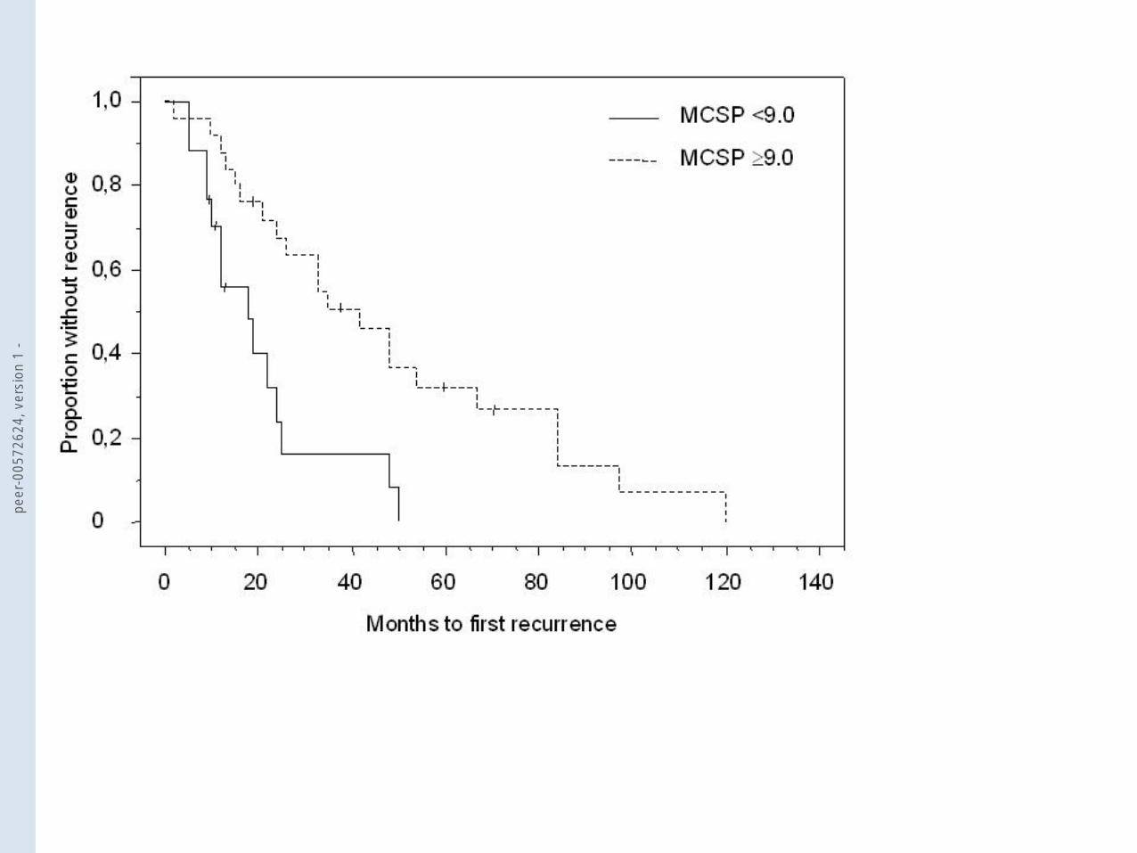
Wald2=8.6). Additionally, tumours with a MCSP-expression <9.0 points IRS had a
shorter recurrence-free interval than tumours with a MCSP-expression ≥9.0 IRS
(Kaplan-Meier analysis; log-rang p=0.002; Chi2=9.7) (figure 3). Analysing the
subgroups, we found that tumours with a lower MCSP-expression had a higher risk to
develop local recurrence and a shorter recurrence-free survival independent from
their clinical characteristics (example for epibulbar / nonepibulbar tumours, p=0.005,
peer
-005
7262
4, v
ersi
on 1
-
12
Chi2=7.8). The same trend was observed in other characteristics as pigmentation
(pigmented/amelanotic; p=0.002; Chi2=9.8), predominant cell type epithelioid
(p=0.02; Chi2=5.1), or occurrence of a second recurrence (p=0.03; Chi2=4.9). We
also observed a trend that tumours with the origin PAM with atypia (mean IRS 7.9 ±
2.7) had a lower mean MCSP expression than tumours appearing de novo (mean
IRS 9.5 ± 2.6). This did not reach statistical significance (p=0.18) but gave us a link to
a clinical explanation for our findings.
For PRAME, we did not find significant correlations to most parameters (data not
shown). In tumours that were confined to the epithelium (pT1, Melanoma in situ),
PRAME-expression was significantly lower than in tumours with subepithelial or
deeper invasion (pT2-pT4) (p=0.04). Tumours classified as pT2, pT3 and pT4 had no
significant difference in PRAME expression between the groups. We observed a
trend that tumours with an IRS-score for PRAME <9.0 points had a lower risk for
metastatic disease (Cox regression, HR=0.44, p=0.3; 95% CI: 0.1-2.1) and tended to
develop metastases later than tumours with an IRS-score ≥9.0 points (log-rang
p=0.3, Chi2=1.2). This reached to no statistical significance, possibly due to the
limited number of cases with metastatic disease in this cohort.
ROC-Analysis and predictive value of MCSP and PRAME
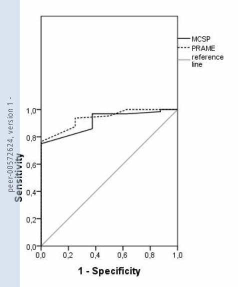
We performed a ROC-analysis in order to assess the power of MCSP and PRAME to
discriminate conjunctival nevi from conjunctival melanoma. For MCSP, the area
under the curve was 91.3% (asymptomatic p-value=0.0002; 95% CI: 0.84-0.99, figure
4). For PRAME, the area under the curve was 93.8% (asymptomatic p-value<0.0001;
95% CI: 0.87-1.0; figure 4). These findings make both parameters suitable to
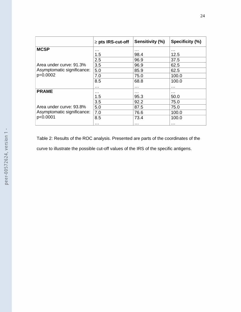
differentiate conjunctival nevi from melanoma. Table 2 shows the cut-off points within
the IRS score revealing that MCSP and PRAME were diagnostically valuable in the
intermediate and high scores (2.5-8.5 pts IRS).
peer
-005
7262
4, v
ersi
on 1
-
13
DISCUSSION:
Our results indicate that MCSP and PRAME are differentially expressed in benign
conjunctival nevi and malignant conjunctival melanomas. Conjunctival melanoma
(CoM) and lymph-node (LN)-metastases of cutaneous melanoma (CM) express both,
MCSP and PRAME, at a similar level, whereas conjunctival nevi (CoN) express both
antigens on a significantly lower level. Both markers can discriminate conjunctival
nevi from melanoma with high significance.
In our cohort MCSP had the surprising effect, that CoM-tumours with lower MCSP-
expression had a higher risk for local recurrence and developed their first recurrence
earlier than tumours with a high MCSP-expression. Factors that had turned out to be
prognostically relevant to the outcome of CoM in clinical studies before did not turn
this trend in our study and might therefore be of subordinate predictive relevance
compared to immunohistochemical characteristics. But despite our results, this can
only be stated as a hypothesis and has to be confirmed by further studies. For
explanation, one has to recall the mechanisms associated with MCSP. MCSP is able
to influence intracellular signal cascades that have an impact on cell adhesion,
motility and invasion.[34] One relevant mechanism in the communication of
melanoma cells to extracellular matrix (ECM) is provided by the α4β1-integrin and its
binding to fibronectin and to the vascular endothelial cell adhesion molecule VCAM
[35] which is also expressed in conjunctival melanoma.[36] It is known that the MCSP
rat-homologue NG2 collaborates with α4β1-integrin for stimulating focal contacts and
stress fibre formation in melanoma cells.[22] This may contribute to the metastatic
potential of a tumour. On the other hand, it may also be possible, that in cases of a
low MCSP expression, the reduced communication of a tumour cell with the ECM
and the immune-cells helps to establish a tumour recurrence in the local environment
peer
-005
7262
4, v
ersi
on 1
-
14
without being “recognised”. This would explain the trend that a lower MCSP-
expression is linked to a higher risk for local recurrence in our cohort of CoM.
Probably, our results could also be explained clinically, as follows: Tumours
originating from PAM with atypia had a lower mean MCSP expression and a 2.4fold
increased risk for recurrence compared to primary tumours in our cohort. The
recurrence-rate may be higher because clinical assessment of PAM with atypia holds
the risk of missing a recurrent transformation to melanoma due to their diffuse and
variable appearance on the ocular surface. It can be assumed, that the lower mean
expression of MCSP in this group leads to our finding that such tumours tend to have
a higher risk for recurrence in the whole cohort.
In conjunctival melanoma PRAME is expressed on a high level. Together with the
very low expression of PRAME in conjunctival nevi, our results correspond to the
reported PRAME-expression in cutaneous melanoma and nevi.[26, 28] Our results
indicate that invasive tumours express higher levels of PRAME than intraepithelial
tumours. This suggests that PRAME-expression increases under melanoma
progression. It is, however, no linear progression but rather seems to divide early
(intraepithelial) from advanced (invasive) tumours. PRAME acts by interfering with
retinoic-acid-–signalling as a dominant inhibitor of the retinoic acid receptor
(RAR).[29] Binding of retinoic acid to its receptor initiates the activation of target
genes that induce proliferation arrest, differentiation and apoptosis. This is the basis
of the antitumoral activity of retinoic acid.[37, 38] Subsequently, the change to, and
increase of, PRAME-expression may support tumour progression as the suppression
of RAR-signalling promotes tumour-growth and inhibits differentiation. The impact of
PRAME on the prognosis of a tumour entity is variable.[39, 40] Our samples showed
a trend towards earlier occurrence of metastases in cases with high PRAME-
peer
-005
7262
4, v
ersi
on 1
-
15
expression combined with an increased risk for metastases. The statistical value was
limited due to the limited number of cases in that subset.
In order to understand the role of PRAME and MCSP in conjunctival melanoma more
completely, further genetic studies are required to elucidate the source and
mechanism of the increased PRAME- and MCSP-expression in conjunctival
melanoma.
MCSP and PRAME have a good diagnostic value to distinguish between conjunctival
nevi and conjunctival melanoma. Whether the antigens are able to improve the
immunohistological diagnosis in difficult cases has to be evaluated further.
In conclusion, in this study we reported on the expression of melanoma chondroitin
sulphate proteoglycan (MCSP) and the preferentially expressed antigen of melanoma
(PRAME) in conjunctival melanoma and benign conjunctival nevi. We showed that
MCSP and PRAME were differentially expressed in conjunctival melanoma and nevi
and can help to differentiate the lesions diagnostically.
The authors have no financial / conflicting interests to disclose.
The Corresponding Author has the right to grant on behalf of all authors and does
grant on behalf of all authors, an exclusive licence (or non exclusive for government
employees) on a worldwide basis to the BMJ Publishing Group Ltd and its licencees,
to permit this article (if accepted) to be published in BJO and any other BMJ Group
products and to exploit all subsidiary rights, as set out in our licence
(http://bjo.bmjjournals.com//ifora/licence.pdf).
peer
-005
7262
4, v
ersi
on 1
-
16
References
1. Seregard S, Kock E. Conjunctival malignant melanoma in Sweden 1969–
1991. Acta Ophthalmol. Scand. 1992;70:289–296.
2. Missotten G, Keijser S, Keizer RJWD, de Wolff-Rouendaal D. Conjunctival
Melanoma in The Netherlands: A Nationwide Study. Invest Ophthalmol Vis Sci
2005;46:75-82.
3. Shields J, Shields C, Mashayekhi A, Marr B, Benavides R, Thangappan A, et
al. Primary acquired melanosis of the conjunctiva: risks for progression to melanoma
in 311 eyes. The 2006 Lorenz E. Zimmerman lecture. Ophthalmology
2008;115(3):511-519.
4. Paridaens AD, Minassian DC, MacCarty CA, Hungerford JL. Prognostic
factors in primary malignant melanoma of the conjunctiva: a clinicopathological study
of 256 cases. Br J Ophthalmol 1994;78(4):252-259.
5. Anastassiou G, Heiligenhaus A, Bechrakis N, Bader E, Bornfeld N, Steuhl K-P.
Prognostic value of clinical and histopathological parameters in conjunctival
melanomas: a retrospective study. Br J Ophthalmol. 2002;86:163-167.
6. Damato B, Coupland S. An audit of conjunctival melanoma treatment in
Liverpool. Eye 2009.
7. Keijser I, Missotten G, Bonfrer J, de Wolff-Rouendaal D, Jager M, de Keizer R.
Immunophenotypic markers to differentiate between benign and malignant
melanocytic lesions. Br J Ophthalmol 2006;90:213–217.
8. Esmaeli B, Wang X, Youssef A, Gershenwald JE. Patterns of regional and
distant metastasis in patients with conjunctival melanoma: experience at a cancer
center over four decades. Ophthalmology 2001;108(11):2101-5.
peer
-005
7262
4, v
ersi
on 1
-
17
9. Iwamoto S, Burrows R, Grossniklaus H, Orcutt J, Kalina R, Boehm M, et al.
Immunophenotype of conjunctival melanomas: comparisons with uveal and
cutaneous melanomas. Arch Ophthalmol. 2002:1625-1629.
10. Hurst E, Harbour J, Cornelius L. Ocular Melanoma. A Review and the
Relationship to Cutaneous Melanoma. Arch Dermatol. 2003;139:1067-1073.
11. Hu D, Yu G, McCormick S, Finger P. Population-based incidence of
conjunctival melanoma in various races and ethnic groups and comparison with other
melanomas. Am J Ophthalmol. 2008;145(3):418-423.
12. Folberg R, Mc Lean IW, Zimmermann LE. Malignant melanoma of the
conjunctiva. Hum Pathol. 1985;16:136-143.
13. Tuomaala S, Eskelin S, Tarkkanen A, Kivela T. Population-Based Assessment
of Clinical Characteristics Predicting Outcome of Conjunctival Melanoma in Whites.
Invest Ophthalmol Vis Sci. 2002;43:3399–3408.
14. Wuestemeyer H, Sauerwein W, Meller D, Chauvel P, Schueler A, Steuhl K-P,
et al. Proton radiotherapy as an alternative to exenteration in the management of
extended conjunctival melanoma. Graefe's Arch Clin Exp Ophthalmol 2006;244:438–
446.
15. Wilson B, Ruberto G, Ferrone S. Immunochemical characterization of a human
high molecular weight melanoma associated antigen identified with monoclonal
antibodies. Cancer Immunol lmmunother 1983;14:196-201.
16. Bemfield M, Gottc M, Park P, Rcizes O, Fitzgerald M, Lincecum J, et al.
Functions of cell surface heparan sulfate proteoglycans. Annu Rev Biochem
1999;68:729-777.
17. Iida J, Pei D, Kang T, Simpson MA, Herlyn M, Furcht LT, et al. Melanome
Chondroitin Sulfate Proteoglycan Regulates Matrix Metalloproteinase-dependent
peer
-005
7262
4, v
ersi
on 1
-
18
Human Melanoma Invasion into Type I Collagen. Journal of Biological Chemistry
2001;276(22):18786-18794.
18. Burg M, Grako K, Stallcup W. Expression of the NG2 proteoglycan enhances
the growth and metastatic properties of melanoma cells. J. Cell. Physiol.
1998;177:299–312.
19. Chekenya M, Rooprai H, Davies D, Levine J, Butt A, Pilkington G. The NG2
chondroitin sulfate proteoglycan: role in malignant progression of human brain
tumours. Int. J. Dev. Neurosci. 1999;17:421–435.
20. Benassi M, Pazzaglia L, Chiechi A, Alberghini M, Conti A, Cattaruzza S, et al.
NG2 expression predicts the metastasis formation in soft-tissue sarcoma patients. J
Orthop Res. 2009;27(1):135-140.
21. Burg M, Grako K, Stallcup W. Expression of the NG2 proteoglycan enhances
the growth and metastatic properties of melanoma cells. J Cell Physiol. 1998.
22. Iida J, Meijne A, Spiro R, Roos E, Furcht L, McCarthy J. Spreading and focal
contact formation of human melanoma cells in response to the stimulation of both
melanoma-associated proteoglycan (NG2) and alpha 4 beta 1 integrin. Cancer Res.
1995;55:2177-2185.
23. Iida J, Wilhelmson K, Ng J, Lee P, Morrison C, Tam E, et al. Cell surface
chondroitin sulfate glycosaminoglycan in melanoma: role in the activation of pro-
MMP-2 (pro-gelatinase A). Biochem. J. 2007;403:553–563.
24. Ferrone S, Temponi M, Gargiulo D, Scassellati G, Cavaliere R, Natali P.
Selection and utilization of monoclonal antibody defined melanoma associated
antigens for immunoscintigraphy in patients with melanoma. In: Srivastava SC,
editor. Radiolabeled monoclonal Antibodies for Imaging and Therapy. New
York/London: Plenum Publishing Corp.; 1988. p. 55–73.
peer
-005
7262
4, v
ersi
on 1
-
19
25. Campoli M, Chang C, Kageshita T, Wang X, McCarthy J, Ferrone S. Human
High Molecular Weight-Melanoma-Associated Antigen (HMW-MAA): A Melanoma
Cell Surface Chondroitin Sulfate Proteoglycan (MSCP) with Biological and Clinical
significance. Critical Riview in Immunology 2004;24(4):267-296.
26. Ikeda H, Lethé B, Lehmann F, van Baren N, Baurain J, de Smet C, et al.
Characterization of an antigen that is recognized on a melanoma showing partial HLA
loss by CTL expressing an NK inhibitory receptor. Immunity 1997;6(2):199–208.
27. Watari K, Tojo A, Nagamura-Inoue T, Nagamura F, Takeshita A, Fukushima T,
et al. Identification of a melanoma antigen, PRAME, as a BCR/ABL-inducible gene.
FEBS Lett. 2000;466(2-3):367-371.
28. Haqq C, Nosrati M, Sudilovsky D, Crothers J, Khodabakhsh D, Pulliam B, et
al. The gene expression signatures of melanoma progression. Proc Natl Acad Sci U
S A 2005;102:6092–6097.
29. Epping M, Wang L, Edel M, Carlée L, Hernandez M, Bernards R. The Human
Tumor Antigen PRAME Is a Dominant Repressor of Retinoic Acid Receptor
Signaling. Cell 2005;122:835–847.
30. Remmele W, Stegner H. Recommendation for uniform definition of an
immunoreactive score (IRS) for immunohistochemical estrogen receptor detection
(ER-ICA) in breast cancer tissue. Pathologe 1987;8(3):138-140.
31. Landis R, Koch G. The measurement of observer agreement for categorical
data. Biometrics 1977;33:159-174.
32. Kohlberger P, Breitenecker F, Kaider A, Lösch A, Gitsch G, Breitenecker G, et
al. Modified true-color computer-assisted image analysis versus subjective scoring of
estrogen receptor expression in breast cancer: a comparison. Anticancer Res.
1999;19(3B):2189-2193.
peer
-005
7262
4, v
ersi
on 1
-
20
33. Remmele W, Schicketanz K. Immunohistochemical determination of estrogen
and progesterone receptor content in human breast cancer. Computer-assisted
image analysis (QIC score) vs. subjective grading (IRS). Pathol Res Pract.
1993;189(8):862-866.
34. Yang J, Price MA, Neudauer CL, Wilson C, Ferrone S, Xia H, et al. Melanoma
chondroitin sulfate proteoglycan enhances FAK and ERK activation by distinct
mechanisms. Journal of Cell Biology 2004;165(6):881-891.
35. Mould A, Askari J, Craig S, Garratt A, Clements J, Humphries M. Integrin
alpha4beta1-mediated melanoma cell adhesion and migration on vascular cell
adhesion molecule-1 (VCAM-1) and the alternatively spliced IIICS region of
fibronectin. J. Biol. Chem. 1994;269:27224–27230.
36. Anastassiou G, Esser M, Bader E, Steuhl K-P, Bornfeld N. Expression of cell
adhesion molecules and tumour infiltration leucocytes in conjunctival melanoma.
Melanoma Research 2004;14:381-285.
37. Altucci L, Gronemeyer H. The promise of retinoids to fight against cancer. Nat.
Rev. Cancer 2001;1:181–193.
38. Freemantle S, Spinella M, Dmitrovsky E. Retinoids in cancer therapy and
chemoprevention: promise meets resistance. Oncogene 2003;22:7305–7315.
39. Steinbach D, Hermann J, Viehmann S, Zintl F, Gruhn B. Clinical implications
of PRAME gene expression in childhood acute myeloid leukemia. Cancer Genet
Cytogenet 2002;133:118–123.
40. Doolan P, Clynes M, Kennedy S, Mehta J, Crown J, O'Driscoll L. Prevalence
and prognostic and predictive relevance of PRAME in breast cancer. Breast Cancer
Res Treat. 2008;109(2):359-365.
peer
-005
7262
4, v
ersi
on 1
-
21
Legends to figures:
Figure 1: Immunohistology of MCSP (A, B, C) and PRAME (D, E, F). Magnification
x200. Both Antibodies showed a cytoplasmic and nuclear immunoreaction. MCSP:
cytoplasmic > nuclear; PRAME: nuclear >> cytoplasmic. This pattern was observed
in conjunctival melanomas, conjunctival nevi, and lymph-node metastases of
cutaneous melanoma in equal measure.
Figure 2: A. Boxplot of results of MCSP-expression in the three diagnosis groups. B:
Boxplot of results of PRAME expression in the three diagnosis groups.
Statistically significant differences are marked by *. Mean IRS and standard deviation
(SD) are given within the box. The horizontal line within the box marks the median.
The box itself spans from the 25th percentile (lower end) to the 75th percentile (upper
end). The T-lines below and above the box mark the 10th percentile (below) and the
90th percentile (above).
Figure 3: Kaplan-Meier analysis for recurrence-free survival in patients with CoM.
The group was divided into MCSP-expression <9.0 points IRS and ≥9.0 points IRS.
Figure 4: ROC-analysis of MCSP (continuous line) and PRAME (dotted line).
Diagonal reference line for orientation. Area under the curve for MCSP: 91.3 %
(p=0.0002); for PRAME: 93.8% (p<0.0001).
peer
-005
7262
4, v
ersi
on 1
-
22
N=70 CoM (%) N=70 CoM (%)
Time of
presentation
40 first diagnosis (57.1%)
Tumour origin
10 Primary (de novo) (14.3%)
22 local recurrences (31.4%) 23 PAM with atypia (32.9%)
8 unknown (11.5%) 16 Nevi (22.9%)
Gender 30 male (42.9%) 10 Recurrence (14.3%)
40 female (57.1%) 11 Unknown (15.6%)
cTNM
18 T1 (25.7%)
Pigmentation
55 Pigmented (78.6%)
18 T2 (25.7%) 10 Amelanotic (14.3%)
17 T3 (24.3%) 5 Unknown (7.1%)
4 T4 (5.7%) Recurrence during
follow-up
36 Yes (51.4%)
13 Tx (18.6%) 17 No (24.3%)
pTNM
3 pT1 (4.3%) 17 Unknown (24.3%)
30 pT2 (42.9%) Predominant cell type
(n=54)
35 Epithelioid (64.8%)
15 pT3 (21.4%) 12 Spindle cell (22.2%)
4 pT4 (5.7%) 7 Pleomorph (13.0%)
18 pTx (25.7%)
Metastases
10 Yes (14.3%)
Location primary
tumour
37 Epibulbar (52.9%) 41 No (58.6%)
21 Nonepibulbar (30.0%) 19 Unknown (27.1%)
12 Unknown (17.1%)
peer
-005
7262
4, v
ersi
on 1
-
23
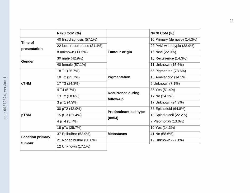
Table 1: Demographic and pathological data of the CoM-group with the number of tumours (n=) of the whole group. CoM: conjunctival
melanoma.
peer
-005
7262
4, v
ersi
on 1
-
24
≥ pts IRS-cut-off Sensitivity (%) Specificity (%)
MCSP
Area under curve: 91.3% Asymptomatic significance: p=0.0002
… 1.5
… 98.4
… 12.5
2.5 96.9 37.5 3.5 96.9 62.5 5.0 85.9 62.5 7.0 75.0 100.0 8.5 …
68.8 …
100.0 …
PRAME
Area under curve: 93.8% Asymptomatic significance: p<0.0001
… 1.5
… 95.3
… 50.0
3.5 92.2 75.0 5.0 87.5 75.0 7.0 76.6 100.0 8.5 …
73.4 …
100.0 …
Table 2: Results of the ROC analysis. Presented are parts of the coordinates of the
curve to illustrate the possible cut-off values of the IRS of the specific antigens.
peer
-005
7262
4, v
ersi
on 1
-

peer
-005
7262
4, v
ersi
on 1
-

peer
-005
7262
4, v
ersi
on 1
-
peer
-005
7262
4, v
ersi
on 1
-
peer
-005
7262
4, v
ersi
on 1
-
Related Documents