Abstract During human pregnancy the placenta produces a variety of proteins like steroid hormones and their receptors that are responsible for the estab- lishment and ongoing of the feto-placental unit. Inhi- bins are dimeric glycoproteins, composed of an a-subunit and one of two possible b-subunits (b A or b B ). Aims of the present study were the determination of the frequency and tissue distribution patterns of the inhibin/activin subunits in human placental tissue of normal pregnancies and pregnancies complicated with fetal growth restriction (IUGR). Slides of paraffin embedded placental tissue were obtained after delivery from patients diagnosed with IUGR (n = 6) and nor- mal term placentas (n = 8). Tissue samples were fixed and incubated with monoclonal antibodies inhibin/ activin-subunits -a,-b A ,-b B . Intensity of immunohis- tochemical reaction on the slides was analysed using a semi-quantitative score and statistical analysis was performed (P < 0.05). A significant lower expression of the inhibin-a subunit in IUGR extravillous trophoblast compared to normal pregnancies was observed, while the inhibin-a immunostaining was significantly upreg- ulated in syncytiotrophoblast. Additionally, a signifi- cant down-regulation of inhibin-b B subunit in extravillous trophoblast cells in IUGR syncytiotroph- oblast cells was demonstrated. A co-localisation of in- hibin-a and the b-subunits was also observed, suggesting a production and secretion of intact inhibin A and inhibin B. Although the precise role of these inhibin/activin subunits in human placenta and IUGR pregnancies is still unclear, they could be involved in autocrine/paracrine signalling, contributing to several aspects like angiogenesis and tissue remodelling. Keywords Inhibin/activin subunits Inhibin-a Inhibin-b A Inhibin-b B Intrauterine growth restriction (IUGR) Extravillous trophoblast Syncytiotrophoblast Introduction Inhibins are dimeric disulphide-linked glycoproteins and belong to the transforming growth factor beta (TGF-b) family of cytokines. They were initially iso- lated from the gonads and identified as modulators of FSH production from the anterior pituitary gland (de Kretser et al. 2002; Vale et al. 1988). These molecules are heterodimers consisting of one a-subunit and one of two possible b-subunits (b A - and b B -subunits). The a-subunit can dimerize with either b A or b B to form inhibin A (a-b A )orB(a-b B ), respectively. Activins are homodimers of b-subunits linked by a disulfide bond. Depending on the combination of the subunits, there are three isoforms of activin, namely activin A (b A –b A ), activin B (b B –b B ) and activin AB (b A –b B ) I. Mylonas (&) B. Schiessl U. Jeschke J. Vogl C. Kuhn S. Kunze S. Schulze F. Kainer K. Friese 1st Department of Obstetrics and Gynaecology, Ludwig- Maximilians-University Munich, Maistrasse 11, Munich 80337, Germany e-mail: [email protected] B. Schiessl School of Surgical and Reproductive Sciences (Obstetrics & Gynaecology), University of Newcastle upon Tyne, Framlington Place, Newcastle upon Tyne NE2 4HH, UK A. Makrigiannakis Department of Obstetrics and Gynecology, University of Crete, Heraklion, Crete, Greece J Mol Hist (2006) 37: 43–52 DOI 10.1007/s10735-006-9029-6 123 ORIGINAL PAPER Expression of inhibin/activin subunits alpha (-a), beta A (-b A ) and beta B (-b B ) in placental tissue of normal and intrauterine growth restricted (IUGR) pregnancies I. Mylonas B. Schiessl U. Jeschke J. Vogl A. Makrigiannakis C. Kuhn S. Kunze S. Schulze F. Kainer K. Friese Received: 29 January 2006 / Accepted: 29 March 2006 / Published online: 3 May 2006 ȑ Springer Science+Business Media B.V. 2006

Welcome message from author
This document is posted to help you gain knowledge. Please leave a comment to let me know what you think about it! Share it to your friends and learn new things together.
Transcript
Abstract During human pregnancy the placenta
produces a variety of proteins like steroid hormones
and their receptors that are responsible for the estab-
lishment and ongoing of the feto-placental unit. Inhi-
bins are dimeric glycoproteins, composed of an
a-subunit and one of two possible b-subunits (bA or
bB). Aims of the present study were the determination
of the frequency and tissue distribution patterns of the
inhibin/activin subunits in human placental tissue of
normal pregnancies and pregnancies complicated with
fetal growth restriction (IUGR). Slides of paraffin
embedded placental tissue were obtained after delivery
from patients diagnosed with IUGR (n = 6) and nor-
mal term placentas (n = 8). Tissue samples were fixed
and incubated with monoclonal antibodies inhibin/
activin-subunits -a, -bA, -bB. Intensity of immunohis-
tochemical reaction on the slides was analysed using a
semi-quantitative score and statistical analysis was
performed (P < 0.05). A significant lower expression of
the inhibin-a subunit in IUGR extravillous trophoblast
compared to normal pregnancies was observed, while
the inhibin-a immunostaining was significantly upreg-
ulated in syncytiotrophoblast. Additionally, a signifi-
cant down-regulation of inhibin-bB subunit in
extravillous trophoblast cells in IUGR syncytiotroph-
oblast cells was demonstrated. A co-localisation of in-
hibin-a and the b-subunits was also observed,
suggesting a production and secretion of intact inhibin
A and inhibin B. Although the precise role of these
inhibin/activin subunits in human placenta and IUGR
pregnancies is still unclear, they could be involved in
autocrine/paracrine signalling, contributing to several
aspects like angiogenesis and tissue remodelling.
Keywords Inhibin/activin subunits Æ Inhibin-a ÆInhibin-bA Æ Inhibin-bB Æ Intrauterine growth
restriction (IUGR) Æ Extravillous trophoblast ÆSyncytiotrophoblast
Introduction
Inhibins are dimeric disulphide-linked glycoproteins
and belong to the transforming growth factor beta
(TGF-b) family of cytokines. They were initially iso-
lated from the gonads and identified as modulators of
FSH production from the anterior pituitary gland (de
Kretser et al. 2002; Vale et al. 1988). These molecules
are heterodimers consisting of one a-subunit and one
of two possible b-subunits (bA- and bB-subunits). The
a-subunit can dimerize with either bA or bB to form
inhibin A (a-bA) or B (a-bB), respectively. Activins
are homodimers of b-subunits linked by a disulfide
bond. Depending on the combination of the subunits,
there are three isoforms of activin, namely activin A
(bA–bA), activin B (bB–bB) and activin AB (bA–bB)
I. Mylonas (&) Æ B. Schiessl Æ U. Jeschke Æ J. Vogl ÆC. Kuhn Æ S. Kunze Æ S. Schulze Æ F. Kainer Æ K. Friese1st Department of Obstetrics and Gynaecology, Ludwig-Maximilians-University Munich, Maistrasse 11, Munich80337, Germanye-mail: [email protected]
B. SchiesslSchool of Surgical and Reproductive Sciences (Obstetrics &Gynaecology), University of Newcastle upon Tyne,Framlington Place, Newcastle upon Tyne NE2 4HH, UK
A. MakrigiannakisDepartment of Obstetrics and Gynecology, University ofCrete, Heraklion, Crete, Greece
J Mol Hist (2006) 37: 43–52
DOI 10.1007/s10735-006-9029-6
123
ORIGINAL PAPER
Expression of inhibin/activin subunits alpha (-a), beta A (-bA)and beta B (-bB) in placental tissue of normal and intrauterinegrowth restricted (IUGR) pregnancies
I. Mylonas Æ B. Schiessl Æ U. Jeschke ÆJ. Vogl Æ A. Makrigiannakis Æ C. Kuhn ÆS. Kunze Æ S. Schulze Æ F. Kainer Æ K. Friese
Received: 29 January 2006 / Accepted: 29 March 2006 / Published online: 3 May 2006� Springer Science+Business Media B.V. 2006
(de Kretser et al. 2002; Vale et al. 1988). Recently,
two additional b-subunits have been identified in hu-
man tissue, determined as bC (Hotten et al. 1995) and
bE (Fang et al. 1996), although their precise role and
functional relationship to the existing subunits re-
mains still unknown.
The expression of inhibin/activin subunits have
been described in different female tissues, including
normal and pathological human endometrium (Mylo-
nas et al. 2003, 2004a, 2006) and placenta (Caniggia
et al. 1997; McCluggage et al. 1998; Petraglia et al.
1991), suggesting different roles such as paracrine
modulators of reproductive function (de Kretser et al.
2002; Welt 2002). During pregnancy, inhibin/activin
subunits are also expressed in placental decidua, the
syncytiotrophoblast (Petraglia et al. 1991) and the
trophoblast (McCluggage et al. 1998). Interestingly,
higher inhibin levels in human serum have been de-
scribed in preeclampsia (Muttukrishna et al. 1997)
and down-syndrome (Aitken et al. 1996), suggesting
that inhibin/activin production by placental cells might
play a major and crucial role in pregnancy-related
pathogenesis.
Normal fetal growth depends on several factors
modulated by the fetus, the placenta and the mother.
In preeclampsia and idiopathic small for gestational
age (SGA) pregnancies, cytotrophoblast invasion is
restricted with a limited remodelling of spiral arteries,
thus resulting in reduced uteroplacental perfusion
(Lim et al. 1997). Small fetuses due of intrauterine
growth restriction (IUGR) are at higher risk for poor
perinatal and long-term outcome (Baschat 2004; Tjoa
et al. 2004), being associated with an increased risk of
heart diseases and type 2 diabetes mellitus (Barker
1998). The most common definition of IUGR is a
birth weight lower than the 10th percentile when ad-
justed to gestational age. In the past years several
molecules have been suggested as predictive markers
of IUGR, including cytokines, neuropeptides, adhe-
sion molecules and glycoproteins such like inhibin A
and activin A (Tjoa et al. 2004). However, limited
data on histological expression of inhibin/activin su-
bunits expression exists. Therefore, aims of the pres-
ent study were:
(a) The determination of the frequency and tissue
distribution patterns of the inhibin/activin
subunits in human placental tissue of normal
pregnancies and pregnancies complicated with
fetal growth restriction (IUGR).
(b) The assessment of a combined expression of in-
hibin-a- and both b-subunits (bA- and bB-subun-
its) using double immunofluorescence technique.
Materials and methods
Tissue samples
Placental tissues were obtained from 12 placentas of
women giving birth at the 1st Department of Obstetrics
and Gynaecology of the LMU Munich. Tissue samples
were obtained from patients diagnosed with IUGR
(n = 6) and normal pregnancies (n = 6) after delivery
(Table 1).
Immunohistochemistry
Immunohistochemistry on paraffin sections (7 lm) of
the different placental tissue specimens was per-
formed by incubating the slides in methanol/H2O2
(30 min) to inhibit endogenous peroxidase activity.
Immunohistochemistry with inhibin-subunits was
performed using a combination of pressure cooker
heating and the standard streptavidin–biotin–peroxi-
dase complex with the use of the mouse-IgG-Vecta-
stain Elite ABC kit (Vector Laboratories,
Burlingame, CA, USA) as previously described
(Mylonas et al. 2004a). Briefly, paraffin-fixed tissue
sections were dewaxed using xylol for 15 min, rehy-
drated in an ascending series of alcohol row (70, 96
and 100%), and subjected to antigen retrieval on a
high setting for 10 min in a pressure cooker in sodium
citrate buffer (pH 6.0), containing citrate acid 0.1 M
and sodium citrate 0.1 M in distillated water. After
cooling, the slides were washed twice in PBS.
Endogenous peroxidase activity was quenched by
immersion in 3% hydrogen peroxide (Merck,
Darmstadt, Germany) in methanol for 20 min. Non-
specific binding of the primary antibodies was blocked
by incubating the sections with diluted normal serum
(10 ml PBS containing 150 ll horse serum; provided
by Vector Laboratories) for 20 min at room temper-
ature. Sections were then incubated at room temper-
ature for 120 min with the primary antibodies
(Table 2). After washing with PBS, the slides were
incubated in diluted biotinylated serum (10 ml
PBS containing 50 ll horse serum; provided by
Vector Laboratories) for another 30 min at room
Table 1 Clinical data of the examined placental tissue. Mean–SD
Control IUGR
Weeks of delivery 38.2 – 3.9 33.0 – 3.0Birth weight 3435 – 412.2 1213.6 – 45.3pH umbilical artery 7.27 – 0.09 7.26 – 0.09APGAR score < 7 at 5 min 9.4 – 0.9 9.0 – 0.7APGAR score < 7 at 10 min 10 – 0 9.6 – 0.5
44 J Mol Hist (2006) 37: 43–52
123
temperature. After incubation with the avidin–biotin
peroxidase complex for another 30 min and repeated
washing steps with PBS, visualisation was performed
with substrate and chromagen 3,3¢-diaminobenzidine
(DAB; Dako, Glostrup, Denmark) for 8–10 min. The
slides where counterstained further with Mayer’s
acidic hematoxylin and washed in an alcohol row (50–
98%). After xylol treatment the slides were covered.
Negative controls were performed by replacing the
primary antibody. Positive cells showed a brownish
colour and negative control as well as unstained cells
appeared blue. The standardisation, dilution and
optimisation of this protocol were primarily tested on
normal premenopausal ovary tissue, while negative
controls included postmenopausal ovarian tissue.
Immunofluorescence double staining reaction
For the immunohistochemical characterisation cryo-
sections from normal placental tissue and from pla-
centas of IUGR pregnancies were examined as
previously described (Jeschke et al. 2002; Mylonas
et al. 2004a, 2006). The used antibodies are listed in
Table 2, whereas the inhibin-a polyclonal antibody was
applied. Briefly, all samples were fixed in 5% buffered
formalin. They were diluted to 10 lg/ml with PBS and
incubated with the slides overnight at 4�C. After
washing, Cy2-labelled goat anti-rabbit IgG and Cy3-
labelled goat anti-mouse IgG, diluted 1:200, served as
second antibody. The slides were finally embedded in
mounting buffer containing 4,6-diamino-2-phenylin-
dole (DAPI) resulting in blue staining of the nucleus.
Slides were examined with a Zeiss (Jena, Germany)
Axiophot photomicroscope. Digital images were ob-
tained with a digital camera system (CF20DXC;
KAPPA Messtechnik, Gleichen, Germany) and saved
on computer.
Immunohistochemical evaluation and statistical
analysis
The intensity and distribution patterns of specific
inhibin/activin subunit immunohistochemical staining
reaction was evaluated by two blinded, independent
observers, including a gynaecological pathologist, using
a semi-quantitative score (IRS score) as previously
described (Mylonas et al. 2000) and used in the eval-
uation of inhibin/activin subunit expression in human
endometrial specimens (Mylonas et al. 2004a, 2006).
Briefly, the IRS score was calculated as follows:
IRS = SI·PP, where SI is the optical staining intensity
(graded as 0 = no staining; 1 = weak staining;
2 = moderate staining and 3 = strong staining) and PP
the percentage of positively stained cells. The PP was
estimated by counting approx. 200 cells (defined as
0 = no staining; 1 = < 10% staining; 2 = 11–50%
staining; 3 = 51–80% staining and 4 = >81% staining).
Digital images were obtained with a digital camera
system (Olympus, Tokyo, Japan) and were saved on
computer. The Mann–Whitney rank-sum test was used
to compare the means of the different IRS scores
(SPSS; Chicago, IL, USA). Significance of differences
was assumed at P £ 0.05.
Results
Immunohistochemical expression of inhibin-a,
-bA and -bB
Immunohistochemical staining was performed using an
appropriate positive control comprising ovaries con-
taining follicular cysts. Inhibin-a stained positive with
ovarian granulosa cells and theca interna cells, while
inhibin-bA and -bB subunits showed also a positive
immunohistochemical staining reaction with human
ovarian tissue as previously described (Mylonas et al.
2004a).
All three inhibin/activin subunits were detected in
human extravillous trophoblast and syncytiotropho-
blast of placental tissue of normal and IUGR preg-
nancies. In normal placental tissue inhibin-a was
primarily expressed in extravillous trophoblast cells,
while immunostaining in syncytiotrophoblast cells was
weaker. In IUGR placental tissue the inhibin-aexpression was decreased in extravillous trophoblast
cells, while syncytiotrophoblast showed a stronger
immunoreactive reaction (Fig. 1a–d).
Table 2 Antibodies used forimmunohistochemicalcharacterisation of placentaltissue samples byimmunohistochemistry andimmunofluorescence
Antibody Clone Isotype Dilution Source
CK 7 Polyclonal Rabbit IgG 1:200 Dianova, Hamburg, GermanyInhibin-a R1 Mouse IgG2a 1:50 Serotec, Oxford, UKInhibin-a Polyclonal Rabbit IgG 1:40 Signet, Dedham, MA, USAInhibin-bA E4 Mouse IgG2b 1:50 Serotec, Oxford, UKInhibin-bB C5 Mouse IgG2a 1:10 Serotec, Oxford, UKCy2 and Cy3 Goat anti-rabbit IgG 1:200 Dianova, Hamburg, GermanyCy2 and Cy3 Goat anti-mouse IgM 1:200 Dianova, Hamburg, Germany
J Mol Hist (2006) 37: 43–52 45
123

The bA-subunit was also detected in placental
extravillous trophoblast and syncytiotrophoblast cells.
While a diffuse immunohistochemical staining reaction
could be observed in IUGR extravillous trophoblast
cells an increased staining intensity reaction was
observed in syncytiotrophoblast placenta (Fig. 2a–d).
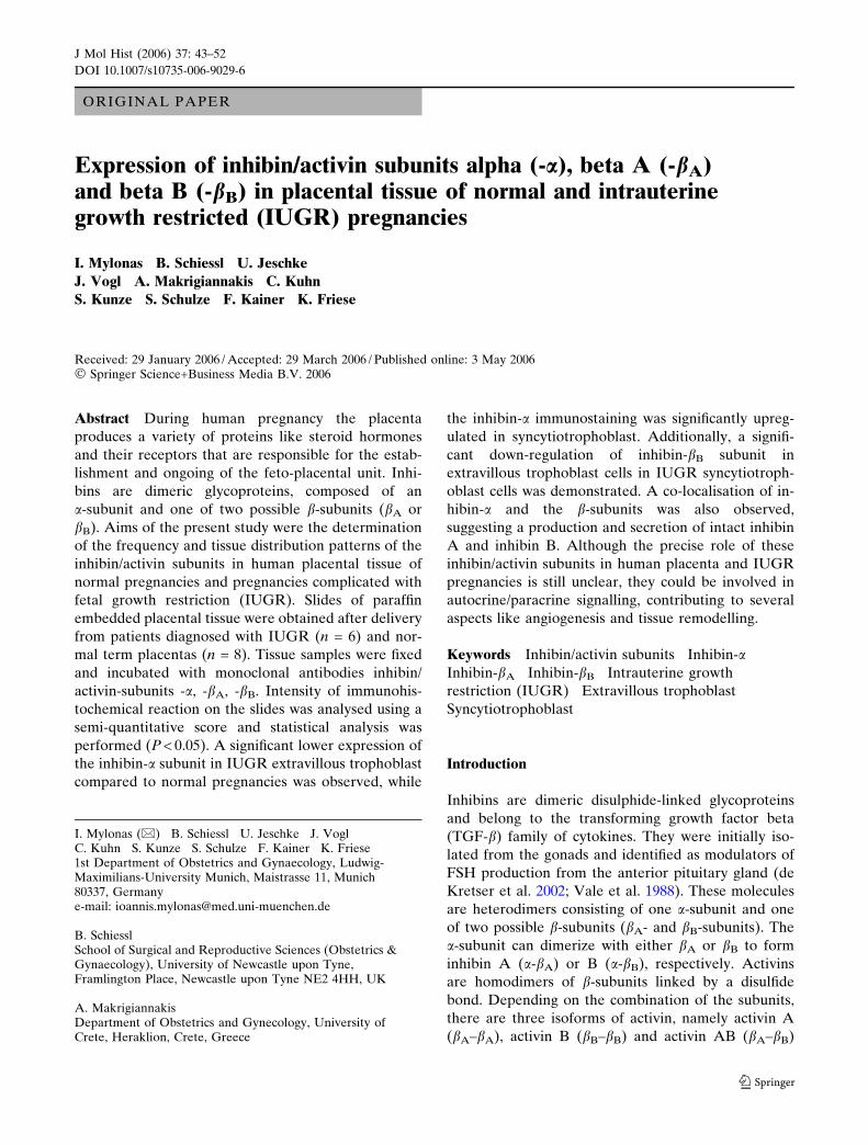
Interestingly, a decrease in the bB-subunit staining
reaction was observed between normal and IUGR
extravillous trophoblast cells. In the syncytiotropho-
blast the staining intensity presented without a distinct
expression pattern between normal and IUGR cells
(Fig. 3a–d).
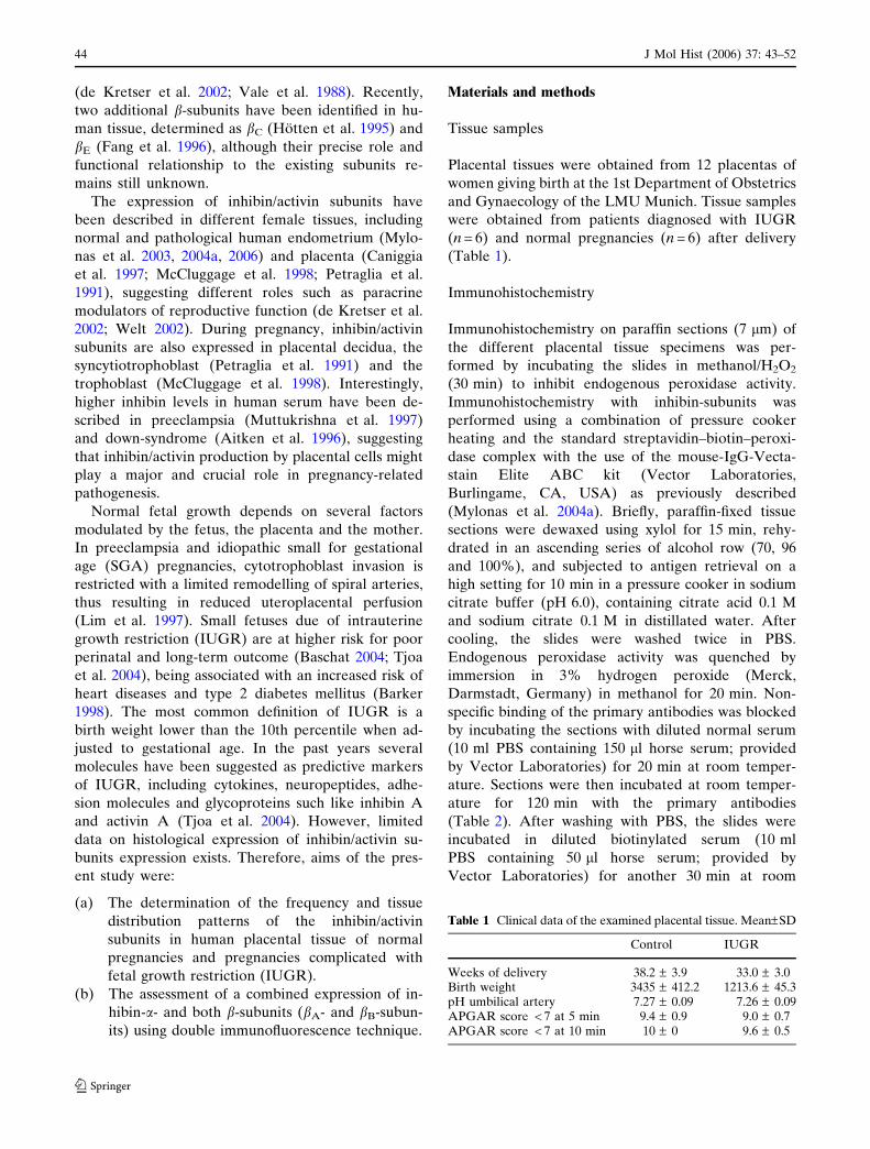
Expression of inhibin-a, -bA and -bB demonstrated
by immunofluorescence
All three inhibin/activin subunits were detected in
placental tissue of normal and IUGR pregnancies,
using immunofluorescence, with a polyclonal inhibin-asubunit antibody and monoclonal antibodies for the
Fig. 1 (a–d)Immunohistochemicalstaining reaction of inhibin-ain placental tissue. In normalplacental tissue inhibin-a wasprimarily expressed inextravillous trophoblast cells(a), while immunostaining insyncytiotrophoblast cells wasweaker (b). In IUGRplacental tissue the inhibin-aexpression was weaker inextravillous trophoblast cells(c), while syncytiotrophoblastshowed an intenseimmunoreactive stainingreaction (d). Magnifications:extravillous trophoblast 250·;syncytiotrophoblast 125·
Fig. 2 (a–d) Immunohistochemical staining reaction of inhibin-bA in placental tissue. The bA-subunit was also detected innormal placenta tissue. Extravillous trophoblast (a) and syncy-tiotrophoblast cells (b) showed intense staining reaction againstthe inhibin-bA antibody. While a diffuse immunohistochemical
staining reaction could be observed in IUGR extravilloustrophoblast cells (c) an increased immunohistochemical stainingreaction was observed in syncytiotrophoblast placenta (d).Magnifications: extravillous trophoblast 250·; syncytiotropho-blast 125·
46 J Mol Hist (2006) 37: 43–52
123
two distinct inhibin-b subunits. The inhibin/activin
subunits were primarily expressed in normal human
extravillous trophoblast as confirmed by co-expression
with cytokeratin antibody (Figs. 4, 5).
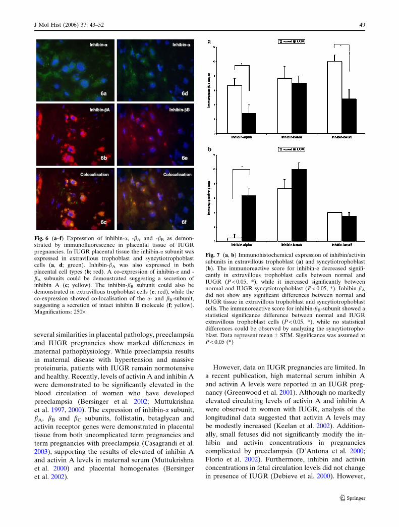
In IUGR placental tissue the inhibin-a subunit was
expressed in extravillous trophoblast and syncytio-
trophoblast cells. Inhibin-bA was also expressed in both
placental cell types. A co-expression of inhibin-a and
-bA subunits could be demonstrated suggesting a
secretion of inhibin A (Fig. 6a–c). The inhibin-bB
subunit could also be demonstrated in extravillous
trophoblast cells, while the co-expression showed
minimal co-localisation of the a- and bB-subunit
(Fig. 6d–f).
Fig. 3 (a–d)Immunohistochemicalstaining reaction of inhibin-bB
in placental tissue.Interestingly, a decrease inthe staining reaction wasobserved between normal (a)and IUGR (c) extravilloustrophoblast cells. In thesyncytiotrophoblast thestaining intensity presentedwithout a distinct expressionpattern between normal (b)and IUGR cells (d).Magnifications: extravilloustrophoblast 250·;syncytiotrophoblast 125·
Fig. 4 Expression of inhibin-a, -bA and -bB demonstratedby immunofluorescence inextravillous trophoblast cellsof normal and IUGRplacental tissue. The inhibin-a(a–c; 125·), inhibin-bA (d–f;250·) and inhibin-bB (g–i;250·) were primarilyexpressed in normal humanextravillous trophoblast asconfirmed by co-expressionwith cytokeratin 7 antibody
J Mol Hist (2006) 37: 43–52 47
123

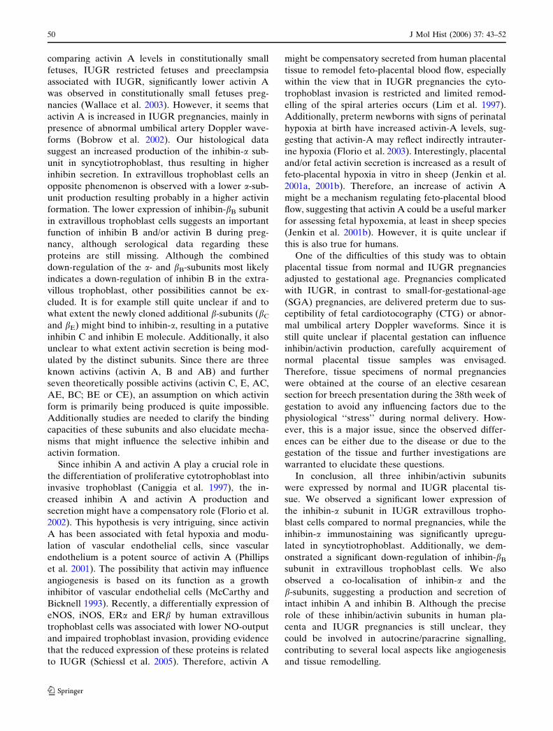
Immunohistochemical evaluation of inhibin-a, -bA
and -bB
The immunoreactive score for inhibin-a decreased
significantly between normal and IUGR placenta in
extravillous trophoblast tissue (P < 0.05), while the
inhibin-bA staining reaction did not show any signifi-
cant differences. Interestingly, inhibin-bB-subunit also
demonstrated a statistical significant decline between
normal and IUGR extravillous trophoblast cells
(Fig. 7a).
In syncytiotrophoblast cells the inhibin-a subunit
showed a significant increase between normal and
IUGR tissue (P < 0.05). Although a more intensive
staining reaction was observed for inhibin-bA in IUGR
syncytiotrophoblast compared to normal control tissue,
no significant differences could be demonstrated. The
immunoreactive score for inhibin-bB did also show no
statistical differences between normal and IUGR
syncytiotrophoblast cells (Fig. 7b).
Discussion
While inhibins/activins were initially characterised as
endocrine and paracrine hormonal regulators of the
hypothalamic–pituitary–gonadal axis, it is now clear
that they are expressed in a wide range of tissues
including human endometrium (Mylonas et al. 2003,
2004b, 2006) and human placenta (Florio et al. 2001;
McCluggage et al. 1998; Petraglia et al. 1991). The
human placenta expresses inhibin/activin mRNA
(Petraglia et al. 1991) and inhibin/activin subunit
protein throughout pregnancy, being the primary
source of maternal circulating inhibin and activin
levels (Florio et al. 2001). We demonstrated by using
immunohistochemistry and immunofluorescence dou-
ble staining technique an expression of inhibin/acti-
vin-subunit in normal and IUGR placental tissues.
Additionally we observed a significant lower expres-
sion of the inhibin-a subunit in extravillous tropho-
blast of IUGR compared to normal pregnancies,
while the inhibin-a immunostaining was significantly
upregulated in syncytiotrophoblast. A significant
down-regulation of inhibin-bB subunit in extravillas
trophoblast cells was also demonstrated, while
inhibin-bA-subunit was more intense in IUGR sync-
tiotrophoblast cells but without any statistical differ-
ences. We also showed a co-localisation of inhibin-aand the b-subunits, suggesting a production and
secretion of intact inhibin A and inhibin B.
In preeclampsia and IUGR pregnancies, cytotroph-
oblast invasion is restrained, resulting in a reduced
uteroplacental perfusion (Lim et al. 1997). Despite
Fig. 5 Expression of inhibin-a, -bA and -bB demonstratedby immunofluorescence inextravillous trophoblast cellsof normal and IUGRplacental tissue. The inhibin-a(a–c; 250·), inhibin-bA (d–f;125·) and inhibin-bB (g–i;125·) were also expressed inhuman extravilloustrophoblast of IUGRpregnancies as confirmed byco-expression withcytokeratin 7 antibody
48 J Mol Hist (2006) 37: 43–52
123
several similarities in placental pathology, preeclampsia
and IUGR pregnancies show marked differences in
maternal pathophysiology. While preeclampsia results
in maternal disease with hypertension and massive
proteinuria, patients with IUGR remain normotensive
and healthy. Recently, levels of activin A and inhibin A
were demonstrated to be significantly elevated in the
blood circulation of women who have developed
preeclampsia (Bersinger et al. 2002; Muttukrishna
et al. 1997, 2000). The expression of inhibin-a subunit,
bA, bB and bC subunits, follistatin, betaglycan and
activin receptor genes were demonstrated in placental
tissue from both uncomplicated term pregnancies and
term pregnancies with preeclampsia (Casagrandi et al.
2003), supporting the results of elevated of inhibin A
and activin A levels in maternal serum (Muttukrishna
et al. 2000) and placental homogenates (Bersinger
et al. 2002).
However, data on IUGR pregnancies are limited. In
a recent publication, high maternal serum inhibin A
and activin A levels were reported in an IUGR preg-
nancy (Greenwood et al. 2001). Although no markedly
elevated circulating levels of activin A and inhibin A
were observed in women with IUGR, analysis of the
longitudinal data suggested that activin A levels may
be modestly increased (Keelan et al. 2002). Addition-
ally, small fetuses did not significantly modify the in-
hibin and activin concentrations in pregnancies
complicated by preeclampsia (D’Antona et al. 2000;
Florio et al. 2002). Furthermore, inhibin and activin
concentrations in fetal circulation levels did not change
in presence of IUGR (Debieve et al. 2000). However,
Fig. 6 (a–f) Expression of inhibin-a, -bA and -bB as demon-strated by immunofluorescence in placental tissue of IUGRpregnancies. In IUGR placental tissue the inhibin-a subunit wasexpressed in extravillous trophoblast and syncytiotrophoblastcells (a, d; green). Inhibin-bA was also expressed in bothplacental cell types (b; red). A co-expression of inhibin-a and -bA subunits could be demonstrated suggesting a secretion ofinhibin A (c; yellow). The inhibin-bB subunit could also bedemonstrated in extravillous trophoblast cells (e; red), while theco-expression showed co-localisation of the a- and bB-subunit,suggesting a secretion of intact inhibin B molecule (f; yellow).Magnifications: 250·
Fig. 7 (a, b) Immunohistochemical expression of inhibin/activinsubunits in extravillous trophoblast (a) and syncytiotrophoblast(b). The immunoreactive score for inhibin-a decreased signifi-cantly in extravillous trophoblast cells between normal andIUGR (P < 0.05, *), while it increased significantly betweennormal and IUGR syncytiotrophoblast (P < 0.05, *). Inhibin-bA
did not show any significant differences between normal andIUGR tissue in extravillous trophoblast and syncytiotrophoblastcells. The immunoreactive score for inhibin-bB-subunit showed astatistical significance difference between normal and IUGRextravillous trophoblast cells (P < 0.05, *), while no statisticaldifferences could be observed by analyzing the syncytiotropho-blast. Data represent mean – SEM. Significance was assumed atP < 0.05 (*)
J Mol Hist (2006) 37: 43–52 49
123
comparing activin A levels in constitutionally small
fetuses, IUGR restricted fetuses and preeclampsia
associated with IUGR, significantly lower activin A
was observed in constitutionally small fetuses preg-
nancies (Wallace et al. 2003). However, it seems that
activin A is increased in IUGR pregnancies, mainly in
presence of abnormal umbilical artery Doppler wave-
forms (Bobrow et al. 2002). Our histological data
suggest an increased production of the inhibin-a sub-
unit in syncytiotrophoblast, thus resulting in higher
inhibin secretion. In extravillous trophoblast cells an
opposite phenomenon is observed with a lower a-sub-
unit production resulting probably in a higher activin
formation. The lower expression of inhibin-bB subunit
in extravillous trophoblast cells suggests an important
function of inhibin B and/or activin B during preg-
nancy, although serological data regarding these
proteins are still missing. Although the combined
down-regulation of the a- and bB-subunits most likely
indicates a down-regulation of inhibin B in the extra-
villous trophoblast, other possibilities cannot be ex-
cluded. It is for example still quite unclear if and to
what extent the newly cloned additional b-subunits (bC
and bE) might bind to inhibin-a, resulting in a putative
inhibin C and inhibin E molecule. Additionally, it also
unclear to what extent activin secretion is being mod-
ulated by the distinct subunits. Since there are three
known activins (activin A, B and AB) and further
seven theoretically possible activins (activin C, E, AC,
AE, BC; BE or CE), an assumption on which activin
form is primarily being produced is quite impossible.
Additionally studies are needed to clarify the binding
capacities of these subunits and also elucidate mecha-
nisms that might influence the selective inhibin and
activin formation.
Since inhibin A and activin A play a crucial role in
the differentiation of proliferative cytotrophoblast into
invasive trophoblast (Caniggia et al. 1997), the in-
creased inhibin A and activin A production and
secretion might have a compensatory role (Florio et al.
2002). This hypothesis is very intriguing, since activin
A has been associated with fetal hypoxia and modu-
lation of vascular endothelial cells, since vascular
endothelium is a potent source of activin A (Phillips
et al. 2001). The possibility that activin may influence
angiogenesis is based on its function as a growth
inhibitor of vascular endothelial cells (McCarthy and
Bicknell 1993). Recently, a differentially expression of
eNOS, iNOS, ERa and ERb by human extravillous
trophoblast cells was associated with lower NO-output
and impaired trophoblast invasion, providing evidence
that the reduced expression of these proteins is related
to IUGR (Schiessl et al. 2005). Therefore, activin A
might be compensatory secreted from human placental
tissue to remodel feto-placental blood flow, especially
within the view that in IUGR pregnancies the cyto-
trophoblast invasion is restricted and limited remod-
elling of the spiral arteries occurs (Lim et al. 1997).
Additionally, preterm newborns with signs of perinatal
hypoxia at birth have increased activin-A levels, sug-
gesting that activin-A may reflect indirectly intrauter-
ine hypoxia (Florio et al. 2003). Interestingly, placental
and/or fetal activin secretion is increased as a result of
feto-placental hypoxia in vitro in sheep (Jenkin et al.
2001a, 2001b). Therefore, an increase of activin A
might be a mechanism regulating feto-placental blood
flow, suggesting that activin A could be a useful marker
for assessing fetal hypoxemia, at least in sheep species
(Jenkin et al. 2001b). However, it is quite unclear if
this is also true for humans.
One of the difficulties of this study was to obtain
placental tissue from normal and IUGR pregnancies
adjusted to gestational age. Pregnancies complicated
with IUGR, in contrast to small-for-gestational-age
(SGA) pregnancies, are delivered preterm due to sus-
ceptibility of fetal cardiotocography (CTG) or abnor-
mal umbilical artery Doppler waveforms. Since it is
still quite unclear if placental gestation can influence
inhibin/activin production, carefully acquirement of
normal placental tissue samples was envisaged.
Therefore, tissue specimens of normal pregnancies
were obtained at the course of an elective cesarean
section for breech presentation during the 38th week of
gestation to avoid any influencing factors due to the
physiological ‘‘stress’’ during normal delivery. How-
ever, this is a major issue, since the observed differ-
ences can be either due to the disease or due to the
gestation of the tissue and further investigations are
warranted to elucidate these questions.
In conclusion, all three inhibin/activin subunits
were expressed by normal and IUGR placental tis-
sue. We observed a significant lower expression of
the inhibin-a subunit in IUGR extravillous tropho-
blast cells compared to normal pregnancies, while the
inhibin-a immunostaining was significantly upregu-
lated in syncytiotrophoblast. Additionally, we dem-
onstrated a significant down-regulation of inhibin-bB
subunit in extravillous trophoblast cells. We also
observed a co-localisation of inhibin-a and the
b-subunits, suggesting a production and secretion of
intact inhibin A and inhibin B. Although the precise
role of these inhibin/activin subunits in human pla-
centa and IUGR pregnancies is still unclear, they
could be involved in autocrine/paracrine signalling,
contributing to several local aspects like angiogenesis
and tissue remodelling.
50 J Mol Hist (2006) 37: 43–52
123

Acknowledgements We would like to thank the medicaldoctors of the 1st Department of Obstetrics and Gynaecol-ogy—Ludwig-Maximilians-University Munich for obtaining theplacental samples. Additionally, we would like to thank Dr. N.Shabani—1st Department of Obsterics and Gynaecology—-Ludwig-Maximilians-University Munich for evaluating theplacental tissue samples and Dr. U. Bierl—DSL DeutschlandGmbH—Sinsheim Germany for his support in conducting thisstudy. This study was financially supported in part by FoFoLe(Nr. 297/03)—Ludwig-Maximilians-University Munich, Munich,Germany and the Weigland Foundation Ludwig-Maximilians-University Munich, Munich, Germany for I. Mylonas.
References
Aitken DA, Wallace EM, Crossley JA, Swanston IA, van Par-eren Y, van Maarle M, Groome NP, Macri JN, Connor JM(1996) Dimeric inhibin A as a marker for Down’s syndromein early pregnancy. N Engl J Med 334:1231–1236
Barker DJP (1998) In utero programming of chronic disease.Clin Sci 95:115–128
Baschat AA (2004) Fetal responses to placental insufficiency: anupdate. BJOG 111:1031–1041
Bersinger NA, Groome N, Muttukrishna S (2002) Pregnancy-associated and placental proteins in the placental tissue ofnormal pregnant women and patients with pre-eclampsia atterm. Eur J Endocrinol 147:785–793
Bobrow CS, Holmes RP, Muttukrishna S, Mohan A, Groome N,Murphy DJ, Soothill PW (2002) Maternal serum activin A,inhibin A, and follistatin in pregnancies with appropriatelygrown and small-for-gestational-age fetuses classified byumbilical artery Doppler ultrasound. Am J Obstet Gynecol186:283–287
Caniggia I, Lye SJ, Cross JC (1997) Activin is a local regulator ofhuman cytotrophoblast cell differentiation. Endocrinology138:3976–3986
Casagrandi D, Bearfield C, Geary J, Redman CW, MuttukrishnaS (2003) Inhibin, activin, follistatin, activin receptors andbeta-glycan gene expression in the placental tissue ofpatients with pre-eclampsia. Mol Hum Reprod 9:199–203
D’Antona D, Reis FM, Benedetto C, Evans LW, Groome NP, deKretser DM, Wallace EM, Petraglia F (2000) Increasedmaternal serum activin A but not follistatin levels in preg-nant women with hypertensive disorders. J Endocrinol165:157–162
de Kretser DM, Hedger MP, Loveland KL, Phillips DJ (2002)Inhibins, activins and follistatin in reproduction. Hum Re-prod Update 8:529–541
Debieve F, Beerlandt S, Hubinont C, Thomas K (2000) Go-nadotropins, prolactin, inhibin A, inhibin B, and activin A inhuman fetal serum from midpregnancy and term pregnancy.J Clin Endocrinol Metab 85:270–274
Fang J, Yin W, Smiley E, Wang SQ, Bonadio J (1996) Molecularcloning of the mouse activin beta E subunit gene. BiochemBiophys Res Commun 228:669–674
Florio P, Ciarmela P, Luisi S, Palumbo MA, Lambert-MesserlianG, Severi FM, Petraglia F (2002) Pre-eclampsia with fetalgrowth restriction: placental and serum activin A and in-hibin A levels. Gynecol Endocrinol 16:365–372
Florio P, Cobellis L, Luisi S, Ciarmela P, Severi FM, Bocchi C,Petraglia F (2001) Changes in inhibins and activin secretionin healthy and pathological pregnancies. Mol Cell Endo-crinol 180:123–130
Florio P, Perrone S, Luisi S, Longini M, Tanganelli D, PetragliaF, Buonocore G (2003) Activin A plasma levels at birth: anindex of fetal hypoxia in preterm newborn. Pediatr Res 6:6
Greenwood C, Impey L, Muttukrishna S, Groome NP, RedmanCW (2001) Intrauterine growth restriction without pre-eclampsia and associated changes in serum inhibin A andactivin A levels: a case report. Placenta 22:617–618
Hotten G, Neidhardt H, Schneider C, Pohl J (1995) Cloning of anew member of the TGF-beta family: a putative new activinbeta C chain. Biochem Biophys Res Commun 206:608–613
Jenkin G, Ward J, Hooper S, O’Connor A, de Kretser D,Wallace E (2001a) Feto-placental hypoxemia regulates therelease of fetal activin A and prostaglandin E(2). Endocri-nology 142:963–966
Jenkin G, Ward J, Loose J, Schneider-Kolsky M, Young R,Canny B, O’Connor A, de Kretser D, Wallace E (2001b)Physiological and regulatory roles of activin A in latepregnancy. Mol Cell Endocrinol 180:131–138
Jeschke U, Richter DU, Hammer A, Briese V, Friese K, KarstenU (2002) Expression of the Thomsen-Friedenreich antigenand of its putative carrier protein mucin 1 in the humanplacenta and in trophoblast cells in vitro. Histochem CellBiol 117:219–226
Keelan JA, Taylor R, Schellenberg JC, Groome NP, MitchellMD, North RA (2002) Serum activin A, inhibin A, andfollistatin concentrations in preeclampsia or small for ges-tational age pregnancies. Obstet Gynecol 99:267–274
Lim KH, Zhou Y, Janatpour M, McMaster M, Bass K, Chun SHet al (1997) Human cytotrophoblast differentiation/invasionis abnormal in preeclampsia. Am J Pathol 151:1809–1818
McCarthy SA, Bicknell R (1993) Inhibition of vascular endo-thelial cell growth by activin-A. J Biol Chem 268:23066–23071
McCluggage WG, Ashe P, McBride H, Maxwell P, Sloan JM(1998) Localization of the cellular expression of inhibin introphoblastic tissue. Histopathology 32:252–256
Muttukrishna S, Knight PG, Groome NP, Redman CW, LedgerWL (1997) Activin A and inhibin A as possible endocrinemarkers for pre-eclampsia. Lancet 349:1285–1288
Muttukrishna S, North RA, Morris J, Schellenberg JC, TaylorRS, Asselin J, Ledger W, Groome N, Redman CW (2000)Serum inhibin A and activin A are elevated prior to theonset of pre-eclampsia. Hum Reprod 15:1640–1645
Mylonas I, Speer R, Makovitzky J, Richter DU, Briese V,Jeschke U, Friese K (2000) Immunohistochemical analysisof steroid receptors and glycodelin A (PP14) in isolatedglandular epithelial cells of normal human endometrium.Histochem Cell Biol 114:405–411
Mylonas I, Jeschke U, Winkler L, Makovitzky J, Richter DU,Briese V, Friese K (2003) Immunohistochemical expressionof inhibin-alpha in human endometrium and the in vitrosecretion of inhibin, estradiol and cortisol in cultured humanendometrial glandular cells. Arch Gynecol Obstet 268:142–150
Mylonas I, Jeschke U, Wiest I, Hoeing A, Vogl J, Shabani N,Kuhn C, Schulze S, Kupka MS, Friese K (2004a) Inhibin/activin subunits alpha, beta-A and beta-B are differentiallyexpressed in normal human endometrium throughout themenstrual cycle. Histochem Cell Biol 122:461–471
Mylonas I, Makovitzky J, Richter DU, Jeschke U, Briese V,Friese K (2004b) Expression of the inhibin-alpha subunit innormal, hyperplastic and malignant endometrial tissue: animmunohistochemical analysis. Gynecol Oncol 93:92–97
Mylonas I, Makovitzky J, Hoeing A, Richter DU, Vogl J,Schulze S, Jeschke U, Briese V, Friese K (2006) Inhibin/activin subunits beta-A (-bA) and beta-B (-bB) are
J Mol Hist (2006) 37: 43–52 51
123
differentially localized in normal, hyperplastic and malig-nant human endometrial tissue. Acta Histochem 108:1–11
Petraglia F, Garuti GC, Calza L, Roberts V, Giardino L,Genazzani AR, Vale W, Meunier H (1991) Inhibin subunitsin human placenta: localization and messenger ribonucleicacid levels during pregnancy. Am J Obstet Gynecol165:750–758
Phillips DJ, Jones KL, Scheerlinck JY, Hedger MP, de KretserDM (2001) Evidence for activin A and follistatin involve-ment in the systemic inflammatory response. Mol CellEndocrinol 180:155–162
Schiessl B, Mylonas I, Hantschmann P, Kuhn C, Kunze S, FrieseK, Jeschke U (2005) Expression of endothelial NO synthase,inducible NO synthase and estrogen receptors alpha andbeta in placental tissue of normal, preeclamptic and intra-uterine growth restricted pregnancies. J Histochem Cyto-chem 53:1441–1449
Tjoa ML, Oudejans CBM, van Vugt JMG, Blankenstein MA,van Wijk IJ (2004) Markers for presymptomatic predictionof preeclampsia and intrauterine growth restriction.Hypertens Pregnancy 23:171–189
Vale W, Rivier C, Hsueh A, Campen C, Meunier H, Bicsak T,Vaughan J, Corrigan A, Bardin W, Sawchenko P et al(1988) Chemical and biological characterization of the in-hibin family of protein hormones. Recent Prog Horm Res44:1–34
Wallace EM, Schneider-Kolsky ME, Edwards A, Baker L, JenkinG (2003) Maternal serum activin A levels in association withintrauterine fetal growth restriction. BJOG 110:306–310
Welt CK (2002) The physiology and pathophysiology of inhibin,activin and follistatin in female reproduction. Curr OpinObstet Gynecol 14:317–323
52 J Mol Hist (2006) 37: 43–52
123
Related Documents


