CLINICAL STUDY Expression of ghrelin and biological activity of specific receptors for ghrelin and des-acyl ghrelin in human prostate neoplasms and related cell lines Paola Cassoni, Corrado Ghe ´ 1 , Tiziana Marrocco, Elena Tarabra 1 , Elena Allia, Filomena Catapano 1 , Romano Deghenghi 2 , Ezio Ghigo 2 , Mauro Papotti and Giampiero Muccioli 1 Department of Biomedical Sciences and Oncology, Division of Pathology, 1 Department of Anatomy, Pharmacology and Forensic Medicine, Division of Pharmacology and 2 Department of Internal Medicine, Division of Endocrinology, University of Turin, Turin, Italy (Correspondence should be addressed to Giampiero Muccioli; Email: [email protected]) Abstract Background: Ghrelin, a natural growth hormone secretagogue (GHS), has been identified in prostate carcinoma cell lines. Objectives: To investigate the presence of ghrelin and its receptors in human prostate tumours and in DU-145, PC-3 and LNCaP prostate carcinoma cell lines, and to assesss the effects of ghrelin and its more abundant circulating form, des-octanoyl ghrelin, on cell proliferation. Methods: Ghrelin and types 1a and 1b GHS receptor (GHS-R) were determined at the mRNA and protein levels by RT-PCR, in situ hybridization, immunohistochemistry and enzyme immunoassay in tissues, cell lines and culture medium. Ghrelin binding was determined by radioreceptor assay. The effects on cell proliferation were evaluated by growth curves. Results: Ghrelin mRNA was found in prostatic carcinomas and benign hyperplasias, but immuno- histochemistry was negative. GHS-R1a and 1b mRNAs were absent from carcinomas, but GHS- R1b mRNA was present in 50% of hyperplasias. Ghrelin peptide and mRNA were present in PC-3 cells exclusively, whereas GHS-R1a and 1b mRNAs were expressed in DU-145 cells only. Specific [ 125 I]Tyr 4 -ghrelin binding was detected in prostate tumour, DU-145 and PC-3 cell membranes and the binding was displaced by ghrelin, synthetic GHS and des-octanoyl ghrelin, which is devoid of GHS-R1a binding affinity and GH-releasing activity. Ghrelin and des-acyl ghrelin inhibited DU-145 cell proliferation, displayed a biphasic effect in PC-3 cells and were ineffective in LNCaP cells. Conclusions: Specific GHS binding sites, other than GHS-R1a and 1b, are present in human prostatic neoplasms. Ghrelin, in addition to des-acyl ghrelin, exerts different effects on cell proliferation in pros- tate carcinoma cell lines. European Journal of Endocrinology 150 173–184 Introduction Ghrelin is a gastrointestinal hormone, identified in the rat and human stomach, that stimulates the release of growth hormone (GH), gut motility and food intake and causes positive energy balance (1 – 3). It is an acyl peptide consisting of 28 amino acids and esterified with octanoic acid on Ser 3 , although a form of 27 amino acids, des-Gln 14 -ghrelin (resulting from alterna- tive splicing of the same gene) has also been isolated (4). Both forms exist in either acylated and, more abun- dantly, des-acylated forms (5), but the latter have been shown to be inactive in terms of GH-releasing activity in vivo (6, 7). Ghrelin expression has also been described in the pituitary gland, the arcuate nucleus of the hypo- thalamus, kidney, placenta, endocrine pancreas, testis (8 –13) and in various endocrine neoplasms of pituitary, gastrointestinal and pancreatic origin (12, 14, 15). The effects of ghrelin are mediated by specific receptors shared by non-natural compounds of the GH secreta- gogue (GHS) family, such as hexarelin and MK-0677 (16, 17). Two types of GHS receptor (GHS-R) have been cloned: the GHS-R type 1a and its inactive isoform, GHS-R1b, which are widely distributed in different regions of the brain, pituitary gland and other periph- eral organs (18 – 20). GHS-R1a mediates the classical GH-releasing effect, in addition to some non-endocrine activities, whereas various uncloned GHS binding sites seem to be involved in mediating other non-endocrine actions, the significance and relevance of which are cur- rently poorly understood (3, 21). The latter include inhibition of cell growth in neoplastic cells, as observed in thyroid, breast and lung tumours (22 – 25). Interest- ingly, the different binding pattern of natural and syn- thetic GHS ligands in some non-tumoural and neoplastic tissues or cell lines seemed to suggest European Journal of Endocrinology (2004) 150 173–184 ISSN 0804-4643 q 2004 Society of the European Journal of Endocrinology Online version via http://www.eje.org

Welcome message from author
This document is posted to help you gain knowledge. Please leave a comment to let me know what you think about it! Share it to your friends and learn new things together.
Transcript
CLINICAL STUDY
Expression of ghrelin and biological activity of specific receptorsfor ghrelin and des-acyl ghrelin in human prostate neoplasmsand related cell linesPaola Cassoni, Corrado Ghe1, Tiziana Marrocco, Elena Tarabra1, Elena Allia, Filomena Catapano1,Romano Deghenghi2, Ezio Ghigo2, Mauro Papotti and Giampiero Muccioli1
Department of Biomedical Sciences and Oncology, Division of Pathology, 1 Department of Anatomy, Pharmacology and Forensic Medicine,Division of Pharmacology and 2 Department of Internal Medicine, Division of Endocrinology, University of Turin, Turin, Italy
(Correspondence should be addressed to Giampiero Muccioli; Email: [email protected])
Abstract
Background: Ghrelin, a natural growth hormone secretagogue (GHS), has been identified in prostatecarcinoma cell lines.Objectives: To investigate the presence of ghrelin and its receptors in human prostate tumours and inDU-145, PC-3 and LNCaP prostate carcinoma cell lines, and to assesss the effects of ghrelin and itsmore abundant circulating form, des-octanoyl ghrelin, on cell proliferation.Methods: Ghrelin and types 1a and 1b GHS receptor (GHS-R) were determined at the mRNA andprotein levels by RT-PCR, in situ hybridization, immunohistochemistry and enzyme immunoassayin tissues, cell lines and culture medium. Ghrelin binding was determined by radioreceptor assay.The effects on cell proliferation were evaluated by growth curves.Results: Ghrelin mRNA was found in prostatic carcinomas and benign hyperplasias, but immuno-histochemistry was negative. GHS-R1a and 1b mRNAs were absent from carcinomas, but GHS-R1b mRNA was present in 50% of hyperplasias. Ghrelin peptide and mRNA were present in PC-3cells exclusively, whereas GHS-R1a and 1b mRNAs were expressed in DU-145 cells only. Specific[125I]Tyr4-ghrelin binding was detected in prostate tumour, DU-145 and PC-3 cell membranes andthe binding was displaced by ghrelin, synthetic GHS and des-octanoyl ghrelin, which is devoid ofGHS-R1a binding affinity and GH-releasing activity. Ghrelin and des-acyl ghrelin inhibited DU-145cell proliferation, displayed a biphasic effect in PC-3 cells and were ineffective in LNCaP cells.Conclusions: Specific GHS binding sites, other than GHS-R1a and 1b, are present in human prostaticneoplasms. Ghrelin, in addition to des-acyl ghrelin, exerts different effects on cell proliferation in pros-tate carcinoma cell lines.
European Journal of Endocrinology 150 173–184
Introduction
Ghrelin is a gastrointestinal hormone, identified in therat and human stomach, that stimulates the release ofgrowth hormone (GH), gut motility and food intakeand causes positive energy balance (1–3). It is an acylpeptide consisting of 28 amino acids and esterifiedwith octanoic acid on Ser3, although a form of 27amino acids, des-Gln14-ghrelin (resulting from alterna-tive splicing of the same gene) has also been isolated (4).Both forms exist in either acylated and, more abun-dantly, des-acylated forms (5), but the latter have beenshown to be inactive in terms of GH-releasing activityin vivo (6, 7). Ghrelin expression has also been describedin the pituitary gland, the arcuate nucleus of the hypo-thalamus, kidney, placenta, endocrine pancreas, testis(8 –13) and in various endocrine neoplasms of pituitary,gastrointestinal and pancreatic origin (12, 14, 15).
The effects of ghrelin are mediated by specific receptorsshared by non-natural compounds of the GH secreta-gogue (GHS) family, such as hexarelin and MK-0677(16, 17). Two types of GHS receptor (GHS-R) havebeen cloned: the GHS-R type 1a and its inactive isoform,GHS-R1b, which are widely distributed in differentregions of the brain, pituitary gland and other periph-eral organs (18–20). GHS-R1a mediates the classicalGH-releasing effect, in addition to some non-endocrineactivities, whereas various uncloned GHS binding sitesseem to be involved in mediating other non-endocrineactions, the significance and relevance of which are cur-rently poorly understood (3, 21). The latter includeinhibition of cell growth in neoplastic cells, as observedin thyroid, breast and lung tumours (22 –25). Interest-ingly, the different binding pattern of natural and syn-thetic GHS ligands in some non-tumoural andneoplastic tissues or cell lines seemed to suggest
European Journal of Endocrinology (2004) 150 173–184 ISSN 0804-4643
q 2004 Society of the European Journal of Endocrinology Online version via http://www.eje.org

the possible existence of specific GHS-R subtypes differ-ent from the GHS-R1a (3, 23, 26, 27). The recent find-ings that breast cancer cell lines, H9c2 cardiomyocytesand guinea pig heart do not express GHS-R1a, but havehigh-affinity binding sites, common for ghrelin anddes-acyl ghrelin, that are involved in mediating theirantiproliferative, cardioprotective and negative cardiacinotropic effects, provide further support to thehypothesis that several receptors for ghrelin and GHSexist in normal and neoplastic tissues (23, 26, 27).Each receptor may then contribute independently tothe wide array of biological activities induced by syn-thetic GHS, ghrelin and endogenous ghrelin-derivedmolecules (3, 28).
At present, no data are available on the expression ofghrelin and GHS-R in human prostate carcinomas or inbenign prostatic hyperplasias. However, a recent studyreported the presence of ghrelin at both mRNA andpeptide levels in various prostate carcinoma cell lines(29). This expression was accompanied by the presenceof GHS-R1a. When the activity of ghrelin on cell pro-liferation was studied in one of the cell lines (PC-3), amitogenic effect was found at low concentrations ofthe peptide (0.1 –20 nmol/l). To clarify a possible auto-crine/paracrine role of ghrelin in prostatic tumours, theeffect of this hormone, and that of its endogenous des-acylated form, on the proliferation of cultured prostaticcarcinoma cells should be validated by using a widerange of concentrations of ghrelin and by examiningthe expression of ghrelin and GHS-R on benign andmalignant human prostatic neoplasms.
On the basis of the foregoing, the aims of the presentstudy were first to investigate the expression of ghrelinin benign (hyperplastic) and malignant (carcinoma-tous) prostatic tissues and in one androgen-dependent(LNCaP) and two androgen-independent (DU-145 andPC-3) prostatic carcinoma cell lines. In addition, inthe above cells and tissues, the presence of GHS-Rwas assessed either by RT-PCR or by binding studies.Finally, we studied the effects of ghrelin, des-octanoylghrelin and some synthetic peptidyl (hexarelin) andnon-peptidyl (MK-0677) GHSs on the proliferation ofDU-145, PC-3 and LNCaP human prostate carcinomacell lines in vitro.
Materials and methods
Chemicals
Human ghrelin(1 – 28), ghrelin (human) enzyme immu-noassay kit and anti-human ghrelin polyclonal anti-body were purchased from Phoenix PharmaceuticalsInc. (Belmont, CA, USA). Hexarelin, MK-0677 andTyr4-ghrelin(1 – 28) were purchased from NeosystemSA (Strasbourg, France), and growth hormone-releas-ing hormone (GHRH)(1 – 29) and somatostatin release-inhibiting factor (SRIF)(1 – 14) were from BachemFeinchemikalien AG (Bubendorf, Switzerland).
[125I]Tyr4-ghrelin (1800 –2100 Ci/mmol) and [3H]thy-midine (specific activity 2000 Ci/mmol) were pur-chased from Amersham Biosciences. Ghrelin wasradioiodinated using a lactoperoxidase method andpurified by reverse-phase high-performance liquid chro-matography. Insulin-like growth factor (IGF)-I, penicil-lin, streptomycin, FCS, trypsin/EDTA solution andother tissue culture reagents were purchased fromSigma Aldrich Srl.
Tissue samples and cell lines
Ten surgical specimens of benign prostatic hyperplasiaand 13 samples of prostatic carcinoma, includingthree neuroendocrine carcinomas, were obtained fromthe Department of Pathology. All patients gave theirinformed consent for the research use of their tissuesand the study obtained approval from an independentlocal Ethics Committee. A small fragment of tissue adja-cent to the portion taken for histopathological examin-ation was immediately frozen at 280 8C and stored for4 –24 months until processed for RNA extraction,immunohistochemistry, in situ hybridization, mem-brane preparation and binding studies.
DU-145, PC-3 and LNCaP cell lines were purchasedfrom the American Tissue Culture Collection (Rockville,MD, USA). Two of them (DU-145 and PC-3) wereandrogen-independent (30, 31), whereas LNCaP wasan androgen-dependent cell line (32). DU-145 cellswere routinely cultured in Iscove, and PC-3 andLNCaP were grown in RPMI 1640 medium sup-plemented with 10% fetal calf serum (FCS) and penicil-lin (5000 U/ml)/streptomycin (15 mg/ml) in a 5% CO2
humidified atmosphere at 37 8C. These cultures wereused for RNA extraction, immunohistochemistry, insitu hybridization and cell proliferation studies, and toobtain cell membranes for binding.
RT-PCR for ghrelin and GHS-R1a and 1b
In 10 cases of prostatic benign hyperplasia and 10cases of prostatic carcinoma, and in the three celllines of prostate carcinoma studied, RT-PCR was per-formed to detect mRNAs for ghrelin and types 1a and1b GHS-R. Total RNA extraction and complementaryDNA transcription were performed as described else-where (33). The primers for ghrelin were synthesizedaccording to the sequence reported by Gualillo et al.(11). The sequences were: 50-TGAGCCCTGAACACCAG-AGAG-30 for the forward primer and 50-AAAGCCAGA-TGAGCGCTTCTA-30 for the reverse primer. Those forGHS-R1a and 1b were synthesized according to Korbo-nits et al. (14) and used for RT-PCR under the sameconditions as described by these authors. The followingsequences were used: 50-TCGTGGGTGCCTCGCT-30 asthe forward primer for both GHS-R1a and GHS-R1b,50-CACCACTACAGCCAGCATTTTC-30 for the GHS-R1areverse primer and 50-GCTGAGACCCACCCAGCA-30 for
174 P Cassoni and others EUROPEAN JOURNAL OF ENDOCRINOLOGY (2004) 150
www.eje.org
the GHS-R1b reverse primer. The reverse primers arespecific for the two different C-terminal sequences ofGHS-R1a and 1b respectively. In particular, the GHS-R1b reverse primer recognizes part of the 74 bpunspliced intron that is characteristic of GHS-R1b tran-script. The expected sizes of the amplicons were 327 bp,65 bp and 66 bp for ghrelin, GHS-R1a and GHS-R1brespectively. Amplification of b2-microglobulin servedas a quality control for the RNA [see details in (12)].Positive controls included normal gastric mucosa forghrelin, and pituitary tissue for GHS-R1a and 1b ampli-fications. Negative controls were carried out byomitting cDNA from the PCR mixture and the reversetranscriptase enzyme during retrotranscription.
To test the specificity of the RT-PCR product further,Southern blot analysis was performed using the probesequence (50-TCCGGTTCAACGCCCCCTTTG-30) pre-viously published by Korbonits et al. (14). Membraneswere hybridized overnight at 42 8C with 25 pmoldigoxigenin-labelled ghrelin and GHS-R oligonucleotideprobes. The membranes were then washed with 2 £SSC –0.1% SDS for 5 min at 42 8C and 0.5 £ SSC –0.1% SDS at 42 8C for 10 min. Digoxigenin-labelledspecific hybridization was visualized using an immuno-logical detection system (Boehringer MannheimGmbh) using anti-digoxigenin antibodies conjugatedwith alkaline phosphatase. Detection was performedusing the chemiluminescent substrate disodium 3-(4-methoxyspiro; 1,2-dioxetane-3,2-(5-chloro) tricyclo(3.3.1.) decan; -4-yl) phenylphosphatase (BoehringerMannheim Gmbh), according to the manufacturer’sinstructions. All blots were exposed to X-ray films atroom temperature for 1–5 h.
In situ hybridization for detection of ghrelinmRNA
Selected cases of benign prostatic hyperplasias (fourcases) and of prostatic carcinomas (four cases) werestudied by means of a non-radioactive in situ hybridiz-ation procedure in order to confirm the RT-PCR findingand to analyse the localization of ghrelin mRNA.Silane-coated slides were hybridized overnight with anequimolar mixture of two 45mer antisense probes cor-responding to nucleotides 90–134 and 421–465 ofthe pre-pro-ghrelin sequence, at the final dilution of33 nmol/l (1). Probes were digoxigenin-labelled withthe Boehringer labelling kit, according to the manufac-turer’s instructions. Prehybridization treatmentsincluded a microwave passage (5 min at 800 W incitrate buffer pH 6.0) and proteinase K digestion(1mg/ml) for 4 min. Hybridization products wererevealed applying the Dako GenPoint kit (Dako A/S,Glostrup, Denmark) as described elsewhere (33), withthe following minor modifications: a 1/5 dilution of tyr-amide was used and the sample was washed with hotPBS after the tyramide incubation (34). Positive con-trols were represented by sections of normal oxyntic
mucosa of the stomach. In order to test the specificityof the antisense probes and to exclude a possible tyr-amide-based background, negative controls were alsorun, such as hybridization with an unrelated probe,omission of the specific probe, and RNase digestion.
Immunohistochemistry for detection ofghrelin in tissue and cell lines
Sections from surgical specimens of benign prostatichyperplasia and prostatic carcinoma were collectedonto poly-L-lysine-coated slides; DU-145, PC-3 andLNCaP cells were grown on glass coverslips and fixedusing methanol (5 min at 220 8C) and acetone (5 s at220 8C), and stained for ghrelin using a standardimmunoperoxidase procedure with streptavidin peroxi-dase (StrAviGen MultiLink kit, BioGenex LaboratoriesInc., San Ramon, CA, USA). Slides were incubatedwith a 1/3000 dilution of an anti-human ghrelin anti-body for 1 h at room temperature and the antigen wasrevealed as described previously (35). Anti-ghrelin anti-body is a rabbit polyclonal antiserum that recognizesthe C-terminal portion of both acylated and des-acy-lated ghrelin. After incubation with the primary anti-body, the slides were incubated for 20 min with thesecondary biotinylated antibody and then with a1/50 dilution of the streptavidin–peroxidase conjugatefor another 20 min. Finally, 3-30-diaminobenzidinechromogen solution (LiquidDAB Substrate Pack, Bio-Genex Laboratories) was used to reveal the final reac-tion product and slides were counterstained inMayer’s haematoxylin (BioGenex Laboratories) for30 s, dehydrated and mounted. Negative control reac-tions for ghrelin included omission of the primary anti-body and preabsorption with a 100-fold excess of theantigen, as described elsewhere (15). The positive con-trol for ghrelin was the endocrine cells of the oxynticmucosa of the stomach.
Enzyme immunoassay for the detection ofghrelin in culture medium
In order to evaluate the secretion of ghrelin by prostatecarcinoma cells, the peptide was assayed in the culturemedium of cancer cell lines immunoreactive for ghre-lin, by means of an enzyme immunoassay (EIA) tech-nique. In a 75 ml flask, cells were grown untilsubconfluent. Medium was collected from the flasksand ghrelin was assayed in accordance with theinstructions for the ghrelin (human) EIA kit. Controlculture medium, in which human ghrelin was addedat concentrations ranging from 10 to 100 pmol/l, wasused as positive control.
Ghrelin binding assay
Ghrelin binding to membranes (30 000 g pellet) iso-lated from tumour tissues or cell line cultures was
Ghrelin and prostate cancer 175EUROPEAN JOURNAL OF ENDOCRINOLOGY (2004) 150
www.eje.org
carried out as described previously (17, 26, 27) using[125I]Tyr4-ghrelin as a radioligand. Tyr4-ghrelin hasbeen reported to have, in vivo, the same GH-releasingpotency as native ghrelin and to be a reliable probefor labelling GHS-R in human tissues (6, 17). For thesingle-point binding assay, cell membranes [corre-sponding to 100mg membrane protein measuredusing the method of Lowry et al. (36)] were incubatedin triplicate at 23 8C for 2 h under constant shakingwith approximately 1 nmol/l [125I]Tyr4-ghrelin in afinal volume of 0.5 ml assay buffer. The compositionof the assay buffer was as follows: 50 mmol/l Tris,2.5 mmol/l EGTA, 0.002% bacitracin, 0.1% BSA,titrated to a final pH of 7.4 with HCl. Parallel incu-bations, in which 2.0mmol/l unlabelled ghrelin wasalso present, were used to determine non-specific bind-ing, which was subtracted from total binding to yieldspecific binding values.
The binding reaction was terminated by the additionof ice-cold assay buffer, followed by filtration throughWhatman GF/B filters. Filters were rinsed three timeswith assay buffer and the radioactivity bound to mem-branes was measured by a Packard auto-g counter.Specific binding was expressed as a percentage of thetotal radioactivity added. Precautions were taken tominimize experimental variations in the binding of[125I]Tyr4-ghrelin to tissue or cell membranes. Thusall binding studies related to one membrane prep-aration were carried out using the same batch of radio-tracer. In some assays, receptor binding saturationstudies were also conducted by incubating tissue mem-branes with increasing concentrations (from 0.03 to3 nmol/l) of radioligand in the absence and in the pre-sence of a fixed amount (2.0mmol/l) of unlabelled ghre-lin. Saturation isotherms were transformed using themethod of Scatchard (37) and the dissociation constant(Kd) and number of binding sites (Bmax) were calculatedwith the Prism 3 program (GraphPad Software, SanDiego, CA, USA). To establish binding site specificity,increasing concentrations of various unlabelledcompetitors were tested in competition assays with[125I]Tyr4-ghrelin. The concentration of a competitoragent causing 50% inhibition of specific radioligandbinding (IC50 value) was derived from the iterativecurve-fitting analysis.
Cell proliferation studies
Cell proliferation was evaluated either by the incorpor-ation of [3H]thymidine into DNA or by counting thenumber of cells after appropriate incubation withdifferent compounds.
Studies of [3H]thymidine incorporation were per-formed as described previously (22 –24). Briefly, starvedDU-145 prostate cancer cells (2 £ 105 cells/ml) wereincubated at 37 8C, with or without 20 ng/ml IGF-I inthe absence or the presence of different concentrations(from 0.1 nmol/l to 1mmol/l) of ghrelin, des-octanoyl
ghrelin, hexarelin, MK-0677 and GHRH. After 20 h,[3H]thymidine 1mCi/well was added and the incu-bation was continued for further 4 h, when the cellswere harvested onto glass-fibre filter strips. Incorpor-ation of [3H]thymidine was measured in a scintillationcounter.
In the experiments involving a study of cell numbers,DU-145, PC-3 and LNCaP cells were seeded in triplicatein 48-multiwell plates (2–4 £ 103 cells/well) in stan-dard culture medium and allowed to become attachedfor 24 h. At 8 h after plating, cells were synchronized,by being left for 36 h in 0.5% FCS. They were thengrown in a standard culture medium for 48 h in theabsence or the presence of different concentrations ofghrelin or des-octanoyl ghrelin (10 pmol/l to1mmol/l). After 48 h of treatment, cells were fixed in2.5% glutaraldehyde, stained with 0.1% crystal violetin 20% methanol and solubilized in 10% acetic acid.Cell growth was evaluated by measuring absorbanceat 590 nm in a microplate reader (Multiskan Bichro-matic, Thermo-Labsystems Oy, Helsinki, Finland). Acalibration curve was set up with known numbers ofcells and a linear correlation between absorbance andcell counts was established up to 1 £ 105 cells. Fortime-course studies, DU-145 and PC-3 cells were trea-ted as in the previous experiment, except that0.5 £ 103 cells were seeded per well and cell growthwas followed in the absence or the presence of low(10 pmol/l) or high (1mmol/l) concentrations of ghre-lin or des-octanoyl ghrelin at more than one timepoint (24, 48, 72 and 96 h after adding the hormone),with a change of medium every 48 h.
Statistical analysis
Values are expressed as median and range unlessotherwise noted. In competition binding experimentsand in cell proliferation studies, data are given asmean^S.E.M. The number of cases or experiments isindicated as n. Significant differences between groupswere assessed by Kruskal–Wallis or Duncan’s multiplerange test. P,0.05 was chosen as the level ofsignificance.
Results
Expression of ghrelin mRNA and peptide
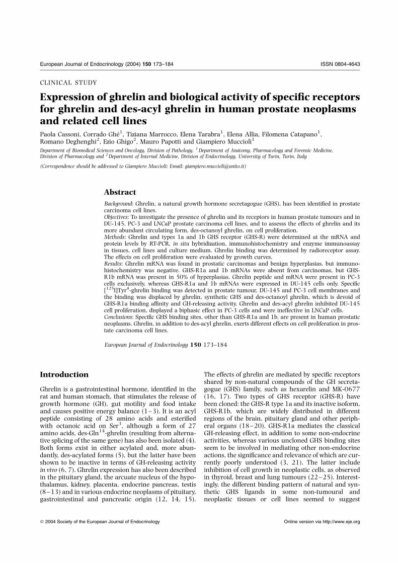
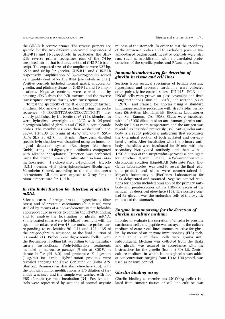
All cases of either benign prostatic hyperplasia or pro-static carcinomas were positive for expression of ghrelinmRNA, as studied by RT-PCR. This is depicted in Fig. 1,in which two representative example are given for bothconditions: lanes 4 and 5, and 6 and 7 respectively. PC-3 cells (lane 3) were also positive, whereas DU-145(lane 1) and LNCaP (lane 2) were negative. Analysisof mRNA by means of in situ hybridization confirmedthe RT-PCR positivity for ghrelin mRNA in bothbenign prostatic hyperplasia (Fig. 2A) and prostatic
176 P Cassoni and others EUROPEAN JOURNAL OF ENDOCRINOLOGY (2004) 150
www.eje.org
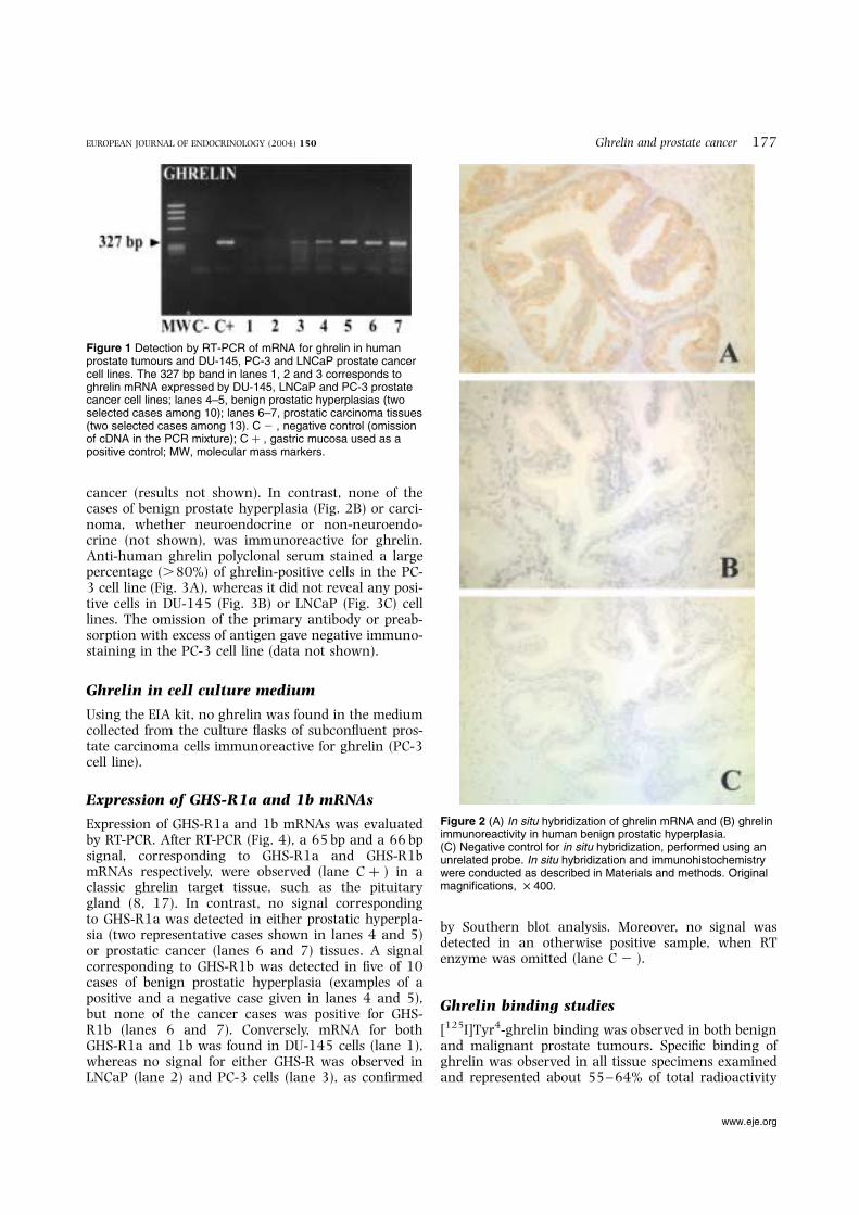
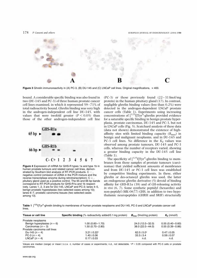
cancer (results not shown). In contrast, none of thecases of benign prostate hyperplasia (Fig. 2B) or carci-noma, whether neuroendocrine or non-neuroendo-crine (not shown), was immunoreactive for ghrelin.Anti-human ghrelin polyclonal serum stained a largepercentage (.80%) of ghrelin-positive cells in the PC-3 cell line (Fig. 3A), whereas it did not reveal any posi-tive cells in DU-145 (Fig. 3B) or LNCaP (Fig. 3C) celllines. The omission of the primary antibody or preab-sorption with excess of antigen gave negative immuno-staining in the PC-3 cell line (data not shown).
Ghrelin in cell culture medium
Using the EIA kit, no ghrelin was found in the mediumcollected from the culture flasks of subconfluent pros-tate carcinoma cells immunoreactive for ghrelin (PC-3cell line).
Expression of GHS-R1a and 1b mRNAs
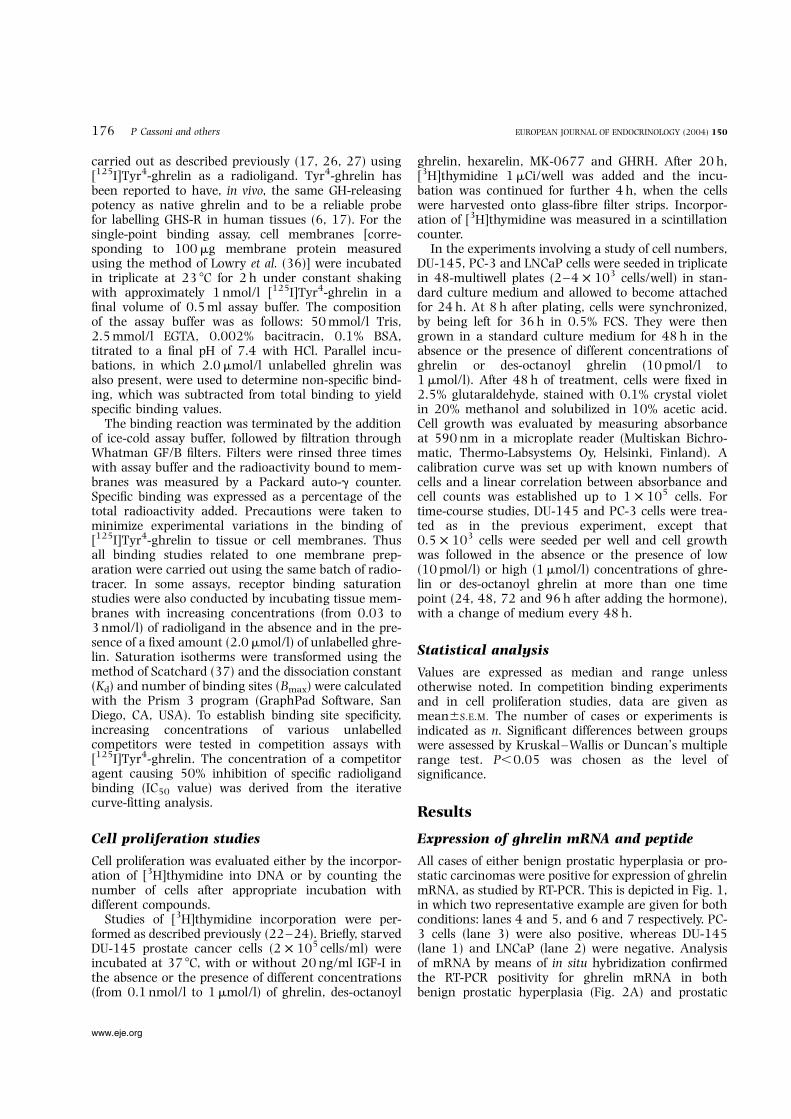
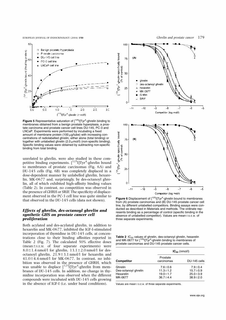
Expression of GHS-R1a and 1b mRNAs was evaluatedby RT-PCR. After RT-PCR (Fig. 4), a 65 bp and a 66 bpsignal, corresponding to GHS-R1a and GHS-R1bmRNAs respectively, were observed (lane C þ ) in aclassic ghrelin target tissue, such as the pituitarygland (8, 17). In contrast, no signal correspondingto GHS-R1a was detected in either prostatic hyperpla-sia (two representative cases shown in lanes 4 and 5)or prostatic cancer (lanes 6 and 7) tissues. A signalcorresponding to GHS-R1b was detected in five of 10cases of benign prostatic hyperplasia (examples of apositive and a negative case given in lanes 4 and 5),but none of the cancer cases was positive for GHS-R1b (lanes 6 and 7). Conversely, mRNA for bothGHS-R1a and 1b was found in DU-145 cells (lane 1),whereas no signal for either GHS-R was observed inLNCaP (lane 2) and PC-3 cells (lane 3), as confirmed
by Southern blot analysis. Moreover, no signal wasdetected in an otherwise positive sample, when RTenzyme was omitted (lane C 2 ).
Ghrelin binding studies
[125I]Tyr4-ghrelin binding was observed in both benignand malignant prostate tumours. Specific binding ofghrelin was observed in all tissue specimens examinedand represented about 55–64% of total radioactivity
Figure 1 Detection by RT-PCR of mRNA for ghrelin in humanprostate tumours and DU-145, PC-3 and LNCaP prostate cancercell lines. The 327 bp band in lanes 1, 2 and 3 corresponds toghrelin mRNA expressed by DU-145, LNCaP and PC-3 prostatecancer cell lines; lanes 4–5, benign prostatic hyperplasias (twoselected cases among 10); lanes 6–7, prostatic carcinoma tissues(two selected cases among 13). C 2 , negative control (omissionof cDNA in the PCR mixture); C þ , gastric mucosa used as apositive control; MW, molecular mass markers.
Figure 2 (A) In situ hybridization of ghrelin mRNA and (B) ghrelinimmunoreactivity in human benign prostatic hyperplasia.(C) Negative control for in situ hybridization, performed using anunrelated probe. In situ hybridization and immunohistochemistrywere conducted as described in Materials and methods. Originalmagnifications, £ 400.
Ghrelin and prostate cancer 177EUROPEAN JOURNAL OF ENDOCRINOLOGY (2004) 150
www.eje.org
bound. A considerable specific binding was also found intwo (DU-145 and PC-3) of three human prostate cancercell lines examined, in which it represented 59–71% oftotal radioactivity bound. Ghrelin binding was very highin the androgen-independent cell line DU-145, withvalues that were twofold greater (P , 0.05) thanthose of the other androgen-independent cell line
(PC-3) or those previously found (22–31 fmol/mgprotein) in the human pituitary gland (17). In contrast,negligible ghrelin binding values (less than 0.2%) weredetected in the androgen-dependent LNCaP prostatecancer cells (Table 1). Experiments using increasingconcentrations of [125I]Tyr4-ghrelin provided evidencefor a saturable specific binding in benign prostate hyper-plasia, prostate carcinomas, DU-145 and PC-3, but notin LNCaP cells (Fig. 5). Scatchard analysis of these data(data not shown) demonstrated the existence of high-affinity sites with limited binding capacity (Bmax) inbenign and malignant neoplasms, and in DU-145 andPC-3 cell lines. No difference in the Kd values wasobserved among prostate tumours, DU-145 and PC-3cells, whereas the number of receptors varied, showinga greater binding capacity in the DU-145 cell line(Table 1).
The specificity of [125I]Tyr4-ghrelin binding to mem-branes from those samples of prostate tumours (carci-nomas) that yielded sufficient amounts of membranesand from DU-145 or PC-3 cell lines was establishedby competitive binding experiments. In these, eitherghrelin or des-octanoyl ghrelin was used, the latteran endogenous ghrelin derivative (5) devoid of bindingaffinity for GHS-R1a (38) and of GH-releasing activityin vivo (6, 7). Some synthetic peptidyl (hexarelin) andnon-peptidyl (MK-0677) GHS, in addition to two hypo-thalamic neuropeptides (GHRH and SRIF) structurally
Figure 4 Expression of mRNA for GHS-R types 1a and type 1b inhuman prostate tumours and related cancer cell lines, demon-strated by Southern blot analysis of RT-PCR products. C 2 ,negative control (omission of cDNA in the PCR mixture and thereverse transcriptase enzyme during retrotranscription); C þ ,pituitary gland used as a positive control. The 65 and 66 bp bandscorrespond to RT-PCR products for GHS-R1a and 1b respect-ively. Lanes 1, 2, 3 are for DU-145, LNCaP and PC-3; lanes 4–5,benign prostatic hyperplasias (two selected cases among 10);lanes 6–7, prostatic carcinoma tissues (two selected casesamong 13).
Table 1 [125I]Tyr4-ghrelin binding to membranes of human prostate neoplasms and DU-145, PC-3 and LNCaP prostate cancer celllines.
Tissue or cell line Specific binding (% radioactivity added/0.1 mg protein) Bmax (fmol/mg protein) Kd (nmol/l)
Prostate neoplasmsBenign hyperplasias (n ¼ 6) 1.00 (0.60–1.70) 24.0 (12.0–32.0) 0.55 (0.40–0.60)Carcinomas (n ¼ 3) 1.50 (0.70–2.80) 38.0 (22.0–44.0) 0.55 (0.39–0.86)
Prostate carcinoma cell linesDu-145 (n ¼ 4) 3.21^0.22* 62.0^5.0* 0.47^0.05PC-3 (n ¼ 4) 1.40^0.06 33.3^3.4 0.59^0.07LNCaP (n ¼ 4) 0.17^0.03 n.d. n.d.
Values are median (range) or mean^S.E.M. n, number of cases or experiments; n.d., not detectable. *P , 0.05 compared with PC-3 cells or prostatecarcinomas.
Figure 3 Ghrelin immunoreactivity in (A) PC-3, (B) DU-145 and (C) LNCaP cell lines. Original magnifications, £ 400.
178 P Cassoni and others EUROPEAN JOURNAL OF ENDOCRINOLOGY (2004) 150
www.eje.org
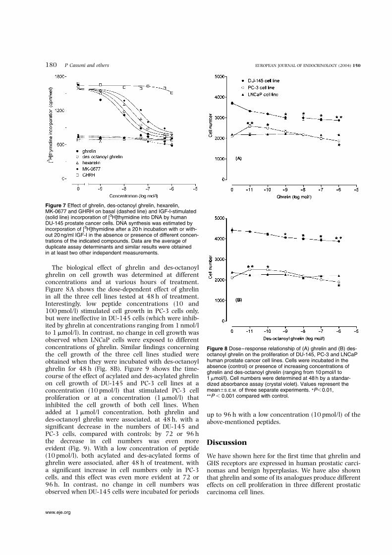
unrelated to ghrelin, were also studied in these com-petitive binding experiments. [125I]Tyr4-ghrelin boundto membranes of prostate carcinomas (Fig. 6A) andDU-145 cells (Fig. 6B) was completely displaced in adose-dependent manner by unlabelled ghrelin, hexare-lin, MK-0677 and, surprisingly, by des-octanoyl ghre-lin, all of which exhibited high-affinity binding values(Table 2). In contrast, no competition was observed inthe presence of GHRH or SRIF. The specificity of displace-ment observed in the PC-3 cell line was quite similar tothat observed in the DU-145 cells (data not shown).
Effects of ghrelin, des-octanoyl ghrelin andsynthetic GHS on prostate cancer cellproliferation
Both acylated and des-acylated ghrelin, in addition tohexarelin and MK-0677, inhibited the IGF-I-stimulatedincorporation of thymidine in DU-145 cells, at concen-trations close to their binding affinities reported inTable 2 (Fig. 7). The calculated 50% effective doses(mean^S.E.M. of four separate experiments) were8.0^1.4 nmol/l for ghrelin, 13.1^2.0 nmol/l for des-octanoyl ghrelin, 21.9^3.1 nmol/l for hexarelin and41.0^4.4 nmol/l for MK-0677. In contrast, no inhi-bition was observed in the presence of GHRH, whichwas unable to displace [125I]Tyr4-ghrelin from mem-branes of DU-145 cells. In addition, no change in thy-midine incorporation was observed when the differentcompounds were incubated with DU-145 cells growingin the absence of IGF-I (i.e. under basal conditions).
Figure 5 Representative saturation of [125I]Tyr4-ghrelin binding tomembranes obtained from a benign prostate hyperplasia, a pros-tate carcinoma and prostate cancer cell lines DU-145, PC-3 andLNCaP. Experiments were performed by incubating a fixedamount of membrane protein (100mg/tube) with increasing con-centrations of radiolabelled ghrelin, either alone (total binding) ortogether with unlabelled ghrelin (2.0mmol/l) (non-specific binding).Specific binding values were obtained by subtracting non-specificbinding from total binding.
Figure 6 Displacement of [125I]Tyr4-ghrelin bound to membranesfrom (A) prostate carcinomas and (B) DU-145 prostate cancer cellline, by different unlabelled competitors. Binding assays were con-ducted as described in Materials and methods. The ordinate rep-resents binding as a percentage of control (specific binding in theabsence of unlabelled competitor). Values are mean^S.E.M. ofthree separate experiments.
Table 2 IC50 values of ghrelin, des-octanoyl ghrelin, hexarelinand MK-0677 for [125I]Tyr4-ghrelin binding to membranes ofprostate carcinomas and DU-145 prostate cancer cells.
IC50 (nmol/l)
CompetitorProstate
carcinomas DU-145 cells
Ghrelin 7.6^0.6 7.9^0.4Des-octanoyl ghrelin 11.3^1.2 15.7^0.9Hexarelin 19.0^1.7 25.0^3.9MK-0677 36.7^4.4 38.9^2.0
Values are mean^S.E.M. of three separate experiments.
Ghrelin and prostate cancer 179EUROPEAN JOURNAL OF ENDOCRINOLOGY (2004) 150
www.eje.org
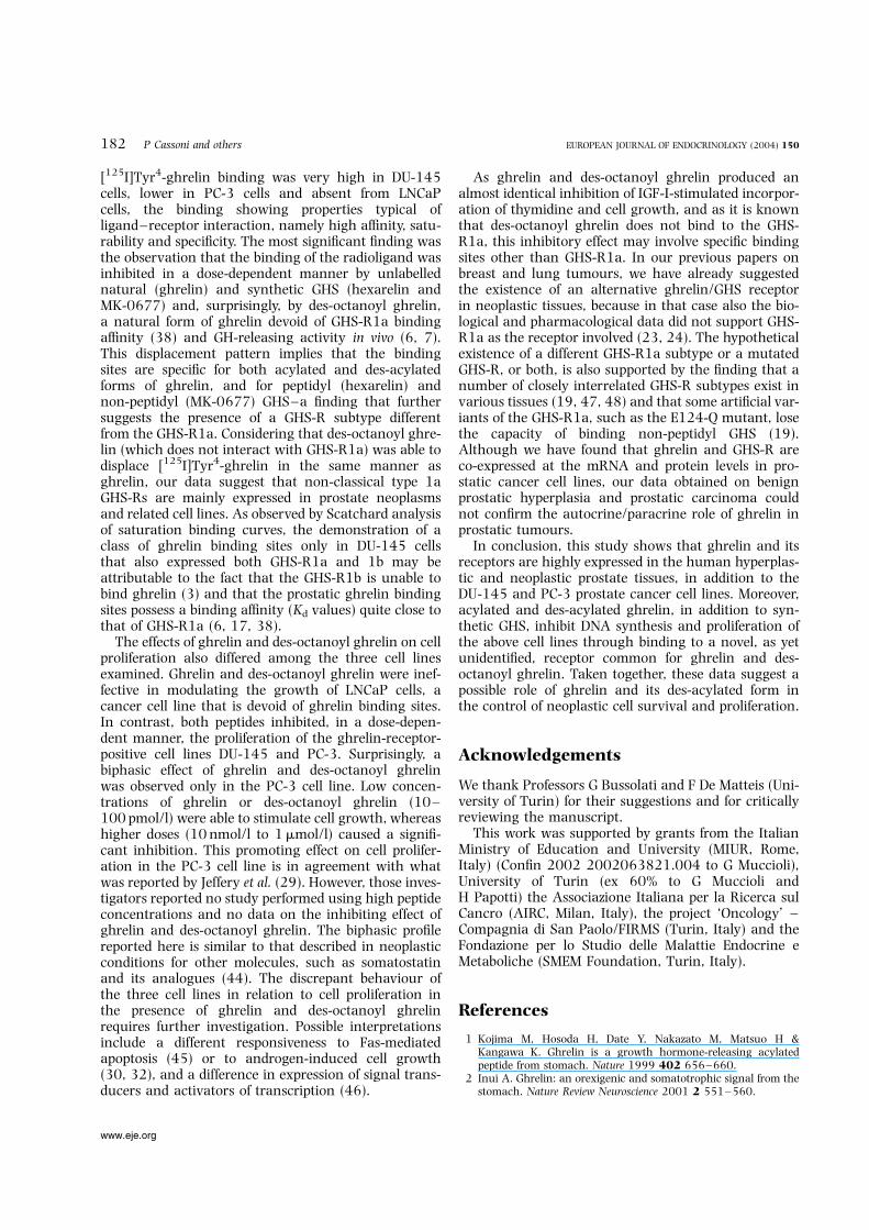
The biological effect of ghrelin and des-octanoylghrelin on cell growth was determined at differentconcentrations and at various hours of treatment.Figure 8A shows the dose-dependent effect of ghrelinin all the three cell lines tested at 48 h of treatment.Interestingly, low peptide concentrations (10 and100 pmol/l) stimulated cell growth in PC-3 cells only,but were ineffective in DU-145 cells (which were inhib-ited by ghrelin at concentrations ranging from 1 nmol/lto 1mmol/l). In contrast, no change in cell growth wasobserved when LNCaP cells were exposed to differentconcentrations of ghrelin. Similar findings concerningthe cell growth of the three cell lines studied wereobtained when they were incubated with des-octanoylghrelin for 48 h (Fig. 8B). Figure 9 shows the time-course of the effect of acylated and des-acylated ghrelinon cell growth of DU-145 and PC-3 cell lines at aconcentration (10 pmol/l) that stimulated PC-3 cellproliferation or at a concentration (1mmol/l) thatinhibited the cell growth of both cell lines. Whenadded at 1mmol/l concentration, both ghrelin anddes-octanoyl ghrelin were associated, at 48 h, with asignificant decrease in the numbers of DU-145 andPC-3 cells, compared with controls; by 72 or 96 hthe decrease in cell numbers was even moreevident (Fig. 9). With a low concentration of peptide(10 pmol/l), both acylated and des-acylated forms ofghrelin were associated, after 48 h of treatment, witha significant increase in cell numbers only in PC-3cells, and this effect was even more evident at 72 or96 h. In contrast, no change in cell numbers wasobserved when DU-145 cells were incubated for periods
up to 96 h with a low concentration (10 pmol/l) of theabove-mentioned peptides.
Discussion
We have shown here for the first time that ghrelin andGHS receptors are expressed in human prostatic carci-nomas and benign hyperplasias. We have also shownthat ghrelin and some of its analogues produce differenteffects on cell proliferation in three different prostaticcarcinoma cell lines.
Figure 7 Effect of ghrelin, des-octanoyl ghrelin, hexarelin,MK-0677 and GHRH on basal (dashed line) and IGF-I-stimulated(solid line) incorporation of [3H]thymidine into DNA by humanDU-145 prostate cancer cells. DNA synthesis was estimated byincorporation of [3H]thymidine after a 20 h incubation with or with-out 20 ng/ml IGF-I in the absence or presence of different concen-trations of the indicated compounds. Data are the average ofduplicate assay determinants and similar results were obtainedin at least two other independent measurements.
Figure 8 Dose–response relationship of (A) ghrelin and (B) des-octanoyl ghrelin on the proliferation of DU-145, PC-3 and LNCaPhuman prostate cancer cell lines. Cells were incubated in theabsence (control) or presence of increasing concentrations ofghrelin and des-octanoyl ghrelin (ranging from 10 pmol/l to1mmol/l). Cell numbers were determined at 48 h by a standar-dized absorbance assay (crystal violet). Values represent themean^S.E.M. of three separate experiments. *P,0.01,
**P , 0.001 compared with control.
180 P Cassoni and others EUROPEAN JOURNAL OF ENDOCRINOLOGY (2004) 150
www.eje.org

We have previously reported a detailed analysis of thedistribution of GHS binding sites in tumoural (22 –24)and non-tumoural human tissues (39–41) using aradiolabelled peptidyl GHS, such as [125I]Tyr-Ala-hexa-relin. More recently, we have demonstrated, using[125I]Tyr4-ghrelin as a ligand, that ghrelin receptorsare present in the human hypothalamus and pituitarygland and that ghrelin needs octanoylation to bindwith high affinity to pituitary GHS-R (6, 17). By using[125I]Tyr4-ghrelin, in addition to RT-PCR and in situlocalization techniques, we have now investigated thepresence of mRNA for ghrelin and types 1a and 1bGHS-R in 10 cases of prostatic benign hyperplasia and
10 cases of prostatic carcinoma, and in prostaticcancer cell lines, two androgen-independent (DU-145and PC-3) and one androgen-dependent (LNCaP).
Interestingly, we found that ghrelin mRNA was pre-sent in all the prostatic benign (hyperplastic) andmalignant (carcinomatous) tissues. However, none ofthese tissues expressed ghrelin at the peptide level.This suggests that hyperplastic and neoplastic prostaticcells either are unable to synthesize ghrelin (despitecontaining the specific mRNA) or do not retain the pep-tide in the cytoplasm once they have synthesized it.Additional possibilities could be insufficient sensitivityof the EIA kit or that the culture conditions usedwere not adequate for the ghrelin cell secretory process.In the cell lines, ghrelin was present (at both the mRNAand the peptide level) exclusively in the PC-3 cells, incontrast to what has previously been reported by Jefferyet al. (29), who also identified ghrelin expression in DU-145 and LNCaP cells. In addition, the concentration ofghrelin in the culture medium of the PC-3 cell lineimmunoreactive for ghrelin was not relevant,suggesting that this cell line is able to synthesize, butdoes not secrete, ghrelin.
On the basis of these observations, the functional sig-nificance of ghrelin in the human prostate remains anopen question: neither prostatic tissue nor cell lineshave significant amounts of ghrelin peptide. Thusonly systemic or exogenous ghrelin could affect thefunctionality of prostatic primary tissue or cancer celllines. However, at present, other sources of ghrelin inthe reproductive tract cannot be excluded. In thissense, it was shown that Leydig cells are the primarysite of expression and production of ghrelin in the rattestis, suggesting that this peptide could influence tes-ticular function, in addition to that of other organs ofthe reproductive system, including the prostate (13).Moreover, a novel proghrelin isoform has been ident-ified in mouse testis (42) and, more recently, in thehuman prostate, where its expression is upregulatedin cancer tissues when compared with normal histo-logical tissue (43). Nevertheless, the functional role ofthis novel peptide remains unexplored.
Regarding the expression of ghrelin receptors, noGHS-R1a mRNA was detected by RT-PCR in any ofthe tissues examined, whereas mRNA for GHS-R1bwas found in 50% of benign prostatic hyperplasias.However, although no mRNA for GHS-R1a wasdetected in the prostatic tissues, a significant bindingwas observed in membranes obtained from both hyper-plastic and cancerous prostatic tissues. These resultssuggest that, in these tumours, a ghrelin receptor sub-type different from the classical GHS-R1a or 1b couldbe present. In agreement with the findings in the pros-tatic cancer tissues, in the prostatic carcinoma cell linesalso, a discrepancy was found between the RT-PCRdata on ghrelin receptors and binding. No mRNA foreither GHS-R1a or 1b was present in LNCaP andin PC-3 cells, whereas it was found in DU-145 cells.
Figure 9 Time-course of the effect of ghrelin (—) and des-octa-noyl ghrelin (- - -) on cell proliferation of (A) DU-145 and (B) PC-3human prostate cancer cell lines. Cells (500/well) were grown for96 h in the absence (control) and presence of the indicated com-pounds; the culture medium was changed every 48 h. The cellnumber was determined at the indicated time by a standardizedabsorbance assay (crystal violet). Values are means mean^S.E.M.
of three separate experiments. *P,0.01, **P , 0.001 comparedwith control.
Ghrelin and prostate cancer 181EUROPEAN JOURNAL OF ENDOCRINOLOGY (2004) 150
www.eje.org
[125I]Tyr4-ghrelin binding was very high in DU-145cells, lower in PC-3 cells and absent from LNCaPcells, the binding showing properties typical ofligand –receptor interaction, namely high affinity, satu-rability and specificity. The most significant finding wasthe observation that the binding of the radioligand wasinhibited in a dose-dependent manner by unlabellednatural (ghrelin) and synthetic GHS (hexarelin andMK-0677) and, surprisingly, by des-octanoyl ghrelin,a natural form of ghrelin devoid of GHS-R1a bindingaffinity (38) and GH-releasing activity in vivo (6, 7).This displacement pattern implies that the bindingsites are specific for both acylated and des-acylatedforms of ghrelin, and for peptidyl (hexarelin) andnon-peptidyl (MK-0677) GHS –a finding that furthersuggests the presence of a GHS-R subtype differentfrom the GHS-R1a. Considering that des-octanoyl ghre-lin (which does not interact with GHS-R1a) was able todisplace [125I]Tyr4-ghrelin in the same manner asghrelin, our data suggest that non-classical type 1aGHS-Rs are mainly expressed in prostate neoplasmsand related cell lines. As observed by Scatchard analysisof saturation binding curves, the demonstration of aclass of ghrelin binding sites only in DU-145 cellsthat also expressed both GHS-R1a and 1b may beattributable to the fact that the GHS-R1b is unable tobind ghrelin (3) and that the prostatic ghrelin bindingsites possess a binding affinity (Kd values) quite close tothat of GHS-R1a (6, 17, 38).
The effects of ghrelin and des-octanoyl ghrelin on cellproliferation also differed among the three cell linesexamined. Ghrelin and des-octanoyl ghrelin were inef-fective in modulating the growth of LNCaP cells, acancer cell line that is devoid of ghrelin binding sites.In contrast, both peptides inhibited, in a dose-depen-dent manner, the proliferation of the ghrelin-receptor-positive cell lines DU-145 and PC-3. Surprisingly, abiphasic effect of ghrelin and des-octanoyl ghrelinwas observed only in the PC-3 cell line. Low concen-trations of ghrelin or des-octanoyl ghrelin (10 –100 pmol/l) were able to stimulate cell growth, whereashigher doses (10 nmol/l to 1mmol/l) caused a signifi-cant inhibition. This promoting effect on cell prolifer-ation in the PC-3 cell line is in agreement with whatwas reported by Jeffery et al. (29). However, those inves-tigators reported no study performed using high peptideconcentrations and no data on the inhibiting effect ofghrelin and des-octanoyl ghrelin. The biphasic profilereported here is similar to that described in neoplasticconditions for other molecules, such as somatostatinand its analogues (44). The discrepant behaviour ofthe three cell lines in relation to cell proliferation inthe presence of ghrelin and des-octanoyl ghrelinrequires further investigation. Possible interpretationsinclude a different responsiveness to Fas-mediatedapoptosis (45) or to androgen-induced cell growth(30, 32), and a difference in expression of signal trans-ducers and activators of transcription (46).
As ghrelin and des-octanoyl ghrelin produced analmost identical inhibition of IGF-I-stimulated incorpor-ation of thymidine and cell growth, and as it is knownthat des-octanoyl ghrelin does not bind to the GHS-R1a, this inhibitory effect may involve specific bindingsites other than GHS-R1a. In our previous papers onbreast and lung tumours, we have already suggestedthe existence of an alternative ghrelin/GHS receptorin neoplastic tissues, because in that case also the bio-logical and pharmacological data did not support GHS-R1a as the receptor involved (23, 24). The hypotheticalexistence of a different GHS-R1a subtype or a mutatedGHS-R, or both, is also supported by the finding that anumber of closely interrelated GHS-R subtypes exist invarious tissues (19, 47, 48) and that some artificial var-iants of the GHS-R1a, such as the E124-Q mutant, losethe capacity of binding non-peptidyl GHS (19).Although we have found that ghrelin and GHS-R areco-expressed at the mRNA and protein levels in pro-static cancer cell lines, our data obtained on benignprostatic hyperplasia and prostatic carcinoma couldnot confirm the autocrine/paracrine role of ghrelin inprostatic tumours.
In conclusion, this study shows that ghrelin and itsreceptors are highly expressed in the human hyperplas-tic and neoplastic prostate tissues, in addition to theDU-145 and PC-3 prostate cancer cell lines. Moreover,acylated and des-acylated ghrelin, in addition to syn-thetic GHS, inhibit DNA synthesis and proliferation ofthe above cell lines through binding to a novel, as yetunidentified, receptor common for ghrelin and des-octanoyl ghrelin. Taken together, these data suggest apossible role of ghrelin and its des-acylated form inthe control of neoplastic cell survival and proliferation.
Acknowledgements
We thank Professors G Bussolati and F De Matteis (Uni-versity of Turin) for their suggestions and for criticallyreviewing the manuscript.
This work was supported by grants from the ItalianMinistry of Education and University (MIUR, Rome,Italy) (Confin 2002 2002063821.004 to G Muccioli),University of Turin (ex 60% to G Muccioli andH Papotti) the Associazione Italiana per la Ricerca sulCancro (AIRC, Milan, Italy), the project ‘Oncology’ –Compagnia di San Paolo/FIRMS (Turin, Italy) and theFondazione per lo Studio delle Malattie Endocrine eMetaboliche (SMEM Foundation, Turin, Italy).
References
1 Kojima M, Hosoda H, Date Y, Nakazato M, Matsuo H &Kangawa K. Ghrelin is a growth hormone-releasing acylatedpeptide from stomach. Nature 1999 402 656–660.
2 Inui A. Ghrelin: an orexigenic and somatotrophic signal from thestomach. Nature Review Neuroscience 2001 2 551–560.
182 P Cassoni and others EUROPEAN JOURNAL OF ENDOCRINOLOGY (2004) 150
www.eje.org

3 Muccioli G, Tschop M, Papotti M, Deghenghi R, Heiman M &Ghigo E. Neuroendocrine and peripheral activities of ghrelin:implications in metabolism and obesity. European Journal ofPharmacology 2002 440 235–254.
4 Hosoda H, Kojima M, Matsuo H & Kangawa K. Purification andcharacterization of rat des-Gln14-ghrelin, a second endogenousligand for the growth hormone secretagogue receptor. Journal ofBiological Chemistry 2000 275 21995–22000.
5 Hosoda H, Kojima M, Matsuo H & Kangawa K. Ghrelin and des-acyl ghrelin: two major forms of rat ghrelin peptide in gastrointes-tinal tissue. Biochemical and Biophysical Research Communications2000 279 909–913.
6 Torsello A, Ghe C, Bresciani E, Catapano F, Ghigo E, Deghenghi Ret al. Short ghrelin peptides neither displace ghrelin binding invitro nor stimulate GH release in vivo. Endocrinology 2002 1431968–1971.
7 Broglio F, Benso A, Gottero C, Prodam F, Gauna C, Filtri L et al.Non-acylated ghrelin does not possess pituitaric and pancreaticendocrine activity of acylated ghrelin in humans. Journal of Endo-crinological Investigation 2003 26 192–196.
8 Kamegai J, Tamura H, Shimizu T, Ishii S, Sugihara H & Oikawa S.Regulation of the ghrelin gene: growth hormone-releasing hor-mone upregulates ghrelin mRNA in the pituitary. Endocrinology2001 142 4154–4157.
9 Cowley MA, Smith RG, Diano S, Tschop M, Pronchuk N, Grove KLet al. The distribution and mechanism of action of ghrelin in theCNS demonstrates a novel hypothalamic circuit regulating energyhomeostasis. Neuron 2003 37 649–661.
10 Mori K, Yoshimoto A, Takaya K, Hosoda K, Ariyasu H, Yahata Ket al. Kidney produces a novel acylated peptide, ghrelin. FEBSLetters 2000 486 213–216.
11 Gualillo O, Caminos J, Blanco M, Garcia-Caballero T, Kojima M,Kangawa K et al. Ghrelin, a novel placental-derived hormone.Endocrinology 2001 142 788–794.
12 Volante M, Allıa E, Gugliotta P, Deghenghi R, Muccioli G, Ghigo Eet al. Expression of ghrelin and GH secretagogue receptorby pancreatic islet cells and related endocrine tumors.Journal of Clinical Endocrinology and Metabolism 2002 871300–1308.
13 Tena-Sempere M, Barreiro ML, Gonzalez LC, Gaytan F, Zhang FP,Caminos JE et al. Novel expression and functional role of ghrelinin rat testis. Endocrinology 2002 143 717–725.
14 Korbonits M, Bustin SA, Kojima M, Jordan S, Adams EF, Lowe DGet al. The expression of the growth hormone secretagogue recep-tor ligand ghrelin in normal and abnormal human pituitary andother neuroendocrine tumors. Journal of Clinical Endocrinology andMetabolism 2001 86 881–887.
15 Papotti M, Cassoni P, Volante M, Deghenghi R, Muccioli G &Ghigo E. Ghrelin-producing endocrine tumors of the stomachand intestine. Journal of Clinical Endocrinology and Metabolism2001 86 5052–5059.
16 Kojima M, Hosoda H, Matsuo H & Kangawa K. Ghrelin: discoveryof the natural endogenous ligand for the growth hormonesecretagogue receptor. Trends in Endocrinology and Metabolism2001 12 118– 122.
17 Muccioli G, Papotti M, Locatelli V, Ghigo E & Deghenghi R.Binding of 125I-labeled ghrelin to membranes from humanhypothalamus and pituitary gland. Journal of EndocrinologicalInvestigation 2001 24 RC7–RC9.
18 Smith RG, Van der Ploeg LH, Howard AD, Feighner SD, Cheng K,Hickey GJ et al. Peptidomimetic regulation of growth hormonesecretion. Endocrine Reviews 1997 18 621–645.
19 Smith RG, Palhya OC, Feighner SD, Tan CP, McKee KK,Hreniuk DR et al. Growth hormone releasing substances:types and their receptors. Hormone Research 1999 51 (Suppl. 3)1–8.
20 Gnanapavan S, Kola B, Bustin SA, Morris DG, McGee P,Fairclough P et al. The tissue distribution of the mRNAof ghrelin and subtypes of its receptor, GHS-R, in humans.Journal of Clinical Endocrinology and Metabolism 2002 872988–2991.
21 Broglio F, Arvat E, Benso A, Gottero C, Prodam F, Granata R et al.Ghrelin: much more than a natural growth hormone secreta-gogue. Israel Medical Association Journal 2002 4 607–613.
22 Cassoni P, Papotti M, Catapano F, Ghe C, Deghenghi R, Ghigo Eet al. Specific binding sites for synthetic growth hormone secreta-gogues in non tumoural and neoplastic human thyroid tissue.Journal of Endocrinology 2000 165 139–146.
23 Cassoni P, Papotti M, Ghe C, Catapano F, Sapino A, Graziani Aet al. Identification, characterization and biological activity ofspecific receptors for natural (ghrelin) and synthetic growth hor-mone secretagogues in human breast carcinomas and cell lines.Journal of Clinical Endocrinology and Metabolism 2001 861738–1745.
24 Ghe C, Cassoni P, Catapano F, Marrocco T, Deghenghi R, Ghigo Eet al. The antiproliferative effect of synthetic peptidyl GH secreta-gogues in human CALU-1 lung carcinoma cells. Endocrinology2002 143 484–491.
25 Volante M, Allia E, Fulcheri E, Cassoni P, Ghigo E, Muccioli G et al.Ghrelin in fetal thyroid and follicular tumors and cell lines:expression and effects on tumor growth. American Journal of Path-ology 2003 162 645–654.
26 Baldanzi G, Filigheddu N, Cutrupi S, Catapano F, Bonissoni S,Fubini A et al. Ghrelin and des-acyl ghrelin inhibit celldeath in cardiomyocytes and endothelial cells throughERK1/2 and PI 3-kinase/AKT. Journal of Cell Biology 2002159 1029–1037.
27 Bedendi I, Alloatti G, Marcantoni A, Malan D, Catapano F, Ghe Cet al. Cardiac effects of ghrelin and its endogenous derivativesdes-octanoyl ghrelin and des-Gln14-ghrelin. European Journal ofPharmacology 2003 476 87–95.
28 Torsello A, Bresciani E, Rossoni G, Avallone R, Tulipano G, Cocchi Det al. Ghrelin plays a minor role in the physiological controlof cardiac function in the rat. Endocrinology 2003 1441787–1792.
29 Jeffery PL, Herington AC & Chopin LK. Expression and action ofthe growth hormone releasing peptide ghrelin and its receptorin prostate cancer cell lines. Journal of Endocrinology 2002 172R7–R11.
30 Lamharzi N, Schally AV, Koppan M & Kroot K. Growth hormone-releasing hormone antagonist MZ-5-156 inhibits growth of DU-145 human androgen-independent prostate carcinoma in nudemice and suppresses the levels of mRNA expression of insulin-like growth factor II in tumors. Proceedings of the National Academyof Sciences of the USA 1998 95 8864–8868.
31 Plonowsky A, Schally AV, Nagy A, Baodong S & Szepeshazi K.Inhibition of PC-3 human androgen-independent prostatecancer and its metastates by cytotoxic somatostatin analogueAN-238. Cancer Research 1999 59 1947– 1953.
32 Gasparian AB, Yao YJ, Kowalczyk D, Lyakh LA, Karseladze A,Slaga TJ et al. The role of IKK in constitutive activation of NF-kB transcription factor in prostate carcinoma cells. Journal ofCell Science 2002 115 141–151.
33 Papotti M, Croce S, Macrı L, Funaro A, Pecchioni C, Schindler Met al. Correlative immunohistochemical and reverse transcrip-tase–polymerase chain reaction analysis of somatostatin receptortype 2 in neuroendocrine tumors of the lung. Diagnostic MolecularPathology 2000 9 47– 57.
34 Volante M, Pecchioni C & Bussolati G. Post-incubation heatingsignificantly improves tyramide signal amplification. Journal ofHistochemistry and Cytochemistry 2000 48 1583–1585.
35 Volante M, Fulcheri E, Allia E, Cerrato M, Pucci A & Papotti M.Ghrelin expression in fetal, infant and adult human lung. Journalof Histochemistry and Cytochemistry 2002 50 1013–1021.
36 Lowry OH, Rosebrough NL, Farr AL & Randall RJ. Proteinmeasurement with the Folin phenol reagent. Journal of BiologicalChemistry 1951 193 265–275.
37 Scatchard G. The attraction of proteins for small molecules andions. Annals of the New York Academy of Sciences 1949 51660–672.
38 Bednarek MA, Feighner SD, Pong SS, McKee KK, Hreniuk DL,Silva MV et al. Structure–function studies on the new growth
Ghrelin and prostate cancer 183EUROPEAN JOURNAL OF ENDOCRINOLOGY (2004) 150
www.eje.org
hormone-releasing peptide, ghrelin: minimal sequence of ghrelinnecessary for activation of growth hormone secretagogue recep-tor 1a. Journal of Medicinal Chemistry 2001 43 4370–4376.
39 Muccioli G, Ghe C, Ghigo MC, Papotti M, Arvat E, Boghen MF et al.Specific receptors for synthetic GH secretagogues in the humanbrain and pituitary gland. Journal of Endocrinology 1998 15799–106.
40 Papotti M, Ghe C, Cassoni P, Catapano F, Deghenghi R, Ghigo Eet al. Growth hormone secretagogue binding sites in peripheralhuman tissues. Journal of Clinical Endocrinology and Metabolism2000 85 3803– 3807.
41 Muccioli G, Broglio F, Valetto MR, Ghe C, Catapano F, Graziani Aet al. Growth hormone-releasing peptides and the cardiovascularsystem. Annales d’Endocrinologie (Paris) 2000 61 27–31.
42 Tanaka M, Hayashida Y, Nakao N, Nakai N & Nakashima K.Testis-specific and developmentally induced expression of a ghre-lin gene-derived trancript that encodes a novel polypeptide in themouse. Biochimica et Biophysica Acta 2001 1522 62–65.
43 Jeffery PL, Herington AC & Chopin LK. The potential autocrine/paracrine roles of ghrelin and its receptor in hormone-dependentcancer. Cytokine and Growth Factor Reviews 2003 14 113–122.
44 Hoelting T, Duh QY, Clark OH & Herfarth C. Somatostatin analogoctreotide inhibits the growth of differentiated thyroid cancer cellsin vitro but not in vivo. Journal of Clinical Endocrinology andMetabolism 1996 81 2638–2641.
45 Gewies A, Rokhlin OW & Cohen MB. Cytochrome c is involved inFas-mediated apoptosis of prostatic carcinoma cell lines. CancerResearch 2000 60 2163–2168.
46 Mora LB, Buettner R, Seigne J, Diaz J, Ahmad N, Garcia R et al.Constitutive activation of Stat3 in human prostate tumorsand cell lines: direct inhibition of Stat3 signaling induces apop-tosis of prostate cancer cells. Cancer Research 2002 626659–6666.
47 McKee KK, Tan CP, Palhya OC, Liu J, Feighner SD, Hreniuk DRet al. Cloning and characterization of two human G-protein-coupled receptor genes (GPR38 and GPR39) related to growthhormone secretagogue and neurotensin receptors. Genomics1997 46 426–434.
48 Tan CP, McKee KK, Liu Q, Palyha OC, Feighner SD, Smith RG et al.Cloning and characterization of a human and murine T-cellorphan G-protein-coupled receptor similar to the growth hor-mone secretagogue and neurotensin receptors. Genomics 199852 223–229.
Received 28 July 2003
Accepted 23 October 2003
184 P Cassoni and others EUROPEAN JOURNAL OF ENDOCRINOLOGY (2004) 150
www.eje.org
Related Documents