Expression of Alternative 2[LGDVH ,QÀXHQFHV Cell Migration $1$ $1'-(/.29,û Tampere University Dissertations 99

Welcome message from author
This document is posted to help you gain knowledge. Please leave a comment to let me know what you think about it! Share it to your friends and learn new things together.
Transcript
Expression of Alternative
Cell Migration
Tampere University Dissertations 99

Tampere University Dissertations 99
ANA ANDJELKOVIĆ
Expression of Alternative Oxidase Influences
Cell Migration
ACADEMIC DISSERTATIONTo be presented, with the permission of
the Faculty Council of the Faculty of Medicine and Health Technologyof Tampere University,
for public discussion in auditorium F115of the ARVO building, Tampere,
on 14.09.2019, at 12 o’clock.
ACADEMIC DISSERTATION Tampere University, Faculty of Medicine and Health Technology Finland Responsible supervisor and Custos
Professor Howard T. Jacobs Tampere University Finland
Pre-examiners Professor Mirka Uhlirova University of Cologne Germany
MD, Professor Navdeep Chandel Northwestern University United States
Opponent Professor Rafael Garesse Autonomous University of Madrid Spain
The originality of this thesis has been checked using the Turnitin OriginalityCheck service. Copyright ©2019 Ana Andjelković Cover design: Roihu Inc. ISBN 978-952-03-1179-7 (print) ISBN 978-952-03-1180-3 (pdf) ISSN 2489-9860 (print) ISSN 2490-0028 (pdf) http://urn.fi/URN:ISBN: 978-952-03-1180-3 PunaMusta Oy – Yliopistopaino Tampere 2019

iii
To Johan

iv
v
ABSTRACT
Cell migration is important in animal development, tissue repair, the functioning of the immune system and for tissue homeostasis in general. Impairments in cell migration are associated with various developmental abnormalities and pathologies. In Drosophila development, cell migration is instrumental during metamorphosis, in the process of thoracic closure. The most studied mammalian model of cell migration at the cellular level is the scratch or wound-healing assay, in which a linear scratch is made in a confluent monolayer of cells, which then migrate to close the gap.
The work presented in this thesis aims to understand how signaling associated with the mitochondrial respiratory chain affects cell migration in different models. As a tool to investigate this relationship, I used model organisms transgenically expressing the alternative oxidase, AOX, from a primitive marine animal, the tunicate Ciona intestinalis. AOX is an accessory component of the mitochondrial respiratory chain, which is found in microbes, plants, and some metazoan phyla, but not in vertebrates or insects. AOX directly oxidizes ubiquinol by molecular oxygen in a non-proton motive reaction, by-passing respiratory chain complexes III and IV. Utilizing AOX from Ciona intestinalis, I perturbed the mitochondrial respiratory chain and investigated the effect on developmental signaling affecting cell migration in the Drosophila and mammalian cell models. To create a control for these studies and test whether the ability of AOX to alleviate tested phenotypes depends on its enzymatic activity, I engineered a mutated variant of AOX in such a way as to abolish this activity.
I observed that co-expression of AOX from Ciona intestinalis was able to alleviate cleft thorax and other dysmorphic phenotypes in Drosophila, brought about by activated GeneSwitch transcription factor, which I hypothesize to interfere in some way with nuclear receptor signaling during development. Using the mutant AOX control, I was able to show that AOX enzymatic activity is instrumental in rescuing developmental lethality or locomotor dysfunction, resulting from cytochrome oxidase deficiency. I proceeded to use mutant AOX to show that the same is true for the rescue of the dysmorphic phenotypes induced by GeneSwitch.
vi
AOX expression also alleviated the cleft thorax phenotype induced by genetic manipulations of the JNK signaling pathway, which regulates the formation of the dorsal thoracic epithelium and governs the migratory behavior of the cell sheets everting from the wing imaginal discs during metamorphosis.
Midline closure defects similar to cleft thorax in the fly are also seen in mammals, for example, in spina bifida, cleft lip and palate and cleft sternum. Considering the well conserved biology between fly and human and the highly conserved JNK signaling pathway, it was possible to use a mammalian cell-culture model to test the generality of the findings from Drosophila, as well as explore the molecular mechanisms underlying the influence of AOX on cell migration. I thus used the mammalian wound-healing model to confirm that AOX expression promotes migration in immortalized (but not primary) mouse embryonic fibroblasts, and rescues pharmacologically induced migration deficiencies through a mechanism involving JNK signaling. Reporter assays showed that AP-1 and its transcriptional activity are not a direct target of AOX. However the data suggest a possible direct involvement of JNK, acting through other targets.
Despite the lack of knowledge on how AOX is regulated in animals, the use of various inhibitors showed that the effect of AOX on cell migration is most likely due to a specific effect on metabolism, possibly due to its thermogenic activity. A full elucidation of the processes that link mitochondrial perturbations with cell migration should be of considerable medical importance and might even enable the design of new and more effective treatments, e.g., for metastatic tumors, tissue injuries, and congenital midline closure defects. A better understanding of the role mitochondria play in mediating cellular signaling is still needed, and will be instrumental to fully understand many fundamental biological processes, causes of disease and enable the design of precision treatments.
Results from this thesis provide us with a new paradigm linking mitochondrial function with developmental cell signaling. They also highlight what can be learned by combining tools and findings from different model organisms. In this case, the tunicate Ciona intestinalis provided a tool to better understand complex developmental processes in Drosophila melanogaster, which was then followed up in mammalian cells with potential relevance to human diseases.
vii
TIIVISTELMÄ
Solujen liikkumisella eli migraatiolla on tärkeä rooli yksilönkehityksessä, kudosvaurioiden korjaamisessa, immuunijärjestelmässä sekä yleisesti kudosten toiminnassa. Solumigraation ongelmat liittyvät moninaisiin kehityshäiriöihin ja sairauksiin. Drosophila melanogaster -banaanikärpäsellä puutteellinen solujen migraatio yksilönkehityksen aikana näkyy muun muassa halkiona kärpäsen keskiruumiissa. Solumigraation tutkituimmassa solutason nisäkäsmallissa yksikerroksiseen solupeitteeseen tehdään lineaarinen viilto, joka umpeutuu solumigraation seurauksena.
Väitöskirjatyöni tavoitteena oli tutkia, miten mitokondrioiden soluhengitykseen liittyvä signalointi vaikuttaa solumigraatioon erilaisissa tutkimusmalleissa. Tutkimuksessani hyödynsin mallieläintä, joka oli geneettisesti muokattu tuottamaan alkukantaisesta Ciona intestinalis -vaippaeläimestä eristettyä vaihtoehtoista oksidaasia (alternative oxidase, AOX). AOX-entsyymiä ei esiinny selkärankaisilla eikä hyönteisillä, mutta muun muassa mikrobeilla, kasveilla ja joillakin monisoluisilla eläimillä se toimii osana mitokondrion hengitysketjua hapettamalla ubikinolin suoraan käyttäen molekulaarista happea. AOX ohittaa entsyymikompleksit III ja IV ilman protonien kuljetusta. Tutkimuksessani muokkasin mitokondrioiden luontaisen hengitysketjun toimintaa hyödyntäen AOX:ia sekä banaanikärpäs- että nisäkässolumallissa, ja tutkin tämän vaikutuksia yksilönkehityksen aikaiseen signalointiin ja solujen migraatioon. Kokeiden kontrolliksi ja testatakseni johtuuko fenotyypin oireiden lieventyminen AOX:n entsymaattisesta aktiivisuudesta, muokkasin AOX:stä mutaation avulla entsymaattisesti toimimattoman version.
Havaitsin, että Ciona intestinalisista saadun AOX:n lisääminen kärpäsmalliin lievensi aktivoidun GeneSwitch transkriptiofaktorin aiheuttamaa keskiruumishalkiota ja muita kehityshäiriöitä. Hypoteesinani on, että aktiivinen GeneSwitch häiritsee jollakin mekanismilla tumareseptorien viestintää kehityksen aikana. Käyttämällä mutatoitua AOX-kontrollia osoitin, että sytokromioksidaasin vajeesta johtuvia letaaleja kehityshäiriöitä ja liikuntakyvyn ongelmia lieventävä vaikutus perustuu AOX:n entsymaattiseen aktiivisuuteen. Jatkoin käyttämällä mutatoitua AOX:ää osoittaakseni saman pätevän myös GeneSwitchin tuottamiin kehityshäiriöihin. AOX:n ilmentäminen lievensi lisäksi keskiruumishalkiota, joka
viii
aiheutui JNK-signalointireitin geneettisestä manipuloinnista. JNK-signalointireitti säätelee keskiruumiin dorsaalisen epiteelin muodostumista ja ohjaa siipiaihioiden solukerrosten migraatiota kärpäsen muodonmuutoksen aikana.
Nisäkkäillä on banaaninkärpäsen keskiruumishalkion kaltaisia keskilinjan kehityshäiriöitä, kuten selkäranka-, huuli-, suulaki- sekä rintalastahalkioita. Monet biologiset toiminnot ja varsinkin JNK-solusignalointireitti ovat säilyneet samankaltaisina ihmisellä ja banaanikärpäsellä, mikä mahdollistaa nisäkässolumallien käytön kärpäsillä saatujen tulosten yleispätevyyden kokeilemiseen sekä AOX:n vaikutusten taustalla olevien molekulaaristen mekanismien tutkimiseen. Näin ollen pystyin hyödyntämään nisäkässoluviljelmään tehtyä lineaarista viiltoa menetelmänä osoittaakseni, että AOX:n ilmentäminen edesauttaa solumigraatiota immortalisoiduissa (mutta ei primaarisissa) hiiren alkion sidekudossoluissa ja korjaa JNK-reitin manipulaation aiheuttamia häiriöitä solujen migraatiossa. Reportterimenetelmät paljastivat, että transkriptiotekijä AP-1 ja sen aktiivisuus eivät ole AOX:n suora kohde, vaan AOX:lla on mahdollisesti suora yhteys JNK-reitin toimintaan jotakin muuta kautta.
Huolimatta siitä, että tietous AOX:n säätelymekanismeista eläimillä on puutteellista, kokeet useilla eri inhibiittoreilla viittasivat siihen, että AOX:n vaikutus solujen migraatioon todennäköisimmin selittyy spesifillä vaikutuksella aineenvaihduntaan; mahdollisesti entsyymin termogeenisellä toiminnalla. Kattavan kokonaiskuvan saaminen prosesseista, joiden kautta mitokondriaaliset häiriöt liittyvät solumigraatioon, tarjoaisi merkittävää lääketieteellistä tietoutta ja saattaisi jopa auttaa muun muassa syövän etäpesäkkeiden, kudosvaurioiden ja synnynnäisten keskilinjan kehityshäiriöiden hoitomuotojen kehittämisessä. Tarvitaan kuitenkin parempaa käsitystä mitokondrioiden osuudesta solusignaloinnissa, ja tämä on olennaista monien biologisten prosessien, sairauksien syiden ymmärtämisen ja kohdennettujen hoitojen suunnittelun kannalta.
Väitöskirjassani esitetyt tutkimustulokset tarjoavat uuden mallin, joka liittää mitokondrioiden toiminnan yksilönkehityksen aikaiseen solusignalointiin. Tuloksissani korostuu myös se, mitä voidaan oppia yhdistelemällä erilaisten mallien tarjoamaa tutkimustietoa ja -menetelmiä. Tässä tapauksessa Ciona intestinalis -vaippaeläimen käyttö auttoi ymmärtämään paremmin Drosophila melanogasterin monimutkaista yksilönkehitystä. Näiden tulosten pohjalta pystyin jatkamaan tutkimusta mitokondrioiden roolista solujen migraatiossa nisäkässolumallissa, millä on potentiaalisesti merkitystä ihmisen sairauksien tutkimisessa.
ix
CONTENTS
1 Introduction .......................................................................................................................... 19
2 Review of the literature ....................................................................................................... 21 2.1 Mitochondria ............................................................................................................ 21
2.1.1 Mitochondria: structure and function ................................................ 21 2.1.2 The mitochondrial respiratory chain .................................................. 25 2.1.3 Mitochondrial dysfunction and disease ............................................. 25 2.1.4 mtROS and their role in cell signaling ............................................... 30
2.1.4.1 ROS-induced molecular damage ...................................... 32 2.1.4.2 ROS defense mechanisms in the cell .............................. 33
2.2 Drosophila development and its regulation ........................................................... 35 2.2.1 Signaling pathways regulating growth and cell migration
during development .............................................................................. 37 2.3 Cell migration ........................................................................................................... 39
2.3.1 Cell migration in Drosophila; single cell migration and epithelial sheet movement ................................................................... 41
2.4 Drosophila dorsal closure and thorax closure; JNK signaling role .................... 41 2.4.1 Differences between dorsal and thorax closure ............................... 44
2.5 JNK signaling in mammals; parallels with the Drosophila .................................. 45 2.5.1 Interplay between JNK signaling and mitochondrial
metabolism ............................................................................................. 47 2.6 Alternative oxidase: proposed roles in living organisms ................................... 48
2.6.1 The role of AOX as gene therapy tool: potential and concerns .................................................................................................. 51
2.7 Drosophila genetic toolkit: The Gal4/UAS system for regulated transgene expression ............................................................................................... 53
3 Aims of the study ................................................................................................................. 57
4 Materials and methods......................................................................................................... 59 4.1 Drosophila stocks and maintenance (I-III) ............................................................ 59 4.2 Cell culture ................................................................................................................ 60
4.2.1 Drosophila S2 cells................................................................................... 60 4.2.2 Mammalian cell lines ............................................................................. 60
4.3 Molecular cloning .................................................................................................... 61 4.4 Gene expression assays ........................................................................................... 61
x
4.4.1 RNA expression analysis by Quantitative Reverse Transcription-PCR (qRT-PCR) .......................................................... 61
4.4.2 Protein analysis by Western blotting .................................................. 61 4.5 Transfection/transduction ..................................................................................... 62 4.6 Migration assays........................................................................................................ 63
4.6.1 Wound-healing assay............................................................................. 63 4.6.2 Single-cell migration assay .................................................................... 63
4.7 Microscopy ................................................................................................................ 63 4.8 Luciferase assays ....................................................................................................... 64
4.8.1 Luciferase reporter assays in S2 cells.................................................. 64 4.8.2 Luciferase reporter assays in mammalian cells ................................. 64
4.9 Respirometry ............................................................................................................. 65 4.10 Experimental methods conducted by co-authors ............................................... 65
5 Results .................................................................................................................................... 67 5.1 Structural modelling and mutagenesis of active site of Ciona intestinalis
AOX (I) ..................................................................................................................... 67 5.2 MutAOX is stably expressed in mammalian cells and flies (I) ......................... 69 5.3 Expression of AOX rescues cleft thorax caused by tubGS/RU486 (II)
..................................................................................................................................... 71 5.4 AOX expression alleviates cleft thorax due to downregulation of JNK
signaling (III) ............................................................................................................. 73 5.5 Is AP-1 the target of AOX? (III) ........................................................................... 77
5.5.1 AOX does not rescue cleft thorax caused by AP-1 transcriptional factor or manipulation of its downstream target Puc ................................................................................................ 77
5.5.2 AOX does not influence AP-1-dependent transcription in cultured cells ........................................................................................... 79
5.5.3 AOX expression does not affect c-Jun phosphorylation at Ser 63/Ser 73 .......................................................................................... 81
5.6 AOX expression can influence mammalian cell migration (III) ...................... 82 5.7 Antimycin A potentiates the migration of AOX-expressing cells (III) .......... 85
6 Discussion .............................................................................................................................. 87 6.1 Overview ................................................................................................................... 87 6.2 Use of the mutAOX control .................................................................................. 88
6.2.1 Validation of mutAOX ......................................................................... 88 6.2.2 Enzymatic activity of AOX is important for phenotypic
rescue ....................................................................................................... 89 6.2.3 Level of AOX activity is important for rescue of cleft
thorax ....................................................................................................... 90 6.3 tubGS induces developmental abnormalities in the fly ...................................... 91 6.4 Model systems: pros and cons ............................................................................... 92
xi
6.4.1 Limitations of the fly model ................................................................ 92 6.4.2 Developmental disturbance in Drosophila resulting from
the GS system ........................................................................................ 93 6.4.3 Limitations of the cell model .............................................................. 94 6.4.4 Limitations of the scratch-wounded confluent monolayer
of fibroblasts .......................................................................................... 94 6.4.5 Limitations of using GFP as a control .............................................. 95
6.5 Possible mechanisms of AOX rescue of cell migration defects ...................... 96 6.5.1 AOX rescue depends on its enzymatic activation ........................... 96 6.5.2 AOX decreases ROS ............................................................................ 97 6.5.3 AOX is a thermogenic protein ........................................................... 97 6.5.4 AOX affects ATP production ............................................................ 98 6.5.5 Potential interplay between AOX and mitochondrially
localized JNK ......................................................................................... 99 6.5.6 AOX and IMM shape........................................................................... 99
6.6 Potential use of AOX in therapy ......................................................................... 100
7 Conclusions ......................................................................................................................... 103
8 Acknowledgements ............................................................................................................ 105
9 References ........................................................................................................................... 107
10 Publications ......................................................................................................................... 157

xii
xiii
ABBREVIATIONS
20E 20-hydroxyecdysone 4-HNE Hydroxy-2-nonenals ADH Alcohol dehydrogenases ADP Adenosine diphosphate AKRs Aldo-keto reductases ALDH Aldehyde dehydrogenase ALS Amyotrophic lateral sclerosis AMPK AMP-activated protein kinase ANOVA Analysis of variance AOX Alternative oxidase AP Apurinic/apyrimidinic sites AP-1 Activator protein 1 AS601245 Inhibitor V; inh V ATP Adenosine triphosphate ATPase Adenosine triphosphatase basket bsk BSA Bovine serum albumin cDNA Complementary DNA ChIP Chromatin immunoprecipitation CiAOX Ciona intestinalis alternative oxidase complex I NADH:ubiquinone oxidoreductase complex II Succinate:ubiquione oxidoreductase complex III Ubiquinol:cytochrome c oxidoreductase complex IV Cytochrome c oxidase complex V ATP synthase COX Cytochrome c oxidase cyt c Cytochrome c DC Dorsal closure dNF-Y Nuclear transcription factor Y dp Dorsal prothoracic disc
xiv
Dpp Decapentaplegic ECM Extracellular matrix EcR Ecdysone receptor EGF Epidermal growth factor EMT Epithelial to mesenchymal transition ERK1/2 Extracellular signal-regulated kinase 1 and 2 ETF Electron-transferring flavoprotein ETF Electron-transferring flavoprotein ETS-1 E26 avian erythroblastosis virus transcription factor GDNF Glial cell line-derived growth factor GPCR G protein coupled receptor GPx Glutathione peroxidases GS GeneSwitch System GSH Gluthatione H2O2 Hyrogen peroxide HClO Hypochlorous acid hemipterus hep HPV16 Human papillomavirus 16 Hsp70 Heat shock protein 70 Hsp90 Heat shock protein 90 hTERT Human telomerase reverse transcriptase Httex1p Q93 Human huntingtin with 93 polyglutamine repeats residue iMEFS Immortalized mouse embryonic fibroblasts IMM Inner mitochondrial membrane JNK c-Jun N-terminal kinase Jra Jun-related antigen kay kayak KD Ketogenic diet kDa Kilodalton, measure of molecular weight or mass KGDHC α-ketoglutarate dehydrogenase enzyme complex LE Leading-edge LPO Lipid peroxidation product MAP3K Mitogen-activated protein kinase kinase kinase MAP4K Mitogen-activated protein kinase kinase kinase kinase MAPK Mitogen-activated protein kinase MAPKAPK Mitogen-activated protein kinase-activated protein kinase
xv
MAPKK Mitogen-activated protein kinase kinase MEFs Mouse embryonic fibroblasts misshapen msn MitoQ Mitoquinone mesylate MMPs Matrix metalloproteinases MnSOD Manganese-dependent superoxide dismutase mtDNA Mitochondrial DNA MTND4 Fourth subunit of NADH dehydrogenase enzyme encoding
gene mutAOX Mutated variant of AOX NAC N-acetyl cysteine NAD+ Nicotinamide adenine dinucleotide (oxidised) NADH Nicotinamide adenine dinucleotide (reduced) NADP+ Nicotinamide adenine dinucleotide phosphate (oxidised) NADPH Nicotinamide adenine dinucleotide phosphate (reduced) Ndi NADH dehydrogenase (alternative) NF-κB Nuclear factor kappa B NRF2 NF-E2-related factor 2 O2 Oxygen O2•− Superoxide ion radical O3 Ozone OH• Hydroxyl radical OMM Outer mitochondrial membrane OPA1 Optic atrophy 1 OXPHOS Oxidative phosphorylation PBS Phosphate-buffered saline PDGF Platelet-derived growth factor PDH Pyruvate dehydrogenase complex PDM Peridroplet mitochondria PGCs Primordial germ cells Pi Inorganic phosphate PM Plasma membrane PMA Phorbol-12-myristate-13-acetate pnr pannier puc puckered PUFAs Polyunsaturated fatty acids
xvi
pvr PDGF and VEGF receptor related Q Ubiquinone QH2 Reduced ubiquinone, ubiquinol RC Respiratory chain Ref-1 Redox factor 1 RNAi Ribonucleic acid interference RO• Alkoxyl radicals ROO• Peroxyl radical ROS Reactive oxygen species RTK Receptor tyrosine kinase RTK/ERK Tyrosine kinase/extracellular regulated kinase RU486 Antiprogestin mifepristone RXR Retinoid X receptor Scrib Scribble complex SOD Superoxide dismutase SOD1 Cytoplasmic superoxide dismutase SOD2 Mitochondrial superoxide dismutase SOM Sister-of-Mammalian Grainyhead TAO Trypanosoma alternative oxidase TbAOX Trypanosoma brucei alternative oxidase TCA Ticarboxylic acid cycle TFAM Mitochondrial transcription factor A TGFβ-1 TGFβ-1 transforming growth factor beta tTG Tissue transglutaminase UAS Upstream activation sequence UPR Mt mitochondrial unfolded protein response Usp Ultraspiracle VEGF Vascular endothelial growth factor w/v Weight/Volume Wg Wingless wt Wild-type
xvii
ORIGINAL PUBLICATIONS
Publication I Andjelković, A., Oliveira, M. T., Cannino, G., Yalgin, C., Dhandapani, P. K., Dufour, E., Rustin, P., Szibor, M., & Jacobs, H. T. (2015). Diiron centre mutations in Ciona intestinalis alternative oxidase abolish enzymatic activity and prevent rescue of cytochrome oxidase deficiency in flies. Scientific Reports. https://doi.org/10.1038/srep18295
Publication II Andjelković, A., Kemppainen, K. K., & Jacobs, H. T. (2016). Ligand-Bound GeneSwitch Causes Developmental Aberrations in Drosophila that Are Alleviated by the Alternative Oxidase . G3: Genes, Genomes, Genetics https://doi.org/10.1534/g3.116.030882
Publication III Andjelković, A., Mordas, A., Bruinsma, L., Ketola, A., Cannino, G., Giordano, L., Dhandapani, P. K., Szibor, M., Dufour, E., & Jacobs, H. T. (2018). Expression of the Alternative Oxidase Influences Jun N-Terminal Kinase Signaling and Cell Migration. Molecular and Cellular Biology. https://doi.org/10.1128/mcb.00110-18

xviii
19
1 INTRODUCTION
Regulation of epithelial sheet migration is a critical component of animal development. For example, dysregulation of epithelial gap closure results in birth defects including spina bifida and cleft palate. Incorrectly regulated cell migration can also lead to impaired wound healing and to metastasis in cancer. The signaling pathways which regulate cell migration are incompletely understood. Previous work by other groups has suggested a role for mitochondrial metabolism in this process. The goal of this research was to identify mitochondria-related targets for manipulating cell migration and epithelial morphogenesis, which may enable future treatments of birth defects and other disorders.
In this work, I applied a genetic approach in the model fruit fly Drosophila melanogaster to dissect the signaling pathways regulating epithelial morphogenesis and explore the effects of altering mitochondrial metabolism. In both humans and fruit flies, the migration of cell sheets is known to be regulated by the JNK signaling pathway. Thorax closure depends upon morphogenetic movements during Drosophila development where migrating epithelial sheets everting from the wing imaginal discs join together to form the dorsal thoracic epithelium. Defects in cell signaling impair this process and produce a cleft thorax phenotype in flies, roughly analogous to spina bifida or cleft palate in humans.
Alternative respiratory pathways are present in many eukaryotic organisms including some arthropods; but are not present in insects or vertebrates. Their roles include (1) to promote thermogenesis, (2) to prevent accumulation of reducing equivalents, and (3) to act as antioxidants and provide resistance to metabolic poisons. One of these alternative pathway enzymes is the alternative oxidase (AOX), which bypasses complexes III and IV of the mitochondrial respiratory chain.
Here, I expressed the AOX gene from the tunicate Ciona intestinalis in flies that had been genetically modified to exhibit cleft thorax. I selected two such models. In the first of these I made use of the steroid-dependent GeneSwitch transcriptional activator, which I observed to induce cleft thorax and other dysmorphic adult phenotypes in the presence of the inducing drug RU486. The project was initiated by the chance observation that AOX expression was able to reverse these effects. In
20
the second model, I generated cleft thorax by specifically downregulating components of the JNK pathway.
To enable these studies, I began by creating an important control for these and all studies involving the expression of an exogenously derived AOX. I engineered mutant AOX lacking enzymatic activity to test if the phenotypic effects of AOX expression are due to enzymatic activity or some other property conferred by AOX.
The identification of effectors that rescue cleft thorax in Drosophila may eventually translate to humans. Therefore, to build upon the Drosophila findings, I also explored the effect of AOX expression on JNK-dependent transcription and wound healing in cultured mammalian cells.
The finding that AOX can influence cell sheet movements in different organisms implies that a general relationship exists between mitochondrial respiratory function and cell migration that might one day find applications in medicine.

21
2 REVIEW OF THE LITERATURE
2.1 Mitochondria
The mitochondrion is an organelle present in most cells of most eukaryotes. The only exceptions are some species of fungi (Margulis, 1981), and some anaerobic microbial eukaryotes, such as Pelomyxa palustris (Brandt & Pappas, 1959) or the oxymonad Monocercomonoides (Karnkowska et al., 2016). In mammals mitochondria are not found in differentiated erythrocytes. Mitochondria were first described as elementary organisms living inside the cell by Altman (1890), who called them bioblasts. The term 'mitochondrion' was first used by Benda (1897), who detailed their unique shape. According to the endosymbiotic theory, eukaryotes evolved from free living oxygen-metabolizing α-protobacteria, which were engulfed by a pre-eukaryotic scavenger cell some 1.6 billion years ago (Zimorski et al., 2014). However, the pre-eukaryotic cell did not digest the α-protobacteria, but rather provided it shelter and nourishment, establishing symbiosis (Margulis, 1981; Lane & Martin, 2010).
2.1.1 Mitochondria: structure and function
The number of mitochondria per cell varies between different cell types and organisms. Kinetoplastida, and some apicomplexans, such as Toxoplasma, have a single mitochondrion, while the oocytes of most animals, as well as the giant amoeba Chaos chaos contain thousands (Bereiter-Hahn & Voth, 1994). In a typical human cell there are 100-1000 mitochondria, depending on cell-type and its metabolic status. Each cell contains about 1000 nucleoids, which are supramolecular assemblies of mitochondrial DNA (mtDNA) compacted by mitochondrial transcription factor A (TFAM) (Brown et al., 2011; Kukat et al., 2015) and associated with other proteins. Mammalian mtDNA encodes only 13 polypeptides, each of which contribute to the oxidative phosphorylation (OXPHOS) complexes.
Each mitochondrion has four interconnected compartments: two membranes, the outer (OMM) and inner (IMM) mitochondrial membranes; the intermembrane
22
space between them and the mitochondrial matrix (MM). The OMM and IMM differ in their composition and permeability (Daum &Vance, 1997; de Kroon et al., 1997; Mejia & Hatch, 2016). The IMM contains the mitochondrial respiratory chain (RC) which is composed of five enzyme complexes.
The OMM includes the following proteins: enzymes involved in the metabolism of amino acids and fatty acids, receptor complexes for protein import (Lill & Neupert, 1996), pore forming proteins (Benz, 1994), proteins controlling mitochondrial morphology (Sogo & Yaffe, 1994), signal transduction components (deKroon et al., 1997), the machinery for the import and export of lipids part of which remains unknown (deKroon et al., 1997), and monoamine oxidases A and B (Schnaitman et al., 1967; Edmondson et al., 2009). In eukaryotes, cardiolipin is the only lipid that is fully synthesized in the mitochondrion. The IMM has several invaginations called cristae. Cristae are dynamic and their number and structure may have functional consequences (Bereiter-Hahn & Jendrach, 2010).
Many mitochondrial enzymes which are involved in the above-mentioned pathways are localized in the matrix but some of them are tightly connected with the IMM, e.g., succinate dehydrogenase, a component of the tricarboxylic acid cycle (TCA cycle), dihydroorotate dehydrogenase, which functions in the pyrimidine nucleotide biosynthesis pathway, and choline dehydrogenase). TCA cycle enzymes are bound to each other and to the IMM forming a supramolecular complex called the metabolon (D’Souza & Srere, 1983; Velot et al., 1997). It was proposed that the organization of these enzymes into such a metabolon allows efficient intermediate transport between active sites. The first structural evidence of substrate channeling in the TCA cycle metabolon was shown by Wu & Minteer (2015). Only part of the energy released by the oxidation of respiratory substrates is used to produce adenosine triphosphate (ATP) and to transport metabolites, while the rest is released as heat. Mitochondria serve as heat generators in brown fat tissue which is found in all mammals (Smith, 1964; Cannon & Nedergaard, 2004). Mitochondria themselves function at temperatures at least 6 to 10 °C higher than the rest of the cell (Chrétien et al., 2018).
Mitochondria are not stationary organelles. They are able to change structurally as well as move to sites of high adenosine triphosphate (ATP) demand to meet the energy requirements of the cell. Locomotion of the mitochondria and changes in their shape reflect the locations of energy consumption in the cell (reviewed by Bereiter-Hahn & Voth, 1994).
Mitochondria have a set of well-defined functions. These differ according to cell type and are performed by mitochondria being able to change their number,
23
localization, shape and molecular components. Mitochondria generate energy in the form of ATP, using a so-called proton-motive force formed by three of the RC complexes (described in the next section).
Mitochondria are responsible for regulating cell death and survival (Tournier et al., 2000; Schell et al., 2014; Morita et al., 2017) and also play vital roles in calcium homeostasis (Rasola et al., 2010; Lim et al., 2008) and steroid synthesis (Miller et al., 2013). There are several important metabolic processes (or parts thereof) localized in the mitochondria including: the TCA, urea cycle, beta-oxidation of fatty acids (though not in all eukaryotes), haem synthesis, glycine cleavage and folic acid metabolism. The generation of iron-sulfur clusters is catalyzed by mitochondrial scaffold proteins and enzymes (Rouault & Tong, 2005). Six classes of steroid hormones (calciferols (Vitamin D), glucocorticoids, mineralocorticoids, estrogens, progestins and androgens), are made from cholesterol in mammalian mitochondria by mitochondrial P450 enzymes (Miller, 2013). Mitochondria achieve all of this by communicating with many different cell organelles including the endoplasmic reticulum and sarcoplasmatic reticulum in tight connection with the cytoskeleton, which is crucial for mitochondrial localization and translocation to the site of action. Mechanobiologists have furthermore proposed that the extracellular matrix mechanics also influence mitochondrial function (Bartolák-Suki et al., 2017; Lyra-Leite et al., 2017). In fat-oxidizing tissues mitochondria associate with lipid droplets as peridroplet mitochondria (PDM). The function of PDMs is still largely unknown. Oocyte mitochondria in many species are organized together with Golgi bodies, endoplasmic reticulum and other organelles into a Balbiani body (Raven, 1961).
Rapid transport of the mitochondria has been shown to occur along neuronal axon microtubules (Boldogh & Pon, 2007). Animal mitochondria rely primarily on microtubules for their transport, although there is evidence that actin filaments are also involved in their motility (Ligon & Steward, 2000). Mitochondrial movement in budding yeast is dependent on the actin cytoskeleton, not microtubules (Boldogh & Pon, 2007). Plants and other fungi also rely on actin filaments for mitochondrial transport. Mitochondrial localization inside the cell is not random and is important in physiology and disease (Campello & Scorrano, 2010). For example, in cell migration, anterior localization of mitochondria is necessary for the cells to migrate faster with greater persistence (Desai et al., 2013). Interestingly, perturbing mitochondrial localization within cells by mutating mitochondrial fusion and fission proteins impacts the distribution of mitochondria, decreases the number of cells with anterior-localized mitochondria and slows cell migration (Desai et al., 2013).
24
In most tissues mitochondria appear to be a dynamic network which can change their shape and location in the cell (Bereiter-Hahn & Voth, 1994). Mitochondrial movement is coordinated with changes in their shape, as shape has to be compatible with their movement (de Vos et al., 2005). Their shape depends on their function, cell-cycle stage, fusion and fragmentation. Mitochondria can change shape from elipsoid to interconnected tubular organelles. Continuously ongoing fusion and fission processes are instrumental in determining mitochondrial shape (Campello & Scorrano, 2010). Mislocalization of the mitochondria is important in a number of neurodegenerative diseases (Ferreirinha, 2004; Baloh, 2007) and it can affect lifespan. For example, decreasing mitochondrial fission has been shown to increase lifespan and fitness of Podospora anserina and Saccharomyces cerevisiae (Scheckhuber et al., 2007). The morphology of mitochondria is important for their function. For example, malformed, 'donut-shaped' mitochondria, a hallmark of the mitochondrial stress (Liu & Hajnóczky, 2011), are associated with abnormally small synaptic contacts, affecting the working memory in rhesus monkeys (Hara et al., 2014).
Mitochondria are crucial to the functions of many differentiated cells in animals and have tissue specific functions. For example mitochondria are involved in regulation of synaptic transmission, brain function and cognition in aging (Sharpley et al., 2012; Hara et al., 2014). Sperm cell metabolism is also highly dependent on mitochondria. Although paternal mitochondria are degraded inside the fertilized egg, sperm mitochondria are critical for fertilization and sperm function (Koppers et al., 2008). Sperm cells are vulnerable to oxidative stress (Mueller & Robaire, 1996; Aitken et al., 2012; Selvaratnam & Robair, 2016) and a correct mitochondrial redox homeostasis balance is crucial for normal sperm motility (Amaral et al., 2013). Moreover, sperm mitochondria in humans are protected in a keratinous structure, the mitochondrial capsule, formed by disulfide bonds between cysteine- and proline-rich selenoproteins, including the sperm-specific phospholipid hydroperoxidase glutathione peroxidase (Ursini et al., 1999; Amaral et al., 2013). Mitochondria are also major components in the glucose-sensing mechanism controlling insulin secretion by pancreatic β-cells (Lowell & Shulman, 2005; Maassen et al., 2004; Maechler & Wollheim, 2001). Another example of a tissue specific function is the production of ketone bodies, as important energetic substrates, occurring exclusively in mammalian liver mitochondria. Finally, mitochondria serve as critical regulators of autophagy via their role in redox and metabolic homeostasis (Engel & Evans, 2006; Scherz-Shouval et al., 2007; Chen et al., 2007; Azad et al., 2009).
25
2.1.2 The mitochondrial respiratory chain
Biochemical energy from nutrients is converted into ATP by glycolysis and the reactions of cellular respiration, combined with ATP synthase, later two making up the system of OXPHOS. OXPHOS is a key functional unit in the mitochondria. Respiration is performed by RC, which comprises NADH:ubiquinone oxidoreductase (complex I), succinate:ubiquione oxidoreductase, often described more simply as succinate dehydrogenase (complex II), ubiquinol:cytochrome c oxidoreductase (bc1 complex; complex III), cytochrome c (Cyt c), and cytochrome c oxidase (CcO; complex IV). In most tissues, the majority of electrons transferred by the RC are derived from NADH and enter the chain via complex I. Electrons derived from FADH2 in complex II feed directly into the ubiquinone (Q) pool. Q is lipid soluble and freely moves through the hydrophobic core of the membrane. Once reduced, (QH2), ubiquinone delivers its electrons to the next complex in the electron transport chain. Electrons are transferred through the RC to oxygen, while the mitochondrial membrane potential is generated by pumping of protons across the IMM. In the final step, ATP synthase (complex V) utilizes the proton gradient to energize the production of ATP from ADP and Pi. Importantly, the respiratory chain also sustains the TCA cycle by re-oxidizing NADH and FADH2.
2.1.3 Mitochondrial dysfunction and disease
The term 'mitochondrial disorders' is used for a collection of clinical syndromes characterized by faulty OXPHOS. The first mitochondrial disorder was described by Luft et al., (1962). He reported a case of a 35 year-old woman with general weakness, excess perspiration, high skin temperature, and inability to gain weight. Mitochondria of the patient exhibited defects in mitochondrial enzyme organization and had densely packed cristae with tubular inner structures (Luft et al., 1962). Mitochondrial disorders can result from deficiency or dysfunction of any OXPHOS component. Over 200 different genes are involved in assembling the OXPHOS complexes (Smeitink et al., 2001), and loss or functional inhibition of any of them can inhibit OXPHOS. Respiratory chain complexes can also be inhibited by various toxins that target the complexes directly, e.g., I (rotenone, Lindahl & Öberg, 1961), III (antimycin, Slater, 1973), IV (cyanide, Van Heyningen, 1935), or V (oligomycin, Bruni & Luciani, 1962).
The incidence and prevalence of mitochondrial disorders is difficult to estimate because of their clinical and genetic variability and the limitations of current
26
diagnostic techniques (Skadal et al., 2003). Mitochondrial disorders manifest as a heterogeneous clinical presentation, from single tissue disorders such as specific neuropathies and myopathies, to multi-system disorders. The onset of the disease can occur at any life stage from neonatal to late adult (Zeviani & Di Donato, 2004). It is estimated that mitochondrial disorders occur with a frequency of 1/5,000 to 1/10,000 births (Smeitink et al., 1998). Mutation analysis is complicated by the complexity of the mitochondrial respiratory chain, which is composed of 13 subunits encoded by mtDNA and over 70 subunits encoded by nuclear DNA. There are over 300 mtDNA mutations associated with mitochondrial disorders (Govindaraj et al., 2011). Mutations of mtDNA can be large-scale rearrangements, partial deletions or duplications, but are usually sporadic, inherited point mutations (Zeviani & Di Donato, 2004). A large number of other nuclear genes are required for mitochondrial protein import and assembly, as well as regulation of mtDNA replication and expression (Shoubridge, 2001). In all, some 1,500 nuclear genes code for mitochondrial proteins (Chinnery & Hudson, 2013), any of which is potentially involved in mitochondrial disease. Thus, mitochondrial disease can be caused by mutations in either the nuclear or mitochondrial genome and can be inherited as autosomal dominant/recessive, X-linked or maternally. Use of next generation sequencing has made the diagnosis and understanding of mitochondrial diseases better, which will lead to treatments (Nightingale, 2013). However, in approximately 50% of adult patients with biochemical and morphological evidence of a mitochondrial disorder, the affected gene remains unidentified (Zeviani & Di Donato, 2004). Even the same mitochondrial disease, such as Leigh syndrome, can be genetically heterogeneous. In some cases it is caused by mtDNA mutations in ATP synthase subunit 6 (Makino et al., 2000) or NADH dehydrogenase subunit 4 (Hadzsiev et al., 2010). In others it is due to autosomal recessive nuclear gene mutations in SURF1, LRPPRC (a mitochondrial mRNA-binding protein), complex I subunits NDUFS7 or NDUFS8 or complex II subunit SDHA (Zeviani & Di Donato, 2004).
Tissues which require energy the most, such as the visual and auditory systems, the CNS and PNS, the heart, muscle, endocrine pancreas, kidney and liver are most sensitive to OXPHOS failure (Zeviani & Di Donato, 2004), and are most commonly affected in mitochondrial disease.
Mitochondria and mtDNA are maternally inherited in all mammals (Hutchinson et al., 1974; Sato & Sato, 2013) and does not undergo germline recombination (Hällberg & Larsson, 2014). However, paternal mitochondria carried by sperm do enter the egg during fertilization (Schwartz & Vissing, 2002). During mammalian
27
zygote formation, sperm mitochondria are removed by either ubiquitination post fertilization (Sutovsky et al., 1996; Sato & Sato, 2013) or potentially during transport through the male reproductive tract (Sutovsky, 2003). However, two reports of paternal transmission of mitochondria, suggest that paternal mtDNA could be passed to the offspring in specific cases (Schwartz & Vissing, 2002; Luo et al., 2018). Mitochondria and their genomes are believed to be randomly distributed to daughter cells during cell division (Jenuth et al., 1996). However, stem-like cells have been observed to distribute mitochondria unequally, based on the age of the mitochondria (Katajisto et al., 2015). This was only observed within the stem-like cells in the culture and it is unknown if this phenomenon occurs in vivo (Sun et al., 2016). However, there is evidence of a corresponding asymmetric inheritance of both mitochondria in asymmetric division in yeast (McFaline-Figueroa et al., 2011).
A repair system not as efficient as that in the nucleus, the proximity of the mtDNA to reactive oxygen species (ROS) production sites and the fact that mtDNA packing by TFAM is not as protective as the nuclear DNA by the histones associations, have been proposed to cause a higher rate of mutation in mtDNA compared to nuclear DNA (LeDoux et al., 2007; Alexeyev et al., 2013). This causes heteroplasmy, where, in a single cell or mitochondrion, wild-type and mutated mtDNA can coexist. Heteroplasmic mutations will not always manifest clinically. Only when mutated gene copies accumulate over a certain threshold, will the effects of the mutation no longer be masked by the co-existing wild-type mtDNA, and disease symptoms will become apparent (Thorbum & Dahl, 2001). However, the critical threshold differs depending on the exact mutation site, tissue, increasing over time in post-mitotic tissues and decreasing in mitotic tissues such as blood. mtDNA deletions give rise to a lower heteroplasmic threshold (~50%) for the appearance of disease symptoms (Rossignol et al., 2003). For many common point mutations, a level of 80-90% mutant mtDNA needs to be reached before symptoms manifest (Chinnery et al., 1997).
Mitochondrial disorders currently have no cure. Mostly they are progressive, leaving the patient with severe disability and shortened lifespan. However, disease progression can also halt and symptoms may stay stable for decades. The complexity of mitochondrial diseases and the fact that they are still incurable necessitates different approaches to treatment. A hallmark of mitochondrial diseases is decreased ATP synthesis. Therefore, some therapeutic interventions aim at increasing the levels of ATP (Viscomi et al., 2015). Antioxidant treatments are also employed with the goal of protecting cells from increased oxidative damage caused by mitochondrial dysfunction (Ni et al., 2016; He et al., 2014). Endurance exercise and small molecule
28
compounds such as vitamins and cofactors have also been used to improve cellular energy metabolism or enhance it indirectly by inducing mitochondrial biogenesis (Reznick & Shulman, 2006; Jager et al., 2007; Holloway, 2009). Exercise improves endurance and muscle function and can also increase the percentage of healthy, non-mutated mtDNA as well as trigger mitochondrial biogenesis (Viscomi et al., 2016). Nutritional therapy, with focus on implementing a ketogenic diet (KD) might be beneficial for mitochondrial function and alleviate mitochondrial disorders (Kerr, 2010; Pfeffer et al., 2013; Peralta et al., 2015). KD is based on high fat, moderate protein and low carbohydrates. Ketone bodies produced from the oxidation of fat are the main source of energy (de Lima et al., 2014). On KD, cells are forced to bypass glycolysis and use oxidative phosphorylation. Preclinical research shows that KD induces mitochondrial biogenesis slowing down disease progression in the Deletor mouse, which has a mutant form of the mitochondrial helicase TWINKLE, which causes progressive external ophthalmoplegia (Ahola‐Erkkila et al., 2010). Ketogenic medium has been shown to kill cybrid cell lines, which carry 100% deleted mtDNA (Santra et al., 2004).
Furthermore, the idea that increasing the amount and/or function of mitochondria could be beneficial to treat mitochondrial disease, has been tested in several ways. Treating fibroblasts from patients with different mitochondrial diseases with a pan-PPAR agonist, bezafibrate (Bastin et al., 2008), corrected respiratory complex deficiency in patient cells (Bastin et al., 2008). Replacing damaged mitochondria with autologous respiration-competent mitochondria has been reported to recover myocardial dysfunction (Emmani & McCully, 2018). In some patients with sparing myopathies, the pathogenic mtDNA is surprisingly absent in terminally fated myogenic precursor cells named satellite cells (Smith et al., 2004). Therefore, myotoxins have been used to destroy mature muscle myofibres harboring the mtDNA mutation, which leads to repopulation of the muscle by satellite cells (Clark et al., 1997). For diseases caused by heteroplasmic mtDNA mutations, one approach seeks to increase the proportion of wild-type mtDNA, using gene therapy to eliminate mutated mtDNA and propagate the wild-type mtDNA (Taylor et al., 2000). Examples of such an approach include the use of a mitochondrially targeted restriction endonuclease designed to preferentially eliminate mutated mtDNA (Bacman et al., 2012). Another version uses sequence-specific nucleic acids which selectively bind and induce mutant mtDNA degradation (Mukherjee et al., 2008). However, despite many ingenious strategies to transfect DNA or RNA into mitochondria (Seibel et al., 1995), none has been shown to work routinely in vivo.
29
Genetic counseling in a family with a history of mitochondrial diseases is important, given the severity of the mitochondrial diseases. However, the inheritance of mutant mtDNA cannot be reliably predicted because of the germ cell bottleneck effect, wherein a limited number of mtDNA molecules are transferred into each oocyte during primary oocyte generation. That means that mothers with pathogenic mtDNA mutations will have offspring with variable levels of mutated mtDNA which cannot be predicted in advance (Chinnery, 2000). However, this can be evaluated using in vitro fertilization followed by preimplantation genetic diagnosis (Herbert & Turnbull, 2018). Women affected with high levels of heteroplasmy or homoplasmy, where nearly all mtDNA is mutated, could in the future undertake a form of germline therapy, mitochondrial replacement therapy, which would greatly decrease the risk of transmitting disease. Mitochondrial replacement therapy requires that an enucleated egg from an unaffected donor to be transplanted with the nuclear genome from the mother affected with the mitochondrial disease (Herbert & Turnbull, 2018). Environmental factors can also trigger or aggravate mitochondrial diseases (Wallace, 2010; Cheng et al., 2010), which adds additional complexity to treatment.
Among these are pharmaceuticals (Wallace, 2010; Stumpf & Copeland, 2014), as well as exposure to environmental chemicals, such as rotenone, cyanide and other RC inhibitors which can interact with the genetic risk factors and trigger or aggravate the disease.
Mitochondrial dysfunction has been implicated in several psychiatric diseases including autism spectrum disorders (Prabakaran et al. 2004; Rossignol & Frye, 2012), and neurological disorders such as Rett syndrome (Dotti et al., 1992). Five percent of children with autism spectrum disorder meet mitochondrial disease criteria (Rossignol & Frye, 2012). Such associations are extremely important as patients may benefit from treatments focused on addressing mitochondrial functionality. Mitochondrial dysfunction has also been linked with infertility (reviewed in Wallace, 1999). mtDNA mutations are found in a wide spectrum of cancers (Polak et al., 1998; Ishikawa et al., 2001; Copeland et al., 2002; Tan et al., 2002), but it remains unclear if the mtDNA mutations influence carcinogenesis and if mtDNA plays a crucial role in the development of cancer. More work is needed to understand the significance of specific mitochondrial mutations in cancer and disease progression. (Ishikawa et al., 2001; Chatterjee et al., 2006). Acquired mtDNA mutations and mitochondrial dysfunction are also proposed to be involved in aging and age-related diseases such as diabetes (Mootha et al., 2003). Mitochondrial dysfunction is also observed in chronic periodontitis (Govindaraj et al., 2001; Pallavi et al., 2016).
30
2.1.4 mtROS and their role in cell signaling
Oxygen and any other compound potentially capable of accepting electrons is described as a pro-oxidant. In biological systems the most important pro-oxidants are ROS. ROS are byproducts of aerobic life (Davies, 2000) and can be split into two groups of compounds, radicals and nonradicals. Radicals include the nitric oxide (NO), superoxide ion (O•−), hydroxyl (OH•), peroxyl (ROO•) and alkoxyl radicals (RO•). The group of nonradical compounds considered as ROS include hypochlorous acid (HClO), hydrogen peroxide (H2O2), organic peroxides, aldehydes, ozone (O3), and O2 itself (Kohen & Nyska, 2002). Low amounts of ROS are necessary to maintain different cell signaling systems (Schieber & Chandel, 2014; Mittler, 2017). In the 1980s, Sies (1985, 1986, 1991) proposed the oxidative stress concept. He described it as an imbalance between oxidants and antioxidants in favor of the former. The high reactivity of ROS makes them perfect signaling molecules (reviewed by D'Autréaux & Toledano, 2007), influencing many biological processes, such as proliferation (Szatrowski & Nathan, 1991; Preston et al., 2001), apoptosis, immunity, defense against microorganisms and autophagy (reviewed by Scherz-Shouval & Elazar, 2007).
ROS produced by mitochondria are termed mtROS. They act as signaling molecules in both normal physiology and in disease etiology (Cadenas & Davies, 2000; Finkel & Holbrook, 2000; Schieber & Chandel 2014; Mittler, 2017).
There are many oxidative stress-responsive transcription factors and genes and some of these have been implicated in influencing the aging processes. The effect of ROS on expression and activity of transcription factors is complex and occurs at multiple levels. For example, although ROS generally cause an increase in AP-1 and NF-κB levels (Pinkus et al., 1996), oxidative stress can at the same time decrease the transcriptional activity of these molecules through the direct oxidation of critical cysteine residues contained within the DNA-binding domain (Schenk et al., 1994; Meyer et al., 2015). The concentration of ambient oxygen influences embryonic development (Allen, 1991) as well as ROS generation (Turrens et al., 1992). Superoxide dismutase (SOD) activity increases during human fetal development in liver, blood and placenta, and during differentiation of monocytes (Allen, 1998). Macrophages and neutrophils create ROS which serve as bactericidal, anti-viral, and anti-tumor agents (Lander, 1997). Autophagy also depends on ROS (reviewed in Scherz-Shouval & Elazar, 2007; Moore, 2008; Azad et al., 2009) and, in turn, serves to decrease oxidative damage (Wu et al., 2009; Yang et al., 2014).
31
The RC is the major source and paradoxically the major target of ROS. Even though the RC is considered to be very efficient in passing electrons, several iron-sulfur clusters within the respiratory chain are exposed and vulnerable to toxic, side reactions with oxygen and can produce superoxide (O2•−) (reviewed by Cadenas & Davies, 2000). As a by-product of normal RC activity, the RC is able to participate in one-electron reduction of oxygen leading to the formation of superoxide anion-radical. As mentioned, the most susceptible sites for ROS damage are thiol groups and iron-sulfur clusters. The latter are specifically damaged by superoxide anion-radicals, which are mainly produced at complex I, which contains ROS-sensitive thiols and many iron-sulfur clusters (Turrens et al., 1980) as well as complex III, which also contains iron-sulfur clusters (Finkel & Holbrook, 2000). Similar damage can also affect complex II (Quinlan et al., 2012; Moreno-Sánchez et al., 2013) and other sites in the mitochondrion. The α-ketoglutarate dehydrogenase enzyme complex (KGDHC) in brain mitochondria produces ROS in the mitochondrial matrix when the NADPH/NADP+ ratio there is elevated (Starkov et al., 2004). Complexes IV and V do not contain flavins or iron-sulfur clusters and thus are not sites of ROS production.
ROS need to be well regulated in order to prevent oxidative damage in the cell. As signaling molecules, ROS can oxidize factors in a variety of pathways that lead to growth and survival, by altering enzyme activity, cellular localization, and protein-protein interactions (reviewed by Schieber & Chandel, 2014). This involves different molecular targets, including protein kinases (Nogueira et al., and phosphatases (Hecht & Zick, 1992; Hamanaka & Chandel, 2010), transcription factors (Kohlgrüber et al., 2017), e.g., nuclear factor kappa B (NF-κB) (Flohé et al., 1997; Pateras et al., 2014), NF-E2-related factor 2 (NRF2) (Mimura & Itoh, 2015), activator protein 1 (AP-1) (Abate et al.,1990; Amstad et al., 1992), E26 avian erythroblastosis virus transcription factor-1 (ETS-1) (Shiu et al., 2018), Sister-of-Mammalian Grainyhead (SOM) involved in neural tube closure, wound healing, and epithelial cell migration, cell-cycle regulators and membrane lipids (Gustavsson et al., 2007; Caddy et al., 2010) and many others.
Redox changes work through oxidation or reduction of protein sulfhydryls which induces conformational changes. In turn this alters the properties of proteins, such as DNA binding activity (Abate et al., 1990, Hecht & Zick, 1992), interactions with regulatory subunits, or the formation of protein complexes, which may be necessary for signal transduction or transcription to proceed.
The mitochondrial RC controls a number of physiological and pathological cellular responses in part by producing mtROS. Much of this superoxide is rapidly
32
converted to H2O2 by mitochondrial superoxide dismutase (SOD2) as well as by cytoplasmic superoxide dismutase (SOD1). H2O2 is able to oxidize cysteine residues in proteins to cysteine sulfenic acid or to form disulfide bonds within or between proteins (Schieber & Chandel, 2014). This can upregulate cell signals generated, for example, by growth factors and hormones such as insulin (Cheng et al., 2010; Loh et al., 2009). Oxidative deactivation of catalytic cysteines in the active sites of protein tyrosine phosphatases like the tumor suppressor PTEN influences cell migration, growth, and survival (Lee et al., 2002; Hopkins et al., 2014).
2.1.4.1 ROS-induced molecular damage
Accumulation of ROS leads to oxidative stress where cellular constituents, including proteins, DNA and lipids are oxidized and suffer damage. The protonated form of the superoxide anion and the hydroxyl radical commonly initiate the process of autocatalytic lipid peroxidation. ROS react with the acyl moiety of lipid molecules. Polyunsaturated fatty acids are more susceptible to ROS attack, since the hydroperoxyl radical reacts more readily with acyl chains compared to a carbon-carbon double bond. 4-hydroxy-2-nonenals (4-HNE) is considered a biomarker for oxidative stress and mediates a number of signaling pathways (Zhong & Yin, 2015). The net result of lipid peroxidation is conversion of unsaturated lipids into polar lipid hydroperoxides, which causes increased membrane fluidity, efflux of cytosolic solutes and loss of membrane-protein activities (Avery, 2011). Brain mitochondria have a higher concentration of lipids with polyunsaturated acyls, which are therefore more sensitive to free radical oxidation than any other lipids (Bolanos et al., 1997). Oxidation of the mitochondrial phospholipid cardiolipin leads to formation of 4-HNE and other oxidation products (Yin & Zhu, 2012; Zhong et al., 2014). Oxidation of cardiolipin in turn has diverse consequences: it regulates apoptosis (Kagan et al., 2005), leads to mitochondrial dysfunction, mitophagy (Chu et al., 2013) and human diseases (Paradies et al., 2009).
Ribo- and deoxyribonucleic acids are also subjected to oxidative damage. DNA damage can be grouped into: strand breaks (single and double), sister chromatid exchange, DNA-DNA and DNA-protein cross-links, and base modifcations. All four bases can be altered which may lead to apurinic/apyrimidinic (AP) sites. ROS-mediated DNA sugar and phosphate damage creates strand breaks (reviewed in Davies, 2000). DNA damage itself can result in elevated ROS generation (Hamanaka & Chandel, 2010; Kang et al., 2012). Direct oxidation of side chains of cysteine, tyrosine, histidine, arginine, and lysine residues, which are among the most
33
susceptible to ROS damage, results in the addition of reactive carbonyl functional groups on proteins, of which aldehydes are the most reactive (Suzuki et al., 2010). Oxidation of enzymes often leads to their inactivation. However, not all enzymes are equally susceptible to oxidation. For instance, glucose 6-phosphate dehydrogenase is considered to be among the most susceptible (Lushchak & Gospodaryov, 2005).
2.1.4.2 ROS defense mechanisms in the cell
ROS production is a by-product of oxidative metabolism. Therefore, all aerobes have had to develop defenses to resist or repair the damaging effects of ROS over the course of the 1.5 billion years of obligate endosymbiotic co-evolution. During this time mitochondria have developed a defense system to deal with oxidative stress similar to that of their cellular host (Zhong & Yin, 2015). However, excessive antioxidant defense when aerobes emerged would have limited subsequent evolution by protecting their DNA from mutations (Harman, 1981). There are four subclasses of antioxidant defense systems: (i) primary antioxidant defense (deals directly with ROS): superoxide dismutase (SOD), glutathione peroxidase (GPx), glutathione reductase (GR) and catalase (CAT), (ii) additional defenses that support primary antioxidant defense, including metal/protein complexes such as ferritin, transferrin, ceruloplasmin, metallothionein and low molecular weight antioxidants such as ascorbate, melanin, melatonin and uric acid, (iii) nonenzymatic dietary antioxidants such as vitamin E, carotenoids and plant polyphenols and (iv) enzymes that repair biomolecules damaged by ROS (Storey, 2004). For example, there are three major detoxification pathways to convert the lipid peroxidation (LPO) product, 4-HNE to less reactive chemical species: aldo-keto reductases (AKRs) or alcohol dehydrogenases (ADH) and aldehyde dehydrogenase (ALDH). AKRs and ADH are present in mitochondria. ALDH2, one member of the ALDH family, is exclusively located in mitochondria. (Zhong & Yin, 2015). Manganese-dependent superoxide dismutase (MnSOD, SOD2) converts superoxide anion into H2O2. This itself can lead to the generation of reactive species which can be damaging to lipids, DNA and proteins. Glutathione (GSH) serves as a buffer to minimize creation of harmful molecules and is one of the most important hydrophilic antioxidants in mitochondria (Hosamani & Muralidhara, 2013; Zhong & Yin, 2015). Glutathionylation has a key role in detoxifying mitochondria from the H2O2 produced by the RC under conditions where complex III is interrupted, thus protecting against the generation of oxidative stress (Garcia-Ruiz et al., 1995). One frequently employed mechanism to decrease mtROS production is to increase the rate of metabolic uncoupling. The
34
uncoupler-like proteins UCP2 and UCP3, located in the IMM, have been found to serve as proton channels through which protons pass from the intermembrane space to the mitochondrial matrix. This results in dissipation of the proton gradient across the membrane, and partial conversion of the membrane potential into heat. Uncoupling proteins (UCPs), notably the canonical UCP1, become abundant in mitochondria of animal tissues during prolonged cold periods, and are also present in heat-producing mitochondria of brown fat. UCP2 and UCP3 are proposed to decrease ROS emission from mitochondria (Guzman et al., 2010; Toime & Brand, 2010; Mailloux et al., 2011).
ROS defense can be compromised by either genetic mutations which impair the activity of the antioxidants, e.g., familial cases of amyotrophic lateral sclerosis (ALS) with a mutation in CuZn SOD (SOD1, Mariani et al., 2005), or by increased radical production (Trushina & McMurray, 2007). In both cases oxidative stress will damage cellular function (Hamran, 1956). Such damage is also associated with a number of age-related diseases, including atherosclerosis, arthritis, muscular dystrophy, macular degeneration, insulin resistance associated with type 2 diabetes, pulmonary dysfunction, various neurological disorders, different types of cancers, cardiovascular diseases, and psychiatric diseases, including depression, autism and schizophrenia (Brieger et al., 2012). Under pathophysiological conditions mtROS can initiate and promote cancer but can also serve as an Achilles’ heel in tumor therapies (Sabharwal & Schumacker, 2014). mtROS can also initiate the production of Aβ (the beta amyloid peptide implicated in Alzheimer’s disease) in vitro and in vivo (Leuner et al., 2012) and aging itself is associated with long term exposure to oxidative stress (Harman, 1956). However, there are reports that oppose the association between increased ROS and aging and this seems paradoxical. A lifespan-promoting role of ROS has been termed as mitohormesis and is believed to be behind increased lifespan mediated by caloric restriction (Ristow & Zarse, 2010). Hormesis is a “process in which a low dose of a chemical agent or environmental factor that is damaging at higher doses induces an adaptive beneficial effect on the cell or organism” (Mattson et al., 2008). The generation of ROS also occurs through exposure to numerous exogenous agents and biological events including ionizing radiation, UV, cytokines, growth factors, chemotherapeutic drugs, environmental toxins and hyperthermia (Salmon et al., 2007).
Changes in the cellular redox equilibrium may also influence developmental pathways in a variety of tissues from phylogenetically diverse organisms (Sohal et al., 1996). Increased ROS generation plays a role in cell differentiation (and a decrease in protein glutathionylation) (Sohal & Allen, 1986; Sohal et al., 1996). SOD activity
35
increases during metamorphosis of the fruit fly Ceradtitis capitatae (Fernandez-Souza & Michelson, 1976). MnSOD activity increases during neonatal rat brain development (Mavelli et al., 1982) and during monocyte transformation into macrophages in humans (Nakagawara et al., 1981). ROS can act as secondary messengers in cellular pathways which protect against, or repair damage (Ristow & Schmeisser, 2011, Yee et al., 2014). Increased mtROS in the cell is believed to induce an adaptive reaction, e.g., the mitochondrial unfolded protein response (UPRmt) which leads to resistance to stress and eventually decreases oxidative stress (Yoneda et al., 2004; Runkel et al., 2013; Nargund et al., 2015).
Ferroptosis is a form of regulated cell death driven by iron-dependent lipid peroxidation with preferential oxidation of phosphatidylethanolamine (Kagan et al., 2017). Loss of lipid repair enzyme, gluthatione peroxidase 4 (GPX4), which reduces hydroperoxides of polyunsaturated fatty acids (PUFA) and phospholipids (Imai & Nakagawa, 2003), leads to accumulation of lipid hydroperoxides and to fatty acid radical generation, causing plasma membrane damage and ferroptotic cell death (Yang et al., 2016; Agmon et al., 2018). Where and how fatty acid radicals are generated is not known (Friedmann Angeli et al., 2014; Gao et al., 2018), but accumulation of lipid hydroperoxides is iron-dependent. Iron chelators are found to block ferroptosis (Dixon et al., 2012), explaining the term chosen for this type of cell death.
It is not known if mitochondria are involved in ferroptosis. What we know is that cells undergoing ferroptosis exibit electron-dense mitochondria and OMM rupture. Both morphological and chemical changes occur in mitochondria and contribute to ferroptosis (Friedmann Angeli et al., 2014). Mitochondrial permeabilization in ferroptosis is not dependent on BAX/BAK and BCL2 which are involved in apoptotic cell death (Dixon et al., 2012). However, when mitochondria are depleted via mitophagy, cells become more resistant to ferroptosis (Gao et al., 2019), demonstrating that the mitochondrion is a crucial player in ferroptosis.
Given that the role of ROS in the cell is complex, the redox effects of cellular manipulations are context- and organism-dependent.
2.2 Drosophila development and its regulation
Metazoan development involves the coordinated activity of signaling pathways to regulate gene expression. This controls and executes the program of cell division, differentiation and cell deaths that shape the animal and its component parts, as it
36
matures. Drosophila is one of the classic model organisms in which this process has been studied.
The Drosophila life cycle comprises embryogenesis, three larval stages, a pupal stage, and the adult stage. Embryogenesis occurs during the first day of the life cycle (after egg laying), and it involves segmentation, gastrulation and organogenesis. During development, a majority of animals first develop small versions of their adult body structures, followed by size increase. The development of Drosophila and of many insects as a whole differs. Embryogenesis gives rise to the larva, which represents a highly modified and typically simplified version of the adult body plan. During the larval stages, precursor structures are set aside as imaginal discs, that will develop later into the adult body components. Clusters of imaginal disc precursor cells invaginate from the embryonic epithelium and form these structures during larval development. The embryonic imaginal-disc precursor cells are in a G0-like quiescent state, but start dividing rapidly during larval growth (Duronio, 1999). Larvae feed and increase cell mass to the point where this triggers reactivation of the cell cycle (Edgar & Lehner, 1996). These precursors give rise to the imaginal discs, which are protected in the larva but do not develop further until the pupal stage, when they do not simply grow, but will be greatly transformed during metamorphosis. Imaginal discs consist of a single-layered epithelium adjoining the epithelium of the larval epidermis (Poodry & Schneiderman, 1970). If imaginal discs are dissociated, the integrity of the epithelium is destroyed (Poodry et al., 1971). There are 20 imaginal discs which develop their internal pattern as the larva grows. At metamorphosis, they evert (turn inside out), extend, and differentiate to form the epidermal layer of the adult fly body, including its appendages (wings, legs, eyes, halteres, and genitals). To form the thorax of the adult, migrating cells from pairs of contralateral discs eventually meet and fuse, joining also to the adjacent ipsilateral discs to form a continuous epithelium (Poodry et al., 1971). Cell identity within imaginal discs is controlled by position along the major body axes, e.g., anterior/posterior. Decapentaplegic (Dpp) and Wingless (Wg) are morphogens that pattern cell types along the dorsal/ventral and anterior/posterior axes, respectively, and are involved in growth and proliferation of imaginal cells (Duronio, 1999). In addition to the discs, other imaginal structures include histoblast nests, which will form the abdominal epidermis, and small groups of cells that will give rise to the gut or salivary glands (Cohen et al., 1993).
Postembryonic development is controlled by fluctuations in the level of the steroid hormone 20-hydroxyecdysone (20E), traditionally called ecdysone. These are known as ecdysone pulses. Each pulse causes different developmental events by
37
activating stage- and tissue-specific gene expression programs (Riddiford, 1993). During the early stages of metamorphosis, most of the larval tissues go through programmed cell death in a process termed histolysis. Histolysis is followed by developing new structures through morphogenesis and differentiation that will make an adult fly (Baehrecke, 1996). Post-embryonically, larvae mature and grow using two modes of cell-cycle progression: the canonical G1-S-G2-M cell division cycle and the endoreduplication, or 'endo', cycle. Larval tissues that will not contribute to adult structures typically grow via the 'endo', cycle where DNA is replicated repeatedly without nuclear or cell division (Duronio, 1999).
2.2.1 Signaling pathways regulating growth and cell migration during development
To shape the structures of the organism, different cells initiate or cease proliferation at defined times during development. Mechanisms that coordinate growth, patterning, and cell proliferation in developing tissues are evolutionarily conserved and regulated by many of the same signaling pathways in vertebrates and Drosophila (Edgar & Lehner,1996), e.g., during mid-embryogenesis. These signaling pathways are highly interconnected, creating a vast diversity of cellular responses that are executed by relatively few signaling pathways: NF-κB Wingless/Wnt, receptor tyrosine kinase/extracellular regulated kinase (RTK/ERK), Jak-STAT, Akt/Tor, Notch, TGF-β, G protein coupled receptors (GPCR), Hedgehog, Toll, and steroid hormone pathways (Friedman & Perrimon, 2007). Output from the downstream transduction network will depend on the context, and the intensity and duration of signaling (Housden & Perrimon, 2014). For simplicity, I will briefly introduce some of the developmental pathways of the fly, which is the model organism used in most of my study, focusing on aspects that are important for the discussion of this thesis. I will also refer, in passing, to relevant studies on other organisms.
Receptor tyrosine kinase (RTK) signaling has many functions during development. Particularly important are RTKs activated by fibroblast growth factors (FGFs), epidermal growth factor (EGF), vascular endothelial growth factor (VEGF), platelet-derived growth factor (PDGF), and glial cell line-derived growth factor (GDNF). Signaling malfunctions in these pathways causes a range of developmental disorders (Basson, 2012) in humans, including cancer.
The mitogen-activated protein kinase (MAPK) cascades are central signaling pathways that regulate proliferation, differentiation, apoptosis, stress responses, cell
38
migration and survival. Each cascade consists of three core protein kinases: MAP kinase kinase kinase (MAP3K), MAP kinase kinase (MAPKK), and MAPK, additionally accompanied by upstream MAP kinase kinase kinase kinase (MAP4K) and downstream MAP kinase-activated protein kinase (MAPKAPK) components. Within each of the cascades, the signal is propagated by phosphorylation and activation of the sequential kinases, eventually leading to the phosphorylation of target regulatory proteins by the MAPK and MAPKAPK components (Flores et al., 2018). There are four different mammalian MAPK cascades named according to their MAPK components, as follows: extracellular signal-regulated kinase 1 and 2 (ERK1/2), c-Jun N-terminal kinase (JNK), p38, and ERK5 (Kyriakis & Avruch, 2001). After stimulation, the MAPK, as well as the MAPKAPK components phosphorylate different substrates in many cellular locations, and these are responsible for initiating diverse cellular processes, such as proliferation, differentiation, fate determination, neuronal plasticity, survival, and under some conditions stress responses and apoptosis (Yoon & Seger, 2006; Plotnikov et al., 2011). The JNK subgroup of MAPKs have been implicated in morphogenetic events in both mouse and Drosophila (Pai et al., 2012) development.
Steroid hormones work by binding to nuclear receptors, which activate transcription in a ligand-dependent manner (Bai et al., 2000). They have a role in stimulating invasive cell behavior, independent of effects on proliferation, as well as many other physiological and developmental processes. Dysregulation of steroid hormones or of nuclear receptor signaling in general, are major underlying factors in diseases such as metastasis of breast and ovarian cancer, because of effects on cell migration (Anzick et al., 1997; Bai et al., 2000). The maturation process in both insects and vertebrates is triggered by rises in hormone titres, and transduced by members of the nuclear-receptor superfamily. These functions are primarily controlled by thyroid hormone and sex steroids in vertebrates, whereas in holometabolous insects such as Drosophila melanogaster, genetic regulation of maturation occurs during metamorphosis and is triggered by rises in the ecdysone. (King-Jones & Thummel, 2005). The ecdysone receptor is a heterodimer composed of Ultraspiracle (Usp) and the Ecdysone receptor (EcR). Usp is a member of the nuclear hormone receptor superfamily, considered as the fly retinoid X receptor (RXR) homolog. Amongst many roles, ecdysone regulates border cell migration. Border cells are groups of follicle cells in the Drosophila ovary and are a model system to the study cell motility (King, 1970; Montell, 1999). Border cells lacking Usp are unable to migrate (Oro et al., 1992) and defects in border cell migration lead to a failure of fertilization (Montell
39
et al. 1992). Recessive lethal usp mutations exhibit a heterozygous phenotype of cleft thorax (Henrich et al., 1994).
Cleft thorax is a midline closure defect arising from a failure of cell migration, affecting the thorax and/or scutellum, the latter being a shield-like plate at the posterior of the mesothoracic tergum. Depending on its causes, the severity of clefting can range from mild to severe. The cleft may arise either from the improper fusion of the discs along the dorsal midline of the notum, or a mutant effect on cells lying along the midline of the mesothoracic disc. Affected flies also exhibit bent and misshapen sensory bristles and severely enlarged legs (Henrich, 1994).
AMP-activated protein kinase (AMPK) is best known as a regulator of cell metabolism, maintaining the balance between ATP production and consumption in all eukaryotic cells because it senses cellular energy levels (Hardie, 2007). In addition, AMPK maintains polarity in epithelial cells (Hardie, 2007) and regulates cell migration by controlling microtubule (Nakano et al., 2010) and actin cytoskeleton dynamics and reorganization at the plasma membrane (Bae et al., 2011; Kondratowicz et al., 2013). AMPK activators are currently in clinical use to inhibit cell migration. However, the underlying mechanism is unknown (Yan et al., 2015).
2.3 Cell migration
Anthony van Leeuwenhoek was the first to observe and report cell movement in 1674 (see Risler, 2009). Cell migration is necessary for the processes in animal development that create the 3D forms of tissues, organs and the whole body, described as morphogenesis. Many such processes take place during embryonic development, which shape the emerging organism (Stossel, 1993). Cell migration is also important in skin and intestine renewal and immune system function. Fibroblast and vascular endothelial cell migration is essential for wound healing (Lauffenburger & Horwitz, 1996; Montell, 1999). The ability of human spermatozoa to migrate is instrumental in fertility (Denissenko et al., 2012). After mammalian fertilization, selected cells of the developing embryo migrate to the uterine wall to establish the placenta (reviewed by Stossel, 1993).
Misregulation of cell migration is implicated in diseases such as birth defects, cancer metastasis, mental retardation, atherosclerosis, osteoporosis and arthritis (Montell, 1999; Davis et al., 2003). It remains a major question in biology to fully understand how cells move, or to use Stossel’s (1993) terminology, how do cells crawl, when do they start their movement and what are the stop signals, once the
40
destination has been reached. To migrate, cells need to be able to polarize and extend two protrusive structures: membrane extensions on the leading edge of the cell called lamellipodia and actin-rich plasma-membrane microspikes that extend beyond the leading edge of lamellipodia in migrating cells and serve as antennae for cells to probe their environment, called filopodia (Ridley et al., 2003; reviewed in Mattila & Lappalainen, 2008). Protrusions are driven by actin polymerization, and adhere to the extracellular matrix or nearby cells via transmembrane receptors (Ananthakrishnan & Ehrlicher, 2007). By protruding in this way, the cell makes a traction site towards the migration point and releases the traction from the rear end. Protrusions are made by membrane movements which are believed to occur by an 'elastic Brownian ratchet' mechanism, in which thermal energy bends the nascent short filaments, storing elastic energy (Mogilner & Oster, 1996; Mogilner & Oster, 2003; Ananthakrishnan et al., 2007). At a point in time when the plasma membrane (PM) is most distant from the filament end, a new monomer of actin can be added. Consequently, the PM is no longer able to return to its former position since the filament is now longer. The filament cannot be pushed backwards by the returning PM as it is locked into the mass of the cell cortex by actin binding proteins. In this way, the PM is permitted to diffuse only in an outward direction (Ridley et al., 2003; Maciver, 2001). Cell migration is a cyclic process of protrusion, adhesion to the substrate, contraction of the cell body, and rear retraction (Abercrombie et al., 1971). Collective migration is defined as the ability of groups of cells to move together while tightly connected and influencing one another on their way (Rørth, 2012; Theveneau & Mayor, 2012). Both epithelial and mesenchymal cells can migrate collectively and interact with their extracellular environment during migration. Leader cells express different genes compared with internal and follower cells (Rørth, 2012). Epithelial cells maintain cell-cell adhesions. Leader cells form protrusions oriented in the direction of migration, whereas followers form smaller cryptic protrusions. Mesenchymal cells migrate directionally as a collective, but they form only transient cell-cell connections. Epiboly, neural tube closure and embryonic wound healing in vertebrates, ventral enclosure in Caenorhabditis elegans, and dorsal closure of the Drosophila embryo are morphogenetic movements that occur because of epithelial cell sheets being able to collectively migrate, spread and fuse (Bard, 1990).
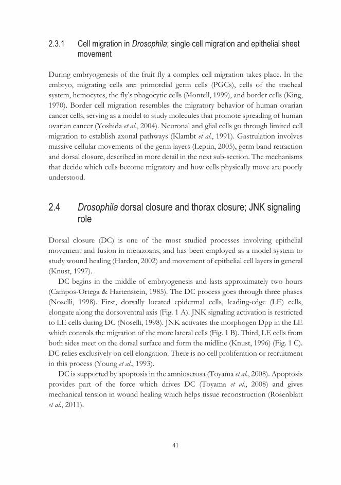
41
2.3.1 Cell migration in Drosophila; single cell migration and epithelial sheet movement
During embryogenesis of the fruit fly a complex cell migration takes place. In the embryo, migrating cells are: primordial germ cells (PGCs), cells of the tracheal system, hemocytes, the fly’s phagocytic cells (Montell, 1999), and border cells (King, 1970). Border cell migration resembles the migratory behavior of human ovarian cancer cells, serving as a model to study molecules that promote spreading of human ovarian cancer (Yoshida et al., 2004). Neuronal and glial cells go through limited cell migration to establish axonal pathways (Klambt et al., 1991). Gastrulation involves massive cellular movements of the germ layers (Leptin, 2005), germ band retraction and dorsal closure, described in more detail in the next sub-section. The mechanisms that decide which cells become migratory and how cells physically move are poorly understood.
2.4 Drosophila dorsal closure and thorax closure; JNK signaling role
Dorsal closure (DC) is one of the most studied processes involving epithelial movement and fusion in metazoans, and has been employed as a model system to study wound healing (Harden, 2002) and movement of epithelial cell layers in general (Knust, 1997).
DC begins in the middle of embryogenesis and lasts approximately two hours (Campos-Ortega & Hartenstein, 1985). The DC process goes through three phases (Noselli, 1998). First, dorsally located epidermal cells, leading-edge (LE) cells, elongate along the dorsoventral axis (Fig. 1 A). JNK signaling activation is restricted to LE cells during DC (Noselli, 1998). JNK activates the morphogen Dpp in the LE which controls the migration of the more lateral cells (Fig. 1 B). Third, LE cells from both sides meet on the dorsal surface and form the midline (Knust, 1996) (Fig. 1 C). DC relies exclusively on cell elongation. There is no cell proliferation or recruitment in this process (Young et al., 1993).
DC is supported by apoptosis in the amnioserosa (Toyama et al., 2008). Apoptosis provides part of the force which drives DC (Toyama et al., 2008) and gives mechanical tension in wound healing which helps tissue reconstruction (Rosenblatt et al., 2011).
42
There are mutations which disturb different stages of DC (Knust, 1996). Mutants with defects in DC are described as 'dorsal open' because the mutant phenotype resembles a hole on the dorsal surface which reflects the failure of the movement of the lateral ectoderm. Mutations in core JNK signaling genes, DJNKK gene hemipterous (hep, JNKK, Glise et al. 1995) and DJNK basket (bsk, JNK, Riesgo-Escovar et al., 1996) disrupt DC because LE cells cannot form filopodial and lamellipodial protrusions. The core signaling module of JNK signaling consists of JNKK (Hep) which when activated phosphorylates JNK (Bsk) (Glise et al., 1995), which gets activated and phosphorylates Jun (Jra) (Riesgo-Escovar et al., 1996; Sluss et al., 1996) and Fos (Ciapponi et al., 2001). Jun (Jra), together with Fos (Kay) form the AP-1 transcription factor which then activates JNK target genes and contributes to expression of TGF-β family protein Dpp in the leading edge of epithelial cells (Hou et al., 1997). Dpp then contributes to elongating the lateral edge of the embryo. During DC, the JNK signaling pathway is activated in the LE by an unknown signal. Another known target of JNK is Drosophila MAPK phosphatase Puc (Martín-Blanco et al., 1998). Puc controls JNK signaling in the LE (Martín-Blanco et al., 1998) by negative feedback, which is important to stop cell movement at the end of the process (Noselli & Agnès, 1999). In addition to specific mutants in JNK signaling that exhibit a dorsal-open phenotype and fail to complete embryogenesis, others do not complete disc eversion or are not able to fuse discs during metamorphosis (Pastor-Pareja et al., 2004).
Apart from the JNK signaling pathway, there are other genes which can be mutated to generate the dorsal-open phenotype. These encode proteins involved in a variety of cellular processes, including components of the cytoskeleton (e.g., zipper), membrane proteins (coracle, yurt), cell adhesion proteins (shotgun, armadillo, myosheroid, scab) and extracellular matrix proteins such as pericardin (Simonova & Burdina, 2009).
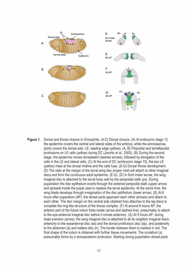
43
Figure 1. Dorsal and thorax closure in Drosophila. (A-C) Dorsal closure. (A) At embryonic stage 13, the epidermis covers the ventral and lateral sides of the embryo, while the amnioserosa (pink) covers the dorsal side. LE- leading edge (yellow). (A, B) Filopodial and lamellipodial protrusions on LE cells (yellow) during DC (Jacinto et al., 2000). (B) During the second stage, the epidermis moves dorsalward (dashed arrows), followed by elongation of the cells in the LE and lateral cells. (C) At the end of DC (embryonic stage 15), the two LE (yellow) meet at the dorsal midline and the cells fuse. (D-G) Dorsal thorax development. (D) The cells at the margin of the larval wing disc proper (red) will attach to other imaginal discs and form the continuous adult epidermis. (E-G). (D) In third instar larvae, the wing imaginal disc is attached to the larval body wall by the peripodial stalk (ps). During pupariation the disc epithelium everts through the widened peripodial stalk (upper arrow) and spreads inside the pupal case to replace the larval epidermis. At the same time, the wing blade develops through invagination of the disc epithelium (lower arrow). (E) At 6 hours after puparation (AP), the dorsal parts approach each other (arrows) and attach to each other. The disc margin on the ventral side (dashed line) attaches to the leg discs to complete the ring-like structure of the thorax complex. (F) At around 8 hours AP, the anterior part of the future notum folds inside (arrow and dashed line), presumably to attach to the eye-antennal imaginal disc before it moves anteriorly. (G) At 9 hours AP, during head eversion (arrow), the wing imaginal disc is attached to all its neighbor imaginal discs: anteriorly to the eyeantennal disc (ea) and the dorsal prothoracic disc (dp), and posteriorly to the abdomen (a) and haltere disc (h). The border between them is marked in red. The final shape of the notum is obtained with further tissue movements. The scutellum (s) presumably forms by a dorsoposterior protrusion. Starting during pupariation dorsal parts
44
of two imaginal wing discs migrate until they finally attach to each other (black arrows). The expression domain of pannier (pnr, green) marks the dorsalmost stripe of the future adult epidermis. The pnrMD237 GAL4 driver used in the experimental work of this thesis has the same expression pattern. Modified from Zeitlinger & Bohmann (1999); Simonova & Burdina (2009).
Along with JNK- and dpp-signaling, the Notch signaling pathway regulates DC. Notch mutants may partially restore the phenotype caused by the abnormalities of the JNK pathway (Zecchini et al., 1999). It is believed that the transmembrane receptor Notch negatively controls one of the probable alternative signaling pathways (probably, MAPK), which is able to partially compensate for the loss of JNK signal in relevant mutants.
2.4.1 Differences between dorsal and thorax closure
Thorax closure starts at the anterior end of the wing disc, proceeds through the most posterior region, and, finally, fills the gap (Martín-Blanco et al., 2000). JNK and dpp signaling mechanisms during thorax closure resemble those involved in DC, but with some differences (Zeitlinger & Bohmann, 1999; Martín-Blanco et al., 2000; Pastor-Pareja et al., 2004). Imaginal cells are brought together by spreading over the larval epidermis in a process mediated by the extension of filopodia, which seem to pull the contralateral epithelial sheets toward the midline. JNK signaling is involved in both cell spreading and imaginal disc fusion (Martín -Blanco et al., 2000), while Dpp participates in the regulation of the actin cytoskeleton of cells at the imaginal LE and the maintenance of cell polarity (Ricos et al., 1999). However, there are some differences between dorsal and thorax closure illustrated in Fig. 2. During embryonic DC, the amnioserosa and the epidermal cells maintain their relative positions and, despite occasional delaminations, amnioserosa cells remain in place until the very end of the process. They detach from the overlying epidermis only when closure is completed, and undergo apoptosis. During disc spreading that leads to thoracic closure, imaginal cells crawl over the larval epidermis (Fig. 2). In this process, larval cells are left below and behind and eventually delaminate from the edges (Martín-Blanco et al., 2000).
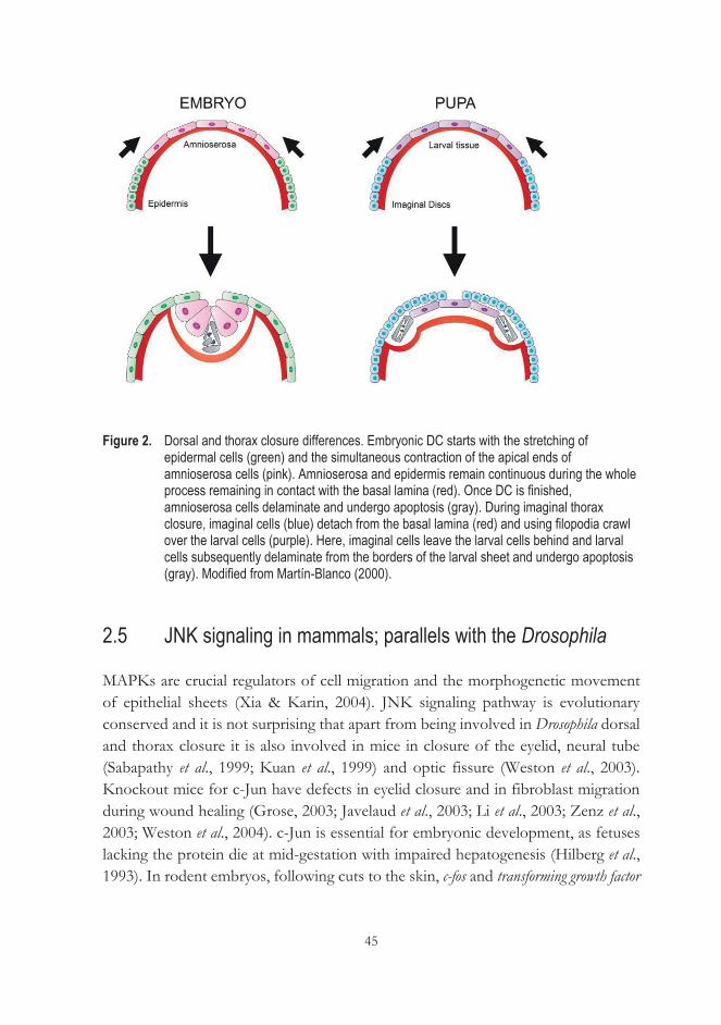
45
Figure 2. Dorsal and thorax closure differences. Embryonic DC starts with the stretching of epidermal cells (green) and the simultaneous contraction of the apical ends of amnioserosa cells (pink). Amnioserosa and epidermis remain continuous during the whole process remaining in contact with the basal lamina (red). Once DC is finished, amnioserosa cells delaminate and undergo apoptosis (gray). During imaginal thorax closure, imaginal cells (blue) detach from the basal lamina (red) and using filopodia crawl over the larval cells (purple). Here, imaginal cells leave the larval cells behind and larval cells subsequently delaminate from the borders of the larval sheet and undergo apoptosis (gray). Modified from Martín-Blanco (2000).
2.5 JNK signaling in mammals; parallels with the Drosophila
MAPKs are crucial regulators of cell migration and the morphogenetic movement of epithelial sheets (Xia & Karin, 2004). JNK signaling pathway is evolutionary conserved and it is not surprising that apart from being involved in Drosophila dorsal and thorax closure it is also involved in mice in closure of the eyelid, neural tube (Sabapathy et al., 1999; Kuan et al., 1999) and optic fissure (Weston et al., 2003). Knockout mice for c-Jun have defects in eyelid closure and in fibroblast migration during wound healing (Grose, 2003; Javelaud et al., 2003; Li et al., 2003; Zenz et al., 2003; Weston et al., 2004). c-Jun is essential for embryonic development, as fetuses lacking the protein die at mid-gestation with impaired hepatogenesis (Hilberg et al., 1993). In rodent embryos, following cuts to the skin, c-fos and transforming growth factor
46
beta (TGFβ-1) which are homologs of Drosophila kayak and dpp are activated at the edge of the wound (Martin & Nobes, 1992).
In vertebrates a phenomenon similar to DC occurs, described as embryonic wound healing. In contrast to mature individuals, this process does not result in scar formation (Martin et al., 1993; Martin, 1997). After a mouse or rat embryo’s skin is injured both c-Fos and TGFβ-1 are activated on the edge of the wound. They are the homologs of the D. melanogaster genes kayak and dpp, respectively (Martin & Nobes, 1992).
Primary c-Jun-/- fibroblasts have a severe proliferation defect and undergo premature senescence in vitro (Johnson et al., 1993; MacLaren et al., 2004). The mammalian AP-1 transcription factor regulates the expression of many genes, such as those encoding matrix metalloproteinases (MMPs), integrins, cytokines, and growth factors (Angel et al., 2001, Yates & Rayner, 2002; Florin et al., 2004), which are involved in wound healing. The Scribble (Scrib) complex contributes to epithelial cell migration and fusion during both development and wound healing in mammals by recruiting the small GTPases Cdc42 and Rac and the serine/threonine kinase Pak to the LE of migrating cells (Murdoch et al., 2003; Zarbalis et al., 2004; Humbert et al., 2006; Dow et al., 2007; Bahri et al., 2010). The JNK-dependent network of transcriptional factors also contributes towards epithelial to mesenchymal transition (EMT), a biological process in which cells lose cell-cell adhesion and become migratory (Sahu et al., 2015). JNK and the Wnt pathway are main regulators of wound repair and tissue regeneration. In wound healing, the actin cytoskeleton (Hall, 1998; Kaibuchi et al., 1999) is a major JNK target, as well as growth factors. Mechanical stretching of the wound-edge cells, loss of cell polarity or necrotic signals are reported to be upstream activators of this signaling pathway (Rämet et al., 2002; Nelson et al., 2005; Igaki et al., 2006). Expression of Wnt target genes is also stimulated by JNK activation to accelerate tumorigenesis (Sancho et al., 2009).
Developmental morphogenesis, tissue injury, and oncogenic transformation can cause the detachment and elimination of epithelial cells by anoikis, a specialized form of apoptosis (Girnius & Davis, 2017). Loss of JNK function by epithelial cells may lead to survival of these cells in luminal spaces and if these cells accumulate additional mutations that may cause cancer. JNK deficiency accordingly enhances tumor formation in a model of breast cancer (She et al., 2002; Cellurale et al., 2012; Hübner et al., 2012). Two upstream components of the JNK pathway (MAP2K4 and MAP3K1) are frequently mutated in human cancer (Stephens et al., 2012, Nik-Zainal et al., 2016). However, an overactive JNK pathway also has been shown to cause cancer (Smeal et al., 1991; Hui et al., 2008; Barbarulo et al., 2013). This means that the
47
JNK pathway performs opposing functions in oncogenic signaling. Downregulation of JNK signaling leads to developmental defects, whilst an overactive JNK pathway can lead to cancers (Bubici & Papa, 2014). Abnormal JNK signaling contributes to many pathological conditions such as stroke (Wu et al., 2000; Borsello et al., 2003), obesity and insulin resistance (Hirosumi et al., 2002), and atherosclerosis (Ricci et al., 2004). JNK activity has also been implicated in Parkinson’s and Alzheimer’s diseases (Zhu et al. 2001; Peng & Andersen, 2003), whilst JNK function is needed for brain development, memory and learning (Raivich & Behrens 2006; Waetzig et al. 2006).
2.5.1 Interplay between JNK signaling and mitochondrial metabolism
There is a multilayered link between JNK signaling and mitochondria. JNK is a major signaling pathway regulating both metabolic (Harris et al. 2002) and apoptotic (Kharbanda et al. 2000; Schroeter et al., 2003) functions in mitochondria, linking cytosolic and mitochondrial energy pathways. For example, it has been shown that, in primary cortical neurons, JNK-induced decline in pyruvate dehydrogenase (PDH) activity resulted in the decline of ATP levels and increased lactate concentration (Zhou et al., 2008). These observations suggest a shift from aerobic metabolism of pyruvate in mitochondria to its anaerobic reduction to lactate in the cytosol with concomitant NAD+ regeneration to support glycolysis. JNK may therefore serve as an important modulator of mitochondrial bioenergetics, anaerobic respiration and cellular ATP levels, regulating mitochondrial electron flow as well as the production of ROS from the RC (Zhou et al., 2008). JNK signaling is associated with the regulation of mitochondrial pathways of apoptosis following viral infection. After reovirus infection, MEKK1 activates JNK, which promotes the release of the proapoptotic factors Smac and cytochrome c (Cyt c) from mitochondria, both of which contribute to apoptosis (Clarke et al., 2004). Mitochondrial H2O2 has been shown to activate JNK in various cell lines upon inhibition of MAPK phosphatase or dissociation of the glutathione transferase-JNK complex (Nemoto et al., 2000; Chen et al., 2001; Foley et al., 2004). Prolonged JNK activation is believed to play a key role in cell injury. For example, apoptosis of auditory neurons as a consequence of oxidative damage has been shown to engage the major downstream target of the JNK signaling cascade, i.e., c-Jun (Scarpidis et al., 2003). This can be prevented by administration of an inhibitory peptide targeted against JNK (Eshraghi et al., 2006). Mitochondria produce H2O2 and NO (Cadenas & Davies, 2000; Poderoso et al., 2000), which are involved in the regulation of redox-sensitive cell signaling through
48
the JNK pathway, thus coordinating functional responses between mitochondria and other cellular processes. For example, H2O2 activates JNK at multiple levels by mechanisms eliciting dissociation of inhibitory complexes (Saitoh et al., 1998) or suppression of phosphatases involved in kinase inactivation (Chen et al., 2001). Nitric oxide inhibits JNK activity by S-nitrosylation of a critical cysteine thiol (Park et al., 2000). In contrast, JNK as a signaling molecule translocates to mitochondria under stress conditions (Kharbanda et al., 1997; Aoki et al., 2002) and has direct metabolic effects in mitochondria, through the activation of phosphorylation cascades (Zhou et al., 2008). This network thus connects cytosolic and mitochondrial processes that control energy levels and the redox environment throughout the cell (Zhou et al., 2008).
2.6 Alternative oxidase: proposed roles in living organisms
The alternative mitochondrial respiratory chain contains the alternative NADH dehydrogenases (NDX) and quinone oxidases (AOX) (McDonald &Gospodaryov, 2018). AOX, which is present in most organisms, is a non-proton motive ubiquinol oxidase (Berthold & Stenmark, 2003), i.e. it catalyzes the oxidation of ubiquinol and consequent reduction of oxygen to water (McDonald & Gospodaryov, 2018), and is a branch point in the respiratory electron transport chain, bypassing complexes III and IV (McDonald et al., 2009) (Fig. 4). By virtue of this property it dramatically decreases ATP production and released energy is dissipated as heat (Rhoads & Subbaiah, 2007). AOX is a membrane bound diiron carboxylate enzyme (Siedow et al., 1995). Depending on species it can occur as a monomer, homodimer or even multimer, with its iron atoms coordinated by glutamic acid residues (Berthold & Stenmark, 2003). AOX activity depends on the membrane-bound concentration and redox state of ubiquinone, as well as cellular oxygen concentration, and is regulated via gene expression or by post-translational modification. In the latter case, cellular redox status affects tertiary complex formation, whilst enzymatic activity is modulated by the action of allosteric effectors such as pyruvate or other metabolites. The ratio of oxidized to reduced protein varies considerably between species and tissues. In plant mitochondria, metabolites such as pyruvate, hydroxypyruvate and glyoxylate can activate AOX (Sluse & Jarmuszkiewicz, 1998).
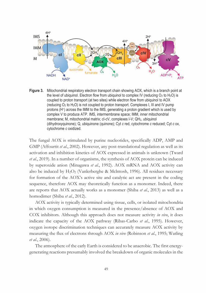
49
Figure 3. Mitochondrial respiratory electron transport chain showing AOX, which is a branch point at the level of ubiquinol. Electron flow from ubiquinol to complex IV (reducing O2 to H2O) is coupled to proton transport (at two sites) while electron flow from ubiquinol to AOX (reducing O2 to H2O) is not coupled to proton transport. Complexes I, III and IV pump protons (H+) across the IMM to the IMS, generating a proton gradient which is used by complex V to produce ATP. IMS, intermembrane space; IMM, inner mitochondrial membrane; M, mitochondrial matrix; cI-cV, complexes I-V; QH2, ubiquinol (dihydroxyquinone); Q, ubiquinone (quinone); Cyt c red, cytochrome c reduced; Cyt c ox, cytochrome c oxidized.
The fungal AOX is stimulated by purine nucleotides, specifically ADP, AMP and GMP (Affourtit et al., 2002). However, any post-translational regulation as well as its activation and inhibition kinetics of AOX expressed in animals is unknown (Tward et al., 2019). In a number of organisms, the synthesis of AOX protein can be induced by superoxide anion (Minagawa et al., 1992). AOX mRNA and AOX activity can also be induced by H2O2 (Vanlerberghe & Mclntosh, 1996). All residues necessary for formation of the AOX’s active site and catalytic act are present in the coding sequence, therefore AOX may theoretically function as a monomer. Indeed, there are reports that AOX actually works as a monomer (Shiba et al., 2013) as well as a homodimer (Shiba et al., 2012).
AOX activity is typically determined using tissue, cells, or isolated mitochondria in which oxygen consumption is measured in the presence/absence of AOX and COX inhibitors. Although this approach does not measure activity in vivo, it does indicate the capacity of the AOX pathway (Ribas-Carbo et al., 1995). However, oxygen isotope discrimination techniques can accurately measure AOX activity by measuring the flux of electrons through AOX in vivo (Robinson et al., 1995; Watling et al., 2006).
The atmosphere of the early Earth is considered to be anaerobic. The first energy-generating reactions presumably involved the breakdown of organic molecules in the
50
absence of oxygen. Since oxygen is a substrate in the formation of toxic reactive species, it is thought that changes in oxygen levels might have caused selective pressure for microorganisms to develop alternative respiratory proteins (McDonald & Gospodaryov, 2018) that are less vulnerable to ROS damage than other oxidoreductases. Many AOX-expressing organisms are subjected to regular changes in oxygen levels (anoxia/reoxygenation), e.g., plant roots that can be overwatered, causing anoxia (Veiga et al., 2000), or sea squirts that experience functional hypoxia (Torre et al., 2014). In addition, many of them are subjected to conditions which compromise synthesis of specific prosthetic groups of respiratory complexes, notably haem and iron-sulphur clusters, which are susceptible to damage by ROS. Since ROS, generated by NADPH oxidases, are frequently used by immune cells to attack pathogens, this may be one reason why many eukaryotic parasites have retained AOX over the course of their evolution whereas mammals developed different mechanisms to buffer such problems, and have lost the gene for AOX.
AOX genes have so far been found only in slow-moving or sessile organisms (McDonald et al., 2009). However, there are sessile organisms which do not have AOX, suggesting that selective pressure to keep AOX is not only related to the sessile state and low ATP demand, but rather to conditions which compromise function of the respiratory chain. AOX is ubiquitously present in plants, where it is most extensively studied, as well as in many microorganisms, fungi and protists, including human/animal parasites such as Trypanosoma brucei, Cryptosporidium parvum, Blastocystis hominis, and Candida albicans (Young et al., 2014). In animals, AOX genes and AOX mRNA have so far been reported in: Placozoa, Porifera, Nematoda, Arthropoda, Mollusca, Annelida, Rotifera, Brachiopoda, Cnidaria, Echinodermata, Hermichordata and Chordata (McDonald et al., 2008; McDonald & Vanlerberghe, 2004; McDonald & Gospodaryov, 2018). It is unclear as to whether the AOX gene is translated into protein in all of these animal species (McDonald et al., 2008). Since AOX is absent in humans and is essential for pathogenic fungi, human parasites and plant pathogenic Nematoda, there is an opportunity for developing species-specific AOX inhibitors which can be tested as potential therapeutics or fungicides (Young et al., 2014). In addition to protection against anoxia, one physiological role of AOX in plants is heat production (Meusse, 1975; Grant et al., 2008; Wang & Zhang, 2015). AOX also has a crucial role in chloroplast protection under extreme environments, such as high light (Xu et al., 2011), as well as tolerance to both biotic and abiotic stress (Saha et al., 2016) especially when resulting in decreased functioning of the cytochrome pathway. Crucially, AOX avoids the generation of excessive ROS resulting from an excess of reducing power accumulated in the ubiquinone pool
51
(Vanlerberghe, 2013). It therefore enables respiration to resist toxins such as NO, CO, CN-, etc., thus fine-tuning ATP production, and the maintenance of mitochondrial and cellular homeostasis (Moore et al., 2013).
2.6.1 The role of AOX as gene therapy tool: potential and concerns
RC dysfunction and ROS overproduction are proposed to be instrumental in cancer, aging, neurodegenerative and mitochondrially inherited diseases. By preventing RC blockade and excess ROS production, AOX has therapeutic potential (El-Khoury et al., 2013), and it has been shown that AOX from a tunicate, Ciona intestinalis, can be safely and ubiquitously expressed in mammalian cells (Hakkaart et al., 2006), flies (Fernandez-Ayala et al., 2009) and mice (El-Khoury et al., 2013).
AOX could potentially be useful as a gene therapy for diseases caused by mutations in genes which encode subunits of complex III and IV of the respiratory chain and assembly factors for these proteins (Dassa et al., 2009; Kemppainen et al., 2014). In the latter case, AOX would allow electron transfer from ubiquinol to the terminal acceptor (molecular oxygen). Without such transfer, mutant cells would not be able to completely oxidize respiratory substrates for several reasons: (1) product inhibition of upstream enzymes in a biochemical pathway - in effect, the entire pathway becomes blocked, (2) potential toxicity or signaling roles of intermediates within a biochemical pathway, (3) physical laws of membrane energetics - inhibition of the enzymes within the respiratory chain would lead to disturbance in membrane potential. In the case of complex III blockade, ubiquinol would accumulate in the membrane and cause overproduction of superoxide anion-radical by reverse electron transport through complex I. Superoxide anion-radical is able to damage other components of the respiratory chain and other macromolecules in its vicinity. In the case of complex IV blockade, both ubiquinol and reduced cytochrome c would accumulate in the respiratory chain. Reduced cytochrome c is also a signaling molecule which may exit the mitochondria and trigger apoptosis. If respiratory substrates accumulate (mostly organic acids formed in other catabolic processes, e.g., pyruvate from glycolysis, alpha-ketoglutarate and oxaloacetate - from amino acid transamination, fatty acids and acetyl-CoA from beta-oxidation) and reducing equivalents (NADH) are not oxidized completely, they become toxic to cells. The simplest mechanism of this toxicity is, for instance, lowering of pH, e.g., lactic acidosis. AOX thus has a potential use in diseases where mitochondrial dysfunction is an intermediate step in the pathological process, rather than the underlying cause.
52
For example, it was shown that expression of Ciona AOX mitigates symptoms of Parkinson’s and Alzheimer’s diseases in Drosophila models (Humphrey et al., 2012; El-Khoury et al., 2016), as well as a lethal inflammatory cascade in a mouse model of sepsis (Mills et al., 2016). Theoretically, it may also ameliorate the symptoms of diseases associated with impaired synthesis of prosthetic groups for respiratory chain complexes. Complexes III and IV, as well as cytochrome c, contain heme. Defects in heme synthesis, iron and copper acquisition would lead to a functional deficiency of complexes III and/or IV.
It is difficult to predict which source of AOX enzyme would have the most benefits if used as a gene therapy tool in humans. AOX from Trypanosoma brucei (TAO) has been suggested to be the most appropriate to be used in human gene therapy (May et al., 2017). It functions at 37 °C within the human blood stream (Chaudhuri et al., 2006) and is the AOX protein that is the best structurally characterized (Shiba et al., 2013). It is also possible and desirable to engineer AOX proteins which can be specifically regulated for the purpose of gene therapy. This would potentially result in an AOX protein that could be turned 'on' and 'off' by cellular pyruvate levels or other effectors, and which could be beneficial under conditions of impaired ROS homeostasis (May et al., 2017).
Constitutive and unregulated functioning of AOX in the whole organism might be problematic, as it would decrease the yield of ATP. However, due to a very low affinity for its substrate, ubiquinol, AOX does not efficiently compete with complex III (Bahr & Bonner, 1973) under standard physiological conditions. For this reason, it is believed that AOX, even if present ubiquitously, functions only when the cytochrome pathway does not. However, if constitutively activated, AOX could be triggered by local metabolic conditions in specific cells, and participate significantly in respiration, it could alter ATP production and other processes dependent on respiratory energy, such as the buffering of calcium. Moreover, an increased metabolic rate with concomitant heat production could be detrimental in some tissues. Saari et al. (2017) reported that Drosophila males expressing AOX had apparent retention of sperm within the testis, which might be attributable to thermogenesis. An interference with cell signaling may also involve ROS. AOX has been reported to decrease mitochondrial ROS production even under the conditions of normal oxidative phosphorylation activity (Fernandez-Ayala et al., 2009; Sanz et al., 2010). This could affect signaling events in which ROS participate. This is why it may be important for gene therapy purposes to engineer an AOX which can be turned 'on' and 'off' as desired.
53
To date, AOX proteins from two different sources have been successfully expressed in human cells, namely from C. intestinalis, expressed in a HEK293T cell derivative (Hakkaart et al., 2006) and from the thermogenic plant Arum concinnatum in HeLa cells (Kakizaki & Ito, 2013). Thus far, the C. intestinalis AOX is the most studied in terms of potential gene therapy applications.
The immune response against transgene products is a critical issue for successful gene therapy. The bigger the phylogenetic distance between organisms, the greater the probability that the proteins of this organism would elicit an immune response. From this point of view, C. intestinalis AOX seems to be a better candidate for expression in humans than plant or trypanosomal AOX.
Although the AOX family contains a highly conserved active site there are remarkable differences in ubiquinol oxidase activity between different AOXs (May et al., 2017). More detailed kinetic and biophysical characterization of different AOX proteins is needed to understand how differences in structure and sequence are related to differences in function. Once the properties of AOX are well established, the sequence of the polypeptide can potentially be manipulated by design to be highly active, have low immunogenicity, and be susceptible to external regulation.
2.7 Drosophila genetic toolkit: The Gal4/UAS system for regulated transgene expression
The Gal4/UAS system (Brand & Perrimon, 1993) is a binary expression system primarily used in Drosophila which makes it possible to study effects of the overexpression or downregulation of any gene in a selected tissue and developmental stage (Brand & Perrimon, 1993). In the driver line, a construct containing a cell-specific promoter drives the expression of the gene encoding the yeast transcription factor Gal4 (Fig. 3 A). In the responder line, a transgene of interest is regulated by a promoter incorporating an upstream activation sequence (UAS). The Gal4 protein binds to the UAS sequence and drives expression of the transgene. Thus, the transgene should only be expressed in cells defined by the promoter regulating Gal4. Through their over-expression, misexpression or downregulation in a tissue- and time-specific manner, this allows different genes to be studied in an internally controlled manner. The two constructs are established in separate fly lines allowing for numerous combinatorial possibilities. The Gal4 protein has basal transcriptional activator activity at 18 °C, higher activity at 25 °C and even higher activity at 29 °C
54
(Busson & Pret, 2007). Temperature thus has a huge effect on the activity of Gal4 and on the expression of transgenes that it controls. It is therefore important to control temperature when performing experiments using this system. However, the Gal4/UAS system is far from perfect. There is variability in Gal4-mediated transcription between cells which cannot be explained (Brand et al., 1994). Gal4 has dosage-dependent (Kramer & Staveley, 2003) biological effects, some of which are detrimental. Expression of high levels of Gal4 in Drosophila can modify the expression of many endogenous genes in a UAS-independent manner, among them components of important signaling pathways (Liu & Lehmann, 2008), even though there is no Gal4 ortholog nor UAS binding site in Drosophila. This potentially complicates the use of this system, since its original premise is that Gal4 is inactive in Drosophila. One of the mechanisms by which Gal4 might influence gene expression is via a selective block of protein transport into the cell nucleus (Uv et al., 2000). Most promoters and enhancers that drive Gal4 expression are active many times throughout development. That is why a modified Gal4/UAS system, the GeneSwitch System (GS), that utilizes a modified Gal4 protein was developed (Osterwalder et al., 2001). It requires antiprogestin, RU486, to activate it, in order to bind the DNA (Fig. 3 B). RU486 can be administered by either feeding or immersing the animals in a solution containing RU486. Reporter protein expression is dependent on the dose of the GeneSwitch activator drug RU486 (Osterwalder et al., 2001). Again, the use of the GS system is justified under the assumption that GS is inert in the fly. However, GS is not completely inert in the absence of RU486, since it can still confer a significant decrease in target gene protein levels when RNA interference (RNAi) transgenes are used (Scialo et al., 2016).
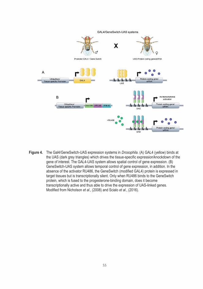
55
Figure 4. The Gal4/GeneSwitch-UAS expression systems in Drosophila. (A) GAL4 (yellow) binds at the UAS (dark grey triangles) which drives the tissue-specific expression/knockdown of the gene of interest. The GAL4-UAS system allows spatial control of gene expression. (B) GeneSwitch-UAS system allows temporal control of gene expression, in addition. In the absence of the activator RU486, the GeneSwitch (modified GAL4) protein is expressed in target tissues but is transcriptionally silent. Only when RU486 binds to the GeneSwitch protein, which is fused to the progesterone-binding domain, does it become transcriptionally active and thus able to drive the expression of UAS-linked genes. Modified from Nicholson et al., (2008) and Scialo et al., (2016).

56
57
3 AIMS OF THE STUDY
The aim of this thesis work was to test whether transgenic expression of AOX from the urochordate Ciona intestinalis in model organisms is able to influence cell migration and other developmental processes.
The more specific aims of this work were:
1. To engineer a catalytically inactive variant of the AOX protein (mutAOX) using site-directed mutagenesis, thus establishing a control for the entire study (I)
2. To create fly lines where AOX and mutAOX are inserted at the same integration site and thus serve as comparable controls for AOX activity in Drosophila using site-specific transgene insertion, (I, II)
3. To investigate the developmental effects of expressing the GeneSwitch transcription factor in Drosophila, and whether these are modified by concomitant AOX expression (II)
4. To test whether tissue-specific knockdown of JNK cascade genes produces cell migration defects during Drosophila development, and whether these can be corrected by concomitant AOX expression (III)
5. To test whether AOX expression influence mammalian cell migration, and if so under what conditions (III)
6. To determine the mechanism behind any such effect (III)
The starting point for the project was a preliminary observation made by my co-author, K. Kemppainen, regarding the interaction between AOX expression and GeneSwitch in Drosophila. This motivated the detailed study (II) undertaken in regard to Aims 3-4, as well as the need for an appropriate control (Aims 1-2, I). The

58
outcome of these studies then justified the more extensive investigation of cell migration and the JNK pathway (Aims 5-7, III).
59
4 MATERIALS AND METHODS
4.1 Drosophila stocks and maintenance (I-III)
Drosophila strains, e.g., drivers, balancers, transgenic recipient, RNAi and various control fly lines and their sources are described in original communications. Flies were maintained in standard high-sugar medium (Fernandez-Ayala et al., 2009) on a 12-h light/12-h dark cycle at 25 °C, except where indicated in the figure legends. Where indicated, medium was supplemented with RU486 (Mifepristone, Sigma) at the concentrations indicated in figures and legends.
ΦC31 recombinase-mediated-site-directed transgenesis was used to generate transgenic fly lines (service provided by BestGene Inc, Chino Hills, CA), using recipient lines with the following integration sites: attP18 (chromosome X), attP40 (chromosome 2) and attP2 (chromosome 3), according to Pfeiffer et al. (2010) employing the wild-type and mutated AOX constructs cloned in pUASTattB and pUASTattB itself as empty-vector control. Following characterization, transgenic lines were maintained over balancers appropriate for chromosome X, 2 or 3, bearing standard markers (FM7, CyO, TM3Sb, respectively). Transgenic lines UAS-AOXF24 and UAS-AOXF6 were described previously (Fernandez-Ayala et al., 2009) and referred to in the thesis and in publications as 'high' expression or 'old' wt AOX lines. Other lines created in the study are described under Results.
Crosses were implemented in triplicate, with flies being tipped into new vials on three successive days after mating. Nine vials per cross were analyzed in most experiments. Morphological abnormalities of the eclosed flies were scored as described in (II) and (III).
60
4.2 Cell culture
4.2.1 Drosophila S2 cells
Drosophila S2 cell line (Schneider, 1972), originally derived from late embryos, was cultured in Schneider's Medium (Sigma-Aldrich) at 25 °C. Cells were passaged every 3-4 days.
4.2.2 Mammalian cell lines
Human embryonic kidney 293 (HEK 293) cells originally derived by treating a human embryonic kidney homogenate with sheared adenovirus DNA (Graham et al., 1997). This cell line has a highly aberrant karyotype and constitutively expresses Ad5 E1A/E1B proteins, which deregulate pRB/p53pathways (Stepanenko & Dmitrenko, 2015). HEK293T (293T, ATCC® CRL-3216™) cells used for the work in I, are the derivative of HEK 293 cells with the difference that they express the SV40 T-antigen.
AOX-positive and -negative mouse embryonic fibroblasts (MEFs), sourced either from embryos transgenic for C. intestinalis AOX, inserted at the Rosa26 locus (Szibor et al., 2017), or from their nontransgenic littermates, were studied at passage 6. MEFs, immortalized by retroviral transduction with HPV16 oncoproteins E6 and E7 (iMEFs) were supplied by P.K. Dhandapani, University of Tampere (III).
The AP-1 reporter HEK293 recombinant cell line (JNK signaling pathway; BPS Bioscience), here abbreviated HEK-AP1, contains a firefly luciferase gene under the control of AP-1-responsive elements that are stably integrated into HEK293 cells (III).
The BJ-5ta human fibroblasts (ATCC CRL-4001) has been immortalized with human telomerase reverse transcriptase (hTERT) (III).
Mammalian cell lines were maintained at 37 °C in 5% CO2 and cultured as detailed in the original publications.
61
4.3 Molecular cloning
The C. intestinalis AOX cDNA (Hakkaart et al., 2006) was recloned into suitable expression vectors for Drosophila (S2 cells and whole flies) and mammalian cells. For details see (I). In vitro mutagenesis of these constructs employed a PCR-based, site-directed mutagenesis approach with customized oligonucleotides using Quick Change Site-Directed Mutagenesis Kit (Agilent Technologies) (I). All constructs were verified by sequencing prior to use.
4.4 Gene expression assays
4.4.1 RNA expression analysis by Quantitative Reverse Transcription-PCR (qRT-PCR)
Extraction of RNA from 2-day old adult flies and S2 cells and measurements of transcript levels relative to an internal standard (RpL32 mRNA) were performed using standard methods (Fernandez-Ayala et al., 2009), as described in (I) and (III).
4.4.2 Protein analysis by Western blotting
To measure transgene expression at the protein level in the adult fly, total protein was extracted from 30 females or 40 males per sample (I, Fig. 2 D and E). Frozen fly samples were used for all experiments except for detecting mutant AOX in females, for which fresh samples were used (I, Fig. 2 D).
For detecting phosphorylation at c-Jun, 300,000 MEFs or HEK-AP1 cells or 250,000 BJ-5ta cells were plated on 6-well plates (CellStar; Greiner Bio-One, III, Fig. 5). After 24 h, the medium was replaced with medium containing either 0.2% DMSO, 20 μM SP600125 in 0.2% DMSO, 20 μM JNK inhibitor V, 8 nM PMA, or no added drug and the plate was incubated for 2 h (or 40 min, in the case of PMA). Cells were carefully rinsed in ice-cold PBS and then scraped on ice using a CytOne cell scraper in resuspension buffer.
62
Primary antibody incubations were carried out overnight at 4 °C. Membranes were then washed 3 x 10 min in PBS-Tween and incubated with HRP-conjugated secondary antibody for 1 h at room temperature in PBS-Tween with 5% nonfat (w/v) milk. After this, the unbound secondary antibody was removed by three successive 10 min washes with PBS-Tween. Protein was detected and visualized using Immun-Star™ HRP Chemiluminescent Kit (Bio-Rad). The chemiluminescence of all blots was documented both with film and by using a Bio-Rad ChemiDoc imager.
Primary antibodies used were: customized rabbit anti-AOX (21st Century Biochemicals, 1:10,000, Dassa et al., 2009), rabbit anti-α-actinin C-20-R used as loading control (Santa Cruz Biotechnology, 1:5,000 (I) 1:7,000 (III), mouse anti-ATP5α used as loading control (Abcam, 1:50,000(I)), phospho-c-Jun (Ser 73) rabbit monoclonal antibody (catalog number 3270; Cell Signaling Technology 1:1,000, III), and phospho-c-Jun (Ser 63) rabbit polyclonal antibody (catalog number 9261; Cell Signaling Technology, III). HRP-conjugated secondary antibodies were: peroxidase-labeled Goat Anti-rabbit IgG (catalog number PI-1000) and Horse Anti-mouse IgG (catalog number PI-2000 (both from Vector Laboratories, 1:10,000 (I, III)).
4.5 Transfection/transduction
Transgenes were introduced into Drosophila and mammalian cell-lines, for various purposes, by plasmid DNA lipofection or by lentivector transduction, as detailed in (I) and (III). Briefly, to assess the expression and activity of AOX and a mutated variant (see Results and I, Fig. 2 A), plasmid DNA was transiently transfected into HEK293T cells using 60 μl Lipofectamine® 2000 (Invitrogen) under manufacturer’s recommended conditions (I). To assay the effects of transgene expression on AP-1 directed transcription using a luciferase reporter approach, test plasmids and an appropriate cocktail of reporter-system plasmids (Chatterjee & Bohmann, 2012) were transiently transfected into Drosophila S2 cells (III, Fig. 7 A-D), using the FuGENE® HD transfection reagent (Promega) according to the manufacturer’s instructions. To conduct similar assays in mammalian cells, as well as for Western blotting to study c-Jun phosphorylation, transgenes were stably introduced into the HEK AP-1 reporter cell-line or into BJ-5ta cells by pWPI-based lentiviral transduction, followed by cell sorting and cloning at limiting dilution (III).
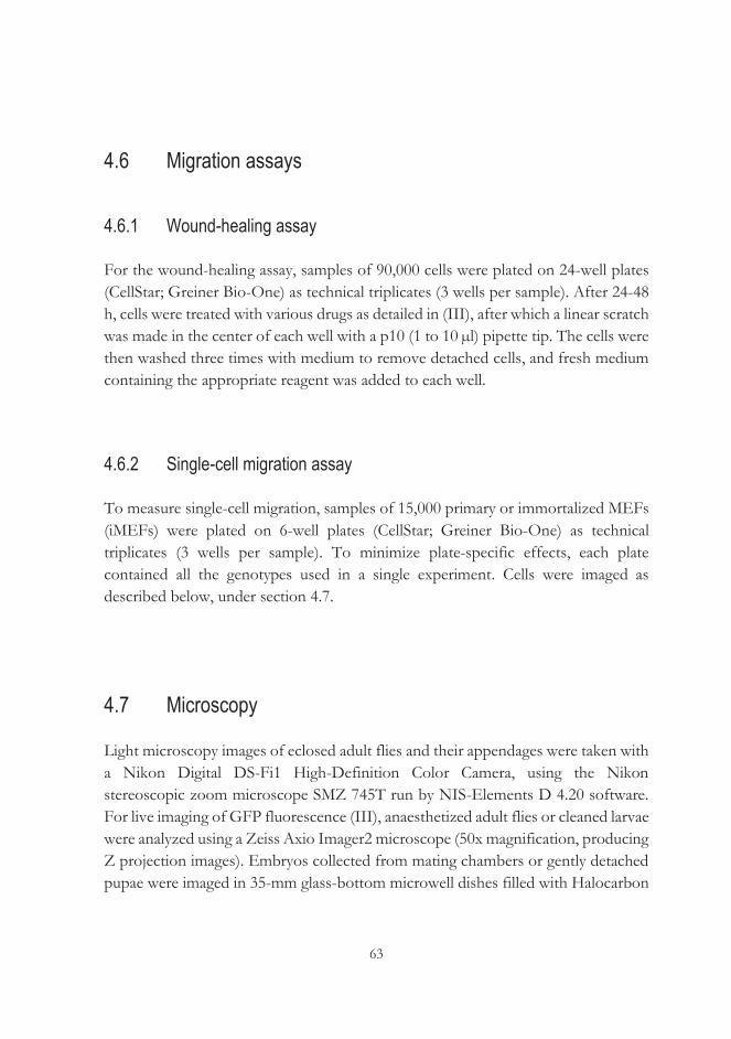
63
4.6 Migration assays
4.6.1 Wound-healing assay
For the wound-healing assay, samples of 90,000 cells were plated on 24-well plates (CellStar; Greiner Bio-One) as technical triplicates (3 wells per sample). After 24-48 h, cells were treated with various drugs as detailed in (III), after which a linear scratch was made in the center of each well with a p10 (1 to 10 μl) pipette tip. The cells were then washed three times with medium to remove detached cells, and fresh medium containing the appropriate reagent was added to each well.
4.6.2 Single-cell migration assay
To measure single-cell migration, samples of 15,000 primary or immortalized MEFs (iMEFs) were plated on 6-well plates (CellStar; Greiner Bio-One) as technical triplicates (3 wells per sample). To minimize plate-specific effects, each plate contained all the genotypes used in a single experiment. Cells were imaged as described below, under section 4.7.
4.7 Microscopy
Light microscopy images of eclosed adult flies and their appendages were taken with a Nikon Digital DS-Fi1 High-Definition Color Camera, using the Nikon stereoscopic zoom microscope SMZ 745T run by NIS-Elements D 4.20 software. For live imaging of GFP fluorescence (III), anaesthetized adult flies or cleaned larvae were analyzed using a Zeiss Axio Imager2 microscope (50x magnification, producing Z projection images). Embryos collected from mating chambers or gently detached pupae were imaged in 35-mm glass-bottom microwell dishes filled with Halocarbon
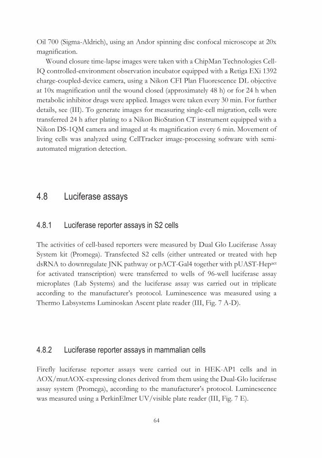
64
Oil 700 (Sigma-Aldrich), using an Andor spinning disc confocal microscope at 20x magnification.
Wound closure time-lapse images were taken with a ChipMan Technologies Cell-IQ controlled-environment observation incubator equipped with a Retiga EXi 1392 charge-coupled-device camera, using a Nikon CFI Plan Fluorescence DL objective at 10x magnification until the wound closed (approximately 48 h) or for 24 h when metabolic inhibitor drugs were applied. Images were taken every 30 min. For further details, see (III). To generate images for measuring single-cell migration, cells were transferred 24 h after plating to a Nikon BioStation CT instrument equipped with a Nikon DS-1QM camera and imaged at 4x magnification every 6 min. Movement of living cells was analyzed using CellTracker image-processing software with semi-automated migration detection.
4.8 Luciferase assays
4.8.1 Luciferase reporter assays in S2 cells
The activities of cell-based reporters were measured by Dual Glo Luciferase Assay System kit (Promega). Transfected S2 cells (either untreated or treated with hep dsRNA to downregulate JNK pathway or pACT-Gal4 together with pUAST-Hepact for activated transcription) were transferred to wells of 96-well luciferase assay microplates (Lab Systems) and the luciferase assay was carried out in triplicate according to the manufacturer’s protocol. Luminescence was measured using a Thermo Labsystems Luminoskan Ascent plate reader (III, Fig. 7 A-D).
4.8.2 Luciferase reporter assays in mammalian cells
Firefly luciferase reporter assays were carried out in HEK-AP1 cells and in AOX/mutAOX-expressing clones derived from them using the Dual-Glo luciferase assay system (Promega), according to the manufacturer’s protocol. Luminescence was measured using a PerkinElmer UV/visible plate reader (III, Fig. 7 E).
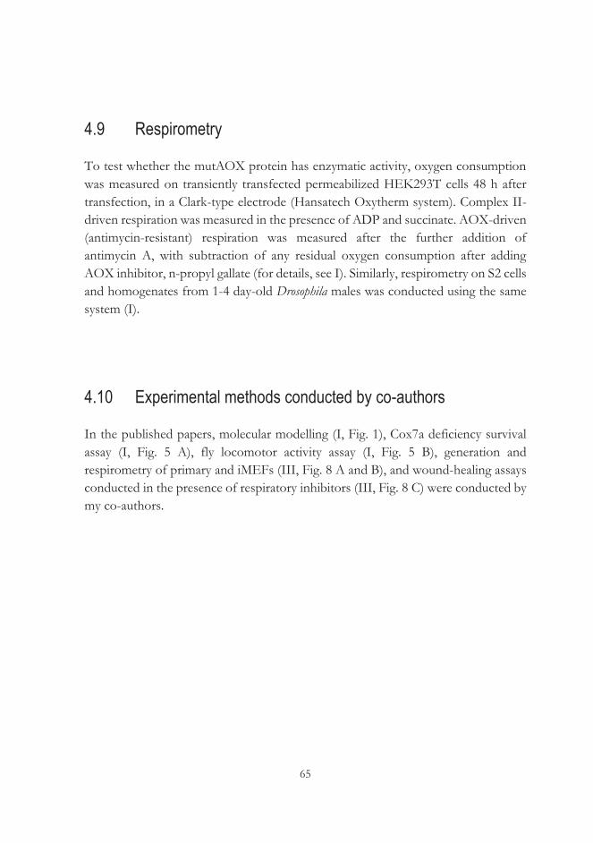
65
4.9 Respirometry
To test whether the mutAOX protein has enzymatic activity, oxygen consumption was measured on transiently transfected permeabilized HEK293T cells 48 h after transfection, in a Clark-type electrode (Hansatech Oxytherm system). Complex II-driven respiration was measured in the presence of ADP and succinate. AOX-driven (antimycin-resistant) respiration was measured after the further addition of antimycin A, with subtraction of any residual oxygen consumption after adding AOX inhibitor, n-propyl gallate (for details, see I). Similarly, respirometry on S2 cells and homogenates from 1-4 day-old Drosophila males was conducted using the same system (I).
4.10 Experimental methods conducted by co-authors
In the published papers, molecular modelling (I, Fig. 1), Cox7a deficiency survival assay (I, Fig. 5 A), fly locomotor activity assay (I, Fig. 5 B), generation and respirometry of primary and iMEFs (III, Fig. 8 A and B), and wound-healing assays conducted in the presence of respiratory inhibitors (III, Fig. 8 C) were conducted by my co-authors.

66
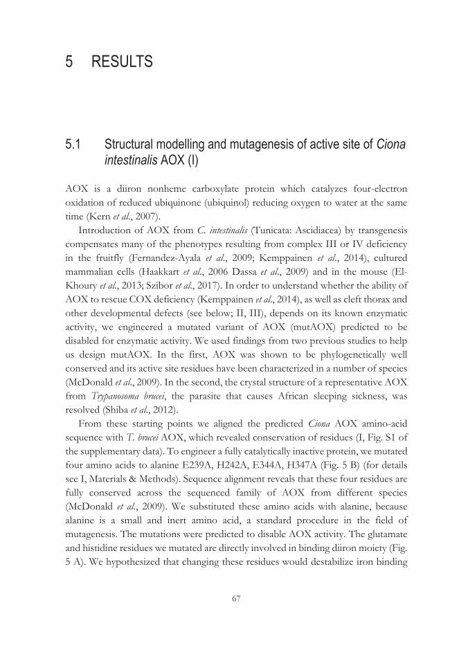
67
5 RESULTS
5.1 Structural modelling and mutagenesis of active site of Ciona intestinalis AOX (I)
AOX is a diiron nonheme carboxylate protein which catalyzes four-electron oxidation of reduced ubiquinone (ubiquinol) reducing oxygen to water at the same time (Kern et al., 2007).
Introduction of AOX from C. intestinalis (Tunicata: Ascidiacea) by transgenesis compensates many of the phenotypes resulting from complex III or IV deficiency in the fruitfly (Fernandez-Ayala et al., 2009; Kemppainen et al., 2014), cultured mammalian cells (Haakkart et al., 2006 Dassa et al., 2009) and in the mouse (El-Khoury et al., 2013; Szibor et al., 2017). In order to understand whether the ability of AOX to rescue COX deficiency (Kemppainen et al., 2014), as well as cleft thorax and other developmental defects (see below; II, III), depends on its known enzymatic activity, we engineered a mutated variant of AOX (mutAOX) predicted to be disabled for enzymatic activity. We used findings from two previous studies to help us design mutAOX. In the first, AOX was shown to be phylogenetically well conserved and its active site residues have been characterized in a number of species (McDonald et al., 2009). In the second, the crystal structure of a representative AOX from Trypanosoma brucei, the parasite that causes African sleeping sickness, was resolved (Shiba et al., 2012).
From these starting points we aligned the predicted Ciona AOX amino-acid sequence with T. brucei AOX, which revealed conservation of residues (I, Fig. S1 of the supplementary data). To engineer a fully catalytically inactive protein, we mutated four amino acids to alanine E239A, H242A, E344A, H347A (Fig. 5 B) (for details see I, Materials & Methods). Sequence alignment reveals that these four residues are fully conserved across the sequenced family of AOX from different species (McDonald et al., 2009). We substituted these amino acids with alanine, because alanine is a small and inert amino acid, a standard procedure in the field of mutagenesis. The mutations were predicted to disable AOX activity. The glutamate and histidine residues we mutated are directly involved in binding diiron moiety (Fig. 5 A). We hypothesized that changing these residues would destabilize iron binding
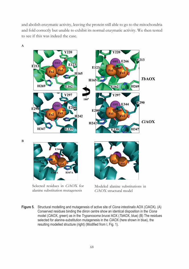
68
and abolish enzymatic activity, leaving the protein still able to go to the mitochondria and fold correctly but unable to exhibit its normal enzymatic activity. We then tested to see if this was indeed the case.
A
B
Figure 5. Structural modelling and mutagenesis of active site of Ciona intestinalis AOX (CiAOX). (A) Conserved residues binding the diiron centre show an identical disposition in the Ciona model (CiAOX, green) as in the Trypanosoma brucei AOX (TbAOX, blue) (B) The residues selected for alanine-substitution mutagenesis in the CiAOX (here shown in blue), the resulting modelled structure (right) (Modified from I, Fig. 1).
Selected residues in CiAOX for alanine substitution mutagenesis
Modeled alanine substitutions in CiAOX structural model
69
5.2 MutAOX is stably expressed in mammalian cells and flies (I)
To study the properties of mutAOX when expressed, I first verified the expression of the mutAOX construct by transient transfection into cultured HEK293T cells. Based on Western blotting (Fig. 6 A), mutAOX protein was the same size and expressed at comparable levels to wild-type (wt) AOX. Next, the mutAOX transgene, under the control of the GAL4-dependent UAS promoter, was introduced into the Drosophila genome by targeted insertion (for details see I, Materials & Methods). Control lines were created, containing wt AOX (described as 'new' wt AOX line in I) and empty vector, inserted at the same site. Insertions were validated by PCR and sequencing. I then measured transgene expression directed by the ubiquitous daGAL4 driver, at both RNA and protein levels, using qRT-PCR (Fig. 6 B, C) and Western blotting (Fig. 6 D, E). In both females (Fig. 6 B) and males (Fig. 6 C), the expression of wt and mutAOX were similar at the RNA level, but 3-4 fold lower than AOX in the previously created transgenic lines, engineered by random P-element insertion ('old' wild-type AOX lines, I). At the protein level, mutAOX has slightly lower expression than wild-type AOX in both sexes, and expression was again less than in the previously created 'old' wt lines. (Fig. 6 D, E).
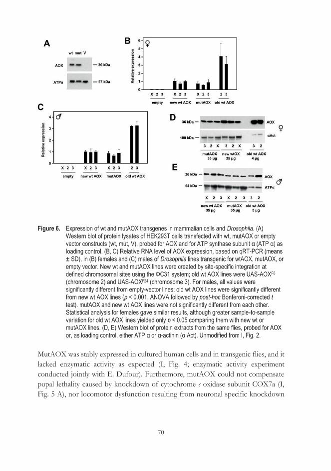
70
Figure 6. Expression of wt and mutAOX transgenes in mammalian cells and Drosophila. (A) Western blot of protein lysates of HEK293T cells transfected with wt, mutAOX or empty vector constructs (wt, mut, V), probed for AOX and for ATP synthase subunit α (ATP α) as loading control. (B, C) Relative RNA level of AOX expression, based on qRT-PCR (means ± SD), in (B) females and (C) males of Drosophila lines transgenic for wtAOX, mutAOX, or empty vector. New wt and mutAOX lines were created by site-specific integration at defined chromosomal sites using the ΦC31 system; old wt AOX lines were UAS-AOXF6 (chromosome 2) and UAS-AOXF24 (chromosome 3). For males, all values were significantly different from empty-vector lines; old wt AOX lines were significantly different from new wt AOX lines (p < 0.001, ANOVA followed by post-hoc Bonferoni-corrected t test). mutAOX and new wt AOX lines were not significantly different from each other. Statistical analysis for females gave similar results, although greater sample-to-sample variation for old wt AOX lines yielded only p < 0.05 comparing them with new wt or mutAOX lines. (D, E) Western blot of protein extracts from the same flies, probed for AOX or, as loading control, either ATP α or α-actinin (α Act). Unmodified from I, Fig. 2.
MutAOX was stably expressed in cultured human cells and in transgenic flies, and it lacked enzymatic activity as expected (I, Fig. 4; enzymatic activity experiment conducted jointly with E. Dufour). Furthermore, mutAOX could not compensate pupal lethality caused by knockdown of cytochrome c oxidase subunit COX7a (I, Fig. 5 A), nor locomotor dysfunction resulting from neuronal specific knockdown
71
of COX7a (I, Fig. 5 B; experiment conducted by C. Yalgin), confirming that the observed rescue depends on the enzymatic activity of AOX.
5.3 Expression of AOX rescues cleft thorax caused by tubGS/RU486 (II)
The GeneSwitch (GS) system allows temporal control of gene expression thanks to a modified GAL4 protein that is active only when the synthetic progesterone analogue, RU486, binds to the fused progesterone steroid receptor. GeneSwitch adds temporal control to the GAL4/UAS system (Yamada et al., 2017). Transgene expression was induced during development by the GS, under the control of a ubiquitously active α-tubulin promoter (tubGS), by feeding RU486 to mothers while laying eggs, and to larvae until pupariation. For experiments involving RU486 we used a diet rich in nutrients (Fernandez-Ayala et al., 2009) because RU486 is not detrimental to fly lifespan on high nutrient food (Yamada et al., 2017).
In the preliminary experiment it was observed that tubGS expression in the presence of RU486 causes cleft thorax which is rescued by AOX (Fig. 7 A). In a follow up study, I further observed, using the same system with induction by 10 μM RU486, extensive pupal lethality and a spectrum of additional malformations such as an enlarged tibia and femur, both fused together preventing movement of the fly in the majority of cases, bristle malformations, cleft abdomen, notches on anterior and posterior wing margins and externalized trachea (Fig. 8). These highly dysmorphic flies were alive nonetheless. For all the experiments throughout this thesis work, only vials with embryo numbers from 50-100 were analyzed to eliminate a crowded population which could affect resource availability during feeding, thus affecting the results.
Coexpression of AOX rescued the phenotype substantially, with over 50% of the eclosing progeny now showing no cleft, and less than 20% having severe cleft. Ndi1 targeted to mitochondria or GFP expressed in the cytoplasm did not rescue the phenotype. Nor did a single copy of AOX, when constitutively expressed under the α-tubulin promoter at a much lower level than when driven by tubGS. However, five doses of the tubAOX transgene gave rescue (II, Fig. 4 C) comparable with the UAS-AOX lines (which express AOX at a higher level than tubAOX and UAS-AOX7.1 fly lines). To test if rescue is due to catalytic activity of AOX we tested mutAOX in the assay. The enzymatically inactivated variant of AOX did not rescue any of the
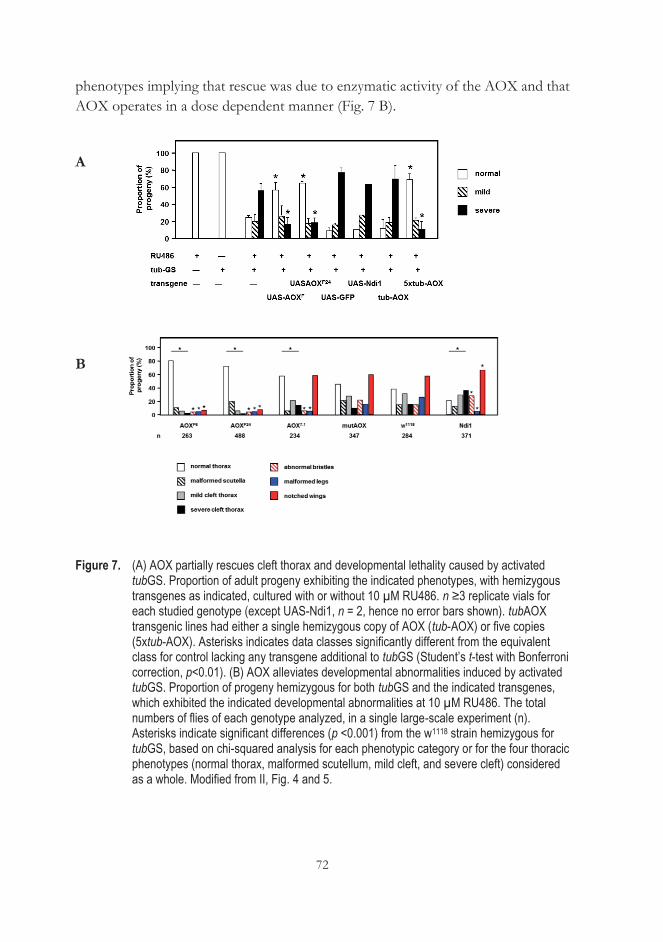
72
phenotypes implying that rescue was due to enzymatic activity of the AOX and that AOX operates in a dose dependent manner (Fig. 7 B).
A
B
Figure 7. (A) AOX partially rescues cleft thorax and developmental lethality caused by activated tubGS. Proportion of adult progeny exhibiting the indicated phenotypes, with hemizygous transgenes as indicated, cultured with or without 10 μM RU486. n ≥3 replicate vials for each studied genotype (except UAS-Ndi1, n = 2, hence no error bars shown). tubAOX transgenic lines had either a single hemizygous copy of AOX (tub-AOX) or five copies (5xtub-AOX). Asterisks indicates data classes significantly different from the equivalent class for control lacking any transgene additional to tubGS (Student’s t-test with Bonferroni correction, p<0.01). (B) AOX alleviates developmental abnormalities induced by activated tubGS. Proportion of progeny hemizygous for both tubGS and the indicated transgenes, which exhibited the indicated developmental abnormalities at 10 μM RU486. The total numbers of flies of each genotype analyzed, in a single large-scale experiment (n). Asterisks indicate significant differences (p <0.001) from the w1118 strain hemizygous for tubGS, based on chi-squared analysis for each phenotypic category or for the four thoracic phenotypes (normal thorax, malformed scutellum, mild cleft, and severe cleft) considered as a whole. Modified from II, Fig. 4 and 5.
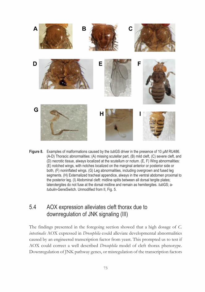
73
Figure 8. Examples of malformations caused by the tubGS driver in the presence of 10 μM RU486. (A-D) Thoracic abnormalities: (A) missing scutellar part, (B) mild cleft, (C) severe cleft, and (D) necrotic tissue, always localized at the scutellum or notum. (E, F) Wing abnormalities: (E) notched wings, with notches localized on the marginal anterior or posterior side or both, (F) noninflated wings. (G) Leg abnormalities, including overgrown and fused leg segments. (H) Externalized tracheal appendice, always in the ventral abdomen proximal to the posterior leg. (I) Abdominal cleft: midline splits between all dorsal tergite plates; laterotergites do not fuse at the dorsal midline and remain as hemitergites. tubGS; a-tubulin-GeneSwitch. Unmodified from II, Fig. 5.
5.4 AOX expression alleviates cleft thorax due to downregulation of JNK signaling (III)
The findings presented in the foregoing section showed that a high dosage of C. intestinalis AOX expressed in Drosophila could alleviate developmental abnormalities caused by an engineered transcription factor from yeast. This prompted us to test if AOX could correct a well described Drosophila model of cleft thorax phenotype. Downregulation of JNK pathway genes, or misregulation of the transcription factors
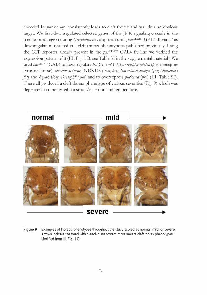
74
encoded by pnr or usp, consistently leads to cleft thorax and was thus an obvious target. We first downregulated selected genes of the JNK signaling cascade in the mediodorsal region during Drosophila development using pnrMD237 GAL4 driver. This downregulation resulted in a cleft thorax phenotype as published previously. Using the GFP reporter already present in the pnrMD237 GAL4 fly line we verified the expression pattern of it (III, Fig. 1 B; see Table S1 in the supplemental material). We used pnrMD237 GAL4 to downregulate PDGF and VEGF receptor related (pvr; a receptor tyrosine kinase), misshapen (msn; JNKKKK) hep, bsk, Jun-related antigen (Jra; Drosophila fos) and kayak (kay; Drosophila jun) and to overexpress puckered (puc) (III, Table S2). These all produced a cleft thorax phenotype of various severities (Fig. 9) which was dependent on the tested construct/insertion and temperature.
Figure 9. Examples of thoracic phenotypes throughout the study scored as normal, mild, or severe. Arrows indicate the trend within each class toward more severe cleft thorax phenotypes. Modified from III, Fig. 1 C.
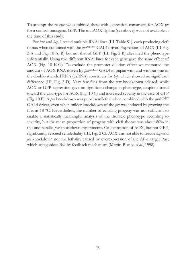
75
To attempt the rescue we combined these with expression constructs for AOX or for a control transgene, GFP. The mutAOX fly line (see above) was not available at the time of this study.
For bsk and hep, I tested multiple RNAi lines (III, Table S1), each producing cleft thorax when combined with the pnrMD237 GAL4 driver. Expression of AOX (III Fig. 2 A and Fig. 10 A, B) but not that of GFP (III, Fig. 2 B) alleviated the phenotype substantially. Using two different RNAi lines for each gene gave the same effect of AOX (Fig. 10 E-G). To exclude the promoter dilution effect we measured the amount of AOX RNA driven by pnrMD237 GAL4 in pupae with and without one of the double-stranded RNA (dsRNA) constructs for hep, which showed no significant difference (III, Fig. 2 D). Very few flies from the msn knockdown eclosed, while AOX or GFP expression gave no significant change in phenotype, despite a trend toward the wild-type for AOX (Fig. 10 C) and increased severity in the case of GFP (Fig. 10 F). A pvr knockdown was pupal semilethal when combined with the pnrMD237 GAL4 driver, even when milder knockdown of the pvr was induced by growing the flies at 18 °C. Nevertheless, the number of eclosing progeny was not sufficient to enable a statistically meaningful analysis of the thoracic phenotype according to severity, but the mean proportion of progeny with cleft thorax was about 80% in this and parallel pvr knockdown experiments. Co-expression of AOX, but not GFP, significantly rescued semilethality (III, Fig. 2 C). AOX was not able to rescue kay and jra knockdown nor the lethality caused by overexpression of the AP-1 target Puc, which antagonizes Bsk by feedback mechanism (Martín-Blanco et al., 1998).
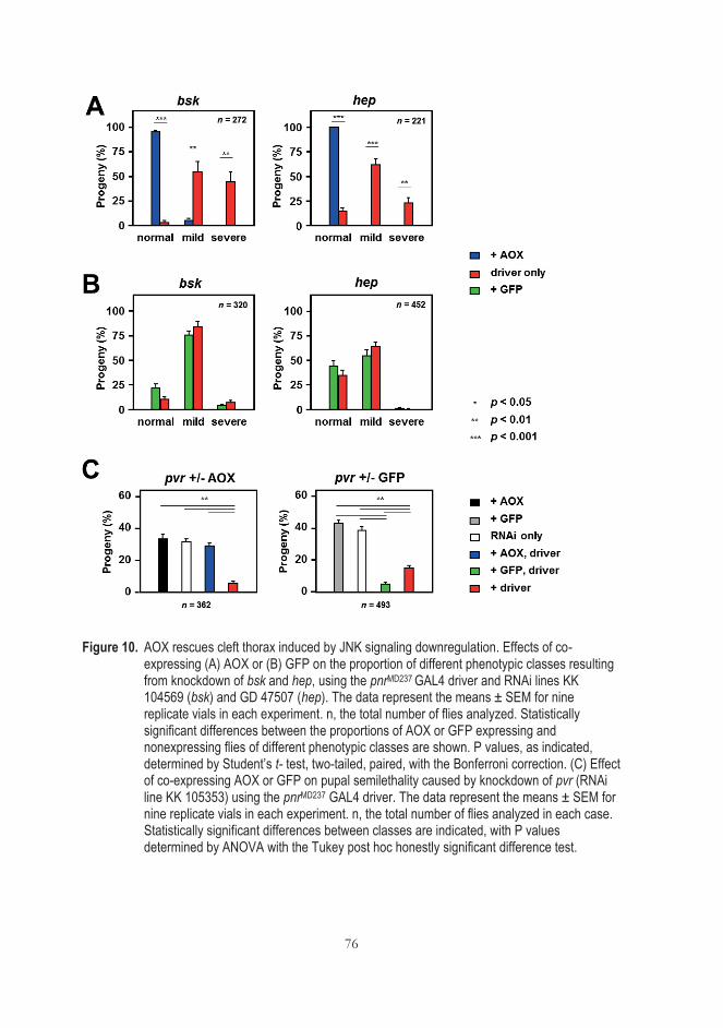
76
Figure 10. AOX rescues cleft thorax induced by JNK signaling downregulation. Effects of co-expressing (A) AOX or (B) GFP on the proportion of different phenotypic classes resulting from knockdown of bsk and hep, using the pnrMD237 GAL4 driver and RNAi lines KK 104569 (bsk) and GD 47507 (hep). The data represent the means ± SEM for nine replicate vials in each experiment. n, the total number of flies analyzed. Statistically significant differences between the proportions of AOX or GFP expressing and nonexpressing flies of different phenotypic classes are shown. P values, as indicated, determined by Student’s t- test, two-tailed, paired, with the Bonferroni correction. (C) Effect of co-expressing AOX or GFP on pupal semilethality caused by knockdown of pvr (RNAi line KK 105353) using the pnrMD237 GAL4 driver. The data represent the means ± SEM for nine replicate vials in each experiment. n, the total number of flies analyzed in each case. Statistically significant differences between classes are indicated, with P values determined by ANOVA with the Tukey post hoc honestly significant difference test.
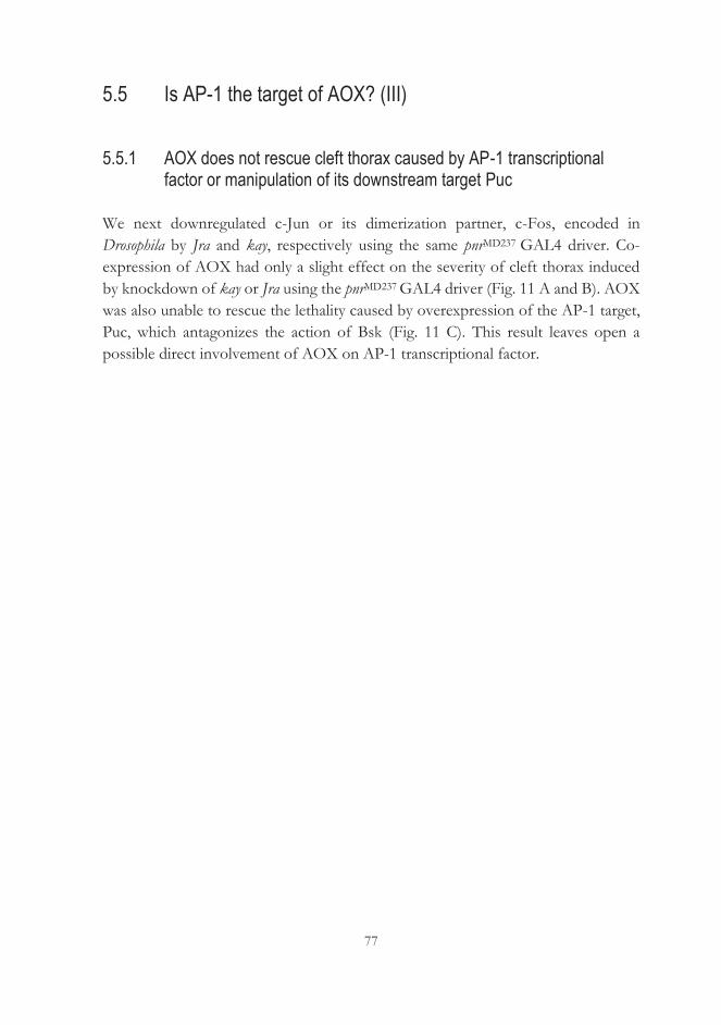
77
5.5 Is AP-1 the target of AOX? (III)
5.5.1 AOX does not rescue cleft thorax caused by AP-1 transcriptional factor or manipulation of its downstream target Puc
We next downregulated c-Jun or its dimerization partner, c-Fos, encoded in Drosophila by Jra and kay, respectively using the same pnrMD237 GAL4 driver. Co-expression of AOX had only a slight effect on the severity of cleft thorax induced by knockdown of kay or Jra using the pnrMD237 GAL4 driver (Fig. 11 A and B). AOX was also unable to rescue the lethality caused by overexpression of the AP-1 target, Puc, which antagonizes the action of Bsk (Fig. 11 C). This result leaves open a possible direct involvement of AOX on AP-1 transcriptional factor.
78
Figure 11. AOX does not rescue cleft thorax induced by altered expression of AP-1 or its target puc. (A, B) Effects of co-expressing AOX on phenotypic classes resulting from knockdown of kay (at 25 °C) and Jra (at 18 °C), using the pnrMD237 GAL4 driver and RNAi lines GD 6212 (kay) and KK 107997 (Jra) (A) and alternate RNAi lines GD 19512 (kay) and GD 10835 (Jra) (B). GD 10835 (Jra) construct is carried on chromosome X, two parallel crosses were required to test the effects of AOX expression in each sex, and statistical analysis was not meaningful in this case. The data represent the means ± SEM for 9 replicate vials in each experiment. n, total number of analyzed flies. Statistically significant differences between the proportions of AOX expressing and nonexpressing flies of different phenotypic classes are shown. P values determined by Student’s t-test, two-tailed, paired, with the Bonferroni correction. Jra knockdown using RNAi line KK 107997 was lethal at 25 °C and AOX did not rescue this lethality. (C) Effect of coexpressing AOX or GFP on pupal lethality caused by overexpression of puc driven by pnrMD237 GAL4 driver. All indicated progeny classes contained the overexpression construct, UAS-puc. The data represent the mean ±SEM for nine replicate vials in each experiment. n, the total number of analyzed flies.
79
5.5.2 AOX does not influence AP-1-dependent transcription in cultured cells
In attempt to reveal the mechanism of the AOX-mediated rescue of cleft thorax, I tested whether AOX expression can change the transcriptional activity of AP-1 in the Drosophila and mammalian system. I set up luciferase reporter-based assays for both S2 and HEK293T cells. I tested whether AOX expression in Drosophila S2 cells was able to alter AP-1-dependent transcription under different conditions of JNK pathway activation, using the reporter system developed by Chatterjee & Bohmann (2012). AOX did not influence AP-1 transcription in the conditions of basal and activated JNK pathway. JNK pathway activation was achieved using the pUAST-Hepact plasmid in combination with pAct-Gal4, which promotes pUAST-Hepact transcription of pUAST-Hepact by constitutive expression of Gal4. (Fig. 12 A-D).
To conduct a similar experiment in mammalian cells, I made use of an AP-1 Reporter HEK293 cell-line derivative (see III, Materials & Methods), here designated HEK AP-1. The cell line contains a stably integrated firefly luciferase gene under the control of AP-1-responsive elements. It was not possible to obtain consistent results with transiently transfected cells (data not presented). I therefore created stable AP-1 reporter cell lines, expressing AOX and mutAOX, using lentiviral constructs in which the transgene is co-expressed with GFP. Cells were cloned at limiting dilution. Different clones had highly variable degrees of AP-1 transcriptional activity, most likely reflecting the diversity of copy number and integration sites, as well as possible karyotypic instability. This is a limitation of the system.
However, I was able to replicate the findings from S2 cells in the mammalian model: AOX expressing clones showed no significant difference in AP-1 transcriptional activity compared with mutAOX expressing clones or parental cells (Fig. 12 E).
Using this model, I also tested the effects of inhibiting JNK pharmacologically with two inhibitors, SP600125 and AS601245 (JNK inhibitor V or inh V) (III). Whilst inh V decreased AP-1 transcription, SP600125 increased it, except when cells were pre-stimulated with PMA (Fig. 12 E), but again no differential effect was seen between AOX expressing, mutAOX expressing and control cells.
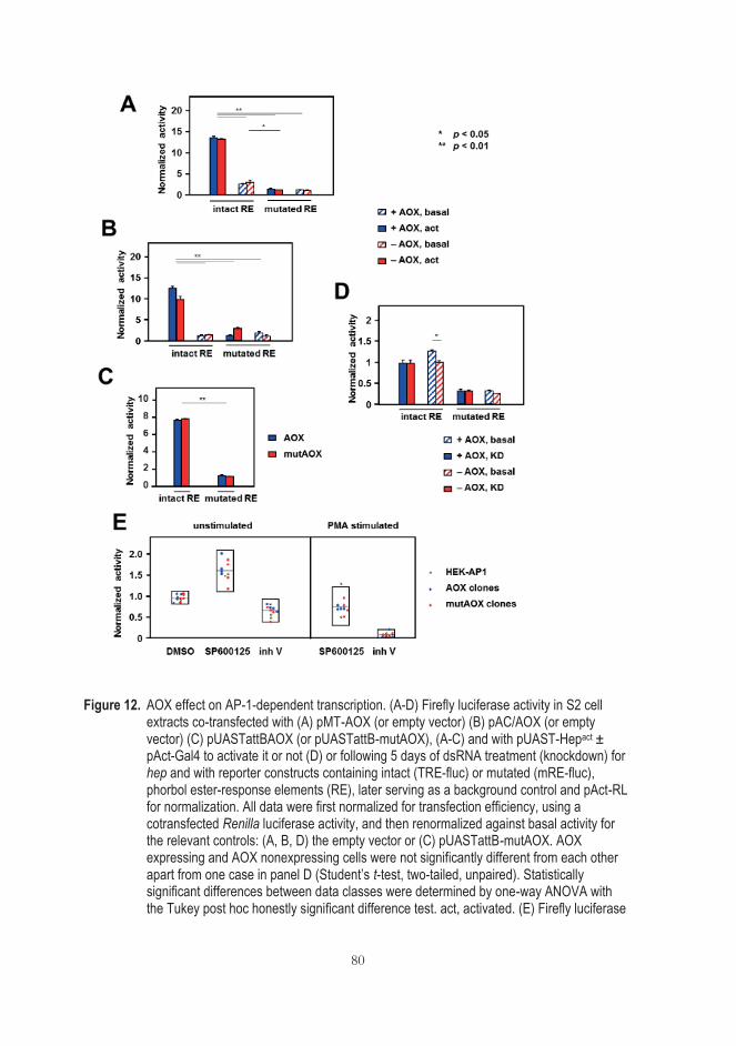
80
Figure 12. AOX effect on AP-1-dependent transcription. (A-D) Firefly luciferase activity in S2 cell extracts co-transfected with (A) pMT-AOX (or empty vector) (B) pAC/AOX (or empty vector) (C) pUASTattBAOX (or pUASTattB-mutAOX), (A-C) and with pUAST-Hepact ± pAct-Gal4 to activate it or not (D) or following 5 days of dsRNA treatment (knockdown) for hep and with reporter constructs containing intact (TRE-fluc) or mutated (mRE-fluc), phorbol ester-response elements (RE), later serving as a background control and pAct-RL for normalization. All data were first normalized for transfection efficiency, using a cotransfected Renilla luciferase activity, and then renormalized against basal activity for the relevant controls: (A, B, D) the empty vector or (C) pUASTattB-mutAOX. AOX expressing and AOX nonexpressing cells were not significantly different from each other apart from one case in panel D (Student’s t-test, two-tailed, unpaired). Statistically significant differences between data classes were determined by one-way ANOVA with the Tukey post hoc honestly significant difference test. act, activated. (E) Firefly luciferase
81
activity in extracts of reporter cells and HEK AP-1 derived clones transduced with lentiviral constructs expressing AOX or mutAOX. Treatments: 0.2% DMSO, 20 μM SP600125 or 20 μM inh V. Data normalized against the values for the corresponding non-treated cells (unstimulated) or for the corresponding cells treated with 8 nM PMA (stimulated). Box plots indicate the means and the 95% confidence intervals for each data set. Modified from III, Fig. 7.
5.5.3 AOX expression does not affect c-Jun phosphorylation at Ser 63/Ser 73
JNK-mediated phosphorylation of c-Jun at Ser 63 and Ser 73 is critical for fibroblast migration (Javelaud et al., 2003). To understand if AOX modulates phosphorylation of c-Jun at Ser 63 and Ser 73, the same set of JNK modulators were tested for their effects on c-Jun phosphorylation at these sites. This was done in iMEFs (Fig. 13 A), as well as in two other cell lines, the HEK AP-1 line used for transcriptional assays (Fig. 13 B) and human fibroblast line BJ-5ta (Fig. 13 C). Only SP600125 decreased the amount of phosphorylated c-Jun, whereas JNK inhibitor V instead increased it, as did PMA. The presence of AOX did not influence c-Jun phosphorylation at these sites in iMEFs (Fig. 13 A), although it potentiated the effect of SP600125 and inhibitor V in an AOX-expressing BJ-5ta cell clone (III, Fig. 13 C).
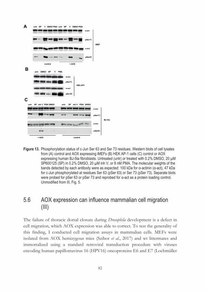
82
Figure 13. Phosphorylation status of c-Jun Ser 63 and Ser 73 residues. Western blots of cell lysates from (A) control and AOX expressing iMEFs (B) HEK AP-1 cells (C) control or AOX expressing human BJ-5ta fibroblasts. Untreated (untr) or treated with 0.2% DMSO, 20 μM SP600125 (SP) in 0.2% DMSO, 20 μM inh V, or 8 nM PMA. The molecular weights of the bands detected by each antibody were as expected: 100 kDa for α-actinin (α-act), 47 kDa for c-Jun phosphorylated at residues Ser 63 (pSer 63) or Ser 73 (pSer 73). Separate blots were probed for pSer 63 or pSer 73 and reprobed for α-act as a protein loading control. Unmodified from III, Fig. 5.
5.6 AOX expression can influence mammalian cell migration (III)
The failure of thoracic dorsal closure during Drosophila development is a defect in cell migration, which AOX expression was able to correct. To test the generality of this finding, I conducted cell migration assays in mammalian cells. MEFs were isolated from AOX hemizygous mice (Szibor et al., 2017) and wt littermates and immortalized using a standard retroviral transduction procedure with viruses encoding human papillomavirus 16 (HPV16) oncoproteins E6 and E7 (Lochmüller
83
et al., 1999; technical steps conducted by co-authors P.K. Dhandapani and L. Giordano). In these cells, AOX is expressed ubiquitously from an incorporated synthetic CAG promoter, the complete construct being targeted into the ubiquitously active Rosa26 locus (Szibor et al., 2017).
The drugs SP600125 and AS601245 have both been shown to decrease cell migration (Jarvelud et al., 2003; Cerbone et al., 2012) and were used here for that purpose. Both drugs are potent, reversible, and an (which express AOX at higher level than tubAOX and UAS-AOX7.1 fly lines)ATP-competitive inhibitor of JNK.
In iMEFs AOX increased the migration rate in all tested conditions in the wound healing essay, except when cells were treated with inh V. The scratch assay conducted on primary MEFs (at passage 6) revealed no difference in migration rate between AOX expressing and control MEFs (Fig. 14 B). All primary lines migrated much slower than iMEFs. Leading edge cells in AOX expressing fibroblasts were able to detach and scout the surface alone, making long protrusions. The wild-type iMEFs leading edge migrated as a whole (data not presented).
To test if the effect of AOX is dependent on cell-cell contact, a single cell migration assay was conducted. The migration speed of single-cell migration of AOX expressing iMEFs was also significantly greater than that of control iMEFs in this assay (Fig. 14 C) indicating that faster cell migration is an intrinsic property of the AOX-expressing iMEFs.
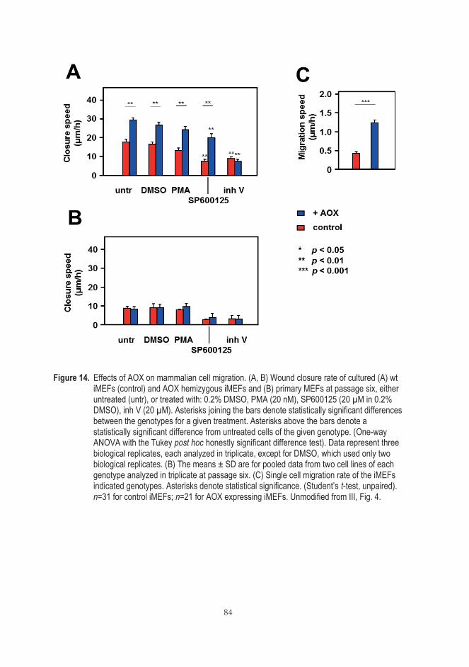
84
Figure 14. Effects of AOX on mammalian cell migration. (A, B) Wound closure rate of cultured (A) wt iMEFs (control) and AOX hemizygous iMEFs and (B) primary MEFs at passage six, either untreated (untr), or treated with: 0.2% DMSO, PMA (20 nM), SP600125 (20 μM in 0.2% DMSO), inh V (20 μM). Asterisks joining the bars denote statistically significant differences between the genotypes for a given treatment. Asterisks above the bars denote a statistically significant difference from untreated cells of the given genotype. (One-way ANOVA with the Tukey post hoc honestly significant difference test). Data represent three biological replicates, each analyzed in triplicate, except for DMSO, which used only two biological replicates. (B) The means ± SD are for pooled data from two cell lines of each genotype analyzed in triplicate at passage six. (C) Single cell migration rate of the iMEFs indicated genotypes. Asterisks denote statistical significance. (Student’s t-test, unpaired). n=31 for control iMEFs; n=21 for AOX expressing iMEFs. Unmodified from III, Fig. 4.
85
5.7 Antimycin A potentiates the migration of AOX-expressing cells (III)
To be able to understand further how AOX influences the migration we tested various metabolic effectors in conjunction with AOX. From various OXPHOS inhibitors, antioxidants and uncouplers, tested, only antimycin A, which binds to quinone reduction site of the cytochrome bc1 complex (Huang et al., 2005), gave differential effect. Antimycin A stimulated the migration of the AOX iMEFs but suppressing the migration of the MEFs (III, Fig. 8 B). To understand these findings we checked respiration in the cell lines tested (III, Fig. 8 C) to observe the effects of AOX under antimycin A. AOX had no significant effect on whole-cell respiration or on permeabilized cell respiration on complex I-, II-, and IV-linked substrates. However, in the presence of antimycin A, AOX enabled almost 80% of the uninhibited respiration rate in permeabilized cells, driven by succinate. This exceeded the measured rate of whole-cell respiration under uninhibited conditions. Capacity for AOX-mediated respiration was sufficient to maintain normal respiratory electron flow in iMEFs in the presence of antimycin A (Experiment conducted jointly with E. Dufour).

86
87
6 DISCUSSION
6.1 Overview
This work aimed to test whether transgenically expressed AOX can affect phenotypes related to the impairment of JNK signaling. We have found that expression of AOX was able to promote cell migration in three different models: in Drosophila, AOX expression corrected thorax defects caused by activated GS (II) and also when the JNK pathway was attenuated at several specific steps (III). It also promoted cell migration in AOX-expressing iMEFs (III). AOX was not able to correct migration defects when either of the Drosophila c-fos and c-jun homologues kay and Jra, respectively, were downregulated, as well as downstream targets of AP-1 such as pnr and puc. AOX was also unable to enhance the migration of primary MEFs. AOX had no effect on AP-1-dependent transcription in proliferating Drosophila (S2) or mammalian (HEK293-derived) cells. The migration of AOX expressing iMEFs compared with that of control iMEFs was differentially stimulated by antimycin A but not by other compounds that affect the mitochondrial respiratory chain, membrane potential, production of ATP or generation of ROS. The summary of the main results from this thesis are illustrated in Figure 15.
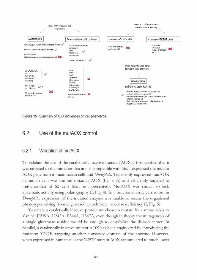
88
Figure 15. Summary of AOX influences on cell phenotype.
6.2 Use of the mutAOX control
6.2.1 Validation of mutAOX
To validate the use of the catalytically inactive mutated AOX, I first verified that it was targeted to the mitochondria and is compatible with life. I expressed the mutant AOX gene both in mammalian cells and Drosophila. Transiently expressed mutAOX in human cells was the same size as AOX (Fig. 6 A) and efficiently targeted to mitochondria of S2 cells (data not presented). MutAOX was shown to lack enzymatic activity using polarography (I, Fig. 4). In a functional assay carried out in Drosophila, expression of the mutated enzyme was unable to rescue the organismal phenotypes arising from engineered cytochrome c oxidase deficiency (I, Fig. 5).
To create a catalytically inactive protein we chose to mutate four amino acids to alanine: E239A, H242A, E344A, H347A, even though in theory the mutagenesis of a single glutamate residue would be enough to destabilize the di-iron center. In parallel, a catalytically inactive mutant AOX has been engineered by introducing the mutation Y297F, targeting another conserved domain of the enzyme. However, when expressed in human cells the Y297F mutant AOX accumulated to much lower
89
levels than the wild-type protein, suggesting that this particular mutant protein is unstable and rapidly degraded (P.K. Dhandapani, personal communication). We showed that targeted mutagenesis described in I resulted in stable but catalytically inactive protein.
6.2.2 Enzymatic activity of AOX is important for phenotypic rescue
MutAOX was not able to rescue locomotor defects in the fly arising from engineered cytochrome oxidase deficiency. MutAOX and the low expression AOX line created in (I), showed that rescue of locomotor activity in the COX 7a knockdown fly is dependent on the enzymatic activity of AOX and is related to the amount of AOX expression/activity. The lower dose of AOX was not able to rescue lethality induced by ubiquitous COX knockdown (data not shown), but was able to rescue locomotor defects from neuronal-specific knockdown to a lesser extent than high doses of AOX (I, Fig. 5 B). This narrows down the mechanistic explanations for the rescue of the COX 7a knockdown phenotype (discussed later). This result also validates the usefulness of the fly lines expressing mutant AOX created in (I).
In flies, the mutAOX transgene, introduced into the genome by targeted ΦC31 insertion, showed 3-4 fold lower expression at the RNA level than the AOX transgenic lines previously engineered by P-element insertion (Fig. 6 B, C). Therefore mutAOX Drosophila lines generated by ΦC31 insertion are not suitable as controls for those expressing AOX at a high level, since different levels of expression may influence the observed phenotype. This is clearly illustrated in the AOX rescue of GS-induced developmental abnormalities (II, Fig. 5), where the mutAOX-expressing flies were compared with transgenic flies generated in parallel, that expressed wild-type AOX from the same insertion site, i.e. at the same low level. In this case, as well as in the case of AOX rescue of locomotor deficit caused by cytochrome oxidase knockdown (I), lower expression of AOX gave an intermediate phenotype between that produced by wild-type AOX and that seen in non-transgenic control flies. mutAOX gave no such rescue at all. Implementing this control was not possible in the case of JNK knockdown, where I found that low-level AOX expression gave only a marginal or insignificant rescue of cleft thorax (unpublished data). It would thus be useful to create flies with high expression levels of mutAOX to test whether the observed rescue can truly be attributed to the catalytic function of the AOX enzyme. Nevertheless, the creation of ΦC31-engineered fly lines containing: AOX, mutAOX and empty vector at the exact same
90
position in the genome allows, in principle, for a rigorous experimental setup to understand if AOX-related phenotypes arise from the catalytic action of AOX, or are simply a hormetic effect due to the presence of a xenotopically expressed protein in the IMM. My findings leave open the possibility that AOX has a yet to be defined function or mode of regulation. The Drosophila lines that were engineered for this work are currently being used by other researchers who are addressing the dose dependence of AOX in other studies. However, I was not able to use them for the JNK knockdown study for the reasons indicated above, given that all the rescue data were obtained using AOX lines exhibiting a higher level of transgene expression (III).
6.2.3 Level of AOX activity is important for rescue of cleft thorax
In a second example, a single copy of AOX, when constitutively expressed under the α-tubulin promoter at a much lower level than when driven by tubGS, could not rescue the GS-induced cleft thorax phenotype. However, five doses of the tubAOX transgene rescued (II, Fig. 4 C) comparable with the UAS-AOX lines (which express AOX at higher level than tubAOX and UAS-AOX7.1 fly lines). Once again, I conclude that AOX provides rescue in a dose-dependent manner. This result is consistent with work on mammalian respiratory membranes where, with increasing AOX supplementation to the membranes, the rate of NADH oxidation increases substantially (Fedor & Hirst, 2018). It would be interesting to test the limits of this effect, and whether too high an expression level of AOX is also detrimental to the rescue of cleft thorax. This would imply a 'Goldilocks' effect, perhaps by regulating ROS, which are also known to operate in such a way (Reczek & Chandel, 2017).
As an alternative to the use of mutAOX as a control, it may be possible to use a small-molecule inhibitor of AOX in such studies. Such an inhibitor would have to be specific for the Ciona AOX and proven to be harmless to the model system(s) employed. Unfortunately, this is not the case for the known AOX inhibitors n-PG or SHAM.
91
6.3 tubGS induces developmental abnormalities in the fly
Transcription factors act in a coordinated manner to direct cell division, growth, and cell death throughout life. During embryonic development, the coordinated action of transcription factors is also needed to drive cell migration and body plan.
pnrMD237 GAL4 driven downregulation of JNK pathway components affects only the notum of the fly (Fig. 9), while GS under the control of the α-tubulin promoter is expressed ubiquitously in the fly. I found that it affects the development of various tissues and appendages, leading to cleft thorax, cleft abdomen, externalization of trachea, malformed bristles and notched wings (Fig. 8). A different GS driver, with a much lower and more restricted expression pattern (III, Fig. S2), affected bristle organization but only very subtly. The major dysmorphologies I observed with GS have been reported previously in a variety of mutants, often in combinations similar to this observation. Cleft thorax together with bent and misshaped sensory bristles and severely enlarged legs have been reported in Ultraspiracle mutants (Henrich et al., 1994), a dimerization partner of the ecdysone receptor and one of the key nuclear receptors which regulates fly development. GS is a steroid-activated chimeric GAL4 protein (GAL4-progesterone-receptor fusion protein) (Fig. 3 B). Thus, it is not surprising that GS can affect so many tissues, given that Drosophila has such a complex development. Legs are derived from the same ancestor cells as the wing discs in early embryogenesis (Cohen et al., 1993). Tracheal placodes and leg primordia arise from a common pool of cells in Drosophila, with differences in their fate controlled by the activation state of the wingless signaling pathway (Franch-Marro, 2006). Tracheal cells in Drosophila always arise in close proximity to the cells that are fated to give rise to the legs (Franch-Marro, 2006). The externalized trachea were always localized ventrally, close to posterior leg (Fig. 8 H).
This all implies that GS might act already in early embryogenesis to produce the observed malformations, for example by interacting with ecdysone receptor signaling or other signaling pathways that impinge on JNK signaling in early development. Cleft thorax together with missing or extra bristles has also been reported in mutants of the GATA transcription factor pannier (Heitzler et al., 1996). Cleft thorax is observed when the nuclear transcription factor Y (dNF-Y) is downregulated. This is due to a decreased level of Bsk, as dNF-Y has been found to bind the bsk (JNK) gene promoter region, which contains a CCAAT box (Yoshioka et al., 2008). The pair-rule transcription factor gene odd regulates the segment polarity genes en and wg in the early embryo and also restricts JNK signaling to medial edge cells of the peripodial membrane for proper thorax closure. Misregulation of odd
92
leads to thorax closure defects (Tripura et al., 2011). Bristle abnormalities similar to those that we observed are also reported in mutants of the gene u-shaped, which is a dimerization partner of pannier (Cubadda et al., 1997). A completely different way in which GS may affect development is by proteotoxicity, as discussed in a later section (6.5.2).
Whether and how the GS effect is related to JNK signaling still remains a puzzle and unraveling this relationship may shed light on the role of JNK signaling in Drosophila development more broadly. It could be that GS interacts with other transcriptional factors at the promoter regions of many developmental selector genes. A chromatin immunoprecipitation (ChIP) assay could be performed to understand whether GS binds in the Drosophila genome to the 5´ flanking regions of any of the genes known to influence development.
6.4 Model systems: pros and cons
6.4.1 Limitations of the fly model
The majority of fundamental biological mechanisms and pathways that control development are conserved across evolution between species (Jennings, 2011). This allows the use of simple model organisms, like yeast, nematodes, Drosophila or the zebrafish, to study complex molecular processes that occur in the human body. It was Charles W. Woodworth, an American entomologist, who first proposed the use of Drosophila melanogaster as a genetic model organism (Sturtevant, 1959). Since Thomas Hunt Morgan’s pioneering work in the 1930s, Drosophila has been extensively used in the study of animal development and behavior, neurobiology, human genetic diseases and drug discovery. We can test the effects of novel therapies by exploiting genetic tools available in this model, one of which is RNA interference (RNAi) which I used (III) to downregulate the expression of genes involved in thorax formation, leading to defective midline closure. I also used another tool, UAS-GAL4, to overexpress the AOX enzyme from C. intestinalis, and observed that this was able to reverse the induced thoracic defect (II, III).
However, Drosophila also has limitations. First, although the fly model helped me to understand which downregulated genes AOX can compensate for, and build a testable hypothesis, it was not possible to use it to elucidate the mechanism by which AOX provides this rescue. Techniques to isolate the specific cells where the JNK
93
pathway was downregulated and test my hypothesis are not currently available. This would require isolation of the limited cell population where JNK is active during the larval and pupal stage from the progeny containing all the needed transgenes for downregulation of the gene and rescue. There is currently no way to distinguish these larvae or pupae from controls, due to the specificity of the crosses and lack of markers. Validating my findings (III) in mammalian cells allowed me to test hypothetical AOX rescue mechanism(s) in a more experimentally malleable system that also has direct translational relevance to human disease. The fact that AOX was found to promote migration in both models, supports but does not prove that it acts by a common mechanism, whether by ROS dampening, heat production or something else.
6.4.2 Developmental disturbance in Drosophila resulting from the GS system
Drosophila is an invaluable model system in research because of its vast and easy to use genetic toolkit, providing over-expression, knockdown, and knockout of particular genes. Also, 75% of Drosophila genes have counterparts in the human genome (Pandey & Nichols, 2011). However, we are not always fully aware of the limitations of the tools available for research. To spatially and temporally control gene expression in a living organism is invaluable, but even an elegant system such as GeneSwitch in Drosophila has potential flaws that must be considered.
The use of the GS system assumes that it is inert in the fly. However, the findings reported here suggest otherwise. I used tubGS and daGS drivers to ubiquitously drive the expression of AOX transgenes during development (II). I identified specific developmental abnormalities arising from the use of tubGS in combination with RU486, including substantial pupal lethality already at 2.5 μM RU486, and from 10 μM dysmorphologies described above (Fig. 7 and 8). This suggests that the tubGS system itself induces such defects, by affecting transcription as hypothesized above. The chimeric GS protein should be inert, as there is no UAS sequence in the Drosophila genome to which it can bind. However, Liu & Lehmann (2008) showed that expression of high levels of the Gal4 protein in Drosophila can cause a genomic response, where modified expression was found, of many genes involved in development. Since GS is a modified Gal4 protein, it is perhaps unsurprising that it induces developmental defects (II), and conversely surprising that this has been previously ignored.
94
My work raises concerns about the use of the tubGS system. To avoid misinterpreting data it is advisable to use it in conjunction with control experiments that carefully monitor the effects of activated GS alone. We cannot rule out that there are other disturbances when using the system, even with other GS drivers and at lower RU486 concentrations than used here. Thus, all studies should control for the effect of the transcription factor plus RU486 alone, when drawing conclusions. There are many studies (e.g., Myllymäki & Rämet, 2013; Cho et al., 2014; Kim et al., 2015) that may have reached misleading conclusions due to a lack of such controls.
6.4.3 Limitations of the cell model
One intriguing issue that arose was the difference in sensitivity to AOX between immortalized and primary MEFs (Fig. 14 A, B). In fact, it was difficult to perform wound healing assays on primary MEFs. Primary MEFs were extremely sensitive to JNK inhibitors, which iMEFs handled without observable problems. The use of human fibroblasts was set aside during the study, after it was discovered that the AOX-transduced BJ-5ta cell line exhibited structurally impaired mitochondria with decreased complex I activity (data not presented). Further investigation using other cell-lines would be needed, to confirm the generality of the effects of AOX on cell migration, and the relationship to immortalization or transformation.
6.4.4 Limitations of the scratch-wounded confluent monolayer of fibroblasts
During re-epithelialisation of the epidermis after injury, cells at the leading edge are able to undertake cytoskeletal changes; extending lamellipodia, filopodia and polarizing in the direction of migration (Wager et al., 2017). Individual cells can take on a leading role in collective migration, forming a cell group termed ‘leader cells’ that have a specific phenotype and profile of gene expression independent of other cells (Wager et al., 2017; Khalil & Friedl, 2010; Riahi et al., 2012). In a simple 2D system, a technique which recapitulates cutaneous wound re-epithelialisation (Wager et al., 2017), I observed that the AOX-expressing wound healing monolayer has single 'leader cells', which were able to detach from other cells and explore the environment, making more protrusions than non-AOX control cells (data not presented). Computational methods could be applied to this data to extract different parameters than those used conventionally to describe collective cell migration. 2D and 3D environments are also different in terms of cell adhesion and migration
95
behaviors (Vu et al., 2015). In vivo, cells move in a complex environment defined by a combination of ECM proteins, proteoglycans, polysaccharides, growth factors, and signaling molecules (Frantz et al., 2010). Here, I tested the effect of AOX on the migration of cells in a purely 2D assay, stripped from any influence of the ECM. It would be interesting to test whether AOX can promote cell migration in models that better mimic the cellular environment in vivo, such as 3D scaffolds manufactured using biomaterials (Yang et al., 2017) or organoid models, e.g., the organotypic culture model of mammary branching morphogenesis (Ewald et al., 2008) before the wound healing is attempted in mice.
6.4.5 Limitations of using GFP as a control
Expression of an exogenous protein can perturb cell structure and function in unpredictable ways. This issue is inherent to any experiment, and is clearly demonstrated by my findings in the case of the GS system (II). This was why I set out to create the catalytically inactive variant of AOX (I), which unfortunately was not available at the start of the JNK study, and anyway proved to be an insufficiently strong expressor. Instead, I initially used GFP expressed in the cytoplasm as a routine control in all AOX expression studies (II, III), which has its own limitations. GFP is a structurally and functionally distinct protein from AOX, thus having different properties when expressed as a foreign protein in cells. Even though it is considered an inert protein in model systems it has been shown that wild-type GFP from the jellyfish, Aequorea victoria, quenches O2•− and has SOD-like activity by competing with cytochrome c for O2•− (Bou-Abdallah & Chasteen, 2006). However, since GFP as used in my experiments resides in the cytosol, it should not have access to mitochondrially generated superoxide, nor should it interact with or compete with cytochrome c. On the other hand, the fact that it is expressed in a separate cellular compartment than AOX, which is an inner mitochondrial membrane protein, means that it may have different non-specific effects on cellular protein homeostasis. Nevertheless, by modulating ROS or interacting with cytochrome c, AOX could influence apoptosis during development, which could impact the mechanism behind any rescue. AOX acts by oxidizing quinol and preventing cytochrome c from reduction, while GFP may restrict cytochrome c oxidation. Thus, they might be predicted to have opposite effects, which I indeed observed when attempting some instances of JNK-pathway rescue with AOX and GFP (III, Fig. 3 F). The JNK
96
signal-transduction pathway appears to be essential for the execution of apoptosis in response to several different stimuli (Davis et al., 2000). Apoptosis is needed in pupae where imaginal cells undergo apoptosis during thorax formation (Martín-Blanco et al., 2000 (Fig. 2). In the case of JNK knockdown AOX could be instrumental in 'replacing' JNK by promoting apoptosis, for which GFP is not so much a control as a parallel experiment.
6.5 Possible mechanisms of AOX rescue of cell migration defects
6.5.1 AOX rescue depends on its enzymatic activation
Based on my findings (I, II), the effects of AOX on Drosophila depend upon on its activation, at least in the case of locomotor impairment resulting from COX 7a knockdown (I) and the developmental abnormalities resulting from activated tubGS (Fig. 7). This is also most likely the case for rescue of JNK knockdown and promotion of cell migration in the wound-healing assay (III). Data obtained from many transgenic models, including human cells (Hakkaart et al., 2006; Dassa et al., 2009), mice (El-Khoury et al., 2013) and flies (Fernandez-Ayala et al., 2009) indicate that the AOX from C. intestinalis becomes activated only when needed, i.e. when the cytochrome pathway is blocked, and there is resulting over-reduction of the quinone pool. This is consistent with previous studies in other organisms (Hoefnagel & Wiskich, 1998; Castro-Guerrero et al., 2004), which showed that AOX activation occurs when ubiquinol accumulates to substantial levels due to respiratory chain inhibition or overload. This would mean that AOX activation during development, when the JNK pathway is inhibited or if GS is active could be due to mimicking this condition. However, not much is known about the activation of AOX in metazoans, leaving open the possibility that the downregulation of JNK signaling or the presence of activated GS leads to the enzymatic activation of AOX by an as yet unknown metabolic mechanism. What we know is that AOX activity in plants increases with cold (Stewart et al., 1990) or oxidative stress (Wagner, 1995). This further implies that there might be disturbances in the redox state of the developing cells of the thoracic epithelium due to JNK downregulation or GS activation, or in a cell monolayer responding to wounding.
97
6.5.2 AOX decreases ROS
It is known that AP-1 mobilizes oxidative defense and it is at the same time sensitive to oxidation and therefore protected by number of proteins. For example, redox factor 1 (Ref-1) is involved in reduction of the critical cysteine residues of Fos and Jun to maintain their DNA-binding activity following oxidative stress (Xanthoudakis & Curran, 1992). MBF1 also protects the critical cysteine residues from oxidation (Jindra et al., 2004) and stimulates AP-1 binding to DNA. AOX could be instrumental in protection of AP-1 from oxidation by generally decreasing mtROS when activated (Dogan et al., 2018). This, however needs to be tested by measuring ROS in the relevant cells, and assessing the status of the cysteine residues of AP-1 by redox proteomics. However, I could not see any change in AP-1 transcriptional readout in AOX-expressing S2 cells, nor in mammalian cells when JNK was modulated. This implies that the mechanism of AOX action is not due to ROS. Furthermore, this conclusion is supported by the lack of any effect of ROS modulators on the AOX-dependent stimulation of migration in the wound-healing assay (III, Fig. 8).
6.5.3 AOX is a thermogenic protein
Temperature is what Richard Feynman called the 'jiggling' of atoms (Sengupta & Garrity, 2013). Thermosensors are molecules whose temperature sensitivity serves as information about the thermal environment and trigger an appropriate physiological or behavioral response (Sengupta & Garrity, 2013). What that means is that temperature alters the configuration of the atoms of biomolecules, affecting their activity. Environmental temperatures and their changes can be read by nucleic acids, lipids and proteins, acting as thermosensors, which then regulate cellular processes, including transcription, protein stability and signal transduction (Sengupta & Garrity, 2013). Such processes could underlie the effects of AOX.
When AOX is active, electrons are passed to oxygen without proton pumping, so that the energy of the reaction is used to produce heat. It is well documented that AOX is thermogenic in plants under specific circumstances; for example in the flowers of Philodendron selloum and Symplocarpus foetidus, which can heat up to 35 °C above ambient temperature (Elthon & Mcintosh, 1987; Wagner et al., 2008; Seymour,
98
2000). We showed in a recent report that Ciona AOX expressed in Drosophila may be functionally thermogenic under specific physiological conditions, providing cold resistance to developing and adult flies (Saari et al., 2018). Consequently, if enzymatically active, AOX could increase the temperature of the developing thorax in Drosophila, which in turn may activate or by-pass JNK signaling by a variety of mechanisms, allowing migration to proceed. One such mechanism could simply be a thermogenic potentiation against oxidative stress, strengthening JNK signaling (Courtial et al., 2017). Increased phosphorylation of JNK was observed in human fibroblasts under thermal stress (Courtial et al., 2017). Another mechanism could involve heat-shock proteins. Heat shock protein 90 (Hsp90) has been reported to stabilize proapoptotic JNK signaling (Nieto-Miguel et al., 2007), whilst heat shock protein 70 (Hsp70) has been shown to promote cell migration by acting as a chaperone to deliver proteins to the leading edge (Boroughs et al., 2011).
As discussed above, a likely way in which GS influences development in Drosophila is via transcription. However, an alternate mechanism could be its accumulation in the cell causing proteotoxicity. The latter hypothesis is tentatively supported by the fact that I observed the same developmental abnormality, cleft thorax, when the human huntingtin protein with an extended polyglutamine repeat of 93 residues (Httex1p Q93) was ubiquitously expressed in Drosophila (unpublished data). A proteotoxic effect could potentially be mitigated by the fact that AOX being thermogenic activates the heat-shock response, which is instrumental in decreasing misfolded proteins, thus providing the rescue. It would be interesting to attempt AOX rescue of the cleft thorax induced by Httex1p Q93.
6.5.4 AOX affects ATP production
AOX activation would mean decreased electron flow through complex III and cytochrome c oxidase and thus, decreased ATP production. This might be exacerbated by increased heat production. These changes could provide the initial stimulus for JNK activation as a stress response. However, treatments that limit mitochondrial ATP production (oligomycin, FCCP and rotenone) did not have differential effect on the migration of AOX-expressing cells in the wound healing assay (III, Fig. 8) showing that ATP modulation is most likely not behind the mechanism of AOX action in that context. In the fly, AOX rescued cleft thorax induced by JNK knockdown, and in iMEFS restored migration when JNK was
99
blocked by SP600125. However, despite their apparent similarity, these two effects of AOX could be via different mechanisms.
6.5.5 Potential interplay between AOX and mitochondrially localized JNK
JNK functions as a signal transducer that conveys cytosolic oxidative stress signals to both the nucleus and mitochondria. JNK phosphorylates c-Jun, p-53, Elk-1, ATF-2 (Johnson & Nakamura, 2007; Weston & Davis, 2007) as well as Bax protein, promoting apoptosis (Bong-Jo et al., 2006). JNK translocates to mitochondria and, by facilitating cytochrome c and SMAC release from the intermembrane space, it can induce apoptosis (Aoki et al., 2002). JNK also phosphorylates pyruvate dehydrogenase (PDH). It has been proposed that JNK kinase may trigger either a phosphorylation cascade across mitochondrial membranes involving unidentified intermediates, or activate a second messenger, such as Ca2+, leading to the activation of PDH (Zhou et al., 2008).
Mitochondria are sources of H2O2 and NO (Cadenas & Davies 2000; Poderoso et al. 2000). Antimycin A increases H2O2 production due to its action on complex III, which in turn results in the translocation of JNK into the mitochondria (Hanawa et al., 2008). The potentiation of cell migration in AOX-expressing iMEFs, but not in the control iMEFs suggests that proximity of JNK when complex III is blocked creates conditions for the AOX to promote cell migration.
6.5.6 AOX and IMM shape
The shape of the inner mitochondrial membrane influences mitochondrial function (Mannella, 2006). For example ATP synthase contributes to the curving of the IMM as result of its oligomerization (Paumard et al., 2002; Strauss et al., 2008). Optic atrophy 1 (OPA1) protein is anchored to the IMM and is another protein involved in mitochondrial shape which affects its functions (Campello & Scorrano, 2010; Belenguer & Pellegrini, 2013). New mitochondria-shaping proteins influencing their function continue to be discovered.
The presence in the IMM of the Ciona AOX protein, which presumably functions as a dimer, might affect inner membrane organization which could have various bioenergetic implications. Altered IMM topology might affect mitochondrial signaling leading to the activation of JNK or of a parallel pathway. However, whilst
100
this might be a plausible mechanism to account for the promotion of iMEF migration by AOX, it cannot explain why AOX corrects cleft thorax and other developmental abnormalities caused by tubGS plus RU486, since this requires the enzymatic activity of AOX, as shown by the mutAOX control (II).
6.6 Potential use of AOX in therapy
Despite sequence conservation, AOXs of different species exhibit considerable diversity in enzymatic properties (May et al., 2017). How Ciona AOX is regulated is still unclear, and there is insufficient data on animal AOX in general to address this issue (McDonald et al., 2009). AOX expressed in animals possess a unique C-terminal of unknown function (McDonald et al., 2009), but also lack an N-terminal cysteine residue which is important for enzyme regulation in plants (McDonald et al., 2009). In plants, AOX protein is stress-regulated (as well as being tissue-specific): for example, it is dramatically increased within 5 hours of addition of antimycin A in tobacco (Vanlerberghe & Mclntosh, 1992). Our own recent work shows that AOX is induced in Ciona by hypoxia and sulfide exposure (Saari et al., 2018). Obviously, such regulation is lost in transgenic models, unless it operates post-transcriptionally. Conversely, the inference that AOX is activated in specific contexts in Drosophila development and in migrating mammalian cells may provide a handle on understanding exactly how and by what the enzyme is activated metabolically, which could help assess better how to use or engineer it therapeutically. In this context, it would be illuminating to test AOXs from other organisms (e.g., plants, fungi or trypanosomes) in the same assays used here.
A similar type of epithelial sheet movement to that seen in thoracic closure in Drosophila occurs in ventral closure in C. elegans, and in neural tube closure, embryonic wound healing and epiboly in vertebrates (Harden, 2002). In addition, key events in cancer progression show remarkable similarities with wound healing (Chang et al., 2004), suggesting that cancer involves deregulation of normal wound healing processes (Dauer et al., 2005). DNA microarray analyses have shown that the gene expression pattern of healing skin resembles that of malignant tumors (Iyer et al., 1999; Chang et al.,2004). Wounds which do not heal could be a risk factor for malignant transformation (Schäfer & Werner, 2008) due to specific changes in gene expression (Chang et al., 2004). This strengthens the idea proposed by Dvorak (1996) that “tumor stroma generation is wound healing gone awry.” Accelerated wound
101
healing is desirable, e.g., in victims of burns, and in people with slow-healing skin lesions as seen in diabetics (Greenhalgh, 2003) or following surgery, At the same time we do not want wound healing to go awry, promoting tumor formation.
Once the mechanism is more clearly established, a follow-up study would be to test AOX in a mouse wound-healing model (Dunn et al., 2013), aiming eventually to apply the knowledge in human diseases such as chronic wounds, where the underlying biology is still not well understood (Nuutila et al., 2014). Translating between models is appropriate in this case because the molecular machinery and mechanisms driving wound healing resemble those found in tissue fusion events during animal development (Kurosaka & Kashina, 2008), including dorsal and thoracic closure in the Drosophila embryo. Based on my findings, AOX could also be applied therapeutically in humans to prevent developmental abnormalities; however, its use in any application could also promote tumor invasion. The outcome may depend on the specific context of where, when and how much AOX is being expressed. AOX may therefore prove most useful as a tool to elucidate the underlying processes and ensure a safe outcome in a wide variety of interventions.

102
103
7 CONCLUSIONS
This is the first report that xenotopic expression of AOX remedies developmental abnormalities that occur without any documented mitochondrial respiratory chain defects. That itself raises the intriguing question of how alterations to nuclear-receptor and JNK signaling may influence mitochondria and vice versa. Using data obtained from two tested models: thorax closure in Drosophila and wound healing in mammalian cells, I am able to propose that mitochondria impact collective cell migration, specifically involving the respiratory chain at the level of complex III and/or IV. Perturbation of the mitochondrial RC affects signaling pathways which control cell migration. The importance of this discovery which combines in vivo model organism experiments and in vitro cell culture should lead to further studies investigating the mechanism whereby mitochondrial respiratory function affects cell migration and other developmental programs. My observation that AOX influences the migration of iMEFs, which are highly dependent on glycolysis, but not that of primary MEFs, suggests a new dimension in considering the links between mitochondrial metabolism and cancer metastasis. Other important questions arising from my work concern the role of ROS and mitochondrial heat production in regulating the intracellular signals responsible for lamellipodia formation, actin cytoskeleton remodeling and focal adhesion turnover. AOX is believed to act, indirectly and perhaps also directly, as a scavenger of mitochondrially generated ROS; but it is also thermogenic. Although a unifying hypothesis would be attractive, there could be different modes of action of AOX affecting cell migration in these different models. A temperature effect could operate in one case, but ROS modulation in another. Using an omics approach could give insight into possible mechanisms of AOX action which can then be subjected to more rigorous experimental testing. Omics could also be used to probe how GS causes such a wide spectrum of malformations in Drosophila, and how this relates to interference with transcription, cell signaling or proteotoxic stress.
The development of in vivo thermal probes for use in Drosophila would enable measuring the temperature in the pupa during thorax formation. Indirectly we could test expression of the heatshock proteins, some of which are known to promote cell migration, e.g., Hsp70 binds the protein cross-linking enzyme tissue

104
transglutaminase (tTG) localizing it to the leading edge in cancer cell migration. (Boroughs et al., 2011). To test the involvement of ROS, we could attempt the rescue of the cleft thorax with catalase or expose flies to agents such as mitoquinone mesylate (MitoQ) or N-acetyl cysteine (NAC).
A full understanding of the mechanism whereby AOX impacts development can potentially enable the design of new treatments for tissue injuries, metastatic tumors and congenital midline closure defects, as well as more 'conventional' effects of mitochondrial dysfunction.

105
8 ACKNOWLEDGEMENTS
This thesis work was carried out at the Faculty of Medicine and Health Technology at the University of Tampere. I want to express gratitude towards the Doctoral Programme in Medicine and Life Sciences and the Finnish Cultural Foundation (grant from Vilho Rossin Fund) for funding my doctoral studies. I wish to acknowledge my co-authors for their contribution to this work. I thank Venk Mallikarjun, PhD and Troy Faithfull, MSc for revising the language of my thesis. I would also like to thank Laura Vesala, PhD, Tiina Salminen, PhD, Suvi Vartiainen, PhD and Ilkka Vartiainen, MA for the help with translation of the abstract to Finnish language.
I wish to express my gratitude to the external reviewers of my thesis, Professor Mirka Uhlirova and Professor Navdeep Chandel, who both provided me with valuable comments and criticism.
I wish to thank Professor Rafael Garesse for agreeing to act as the opponent in the public defense of this dissertation.
I wish to express my ultimate gratitude to Professor Howard Jacobs for his mentorship and for providing a nurturing atmosphere for becoming a scientist. I want to thank him for introducing me to the power of networking in science, for pushing me to pursue my own ideas and adjust to failures when the data did not match expectations. Mainly, I want to thank him for giving me a difficult project to pursue. For me, the PhD experience was akin to a military camp. A lot of hard work and discipline. I felt like an SAS recruit, constantly being tested to quit the challenge with each failed experiment. Alongside receiving encouragement to persevere, I felt I was being given training to become a top level scientist prepared for the world stage.
I thank all the Howylab members for creating a truly unique and warm working environment. I especially thank Suvi Vartiainen, PhD for practical help in surviving Finland and valuable friendship. I also thank Tea Tuomela, BSc for sharing her vast Drosophila knowledge, Kia Kemppainen, PhD for introducing me to the project and her encouragement along the way, Giuseppe Cannino, PhD and Professor Marcos Oliveira for showing me the tools to develop the research project which resulted in many publications. I thank Ines Anderl, PhD for introducing me to the world of
106
Drosophila immunology. I wish to thank my thesis committee members Alberto Sanz, PhD, Professor Tapio Visakorpi and Professor Dan Hultmark for their time to monitor my progress and critically guide the next phase of the work. I thank Professor Vesa Hytönen, whose doors were always open for discussions on how to create a perfect mutant AOX.
I thank Teemu Ihalainen, PhD and Outi Paloheimo, MSc for always being there to help out with the microscopy. I thank Outi for help with the illustrations in this thesis.
During my PhD work I was privileged to supervise several hard working and motivated students who contributed to the work of this thesis. I wish to thank Anikka Ketola, MSc, Amelia Mordas, MSc, Lyon Bruinsma, MSc, Henri Virtanen, MSc, Samuli Hartikainen, MSc and Arto Alatalo, MSc for their contribution.
I want to thank Professor Dmytro Gospodaryov and Eric Dufour, PhD for unconditional teaching. I admire their passion for science and drive for to understand basic biological questions, which is the only way to push science and the world forward.
I thank the Helamaa & Heiskanen Architects for the ARVO building where I spent long hours writing this thesis enjoying the noise of the researchers, but also the oft needed silence during this process. ARVO is a truly unique research environment.
I gratefully acknowledge the input and friendship of many colleagues from the BioMediTech institute in shaping ideas and their practical help. I thank Jack George, MSc, Laura Vesala, PhD, Suvi Kalliokoski, PhD, Laura Airaksinen, MSc, Juliana Cerqueira, MSc and Heidi Kontro, PhD for being tremendously supportive with the very challenging Helsinki-Tampere adventure when approaching the finish line.
I thank my parents and my sister for their unconditional love. I thank Johan who was observing the process of me becoming a scientist. At the time of writing Johan is surviving glioblastoma multiforme grade IV tumor. He would always say: “You need to finish your work because that’s where you belong, as a scientist in a lab with other scientists finding cures for the diseases.” Now, that is my next mission.
Tampere, January 2019
107
9 REFERENCES
Abate, C., Patel, L., Rauscher 3rd, F.J., Curran, T. (1990). Redox Regulation of
Fos and Jun DNA-Binding Activity in Vitro. Science, 49(4973), 1157-61. https://doi.org/10.1126/science.2118682
Abercrombie, M., Heaysman, J. E. M., & Pegrum, S. M. (1971). The locomotion
of fibroblasts in culture. IV. Electron microscopy of the leading lamella. Experimental Cell Research, 67(2), 359-67. https://doi.org/10.1016/0014-4827(71)90420-4
Abreu-Blanco, M. T., Watts, J. J., Verboon, J. M., & Parkhurst, S. M. (2012).
Cytoskeleton responses in wound repair. Cellular and Molecular Life Sciences 69 (15) 2469-83. https://doi.org/10.1007/s00018-012-0928-2
Agmon, E., Solon, J., Bassereau, P., & Stockwell, B. R. (2018). Modeling the
effects of lipid peroxidation during ferroptosis on membrane properties. Scientific Reports, 8(1). https://doi.org/10.1038/s41598-018-23408-0
Aitken, R. J., Jones, K. T., & Robertson, S. A. (2012). Reactive oxygen species
and sperm function-in sickness and in health 33(6), 1096-106. Journal of Andrology. https://doi.org/10.2164/jandrol.112.016535
Albury, M. S., Elliott, C., & Moore, A. L. (2010). Ubiquinol-binding site in the
alternative oxidase: Mutagenesis reveals features important for substrate binding and inhibition. Biochimica et Biophysica Acta, 1797(12), 1933-9. https://doi.org/10.1016/j.bbabio.2010.01.013
Alexeyev, M., Shokolenko, I., Wilson, G., & LeDoux, S. (2013). The maintenance
of mitochondrial DNA integrity - Critical analysis and update. Cold Spring Harbor Perspectives in Biology, 5(5), a012641.
https://doi.org/10.1101/cshperspect.a012641 Allen, R. G. (1991). Oxygen-Reactive Species and Antioxidant Responses during
Development: The Metabolic Paradox of Cellular Differentiation. Experimental Biology and Medicine, 196(2), 117-29.
108
https://doi.org/10.3181/00379727-196-43171A Allen, R. G. (1998). Oxidative stress and superoxide dismutase in development,
aging and gene regulation. Age, 21(2), 47-76. https://doi.org/10.1007/s11357-998-0007-7 Allen, R. G., & Balin, A. K. (1989). Oxidative influence on development and
differentiation: an overview of a free radical theory of development. Free Radical Biology & Medicine, 6(6), 631-61. https://doi.org/10.1016/0891-5849(89)90071-3
Allen, R. G., & Tresini, M. (2000). Oxidative stress and gene regulation. Free
Radical Biology & Medicine, 28(3), 463-99. https://doi.org/10.1016/S0891-5849(99)00242-7
Amaral, A., Lourenço, B., Marques, M., & Ramalho-Santos, J. (2013).
Mitochondria functionality and sperm quality. Reproduction, 146(5), 163-74. https://doi.org/10.1530/REP-13-0178
Amstad, P. A., Krupitza, G., & Cerutti, P. A. (1992). Mechanism of c-fos
Induction by Active Oxygen. Cancer Research, 52(14), 3952-60. Ananthakrishnan, R., & Ehrlicher, A. (2007). The forces behind cell movement.
International Journal of Biological Sciences, 3(5), 303-17. https://doi.org/10.7150/ijbs.3.303 Anzick, S.L., Kononen. J., Walker. R.L., Azorsa. D.O., Tanner. M.M., Guan.
X.Y., Sauter. G., Kallioniemi. O.P., Trent. J.M., Meltzer, P.S. (1997). AIB1, a steroid receptor coactivator amplified in breast and ovarian cancer. Science, 277(5328), 965-8. https://doi.org/10.1126/science.277.5328.965
Aoki, H., Kang, P. M., Hampe, J., Yoshimura, K., Noma, T., Matsuzaki, M., &
Izumo, S. (2002). Direct activation of mitochondrial apoptosis machinery by c-Jun n-terminal kinase in adult cardiac myocytes. The Journal of Biological Chemistry, 277(12), 10244-50. https://doi.org/10.1074/jbc.M112355200
Avery, S. V. (2011). Molecular targets of oxidative stress. Biochemical Journal,
434(2), 201-10. https://doi.org/10.1042/BJ20101695 Azad, M. B., Chen, Y., & Gibson, S. B. (2009).Regulation of Autophagy by
Reactive Oxygen Species (ROS): Implications for Cancer Progression and Treatment. Antioxidants & Redox Signaling 11(4):777-90.
109
http://doi.org/10.1089/ars.2008.2270 Bacman, S. R., Williams, S. L., Duan, D., & Moraes, C. T. (2012). Manipulation
of mtDNA heteroplasmy in all striated muscles of newborn mice by AAV9-mediated delivery of a mitochondria-targeted restriction endonuclease. Gene Therapy, 19(11), 1101-6. https://doi.org/10.1038/gt.2011.196
Bae, H. B., Zmijewski, J. W., Deshane, J. S., Tadie, J. M., Chaplin, D. D.,
Takashima, S., & Abraham, E. (2011). AMP-activated protein kinase enhances the phagocytic ability of macrophages and neutrophils. The FASEB Journal (12), 4358-68. https://doi.org/10.1096/fj.11-190587
Baehrecke, E. H. (1996). Ecdysone signaling cascade and regulation of
Drosophila metamorphosis. Archives of Insect Biochemistry and Physiology 33(3-4), 231-44. https://doi.org/10.1002/(SICI)1520-6327(1996)33:3/
4<231::AID-ARCH5>3.0.CO;2-V Bai, J., Uehara, Y., & Montell, D. J. (2000). Regulation of invasive cell behavior
by taiman, a Drosophila protein related to AIB1, a steroid receptor coactivator amplified in breast cancer. Cell, 103(7), 1047-58.
https://doi.org/10.1016/S0092-8674(00)00208-7 Baloh, R. H., Schmidt, R. E., Pestronk, A., & Milbrandt, J. (2007). Altered Axonal
Mitochondrial Transport in the Pathogenesis of Charcot-Marie-Tooth Disease from Mitofusin 2 Mutations. Journal of Neuroscience 27(2), 422-30. https://doi.org/10.1523/JNEUROSCI.4798-06.2007
Barbarulo, A., Iansante, V., Chaidos, A., Naresh, K., Rahemtulla, A., Franzoso,
G., … Bubici, C. (2013). Poly(ADP-ribose) polymerase family member 14 (PARP14) is a novel effector of the JNK2-dependent pro-survival signal in multiple myeloma. Oncogene, 32(36), 4231-42.
https://doi.org/10.1038/onc.2012.448 Bartolák-Suki, E., Imsirovic, J., Nishibori, Y., Krishnan, R., & Suki, B. (2017).
Regulation of mitochondrial structure and dynamics by the cytoskeleton and mechanical factors. International Journal of Molecular Sciences, 18(8), 1812. https://doi.org/10.3390/ijms18081812
Basson, A. M. (2012). Signaling in cell differentiation and morphogenesis. Cold
Spring Harbor Perspectives in Biology (4). https://doi.org/10.1101/cshperspect.a008151
110
Bastin, J., Aubey, F., Rötig, A., Munnich, A., & Djouadi, F. (2008). Activation of peroxisome proliferator-activated receptor pathway stimulates the mitochondrial respiratory chain and can correct deficiencies in patients’ cells lacking its components. Journal of Clinical Endocrinology and Metabolism, 3(4), 1433-41. https://doi.org/10.1210/jc.2007-1701
Bayer, S. B., Maghzal, G., Stocker, R., Hampton, M. B., & Winterbourn, C. C.
(2013). Neutrophil-mediated oxidation of erythrocyte peroxiredoxin 2 as a potential marker of oxidative stress in inflammation. FASEB Journal, 27(8), 3315-22. https://doi.org/10.1096/fj.13-227298
Beard, M. E., & Holtzman, E. (1987). Peroxisomes in wild-type and rosy mutant
Drosophila melanogaster. Proceedings of the National Academy of Sciences of the United States of America, 84(21), 7433-37.
Beira, J. V., & Paro, R. (2016). The legacy of Drosophila imaginal discs.
Chromosoma, 125(4), 573-92. https://doi.org/10.1007/s00412-016-0595-4 Belenguer, P., & Pellegrini, L. (2013). The dynamin GTPase OPA1: More than
mitochondria? Biochimica et Biophysica Acta - Molecular Cell Research, 1833(1), 176-83. https://doi.org/10.1016/j.bbamcr.2012.08.004
Benz, R. (1994). Permeation of hydrophilic solutes through mitochondrial outer
membranes: review on mitochondrial porins. Biochimica et Biophysica Acta, 1197(2), 167-196. https://doi.org/10.1016/0304-4157(94)90004-3
Bereiter-Hahn, J. (1990). Behavior of Mitochondria in the Living Cell. International
Review of Cytology, 122, 1-63. https://doi.org/10.1016/S0074-7696(08)61205-X Bereiter-Hahn, J., & Jendrach, M. (2010). Mitochondrial Dynamics. International
Review of Cell and Molecular Biology, 284, 1-65. https://doi.org/10.1016/S1937-6448(10)84001-8 Bereiter-Hahn, J., & Voth, M. (1994). Dynamics of Mitochondria in Living Cells:
Shape Changes, Dislocations, Fusion, and Fission of Mitochondria. Microscopy Research and Technique, 27(3), 198-219.
https://doi.org/10.1002/jemt.1070270303 Berthold, D. A., & Stenmark, P. (2003). Membrane-bound diiron carboxilate
proteins. Annual Review of Plant Biology, 54, 497-517. https://doi.org/10.1146/annurev.arplant.54.031902.134915
111
Boldogh, I. R., & Pon, L. A. (2007). Mitochondria on the move. Trends in Cell
Biology 17(10), 502-10. https://doi.org/10.1016/j.tcb.2007.07.008 Boroughs, L. K., Antonyak, M. A., Johnson, J. L., & Cerione, R. A. (2011). A
Unique Role for Heat Shock Protein 70 and Its Binding Partner Tissue Transglutaminase in Cancer Cell Migration. The Journal of Biological Chemistry, 286, 37094-107. https://doi.org/10.1074/jbc.M111.242438
Borsello, T., Clarkel, P. G. H., Hirt, L., Vercelli, A., Repici, M., Schorderet, D. F.,
… Bonny, C. (2003). A peptide inhibitor of c-Jun N-terminal kinase protects against excitotoxicity and cerebral ischemia. Nature Medicine, 9, 1180-86. https://doi.org/10.1038/nm911
Bou-Abdallah, F., Chasteen, N. D., & Lesser, M. P. (2006). Quenching of
Superoxide Radicals by Green Fluorescent Protein. Biochimica et Biophysica Acta, 1760(11), 1690-5. https://doi.org/10.1016/j.bbagen.2006.08.014
Bough, K. J., Wetherington, J., Hassel, B., Pare, J. F., Gawryluk, J. W., Greene, J.
G., … Dingledine, R. J. (2006). Mitochondrial biogenesis in the anticonvulsant mechanism of the ketogenic diet. Annals of Neurology, 60(2), 223-35. https://doi.org/10.1002/ana.20899
Boutros, M., Paricio, N., Strutt, D. I., & Mlodzik, M. (1998). Dishevelled activates
JNK and discriminates between JNK pathways in planar polarity and wingless signaling. Cell, 94(1), 109-18. https://doi.org/10.1016/S0092-8674(00)81226-X
Bradford, M. M. (1976). A Rapid and Sensitive Method for the Quantitation of
Microgram Quantities of Protein Utilizing the Principle of Protein-Dye Binding. Analytical Biochemistry, 72(1-2), 248-54.
https://doi.org/10.1016/0003-2697(76)90527-3 Brand et al. (1994). Drosophila melanogaster: Practical Uses in Cell and Molecular Biology.
(Leslie Wilson Paul Matsudaira, Ed.) (1st ed.). Academic Press, Inc. Brieger, K., Schiavone, S., Miller, F. J., & Krause, K.-H. (2012). Reactive oxygen
species: from health to disease. Swiss Medical Weekly, 142, w13659. https://doi.org/10.4414/smw.2012.13659 Brandt, P. W., & Pappas, G. D. (1959). Mitochondria. I. Fine structure of the
complex patterns in the mitochondria of Pelomyxa carolinensis Wilson
112
(Chaos chaos L.). Journal of Cell Biology, 6(1), 85-90. https://doi.org/10.1083/jcb.6.1.91
Bruni, A., & Luciani, S. (1962). Effects of Atractyloside and Oligomycin on
Magnesium-stimulated Adenosine Triphosphatase and on Adenosine Triphosphate-induced Contraction of Swollen Mitochondria. Nature, 196, 578-80.
Bubici, C., & Papa, S. (2014). JNK signalling in cancer: In need of new, smarter
therapeutic targets. British Journal of Pharmacology, 171(1), 24-37. https://doi.org/10.1111/bph.12432 Burgess, S. M., Delannoy, M., & Jensen, R. E. (1994). MMM1 encodes a
mitochondrial outer membrane protein essential for establishing and maintaining the structure of yeast mitochondria. Journal of Cell Biology, 126(6), 1375-91. https://doi.org/10.1083/jcb.126.6.1375
Caddy, J., Wilanowski, T., Darido, C., Dworkin, S., Ting, S. B., Zhao, Q., … Jane,
S. M. (2010). Epidermal Wound Repair is Regulated by the Planar Cell Polarity Signaling Pathway. Developmental Cell, 19(1), 138-47.
https://doi.org/10.1016/j.devcel.2010.06.008 Cadenas, E., & Davies, K. J. A. (2000). Mitochondrial free radical generation,
oxidative stress, and aging. Free Radical Biology & Medicine, 29(3-4), 222-30. https://doi.org/10.1016/S0891-5849(00)00317-8
Campello, S., & Scorrano, L. (2010). Mitochondrial shape changes: Orchestrating
cell pathophysiology. EMBO Reports, 11(9), 678-84. https://doi.org/10.1038/embor.2010.115 Campos-Ortega, Jose A., Hartenstein, V. (1985). The Embryonic Development of
Drosophila melanogaster. Berlin, Heidelberg: Springer. Cannino, G., El-Khoury, R., Pirinen, M., Hutz, B., Rustin, P., Jacobs, H. T., &
Dufour, E. (2012). Glucose Modulates Respiratory Complex I Activity in Response to Acute Mitochondrial Dysfunction. The Journal of Biological Chemistry, 287(46), 38729-40.
https://doi.org/10.1074/jbc.M112.386060 Cannon, B. (2004). Brown Adipose Tissue: Function and Physiological
Significance. Physiological Reviews, 84(1), 277-359. https://doi.org/10.1152/physrev.00015.2003
113
Cannon, B., Shabalina, I. G., Kramarova, T. V., Petrovic, N., & Nedergaard, J.
(2006). Uncoupling proteins: A role in protection against reactive oxygen species-or not? Biochimica et Biophysica Acta, 1757(5-6), 449-58.
https://doi.org/10.1016/j.bbabio.2006.05.016 Carboni, S. (2004). AS601245 (1,3-Benzothiazol-2-yl (2-{[2-(3-pyridinyl) ethyl]
amino}-4 pyrimidinyl) Acetonitrile): A c-Jun NH2-Terminal Protein Kinase Inhibitor with Neuroprotective Properties. Journal of Pharmacology and Experimental Therapeutics, 310(1), 25-32.
https://doi.org/10.1124/jpet.103.064246 Cardoso, A. R., Kakimoto, P. A. H. B., & Kowaltowski, A. J. (2013). Diet-
Sensitive Sources of Reactive Oxygen Species in Liver Mitochondria: Role of Very Long Chain Acyl-CoA Dehydrogenases. PLoS ONE, 8(10), e77088. https://doi.org/10.1371/journal.pone.0077088
Castro-Guerrero, N. A., Krab, K., & Moreno-Sánchez, R. (2004). The Alternative
Respiratory Pathway of Euglena Mitochondria. Journal of Bioenergetics and Biomembranes, 36(5), 459-69.
https://doi.org/10.1023/B:JOBB.0000047328.82733.ef Cerbone, A., Toaldo, C., Minelli, R., Ciamporcero, E., Pizzimenti, S., Pettazzoni,
P., … Barrera, G. (2012). Rosiglitazone and AS601245 decrease cell adhesion and migration through modulation of specific gene expression in human colon cancer cells. PLoS ONE, 7(6), e40149.
https://doi.org/10.1371/journal.pone.0040149 Cereghetti, G. M., Costa, V., & Scorrano, L. (2010). Inhibition of Drp1-
dependent mitochondrial fragmentation and apoptosis by a polypeptide antagonist of calcineurin. Cell Death and Differentiation 17, 1785-94.
https://doi.org/10.1038/cdd.2010.61 Chakrabarty, S., & Martin, J. (2011). Postnatal refinement of proprioceptive
afferents in the cat cervical spinal cord. European Journal of Neuroscience, 33(9), 1656-66. https://doi.org/10.1111/j.1460-9568.2011.07662.x
Chambers, J. W., Cherry, L., Laughlin, J. D., Figuera-Losada, M., & Lograsso, P.
V. (2011). Selective inhibition of mitochondrial JNK signaling achieved using peptide mimicry of the sab kinase interacting motif-1 (KIM1). ACS Chemical Biology, 6(8), 808-18. https://doi.org/10.1021/cb200062a
114
Chang, H. Y., Sneddon, J. B., Alizadeh, A. A., Sood, R., West, R. B., Montgomery, K., … Brown, P. O. (2004). Gene expression signature of fibroblast serum response predicts human cancer progression: Similarities between tumors and wounds. PLoS Biology, 2(2), e7.
https://doi.org/10.1371/journal.pbio.0020007 Chatterjee, N., & Bohmann, D. (2012). A versatile φC31 based reporter system
for measuring AP-1 and NRF2 signaling in Drosophila and in tissue culture. PLoS ONE, 7(4), e34063. https://doi.org/10.1371/journal.pone.0034063
Chaudhuri, M., Ott, R. D., & Hill, G. C. (2006). Trypanosome alternative oxidase:
from molecule to function. Trends in Parasitology 22(10), 484-91. https://doi.org/10.1016/j.pt.2006.08.007
Chaudhuri, M., Ott, R. D., Saha, L., Williams, S., & Hill, G. C. (2005). The
trypanosome alternative oxidase exists as a monomer in Trypanosoma brucei mitochondria. Parasitology Research, 96(3), 178-83.
https://doi.org/10.1007/s00436-005-1337-3 Chen, Y., McMillan-Ward, E., Kong, J., Israels, S. J., & Gibson, S. B. (2007).
Mitochondrial electron-transport-chain inhibitors of complexes I and II induce autophagic cell death mediated by reactive oxygen species. Journal of Cell Science, 20(23), 4155-66. https://doi.org/10.1242/jcs.011163
Cheng, J. C., Klausen, C., & Leung, P. C. K. (2010). Hydrogen Peroxide Mediates
EGF-Induced Down-Regulation of E-Cadherin Expression via p38 MAPK and Snail in Human Ovarian Cancer Cells. Molecular Endocrinology, 24(8), 569-80. https://doi.org/10.1210/me.2010-0034
Cheng, W. Y., Tong, H., Miller, E. W., Chang, C. J., Remington, J., Zucker, R.
M., … Hofer, T. P. J. (2010). An integrated imaging approach to the study of oxidative stress generation by mitochondrial dysfunction in living cells. Environmental Health Perspectives, 118(7), 902-8.
https://doi.org/10.1289/ehp.0901811 Chinnery, P. F., & Hudson, G. (2013). Mitochondrial genetics. British Medical
Bulletin, 106(1), 135-59. https://doi.org/10.1093/bmb/ldt017 Chinnery, P. F., Thorburn, D. R., Samuels, D. C., White, S. L., Dahl, H. H. M.,
Turnbull, D. M., … Howell, N. (2000). The inheritance of mitochondrial DNA heteroplasmy: Random drift, selection or both? Trends in Genetics, 16(11), 500-5. https://doi.org/10.1016/S0168-9525(00)02120-X
115
Cho, K. H., Daubnerová, I., Park, Y., Zitnan, D., & Adams, M. E. (2014).
Secretory competence in a gateway endocrine cell conferred by the nuclear receptor βFTZ-F1 enables stage-specific ecdysone responses throughout development in Drosophila. Developmental Biology, 385(2), 253-62.
https://doi.org/10.1016/j.ydbio.2013.11.003 Chrétien, D., Bénit, P., Ha, H. H., Keipert, S., El-Khoury, R., Chang, Y. T., …
Rak, M. (2018). Mitochondria are physiologically maintained at close to 50 °C. PLoS Biology, 16(1), e2003992.
https://doi.org/10.1371/journal.pbio.2003992 Chu, C. T., Ji, J., Dagda, R. K., Jiang, J. F., Tyurina, Y. Y., Kapralov, A. A., …
Kagan, V. E. (2013). Cardiolipin externalization to the outer mitochondrial membrane acts as an elimination signal for mitophagy in neuronal cells. Nature Cell Biology, 15(10), 1197-205. https://doi.org/10.1038/ncb2837
Ciapponi, L., Jackson, D. B., Mlodzik, M., & Bohmann, D. (2001). Drosophila
Fos mediates ERK and JNK signals via distinct phosphorylation sites. Genes and Development, 15(12), 1540-53. https://doi.org/10.1101/gad.886301
Clark, K,M,, Bindoff, L.A., Lightowlers. R.N., Andrews. R.M., Griffiths. P.G.,
Johnson. M.A., Brierley. E.J., Turnbull, D.M. (1997). Reversal of a mitochondrial DNA defect in human skeletal muscle. Nature Genetics, 16(3), 222-4.
https://doi.org/10.1038/ng0797-222 Clarke, P., Meintzer, S. M., Wang, Y., Moffitt, L. A., Richardson-Burns, S. M.,
Johnson, G. L., & Tyler, K. L. (2004). JNK Regulates the Release of Proapoptotic Mitochondrial Factors in Reovirus-Infected Cells. Journal of Virology, 78(23), 13132-38. https://doi.org/10.1128/JVI.78.23.13132-13138.2004
Cohen, B., Simcox A. A., Cohen, S. M. (1993). Allocation of the thoracic imaginal
primordia in the Drosophila embryo. Development, 117, 597-608. Copeland, W. C., Wachsman, J. T., Johnson, F. M., & Penta, J. S. (2002).
Mitochondrial DNA alterations in cancer. Cancer Investigation, 20(4), 557-69. https://doi.org/10.1081/CNV-120002155
Courtial, L., Picco, V., Grover, R., Cormerais, Y., Rottier, C., Labbe, A., …
Ferrier-Pagès, C. (2017). The c-Jun N-Terminal kinase prevents oxidative
116
stress induced by UV and thermal stresses in corals and human cells. Scientific Reports, 7, 45713. https://doi.org/10.1038/srep45713
D’Autréaux, B., & Toledano, M. B. (2007). ROS as signalling molecules:
Mechanisms that generate specificity in ROS homeostasis. Nature Reviews Molecular Cell Biology, 8(10), 813-24. https://doi.org/10.1038/nrm2256
D’Souza, S. F., & Srere, P. A. (1983). Binding of citrate synthase to mitochondrial
inner membranes. The Journal of Biological Chemistry, 258(8), 4706-9. Dassa, E. P., Dufour, E., Gonçalves, S., Paupe, V., Hakkaart, G. A. J., Jacobs, H.
T., & Rustin, P. (2009). Expression of the alternative oxidase complements cytochrome c oxidase deficiency in human cells. EMBO Molecular Medicine, 1(1), 30-6. https://doi.org/10.1002/emmm.200900001
Dauer, D. J., Ferraro, B., Song, L., Yu, B., Mora, L., Buettner, R., … Haura, E.
B. (2005). Stat3 regulates genes common to both wound healing and cancer. Oncogene, 24(21), 3397-408. https://doi.org/10.1038/sj.onc.1208469
Daum, G., & Vance, J. E. (1997). Import of lipids into mitochondria. Progress in
Lipid Research, 36(2-3), 103-30. https://doi.org/10.1016/S0163-7827(97)00006-4 Davies, K. J. A. (2000). Oxidative stress, antioxidant defenses, and damage
removal, repair, and replacement systems. IUBMB Life, 50(4-5), 279-89. https://doi.org/10.1080/15216540051081010 Davis, R. J. (2000). Signal transduction by the JNK group of MAP kinases. Cell,
103(2), 239-52. https://doi.org/10.1016/S0092-8674(00)00116-1 Denissenko, P., Kantsler, V., Smith, D. J., & Kirkman-Brown, J. (2012). Human
spermatozoa migration in microchannels reveals boundary-following navigation. Proceedings of the National Academy of Sciences, 109(21), 8007-10. https://doi.org/10.1073/pnas.1202934109
Desai, S. P., Bhatia, S. N., Toner, M., & Irimia, D. (2013). Mitochondrial
localization and the persistent migration of epithelial cancer cells. Biophysical Journal, 104(9), 2077-88. https://doi.org/10.1016/j.bpj.2013.03.025
Di Mauro, S., & De Vivo, D. C. (1996). Genetic heterogeneity in Leigh syndrome.
Annals of Neurology, 40(1), 5-7. https://doi.org/10.1002/ana.410400104
117
Dixon, S. J., Lemberg, K. M., Lamprecht, M. R., Skouta, R., Zaitsev, E. M., Gleason, C. E., … Stockwell, B. R. (2012). Ferroptosis: An iron-dependent form of nonapoptotic cell death. Cell, 149(5), 1060-72. https://doi.org/10.1016/j.cell.2012.03.042
Dogan, S. A., Cerutti, R., Benincá, C., Brea-Calvo, G., Jacobs, H. T., Zeviani, M.,
… Viscomi, C. (2018). Perturbed Redox Signaling Exacerbates a Mitochondrial Myopathy. Cell Metabolism, 28(5), 764-75.
https://doi.org/10.1016/j.cmet.2018.07.012 Dotti, M. T., Manneschi, L., Malandrini, A., De Stefano, N., Caznerale, F., &
Federico, A. (1993). Mitochondrial dysfunction in Rett syndrome An ultrastructural and biochemical study. Brain & Development, 15(103). 103-6. https://doi.org/10.1016/0387-7604(93)90045-A
Dunn, L., Prosser, H. C. G., Tan, J. T. M., Vanags, L. Z., Ng, M. K. C., & Bursill,
C. A. (2013). Murine Model of Wound Healing. Journal of Visualized Experiments, 28(75) e50265. https://doi.org/10.3791/50265
Duronio, R. (1999). Establishing links between developmental signaling
pathways and cell-cycle regulation in Drosophila. Current Opinion in Genetics & Development, 9, 81-8. https://doi.org/10.1016/S0959-437X(99)80012-4
Edgar, B. A., & Lehner, C. F. (1996). Developmental Control of Cell Cycle
Regulators: A Fly’s Perspective. Science, 274(5293), 1646-52. https://doi.org/10.1126/science.274.5293.1646 Edmondson, D. E., Binda, C., Wang, J., Upadhyay, A. K., & Mattevi, A. (2009).
Molecular and mechanistic properties of the membrane-bound mitochondrial monoamine oxidases. Biochemistry, 48(20), 4220-30. https://doi.org/10.1021/bi900413g
El-Khoury, R., Dufour, E., Rak, M., Ramanantsoa, N., Grandchamp, N., Csaba,
Z., … Rustin, P. (2013). Alternative Oxidase Expression in the Mouse Enables Bypassing Cytochrome c Oxidase Blockade and Limits Mitochondrial ROS Overproduction. PLoS Genetics, 9(1), e1003182.
https://doi.org/10.1371/journal.pgen.1003182 El-Khoury, R., Kaulio, E., Lassila, K. A., Crowther, D. C., Jacobs, H. T., &
Rustin, P. (2016). Expression of the alternative oxidase mitigates beta-amyloid production and toxicity in model systems. Free Radical Biology and Medicine, 96, 57-66. https://doi.org/10.1016/j.freeradbiomed.2016.04.006
118
Elthon, T. E., & Mcintosh, L. (1987). Identification of the alternative terminal
oxidase of higher plant mitochondria (cyanide-resistant respiration/thermogenicity). Proceedings of the National Academy of Sciences of the United States of America, 84(23), 8399-403.
Emani, S. M., McCully, J. D. (2018). Mitochondrial transplantation: applications
for pediatric patients with congenital heart disease. Translational Pediatrics, 7(2), 169-75. https://doi.org/10.21037/tp.2018.02.02
Engel, R. H., & Evens, A. M. (2006). Oxidative stress and apoptosis: a new
treatment paradigm in cancer. Frontiers in Bioscience, 11, 300-12. Eriksson, L. (1987). Growth Hormone in Human Pregnancy: Maternal 24-hour
Serum Profiles and Experimental Effects of Continuous GH Secretion. Acta Obstetricia et Gynecologica Scandinavica, 68(147), 6-38.
https://doi.org/10.3109/00016348709156496 Eshraghi, A. A., & Van De Water, T. R. (2006). Cochlear implantation trauma
and noise-induced hearing loss: Apoptosis and therapeutic strategies. Anatomical Record - Part A Discoveries in Molecular, Cellular, and Evolutionary Biology, 288(4), 473-81. https://doi.org/10.1002/ar.a.20305
Etienne-Manneville, S., & Hall, A. (2002). Rho GTPases in Cell Biology. Nature,
420(6916), 629-35. https://doi.org/ 10.1038/nature01148 Ewald, A. J., Brenot, A., Duong, M., Chan, B. S., & Werb, Z. (2008). Collective
Epithelial Migration and Cell Rearrangements Drive Mammary Branching Morphogenesis. Developmental Cell, 14(4), 570-81.
https://doi.org/10.1016/j.devcel.2008.03.003 Eyers, C. E., Vonderach, M., Ferries, S., Jeacock, K., & Eyers, P. A. (2018).
Understanding protein-drug interactions using ion mobility-mass spectrometry. Current Opinion in Chemical Biology, 42, 167-76.
https://doi.org/10.1016/j.cbpa.2017.12.013 Faust, J. E., Verma, A., Peng, C., & Mcnew, J. A. (2012). An inventory of
peroxisomal proteins and pathways in Drosophila melanogaster. Traffic, 13(10), 1378-92. https://doi.org/10.1111/j.1600-0854.2012.01393.x
Fedor, J. G., & Hirst, J. (2018). Mitochondrial Supercomplexes Do Not Enhance
119
Catalysis by Quinone Channeling. Cell Metabolism, 28(3), 525-53. https://doi.org/10.1016/j.cmet.2018.05.024 Fernandez-Ayala, D. J. M., Sanz, A., Vartiainen, S., Kemppainen, K. K.,
Babusiak, M., Mustalahti, E., … Jacobs, H. T. (2009). Expression of the Ciona intestinalis Alternative Oxidase (AOX) in Drosophila Complements Defects in Mitochondrial Oxidative Phosphorylation. Cell Metabolism, 9(5), 449-60. https://doi.org/10.1016/j.cmet.2009.03.004
Fernandez-Souza, J. M., & Michelson, A. M. (1976). Variation of superoxide
dismutases during the development of the fruitfly Ceratitis capirata. Biochemical and Biophysical Research Communications, 73(2), 217-23. https://doi.org/10.1016/0006-291X(76)90696-3
Ferreirinha, F., Ballabio, A., & Rugarli, E. I. (2004). Axonal degeneration in
paraplegin-deficient mice is associated with abnormal mitochondria and impairment of axonal transport. The Journal of Clinical Investigation, 113(2), 231-42. https://doi.org/10.1172/JCI20138
Finkel, T. (2003). Oxidant signals and oxidative stress. Current Opinion in Cell
Biology, 15(2), 247-54. https://doi.org/10.1016/S0955-0674(03)00002-4 Flohé, L., Brigelius-Flohé, R., Saliou, C., Traber, M. G., & Packer, L. (1997).
Redox regulation of NF-kappa B activation. Free Radical Biology and Medicine, 22(6), 1115-26. https://doi.org/10.1016/S0891-5849(96)00501-1
Franch-Marro, X. (2006). Association of tracheal placodes with leg primordia in
Drosophila and implications for the origin of insect tracheal systems. Development, 133(5), 785-90. https://doi.org/10.1242/dev.02260
Frantz, C., Stewart, K. M., & Weaver, V. M. (2010). The extracellular matrix at a
glance. Journal of Cell Science, 123(24), 4195-420. https://doi.org/10.1242/jcs.023820 Friedl, P., & Gilmour, D. (2009). Collective cell migration in morphogenesis,
regeneration and cancer. Nature Reviews Molecular Cell Biology, 10(7), 445-57. https://doi.org/10.1038/nrm2720 Friedman, A., & Perrimon, N. (2007). Genetic Screening for Signal Transduction
in the Era of Network Biology. Cell, 128(2), 225-31. https://doi.org/10.1016/j.cell.2007.01.007
120
Friedmann Angeli, J. P., Schneider, M., Proneth, B., Tyurina, Y. Y., Tyurin, V. A., Hammond, V. J., … Conrad, M. (2014). Inactivation of the ferroptosis regulator Gpx4 triggers acute renal failure in mice. Nature Cell Biology, 16(12), 1180-91. https://doi.org/10.1038/ncb3064
Gadaleta, R. M., & Magnani, L. (2013). Nuclear receptors and chromatin: An
inducible couple. Journal of Molecular Endocrinology, 52(2), 137-49. https://doi.org/10.1530/JME-13-0170 Gao, M., Yi, J., Zhu, J., Minikes, A. M., Monian, P., Thompson, C. B., & Jiang,
X. (2019b). Role of Mitochondria in Ferroptosis. Molecular Cell, 73(2), 354-63.e3. https://doi.org/10.1016/j.molcel.2018.10.042
García-Ruiz C, Colell A, Morales A, Kaplowitz N., & Fernández-Checa, J. C.
(1995). Role of oxidative stress generated from the mitochondrial electron transport chain and mitochondrial glutathione status in loss of mitochondrial function and activation of transcription factor nuclear factor-kappa B: studies with isolated mitochondria and rat hepatocytes. Molecular Pharmacology, 48(5), 825-34.
Girnius, N., & Davis, R. J. (2017). JNK Promotes Epithelial Cell Anoikis by
Transcriptional and Post-translational Regulation of BH3-Only Proteins. Cell Reports, 21(7), 1910-21. https://doi.org/10.1016/j.celrep.2017.10.067
Glise, B., Bourbon, H., & Noselli, S. (1995). hemipterous encodes a novel
Drosophila MAP kinase kinase, required for epithelial cell sheet movement. Cell, 83(3), 451-61. https://doi.org/10.1016/0092-8674(95)90123-X
Govindaraj, P., Khan, N. A., Gopalakrishna, P., Chandra, R. V., Vanniarajan, A.,
Reddy, A. A., … Thangaraj, K. (2011). Mitochondrial dysfunction and genetic heterogeneity in chronic periodontitis. Mitochondrion, 11(3), 504-12. https://doi.org/10.1016/j.mito.2011.01.009
Graham, F. L., Smiley, J., Russell, W. C., & Nairn, R. (1977). Characteristics of a
human cell line transformed by DNA from human adenovirus type 5. Journal of General Virology, 36(1), 59-74. https://doi.org/10.1099/0022-1317-36-1-59
Grant, N. M., Miller, R. E., Watling, J. R., & Robinson, S. A. (2008). Synchronicity
of thermogenic activity, alternative pathway respiratory flux, AOX protein content, and carbohydrates in receptacle tissues of sacred lotus during floral development. Journal of Experimental Botany 59(3), 705-14.
121
https://doi.org/10.1093/jxb/erm333 Gupta, S., Barrett, T., Whitmarsh, A. J., Cavanagh, J., Sluss, H. K., Derijard, B.,
& Davis1, R. J. (1996). Selective interaction of JNK protein kinase isoforms with transcription factors. The EMBO Journal, 15(11), 2760-70.
Gustavsson, P., Greene, N. D. E., Lad, D., Pauws, E., de Castro, S. C. P., Stanier,
P., & Copp, A. J. (2007). Increased expression of Grainyhead-like-3 rescues spina bifida in a folate-resistant mouse model. Human Molecular Genetics, 6(21), 2640-6. https://doi.org/10.1093/hmg/ddm221
Guzman, J. N., Sanchez-Padilla, J., Wokosin, D., Kondapalli, J., Ilijic, E.,
Schumacker, P. T., & Surmeier, D. J. (2010). Oxidant stress evoked by pacemaking in dopaminergic neurons is attenuated by DJ-1. Nature, 468, 696-700. https://doi.org/10.1038/nature09536
Hadzsiev, K., Maasz, A., Kisfali, P., Kalman, E., Gomori, E., Pal, E., … Melegh,
B. (2010). Mitochondrial DNA 11777C>A mutation associated leigh syndrome: Case report with a review of the previously described pedigrees. NeuroMolecular Medicine, 12(3), 277-84. https://doi.org/10.1007/s12017-010-8115-9
Hakkaart, G. A. J., Dassa, E. P. E. P., Jacobs, H. T., & Rustin, P. (2006). Allotopic
expression of a mitochondrial alternative oxidase confers cyanide resistance to human cell respiration. EMBO Reports, 7(3), 341-45.
https://doi.org/10.1038/sj.embor.7400601 Hamanaka, R. B., & Chandel, N. S. (2010). Mitochondrial reactive oxygen species
regulate cellular signaling and dictate biological outcomes. Trends in Biochemical Sciences, 35(9), 505-13.
https://doi.org/10.1016/j.tibs.2010.04.002 Hancock, J. T., Desikan, R., & Neil, S. J. (2001). Role of reactive oxygen species
in cell signalling pathways. Biochemical Society Transactions 29(2), 345-50. https://doi.org/10.1042/bst0290345
Hara, Y., Yuk, F., Puri, R., Janssen, W. G. M., Rapp, P. R., & Morrison, J. H.
(2014). Presynaptic mitochondrial morphology in monkey prefrontal cortex correlates with working memory and is improved with estrogen treatment. Proceedings of the National Academy of Sciences, 111(1), 486-91.
https://doi.org/10.1073/pnas.1311310110
122
Harden, N. (2002). Signaling pathways directing the movement and fusion of epithelial sheets: Lessons from dorsal closure in Drosophila. Differentiation, 70(4-5), 181-203. https://doi.org/10.1046/j.1432-0436.2002.700408.x
Hardie, D. G. (2007). AMP-activated/SNF1 protein kinases: Conserved
guardians of cellular energy. Nature Reviews Molecular Cell Biology, 8(10), 774-85. https://doi.org/10.1038/nrm2249
Harman, D. (1981). The aging process. Proceedings of the National Academy of Sciences,
78(11) 7124-28. https://doi.org/10.1073/pnas.78.11.7124 Harman, D. (1956). Aging: a theory based on free radical and radiation chemistry.
Journal of Gerontology, 11(3), 298-300. https://doi.org/10.1093/geronj/11.3.298 Harris, C. A., Deshmukh, M., Tsui-Pierchala, B., Maroney, A. C., & Johnson, E.
M. (2001). Inhibition of the c-Jun N-Terminal Kinase Signaling Pathway by the Mixed Lineage Kinase Inhibitor CEP-1347 (KT7515) Preserves Metabolism and Growth of Trophic Factor-Deprived Neurons. The Journal of Neuroscience, 22(1), 103-13. https://doi.org/10.1523/JNEUROSCI.22-01-00103.2002
Hayes, J. D., & McLellan, L. I. (1999). Glutathione and glutathione-dependent
enzymes represent a co-ordinately regulated defence against oxidative stress. Free Radical Research, 31(4), 273-300.
https://doi.org/10.1080/10715769900300851 He, Q., Harris, N., Ren, J., & Han, X. (2014). Mitochondria-targeted antioxidant
prevents cardiac dysfunction induced by tafazzin gene knockdown in cardiac myocytes. Oxidative Medicine and Cellular Longevity 2014, 654198. https://doi.org/10.1155/2014/654198
Hecht, D., & Zick, Y. (1992a). Selective inhibition of protein tyrosine
phosphatase activities by H2O2 and vanadate in vitro. Biochemical and Biophysical Research Communications, 188(2), 773-9.
https://doi.org/10.1016/0006-291X(92)91123-8 Heitzler, P., Haenlin, M., Ramain, P., Calleja, M., & Simpson, P. (1996). A genetic
analysis of pannier, a gene necessary for viability of dorsal tissues and bristle positioning in Drosophila. Genetics, 143(3), 1271-86.
https://doi.org/10.1162/DESI_r_00409
123
Henrich, V. C., Szekely, A. A., Kim, S. J., Brown, N. E., Antoniewski, C., Hayden, M. A., … Gilbert, L. I. (1994). Expression and Function of the ultraspiracle (usp) Gene during Development of Drosophila melanogaster. Developmental Biology, 165(1), 38-52. https://doi.org/10.1006/dbio.1994.1232
Herbert, M., & Turnbull, D. (2018). Progress in mitochondrial replacement
therapies. Nature Reviews Molecular Cell Biology, 19(2), 71-2. https://doi.org/10.1038/nrm.2018.3 Hilberg, F., Aguzzi, A., Howells, N., & Wagner, E. F. (1993). c-jun is essential
for normal mouse development and hepatogenesis. Nature, 365(6442), 179-81. https://doi.org/10.1038/365179a0
Hirosumi, J., Rol Tuncman, G., Chang, L., Gö Rgü, C. Z., Uysal, K. T., Maeda,
K., … Hotamisligil, S. (2002). A central role for JNK in obesity and insulin resistance. Nature, 420(6913), 333-6.
Hoefnagel, M. H. N., & Wiskich, J. T. (1998). Activation of the plant alternative
oxidase by high reduction levels of the Q-pool and pyruvate. Archives of Biochemistry and Biophysics, 355(2), 262-70.
https://doi.org/10.1006/abbi.1998.0737 Holloway, G. P. (2009). Mitochondrial function and dysfunction in exercise and
insulin resistance. Applied Physiology, Nutrition, and Metabolism, 34(3), 440-6. https://doi.org/10.1139/H09-028
Hopkins, B. D., Hodakoski, C., Barrows, D., Mense, S. M., & Parsons, R. E.
(2014). PTEN function: The long and the short of it. Trends in Biochemical Sciences, 39(4), 183-90. https://doi.org/10.1016/j.tibs.2014.02.006
Hosamani, R., & Muralidhara (2013). Acute exposure of Drosophila
melanogaster to paraquat causes oxidative stress and mitochondrial dysfunction. Archives of Insect Biochemistry and Physiology, 83(1), 25-40.
https://doi.org/10.1002/arch.21094 Hou, X. S., Goldstein, E. S., & Perrimon, N. (1997). Drosophila jun relays the
jun amino-terminal kinase signal transduction pathway to the decapentaplegic signal transduction pathway in regulating epithelial cell sheet movement. Genes and Development, 11(13), 1728-37.
https://doi.org/10.1101/gad.11.13.1728 Housden, B. E., & Perrimon, N. (2014). Spatial and temporal organization of
124
signaling pathways. Trends in Biochemical Sciences, 39(10), 457-64. https://doi.org/10.1016/j.tibs.2014.07.008 Huang, L.S., Cobessi, D., Tung, E. Y., & Berry, E. A. (2005). Binding of the
respiratory chain inhibitor antimycin to the mitochondrial bc 1 complex: a new crystal strycture reveals an altered intramolecular hydrogen-bonding pattern. Journal of Molecular Biology 351(3), 573-97.
https://doi.org/10.1016/j.jmb.2005.05.053 Hui, L., Zatloukal, K., Scheuch, H., Stepniak, E., & Wagner, E. F. (2008).
Proliferation of human HCC cells and chemically induced mouse liver cancers requires JNK1-dependent p21 downregulation. Journal of Clinical Investigation, 118(12), 3943-53. https://doi.org/10.1172/JCI37156
Humphrey, D. M., Parsons, R. B., Ludlow, Z. N., Riemensperger, T., Esposito,
G., Verstreken, P., … Hirth, F. (2012). Alternative oxidase rescues mitochondria-mediated dopaminergic cell loss in Drosophila. Human Molecular Genetics, 21(12), 2698-712. https://doi.org/10.1093/hmg/dds096
Hutchison C.A. 3rd, Newbold J.E., Potter S.S., Edgell M.H. (1974). Maternal
inheritance of mammalian mitochondrial DNA. Nature, 251(5475), 536-38. https://doi.org/10.1038/251536a0
Ilina, O., & Friedl, P. (2009). Mechanisms of collective cell migration at a glance.
Journal of Cell Science, 122(18), 3203-08. https://doi.org/10.1242/jcs.036525 Imai, H. & Nakagawa, Y. (2003). Biological significance of phospholipid
hydroperoxide glutathione peroxidase (PHGPx, GPx4) in mammalian cells. Free Radical Biology and Medicine, 34(2), :145-69.
https://doi.org/https://doi.org/10.1016/S0891-5849(02)01197-8 Ip, T., & Davis, R. J. (1998). Signal transduction by the c-Jun N-terminal kinase
(JNK)-from inflammation to development. Current Opinion in Cell Biology. 10(2), 205-19. https://doi.org/10.1016/S0955-0674(98)80143-9
Jacinto, A., Wood, W., Balayo, T., Turmaine, M., Martinez-Arias, A., & Martin,
P. (2000). Dynamic actin-based epithelial adhesion and cell matching during Drosophila dorsal closure. Current Biology, 10(22), 1420-6.
https://doi.org/10.1016/S0960-9822(00)00796-X Jager, S., Handschin, C., St.-Pierre, J., & Spiegelman, B. M. (2007). AMP-
activated protein kinase (AMPK) action in skeletal muscle via direct
125
phosphorylation of PGC-1. Proceedings of the National Academy of Sciences of the United States of America, 104(29), 12017-22.
https://doi.org/10.1073/pnas.0705070104 Jenuth, J. P., Peterson, A. C., Fu, K., & Shoubridge, E. A. (1996). Random genetic
drift in the female germline explains the rapid segregation of mammalian mitochondrial DNA. Nature Genetics, 14(2), 146-51.
https://doi.org/10.1038/ng1096-146 Jindra, M., Gaziova, I., Uhlirova, M., Okabe, M., Hiromi, Y., & Hirose, S. (2004).
Coactivator MBF1 preserves the redox-dependent AP-1 activity during oxidative stress in Drosophila. EMBO Journal, 23(17), 3538-47.
https://doi.org/10.1038/sj.emboj.7600356 Johnson, R. S., Van Lingen, B., Papaioannou, V. E., & Spiegelman, B. M. (1993).
A null mutation at the c-jun locus causes embryonic lethality and retarded cell growth in culture. Genes and Development, 7(7B), 1309-17.
https://doi.org/10.1101/gad.7.7b.1309 Johnson, R., Spiegelman, B., Hanahan, D., & Wisdom, R. (1996). Cellular
Transformation and Malignancy Induced by ras Require c-jun. Molecular and Cellular Biology, 16(8), 4504-11.
Jules, R. S., Beard, M., & Holtzman, E. (1989). Cytochemical localization of a D-
amino acid oxidizing enzyme in peroxisomes of Drosophila melanogaster. Tissue and Cell, 21(5), 661-71.
https://doi.org/10.1016/0040-8166(89)90077-3 Kagan, V. E., Mao, G., Qu, F., Angeli, J. P. F., Doll, S., Croix, C. S., … Baylr, H.
(2017). Oxidized arachidonic and adrenic PEs navigate cells to ferroptosis. Nature Chemical Biology, 13(1), 81-90.
https://doi.org/10.1038/nchembio.2238 Kakizaki, Y., & Ito, K. (2013). Engineering plant alternative oxidase function in
mammalian cells: Substitution of the motif-like sequence ENV for QDT diminishes catalytic activity of Arum concinnatum AOX1a expressed in HeLa cells. Applied Biochemistry and Biotechnology, 170(5), 1229-40.
https://doi.org/10.1007/s12010-013-0235-x Kang, M. A., So, E. Y., Simons, A. L., Spitz, D. R., & Ouchi, T. (2012). DNA
damage induces reactive oxygen species generation through the H2AX-Nox1/Rac1 pathway. Cell Death and Disease, 3, e249.
126
https://doi.org/10.1038/cddis.2011.134 Karnkowska, A., Vacek, V., Zubáčová, Z., Treitli, S. C., Petrželková, R., Eme, L.,
… Hampl, V. (2016). A eukaryote without a mitochondrial organelle. Current Biology, 26(10), 1274-84. https://doi.org/10.1016/j.cub.2016.03.053
Katajisto, P., Döhla, J., Chaffer, C. L., Pentinmikko, N., Marjanovic, N., Iqbal,
S., … Sabatini, D. M. (2015). Asymmetric apportioning of aged mitochondria between daughter cells is required for stemness. Science, 348(6232), 340-3. https://doi.org/10.1126/science.1260384
Kemppainen, K. K., Rinne, J., Sriram, A., Lakanmaa, M., Zeb, A., Tuomela, T.,
… Jacobs, H. T. (2014). Expression of alternative oxidase in Drosophila ameliorates diverse phenotypes due to cytochrome oxidase deficiency. Human Molecular Genetics, 23(8), 2078-93.
https://doi.org/10.1093/hmg/ddt601 Kenneth B. Storey (Ed.). (2004). Functional Metabolism: Regulation and Adaptation.
Wiley-Liss, Hoboken, NJ. Kern, A., Hartner, F. S., Freigassner, M., Spielhofer, J., Rumpf, C., Leitner, L., …
Glieder, A. (2007). Pichia pastoris “just in time” alternative respiration. Microbiology, 153(4), 1250-60. https://doi.org/10.1099/mic.0.2006/0014040
Khalil, A. A., & Friedl, P. (2010). Determinants of leader cells in collective cell
migration. Integrative Biology, 2(11-12), 568-74. https://doi.org/10.1039/c0ib00052c Kharbanda, S., Pandey, P., Schofield, L., Israels, S., Roncinske, R., Yoshida, K.,
… Kufe, D. (1997). Role for Bcl-xL as an inhibitor of cytosolic cytochrome C accumulation in DNA damage-induced apoptosis. Proceedings of the National Academy of Sciences, 94(13), 6939-42.
https://doi.org/10.1073/pnas.94.13.6939 Kharbanda, S., Saxena, S., Yoshida, K., Pandey, P., Kaneki, M., Wang, Q., …
Kufe, D. (2000). Translocation of SAPK/JNK to Mitochondria and Interaction with Bcl-x L in Response to DNA Damage. Journal of Biological
Chemistry, 275(1), 322-7. https://doi.org/110.1074/jbc.275.1.322 Kim, G. H., Lee, Y. E., Lee, G. H., Cho, Y. H., Lee, Y. N., Jang, Y., … Park, J.
J. (2015). Overexpression of malic enzyme in the larval stage extends Drosophila lifespan. Biochemical and Biophysical Research Communications,
127
456(2), 676-82. https://doi.org/10.1016/j.bbrc.2014.12.020 Kim, M. Clark, Laurence A. Bindoff, Robert N. Lightowlers, Richard M.
Andrews, Philip G. Griffiths, Margaret A. Johnson, E. J. B. & D. M. T. (1997). Reversal of a mitochondrial DNA defect in human skeletal muscle. Nature Genetics, 16, 222-4.
King-Jones, K., & Thummel, C. S. (2005). Nuclear receptors - A perspective
from Drosophila. Nature Reviews Genetics, 6(4), 311-23. https://doi.org/10.1038/nrg1581 King, R. C. (1970). Ovarian development in Drosophila melanogaster. New York :
Academic Press. Kohen, R., & Nyska, A. (2002). Oxidation of Biological Systems: Oxidative Stress
Phenomena, Antioxidants, Redox Reactions, and Methods for Their Quanti cation. Toxicologic Pathology, 30(6), 620-50.
https://doi.org/10.1080/0192623029016672 Kohlgrüber, S., Upadhye, A., Dyballa-Rukes, N., McNamara, C. A., &
Altschmied, J. (2017). Regulation of Transcription Factors by Reactive Oxygen Species and Nitric Oxide in Vascular Physiology and Pathology. Antioxidants & Redox Signaling, 26(13), 679-99.
https://doi.org/10.1089/ars.2016.6946 Kondratowicz, A. S., Hunt, C. L., Davey, R. A., Cherry, S., & Maury, W. J. (2013).
AMP-Activated Protein Kinase Is Required for the Macropinocytic Internalization of Ebolavirus. Journal of Virology, 87(2), 746-55.
https://doi.org/10.1128/JVI.01634-12 Koppers, A. J., De Iuliis, G. N., Finnie, J. M., McLaughlin, E. A., & Aitken, R. J.
(2008). Significance of mitochondrial reactive oxygen species in the generation of oxidative stress in spermatozoa. Journal of Clinical Endocrinology and Metabolism, 93(8), 3199-207. https://doi.org/10.1210/jc.2007-2616
Kramer, J. M., & Staveley, B. E. (2003). GAL4 causes developmental defects and
apoptosis when expressed in the developing eye of Drosophila melanogaster. Genetics and Molecular Research, 2(1), 43-7.
Kroemer, G., Galluzzi, L., & Brenner, C. (2007). Mitochondrial Membrane
Permeabilization in Cell Death. Physiological Reviews 87(1), 99-163.
128
https://doi.org/10.1152/physrev.00013.2006 Kuan, C.-Y., Yang, D. D., Samanta Roy, D. R., Davis, R. J., Rakic, P., & Flavell,
R. A. (1999). The Jnk1 and Jnk2 Protein Kinases Are Required for Regional Specific Apoptosis during Early Brain Development. Neuron, 22, 667-76. https://doi.org/10.1016/S0896-6273(00)80727-8
Kukat, C., Davies, K. M., Wurm, C. A., Spåhr, H., Bonekamp, N. A., Kühl, I., …
Larsson, N.G. (2015). Cross-strand binding of TFAM to a single mtDNA molecule forms the mitochondrial nucleoid. Proceedings of the National Academy of Sciences of the United States of America, 112(36), 11288-93. https://doi.org/10.1073/pnas.1512131112
Kurosaka, S., & Kashina, A. (2008). Cell biology of embryonic migration. Birth
Defects Research Part C - Embryo Today: Reviews. 84(2), 102-22. https://doi.org/10.1002/bdrc.20125 Kyriakis, J. M., & Avruch, J. (2001). Mammalian Mitogen-Activated Protein
Kinase Signal Transduction Pathways Activated by Stress and Inflammation. Physiological Reviews, 81(2), 807-69.
https://doi.org/10.1152/physrev.2001.81.2.807 Lane, N., & Martin, W. (2010). The energetics of genome complexity. Nature,
467, 929-34. https://doi.org/10.1038/nature09486 Lauffenburger, D. A., & Horwitz, A. F. (1996). Cell migration: A physically
integrated molecular process. Cell, 84(3), 359-69. https://doi.org/10.1016/S0092-8674(00)81280-5 LeDoux, S. P., Druzhyna, N. M., Hollensworth, S. B., Harrison, J. F., & Wilson,
G. L. (2007). Mitochondrial DNA repair: A critical player in the response of cells of the CNS to genotoxic insults. Neuroscience, 145(4), 1249-59.
https://doi.org/10.1016/j.neuroscience.2006.10.002 Lee, S. R., Yang, K. S., Kwon, J., Lee, C., Jeong, W., & Rhee, S. G. (2002).
Reversible inactivation of the tumor suppressor PTEN by H2O2. The Journal of Biological Chemistry, 277(23), 20336-42.
https://doi.org/10.1074/jbc.M111899200 Leptin, M. (2005). Gastrulation movements: The logic and the nuts and bolts.
Developmental Cell, 8(2), 305-20. https://doi.org/10.1016/j.devcel.2005.02.007
129
Leuner, K., Schütt, T., Kurz, C., Eckert, S. H., Schiller, C., Occhipinti, A., …
Müller, W. E. (2012). Mitochondrion-Derived Reactive Oxygen Species Lead to Enhanced Amyloid Beta Formation. Antioxidants & Redox Signaling, 16(12), 1421-33. https://doi.org/10.1089/ars.2011.4173
Ligon, L. A., & Steward, O. (2000). Role of microtubules and actin filaments in
the movement of mitochondria in the axons and dendrites of cultured hippocampal neurons. Journal of Comparative Neurology, 427(3), 351-61.
https://doi.org/10.1002/1096-9861(20001120)427:3<351::AID-CNE3>3.0.CO;2-R
Lill, R., & Neupert, W. (1996). Mechanisms of protein import across the
mitochondrial outer membrane. Trends in Cell Biology, 6(2), 56-61. https://doi.org/10.1016/0962-8924(96)81015-4 Lim, D., Fedrizzi, L., Tartari, M., Zuccato, C., Cattaneo, E., Brini, M., & Carafoli,
E. (2008). Calcium homeostasis and mitochondrial dysfunction in striatal neurons of Huntington disease. The Journal of Biological Chemistry, 283(9), 5780-9. https://doi.org/10.1074/jbc.M704704200
Lima, P., Sampaio, L., & Damasceno, N. (2014). Neurobiochemical mechanisms
of a ketogenic diet in refractory epilepsy. Clinics, 69(10), 699-705. https://doi.org/10.6061/clinics/2014(10)09 Lindahl, P. E., & Oberg, K. E. (1961). The effect of rotenone on respiration and
its point of atack. Expertmental Cell Research, 23(2), 228-37. https://doi.org/10.1016/0014-4827(61)90033-7
Liu, J., & Lin, A. (2005). Role of JNK activation in apoptosis: A double-edged
sword. Cell Research, 15(1) 36-42. https://doi.org/10.1038/sj.cr.7290262 Liu, X., & Hajnóczky, G. (2011). Altered fusion dynamics underlie unique
morphological changes in mitochondria during hypoxia-reoxygenation stress. Cell Death and Differentiation, 18(10), 1561-72.
https://doi.org/10.1038/cdd.2011.13 Liu, Y., & Lehmann, M. (2008). A genomic response to the yeast transcription
factor GAl4 in Drosophila. Fly, 2(2), 92-8. https://doi.org/10.4161/fly.6311 Liu, Y., & Montell, D. J. (1999). Identification of mutations that cause cell
130
migration defects in mosaic clones. Development 126, 1869-78. Lochmüller, H., Johns, T., & Shoubridge, E. A. (1999). Expression of the E6 and
E7 genes of human papillomavirus (HPV16) extends the life span of human myoblasts. Experimental Cell Research, 248(1), 186-93.
https://doi.org/10.1006/excr.1999.4407 Loh, K., Deng, H., Fukushima, A., Cai, X., Boivin, B., Galic, S., … Tiganis, T.
(2009). Reactive Oxygen Species Enhance Insulin Sensitivity. Cell Metabolism, 10(4), 260-72. https://doi.org/10.1016/j.cmet.2009.08.009
Luft, R., Ikkos, D., Palmieri, G., Ernster, L., & Afzelius, B. (1962). A case of
severe hypermetabolism of nonthyroid origin with a defect in the maintenance of mitochondrial respiratory control: a correlated clinical, biochemical, and morphological study. Journal of Clinical Investigation, 41, 1776-804. https://doi.org/10.1172/JCI104637
Luo, S., Valencia, C. A., Zhang, J., Lee, N.-C., Slone, J., Gui, B., … Huang, T.
(2018). Biparental Inheritance of Mitochondrial DNA in Humans. Proceedings of the National Academy of Sciences, 115(51), 13039-44.
https://doi.org/10.1073/pnas.1810946115 Lyra-Leite, D. M., Andres, A. M., Petersen, A. P., Ariyasinghe, N. R., Cho, N.,
Lee, J. A., … McCain, M. L. (2017). Mitochondrial function in engineered cardiac tissues is regulated by extracellular matrix elasticity and tissue alignment. American Journal of Physiology - Heart and Circulatory Physiology, 313(4), 757-67. https://doi.org/10.1152/ajpheart.00290.2017
Maclaren, A., Black, E. J., Clark, W., & Gillespie, D. A. F. (2004). c-Jun-Deficient
Cells Undergo Premature Senescence as a Result of Spontaneous DNA Damage Accumulation. Molecular and Cellular Biology, 24(20), 9006-18. https://doi.org/10.1128/MCB.24.20.9006-9018.2004
Mahata, B., Mukherjee, S., Mishra, S., Bandyopadhyay, A., & Adhya, S. (2006).
Functional delivery of a cytosolic tRNA info mutant mitochondria of human cells. Science, 314(5798), 471-4.
https://doi.org/10.1126/science.1129754 Mailloux, R. J., Seifert, E. L., Bouillaud, F., Aguer, C., Collins, S., & Harper, M.
E. (2011). Glutathionylation acts as a control switch for uncoupling proteins UCP2 and UCP3. The Journal of Biological Chemistry, 286(24), 21865-75.
https://doi.org/10.1074/jbc.M111.240242
131
Makino, M., Horai, S., Goto, Y. I., & Nonaka, I. (2000). Mitochondrial DNA
mutations in Leigh syndrome and their phylogenetic implications. Journal of Human Genetics, 45(2), 69-75. https://doi.org/10.1007/s100380050014
Mannella, C. A. (2006). Structure and dynamics of the mitochondrial inner
membrane cristae. Biochimica et Biophysica Acta, 1763(5-6), 542-8. https://doi.org/10.1016/j.bbamcr.2006.04.006
Manning, A. M., & Davis, R. J. (2003). Targeting JNK for therapeutic benefit:
From junk to gold? Nature Reviews Drug Discovery, 2(7), 554-65. https://doi.org/10.1038/nrd1132 Martín-Blanco, E., Gampel, A., Ring, J., Virdee, K., Kirov, N., Tolkovsky, A. M.,
& Martinez-Arias, A. (1998). puckered encodes a phosphatase that mediates a feedback loop regulating JNK activity during dorsal closure in Drosophila. Genes & Development, 12(4), 557-70.
Martín-Blanco, E., Pastor-Pareja, J. C., & García-Bellido, A. (2000). JNK and
decapentaplegic signaling control adhesiveness and cytoskeleton dynamics during thorax closure in Drosophila. Proceedings of the National Academy of Sciences of the United States of America 97(14), 7888-93.
https://doi.org/10.1073/pnas.97.14.7888 Martin, P. (1997). Wound Healing-Aiming for Perfect Skin Regeneration. Science,
276(5309), 75-81. https://doi.org/10.1126/science.276.5309.75 Martin, P., & Nobes, C. D. (1992). An early molecular component of the wound
healing response in rat embryos-induction of c-fos protein in cells at the epidermal wound margin. Mechanisms of Development, 38(3), 209-15.
https://doi.org/10.1016/0925-4773(92)90054-N Massey, V., Strickland, S., Mayhew, S. G., Howell, L. G., Engel, P. C., Matthews,
R. G., … Sullivan, P. A. (1969). The production of superoxide anion radicals in the reaction of reduced flavins and flavoproteins with molecular oxygen. Biochemical and Biophysical Research Communications, 36(6), 891-97.
https://doi.org/10.1016/0006-291X(69)90287-3 Matsushima, Y., Adán, C., Garesse, R., & Kaguni, L. S. (2007). Functional
Analysis by Inducible RNA Interference in Drosophila melanogaster. Methods in Molecular Biology, 372, 207-17 https://doi.org/10.1007/978-1-59745-365-3_15
132
Mattila PK, L. P. (2008). Filopodia: Molecular architecture and cellular functions.
Nature Reviews Molecular Cell Biology. https://doi.org/10.1038/nrm2406 Mattson, M. P. (2008). Hormesis defined. Ageing Research Reviews, 7(1), 1-7.
https://doi.org/10.1016/j.arr.2007.08.007 Maundrell, K., Antonsson, B., Magnenat, E., Camps, M., Muda, M., Chabert, C.,
… Arkinstall, S. (1997). Bcl-2 Undergoes Phosphorylation by c-Jun N-terminal Kinase/ Stress-activated Protein Kinases in the Presence of the Constitutively Active GTP-binding Protein Rac1. The Journal of Biological Chemistry, 272(40), 25238-42. https://doi.org/10.1074/jbc.272.40.25238
Mavelli I, Rigo A, Federico R, Ciriolo M. R., Rotilio, G. (1982). Superoxide
dismutase, glutathione peroxidase and catalase in developing rat brain. Mechanisms of Ageing and Development, 204(2), 535-40. https://doi.org/10.1042/bj2040535
May, B., Young, L., & Moore, A. L. (2017). Structural insights into the alternative
oxidases: are all oxidases made equal? Biochemical Society Transactions, 45(3), 731-40. https://doi.org/10.1042/BST20160178
Mcdonald, A. E. (2016). Taxonomic Distribution and Characterization of the
Alternative Oxidase in Animals Isolation and crystallization of the pathological mutants of human dihydrolipoamide dehydrogenase Effect of Hofmeister cosolutes on the photocycle of bacteriorhodopsin Phylogenomic analysis of the type I NADH:quinone-oxidoreductase. Biochimica et Biophysica Acta, 1857, e42.
https://doi.org/10.1016/j.bbabio.2016.04.312 McDonald, A. E., & Gospodaryov, D. V. (2018). Alternative NAD(P)H
dehydrogenase and alternative oxidase: Proposed physiological roles in animals. Mitochondrion, 1567-7249(17), 30107-1.
https://doi.org/10.1016/j.mito.2018.01.009 McDonald, A. E., & Vanlerberghe, G. C. (2004). Branched mitochondrial
electron transport in the animalia: Presence of alternative oxidase in several animal phyla. IUBMB Life, 56(6), 333-41. https://doi.org/10.1080/1521-6540400000876
McDonald, A. E., Vanlerberghe, G. C., & Staples, J. F. (2009). Alternative oxidase
in animals: unique characteristics and taxonomic distribution. Journal of
133
Experimental Biology, 212(16), 2627-34. https://doi.org/10.1242/jeb.032151 McFaline-Figueroa, J. R., Vevea, J., Swayne, T. C., Zhou, C., Liu, C., Leung, G.,
… Pon, L. A. (2011). Mitochondrial quality control during inheritance is associated with lifespan and mother-daughter age asymmetry in budding yeast. Aging Cell, 10(5), 885-95.
https://doi.org/10.1111/j.1474-9726.2011.00731.x McGuire, S. E., Mao, Z., & Davis, R. L. (2004). Spatiotemporal Gene Expression
Targeting with the TARGET and Gene-Switch Systems in Drosophila. Science Signaling, 2004(220), pl6. https://doi.org/10.1126/stke.2202004pl6
Meeuse, B. J. D. (1975). Thermogenic Respiration in Aroids. Annual Review of
Plant Physiology, 26, 117-26. https://doi.org/10.1146/annurev.pp.26.060175.001001 Mejia, E. M., & Hatch, G. M. (2016). Mitochondrial phospholipids: role in
mitochondrial function. Journal of Bioenergetics and Biomembranes, 48(2), 99-112. https://doi.org/10.1007/s10863-015-9601-4
Meyer, M., Schreck, R., & Baeuerle, P. A. (1993). H202 and antioxidants have
opposite effects on activation of NF-xB and AP-1 in intact cells: AP-1 as secondary antioxidant-responsive factor. The EMBO Journal 12(5), 2005-15.
Millar, A. H., Wiskich, J. T., Whelan, J., & Day, D. A. (1993). Organic acid
activation of the alternative oxidase of plant mitochondria. FEBS Letters, 329(3) 259-62. https://doi.org/10.1016/0014-5793(93)80233-K
Miller, F. J. (2003). Precise determination of mitochondrial DNA copy number
in human skeletal and cardiac muscle by a PCR-based assay: lack of change of copy number with age. Nucleic Acids Research, 31(11), e61.
https://doi.org/10.1093/nar/gng060 Miller, W. L. (2013). Steroid hormone synthesis in mitochondria. Molecular and
Cellular Endocrinology, 379(1-2), 62-73. https://doi.org/10.1016/j.mce.2013.04.014 Milne, D. M., Campbell, L. E., Campbell, D. G., & Meek, D. W. (1995). p53 is
phosphorylated in vitro and in vivo by an ultraviolet radiation- induced protein kinase characteristic of the c-Jun kinase, JNK1. The Journal of Biological Chemistry, 270(10), 5511-8.
https://doi.org/10.1074/jbc.270.10.5511
134
Mimura, J., & Itoh, K. (2015). Role of Nrf2 in the pathogenesis of atherosclerosis.
Free Radical Biology and Medicine, 88(Pt B), 221-32. https://doi.org/10.1016/j.freeradbiomed.2015.06.019 Minagawa, N., Koga, S., Nakano, M., Sakajo, S., & Yoshimoto, A. (1992).
Possible involvement of superoxide anion in the induction of cyanide-resistant respiration in Hansenula anomala. FEBS Letters, 302(3), 217-9. https://doi.org/10.1016/0014-5793(92)80444-L
Mitchison, T. J., & Cramer, L. P. (1996). Actin-based cell motility and cell
locomotion. Cell, 84(3), 371-9. https://doi.org/10.1016/S0092-8674(00)81281-7
Mittler, R. (2017). ROS Are Good. Trends in Plant Science, 22(1), 11-9. https://doi.org/10.1016/j.tplants.2016.08.002 Mogilner, A., & Oster, G. (1996). Cell motility driven by actin polymerization.
Biophysical Journal, 71(6), 3030-45. https://doi.org/10.1080/00103628209367255 Mogilner, A., & Oster, G. (2003). Force generation by actin polymerization II:
The elastic ratchet and tethered filaments. Biophysical Journal, 84(3), 1591-605. https://doi.org/10.1016/S0006-3495(03)74969-8
Montell, D. J. (1999). The genetics of cell migration in Drosophila melanogaster
and Caenorhabditis elegans development. Development, 126, 3035-46. Montell, D. J., Rorth, P., & Spradling, A. C. (1992). slow border cells, a locus
required for a developmentally regulated cell migration during oogenesis, encodes Drosophila C/EBP. Cell, 71(1), 51-62.
https://doi.org/10.1016/0092-8674(92)90265-E Moore, A. L., Shiba, T., Young, L., Harada, S., Kita, K., & Ito, K. (2013).
Unraveling the Heater: New Insights into the Structure of the Alternative Oxidase. Annual Review of Plant Biology, 64, 637-63. https://doi.org/10.1146/annurev-arplant-042811-105432
Moore, M. N. (2008). Autophagy as a second level protective process in
conferring resistance to environmentally-induced oxidative stress. Autophagy, 4(2), 254-6. https://doi.org/10.4161/auto.5528
135
Mootha VK, Lindgren CM, Eriksson KF, Subramanian A, Sihag S, Lehar J, Puigserver P, Carlsson E, Ridderstråle M, Laurila E, Houstis N, Daly MJ, Patterson N, Mesirov JP, Golub TR, Tamayo P, Spiegelman B, Lander ES, Hirschhorn JN, Altshuler D, G. L. (2003). PGC-1alpha-responsive genes involved in oxidative phosphorylation are coordinately downregulated in human diabetes. Nature Genetics, 34(3), 267-73.
https://doi.org/10.1038/ng1180 Morita, M., Prudent, J., Basu, K., Goyon, V., Katsumura, S., Hulea, L., …
Sonenberg, N. (2017). mTOR Controls Mitochondrial Dynamics and Cell Survival via MTFP1. Molecular Cell, 67(6), 922-35.e5.
https://doi.org/10.1016/j.molcel.2017.08.013 Mueller, A., Hermo, L., & Robaire, B. (n.d.). The Effects of Aging on the
Expression of Glutathione S-Transferases in the Testis and Epididymis of the Brown Norway Rat. Journal of Andrology 19(4), 450-65. https://doi.org/10.1002/j.1939-4640.1998.tb02039.x
Mukherjee, S., Mahata, B., Mahato, B., & Adhya, S. (2008). Targeted mRNA
degradation by complex-mediated delivery of antisense RNAs to intracellular human mitochondria. Human Molecular Genetics, 17(9), 1292-8.
https://doi.org/10.1093/hmg/ddn017 Myllymäki, H., & Rämet, M. (2013). Transcription factor zfh1 downregulates
Drosophila Imd pathway. Developmental and Comparative Immunology, 39(3), 188-97. https://doi.org/10.1016/j.dci.2012.10.007
Nakagawara, A., Nathan, C. F., & Cohn, Z. A. (1981). Hydrogen peroxide
metabolism in human monocytes during differentiation in vitro. Journal of Clinical Investigation, 68(5), 1243-52. https://doi.org/10.1172/JCI110370
Nakano, A., Kato, H., Watanabe, T., Min, K. D., Yamazaki, S., Asano, Y., …
Takashima, S. (2010). AMPK controls the speed of microtubule polymerization and directional cell migration through CLIP-170 phosphorylation. Nature Cell Biology, 12(6), 583-90.
https://doi.org/10.1038/ncb2060 Nargund, A. M., Fiorese, C. J., Pellegrino, M. W., Deng, P., & Haynes, C. M.
(2015). Mitochondrial and nuclear accumulation of the transcription factor ATFS-1 promotes OXPHOS recovery during the UPRmt. Molecular Cell, 58(1), 123-33. https://doi.org/10.1016/j.molcel.2015.02.008
136
Nemoto, S., Takeda, K., Yu, Z.-X., Ferrans, V. J., & Finkel, T. (2000). Role for Mitochondrial Oxidants as Regulators of Cellular Metabolism. Molecular and Cellular Biology, 20(19), 7311-8. https://doi.org/10.1128/MCB.20.19.7311-7318.2000
Ni, R., Cao, T., Xiong, S., Ma, J., Fan, G. C., Lacefield, J. C., … Peng, T. (2016).
Therapeutic inhibition of mitochondrial reactive oxygen species with mito-TEMPO reduces diabetic cardiomyopathy. Free Radical Biology and Medicine, 90, 12-23. https://doi.org/10.1016/j.freeradbiomed.2015.11.013
Nicholson, L., Singh, G. K., Osterwalder, T., Roman, G. W., Davis, R. L., &
Keshishian, H. (2008). Spatial and temporal control of gene expression in drosophila using the inducible geneSwitch GAL4 system. I. Screen for larval nervous system drivers. Genetics, 178(1), 215-34.
https://doi.org/10.1534/genetics.107.081968 Nightingale, H., Pfeffer, G., Bargiela, D., Horvath, R., & Chinnery, P. F. (2016).
Emerging therapies for mitochondrial disorders. Brain, 139(Pt 6), 1633-48. https://doi.org/10.1093/brain/aww081
Nishikawa, M., Nishiguchi, S., Shiomi, S., Tamori, A., Koh, N., Takeda, T., …
Inoue, M. (2001). Somatic mutation of mitochondrial DNA in cancerous and noncancerous liver tissue in individuals with hepatocellular carcinoma. Cancer Research, 61(5), 1843-5.
Nogueira, V., Park, Y., Chen, C. C., Xu, P. Z., Chen, M. L., Tonic, I., … Hay, N.
(2008). Akt Determines Replicative Senescence and Oxidative or Oncogenic Premature Senescence and Sensitizes Cells to Oxidative Apoptosis. Cancer Cell, 14(6), 458-70.
https://doi.org/10.1016/j.ccr.2008.11.003 Noselli, S. (1998). JNK signaling and morphogenesis in Drosophila. Trends in
Genetics, 14(1), 33-8. https://doi.org/10.1016/S0168-9525(97)01320-6 Noselli, S., & Agnès, F. (1999). Roles of the JNK signaling pathway in Drosophila
morphogenesis. Current Opinion in Genetics & Development, 9(4), 466-72. https://doi.org/10.1016/S0959-437X(99)80071-9
Nuutila, K., Katayama, S., Vuola, J., & Kankuri, E. (2014). Human Wound-
Healing Research: Issues and Perspectives for Studies Using Wide-Scale Analytic Platforms. Advances in Wound Care, 3(3), 264-71.
https://doi.org/10.1089/wound.2013.0502
137
Ormazabal, A., Casado, M., Molero-Luis, M., Montoya, J., Rahman, S., Aylett, S.
B., … Artuch, R. (2015). Can folic acid have a role in mitochondrial disorders? Drug Discovery Today, 20(11), 1349-54.
https://doi.org/10.1016/j.drudis.2015.07.002 Oro, A. E., Mckeown, M., & Evans, R. M. (1992). The Drosophila retinoid X
receptor homolog ultraspiracle functions in both female reproduction and eye morphogenesis. Development 115(2), 449-62.
Osterwalder, T., Yoon, K. S., White, B. H., & Keshishian, H. (2001). A
conditional tissue-specific transgene expression system using inducible GAL4. Proceedings of the National Academy of Sciences of the United States of America, 98(22), 12596-601.
https://doi.org/10.1073/pnas.221303298 Palade, G. E. (1953). An electron microscope study of the mitochondrial
structure. Journal of Histochemistry & Cytochemistry 1(4), 188-211. https://doi.org/10.1038/nrm2406
Pallavi, T., Chandra, R. V., Reddy, A. A., Reddy, B. H., & Naveen, A. (2016).
Identical mitochondrial somatic mutations unique to chronic periodontitis and coronary artery disease. Journal of Indian Society of Periodontology, 20(1), 17-21. https://doi.org/10.4103/0972-124X.168495
Pandey, U. B., & Nichols, C. D. (2011). Human Disease Models in Drosophila
melanogaster and the Role of the Fly in Therapeutic Drug Discovery. Pharmacological Reviews, 63(2), 411-36.
https://doi.org/10.1124/pr.110.003293 Brandt, P. W., & Pappas, G. D. (1959). Mitochondria. I. Fine structure of the
complex patterns in the mitochondria of Pelomyxa carolinensis Wilson (Chaos chaos L.). Journal of Cell Biology 6(1), 85-90.
https://doi.org/10.1083/jcb.6.1.91 Paradies, G., Petrosillo, G., Paradies, V., & Ruggiero, F. M. (2009). Role of
cardiolipin peroxidation and Ca2+in mitochondrial dysfunction and disease. Cell Calcium, 45(6), 643-50.
https://doi.org/10.1016/j.ceca.2009.03.012 Park, H.-S., Huh, S.-H., Kim, M.-S., Lee, S. H., & Choi, E.-J. (2000). Nitric oxide
negatively regulates c-Jun N-terminal kinasestress-activated protein kinase
138
by means of S-nitrosylation. Proceedings of the National Academy of Sciences of the United States of America, 97(26), 14382-7.
https://doi.org/10.1073/pnas.97.26.14382 Pastor-Pareja, J. C., Grawe, F., Martín-Blanco, E., & García-Bellido, A. (2004).
Invasive cell behavior during Drosophila imaginal disc eversion is mediated by the JNK signaling cascade. Developmental Cell, 7(3), 387-99.
https://doi.org/10.1016/j.devcel.2004.07.022 Pateras, I., Giaginis, C., Tsigris, C., Patsouris, E., & Theocharis, S. (2014). NF-
κB signaling at the crossroads of inflammation and atherogenesis: searching for new therapeutic links. Expert Opinion on Therapeutic Targets, 18(9), 1089-101. https://doi.org/10.1517/14728222.2014.938051
Patterson, H. C., Gerbeth, C., Thiru, P., Vögtle, N. F., Knoll, M., Shahsafaei, A.,
… Lodish, H. F. (2015). A respiratory chain controlled signal transduction cascade in the mitochondrial intermembrane space mediates hydrogen peroxide signaling. Proceedings of the National Academy of Sciences of the United States of America, 112(42), e5679-88.
https://doi.org/10.1073/pnas.1517932112 Pfeiffer, B. D., Ngo, T. T. B., Hibbard, K. L., Murphy, C., Jenett, A., Truman, J.
W., & Rubin, G. M. (2010). Refinement of tools for targeted gene expression in Drosophila. Genetics, 86(2), 735-55.
https://doi.org/10.1534/genetics.110.119917 Phelan, J. (2013). Morgan, Thomas Hunt. Brenner’s Encyclopedia of Genetics: Second
Edition. https://doi.org/10.1016/B978-0-12-374984-0.00974-8 Pinkus, R., Weiner, L. M., & Daniel, V. (1996). Role of Oxidants and
Antioxidants in the Induction of AP-1, NF- kappa B, and Glutathione S-Transferase Gene Expression. The Journal of Biological Chemistry. 271(23), 13422-9. https://doi.org/10.1074/jbc.271.23.13422
Plotnikov, A., Zehorai, E., Procaccia, S., & Seger, R. (2011). The MAPK
cascades: Signaling components, nuclear roles and mechanisms of nuclear translocation. Biochimica et Biophysica Acta, 1813(9), 1619-33. https://doi.org/10.1016/j.bbamcr.2010.12.012
Poirier, L., Shane, A., Zheng, J., & Seroude, L. (2008). Characterization of the
Drosophila Gene-Switch system in aging studies: A cautionary tale. Aging Cell, 7(5), 758-70. https://doi.org/10.1111/j.1474-9726.2008.00421.x
139
Pollard, T. D., & Borisy, G. G. (2003). Cellular motility driven by assembly and
disassembly of actin filaments. Cell, 112(4), 453-65. https://doi.org/10.1016/S0092-8674(03)00120-X
Polyak, K., Li, Y., Zhu, H., Lengauer, C., Willson, J. K. V, Markowitz, S. D., …
Vogelstein, B. (1998). Somatic mutations of the mitochondrial genome in human colorectal tumours. Nature Genetics , 20, 291-93.
https://doi.org/10.1038/3108 Poodry, C. A., Bryant, P. J., & Schneiderman, H. A. (1971). The mechanism of
pattern reconstruction by dissociated imaginal discs of Drosophila melanogaster. Developmental Biology, 26(3), 464-77.
https://doi.org/10.1016/0012-1606(71)90076-5 Poodry, C. A., & Schneiderman, H. A. (1970). The ultrastructure of the
developing leg of Drosophila melanogaster. Development Genes and Evolution, 166(1), 1-44. https://doi.org/10.1007/BF00576805
Prabakaran, S., Swatton, J. E., Ryan, M. M., Huffaker, S. J., Huang, J. T. J.,
Griffin, J. L., … Bahn, S. (2004). Mitochondrial dysfunction in schizophrenia: Evidence for compromised brain metabolism and oxidative stress. Molecular Psychiatry, 9(7), 684-97.
https://doi.org/10.1038/sj.mp.4001532 Preston, T. J., Muller, W. J., & Singh, G. (2001). Scavenging of Extracellular
H2O2by Catalase Inhibits the Proliferation of HER-2/Neu-transformed Rat-1 Fibroblasts through the Induction of a Stress Response. The Journal of Biological Chemistry, 276(12), 9558-64.
https://doi.org/10.1074/jbc.M004617200 Pulverer, B. J., Kyriakis, J. M., Avruch, J., Nikolakaki, E., & Woodgett, J. R.
(1991). Phosphorylation of c-jun mediated by MAP kinases. Nature, 353(6345), 670-4. https://doi.org/10.1038/353670a0
Quinlan, C. L., Orr, A. L., Perevoshchikova, I. V., Treberg, J. R., Ackrell, B. A.,
& Brand, M. D. (2012). Mitochondrial complex II can generate reactive oxygen species at high rates in both the forward and reverse reactions. The Journal of Biological Chemistry, 287(32), 27255-64.
https://doi.org/10.1074/jbc.M112.374629 Quinlan, C. L., Perevoshchikova, I. V., Hey-Mogensen, M., Orr, A. L., & Brand,
140
M. D. (2013). Sites of reactive oxygen species generation by mitochondria oxidizing different substrates. Redox Biology, 1, 304-12.
https://doi.org/10.1016/j.redox.2013.04.005 Rasola, A., Sciacovelli, M., Chiara, F., Pantic, B., Brusilow, W. S., & Bernardi, P.
(2010). Activation of mitochondrial ERK protects cancer cells from death through inhibition of the permeability transition. Proceedings of the National Academy of Sciences of the United States of America, 107(2), 726-31.
https://doi.org/10.1073/pnas.0912742107 Reczek, C. R., & Chandel, N. S. (2017). The Two Faces of Reactive Oxygen
Species in Cancer. Annual Review of Cancer Biology, 1, 79-98. https://doi.org/10.1146/annurev-cancerbio-041916-065808
Reczek, C. R., & Chandel, N. S. (2018). ROS Promotes Cancer Cell Survival
through Calcium Signaling. Cancer Cell, 33(6), 949-51. https://doi.org/10.1016/j.ccell.2018.05.010
Rezával, C., Werbajh, S., & Ceriani, M. F. (2007). Neuronal death in Drosophila
triggered by GAL4 accumulation. European Journal of Neuroscience, 25(3), 683-94. https://doi.org/10.1111/j.1460-9568.2007.05317.x
Reznick, R. M., & Shulman, G. I. (2006). The role of AMP-activated protein
kinase in mitochondrial biogenesis. Journal of Physiology, 574(Pt 1), 33-9. https://doi.org/10.1113/jphysiol.2006.109512
Rhoads, D. M., & Subbaiah, C. C. (2007). Mitochondrial retrograde regulation in
plants. Mitochondrion, 7(3), 177-94. https://doi.org/10.1016/j.mito.2007.01.002 Riahi, R., Yang, Y., Zhang, D. D., & Wong, P. K. (2012). Advances in wound-
healing assays for probing collective cell migration. Journal of Laboratory Automation, 17(1), 59-65. https://doi.org/10.1177/2211068211426550
Ribas-Carbo, M., Berry, J. A., Yakir, D., Giles, L., Robinson, S. A., Lennon, A.
M., & Siedow, J. N. (1995). Electron Partitioning between the Cytochrome and Alternative Pathways in Plant Mitochondria. Plant Physiology, 109(3), 829-37. https://doi.org/10.1104/pp.109.3.829
Ricci, R., Sumara, G., Sumara, I., Rozenberg, I., Kurrer, M., Akhmedov, A.,
Hersberger, M., Eriksson, U., Eberli, F.R., Becher, B., Borén, J., Chen, M., Cybulsky, M. I., Moore, K. J., Freeman, M. W., Wagner, E. F., Matter, C.
141
M., Lüscher, T. F. (2004). Requirement of JNK2 for Scavenger Receptor A-Mediated Foam Cell Formation in Atherogenesis. Science, 306(5701), 1558-61. https://doi.org/10.1126/science.1101909
Ricos, M. G., Harden, N., Sem, K. P., Lim, L., Chia, W. (1999). Dcdc42 acts in
TGF-beta signaling during Drosophila morphogenesis: distinct roles for the Drac1/JNK and Dcdc42/TGF-beta cascades in cytoskeletal regulation. Journal of Cell Science, 112, 1225-35.
Riddiford, L. M. (1993). The Development of Drosophila melanogaster. (Bate, M., &
Martinez-Arias, A. Ed.). Cold Spring Harbor: Cold Spring Harbor Laboratory Press.
Ridley A. J., Schwartz M. A., Burridge, K., Firtel, R. A., Ginsberg, M. H., Borisy,
G., Parsons, J. T., Horwitz, A. R. (2003). Cell Migration: Integrating Signals from Front to. Back. Science, 302(5651), 1704-9
https://doi.org/10.1126/science.1092053 Riesgo-Escovar, J. R., Jenni, M., Fritz, A., & Hafen, E. (1996). The Drosophila
jun-N-terminal kinase is required for cell morphogenesis but not for DJun-dependent cell fate specification in the eye. Genes and Development, 10(21), 2759-68. https://doi.org/10.1101/gad.10.21.2759
Ring, J. M., Martinez Arias, A. (1993). puckered, a gene involved in position-
specific cell differentiation in the dorsal epidermis of the Drosophila larva. Development, 251-59.
Ríos-Barrera, L. D., Gutiérrez-Pérez, I., Domínguez, M., & Riesgo-Escovar, J. R.
(2015). acal is a Long Non-coding RNA in JNK Signaling in Epithelial Shape Changes during Drosophila Dorsal Closure. PLoS Genetics, 11(2), e1004927. https://doi.org/10.1371/journal.pgen.1004927
Risler, T. (2009). Cytoskeleton and Cell Motility. In Encyclopedia of Complexity and
System Science, 1738-74. Springer New York . https://doi.org/10.1007/978-0-387-30440-3_112 Ristow, M., & Zarse, K. (2010). How increased oxidative stress promotes
longevity and metabolic health: The concept of mitochondrial hormesis (mitohormesis). Experimental Gerontology, 45(6), 410-8.
https://doi.org/10.1016/j.exger.2010.03.014 Rommerswinkel, N., Niggemann, B., Keil, S., Zänker, K. S., & Dittmar, T.
142
(2014). Analysis of Cell Migration within a Three-dimensional Collagen Matrix. Journal of Visualized Experiments, (92), e51963.
https://doi.org/10.3791/51963 Rorth, P. (2012). Fellow travellers: Emergent properties of collective cell
migration. EMBO Reports, 13(11), 984-91. https://doi.org/10.1038/embor.2012.149 Rosenblatt, J., Raff, M. C., & Cramer, L. P. (2001). An epithelial cell destined for
apoptosis signals its neighbors to extrude it by an actin- and myosin-dependent mechanism. Current Biology, 11(23), 1847-57.
https://doi.org/10.1016/S0960-9822(01)00587-5 Rossignol, D. A., & Frye, R. E. (2012). Mitochondrial dysfunction in autism
spectrum disorders: A systematic review and meta-analysis. Molecular Psychiatry, 17(3), 290-314. https://doi.org/10.1038/mp.2010.136
Rossignol, D., & Frye, R. (2011). Mitochondrial dysfunction in autism spectrum
disorders: a systematic review and meta-analysis. Molecular Psychiatry, 17, 290–314. https://doi.org/10.1038/mp.2010.136
Rossignol, R., Faustin, B., Rocher, C., Malgat, M., Mazat, J. P., & Letellier, T.
(2003). Mitochondrial threshold effects. Biochemical Journal, 370(Pt 3), 751-62. https://doi.org/10.1042/bj20021594
Rouault, T. A., & Tong, W. H. (2005). Iron-sulphur cluster biogenesis and
mitochondrial iron homeostasis. Nature Reviews Molecular Cell Biology, 6(4), 345-51. https://doi.org/10.1038/nrm1620
Runkel, E. D., Liu, S., Baumeister, R., Schulze, E. (2013). Surveillance-Activated
Defenses Block the ROS-Induced Mitochondrial Unfolded Protein Response. Plos Genetics, 9(3), e1006377.
https://doi.org/10.1371/journal.pgen.1003346 Saari, S., Andjelković, A., Garcia, G. S., Jacobs, H. T., & Oliveira, M. T. (2017).
Expression of Ciona intestinalis AOX causes male reproductive defects in Drosophila melanogaster. BMC Developmental Biology, 17(1), 9.
https://doi.org/10.1186/s12861-017-0151-3 Sabapathy K, Jochum W, Hochedlinger K, Chang L, Karin M, W. E. (1999).
Defective neural tube morphogenesis and altered apoptosis in the absence of both JNK1 and JNK2. Mechanisms of Development, 89(1-2), 115-24.
143
https://doi.org/10.1016/S0925-4773(99)00213-0 Sabharwal, S. S., & Schumacker, P. T. (2014). Mitochondrial ROS in cancer:
Initiators, amplifiers or an Achilles’ heel? Nature Reviews Cancer, 14(11), 709-21. https://doi.org/10.1038/nrc3803
Saha, B., Borovskii, G., & Panda, S. K. (2016). Alternative oxidase and plant
stress tolerance. Plant Signaling & Behavior, 11(12), e1256530. https://doi.org/10.1080/15592324.2016.1256530 Sahu, S. K., Garding, A., Tiwari, N., Thakurela, S., Toedling, J., Gebhard, S., …
Tiwari, V. K. (2015). JNK-dependent gene regulatory circuitry governs mesenchymal fate. The EMBO Journal, 34(16), 2162-81.
https://doi.org/10.15252/embj.201490693 Saitoh, M., Nishitoh, H., Fujii, M., Takeda, K., Tobiume, K., Sawada, Y., …
Ichijo, H. (1998). Mammalian thioredoxin is a direct inhibitor of apoptosis signal-regulating kinase (ASK) 1. The EMBO Journal 17(9), 2596-606. https://doi.org/10.1093/emboj/17.9.2596
Saneto, R., & Ruhoy, I. (2014). The genetics of Leigh syndrome and its
implications for clinical practice and risk management. The Application of Clinical Genetics, 7, 221-34. https://doi.org/10.2147/TACG.S46176
Santra, S., Gilkerson, R. W., Davidson, M., & Schon, E. A. (2004). Ketogenic
treatment reduces deleted mitochondrial DNAs in cultured human cells. Annals of Neurology, 56(5), 662-9. https://doi.org/10.1002/ana.20240
Sanz, A., Fernández-Ayala, D. J. M., Stefanatos, R. K. A., & Jacobs, H. T. (2010).
Mitochondrial ROS production correlates with, but does not directly regulate lifespan in Drosophila. Aging, 2(4), 200-23.
https://doi.org/10.18632/aging.100137 Sato, M., & Sato, K. (2013). Maternal inheritance of mitochondrial DNA by
diverse mechanisms to eliminate paternal mitochondrial DNA. Biochimica et Biophysica Acta - Molecular Cell Research, 1833, 1979-84.
https://doi.org/10.1016/j.bbamcr.2013.03.010 Scarpidis, B. C., Madnani, D., Shoemaker, C., Fletcher, C. H., Kojima, K.,
Eshraghi, A. A., …Van De Water, T. R. (2003). Arrest of apoptosis in auditory neurons: implications for sensorineural preservation in cochlear implantation. Otology and Neurotology , 24, 409-17.
144
https://doi.org/10.1097/00129492-200305000-00011 Scheckhuber, C. Q., Erjavec, N., Tinazli, A., Hamann, A., Nyström, T., &
Osiewacz, H. D. (2007). Reducing mitochondrial fission results in increased life span and fitness of two fungal ageing models. Nature Cell Biology, 9(1), 99-105. https://doi.org/10.1038/ncb1524
Schell, J. C., Olson, K. A., Jiang, L., Hawkins, A. J., VanVranken, J. G., Xie, J.,
… Rutter, J. (2014). A role for the mitochondrial pyruvate carrier as a repressor of the warburg effect and colon cancer cell growth. Molecular Cell, 56(3), 400-13. https://doi.org/10.1016/j.molcel.2014.09.026
Schenk, H., Klein, M., Erdbrügger, W., Dröge, W., Schulze-Osthoff, K. (1994).
Distinct effects of thioredoxin and antioxidants on the activation of transcription factors NF-kappa B and AP-1. Proceedings of the National Academy of Sciences of the United States of America, 91(5), 1672-6.
https://doi.org/10.1073/pnas.91.5.1672 Scherz-Shouval, R., & Elazar, Z. (2007). ROS, mitochondria and the regulation
of autophagy. Trends in Cell Biology, 17(9), 422-7. https://doi.org/10.1016/j.tcb.2007.07.009 Scherz-Shouval, R., Shvets, E., Fass, E., Shorer, H., Gil, L., & Elazar, Z. (2007).
Reactive oxygen species are essential for autophagy and specifically regulate the activity of Atg4. The EMBO Journal, 26(7), 1749-60.
https://doi.org/10.1038/sj.emboj.7601623 Schieber, M., & Chandel, N. S. (2014). ROS function in redox signaling and
oxidative stress. Current Biology, 24(10), 453-62. https://doi.org/10.1016/j.cub.2014.03.034 Schneider, I. (1972). Cell lines derived from late embryonic stages of Drosophila
melanogaster. Journal of Embryology and Experimental Morphology 27(2), 353-65. Schroeter, H., Boyd, S. C., Ahmed, R., Spencer, E. J. P., Duncan, R. F., Rice-
Evans, C., & Cadenas, E. (2003). c-Jun N-terminal kinase (JNK)-mediated modulation of brain mitochondria function: new target proteins for JNK signalling in mitochondrion-dependent apoptosis. Biochemical Journal, 372(Pt 2), 359-69. https://doi.org/10.1042/bj20030201
Schwartz, M. & Vissing, J. (2002). Paternal Inheritance of Mitochondrial DNA.
The New England Journal of Medicine, 347(8), 576-80.
145
https://doi.org/10.1056/NEJMoa020350 Scialo, F., Sriram, A., Stefanatos, R., & Sanz, A. (2016). Practical
recommendations for the use of the geneswitch Gal4 system to knock-down genes in Drosophila melanogaster. PLoS ONE, 11(8), e0161817. https://doi.org/10.1371/journal.pone.0161817
Seibel, P., Trappe, J., Villani, G., Klopstock, T., Papa, S., & Reichmann, H.
(1995). Transfection of mitochondria: Strategy towards a gene therapy of mitochondrial DNA diseases. Nucleic Acids Research, 23(1), 10-7.
https://doi.org/10.1093/nar/23.1.10 Selvaratnam, J. S., & Robaire, B. (2016). Effects of Aging and Oxidative Stress
on Spermatozoa of Superoxide-Dismutase 1- and Catalase-Null Mice. Biology of Reproduction, 95(3), 60.
https://doi.org/10.1095/biolreprod.116.141671 Sengupta, P., & Garrity, P. (2013). Sensing temperature. Current Biology, 23(8),
304-7. https://doi.org/10.1016/j.cub.2013.03.009 Serrano, M., Pérez-Dueñas, B., Montoya, J., Ormazabal, A., & Artuch, R. (2012).
Genetic causes of cerebral folate deficiency: Clinical, biochemical and therapeutic aspects. Drug Discovery Today, 17(23-24), 1299-306.
https://doi.org/10.1016/j.drudis.2012.07.008 Seymour, R. S. (2001). Biophysics and Physiology of Temperature Regulation in
Thermogenic Flowers. Bioscience Reports 21(2), 223-36. https://doi.org/10.1023/A:1013608627084 Sharpley, M. S., Marciniak, C., Eckel-Mahan, K., McManus, M., Crimi, M.,
Waymire, K., … Wallace, D. C. (2012). Heteroplasmy of mouse mtDNA is genetically unstable and results in altered behavior and cognition. Cell, 151(2), 333-43. https://doi.org/10.1016/j.cell.2012.09.004
She, Q.-B., Chen, N., Bode, A. M., Flavell, R. A., & Dong, Z. (2002). Deficiency
of c-Jun-NH 2-terminal Kinase-1 in Mice Enhances Skin Tumor Development by 12-O-Tetradecanoylphorbol-13-Acetate 1. Cancer Research, 62(5), 1343-8.
Shiba, T., Kido, Y., Sakamoto, K., Inaoka, D. K., Tsuge, C., Tatsumi, R., … Kita,
K. (2013). Structure of the trypanosome cyanide-insensitive alternative oxidase. Proceedings of the National Academy of Sciences of the United States of
146
America, 110(12), 4580-5. https://doi.org/10.1073/pnas.1218386110 Shiu, Y.-T., & Jaimes, E. (2018). Transcription Factor ETS-1 and Reactive
Oxygen Species: Role in Vascular and Renal Injury. Antioxidants, 7(7), 84. https://doi.org/10.3390/antiox7070084
Siedow, J. N., Umbach, A. L., & Moore, A. L. (1995). The active site of the
cyanide-resistant oxidase from plant mitochondria contains a binuclear iron center. FEBS Letters, 362(1), 10-4.
https://doi.org/10.1016/0014-5793(95)00196-G Sies, H. (1985). Oxidative Stress: Introductory Remarks. Oxidative Stress (pp. 1 8).
London: Elsevier Ltd. https://doi.org/10.1016/B978-0-12-642760-8.50005-3
Sies, H., & de Groot, H. (1992). Role of reactive oxygen species in cell toxicity.
Toxicology Letters, 64-65, 547-51. https://doi.org/10.1016/0378-4274(92)90230-H Simonova, O. B., & Burdina, N. V. (2009). Morphogenetic movement of cells in
embryogenesis of Drosophila melanogaster: Mechanism and genetic control. Russian Journal of Developmental Biology, 40(5), 355-72.
https://doi.org/10.1134/S1062360409050038 Skladal, D., Halliday, J., & Thorburn, D. R. (2003). Minimum birth prevalence of
mitochondrial respiratory chain disorders in children. Brain, 126(Pt 8), 1905-12. https://doi.org/10.1093/brain/awg170
Skulachev, V. P. (1996). Role of uncoupled and non-coupled oxidations in
maintenance of safely low levels of oxygen and its one-electron reductants. Quarterly Reviews of Biophysics, 29(2), 169-202.
Slater, E. C. (1973). The mechanism of action of the respiratory inhibitor,
antimycin. Biochimica et Biophysica Acta, 301(2), 129-54. https://doi.org/10.1016/0304-4173(73)90002-5 Sluse, F. E., & Jarmuszkiewicz, W. (1998). Alternative oxidase in the branched
mitochondrial respiratory network: An overview on structure, function, regulation, and role. Brazilian Journal of Medical and Biological Research, 31(6), 733-47. https://doi.org/10.1590/S0100-879X1998000600003
Sluss, H. K., & Davis, R. (1997). Embryonic morphogenesis signaling pathway
147
mediated by JNK targets the transcription factor JUN and the TGF-beta homologue decapentaplegic. Journal of Cellular Biochemistry, 67(1), 1-12.
https://doi.org/10.1002/(SICI)1097-4644(19971001)67:1<1::AID-JCB1>3.0.CO;2-1
Small, J. V. (2013). Cell Migration. Encyclopedia of Biological Chemistry: Second Edition,
pp 430-5 https://doi.org/10.1016/B978-0-12-378630-2.00420-5 Smeal, T., Binetruy, B., Mercola, D. A., Birrer, M., & Karin, M. (1991). Oncogenic
and transcriptional cooperation with Ha-Ras requires phosphorylation of c-Jun on serines 63 and 73. Nature, 354(6353), 494-6.
https://doi.org/10.1038/354494a0 Smeitink J, van den Heuvel L, & DiMauro, S. (2001). The genetics and pathology
of oxidative phosphorylation. Nature Reviews Genetics, 2(5), 342-52. https://doi.org/10.1038/35072063
Smeitink, J. A. M., Loeffen, J. L. C. M., Triepels, R. H., Smeets, R. J. P., Trijbels,
J. M. F., & van den Heuvel, L. P. (1998). Nuclear genes of human complex I of the mitochondrial electron transport chain: state of the art. Human Molecular Genetics, 7(10), 1573-9.
Smith, P. M., Ross, G. F., Taylor, R. W., Turnbull, D. M., & Lightowlers, R. N.
(2004). Strategies for treating disorders of the mitochondrial genome. Biochimica et Biophysica Acta - Bioenergetics, 1659(2-3), 232-9.
https://doi.org/10.1016/j.bbabio.2004.09.003 Smith, R. E. (1964). Brown fat in the rat: adaptive changes in cold. Helgoland
Marine Research, 9(1-4), 187-96. https://doi.org/10.1007/BF01610032 Sogo, L. F., & Yaffe, M. P. (1994). Regulation of mitochondrial morphology and
inheritance by Mdm10p, a protein of the mitochondrial outer membrane. Journal of Cell Biology, 126(6), 1361. https://doi.org/10.1083/jcb.126.6.1361
Sohal, R. S., & Allen, R. G. (1986). Relationship between oxygen metabolism,
aging and development. Advances in Free Radical Biology and Medicine, 2(1), 117-60. https://doi.org/10.1016/S8755-9668(86)80026-6
Sohal, R. S., Allen, R. G., & Nations, C. (1986). Oxygen free radicals play a role
in cellular differentiation: An hypothesis. Journal of Free Radicals i n Siology & Medicine, 2, 115-8.
148
Sopko, R., & Perrimon, N. (2013). Receptor tyrosine kinases in Drosophila development. Cold Spring Harbor Perspectives in Biology, 5(6). pii: a009050. https://doi.org/10.1101/cshperspect.a009050
Starkov, A. A. (2004). Mitochondrial -Ketoglutarate Dehydrogenase Complex
Generates Reactive Oxygen Species. Journal of Neuroscience, 115, 136-45. https://doi.org/10.1523/JNEUROSCI.1899-04.2004
Stepanenko, A. A., & Dmitrenko, V. V. (2015). HEK293 in cell biology and
cancer research: Phenotype, karyotype, tumorigenicity, and stress-induced genome-phenotype evolution. Gene, ;569(2), 182-90.
https://doi.org/10.1016/j.gene.2015.05.065 Stossel, P. T. (1993). On the crawling of animal cells. Science, 260(5111), 1086-
109. https://doi.org/10.1126/science.8493552 Strauss, M., Hofhaus, G., Schröder, R. R., & Kühlbrandt, W. (2008). Dimer
ribbons of ATP synthase shape the inner mitochondrial membrane. EMBO Journal, 27(7), 1154-60. https://doi.org/10.1038/emboj.2008.35
Stumpf, J. D., & Copeland, W. C. (2014). MMS Exposure Promotes Increased
MtDNA Mutagenesis in the Presence of Replication-Defective Disease-Associated DNA Polymerase γ Variants. PLoS Genetics, 10(10), e1004748. https://doi.org/10.1371/journal.pgen.1004748
Sun, N., Youle, R. J., & Finkel, T. (2016). The Mitochondrial Basis of Aging.
Molecular Cell, 61(5), 654-66. https://doi.org/10.1016/j.molcel.2016.01.028 Sutovsky, P. (2003). Ubiquitin-dependent proteolysis in mammalian
spermatogenesis, fertilization, and sperm quality control: Killing three birds with one stone. Microscopy Research and Technique, 61(1), 88-102. https://doi.org/10.1002/jemt.10319
Sutovsky, P., Moreno, R. D., Ramalho-Santos, J., Dominko, T., Simerly, C., &
Schatten, G. (1999). Ubiquitin tag for sperm mitochondria. Nature. https://doi.org/10.1038/46466
Sutovsky, P., Navara, C. S., & Schatten, G. (1996). Fate of the Sperm
Mitochondria, and the Incorporation, Conversion, and Disassembly of the Sperm Tail Structures during Bovine Fertilization’. Biology of Reproduction, 55, 1195-205.
149
Suzuki, Y. J., Carini, M., & Butterfield, D. A. (2010). Protein Carbonylation. Antioxidants & Redox Signaling, 12(3), 323-5.
https://doi.org/10.1089/ars.2009.2887 Szatrowski, T. P., & Nathan, C. F. (1991). Production of Large Amounts of
Hydrogen Peroxide by Human Tumor Cells, 51(3), 794-8. Cancer Research. https://doi.org/10.1158/0008-5472.can-13-3456
Szibor, M., Dhandapani, P. K., Dufour, E., Holmström, K. M., Zhuang, Y.,
Salwig, I., … Braun, T. (2017). Broad AOX expression in a genetically tractable mouse model does not disturb normal physiology. Disease Models & Mechanisms, 10(2), 63-71. https://doi.org/10.1242/dmm.027839
Tait, S. W. G., & Green, D. R. (2012). Mitochondria and cell signalling. Journal of
Cell Science, 125, 807-15. https://doi.org/10.1242/jcs.099234 Tan, D. J., Bai, R. K., & Wong, L. J. C. (2002). Comprehensive scanning of
somatic mitochondrial DNA mutations in breast cancer, Cancer Research, 62(4), 972-6.
Tanemura, S., Momose, H., Shimizu, N., Kitagawa, D., Seo, J., Yamasaki, T., …
Nishina, H. (2009). Blockage by SP600125 of Fcε receptor-induced degranulation and cytokine gene expression in mast cells is mediated through inhibition of phosphatidylinositol 3-kinase signalling pathway. Journal of Biochemistry, 145(3), 345-54. https://doi.org/10.1093/jb/mvn172
Taylor, R. W., Chinnery, P. F., Turnbull, D. M., & Lightowlers, R. N. (2000). In-
vitro genetic modification of mitochondrial function. Human Reproduction, 15 Suppl 2, 79-85. https://doi.org/10.1093/humrep/15.suppl_2.79
Taylor, R. W., & Turnbull, D. M. (2005). Mitochondrial DNA mutations in
human disease. Nature Reviews Genetics, 6(5), 389-402. https://doi.org/10.1038/nrg1606 Theveneau, E., & Mayor, R. (2012). Cadherins in collective cell migration of
mesenchymal cells, 15 Suppl 2, 79-85. Current Opinion in Cell Biology. https://doi.org/10.1016/j.ceb.2012.08.002 Thummel, C. S. (1995). From embryogenesis to metamorphosis: The regulation
and function of Drosophila nuclear receptor superfamily members. Cell, 83(6), 871-7. https://doi.org/10.1016/0092-8674(95)90203-1
150
Toime, L. J., & Brand, M. D. (2010). Uncoupling protein-3 lowers reactive oxygen species production in isolated mitochondria. Free Radical Biology and Medicine, 49(4), 606-11.
https://doi.org/10.1016/j.freeradbiomed.2010.05.010 Torre, L., Abele, D., Lagger, C., Momo, F., & Sahade, R. (2014). When shape
matters: Strategies of different Antarctic ascidians morphotypes to deal with sedimentation. Marine Environmental Research, 99, 179-87.
https://doi.org/10.1016/j.marenvres.2014.05.014 Tournier, C, Hess, P., Yang, D. D., Xu., J, Turner, T. K., Nimnual, A., Bar-Sagi,
D., Jones, S. N., Flavell, R. A., Davis, R. J. (2000). Requirement of JNK for Stress Induced Activation of the Cytochrome c-Mediated Death Pathway. Science, 288(5467), 870-4. https://doi.org/10.1126/science.288.5467.870
Tournier, C. (2013). The 2 Faces of JNK Signaling in Cancer. Genes and Cancer,
4(9-10), 397-400. https://doi.org/10.1177/1947601913486349 Toyama, Y., Peralta, X. G., Wells, A. R., Kiehart, D. P., & Edwards, G. S. (2008).
Apoptotic force and tissue dynamics during Drosophila embryogenesis. Science, 321(5896), 1683-6. https://doi.org/10.1126/science.1157052
Tripura, C., Chandrika, N. P., Susmitha, V. N., Noselli, S., & Shashidhara, L.
(2011). Regulation and activity of JNK signaling in the wing disc peripodial membrane during adult morphogenesis in Drosophila. International Journal of Developmental Biology, 55(6), 583-90. https://doi.org/10.1387/ijdb.103275ct
Trushina, E., & McMurray, C. T. (2007). Oxidative stress and mitochondrial
dysfunction in neurodegenerative diseases. Neuroscience, 74, 101-10. https://doi.org/10.1016/j.neuroscience.2006.10.056 Turrens, J. F. (1997). Superoxide Production by the Mitochondrial Respiratory
Chain. Bioscience Reports, 17(1), 3-8. https://doi.org/10.1023/A:1027374931887 Turrens, J. F., & Boveris, A. (1980). Generation of superoxide anion by the
NADH dehydrogenase of bovine heart mitochondria. Biochemical Journal, 191(2), 421-7. https://doi.org/10.1042/bj1910421
Turrens, J. F., Freeman, B. A., Levitt, J. G., & Crapo, J. D. (1982). The effect of
hyperoxia on superoxide production by lung submitochondrial particles. Archives of Biochemistry and Biophysics, 217(2), 401-10.
151
https://doi.org/10.1016/0003-9861(82)90518-5 Tward, C. E., Singh, J., Cygelfarb, W., & McDonald, A. E. (2019). Identification
of the alternative oxidase gene and its expression in the copepod Tigriopus californicus. Comparative Biochemistry and Physiology Part - B: Biochemistry and Molecular Biology, 228, 41-50. https://doi.org/10.1016/j.cbpb.2018.11.003
Umbach, A., & Siedow, J. N. (1993). Covalent and Noncovalent Dimers of the
Cyanide-Resistant Alternative Oxidase Protein in Higher Plant Mitochondria and Their Relationship to Enzyme Activity’. Plant Physiol, 103, 845-54. Retrieved from www.plantphysiol.org
Umbach, A. L., Ng, V. S., & Siedow, J. N. (2006). Regulation of plant alternative
oxidase activity: A tale of two cysteines, 1757(2), 135-42. Biochimica et Biophysica Acta - Bioenergetics. https://doi.org/10.1016/j.bbabio.2005.12.005
Ursini F, Heim S, Kiess M, Maiorino M, Roveri A, Wissing J, F. L. (1999). Dual
Function of the Selenoprotein PHGPx During Sperm Maturation. Science, 285(5432), 1393-6. https://doi.org/10.1126/science.285.5432.1393
Usatyuk, P. V., Fu, P., Mohan, V., Epshtein, Y., Jacobson, J. R., Gomez-
Cambronero, J., … Natarajan, V. (2014). Role of c-Met/phosphatidylinositol 3-kinase (PI3k)/Akt signaling in Hepatocyte growth factor (HGF)-mediated lamellipodia formation, reactive oxygen species (ROS) generation, and motility of lung endothelial cells. The Journal of Biological Chemistry, 289(19), 13476-91.
https://doi.org/10.1074/jbc.M113.527556 Uv, A. E., Roth, P., Xylourgidis, N., Wickberg, A., Cantera, R., & Samakovlis, C.
(2000). Members only encodes a Drosophila nucleoporin required for Rel protein import and immune response activation. Genes and Development, 14(15), 1945-57. https://doi.org/10.1101/gad.14.15.1945-1957.2000
Van Der Kooi, A. J., Van Langen, I. M., Aronica, E., Van Doorn, P. A., Wokke,
J. H. J., Brusse, E., … De Visser, M. (2008). Extension of the clinical spectrum of Danon disease. Neurology, 70(16), 1358-9.
https://doi.org/10.1212/01.wnl.0000309219.61785.b3 Van Heyningen, W. E. (1935). The Inhibition of Respiration by Cyanide.
Biochemical Journal , 29(9), 2036-9. Vanlerberghe, C. C., & Mclntosh, L. (1996). Signals Regulating the Expression
152
of the Nuclear Gene Encoding Alternative Oxidase of Plant Mitochondria. Plant Physiol, 111(2), 589-95.
Vanlerberghe, G. C. (2013). Alternative oxidase: A mitochondrial respiratory
pathway to maintain metabolic and signaling homeostasis during abiotic and biotic stress in plants. International Journal of Molecular Sciences, 14(4), 6805-47. https://doi.org/10.3390/ijms14046805
Vélot, C., Mixon, M. B., Teige, M., & Srere, P. A. (1997). Model of a Quinary
Structure between Krebs TCA Cycle Enzymes: A Model for the Metabolon . Biochemistry, 36(47), 14271-76. https://doi.org/10.1021/bi972011j
Venken, K. J. T., He, Y., Hoskins, R. A., & Bellen, H. J. (2006). P[acman]: A BAC
Transgenic Platform for Targeted Insertion of Large DNA Fragments in D. melanogaster. Science, 314(5806), 1747-51.
https://doi.org/10.1126/science.1134426 Vettraino, J., Buck, S., & Arking, R. (2001). Direct Selection for Paraquat
Resistance in Drosophila Results in a Different Extended Longevity Phenotype. Journal of Gerontology: Biological Sciences, 56(10), 415-25.
Viscomi, C., Bottani, E., & Zeviani, M. (2015). Emerging concepts in the therapy
of mitochondrial disease. Biochimica et Biophysica Acta - Bioenergetics, 1847(6-7), 544-57. https://doi.org/10.1016/j.bbabio.2015.03.001
Vu, L. T., Jain, G., Veres, B. D., & Rajagopalan, P. (2015). Cell Migration on
Planar and Three-Dimensional Matrices: A Hydrogel-Based Perspective. Tissue Engineering Part B: Reviews, 21(1), 67-74.
https://doi.org/10.1089/ten.teb.2013.0782 Wager, L. J., Murray, R. Z., Thompson, E. W., & Leavesley, D. I. (2017). A fence
barrier method of leading edge cell capture for explorative biochemical research. Cell Adhesion and Migration, 11(5-6), 496-503.
https://doi.org/10.1080/19336918.2016.1269997 Wagner, A. M., Krab, K., Wagner, M. J., & Moore, A. L. (2008). Regulation of
thermogenesis in flowering Araceae: The role of the alternative oxidase. Biochimica et Biophysica Acta, 1777(7-8), 993-1000.
https://doi.org/10.1016/j.bbabio.2008.04.001 Wang, R., & Zhang, Z. (2015). Floral thermogenesis: An adaptive strategy of
pollination biology in Magnoliaceae. Communicative and Integrative Biology, 8(1):
153
e992746. https://doi.org/10.4161/19420889.2014.992746 Watling, J. R. (2006). Contribution of the Alternative Pathway to Respiration
during Thermogenesis in Flowers of the Sacred Lotus. Plant Physiology, 140(4), 1367-73. https://doi.org/10.1104/pp.105.075523
Weijer, C. J. (2009). Collective cell migration in development. Journal of Cell Science,
122(18), 3215-23. https://doi.org/10.1242/jcs.036517 West, A. P., Brodsky, I. E., Rahner, C., Woo, D. K., Erdjument-Bromage, H.,
Tempst, P., … Ghosh, S. (2011). TLR signalling augments macrophage bactericidal activity through mitochondrial ROS. Nature, 472(7344), 476-80. https://doi.org/10.1038/nature09973
Weston, C. R., Wong, A., Hall, J. P., Goad, M. E. P., Flavell, R. A., & Davis, R.
J. (2003). JNK initiates a cytokine cascade that causes Pax2 expression and closure of the optic fissure. Genes and Development, 17, 1271-80. https://doi.org/10.1101/gad.1087303
White, S. R., Tse, R., & Marroquin, B. A. (2005). Stress-activated protein kinases
mediate cell migration in human airway epithelial cells. American Journal of Respiratory Cell and Molecular Biology, 32(4), 301-10.
https://doi.org/10.1165/rcmb.2004-0118OC Whitmarsh, A. J., & Davis, R. J. (1996). Transcription factor AP-1 regulation by
mitogen-activated protein kinase signal transduction pathways. Journal of Molecular Medicine, 74(10), 589-607.
https://doi.org/10.1007/s001090050063 Wu, D. C., Ye, W., Che, X. M., & Yang, G. Y. (2000). Activation of mitogen-
activated protein kinases after permanent cerebral artery occlusion in mouse brain. Journal of Cerebral Blood Flow and Metabolism, 20(9), 1320-30. https://doi.org/10.1097/00004647-200009000-00007
Wu, F., & Minteer, S. (2015). Krebs cycle metabolon: Structural evidence of
substrate channeling revealed by cross-linking and mass spectrometry. Angewandte Chemie - International Edition, 54(6), 1851-4.
https://doi.org/10.1002/anie.201409336 Wu, H., Wang, M. C., & Bohmann, D. (2009). JNK protects Drosophila from
oxidative stress by trancriptionally activating autophagy. Mechanisms of Development, 126(8-9), 624-37. https://doi.org/10.1016/j.mod.2009.06.1082
154
Xanthoudakis, S., & Curran’, T. (1992). Identification and characterization of
Ref-1, a nuclear protein that facilitates AP-1 DNA-binding activity. The EMBO Journal, 11(2), 653-65.
Xia, Y., & Karin, M. (2004). The control of cell motility and epithelial
morphogenesis by Jun kinases. Trends in Cell Biology, 14(2), 94-101. https://doi.org/10.1016/j.tcb.2003.12.005
Xu, F., Yuan, S., & Lin, H. H. (2011). Response of mitochondrial alternative
oxidase (AOX) to light signals. Plant Signaling and Behavior, 6(1), 55-8. https://doi.org/10.4161/psb.6.1.14192
Yamada, R., Deshpande, S. A., Keebaugh, E. S., Ehrlich, M. R., Soto Obando,
A., & Ja, W. W. (2017). Mifepristone Reduces Food Palatability and Affects Drosophila Feeding and Lifespan. The Journals of Gerontology. Series A, Biological Sciences and Medical Sciences, 72(2), 173-80.
https://doi.org/10.1093/gerona/glw072 Yan, Y., Tsukamoto, O., Nakano, A., Kato, H., Kioka, H., Ito, N., … Takashima,
S. (2015). Augmented AMPK activity inhibits cell migration by phosphorylating the novel substrate Pdlim5. Nature Communications, 6, 6137. https://doi.org/10.1038/ncomms7137
Yang, D., Zhao, Z., Bai, F., Wang, S., Tomsia, A. P., & Bai, H. (2017). Promoting
Cell Migration in Tissue Engineering Scaffolds with Graded Channels. Advanced Healthcare Materials, 6(18).
https://doi.org/10.1002/adhm.201700472 Yang, W. S., Kim, K. J., Gaschler, M. M., Patel, M., Shchepinov, M. S., &
Stockwell, B. R. (2016). Peroxidation of polyunsaturated fatty acids by lipoxygenases drives ferroptosis. Proceedings of the National Academy of Sciences, 113(34), E4966-E4975.
https://doi.org/10.1073/pnas.1603244113 Yang, Y. H., Li, B., Zheng, X. F., Chen, J. W., Chen, K., Jiang, S. D., & Jiang, L.
S. (2014). Oxidative damage to osteoblasts can be alleviated by early autophagy through the endoplasmic reticulum stress pathway - Implications for the treatment of osteoporosis. Free Radical Biology and Medicine, 77, 10-20. https://doi.org/10.1016/j.freeradbiomed.2014.08.028
Yin, H., & Zhu, M. (2012). Free radical oxidation of cardiolipin: chemical
155
mechanisms, detection and implication in apoptosis, mitochondrial dysfunction and human diseases. Free Radical Research, 46(8), 959-74. https://doi.org/10.3109/10715762.2012.676642
Yoneda, T. (2004). Compartment-specific perturbation of protein handling
activates genes encoding mitochondrial chaperones. Journal of Cell Science, 117(Pt 18), 4055-66. https://doi.org/10.1242/jcs.01275
Yoon, S., & Seger, R. (2006). The extracellular signal-regulated kinase: Multiple
substrates regulate diverse cellular functions. Growth Factors, 24(1), 21-44. https://doi.org/10.1159/000094762
Yoshida, H., Cheng, W., Hung, J., Montell, D., Geisbrecht, E., Rosen, D., …
Naora, H. (2004). Lessons from border cell migration in the Drosophila ovary: A role for myosin VI in dissemination of human ovarian cancer Proceedings of the National Academy of Sciences of the United States of America,. 101(21), 8144-9. https://doi.org/10.1073/pnas.0400400101
Young, L., May, B., Pendlebury-Watt, A., Shearman, J., Elliott, C., Albury, M. S.,
… Moore, A. L. (2014). Probing the ubiquinol-binding site of recombinant Sauromatum guttatum alternative oxidase expressed in E. coli membranes through site-directed mutagenesis. Biochimica et Biophysica Acta - Bioenergetics, 1837(7), 1219-25. https://doi.org/10.1016/j.bbabio.2014.01.027
Young, P. E., Richman, A. M., Ketchum, A. S., & Kiehart, D. P. (1993).
Morphogenesis in Drosophila requires nonmuscle myosin heavy chain function. Genes and Development, 7(1), 29-41.
https://doi.org/10.1101/gad.7.1.29 Zeitlinger, J., & Bohmann, D. (1999). Thorax closure in Drosophila: involvement
of Fos and the JNK pathway. Development, 126(17), 3947-56. Zeviani, M., & Di Donato, S. (2004). Mitochondrial disorders. Brain, 127(10),
2153-72. https://doi.org/10.1093/brain/awh259 Zeviani, M., Moraes, C., DiMauro, S., Nakase, H., Bonilla, E., Schon, E., &
Rowland, L. (1998). Deletions of mitochondrial DNA in Kearns-Sayre syndrome. Neurology, 38(9), 1339-46.
https://doi.org/10.1212/WNL.51.6.1525-a Zhong, H., Lu, J., Xia, L., Zhu, M., & Yin, H. (2014). Formation of electrophilic
oxidation products from mitochondrial cardiolipin in vitro and in vivo in
156
the context of apoptosis and atherosclerosis. Redox Biology, 2, 878-83. https://doi.org/10.1016/j.redox.2014.04.003
Zhou, Q., Gui, S., & Wang, Y. (2014). Melatonin Inhibits the Migration of
Human Lung Adenocarcinoma A549 Cell Lines Involving JNK/MAPK Pathway. PLoS ONE, 9(7), e101132.
https://doi.org/10.1371/journal.pone.0101132 Zhou, Q., Lam, P. Y., Han, D., & Cadenas, E. (2008). c-Jun N-terminal kinase
regulates mitochondrial bioenergetics by modulating pyruvate dehydrogenase activity in primary cortical neurons. Journal of Neurochemistry, 2, 325-35. https://doi.org/10.1111/j.1471-4159.2007.04957.x
Zimorski, V., Ku, C., Martin, W. F., & Gould, S. B. (2014). Endosymbiotic theory
for organelle origins. Current Opinion in Microbiology, 22, 38-48. https://doi.org/10.1016/j.mib.2014.09.008

157
10 PUBLICATIONS

158
PUBLICATION I
Diiron centre mutations in Ciona intestinalis alternative oxidase abolish enzymatic activity and prevent rescue of cytochrome oxidase deficiency in
flies
Ana Andjelkovi , Marcos T. Oliveira, Giuseppe Cannino, Cagri Yalgin, Praveen K. Dhandapani, Eric Dufour, Pierre Rustin, Marten Szibor & Howard T.
Jacobs Scientific Reports, volume 5, article number: 18295
https://doi.org/ 10.1038/srep18295
Publication reprinted with the permission of the copyright holders.

1SCIENTIFIC REPORTS
Diiron centre mutations in Ciona intestinalis alternative oxidase abolish enzymatic activity and prevent rescue of cytochrome
Ana , Marcos T. Oliveira , , Giuseppe Cannino , Cagri Yalgin , Praveen K. Dhandapani , , Eric Dufour , Pierre Rustin , Marten Szibor , & Howard T. Jacobs ,
The mitochondrial alternative oxidase, AOX, carries out the non proton-motive re-oxidation of
of AOX from Ciona instestinalis, carrying mutations at conserved residues predicted to be required for
for a subunit of cytochrome oxidase. The mutated AOX transgene is thus a potentially useful tool in
The mitochondrial alternative oxidase, AOX, carries out the non proton-motive re-oxidation of ubiquinol by molecular oxygen. Terminal electron transfer by AOX constitutes a parallel system to that provided by OXPHOS complexes III and IV in plants, fungi, protists and many animal phyla1. AOX is believed to become activated under stress conditions, when the OXPHOS cytochrome chain is overloaded or unavailable.
In many organisms this is achieved, at least in part, via the regulated expression of the AOX gene, which is induced by a variety of stresses relevant to OXPHOS dysfunction2,3. The enzyme is also inherently responsive to the metabolic signature of such stresses in different organisms. Firstly, it is activated by high levels of its reduced substrate, ubiquinol4,5, which is assumed to reflect a lower affinity for the substrate than that exhibited by OXPHOS complex III, with which it competes. Thus, under normal physiological conditions, most of the electron flow from ubiquinol to oxygen is channelled through complexes III and IV, even if AOX is physically present. Only if ubiquinol levels increase, for example, if the enzymatic capacity of complexes III and IV becomes limiting, will AOX become functionally significant. In addition, AOX is allosterically activated in many organisms by metabo-lites whose levels increase under conditions of OXPHOS insufficiency, for example pyruvate3, as well as by other metabolites indicative of cellular redox state.
Although the AOX gene has been lost, during the course of evolution, in the lineages leading to the most com-plex and advanced metazoan groups, including mammals1, we reasoned that its reintroduction by transgenesis should enable such animals to buffer many of the pathological stresses resulting from OXPHOS dysfunction6. Thus AOX could become a therapeutic tool for treating mitochondrial diseases and other conditions mediated by OXPHOS dysfunction7. Preliminary tests in model organisms, including cultured human cells8,9, Drosophila10,11 and the mouse12, support this concept. In particular, the expression of AOX from the tunicate Ciona intestinalis, was shown to compensate many of the phenotypes resulting from cytochrome oxidase (COX, complex IV) deficiency in Drosophila, including the knockdown of structurally essential subunits of the complex11. However, if AOX is to be of value in eventual therapy, the mechanism of this compensation needs to be established. The hypothesized
Departamento de Tecnologia, Faculdade de Ciências Agrárias e Veterinárias, Universidade Estadual Paulista “Júlio de Mesquita Filho”,
A
P
OPEN
www.nature.com/scientificreports/
2SCIENTIFIC REPORTS
enzymatic by-pass is only one of several possible such mechanisms. Expression of an inert transgene, such as GFP, in place of AOX, was unable to rescue the phenotypes produced by engineered deficiency of cytochrome oxidase10,11. However, this control cannot be unambiguously interpreted, since the expressed GFP was not targeted to mitochondria, and even if it were, does not possess other structural features of AOX that enable it to insert into the inner mitochondrial membrane in a specific fashion and interact with other components thereof.
In order to provide a more applicable test of whether the ability of AOX to rescue COX deficiency depends on its primary enzymatic activity, we sought to engineer the AOX in such a way as to destroy this activity, whilst producing only a minimal effect on the overall structure, stability and expression of the protein. To do this, we took advantage of the fact that AOX is well conserved phylogenetically, that the residues contributing to its active site have been characterized in a number of species, and that the structure of a representative AOX, from the protistan parasite Trypanosoma brucei, has recently been published13. Using currently available bioinformatics tools, we modelled the structure of the Ciona intestinalis enzyme against this template, predicted amino-acids required for binding the catalytically essential diiron moiety at the active site, and proceeded via alanine-substitution mutagen-esis to create an expressible version of the enzyme expected to lack enzymatic activity, despite being predicted to fold to a similar overall structure. In several different contexts (cultured human and Drosophila cells, as well as whole animals), we found that the mutated AOX was stably expressed but devoid of detectable enzymatic activity. Furthermore, expression of the transgene encoding the mutated AOX was unable to rescue engineered COX defi-ciency in the fly, confirming that this rescue indeed depends on the enzymatic activity of AOX.
Materials and MethodsSequence alignments and molecular modelling. The sequences of AOX homologues found by BlastP searching were aligned using the MUSCLE algorithm built into the software MEGA614, with default parameters. A homologous model of the structure of one subunit of the C. intestinalis AOX was generated using the software I-TASSER15, based on the crystal structure of the Trypanosoma brucei AOX (PDB 3VV9:A)13 as template and the multiple sequence alignment described above as input restraint. Other parameters were set as default. Selection of the model was based upon the best accuracy estimations provided by the C-scores, estimated TM-scores and RMSD values. Because the N-terminal region (M1-K103) of the C. intestinalis AOX structure could not be mod-elled with high accuracy, this region was eliminated from the analysis. The dimeric model of C. intestinalis AOX and the positioning of the two diiron centres (one per subunit) were built by overlapping two copies of the model generated by I-TASSER into the crystal structure of the dimeric T. brucei AOX using Pymol (www.pymol.org). Pymol was also used to analyze all structure models and to produce the figures.
Cloning procedures and mutagenesis. For Drosophila expression, the C. intestinalis AOX coding sequence, including its natural stop codon, was recloned from the pMT/V5-His B vector (Invitrogen), in which it had been previously propagated, into the EcoRI site of pUASTattB16. Based on the multiple sequence alignment shown in Fig. S1, and the results of molecular modelling (see Results), PCR-based alanine substitution mutagenesis and recloning were carried out according to the scheme of Fig. S2. Mutations E239A, H242A, E344A and H347A were introduced, using the plasmid-borne AOX cDNA as template, Pfu DNA polymerase (Stratagene) and oligonucleo-tides (all shown 5′ to 3′ ) as follows: GAAGCTGAAAATGcGAGAATGgcCTTAATGACTGCG and CGCAGTCA TTAAGgcCATTCTCgCATTTTCAGCTTC to create E239A/H242A, followed by ATCTGAGCTGAT GcAGCACATgcCAGATCAGTCAAC and GTTGACTGATCTGgcATGTGCTgCATCAGCTCGGAT to create E344A/H347A (lowercase letters indicate the sites of introduced mutations). For expression in S2 cells, constructs containing the original and mutated AOX cDNA inserts, again using the natural stop codon, were recloned into the EcoRI site of pAc5.1/V5-His B (Invitrogen, USA) to create pAC/AOX17 and pAC/mutAOX. For transient mammalian expression, the wild-type and mutated AOX coding sequences were recloned, respectively, into a pBR322-derived kanR plasmid containing the CAG promoter18 and bovine growth hormone poly(A) signal, together with other elements not relevant to the present study (copies of the tet operator, loxP sites, insulator elements and portions of the porcine Ggta1 gene), to create the expression constructs pCAG-AOX and pCAG-mutAOX. The nucleotide sequences of all clones were confirmed by Sanger sequencing using the Big Dye Terminator v3.1 kit (Life Technologies) and an ABI3130xl Genetic Analyzer, according to the manufacturer’s specifications.
Drosophila stocks and maintenance. Except where stated, flies were maintained and grown on standard medium at 25°C, using a 12 h light/dark cycle, as previously10,19. Balancers, recipient line w1118, the RNAi line for CG9603 (Vienna Drosophila RNAi Center line 106661), the ubiquitous da-GAL4 driver (Bloomington line 8641) and the driver line bearing elavC155-GAL4 on chromosome X and UAS-Dcr2 on chromosome 2 (Bloomington line 25750), were obtained from stock centres. Φ C31 recombinase-mediated-site-directed transgenesis was used to generate transgenic fly lines (service provided by BestGene Inc, Chino Hills, CA), using recipient lines with the following integration sites: attP18 (chromosome X), attP40 (chromosome 2) and attP2 (chromosome 3), according to Pfeiffer et al.20, employing the wild-type and mutated AOX constructs cloned in pUASTattB and pUASTattB itself as empty-vector control. Following characterization, transgenic lines were maintained over balancers appro-priate for chromosome X, 2 or 3, bearing standard markers (FM7, CyO, TM3Sb, respectively). Transgenic lines UAS-AOXF24 and UAS-AOXF6 were described previously10.
Cell culture and transfection. HEK293T cells were cultured as previously21. Plates of 3 × 10⁶ cells were trans-fected with 24 μ g of the pCAG-AOX or pCAG-mutAOX plasmids or, as control, empty vector (pWPI, Addgene), using 60 μ l Lipofectamine® 2000 (Invitrogen) under manufacturer’s recommended conditions. Drosophila S2 cells were grown and transfected with pAc5.1/V5-His B or derivatives as previously17.
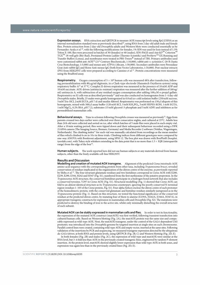
www.nature.com/scientificreports/
3SCIENTIFIC REPORTS
Expression assays. RNA extraction and QRTPCR to measure AOX transcript levels using RpL32 RNA as an internal normalization standard were as previously described10, using RNA from 2 day-old adult male and female flies. Protein extraction from 2 day-old Drosophila adults and Western blots were conducted essentially as by Fernandez-Ayala et al.10, with the following modifications: for females, 1% SDS was used for lysis instead of 1.5% Triton X-100, flies were processed in batches of 30 (females) or 40 (males), SDS-PAGE used Any kD™ Criterion™ TGX™ 18-well gels (Bio-Rad), Prestained Protein Ladder (Thermo-Scientific) and ProSieveTM EX Running and Transfer Buffers (Lonza), and membranes were treated in PBS-Tween® instead of TBS. Primary antibodies used were customized rabbit anti-AOX10 (21st Centrury Biochemicals, 1:10,000), rabbit anti-α -actininin C-20-R (Santa Cruz Biotechnology, 1:5,000) and mouse anti-ATP5A (Abcam, 1:50,000). Secondary antibodies were Peroxidase Goat Anti-rabbit IgG and Horse Anti-mouse IgG (both from Vector Laboratories, 1:10,000). Post-nuclear extracts (PN) from HEK293T cells were prepared according to Cannino et al.21. Protein concentrations were measured using the Bradford assay.
Respirometry. Oxygen consumption of 5 × 10⁶ human cells was measured 48 h after transfection, follow-ing permeabilization with 80 μ g/ml digitonin, in a Clark-type electrode (Hansatech Oxytherm system) using respiratory buffer A22 at 37 °C. Complex II-driven respiration was measured in the presence of 10 mM ADP and 10 mM succinate. AOX-driven (antimycin-resistant) respiration was measured after the further addition of (60 ng/ml) antimycin A, with subtraction of any residual oxygen consumption after adding 100 μ M n-propyl gallate. Respirometry on S2 cells was as described previously17 and was also conducted on homogenates from 1–4 day-old Drosophila males. Briefly, 25 males were gently homogenized in 0.8 ml ice-cold isolation buffer (250 mM sucrose, 5 mM Tris-HCl, 2 mM EGTA, pH 7.4) and muslin-filtered. Respirometry was performed on 150 μ l aliquots of this homogenate, mixed with 500 μ l assay buffer (120 mM KCl, 5 mM KH2PO4, 3 mM HEPES-KOH, 1 mM EGTA, 1 mM MgCl2, 0.2% BSA, pH 7.2), substrates (15 mM glycerol-3-phosphate and 5 mM ADP) and inhibitors as for permeabilized mammalian cells.
Behavioural assays. Time to eclosion following Drosophila crosses was measured as previously23. Eggs from parents crossed two days earlier were collected over three consecutive nights, and cultured at 25°C. Adults less than 24 h old were collected and sorted on ice, after which batches of 5 male flies were placed in each empty vial. After a 10 min waiting period, flies were tipped down and their subsequent behaviour recorded using a DFK 21AF04 camera (The Imaging Source, Bremen, Germany) and Media Recorder 2 software (Noldus, Wageningen, Netherlands). The climbing index11 for each vial was manually calculated from recordings as the mean number of flies which climbed 6 cm in 10 s in three trials. Climbing indices from different genotypes were compared by one-way ANOVA with Bonferoni adjustment, using SPSS 12. The box plot was drawn with BoxPlotR (boxplot.tyerslab.com), with Tukey style whiskers extending to the data point that is no more than 1.5 × IQR (interquartile range) from the edge of the box24.
Human subjects. The work reported here did not use human subjects or any materials derived from human subjects, other than the freely available cell-line HEK293T.
Results and DiscussionModelling and creation of mutated AOX transgene. Alignment of the predicted Ciona intestinalis AOX amino-acid sequence with the corresponding protein from other taxa, including Trypanosoma brucei, revealed conservation of residues implicated in the organization of the diiron centre of the enzyme, as previously reported by Shiba et al.13. The four invariant glutamate residues and two histidines correspond in Ciona AOX with E200, E239, E290, E344, H242 and H347 (Fig. S1), numbered from the first methionine of the putative preprotein. In the Trypanosoma AOX structure, the conserved histidines participate in a hydrogen bond network that also includes a conserved tyrosine, Y297 in Ciona AOX (Fig. S1). Structural modelling (Fig. 1) showed that Ciona AOX can fold to an almost identical structure as its Trypanosoma counterpart, ignoring the poorly conserved N-terminal region (residues 1–103 of the Ciona protein, Fig. S1). Four alpha-helices enclose the diiron centre of each protomer of the homodimeric protein, with the conserved glutamate and histidine residues similarly juxtaposed as in the Trypanosoma protein (Fig. 1). Based on this structure, we tested the functional significance of the conserved residues at the predicted diiron centre, by mutating four of them to alanine (E239A, H242A, E344A, H347A), in appropriate transgenic constructs for expression in mammalian cells and Drosophila (Fig. S2). The mutations were predicted to destroy the binding of iron to the active site, whilst only minimally disturbing the overall structure of each subunit.
In order to test its functionality, the expression of the mutated AOX construct (mutAOX) was first verified, following transient transfection into cultured human cells. Based on Western blotting (Fig. 2A), the mutAOX protein was the same size and compa-rably expressed as wild-type AOX. Next, the mutAOX transgene, under the control of the GAL4-dependent UAS promoter, was introduced into the Drosophila genome by targeted insertion at single sites on each chromosome. Parallel control lines were created, containing wild-type AOX and empty vector, inserted at the same sites. Following validation of the insertions by PCR and sequencing, we measured transgene expression directed by the ubiquitous da-GAL4 driver, at both RNA and protein levels, using QRTPCR (Fig. 2B, C) and Western blotting (Fig. 2D, E).
In both females (Fig. 2B) and males (Fig. 2C), the expression of wild-type and mutAOX were similar at the RNA level, but 3–4 fold less than AOX in the previously created transgenic lines, engineered by random P-element insertion. At the protein level, mutAOX showed slightly lower expression than wild-type AOX in both sexes, and expression was again less than in the previously created lines (Fig. 2D, E).
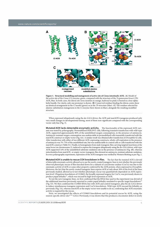
www.nature.com/scientificreports/
4SCIENTIFIC REPORTS
When expressed ubiquitously using the da-GAL4 driver, the AOX and mutAOX transgenes produced only very small changes in developmental timing, most of them non-significant compared with the corresponding vector-only line (Fig. 3).
Mutated AOX lacks detectable enzymatic activity. The functionality of the expressed AOX vari-ants was tested by polarography. Permeabilized HEK293T cells, following transient transfection with wild-type AOX, supported approximately 80% of the uninhibited oxygen consumption, in the presence of antimycin. Antimycin-resistant oxygen consumption was undetectable in permeabilized cells transiently transfected with the mutAOX construct or empty vector (Fig. 4A). A similar result was obtained after transfection of Drosophila S2 cells. After transfection with either of two different AOX-expressing constructs, whole-cell respiration in the presence of antimycin was 70–73% of the uninhibited rate, but was undetectable in control cells or cells transfected with the mutAOX construct (Table S1). Finally, in homogenates from male transgenic flies carrying targeted insertions at the same locus (on chromosome 2), induced to express the transgene ubiquitously using the da-GAL4 driver, wild-type AOX supported 14% of the uninhibited substrate oxidation rate in the presence of antimycin (Fig. 4B), whereas mitochondria from mutAOX- or empty vector-transgenic flies showed no antimycin-resistant substrate oxidation. In every polarography experiment, expression of the AOX transgene was verified by Western blotting as per Fig. 2.
The fact that the mutated AOX is devoid of detectable enzymatic activity allowed us to use the newly created transgenic lines to test whether the previously observed phenotypic rescue of flies knocked down for a subunit of cytochrome oxidase (Cox7a) was due to the enzymatic activity of AOX or some other property conferred by the AOX protein, when expressed in Drosophila. Moreover, the fact that the newly created transgenic lines express AOX at only about 30% of the level of the lines previously studied, allowed us to test whether phenotypic rescue was quantitatively dependent on AOX expres-sion level. Ubiquitous knockdown of CG9603, the broadly expressed isogene for Cox7a, was previously shown to produce pupal lethality11, which was rescued by high-level expression of AOX.
To test the new transgenic lines, we first confirmed that the RNAi line used in the experiment was devoid of the additional insertion previously reported to confer pupal lethality unrelated to specific target knockdown25 (Fig. S3). We then combined the CG9603 RNAi line with AOX and control transgenes, plus the da-GAL4 driver to induce simultaneous transgene expression and Cox7a knockdown. Wild-type AOX rescued the lethality, as previously (Fig. 5A), whereas mutAOX or the empty vector were unable to do so, confirming that AOX enzymatic activity is required for the rescue.
Next, we investigated the effects of CG9603 knockdown and its potential rescue by AOX, using the neuron-specific driver elavC155-GAL4. Previously, it was shown that this produces a locomotor defect in newly
Figure 1. Structural modelling and mutagenesis of active site of Ciona intestinalis AOX. (A) Model of the active site of the Ciona (Ci) enzyme, green, compared with the structure of the Trypanosoma brucei (Tb) AOX, blue. In both cases, the diiron site (iron moieties in orange, hydroxyl in pink) is buried in a four alpha-helix bundle. For clarity, only one protomer is shown. (B) Conserved residues binding the diiron centre show an identical arrangement in the Ci model (green) as in the Tb structure (blue). (C) The residues selected for alanine-substitution mutagenesis in the Ci enzyme (here shown in blue), alongside the resulting modelled structure.
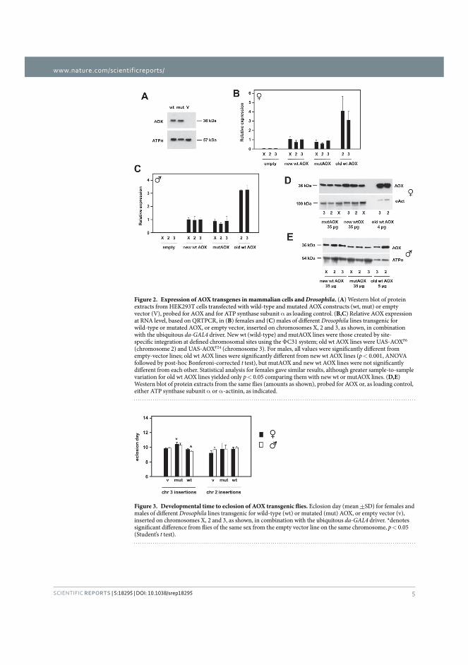
www.nature.com/scientificreports/
5SCIENTIFIC REPORTS
Figure 2. Expression of AOX transgenes in mammalian cells and Drosophila. (A) Western blot of protein extracts from HEK293T cells transfected with wild-type and mutated AOX constructs (wt, mut) or empty vector (V), probed for AOX and for ATP synthase subunit α as loading control. (B,C) Relative AOX expression at RNA level, based on QRTPCR, in (B) females and (C) males of different Drosophila lines transgenic for wild-type or mutated AOX, or empty vector, inserted on chromosomes X, 2 and 3, as shown, in combination with the ubiquitous da-GAL4 driver. New wt (wild-type) and mutAOX lines were those created by site-specific integration at defined chromosomal sites using the Φ C31 system; old wt AOX lines were UAS-AOXF6 (chromosome 2) and UAS-AOXF24 (chromosome 3). For males, all values were significantly different from empty-vector lines; old wt AOX lines were significantly different from new wt AOX lines (p < 0.001, ANOVA followed by post-hoc Bonferoni-corrected t test), but mutAOX and new wt AOX lines were not significantly different from each other. Statistical analysis for females gave similar results, although greater sample-to-sample variation for old wt AOX lines yielded only p < 0.05 comparing them with new wt or mutAOX lines. (D,E) Western blot of protein extracts from the same flies (amounts as shown), probed for AOX or, as loading control, either ATP synthase subunit α or α -actinin, as indicated.
Figure 3. Developmental time to eclosion of AOX transgenic flies. Eclosion day (mean +SD) for females and males of different Drosophila lines transgenic for wild-type (wt) or mutated (mut) AOX, or empty vector (v), inserted on chromosomes X, 2 and 3, as shown, in combination with the ubiquitous da-GAL4 driver. *denotes significant difference from flies of the same sex from the empty vector line on the same chromosome, p < 0.05 (Student’s t test).
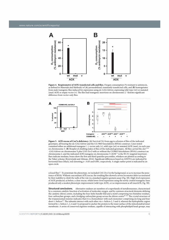
www.nature.com/scientificreports/
6SCIENTIFIC REPORTS
eclosed flies11. To potentiate the phenotype, we included UAS-Dcr2 in the background, so as to increase the pene-trance of RNAi. Without concomitant AOX rescue, the resulting flies showed a severe locomotor defect as measured by their inability to climb the walls of the vial, in a standard negative geotaxis assay (Fig. 5B). High-level expression of AOX produced, as before, a clear rescue, whilst lower-level expression using the newly created transgenic lines produced only a modest phenotypic improvement (wild-type AOX), or no improvement at all (mutAOX, Fig. 5B).
Structural conclusions. Alternative oxidases are members of a superfamily of metalloenzymes, characterized by a common catalytic function of activation of molecular oxygen, and by common structural elements defining the catalytic diiron centre, including the four-helix bundle fold and a motif comprising two histidine residues, four carboxylate groups, and a bridging carboxylate group across the diiron centre26–28. The crystal structure of the trypanosomal enzyme indicates that it is a homodimer with each monomer comprising six long and four short α -helices13. The subunits interact with each other via α -helices 2, 3 and 4, whereas the hydrophobic region formed by α -helices 1, 2, 4 and 5 is proposed to anchor the protein to the inner surface of the mitochondrial inner membrane. A series of conserved arginine residues, capable of interacting with phospholipid head-groups, may
Figure 4. Respirometry of AOX-transfected cells and flies. Oxygen consumption (% resistant to antimycin, as defined in Materials and Methods) of (A) permeabilized, transiently transfected cells, and (B) homogenates from male transgenic flies induced for expression using da-GAL4 driver, expressing wild-type (wt) or mutated (mut) AOX or empty vector (v). The flies had transgenic insertions on chromosome 2. *denotes significant difference from vector-only flies.
Figure 5. AOX rescue of Cox7a deficiency. (A) Survival (%) from egg to eclosion of flies of the indicated genotypes, all bearing the da-GAL4 driver and the CG-9603 knockdown (RNAi) construct. Lines tested contained either no additional transgene (–), vector only (v), wild-type (wt) or mutated AOX (mut), in each case on chromosome 3. (B) Boxplot of climbing index of flies of the indicated genotypes. All flies carried the elavC155 -GAL4 driver on chromosome X plus UAS-Dcr2 with or without the CG9603 knockdown (RNAi) construct on chromosome 2, and the indicated AOX transgene on chromosome 3 (AOX7.1 is the Φ C31-targeted insertion). Bars indicate medians, boxes show the first and third quartiles percentiles, whiskers are plotted according to the Tukey scheme (Krzywinski and Altman, 2014). Significant differences based on ANOVA are indicated by horizontal lines (black, red) denoting p < 0.05 and 0.001, respectively. A single outlier point is indicated by an open circle.
www.nature.com/scientificreports/
7SCIENTIFIC REPORTS
assist inner membrane anchorage13. Our structure modelling of the C. intestinalis AOX suggests that the same structural elements are conserved in animal AOXs, and that the enzyme is also a homodimer inserted into the mitochondrial inner membrane.
In addition, the model predicts that the active site, and therefore the mechanism of oxygen activation, are also conserved in animal AOXs. The four-helix bundle, which acts as a structural platform for the binding of the two iron atoms, buries the active site deep in a hydrophobic environment. In T. brucei AOX, glutamate residues 123, 162, 213 and 266, in addition to a hydroxo-bridge, are responsible for directly coordinating the diiron centre. The centre is further stabilized by a redox-active tyrosine residue29,30, Y220, and two histidine residues (H165 and H269), which are within hydrogen-bond distances of E123, E169 and E213. The C. intestinalis AOX model indicates that the homologous residues E200, E239, E290, E344, Y297, H242 and H347 organize the active site in the same way.
Functional conclusions. In theory, the mutagenesis of a single glutamate residue should be enough to dest-abilize the diiron centre31. However, taking advantage of the proximity in the DNA sequence of the codons for E239 and H242 and of those for E344 and H347, we were able to create alanine substitutions for four important active site residues simultaneously. According to our model, these mutations should disrupt iron binding, thus generating a mutant devoid of catalytic activity, without any major disturbance to the overall protein structure. These predictions are supported by the fact that the mutant and wild-type proteins were expressed at comparable levels in mammalian cells and in flies, but that no enzymatic activity could be detected.
Importantly, the mutated enzyme was unable to rescue the organismal phenotypes arising from engineered cytochrome oxidase deficiency. In theory, the action of a foreign protein in attenuating such phenotypes could be due to any of several different mechanisms, of which the provision of an enzymatic by-pass for ubiquinol oxidation is only one. In previous work we found that Ciona AOX, when expressed in Drosophila mitochondria, decreased the net production of mitochondrial ROS even under non-inhibited conditions10,32. The mechanism of this remains unknown, but one possibility is that AOX is able to act directly or indirectly as an antioxidant, e.g. by binding and quenching quinone radicals via some other mechanism. Studies in various organisms have supported the idea that a hydrophobic pocket, located between α -helices 2 and 3, binds and channels ubiquinone to the active site33, which might be involved in such an activity.
A second possibility would be a hormetic response to disruption of the inner mitochondrial membrane or its protein complexes by the foreign protein. The induction of a variety of defence pathways to protect cells from increased ROS, disturbed protein, lipid or redox homeostasis, or altered mitochondrial turnover or dynamics, might equip the organism to cope with the additional but related stresses of respiratory insufficiency. Many stud-ies in model organisms support this concept of ‘mitohormesis’34. Whilst we cannot rule out that such effects are material in other contexts, our findings do exclude them in regard to the developmental lethality produced by global cytochrome oxidase knockdown, or the locomotor dysfunction resulting from its knockdown specifically in neurons11. Based on our findings, that mutAOX cannot compensate these phenotypes, we infer that the rescue of these effects of cytochrome oxidase deficiency by AOX is almost certainly due to its enzymatic activity as a quinol oxidase, though formally we cannot exclude other, unknown effects of iron binding. A requirement for enzymatic activity might not be true of every phenotypic feature conferred by AOX in model organisms. Our findings indicate a robust way to test this in regard to all potential such phenotypes, allowing the mechanisms by which AOX acts to be probed, controlled or verified.
Several quantitative issues are also addressed by our findings. The first is that the extent of phenotypic rescue depends in some instances on the AOX expression level, but in other cases, such as the rescue of the developmental lethality caused by ubiquitous COX knockdown, is an all-or-none phenomenon. We suggest that this reflects a threshold effect wherein even the three-fold lower expression level of AOX, when integrated at specific sites by Φ C31-mediated recombination (in comparison with P element-mediated integrants created previously), exceeds a threshold value required to maintain metabolic homeostasis and complete development. In contrast, the lower expression level of the targeted integrants gave a clearly weaker rescue of locomotor dysfunction, when COX was knocked down only in neurons, roughly in proportion to the decreased expression level.
It may also be noted that the amount of antimycin-resistance conferred upon respiration in homogenates from the targeted integrants was still approximately 14%, compared with approximately 20% for the P element-mediated integrants, even though they are expressed at a much higher level. The level of respiratory antimycin-resistance in the fly may vary between tissues, and this 20% maximum may reflect only the properties of the predominant class of mitochondria. Most of the respiratory capacity in adult flies is vested in the flight muscles, where mitochondria make up almost one-third of the total tissue mass35. The apparent upper limit of how much electron flow can be diverted through AOX probably reflects specific features of this tissue and its energetic needs. The limit could be dictated by the constraints of membrane architecture, for example, if much of the ubiquinone pool is channelled directly from complex I to complex III via respiratory supercomplexes, such that it equilibrates only slowly with free ubiquinones available to AOX36. Most of the respiratory activity in adult Drosophila indeed resides in super-complexes37. Such a phenomenon may account for the inferred threshold effect on the rescue of developmental lethality. Conversely, the organization of the respiratory chain may differ in other tissues, such as in neurons, where a more graded response to the AOX expression level is evident.
In conclusion, mutAOX offers a useful tool for future studies of the mechanism(s) whereby expression of Ciona AOX modifies the phenotypes of model organisms, potentially contributing the eventual development of AOX-based therapies.
References1. McDonald, A. E., Vanlerberghe, G. C. & Staples, J. F. Alternative oxidase in animals: unique characteristics and taxonomic distribution.
J. Exp. Biol. 212, 2627–2634 (2009).2. Feng, H. et al. Expression and signal regulation of the alternative oxidase genes under abiotic stresses. Acta. Biochim. Biophys. Sin.
45, 985–994 (2013).
www.nature.com/scientificreports/
8SCIENTIFIC REPORTS
3. Vanlerberghe, G. C. Alternative oxidase: a mitochondrial respiratory pathway to maintain metabolic and signaling homeostasis during abiotic and biotic stress in plants. Int. J. Mol. Sci. 14, 6805–6847 (2013).
4. Hoefnagel, M. H. & Wiskich, J. T. Activation of the plant alternative oxidase by high reduction levels of the Q-pool and pyruvate. Arch. Biochem. Biophys. 355, 262–270 (1998).
5. Castro-Guerrero, N. A., Krab, K. & Moreno-Sánchez, R. The alternative respiratory pathway of euglena mitochondria. J Bioenerg. Biomembr. 36, 459–469 (2004).
6. Rustin, P. & Jacobs, H. T. Respiratory chain alternative enzymes as tools to better understand and counteract respiratory chain deficiencies in human cells and animals. Physiol. Plant 137, 362–370 (2009).
7. El-Khoury, R. et al. Engineering the alternative oxidase gene to better understand and counteract mitochondrial defects: state of the art and perspectives. Br. J. Pharmacol. 171, 2243–2249 (2014).
8. Hakkaart, A., Dassa, E. P., Jacobs, H. T. & Rustin, P. Allotopic expression of a mitochondrial alternative oxidase confers cyanide resistance to human cell respiration. EMBO Rep. 7, 341–345 (2006).
9. Dassa, E. P. et al. Expression of the alternative oxidase complements cytochrome c oxidase deficiency in human cells. EMBO Mol. Med. 1, 30–36 (2009).
10. Fernandez-Ayala, D. J. et al. Expression of the Ciona intestinalis alternative oxidase (AOX) in Drosophila complements defects in mitochondrial oxidative phosphorylation. Cell Metab. 9, 449–460 (2009).
11. Kemppainen, K. K. et al. Expression of alternative oxidase in Drosophila ameliorates diverse phenotypes due to cytochrome oxidase deficiency. Hum. Mol. Genet. 23, 2078–2093 (2014).
12. El-Khoury, R. et al. Alternative oxidase expression in the mouse enables bypassing cytochrome c oxidase blockade and limits mitochondrial ROS overproduction. PLoS Genet. 9, e1003182 (2013).
13. Shiba, T. et al. Structure of the trypanosome cyanide-insensitive alternative oxidase. Proc. Natl. Acad. Sci. USA 110, 4580–4585 (2013).14. Tamura, K., Stecher, G., Peterson, D., Filipski, A. & Kumar, S. MEGA6: Molecular Evolutionary Genetics Analysis version 6.0. Mol.
Biol. Evol. 30, 2725–2729 (2013).15. Bazzoli, A., Tettamanzi, A. G. & Zhang, Y. Computational protein design and large-scale assessment by I-TASSER structure assembly
simulations. J Mol. Biol. 407, 764–776 (2011).16. Bischof, J., Maeda, R. K., Hediger, M., Karch, F. & Besler, K. An optimized transgenesis system for Drosophila using germ-line-specific ϕ C31 integrases. Proc. Natl. Acad. Sci. USA 104, 3312–3317 (2007).
17. Fukuoh, A. et al. Screen for mitochondrial DNA copy number maintenance genes reveals essential role for ATP synthase. Mol. Syst. Biol. 10, 734 (2014).
18. Niwa, H., Yamamura, K. & Miyazaki, J. Efficient selection for high-expression transfectants with a novel eukaryotic vector. Gene 108, 193–199 (1991).
19. Sanz, A. et al. Expression of the yeast NADH dehydrogenase Ndi1 in Drosophila confers increased lifespan independently of dietary restriction. Proc. Natl. Acad. Sci. USA 107, 9105–9110 (2010).
20. Pfeiffer, B. D. et al. Refinement of tools for targeted gene expression in Drosophila. Genetics 186, 735–755 (2010).21. Cannino, G. et al. Glucose modulates respiratory complex I activity in response to acute mitochondrial dysfunction. J Biol. Chem.
287, 38729–38740 (2012).22. Chretien, D. et al. Reference charts for respiratory chain activities in human tissues. Clin. Chim. Acta. 228, 53–70 (1994).23. Toivonen, J. M. et al. technical knockout, a Drosophila model of mitochondrial deafness. Genetics 159, 241–254 (2001).24. Krzywinski, M. & Altman, N. Points of Significance: Visualizing samples with box plots. Nature Methods 11, 119–120 (2014).25. Green, E. W., Fedele, G., Giorgini, F. & Kyriacou, C. P. A Drosophila RNAi collection is subject to dominant phenotypic effects. Nat.
Methods 11, 222–223 (2014).26. Berthold, D. A., Voevodskaya, N., Stenmark, P., Gräslund, A. & Nordlund, P. EPR studies of the mitochondrial alternative oxidase.
Evidence for a diiron carboxylate center. J Biol. Chem. 277, 43608–43614 (2002).27. Berthold, D. A. & Stenmark, P. Membrane-bound di-iron carboxylate proteins. Annu. Rev. Plant Biol. 54, 497–517 (2003).28. Simone, F., Reisner, E. & Lippard, S. J. Current challenges of modeling diiron enzyme active sites for dioxygen activation by biomimetic
synthetic complexes. Chem. Soc. Rev. 39, 2768–2779 (2010).29. Albury, M. S., Affourtit, C., Crichton, P. G. & Moore, A. L. Structure of the plant alternative oxidase. Site-directed mutagenesis provides
new information on the active site and membrane topology. J Biol. Chem. 277, 1190–1194 (2002).30. Affourtit, C., Albury, M. S., Crichton, P. G. & Moore, A. L. Exploring the molecular nature of alternative oxidase regulation and
catalysis. FEBS Lett. 510, 121–126 (2002).31. Ajayi, W. U., Chaudhuri, M. & Hill, G. C. Site-directed mutagenesis reveals the essentiality of the conserved residues in the putative
diiron active site of the trypanosome alternative oxidase. J Biol. Chem. 277, 8187–8193 (2002).32. Sanz, A., Fernández-Ayala, D. J., Stefanatos, R. K. & Jacobs, H. T. Mitochondrial ROS production correlates with, but does not directly
regulate lifespan in Drosophila. Aging 2, 200–223 (2010).33. Albury, M. S., Elliott, C. & Moore, A. L. Towards a structural elucidation of the alternative oxidase in plants. Physiol. Plant 137,
316–327 (2009).34. Yun, J. & Finkel, T. Mitohormesis. Cell Metab. 19, 757–766 (2014).35. Levenbook, L. & Williams, C. M. Mitochondria in the flight muscles of insects III. Mitochondrial cytochrome c in relation to the
aging and wing beat frequency of flies. J. Gen. Physiol. 39, 497–512 (1956).36. Genova, M. L. & Lenaz, G. Functional role of mitochondrial respiratory supercomplexes. Biochim. Biophys. Acta. 1837, 427–443
(2014).37. Celotto, A. M., Chiu, W. K., Van Voorhies, W. & Palladino, M. J. Modes of metabolic compensation during mitochondrial disease
using the Drosophila model of ATP6 dysfunction. PLoS One 6, e25823 (2011).
AcknowledgementsWe thank Tony Moore for useful discussions, Filippo Scialo for the construction of the original AOX plasmid for expression in S2 cells, Dmitro Gospodaryov for critical reading of the manuscript and Samuli Hartikainen, Eveliina Kaulio, Tea Tuomela, Essi Kiviranta, Outi Kurronen, Merja Jokela and Maarit Myöhänen for technical assistance. Funding was provided by Academy of Finland (CoE grant 272376), the European Research Council (advanced grant 232738 to HTJ), the EU (Marie Curie International Incoming Fellowship 328988 to MTO), Tampere University Hospital Medical Research Fund, and the Sigrid Juselius Foundation.
Author ContributionsA.A., M.T.O., H.T.J. and P.R. conceived and planned the project. A.A., M.T.O., G.C., C.Y. and P.K.D. conducted the laboratory work and analysis. H.T.J., M.S. and E.D. supervised the laboratory work and contributed analysis and insights. H.T.J. and M.T.O. compiled the figures and drafted the manuscript.
Additional InformationSupplementary information accompanies this paper at http://www.nature.com/srep

www.nature.com/scientificreports/
9SCIENTIFIC REPORTS
Competing financial interests: The authors declare no competing financial interests.How to cite this article: Andjelković, A. et al. Diiron centre mutations in Ciona intestinalis alternative oxidase abolish enzymatic activity and prevent rescue of cytochrome oxidase deficiency in flies. Sci. Rep. 5, 18295; doi: 10.1038/srep18295 (2015).
This work is licensed under a Creative Commons Attribution 4.0 International License. The images or other third party material in this article are included in the article’s Creative Commons license,
unless indicated otherwise in the credit line; if the material is not included under the Creative Commons license, users will need to obtain permission from the license holder to reproduce the material. To view a copy of this license, visit http://creativecommons.org/licenses/by/4.0/

Ciona intestinalis
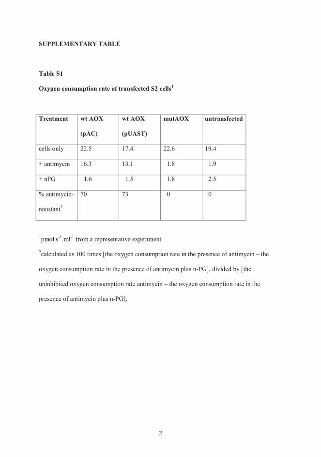
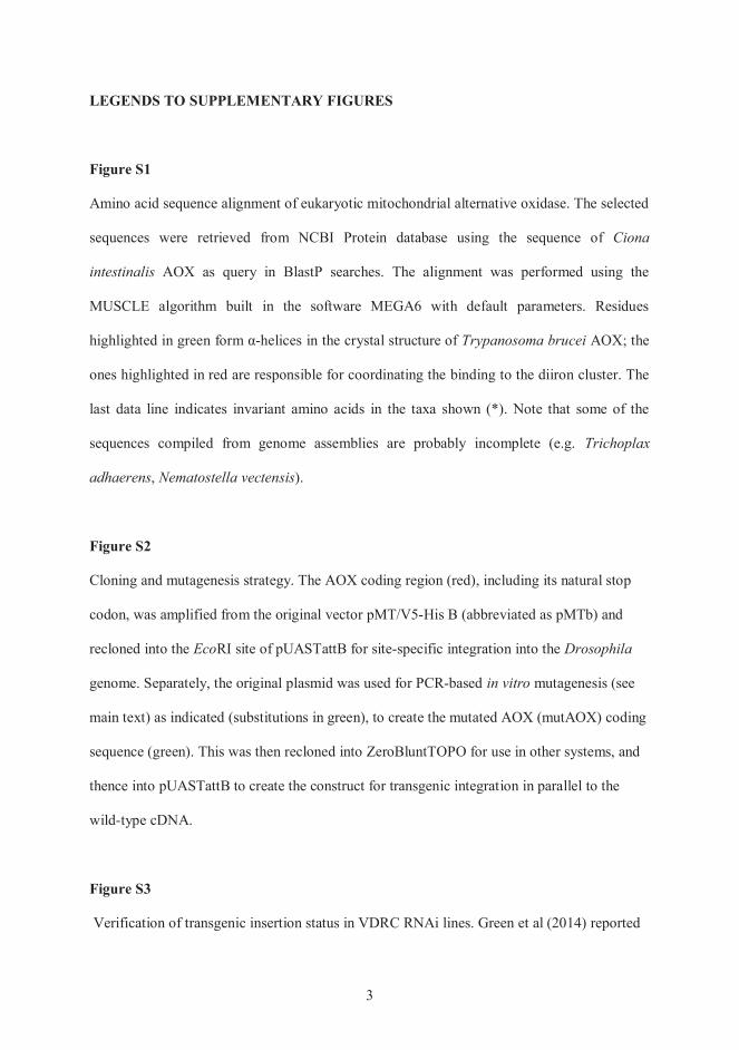
Ciona
intestinalis
Trypanosoma brucei
Trichoplax
adhaerens Nematostella vectensis
Eco Drosophila
in vitro
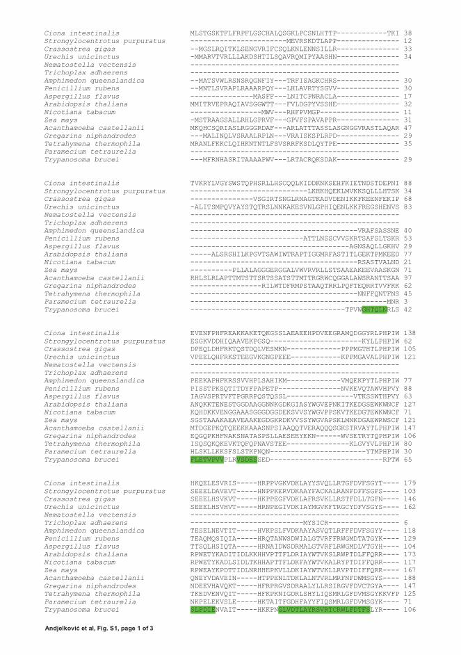
Ciona intestinalis MLSTGSKTFLFRPFLGSCHALQSGKLPCSNLHTTP------------TKI 38Strongylocentrotus purpuratus -----------------------MEVRSKDTLAPP--------------- 12Crassostrea gigas --MGSLRQITKLSENGVRIFCSQLKNLENNSILLR--------------- 33Urechis unicinctus -MMARVTVRLLLAKDSHTILSQAVRQMIPYAASHN--------------- 34Nematostella vectensis --------------------------------------------------Trichoplax adhaerens --------------------------------------------------Amphimedon queenslandica --MATSVWLRSNSRQGNFIY---TRFISAGKCHRS--------------- 30Penicillium rubens --MNTLSVRAPLRAAARPQY---LHLAVRTYSGVV--------------- 30Aspergillus flavus ---------------MASFF---LNITCPNRACLA--------------- 17Arabidopsis thaliana MMITRVEPRAQIAVSGGWTT---FVLDGPYVSSHE--------------- 32Nicotiana tabacum -----------------MWV---RHFPVMGP------------------- 11Zea mays -MSTRAAGSALLRHLGPRVF---GPVFSPAVAPPR--------------- 31Acanthamoeba castellanii MKQHCSQRIASLRGGGRDAF---ARLATTTASSLASGNGGVRASTLAQAR 47Gregarina niphandrodes ---MALINQLVSRAALRPLN---VRAISKSPLRPD--------------- 29Tetrahymena thermophila MRANLFKKCLQIHKNTNTLFSVSRRFKSDLQYTPE--------------- 35Paramecium tetraurelia --------------------------------------------------Trypanosoma brucei ---MFRNHASRITAAAAPWV---LRTACRQKSDAK--------------- 29
Ciona intestinalis TVKRYLVGYSWSTQPHSRLLHSCQQLKIDDKNKSEHFKIETNDSTDEPNI 88Strongylocentrotus purpuratus ----------------------------LKHKHQEKLMVKKSQLLLHTSK 34Crassostrea gigas ---------------VSGIRTSNGLRNAGTKADVDENIKKFKEENFEKIP 68Urechis unicinctus -ALITSMPQVYAYSTQTRSLNNKAKESVNLGPHIQENLKKFREGSHENVS 83Nematostella vectensis --------------------------------------------------Trichoplax adhaerens --------------------------------------------------Amphimedon queenslandica ----------------------------------------VRAFSASSNE 40Penicillium rubens ---------------------------ATTLNSSCVVSKRTSAFSLTSKR 53Aspergillus flavus --------------------------------------AGNSAQLLGKHV 29Arabidopsis thaliana -----ALSRSHILKPGVTSAWIWTRAPTIGGMRFASTITLGEKTPMKEED 77Nicotiana tabacum ----------------------------------------RSASTVALND 21Zea mays ----------PLLALAGGGERGGALVWVRVRLLSTSAAEAKEEVAASKGN 71Acanthamoeba castellanii RHLSLRLAPTTMTSTTSRTSSATSTTMTTRGRWCQGGALAWSRANTTSAA 97Gregarina niphandrodes -----------------RILWTDFRMPSTAAQTRRLPQFTEQRRTVVFKK 62Tetrahymena thermophila ----------------------------------------NNFFQNTFNS 45Paramecium tetraurelia -----------------------------------------------MNR 3Trypanosoma brucei -------------------------------------TPVWGHTQLNRLS 42
Ciona intestinalis EVENFPHFREAKKAKETQKGSSLAEAEEHPDVEEGRAMQDGGYRLPHPIW 138Strongylocentrotus purpuratus ESGKVDDHIQAAVEKPGSQ----------------------KYLLPHPIW 62Crassostrea gigas DPEQLDHFRKTQSTDQLVESMKN-------------PPPMGTHTLPHPIW 105Urechis unicinctus VPEELQHFRKSTEEGVKGNGPEEE------------KPPMGAVALPHPIW 121Nematostella vectensis --------------------------------------------------Trichoplax adhaerens --------------------------------------------------Amphimedon queenslandica PEEKAPHFKRSSVVHPLSAHIKM-------------VMQEKPYTLPHPIW 77Penicillium rubens PISSTPKSQTITDYFPAPETP---------------NVKEVQTAWVHPVY 88Aspergillus flavus IAGVSPRTVFTPGRRPQSTQSSL----------------VTKSSWTHPVY 63Arabidopsis thaliana ANQKKTENESTGGDAAGGNNKGDKGIASYWGVEPNKITKEDGSEWKWNCF 127Nicotiana tabacum KQHDKKVENGGAAASGGGDGGDEKSVVSYWGVPPSKVTKEDGTEWKWNCF 71Zea mays SGSTAAAKAEAVEAAKEGDGKRDKVVSSYWGVAPSKLMNKDGAEWRWSCF 121Acanthamoeba castellanii MTDGEPKQTQEEKKAAASNPSIAAQQTVERAQQQSGKSTRVAYTLPHPIW 147Gregarina niphandrodes EQGQPKHFNAKSNATASPSLLAESEEYEKN------WVSETRYTQPHPIW 106Tetrahymena thermophila ISQSQKQKEVKTQFQPNAVSTEE---------------KLGVYVLPHPIW 80Paramecium tetraurelia HLSKLLKKSFSLSTKPNQN-----------------------YTMPHPIW 30Trypanosoma brucei FLETVPVVPLRVSDESSED---------------------------RPTW 65
Ciona intestinalis HKQELESVRIS-----HRPPVGKVDKLAYYSVQLLRTGFDVFSGYT---- 179Strongylocentrotus purpuratus SEEELDAVEVT-----HNPPKERVDKAAYFACKALRANFDFFSGFS---- 103Crassostrea gigas SEEELHSVKVT-----HKPPEGFVDKLAFRSVKLLRSTFDLLTGFN---- 146Urechis unicinctus SEEELHSVHVT-----HRNPEGIVDKIAYMGVKFTRGCYDFVSGYS---- 162Nematostella vectensis --------------------------------------------------Trichoplax adhaerens ---------------------------MYSICR----------------- 6Amphimedon queenslandica TESELNEVTIT-----HVKPSLFVDKAAYASVQTLRFFFDVFSGYY---- 118Penicillium rubens TEAQMQSIQIA-----HRQTANWSDWIALGTVRFFRWGMDTATGYK---- 129Aspergillus flavus TTSQLHSIQTA-----HRNAIDWSDRMALGTVRFLRWGMDLVTGYH---- 104Arabidopsis thaliana RPWETYKADITIDLKKHHVPTTFLDRIAYWTVKSLRWPTDLFFQRR---- 173Nicotiana tabacum RPWETYKADLSIDLTKHHAPTTFLDKFAYWTVKALRYPTDIFFQRR---- 117Zea mays RPWEAYKPDTTIDLNRHHEPKVLLDKIAYWTVKLLRVPTDIFFQRR---- 167Acanthamoeba castellanii QNEYVDAVEIN-----HTPPENLTDKLALNTVRLMRFNFDWMSGYS---- 188Gregarina niphandrodes NDEEVHAVQKT-----HFRPRGVSDRAALYLLRSIRGVFDVCTGYA---- 147Tetrahymena thermophila TKEDVENVQIT-----HFKPKNIGDRLSHYLIQSMRLGFDVMSGYKKVFP 125Paramecium tetraurelia NKPELEKVSLE-----HKTAITFGDHFAYYFIQSMRLGFDVMSGYK---- 71Trypanosoma brucei SLPDIENVAIT-----HKKPNGLVDTLAYRSVRTCRWLFDTFSLYR---- 106
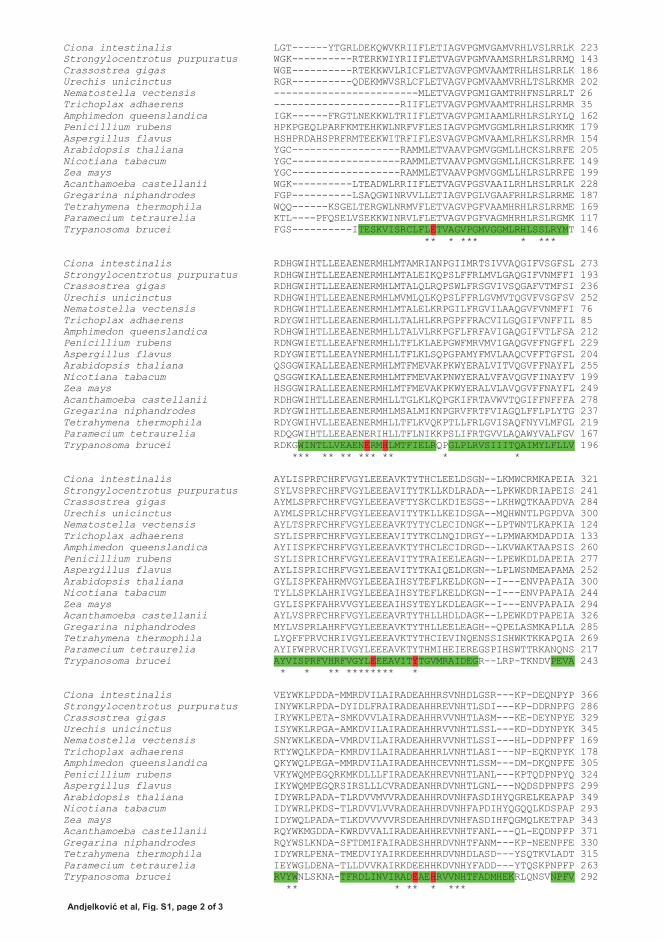
Ciona intestinalis LGT------YTGRLDEKQWVKRIIFLETIAGVPGMVGAMVRHLVSLRRLK 223Strongylocentrotus purpuratus WGK----------RTERKWIYRIIFLETVAGVPGMVAAMSRHLRSLRRMQ 143Crassostrea gigas WGE----------RTEKKWVLRICFLETVAGVPGMVAAMTRHLHSLRRLK 186Urechis unicinctus RGR----------QDEKMWVSRLCFLETVAGVPGMVAAMVRHLTSLRKMR 202Nematostella vectensis ------------------------MLETVAGVPGMIGAMTRHFNSLRRLT 26Trichoplax adhaerens ---------------------RIIFLETVAGVPGMVAAMTRHLHSLRRMR 35Amphimedon queenslandica IGK------FRGTLNEKKWLTRIIFLETVAGVPGMIAAMLRHLRSLRYLQ 162Penicillium rubens HPKPGEQLPARFKMTEHKWLNRFVFLESIAGVPGMVGGMLRHLRSLRKMK 179Aspergillus flavus HSHPRDAHSPRFRMTEEKWITRFIFLESVAGVPGMVAAMLRHLKSLRRMR 154Arabidopsis thaliana YGC------------------RAMMLETVAAVPGMVGGMLLHCKSLRRFE 205Nicotiana tabacum YGC------------------RAMMLETVAAVPGMVGGMLLHCKSLRRFE 149Zea mays YGC------------------RAMMLETVAAVPGMVGGMLLHLRSLRRFE 199Acanthamoeba castellanii WGK----------LTEADWLRRIIFLETVAGVPGSVAAILRHLHSLRRLK 228Gregarina niphandrodes FGP----------LSAQGWINRVVLLETIAGVPGLVGAAFRHLRSLRRME 187Tetrahymena thermophila WQQ------KSGELTERGWLNRMVFLETVAGVPGFVAAMHRHLRSLRRME 169Paramecium tetraurelia KTL----PFQSELVSEKKWINRVLFLETVAGVPGFVAGMHRHLRSLRGMK 117Trypanosoma brucei FGS----------ITESKVISRCLFLETVAGVPGMVGGMLRHLSSLRYMT 146
** * *** * ***
Ciona intestinalis RDHGWIHTLLEEAENERMHLMTAMRIANPGIIMRTSIVVAQGIFVSGFSL 273Strongylocentrotus purpuratus RDHGWIHTLLEEAENERMHLMTALEIKQPSLFFRLMVLGAQGIFVNMFFI 193Crassostrea gigas RDHGWIHTLLEEAENERMHLMTALQLRQPSWLFRSGVIVSQGAFVTMFSI 236Urechis unicinctus RDHGWIHTLLEEAENERMHLMVMLQLKQPSLFFRLGVMVTQGVFVSGFSV 252Nematostella vectensis RDHGWIHTLLEEAENERMHLMTALELKRPGILFRGVILAAQGVFVNMFFI 76Trichoplax adhaerens RDYGWIHTLLEEAENERMHLLTALHLKRPGPFFRACVILGQGIFVNFFIL 85Amphimedon queenslandica RDHGWIHTLLEEAENERMHLLTALVLRKPGFLFRFAVIGAQGIFVTLFSA 212Penicillium rubens RDNGWIETLLEEAFNERMHLLTFLKLAEPGWFMRVMVIGAQGVFFNGFFL 229Aspergillus flavus RDYGWIETLLEEAYNERMHLLTFLKLSQPGPAMYFMVLAAQCVFFTGFSL 204Arabidopsis thaliana QSGGWIKALLEEAENERMHLMTFMEVAKPKWYERALVITVQGVFFNAYFL 255Nicotiana tabacum QSGGWIKALLEEAENERMHLMTFMEVAKPNWYERALVFAVQGVFINAYFV 199Zea mays HSGGWIRALLEEAENERMHLMTFMEVAKPKWYERALVLAVQGVFFNAYFL 249Acanthamoeba castellanii RDHGWIHTLLEEAENERMHLLTGLKLKQPGKIFRTAVWVTQGIFFNFFFA 278Gregarina niphandrodes RDYGWIHTLLEEAENERMHLMSALMIKNPGRVFRTFVIAGQLFFLPLYTG 237Tetrahymena thermophila RDYGWIHVLLEEAENERMHLLTFLKVQKPTLLFRLGVISAQFNYVLMFGL 219Paramecium tetraurelia RDQGWIHTLLEEAENERIHLLTFLNIKKPSLIFRTGVVLAQAWYVALFGV 167Trypanosoma brucei RDKGWINTLLVEAENERMHLMTFIELRQPGLPLRVSIIITQAIMYLFLLV 196
*** ** ** *** ** * *
Ciona intestinalis AYLISPRFCHRFVGYLEEEAVKTYTHCLEELDSGN--LKMWCRMKAPEIA 321Strongylocentrotus purpuratus SYLVSPRFCHRFVGYLEEEAVITYTKLLKDLRADA--LPKWKDRIAPEIS 241Crassostrea gigas AYMLSPRFCHRFVGYLEEEAVFTYSKCLKDIESGS--LKHWQTKAAPDVA 284Urechis unicinctus AYMLSPRLCHRFVGYLEEEAVITYTKLLKEIDSGA--MQHWNTLPGPDVA 300Nematostella vectensis AYLTSPRFCHRFVGYLEEEAVKTYTYCLECIDNGK--LPTWNTLKAPKIA 124Trichoplax adhaerens SYLISPRFCHRFVGYLEEEAVITYTKCLNQIDRGY--LPMWAKMDAPDIA 133Amphimedon queenslandica AYIISPKFCHRFVGYLEEEAVKTYTHCLECIDRGD--LKVWAKTAAPSIS 260Penicillium rubens SYLISPRICHRFVGYLEEEAVITYTRAIEELEAGN--LPEWKDLDAPEIA 277Aspergillus flavus AYLISPRICHRFVGYLEEEAVITYTKAIQELDKGN--LPLWSNMEAPAMA 252Arabidopsis thaliana GYLISPKFAHRMVGYLEEEAIHSYTEFLKELDKGN--I---ENVPAPAIA 300Nicotiana tabacum TYLLSPKLAHRIVGYLEEEAIHSYTEFLKELDKGN--I---ENVPAPAIA 244Zea mays GYLISPKFAHRVVGYLEEEAIHSYTEYLKDLEAGK--I---ENVPAPAIA 294Acanthamoeba castellanii AYLVSPRFCHRFVGYLEEEAVRTYTHLLHDLDAGK--LPEWKDTPAPEIA 326Gregarina niphandrodes MYLVSPRLAHRFVGYLEEEAVKTYTHLLEELEAGH--QPELASMKAPLLA 285Tetrahymena thermophila LYQFFPRVCHRIVGYLEEEAVKTYTHCIEVINQENSSISHWKTKKAPQIA 269Paramecium tetraurelia AYIFWPRVCHRIVGYLEEEAVKTYTHMIHEIEREGSPIHSWTTRKANQNS 217Trypanosoma brucei AYVISPRFVHRFVGYLEEEAVITYTGVMRAIDEGR--LRP-TKNDVPEVA 243
* * ** ******** *
Ciona intestinalis VEYWKLPDDA-MMRDVILAIRADEAHHRSVNHDLGSR---KP-DEQNPYP 366Strongylocentrotus purpuratus INYWKLRPDA-DYIDLFRAIRADEAHHREVNHTLSDI---KP-DDRNPFG 286Crassostrea gigas IRYWKLPETA-SMKDVVLAIRADEAHHRVVNHTLASM---KE-DEYNPYE 329Urechis unicinctus ISYWKLRPGA-AMKDVILAIRADEAHHRVVNHTLSSL---KD-DDYNPYK 345Nematostella vectensis SNYWKLKEDA-VMRDVILAIRADEAHHRVVNHTLSSI---HL-DDPNPFF 169Trichoplax adhaerens RTYWQLKPDA-KMRDVILAIRADEAHHRLVNHTLASI---NP-EQKNPYK 178Amphimedon queenslandica QKYWQLPEGA-MMRDVILAIRADEAHHCEVNHTLSSM---DM-DKQNPFE 305Penicillium rubens VKYWQMPEGQRKMKDLLLFIRADEAKHREVNHTLANL---KPTQDPNPYQ 324Aspergillus flavus IKYWQMPEGQRSIRSLLLCVRADEANHRDVNHTLGNL---NQDSDPNPFS 299Arabidopsis thaliana IDYWRLPADA-TLRDVVMVVRADEAHHRDVNHFASDIHYQGRELKEAPAP 349Nicotiana tabacum IDYWRLPKDS-TLRDVVLVVRADEAHHRDVNHFAPDIHYQGQQLKDSPAP 293Zea mays IDYWQLPADA-TLKDVVVVVRSDEAHHRDVNHFASDIHFQGMQLKETPAP 343Acanthamoeba castellanii RQYWKMGDDA-KWRDVVALIRADEAHHREVNHTFANL---QL-EQDNPFP 371Gregarina niphandrodes RQYWSLKNDA-SFTDMIFAIRADESHHRDVNHTFANM---KP-NEENPFE 330Tetrahymena thermophila IDYWRLPENA-TMEDVIYAIRKDEEHHRDVNHDLASD---YSQTKVLADT 315Paramecium tetraurelia IEYWGLDENA-TLLDVVKAIRKDEEHHKDVNHYFADD---YTQSKPNPFP 263Trypanosoma brucei RVYWNLSKNA-TFRDLINVIRADEAEHRVVNHTFADMHEKRLQNSVNPFV 292
** * ** * ***

Ciona intestinalis ----------PGQ------------------------- 369Strongylocentrotus purpuratus ----------PGE------------------------- 289Crassostrea gigas ----------PGK------------------------- 332Urechis unicinctus ----------PGQ------------------------- 348Nematostella vectensis ----------PGQRKL---------------------- 175Trichoplax adhaerens ----------PGE------------------------- 181Amphimedon queenslandica ----------PGK------------------------- 308Penicillium rubens IEYADLSVSHPTKGIDNLRPEGWDRNEIFMGKARTEKS 362Aspergillus flavus AKFRNALKE-ASQPLSPVKEHR---------------- 320Arabidopsis thaliana ----------IGYH------------------------ 353Nicotiana tabacum ----------IGYH------------------------ 297Zea mays ----------IEYH------------------------ 347Acanthamoeba castellanii ----------PGH------------------------- 374Gregarina niphandrodes ----------PGH------------------------- 333Tetrahymena thermophila ----------TQDEHYI--------------------- 322Paramecium tetraurelia ----------PGK------------------------- 266Trypanosoma brucei VLKKNPEEMYSNQPSGKTRTDFGSEGAKTASNVNKHV- 329
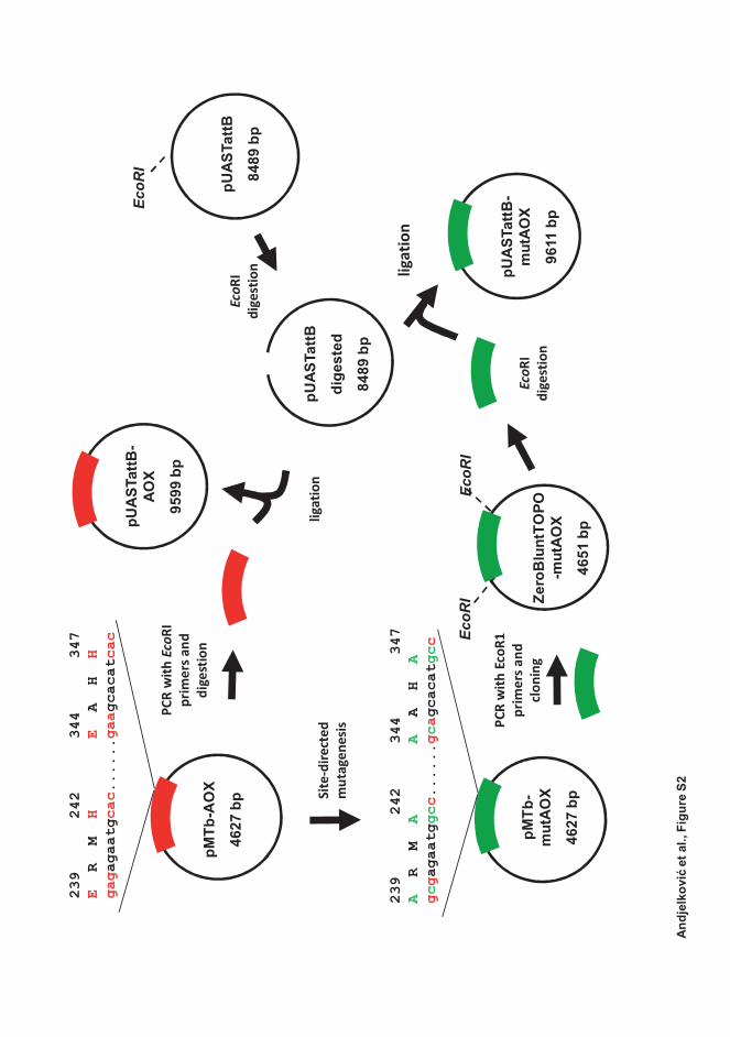
EcoR
I
EcoR
IEc
oRI
239
242
34
4
3
47E
R M
HE
A H
Hgaga
gaat
gcac
....
..gaag
caca
tcac
239
24
2
34
4
347
AR M
AA
A H
Agcgagaat
ggcc
....
..gcag
caca
tgcc
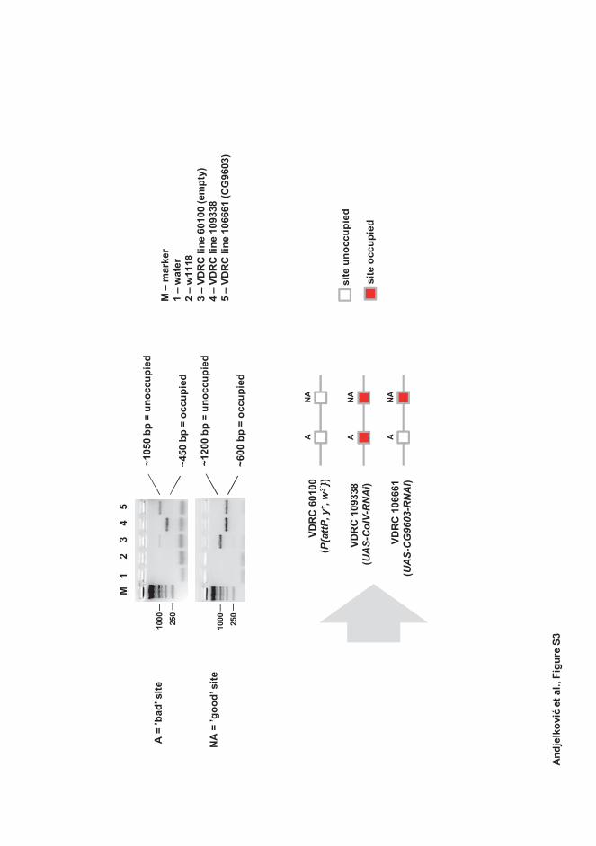
P{at
tP, y
+ , w
3`}
UA
S-C
G96
03-R
NA
i
UA
S-C
oIV-
RN
Ai
PUBLICATION II
Ligand-Bound GeneSwitch Causes Developmental Aberrations in Drosophila that Are Alleviated by the Alternative Oxidase
Ana Andjelkovi , Kia K. Kemppainen & Howard T. Jacobs
G3: Genes, Genomes, Genetics, volume 6, 2839–2846 https://doi.org/ 10.1534/g3.116.030882
Publication reprinted with the permission of the copyright holders.

INVESTIGATION
Ligand-Bound GeneSwitch Causes DevelopmentalAberrations in Drosophila that Are Alleviated bythe Alternative OxidaseAna Andjelkovi�c,*,† Kia K. Kemppainen,*,† and Howard T. Jacobs*,†,‡,1
*BioMediTech, FI-33520 and †Tampere University Hospital, FI-33014, University of Tampere, Finland and ‡Institute ofBiotechnology, University of Helsinki, FI-00014, Finland
ABSTRACT Culture of Drosophila expressing the steroid-dependent GeneSwitch transcriptional activatorunder the control of the ubiquitous a-tubulin promoter was found to produce extensive pupal lethality, aswell as a range of dysmorphic adult phenotypes, in the presence of high concentrations of the inducingdrug RU486. Prominent among these was cleft thorax, seen previously in flies bearing mutant alleles of thenuclear receptor Ultraspiracle and many other mutants, as well as notched wings, leg malformations, andbristle abnormalities. Neither the a-tubulin-GeneSwitch driver nor the inducing drug on their own producedany of these effects. A second GeneSwitch driver, under the control of the daughterless promoter, whichgave much lower and more tissue-restricted transgene expression, exhibited only mild bristle abnormalitiesin the presence of high levels of RU486. Coexpression of the alternative oxidase (AOX) from Ciona intestinalisproduced a substantial shift in the developmental outcome toward a wild-type phenotype, which was depen-dent on the AOX expression level. Neither an enzymatically inactivated variant of AOX, nor GFP, or thealternative NADH dehydrogenase Ndi1 from yeast gave any such rescue. Users of the GeneSwitch systemshould be aware of the potential confounding effects of its application in developmental studies.
KEYWORDS
inducibletransgenes
nuclear receptorDrosophilacleft thoraxnotched wings
TheGeneSwitch (GS) system is commonlyused toactivate transgenes inDrosophila in a graded fashion. GS comprises a modified form of theyeast transcriptional activator Gal4, which is covalently linked to thehormone-binding fragment of the progesterone receptor, rendering itstranscriptional activity dependent on an exogenously supplied proges-terone analog, RU486 or mifepristone (Osterwalder et al. 2001). Anytransgene governed by the UAS promoter element, rendering it Gal4-responsive, may be induced by the combination of GS and RU486 in adose-dependent manner. Depending on the promoter to which GS isitself combined, plus its insertion site in the fly genome, drug-inducibletransgene expression can be achieved in a wide variety of developmen-tal patterns, cell-types, and overall strengths. Thus, the widely used
a-tubulin-GS (tubGS) and actin5C-GS drivers confer ubiquitous,RU486-dependent transgene expression when crossed to lines bearinga UAS-governed transgene. Tissue-specific drivers such as the neuron-specific elav-GS enable transgene expression in just one tissue, but againat a level and timing that can bemanipulated over a wide range. The useof this system is predicated on the assumption that the expression ofGeneSwitch and exposure to RU486 do not themselves produce mea-surable effects on fly physiology and development, which is supportedby controls in many studies.
Our laboratoryhasmadeuseof this system, for example toexpress, inDrosophila, foreign transgenes coding for nonproton-motive alterna-tive respiratory chain enzymes derived from simpler eukaryotes, suchas the alternative oxidase (AOX) from Ciona intestinalis (Fernandez-Ayala et al. 2009; Kemppainen et al. 2014a). When supplied to adultDrosophila bearing both tubGS and a UAS-AOX transgene, RU486produced dose-dependent transgene expression that saturated at drugconcentrations (in fly food) of 100–200mM (Kemppainen et al. 2014a).However, when supplied throughout development, RU486 concentra-tions two orders of magnitude lower were sufficient to induce maximalexpression (Fernandez-Ayala et al. 2009). The precise reasons for thisdiscrepancy in required dose are unclear, although early larvae, whichare very rapidly growing (Church and Robertson 1966; Watts et al.2006), must absorb larger amounts of drugs added to fly food than
Copyright © 2016 Andjelkovic et al.doi: 10.1534/g3.116.030882Manuscript received April 5, 2016; accepted for publication July 6, 2016; publishedEarly Online July 12, 2016.This is an open-access article distributed under the terms of the CreativeCommons Attribution 4.0 International License (http://creativecommons.org/licenses/by/4.0/), which permits unrestricted use, distribution, and reproductionin any medium, provided the original work is properly cited.Supplemental material is available online at www.g3journal.org/lookup/suppl/doi:10.1534/g3.116.030882/-/DC11Corresponding author: Institute of Biotechnology, FI-00014 University of Helsinki,Finland. E-mail: [email protected]
Volume 6 | September 2016 | 2839
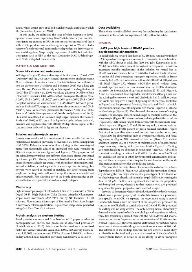
adults, which do not grow at all and even lose weight during early adultlife (Fernandez-Ayala et al. 2009).
In this study, we addressed the issue of what happens to devel-opment when larvae expressing GeneSwitch drivers (but no othertransgene) are exposed to RU486 concentrations in excess of thosesufficient to produce maximal transgene expression. We detected avariety of developmental abnormalities dependent on driver expres-sion and drug dose. Surprisingly, expression of AOX, but not othertransgenes such as GFP or the yeast alternative NADH dehydroge-nase Ndi1, mitigated these effects.
MATERIALS AND METHODS
Drosophila stocks and maintenanceWild-type (Oregon R), standard transgenic host strainsw1118 andwDAH
(Dahomey) and the UAS-GFP (Stinger) line (insertion on chromosome2) were obtained from stock centers. The tubGS driver line with inser-tion on chromosome 3 (Sykiotis and Bohmann 2008) was a kind giftfrom Dr Scott Pletcher (University of Michigan). The daughterless-GS(daGS) line (Tricoire et al. 2009) was a kind gift from Dr Alberto Sanz(Newcastle University, UK). AOX andNdi1 transgenic flies [linesUAS-AOXF6, UAS-AOXF24, tub-AOX7, tub-AOX35 tub-AOX50, UAS-AOX7.1
(targeted insertion on chromosome 3) UAS-AOXmut (denoted previ-ously as UAS-AOX4.1, targeted insertion on chromosome 3), and UAS-Ndi1B20] were as described previously (Fernandez-Ayala et al. 2009;Sanz et al. 2010b; Kemppainen et al. 2014b; Andjelkovi�c et al. 2015).Flies were maintained in standard high-sugar medium (Fernandez-Ayala et al. 2009) at 25�, on a 12 hr light/dark cycle. Where indicated,medium was supplemented with RU486 (Mifepristone, Sigma) at theconcentrations indicated in figures and legends.
Eclosion and phenotypic assaysCrosses were conducted in a minimum of three, usually four to fivereplicates, as described previously (Toivonen et al. 2001; Kemppainenet al. 2009). Either the number of flies eclosing or the percentage ofpupae that successfully eclosed in individual vials were recorded indifferent experiments (see figures and legends). The proportion ofthe eclosed progeny falling into different phenotypic classes was scoredby microscopy. Cleft thorax, where subclassified, was scored as mild orsevere (heminota clearly separated), with the mildest abnormality, mal-formed scutellum, scored separately in some experiments. Wing phe-notypes were scored as normal or notched, the latter ranging fromsingle notches to grossly malformed wings that in some cases did notinflate properly. Flies showing any of the bristle abnormalities as de-scribed below were generally scored as a single category.
MicroscopyLight microscopy images of eclosed adult flies were taken with a NikonDigital DS-Fi1 High-Definition Color Camera, using the Nikon stereo-scopic zoom microscope SMZ 745T run by NIS-Elements D 4.20software. Fluorescence microscopy of flies used a Zeiss Axio Imager2 microscope (50 ·magnification). Z projection images were generatedusing Carl Zeiss Zen 2012 software.
Protein analysis by western blottingTotal protein was extracted from batches of 20 pupae crushed inhomogenization buffer, and processed as described previously(Andjelkovi�c et al. 2015). Primary antibodies used were customizedrabbit anti-AOX (Fernandez-Ayala et al. 2009; 21st Centrury Biochem-icals, 1:10,000), and mouse anti-ATP5A (Abcam, 1:100,000), with sec-ondary antibodies as described previously (Andjelkovi�c et al. 2015).
Data availabilityThe authors state that all data necessary for confirming the conclusionspresented in the article are represented fully within the article.
RESULTS
tubGS plus high levels of RU486 producedevelopmental abnormalitiesIn initial trials, we noticed that doses of RU486 used routinely to induceUAS-dependent transgene expression in Drosophila, in combinationwith the tubGS driver in adult flies (200–500 mM; Kemppainen et al.2014a), were lethal when present throughout development. In order toinvestigate possible mechanisms of this lethality, we reared flies atRU486 doses intermediate between this lethal level, and levels sufficientto induce full dose-dependent transgene expression, which in larvaewas only 1–2 mM. In combination with tubGS, RU486 at 100 mM wasstill lethal (Figure 1A), whereas tubGS flies reared without drug,or wild-type flies reared at this concentration of RU486, developednormally. At intermediate drug concentrations (5–50 mM, Figure 1,A and B), we observed dose-dependent semilethality, althoughmany ofthe eclosing flies were very weak and died within 1 d. In addition, eventhe viable flies displayed a range of dysmorphic phenotypes, illustratedin Figure 2 and Supplemental Material, Figure S1 and File S1, of whichthe commonest andmost striking were cleft thorax (Figure 2, B–D) andnotched wings (Figure 2E). The observed phenotypes were of varyingseverity. For example, some flies had single or multiple notches at thewingmargin (Figure 2E), whereas others had wings that failed to inflate(Figure 2F). Cleft thorax ranged from severe, with the heminota com-pletely separated (Figure 2, C and D), to very mild, showing only anabnormal, parted bristle pattern or just a reduced scutellum (Figure2A). A minority of flies also showed necrotic tissue in the notum area(Figure 2D), leg abnormalities such as overgrown, reduced, and fusedleg segments (Figure 2G), externalized trachea (Figure 2H), cleftedabdomen (Figure 2I), or a variety of malformations of macrochaetae(supernumerary, missing, kinked, or short bristles, Figure S1). Cleftingalso extended along the abdomen in some cases (Figure 2I). tubGS fliesreared without drug, or cultured in RU486 in the absence of tubGS, didnot exhibit cleft thorax or other developmental abnormalities, indicat-ing that these teratogenic effects require the combination of the mod-ified transcription factor plus the inducing steroid.
We quantified themain classes of abnormality and observed a dose-dependence on RU486 (Figure 3A). Although the proportion of prog-eny showing the two major dysmorphic phenotypes of cleft thorax ornotched wings was already substantial at 10mMRU486, increasing thedose to 30 mM resulted in a significant increase in the proportionexhibiting cleft thorax, whereas a further increase to 50 mM produceda significantly greater proportion with notched wings.
In order to determine whether the induction of these developmentaldefects was a general property of GeneSwitch drivers, or a phenome-non specific to tubGS, we repeated the experiment using a secondGeneSwitch driver under the control of the daughterless promoter. Incontrast to tubGS, daGS in combination with 10 mM RU486 producedno clefting and no wing defects. The only developmental abnormalitydetected was in regard to bristle morphology and organization which,while less frequently observed than with the tubGS driver, did show atendency to rise in frequency as the concentration of RU486 was in-creased (Figure 3C). However, neither cleft thorax nor notched wingswere seen at these elevated drug concentrations, nor even at 100 mM.The difference in the findings between the two drivers is most likelyattributable to the level and pattern of expression of the GeneSwitchtranscription factor, as reflected in its ability to drive transgene
2840 | A. Andjelkovi�c, K. K. Kemppainen, and H. T. Jacobs
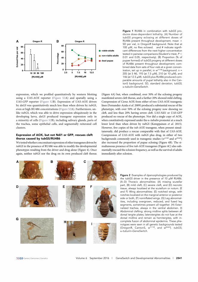
expression, which we profiled quantitatively by western blottingusing a UAS-AOX reporter (Figure S2A) and spatially using aUAS-GFP reporter (Figure S2B). Expression of UAS-AOX drivenby daGS was quantitatively much less than when driven by tubGS,even at high RU486 concentrations (Figure S2A). Furthermore, un-like tubGS, which was able to drive expression ubiquitously in thedeveloping larva, daGS produced transgene expression only ina minority of cells (Figure S2B), including salivary glands, parts ofthe trachea, some epithelial cells, and segmentally reiterated cellclusters.
Expression of AOX, but not Ndi1 or GFP, rescues cleftthorax caused by tubGS/RU486Wetestedwhetherconcomitantexpressionofother transgenesdrivenbytubGS in the presence of RU486 was able to modify the developmentalphenotypes resulting from the driver and drug alone (Figure 4). Onceagain, neither tubGS nor the drug on its own produced cleft thorax
(Figure 4A) but, when combined, over 50% of the eclosing progenymanifested severe cleft thorax, and a further 20% showed mild clefting.Coexpression of Ciona AOX from either of two UAS-AOX transgeniclines (Fernandez-Ayala et al. 2009) produced a substantial rescue of thephenotype, with over 50% of the eclosing progeny now showing nocleft, and less than 20% having severe cleft. UAS-Ndi1 or UAS-GFPproduced no rescue of the phenotype. Nor did a single copy of AOX,when constitutively expressed under the a-tubulin promoter at a muchlower level than when driven by tubGS (Kemppainen et al. 2015).However, five copies of the tub-AOX transgene, when present simul-taneously, did produce a rescue comparable with that of UAS-AOX.Coexpression of UAS-AOX with tubGS plus drug, in either of twobackgrounds commonly used in transgenic studies (w1118 and wDAH)also increased the proportion of pupae eclosing (Figure 4B). The si-multaneous presence of five tub-AOX transgenes (Figure 4C) also sub-stantially rescued the eclosion frequency, as well as the survival of adultsimmediately after eclosion.
Figure 1 RU486 in combination with tubGS pro-duces dose-dependent lethality. (A) Number oftubGS progeny eclosing at different doses ofRU486 present throughout development, mean 6SD per vial, in OregonR background. Note that at100 mM, no flies eclosed. � and # indicate signifi-cant differences from the next higher concentrationtested in pairwise comparisons (Student’s t-test, P,0.01 and 0.05, respectively). (B) Proportion (% ofpupae formed) of tubGS progeny at different dosesof RU486 present throughout development; com-bined data from sets of four vials at a given concen-tration, set up in parallel, in w1118 background. n =205 (at 5 M), 193 (at 7.5 mM), 210 (at 10 mM), and146 (at 12.5 mM). tubGS plus RU486 produced com-parable amounts of pupal lethality also in the Can-tonS background. SD, standard deviation; tubGS;a-tubulin-GeneSwitch.
Figure 2 Examples of dysmorphologies produced bythe tubGS driver in the presence of 10 mM RU486.(A–D) Thoracic abnormalities: (A) missing scutellarpart, (B) mild cleft, (C) severe cleft, and (D) necrotictissue, always localized at the scutellum or notum. (Eand F) Wing abnormalities: (E) notched wings, withnotches localized on the marginal anterior or posteriorside or both, (F) noninflated wings. (G) Leg abnormal-ities, including overgrown, reduced, and fused legsegments, sometimes present all together. (H) Exter-nalized trachea, always in the ventral abdomen. (I)Abdominal clefting: strong midline splits between alldorsal tergite plates; laterotergites do not fuse at thedorsal midline and remain as hemitergites, with in-complete fusion of abdominal epidermis. These phe-notypes were seen in all genetic backgrounds tested(OregonR, CantonS, w1118, and wDAH). tubGS;a-tubulin-GeneSwitch.
Volume 6 September 2016 | GeneSwitch and Developmental Abnormalities | 2841
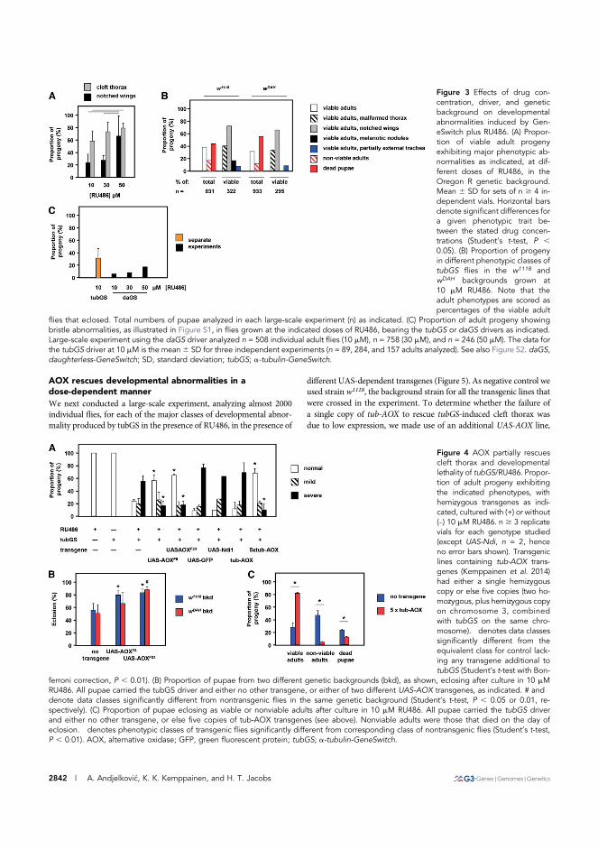
AOX rescues developmental abnormalities in adose-dependent mannerWe next conducted a large-scale experiment, analyzing almost 2000individual flies, for each of the major classes of developmental abnor-mality produced by tubGS in the presence of RU486, in the presence of
different UAS-dependent transgenes (Figure 5). As negative control weused strain w1118, the background strain for all the transgenic lines thatwere crossed in the experiment. To determine whether the failure ofa single copy of tub-AOX to rescue tubGS-induced cleft thorax wasdue to low expression, we made use of an additional UAS-AOX line,
Figure 3 Effects of drug con-centration, driver, and geneticbackground on developmentalabnormalities induced by Gen-eSwitch plus RU486. (A) Propor-tion of viable adult progenyexhibiting major phenotypic ab-normalities as indicated, at dif-ferent doses of RU486, in theOregon R genetic background.Mean 6 SD for sets of n $ 4 in-dependent vials. Horizontal barsdenote significant differences fora given phenotypic trait be-tween the stated drug concen-trations (Student’s t-test, P ,0.05). (B) Proportion of progenyin different phenotypic classes oftubGS flies in the w1118 andwDAH backgrounds grown at10 mM RU486. Note that theadult phenotypes are scored aspercentages of the viable adult
flies that eclosed. Total numbers of pupae analyzed in each large-scale experiment (n) as indicated. (C) Proportion of adult progeny showingbristle abnormalities, as illustrated in Figure S1, in flies grown at the indicated doses of RU486, bearing the tubGS or daGS drivers as indicated.Large-scale experiment using the daGS driver analyzed n = 508 individual adult flies (10 mM), n = 758 (30 mM), and n = 246 (50 mM). The data forthe tubGS driver at 10 mM is the mean6 SD for three independent experiments (n = 89, 284, and 157 adults analyzed). See also Figure S2. daGS,daughterless-GeneSwitch; SD, standard deviation; tubGS; a-tubulin-GeneSwitch.
Figure 4 AOX partially rescuescleft thorax and developmentallethality of tubGS/RU486. Propor-tion of adult progeny exhibitingthe indicated phenotypes, withhemizygous transgenes as indi-cated, cultured with (+) or without(–) 10 mM RU486. n $ 3 replicatevials for each genotype studied(except UAS-Ndi, n = 2, henceno error bars shown). Transgeniclines containing tub-AOX trans-genes (Kemppainen et al. 2014)had either a single hemizygouscopy or else five copies (two ho-mozygous, plus hemizygous copyon chromosome 3, combinedwith tubGS on the same chro-mosome). � denotes data classessignificantly different from theequivalent class for control lack-ing any transgene additional totubGS (Student’s t-test with Bon-
ferroni correction, P , 0.01). (B) Proportion of pupae from two different genetic backgrounds (bkd), as shown, eclosing after culture in 10 mMRU486. All pupae carried the tubGS driver and either no other transgene, or either of two different UAS-AOX transgenes, as indicated. # and �
denote data classes significantly different from nontransgenic flies in the same genetic background (Student’s t-test, P , 0.05 or 0.01, re-spectively). (C) Proportion of pupae eclosing as viable or nonviable adults after culture in 10 mM RU486. All pupae carried the tubGS driverand either no other transgene, or else five copies of tub-AOX transgenes (see above). Nonviable adults were those that died on the day ofeclosion. � denotes phenotypic classes of transgenic flies significantly different from corresponding class of nontransgenic flies (Student’s t-test,P , 0.01). AOX, alternative oxidase; GFP, green fluorescent protein; tubGS; a-tubulin-GeneSwitch.
2842 | A. Andjelkovi�c, K. K. Kemppainen, and H. T. Jacobs
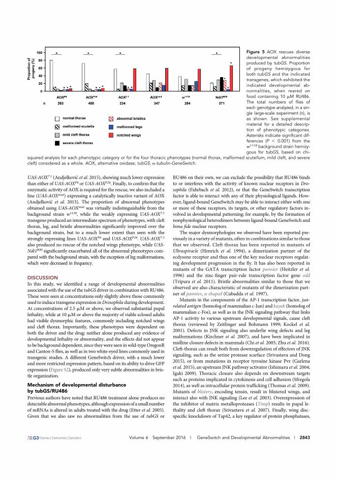
UAS-AOX7.1 (Andjelkovi�c et al. 2015), showing much lower expressionthan either ofUAS-AOXF6 orUAS-AOXF24. Finally, to confirm that theenzymatic activity of AOX is required for the rescue, we also included aline (UAS-AOXmut) expressing a catalytically inactive variant of AOX(Andjelkovi�c et al. 2015). The proportion of abnormal phenotypesobtained using UAS-AOXmut was virtually indistinguishable from thebackground strain w1118, while the weakly expressing UAS-AOX7.1
transgene produced an intermediate spectrum of phenotypes, with cleftthorax, leg, and bristle abnormalities significantly improved over thebackground strain, but to a much lower extent than seen with thestrongly expressing lines UAS-AOXF6 and UAS-AOXF24. UAS-AOX7.1
also produced no rescue of the notched wings phenotype, while UAS-Ndi1B20 significantly exacerbated all of the abnormal phenotypes com-pared with the background strain, with the exception of leg malformations,which were decreased in frequency.
DISCUSSIONIn this study, we identified a range of developmental abnormalitiesassociated with the use of the tubGS driver in combination with RU486.These were seen at concentrations only slightly above those commonlyused to induce transgene expression inDrosophila during development.At concentrations of 2.5 mM or above, we observed substantial pupallethality, while at 10 mM or above the majority of viable eclosed adultshad visible dysmorphic features, commonly including notched wingsand cleft thorax. Importantly, these phenotypes were dependent onboth the driver and the drug: neither alone produced any evidence ofdevelopmental lethality or abnormality, and the effects did not appearto be background dependent, since theywere seen inwild-typeOregonRand Canton-S flies, as well as in two white-eyed lines commonly used intransgenic studies. A different GeneSwitch driver, with a much lowerand more restricted expression pattern, based on its ability to drive GFPexpression (Figure S2), produced only very subtle abnormalities in bris-tle organization.
Mechanism of developmental disturbanceby tubGS/RU486Previous authors have noted that RU486 treatment alone produces nodetectable abnormalphenotypes, althoughexpressionof a smallnumberof mRNAs is altered in adults treated with the drug (Etter et al. 2005).Given that we also saw no abnormalities from the use of tubGS or
RU486 on their own, we can exclude the possibility that RU486 bindsto or interferes with the activity of known nuclear receptors in Dro-sophila (Fahrbach et al. 2012), or that the GeneSwitch transcriptionfactor is able to interact with any of their physiological ligands. How-ever, ligand-bound GeneSwitch may be able to interact either with oneor more of these receptors, its targets, or other regulatory factors in-volved in developmental patterning; for example, by the formation ofnonphysiological heterodimers between ligand-bound GeneSwitch andbona fide nuclear receptors.
The major dysmorphologies we observed have been reported pre-viously in a variety ofmutants, often in combinations similar to thosethat we observed. Cleft thorax has been reported in mutants ofUltraspiracle (Henrich et al. 1994), a dimerization partner of theecdysone receptor and thus one of the key nuclear receptors regulat-ing development progression in the fly. It has also been reported inmutants of the GATA transcription factor pannier (Heitzler et al.1996) and the zinc-finger pair-rule transcription factor gene odd(Tripura et al. 2011). Bristle abnormalities similar to those that weobserved are also characteristic of mutants of the dimerization part-ner of pannier, u-shaped (Cubadda et al. 1997).
Mutants in the components of the AP-1 transcription factor, jun-related antigen (homolog of mammalian c-Jun) and kayak (homolog ofmammalian c-Fos), as well as in the JNK signaling pathway that linksAP-1 activity to various upstream developmental signals, cause cleftthorax (reviewed by Zeitlinger and Bohmann 1999; Kockel et al.2001). Defects in JNK signaling also underlie wing defects and legmalformations (Kirchner et al. 2007), and have been implicated inmidline closure defects in mammals (Chi et al. 2005; Zhu et al. 2016).Cleft-thorax can result both from downregulation of effectors of JNKsignaling, such as the serine protease scarface (Srivastava and Dong2015), or from mutations in receptor tyrosine kinase Pvr (Garlenaet al. 2015), an upstream JNK pathway activator (Ishimaru et al. 2004;Igaki 2009). Thoracic closure also depends on downstream targetssuch as proteins implicated in cytokinesis and cell adhesion (Sfregola2014), as well as intracellular protein trafficking (Thomas et al. 2009).Mutants of blistery, encoding tensin, result in blistered wings, andinteract also with JNK signaling (Lee et al. 2003). Overexpression ofthe inhibitor of matrix metalloproteases (Timp) results in pupal le-thality and cleft thorax (Srivastava et al. 2007). Finally, wing disc-specific knockdown of Tap42, a key regulator of protein phosphatases,
Figure 5 AOX rescues diversedevelopmental abnormalitiesproduced by tubGS. Proportionof progeny hemizygous forboth tubGS and the indicatedtransgenes, which exhibited theindicated developmental ab-normalities, when reared onfood containing 10 mM RU486.The total numbers of flies ofeach genotype analyzed, in a sin-gle large-scale experiment (n), isas shown. See supplementalmaterial for a detailed descrip-tion of phenotypic categories.Asterisks indicate significant dif-ferences (P , 0.001) from thew1118 background strain hemizy-gous for tubGS, based on chi-
squared analysis for each phenotypic category or for the four thoracic phenotypes (normal thorax, malformed scutellum, mild cleft, and severecleft) considered as a whole. AOX, alternative oxidase; tubGS; a-tubulin-GeneSwitch.
Volume 6 September 2016 | GeneSwitch and Developmental Abnormalities | 2843
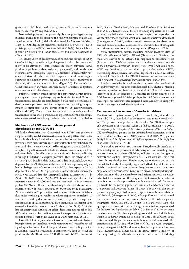
gives rise to cleft thorax and to wing abnormalities similar to somethat we observed (Wang et al. 2012).
Notched wings are another previously observed phenotype in manymutants, including those affecting the highly pleiotropic intercellularsignaling factor Notch (originally discovered by Morgan; Welshons1958), SNARE-dependent membrane trafficking (Stewart et al. 2001),protein phosphatase PP2A (Kunttas-Tatli et al. 2009), the RNA-bind-ing fragile X protein FMR1 (Wan et al. 2000), and histone deacetylation(Pile et al. 2001).
The exact pattern of developmental abnormalities brought about byGeneSwitch together with its ligand appears to reflect the tissue spec-ificity of its expression. Thus, whereas the widely expressed tubGSproduces a plethora of abnormal phenotypes, daGS, with much morerestricted larval expression (Figure S2), primarily in segmentally reit-erated clusters of cells that might represent larval sense organs(Brewster and Bodmer 1995), has only a single visible phenotype inthe adult, affecting the sensory bristles (Figure 3C). The use of otherGeneSwitch drivers may help to further clarify how its level and patternof expression affect the phenotypic outcome.
Finding a common thread through this rather bewildering array ofphenotypes and genetic pathwaysmay not be straightforward. However,transcriptional cascades are considered to be the main determinants ofdevelopmental processes, and the key system for regulating morpho-genesis at pupal stage is the steroid hormone 20-hydroxyecdysone(Riddiford 1993). Thus, an interference with ecdysteroid-dependenttranscription is the most parsimonious explanation for the pleiotropiceffects we observed, even thoughmolecular details remain to be filled in.
Mechanism of AOX rescue of developmentaldisturbance by tubGS/RU486While the observation that GeneSwitch-plus-RU486 can produce arange of developmental abnormalities may be unexpected, their rescueby a mitochondrially localized electron-transfer protein from anotherphylum is even more surprising. It is important to note that, while theabnormal phenotypes were produced by using an engineered (and thusnonphysiological) transcription factor, andwere rescued by a gene froma distant phylum, the effects were systematic in both cases, indicatingmeaningful underlying biological processes. Thus, the extent of AOXrescue of pupal lethality, cleft thorax, and other dysmorphologies wasdependenton theAOXexpression level, since strains expressingonlyatalow level (single-copy of constitutive tub-AOX, or low-expressor GAL4-dependent lineUAS-AOX7.1) produced a less dramatic alleviation of thephenotypes studied than the corresponding high-expressors (5 · tub-AOX, UAS-AOXF24, and UAS-AOXF6). Rescue was dependent on theenzymatic activity of AOX and was not seen with an inert reporterprotein (GFP) or a different mitochondrially localized electron-transferprotein, yeast Ndi, which appeared to exacerbate some phenotypes.AOX maintains ATP production, redox homeostasis, and metabolicflux under physiological conditions where respiratory complexes IIIand IV are limiting due to overload, toxins, or genetic damage, andconcomitantly limits mitochondrial ROS production consequent uponoverreduction of the quinone pool (El-Khoury et al. 2014). AOX alsohas an unexplained antioxidant effect, decreasing net mitochondrialROS output even under conditions where the respiratory chain is func-tioning normally (Fernandez-Ayala et al. 2009; Sanz et al. 2010a).
How this links to a global alleviation of developmental perturbationsbrought about by interference with transcriptional cascades or cellsignaling is far from clear. In a general sense, our findings hint ata common metabolic regulation of transcription, such as evidencedpreviously byAMPKsirtuins or PARP (Kraus and Lis 2003; Ghosh et al.
2010; Gut and Verdin 2013; Schiewer and Knudsen 2014; Salminenet al. 2016), although none of these is obviously implicated, so a novelpathwaymay be involved. Inmice, nuclear receptors are responsive to avariety ofmetabolic effectors, which can also bemicrobiome-dependent(Montagner et al. 2016), while cross-talk between nutrient-based sen-sors and nuclear receptors is dependent on mitochondrial stress signalsand influences mitochondrial gene expression (Kang et al. 2015).
Many transcription factors, including nuclear receptors such asLXRa (Serviddio et al. 2013) or NR4A1 (Shimizu et al. 2015) in mam-mals, are known to be activated in response to oxidative stress(Lavrovsky et al. 2000), and redox regulation of nuclear receptors suchas the glucocorticoid receptor (Tanaka et al. 1999) is well established.AOX may therefore act by providing a general dampening of ROS,normalizing developmental outcomes dependent on such receptors,with which GeneSwitch plus RU486 interferes. An exhaustive studyusing different ROS scavengers may shed further light on this.
Another possibility is based on the observation that synthesis of20-hydroxyecdysone requires mitochondrial Fe-S cluster-containingproteins dependent on frataxin (Palandri et al. 2015) and mitoferrin(Llorens et al. 2015). Because Fe-S proteins are highly susceptible toROS damage, a general ROS dampening effect of AOXmay counteracttranscriptional interference from ligand-bound GeneSwitch, simply byboosting endogenous ecdysteroid synthesis.
Recommendations on use of GeneSwitch driversThe GeneSwitch system was originally elaborated using other driversthan tubGS, i.e., those linked to the neuron- and muscle-specific elavand Mhc promoters, respectively (Osterwalder et al. 2001), or for spe-cific expression in other tissues such as the fat body (Roman et al. 2001).Subsequently, the “ubiquitous”GS drivers (such as tubGS andActin5C-GS) have been brought into use for inducing broad expression, both inadults and larvae (Ford et al. 2007; Waskar et al. 2009; Wigby et al.2011; Paik et al. 2012; Kuo et al. 2012; Kemppainen et al. 2014a,b; Sunet al. 2014; Da-Rè et al. 2014).
Our work raises at least two concerns. First, the visible interferencewith developmental processes at saturating or near-saturating drugconcentrations, using the tubGS driver, indicates the need for rigorouscontrols and cautious interpretation of all data obtained using thisdriver during development. Furthermore, we obviously cannot ruleout subtler but also biologically significant effects that did not havevisible manifestations, even at lower drug concentrations than thoseemployed here. Second, other GeneSwitch drivers activated during de-velopment may also be vulnerable to such effects, since our data indi-cate that they depend on the drug and the transcription factor incombination, which applies wherever they are colocated. An exam-ple would be the recently published use of a GeneSwitch driver tooverexpress malic enzyme (Kim et al. 2015). The driver in this exam-ple was originally reported to induce expression in the adult abdom-inal fat body (Hwangbo et al. 2004), although Kim et al. (2015) foundthat expression in larvae was instead driven in the salivary glands,Malpighian tubule, and part of the gut. In this particular paper, theappropriate controls without the transgene were indeed implementedfor the adult (see Supplementary Table 1C of Kim et al. 2015), but somequestions remain. The driver plus drug alone did not affect the bodyweight of L3 larvae (Figure 3A of Kim et al. 2015), but effects on stressresistance and lifespan in such controls were not documented. Theconcentrations of RU486 used by Kim et al. (2015), i.e., 2.5–10 mg/ml,corresponding with 5.8–23 mM, were within the range in which we sawmajor developmental effects using the tubGS driver. Similarly, inflies expressing GeneSwitch in specific endocrine cells during
2844 | A. Andjelkovi�c, K. K. Kemppainen, and H. T. Jacobs
development, using a customized driver and RU486 at even higherconcentrations from larval L2 stage onwards (Cho et al. 2014), cleardevelopmental abnormalities were attributed to knockdown of a nu-clear receptor, although driver-plus-drug controls were not includedin all of the experiments reported. Some phenotypes observed (Figure 3of Cho et al. 2014) resemble those that we report here (pupal lethality,uninflated wings, abdominal clefting, and leg malformations). Whiletheir interpretation that these are due to disrupted ecdysone signalingmay be correct, an effect of GeneSwitch plus RU486 in the target cellscannot be excluded. Phenotypic rescue by injected ETH (Table 2 ofCho et al. 2014) confirmed the involvement of disrupted ecdysis, butnot the underlying causes thereof. A further possible example alreadyreported in the literature is the effect of the abdominal fat body-specific GeneSwitch driver on lifespan, when RU486-containing foodwas supplied in the adult to drive the supposedly inert GFP transgene(Ren andHughes, 2014. RU486-dependent lethality in larvae containingthe Elav-GeneSwitch driver (Shen et al. 2009), and embryonic lethal-ity produced by either the Elav- or Actin5C-GeneSwitch drivers plusmaternal RU486 (Landis et al. 2015), have been previously reported.
We would recommend that future users of all GeneSwitch driversshould routinely include otherwise nontransgenic controls bearing thedrivers, plus and minus drug, in all experiments. Based on our findings(Figure S2B), the daGS driver is clearly not ubiquitous, despite the factthat the daughterless gene itself, as well as the “standard” daGAL4drivers, do show widespread expression.
ACKNOWLEDGMENTSWe thank Alberto Sanz and Scott Pletcher for kindly supplying Dro-sophila strains, Dmytro Gospodaryov for valuable discussions, andAnnika Ketola, Ville Someri, and Tea Tuomela for technical assistance.This work was supported by funding from the European ResearchCouncil (advanced grant 232738 to H.T.J.), Academy of Finland; Uni-versity of Tampere; Tampere University Hospital Medical ResearchFund; the Sigrid Juselius Foundation, and the Finnish Cultural Foun-dation (grant to A.A.). The authors declare no conflict of interest.
LITERATURE CITEDAndjelkovi�c, A., M. T. Oliveira, G. Cannino, C. Yalgin, P. K. Dhandapani
et al., 2015 Diiron centre mutations in Ciona intestinalis alternativeoxidase abolish enzymatic activity and prevent rescue of cytochromeoxidase deficiency in flies. Sci. Rep. 5: 18295.
Brewster, R., and R. Bodmer, 1995 Origin and specification of type IIsensory neurons in Drosophila. Development 121: 2923–2936.
Chi, H., M. R. Sarkisian, P. Rakic, and R. A. Flavell, 2005 Loss of mitogen-activated protein kinase kinase kinase 4 (MEKK4) results in enhancedapoptosis and defective neural tube development. Proc. Natl. Acad. Sci.USA 102: 3846–3851.
Cho, K. H., I. Daubnerová, Y. Park, D. Zitnan, and M. E. Adams,2014 Secretory competence in a gateway endocrine cell conferred by thenuclear receptor bFTZ-F1 enables stage-specific ecdysone responsesthroughout development in Drosophila. Dev. Biol. 385: 253–262.
Church, R. B., and F. W. Robertson, 1966 A biochemical study of thegrowth of Drosophila melanogaster. J. Exp. Zool. 162: 337–352.
Cubadda, Y., P. Heitzler, R. P. Ray, M. Bourouis, P. Ramain et al.,1997 u-shaped encodes a zinc finger protein that regulates the proneuralgenes achaete and scute during the formation of bristles in Drosophila.Genes Dev. 11: 3083–3095.
Da-Rè, C., S. von Stockum, A. Biscontin, C. Millino, P. Cisotto et al.,2014 Leigh syndrome in Drosophila melanogaster: morphological andbiochemical characterization of Surf1 post-transcriptional silencing.J. Biol. Chem. 289: 29235–29246.
El-Khoury, R., K. K. Kemppainen, E. Dufour, M. Szibor, H. T. Jacobs et al.,2014 Engineering the alternative oxidase gene to better understand
and counteract mitochondrial defects: state of the art and perspectives.Br. J. Pharmacol. 171: 2243–2249.
Etter, P. D., R. Narayanan, Z. Navratilova, C. Patel, D. Bohmann et al.,2005 Synaptic and genomic responses to JNK and AP-1 signaling inDrosophila neurons. BMC Neurosci. 6: 39.
Fahrbach, S. E., G. Smagghe, and R. A. Velarde, 2012 Insect nuclear re-ceptors. Annu. Rev. Entomol. 57: 83–106.
Fernandez-Ayala, D. J., A. Sanz, S. Vartiainen, K. K. Kemppainen, M. Babusiaket al., 2009 Expression of the Ciona intestinalis alternative oxidase(AOX) in Drosophila complements defects in mitochondrial oxidativephosphorylation. Cell Metab. 9: 449–460.
Ford, D., N. Hoe, G. N. Landis, J. Tozer, A. Luu et al., 2007 Alteration ofDrosophila life span using conditional, tissue-specific expression oftransgenes triggered by doxycyline or RU486/Mifepristone. Exp. Gerontol.42: 483–497.
Garlena, R. A., A. L. Lennox, L. R. Baker, T. E. Parsons, S. M. Weinberg et al.,2015 The receptor tyrosine kinase Pvr promotes tissue closure by co-ordinating corpse removal and epidermal zippering. Development 142:3403–3415.
Ghosh, S., S. George, U. Roy, D. Ramachandran, and U. Kolthur-Seetharam,2010 NAD: a master regulator of transcription. Biochim. Biophys.Acta 1799: 681–693.
Gut, P., and E. Verdin, 2013 The nexus of chromatin regulation and in-termediary metabolism. Nature 502: 489–498.
Heitzler, P., M. Haenlin, P. Ramain, M. Calleja, and P. Simpson, 1996 Agenetic analysis of pannier, a gene necessary for viability of dorsal tissuesand bristle positioning in Drosophila. Genetics 143: 1271–1286.
Henrich, V. C., A. A. Szekely, S. J. Kim, N. E. Brown, C. Antoniewski et al.,1994 Expression and function of the ultraspiracle (usp) gene duringdevelopment of Drosophila melanogaster. Dev. Biol. 165: 38–52.
Hwangbo, D. S., B. Gershman, M. P. Tu, M. Palmer, and M. Tatar,2004 Drosophila dFOXO controls lifespan and regulates insulinsignalling in brain and fat body. Nature 429: 562–566.
Igaki, T., 2009 Correcting developmental errors by apoptosis: lessons fromDrosophila JNK signaling. Apoptosis 14: 1021–1028.
Ishimaru, S., R. Ueda, Y. Hinohara, M. Ohtani, and H. Hanafusa, 2004 PVRplays a critical role via JNK activation in thorax closure during Dro-sophila metamorphosis. EMBO J. 23: 3984–3989.
Kang, Y. K., N. Putluri, S. Maity, A. Tsimelzon, O. Ilkayeva et al.,2015 CAPER is vital for energy and redox homeostasis by integratingglucose-induced mitochondrial functions via ERR-a-Gabpa and stress-induced adaptive responses via NF-kB-cMYC. PLoS Genet. 11:e1005116.
Kemppainen, E., D. J. Fernández-Ayala, L. C. Galbraith, K. M. C. O’Dell, andH. T. Jacobs, 2009 Phenotypic suppression of the Drosophila mito-chondrial disease-like mutant tko(25t) by duplication of the mutant genein its natural chromosomal context. Mitochondrion 9: 353–363.
Kemppainen, K. K., E. Kemppainen, and H. T. Jacobs, 2014a The alterna-tive oxidase AOX does not rescue the phenotype of tko25t mutant flies.G3 (Bethesda) 4: 2013–2021.
Kemppainen, K. K., J. Rinne, A. Sriram, M. Lakanmaa, A. Zeb et al.,2014b Expression of alternative oxidase in Drosophila ameliorates di-verse phenotypes due to cytochrome oxidase deficiency. Hum. Mol.Genet. 23: 2078–2093.
Kim, G. H., Y. E. Lee, G. H. Lee, Y. H. Cho, Y. N. Lee et al., 2015 Over-expression of malic enzyme in the larval stage extends Drosophila life-span. Biochem. Biophys. Res. Commun. 456: 676–682.
Kirchner, J., S. Gross, D. Bennett, and L. Alphey, 2007 The nonmusclemyosin phosphatase PP1beta (flapwing) negatively regulates JunN-terminal kinase in wing imaginal discs of Drosophila. Genetics 175:1741–1749.
Kockel, L., J. G. Homsy, and D. Bohmann, 2001 Drosophila AP-1: lessonsfrom an invertebrate. Oncogene 20: 2347–2364.
Kraus, W. L., and J. T. Lis, 2003 PARP goes transcription. Cell 113: 677–683.
Kunttas-Tatli, E., A. Bose, B. Kahali, and C. P. Bishop, and A. P. Bidwai,2009 Functional dissection of Timekeeper (Tik) implicates opposite
Volume 6 September 2016 | GeneSwitch and Developmental Abnormalities | 2845
roles for CK2 and PP2A during Drosophila neurogenesis. Genesis 47:647–658.
Kuo, T. H., T. Y. Fedina, I. Hansen, K. Dreisewerd, H. A. Dierick et al.,2012 Insulin signaling mediates sexual attractiveness in Drosophila.PLoS Genet. 8: e1002684.
Landis, G. N., M. P. Salomon, D. Keroles, N. Brookes, T. Sekimura et al.,2015 2015 The progesterone antagonist mifepristone/RU486 blocks thenegative effect on life span caused by mating in female Drosophila. Aging(Albany, N.Y.) 7: 53–69.
Lavrovsky, Y., B. Chatterjee, R. A. Clark, and A. K. Roy, 2000 Role of redox-regulated transcription factors in inflammation, aging and age-relateddiseases. Exp. Gerontol. 35: 521–532.
Lee, S. B., K. S. Cho, E. Kim, and J. Chung, 2003 blistery encodes Drosophilatensin protein and interacts with integrin and the JNK signaling path-way during wing development. Development 130: 4001–4010.
Llorens, J. V., C. Metzendorf, F. Missirlis, and M. I. Lind, 2015 Mito-chondrial iron supply is required for the developmental pulse of ecdysonebiosynthesis that initiates metamorphosis in Drosophila melanogaster.J. Biol. Inorg. Chem. 20: 1229–1238.
Montagner, A., A. Korecka, and A. Polizzi, Y. Lippi, Y. Blum et al.,2016 Hepatic circadian clock oscillators and nuclear receptors integratemicrobiome-derived signals. Sci. Rep. 6: 20127.
Osterwalder, T., K. S. Yoon, B. H. White, and H. Keshishian, 2001 Aconditional tissue-specific transgene expression system using inducibleGAL4. Proc. Natl. Acad. Sci. USA 98: 12596–12601.
Paik, D. Y. G., Y. E. Jang, Y. N. Lee, R. Lee, R. Yamamoto et al.,2012 Misexpression screen delineates novel genes controlling Dro-sophila lifespan. Mech. Ageing Dev. 133: 234–245.
Palandri, A., D. L’hôte, J. Cohen-Tannoudji, H. Tricoire, and V. Monnier,2015 Frataxin inactivation leads to steroid deficiency in flies and humanovarian cells. Hum. Mol. Genet. 24: 2615–2626.
Pile, L. A., F. W. Lee, and D. A. Wassarman, 2001 The histone deacetylaseinhibitor trichostatin A influences the development of Drosophilamelanogaster. Cell. Mol. Life Sci. 58: 1715–1718.
Ren, Y., and K. A. Hughes, 2014 Vitellogenin family gene expression doesnot increase Drosophila lifespan or fecundity. F1000 Res. 3: 125.
Riddiford, L. M., 1993 Hormones and Drosophila development, pp. 899–939 in The Development of Drosophila melanogaster, volume 2, edited byM. Bates and A. Martinez-Arias. Cold Spring Harbor Laboratory Press,New York.
Roman, G., K. Endo, L. Zong, and R. L. Davis, 2001 P{Switch}, a system forspatial and temporal control of gene expression in Drosophila melanogaster.Proc. Natl. Acad. Sci. USA 98: 12602–12607.
Salminen, A., A. Kauppinen, and K. Kaarniranta, 2016 AMPK/Snf1 sig-naling regulates histone acetylation: Impact on gene expression andepigenetic functions. Cell Signal 28: 887–895.
Sanz, A., D. J. Fernández-Ayala, R. K. Stefanatos, and H. T. Jacobs,2010a Mitochondrial ROS production correlates with, but does notdirectly regulate lifespan in Drosophila. Aging 2: 200–223.
Sanz, A., M. Soikkeli, M. Portero-Otín, A. Wilson, E. Kemppainen et al.,2010b Expression of the yeast NADH dehydrogenase Ndi1 in Dro-sophila confers increased lifespan independently of dietary restriction.Proc. Natl. Acad. Sci. USA 107: 9105–9110.
Schiewer, M. J., and K. E. Knudsen, 2014 Transcriptional roles of PARP1 incancer. Mol. Cancer Res. 12: 1069–1080.
Serviddio, G., M. Blonda, F. Bellanti, R. Villani, L. Iuliano et al., 2013 Ox-ysterols and redox signaling in the pathogenesis of non-alcoholic fatty liverdisease. Free Radic. Res. 47: 881–893.
Sfregola, M., 2014 Centralspindlin is required for thorax developmentduring Drosophila metamorphosis. Genesis 52: 387–398.
Shen, J., C. Curtis, S. Tavare, and J. Tower, 2009 A screen of apoptosis andsenescence regulatory genes for life span effects when over-expressed inDrosophila. Aging (Albany, N.Y.) 1: 191–211.
Shimizu, Y., R. Miyakura, and Y. Otsuka, 2015 Nuclear receptor subfamily4, group A, member 1 inhibits extrinsic apoptosis and reduces caspase-8activity in H2O2-induced human HUC-F2 fibroblasts. Redox Rep. 20:81–88.
Srivastava, A., and Q. Dong, 2015 Regulation of a serine protease homologby the JNK pathway during thoracic development of Drosophila melanogaster.FEBS Open Bio 5: 117–123.
Stewart, B. A., M. Mohtashami, L. Zhou, W. S. Trimble, and G. L. Boulianne,2001 SNARE-dependent signaling at the Drosophila wing margin. Dev.Biol. 234: 13–23.
Sun, X., C. T. Wheeler, J. Yolitz, M. Laslo, T. Alberico et al., 2014 A mi-tochondrial ATP synthase subunit interacts with TOR signaling tomodulate protein homeostasis and lifespan in Drosophila. Cell Reports 8:1781–1792.
Sykiotis, G. P., and D. Bohmann, 2008 Keap1/Nrf2 signaling regulates ox-idative stress tolerance and lifespan in Drosophila. Dev. Cell 14: 76–85.
Tanaka, H., Y. Makino, K. Okamoto, T. Iida, K. Yan et al., 1999 Redoxregulation of the glucocorticoid receptor. Antioxid. Redox Signal. 1:403–423.
Thomas, C., R. Rousset, and S. Noselli, 2009 JNK signalling influences in-tracellular trafficking during Drosophila morphogenesis through regula-tion of the novel target gene Rab30. Dev. Biol. 331: 250–260.
Toivonen, J. M., K. M. O’Dell, N. Petit, S. C. Irvine, G. K. Knight et al.,2001 Technical knockout, a Drosophila model of mitochondrial deaf-ness. Genetics 159: 241–254.
Tricoire, H., V. Battisti, S. Trannoy, C. Lasbleiz, A. M. Pret et al., 2009 Thesteroid hormone receptor EcR finely modulates Drosophila lifespanduring adulthood in a sex-specific manner. Mech. Ageing Dev. 130:547–552.
Tripura, C., C. Nulu-Prafulla, V. N. Susmitha, S. Noselli, and L. S. Shashidhara,2011 Regulation and activity of JNK signaling in the wing disc peripodialmembrane during adult morphogenesis in Drosophila. Int. J. Dev. Biol. 55:583–590.
Wan, L., T. C. Dockendorff, T. A. Jongens, and G. Dreyfuss,2000 Characterization of dFMR1, a Drosophila melanogaster homo-log of the fragile X mental retardation protein. Mol. Cell. Biol. 20:8536–8547.
Wang, N., H. T. Leung, M. D. Mazalouskas, and G. R. Watkins,R. J. Gomezet al., 2012 Essential roles of the Tap42-regulated proteinphosphatase 2A (PP2A) family in wing imaginal disc development ofDrosophila melanogaster. PLoS One 7: e38569.
Waskar, M. G. N., J. Landis, C. Shen, C. K. Curtis, K. Tozer et al., 2009 Dro-sophila melanogaster p53 has developmental stage-specific and sex-specificeffects on adult life span indicative of sexual antagonistic pleiotropy. Aging 1:903–936.
Watts, T., H. A. Woods, S. Hargand, J. J. Elser, and T. A. Markow,2006 Biological stoichiometry of growth in Drosophila melanogaster.J. Insect Physiol. 52: 187–193.
Welshons, W. J., 1958 The analysis of a pseudoallelic recessive lethal systemat the notch locus of Drosophila melanogaster. Cold Spring Harb. Symp.Quant. Biol. 23: 171–176.
Wigby, S., C. Slack, S. Grönke, P. Martinez, F. C. Calboli et al., 2011 Insulinsignalling regulates remating in female Drosophila. Proc. Biol. Sci. 278:424–431.
Zeitlinger, J., and D. Bohmann, 1999 Thorax closure in Drosophila: in-volvement of Fos and the JNK pathway. Development 126: 3947–3956.
Zhu, X. J., Y. Liu, X. Yuan, M. Wang, W. Zhao et al., 2016 Ectodermal Wntcontrols nasal pit morphogenesis through modulation of the BMP/FGF/JNK signaling axis. Dev. Dyn. 245: 414–426.
Communicating editor: C. Gonzalez
2846 | A. Andjelkovi�c, K. K. Kemppainen, and H. T. Jacobs
Drosophila
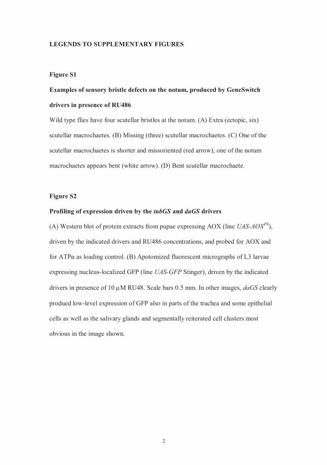
tubGS daGS
UAS-AOXF6
UAS-GFP
daGS
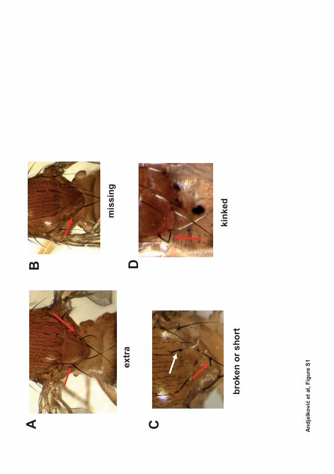
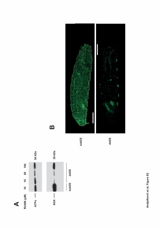
PUBLICATION III
Expression of the Alternative Oxidase Influences Jun N-Terminal Kinase Signaling and Cell Migration
Ana Andjelkovi , Amelia Mordas, Lyon Bruinsma, Annika Ketola, Giuseppe Cannino, Luca Giordano, Praveen K. Dhandapani, Marten Szibor, Eric Dufour &
Howard T. Jacobs Molecular and Cellular Biology, volume 38, pages 2839–2846
https://doi.org/ 10.1128/MCB.00110-18
Publication reprinted with the permission of the copyright holders.

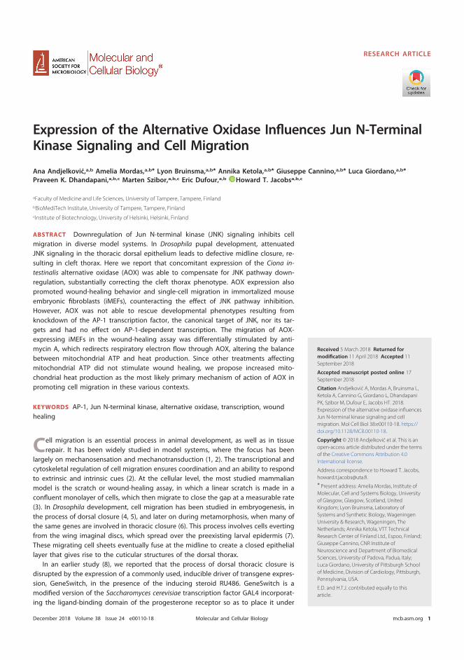
Expression of the Alternative Oxidase Influences Jun N-TerminalKinase Signaling and Cell Migration
Ana Andjelkovic,a,b Amelia Mordas,a,b* Lyon Bruinsma,a,b* Annika Ketola,a,b* Giuseppe Cannino,a,b* Luca Giordano,a,b*Praveen K. Dhandapani,a,b,c Marten Szibor,a,b,c Eric Dufour,a,b Howard T. Jacobsa,b,c
aFaculty of Medicine and Life Sciences, University of Tampere, Tampere, Finland
bBioMediTech Institute, University of Tampere, Tampere, Finland
cInstitute of Biotechnology, University of Helsinki, Helsinki, Finland
ABSTRACT Downregulation of Jun N-terminal kinase (JNK) signaling inhibits cellmigration in diverse model systems. In Drosophila pupal development, attenuatedJNK signaling in the thoracic dorsal epithelium leads to defective midline closure, re-sulting in cleft thorax. Here we report that concomitant expression of the Ciona in-testinalis alternative oxidase (AOX) was able to compensate for JNK pathway down-regulation, substantially correcting the cleft thorax phenotype. AOX expression alsopromoted wound-healing behavior and single-cell migration in immortalized mouseembryonic fibroblasts (iMEFs), counteracting the effect of JNK pathway inhibition.However, AOX was not able to rescue developmental phenotypes resulting fromknockdown of the AP-1 transcription factor, the canonical target of JNK, nor its tar-gets and had no effect on AP-1-dependent transcription. The migration of AOX-expressing iMEFs in the wound-healing assay was differentially stimulated by anti-mycin A, which redirects respiratory electron flow through AOX, altering the balancebetween mitochondrial ATP and heat production. Since other treatments affectingmitochondrial ATP did not stimulate wound healing, we propose increased mito-chondrial heat production as the most likely primary mechanism of action of AOX inpromoting cell migration in these various contexts.
KEYWORDS AP-1, Jun N-terminal kinase, alternative oxidase, transcription, woundhealing
Cell migration is an essential process in animal development, as well as in tissuerepair. It has been widely studied in model systems, where the focus has been
largely on mechanosensation and mechanotransduction (1, 2). The transcriptional andcytoskeletal regulation of cell migration ensures coordination and an ability to respondto extrinsic and intrinsic cues (2). At the cellular level, the most studied mammalianmodel is the scratch or wound-healing assay, in which a linear scratch is made in aconfluent monolayer of cells, which then migrate to close the gap at a measurable rate(3). In Drosophila development, cell migration has been studied in embryogenesis, inthe process of dorsal closure (4, 5), and later on during metamorphosis, when many ofthe same genes are involved in thoracic closure (6). This process involves cells evertingfrom the wing imaginal discs, which spread over the preexisting larval epidermis (7).These migrating cell sheets eventually fuse at the midline to create a closed epitheliallayer that gives rise to the cuticular structures of the dorsal thorax.
In an earlier study (8), we reported that the process of dorsal thoracic closure isdisrupted by the expression of a commonly used, inducible driver of transgene expres-sion, GeneSwitch, in the presence of the inducing steroid RU486. GeneSwitch is amodified version of the Saccharomyces cerevisiae transcription factor GAL4 incorporat-ing the ligand-binding domain of the progesterone receptor so as to place it under
Received 5 March 2018 Returned for
modification 11 April 2018 Accepted 11
September 2018
Accepted manuscript posted online 17
September 2018
Citation Andjelkovic A, Mordas A, Bruinsma L,
Ketola A, Cannino G, Giordano L, Dhandapani
PK, Szibor M, Dufour E, Jacobs HT. 2018.
Expression of the alternative oxidase influences
Jun N-terminal kinase signaling and cell
migration. Mol Cell Biol 38:e00110-18. https://
doi.org/10.1128/MCB.00110-18.
Copyright © 2018 Andjelkovic et al. This is an
open-access article distributed under the terms
of the Creative Commons Attribution 4.0
International license.
Address correspondence to Howard T. Jacobs,
* Present address: Amelia Mordas, Institute of
Molecular, Cell and Systems Biology, University
of Glasgow, Glasgow, Scotland, United
Kingdom; Lyon Bruinsma, Laboratory of
Systems and Synthetic Biology, Wageningen
University & Research, Wageningen, The
Netherlands; Annika Ketola, VTT Technical
Research Center of Finland Ltd., Espoo, Finland;
Giuseppe Cannino, CNR Institute of
Neuroscience and Department of Biomedical
Sciences, University of Padova, Padua, Italy;
Luca Giordano, University of Pittsburgh School
of Medicine, Division of Cardiology, Pittsburgh,
Pennsylvania, USA.
E.D. and H.T.J. contributed equally to this
article.
RESEARCH ARTICLE
crossm
December 2018 Volume 38 Issue 24 e00110-18 mcb.asm.org 1Molecular and Cellular Biology
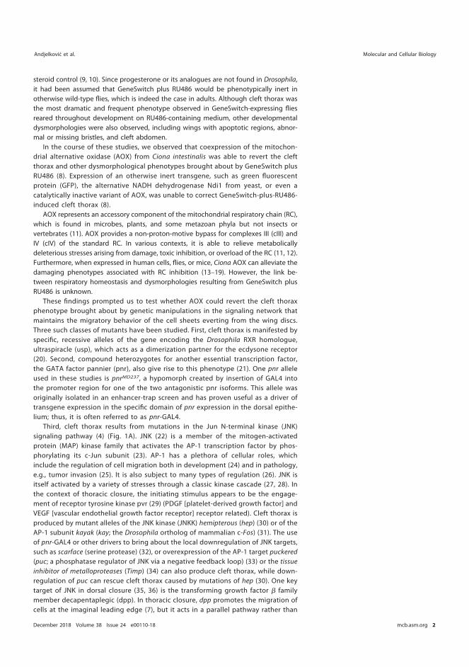
steroid control (9, 10). Since progesterone or its analogues are not found in Drosophila,it had been assumed that GeneSwitch plus RU486 would be phenotypically inert inotherwise wild-type flies, which is indeed the case in adults. Although cleft thorax wasthe most dramatic and frequent phenotype observed in GeneSwitch-expressing fliesreared throughout development on RU486-containing medium, other developmentaldysmorphologies were also observed, including wings with apoptotic regions, abnor-mal or missing bristles, and cleft abdomen.
In the course of these studies, we observed that coexpression of the mitochon-drial alternative oxidase (AOX) from Ciona intestinalis was able to revert the cleftthorax and other dysmorphological phenotypes brought about by GeneSwitch plusRU486 (8). Expression of an otherwise inert transgene, such as green fluorescentprotein (GFP), the alternative NADH dehydrogenase Ndi1 from yeast, or even acatalytically inactive variant of AOX, was unable to correct GeneSwitch-plus-RU486-induced cleft thorax (8).
AOX represents an accessory component of the mitochondrial respiratory chain (RC),which is found in microbes, plants, and some metazoan phyla but not insects orvertebrates (11). AOX provides a non-proton-motive bypass for complexes III (cIII) andIV (cIV) of the standard RC. In various contexts, it is able to relieve metabolicallydeleterious stresses arising from damage, toxic inhibition, or overload of the RC (11, 12).Furthermore, when expressed in human cells, flies, or mice, Ciona AOX can alleviate thedamaging phenotypes associated with RC inhibition (13–19). However, the link be-tween respiratory homeostasis and dysmorphologies resulting from GeneSwitch plusRU486 is unknown.
These findings prompted us to test whether AOX could revert the cleft thoraxphenotype brought about by genetic manipulations in the signaling network thatmaintains the migratory behavior of the cell sheets everting from the wing discs.Three such classes of mutants have been studied. First, cleft thorax is manifested byspecific, recessive alleles of the gene encoding the Drosophila RXR homologue,ultraspiracle (usp), which acts as a dimerization partner for the ecdysone receptor(20). Second, compound heterozygotes for another essential transcription factor,the GATA factor pannier (pnr), also give rise to this phenotype (21). One pnr alleleused in these studies is pnrMD237, a hypomorph created by insertion of GAL4 intothe promoter region for one of the two antagonistic pnr isoforms. This allele wasoriginally isolated in an enhancer-trap screen and has proven useful as a driver oftransgene expression in the specific domain of pnr expression in the dorsal epithe-lium; thus, it is often referred to as pnr-GAL4.
Third, cleft thorax results from mutations in the Jun N-terminal kinase (JNK)signaling pathway (4) (Fig. 1A). JNK (22) is a member of the mitogen-activatedprotein (MAP) kinase family that activates the AP-1 transcription factor by phos-phorylating its c-Jun subunit (23). AP-1 has a plethora of cellular roles, whichinclude the regulation of cell migration both in development (24) and in pathology,e.g., tumor invasion (25). It is also subject to many types of regulation (26). JNK isitself activated by a variety of stresses through a classic kinase cascade (27, 28). Inthe context of thoracic closure, the initiating stimulus appears to be the engage-ment of receptor tyrosine kinase pvr (29) (PDGF [platelet-derived growth factor] andVEGF [vascular endothelial growth factor receptor] receptor related). Cleft thorax isproduced by mutant alleles of the JNK kinase (JNKK) hemipterous (hep) (30) or of theAP-1 subunit kayak (kay; the Drosophila ortholog of mammalian c-Fos) (31). The useof pnr-GAL4 or other drivers to bring about the local downregulation of JNK targets,such as scarface (serine protease) (32), or overexpression of the AP-1 target puckered(puc; a phosphatase regulator of JNK via a negative feedback loop) (33) or the tissueinhibitor of metalloproteases (Timp) (34) can also produce cleft thorax, while down-regulation of puc can rescue cleft thorax caused by mutations of hep (30). One keytarget of JNK in dorsal closure (35, 36) is the transforming growth factor � familymember decapentaplegic (dpp). In thoracic closure, dpp promotes the migration ofcells at the imaginal leading edge (7), but it acts in a parallel pathway rather than
Andjelkovic et al. Molecular and Cellular Biology
December 2018 Volume 38 Issue 24 e00110-18 mcb.asm.org 2
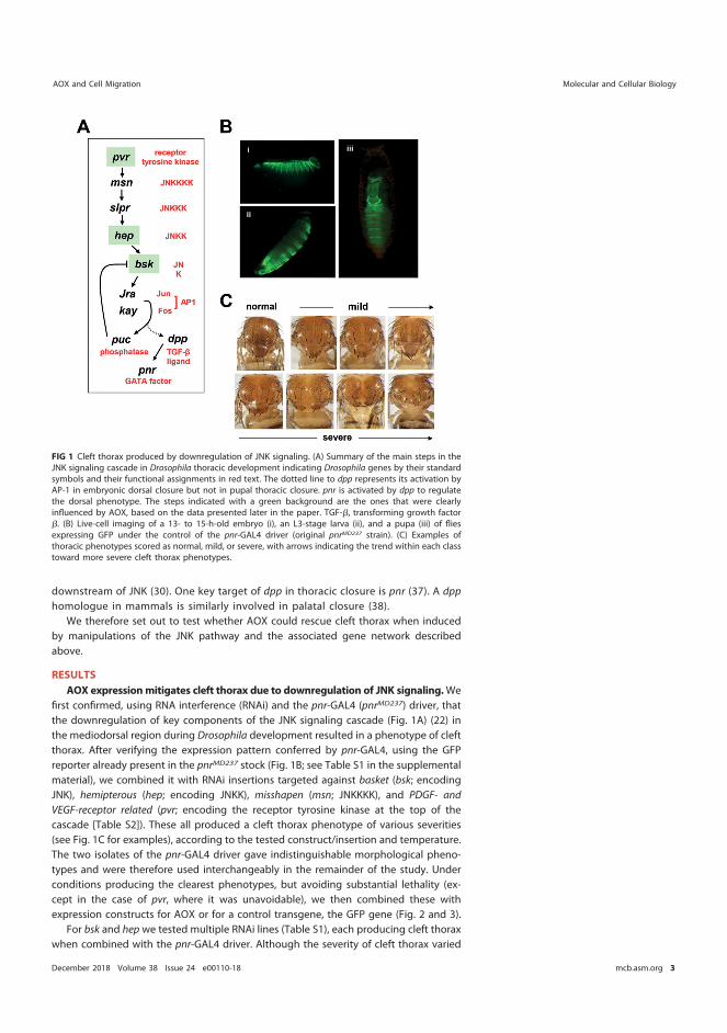
downstream of JNK (30). One key target of dpp in thoracic closure is pnr (37). A dpphomologue in mammals is similarly involved in palatal closure (38).
We therefore set out to test whether AOX could rescue cleft thorax when inducedby manipulations of the JNK pathway and the associated gene network describedabove.
RESULTS
AOX expressionmitigates cleft thorax due to downregulation of JNK signaling.Wefirst confirmed, using RNA interference (RNAi) and the pnr-GAL4 (pnrMD237) driver, thatthe downregulation of key components of the JNK signaling cascade (Fig. 1A) (22) inthe mediodorsal region during Drosophila development resulted in a phenotype of cleftthorax. After verifying the expression pattern conferred by pnr-GAL4, using the GFPreporter already present in the pnrMD237 stock (Fig. 1B; see Table S1 in the supplementalmaterial), we combined it with RNAi insertions targeted against basket (bsk; encodingJNK), hemipterous (hep; encoding JNKK), misshapen (msn; JNKKKK), and PDGF- andVEGF-receptor related (pvr; encoding the receptor tyrosine kinase at the top of thecascade [Table S2]). These all produced a cleft thorax phenotype of various severities(see Fig. 1C for examples), according to the tested construct/insertion and temperature.The two isolates of the pnr-GAL4 driver gave indistinguishable morphological pheno-types and were therefore used interchangeably in the remainder of the study. Underconditions producing the clearest phenotypes, but avoiding substantial lethality (ex-cept in the case of pvr, where it was unavoidable), we then combined these withexpression constructs for AOX or for a control transgene, the GFP gene (Fig. 2 and 3).
For bsk and hep we tested multiple RNAi lines (Table S1), each producing cleft thoraxwhen combined with the pnr-GAL4 driver. Although the severity of cleft thorax varied
FIG 1 Cleft thorax produced by downregulation of JNK signaling. (A) Summary of the main steps in theJNK signaling cascade in Drosophila thoracic development indicating Drosophila genes by their standardsymbols and their functional assignments in red text. The dotted line to dpp represents its activation byAP-1 in embryonic dorsal closure but not in pupal thoracic closure. pnr is activated by dpp to regulatethe dorsal phenotype. The steps indicated with a green background are the ones that were clearlyinfluenced by AOX, based on the data presented later in the paper. TGF-�, transforming growth factor�. (B) Live-cell imaging of a 13- to 15-h-old embryo (i), an L3-stage larva (ii), and a pupa (iii) of fliesexpressing GFP under the control of the pnr-GAL4 driver (original pnrMD237 strain). (C) Examples ofthoracic phenotypes scored as normal, mild, or severe, with arrows indicating the trend within each classtoward more severe cleft thorax phenotypes.
AOX and Cell Migration Molecular and Cellular Biology
December 2018 Volume 38 Issue 24 e00110-18 mcb.asm.org 3
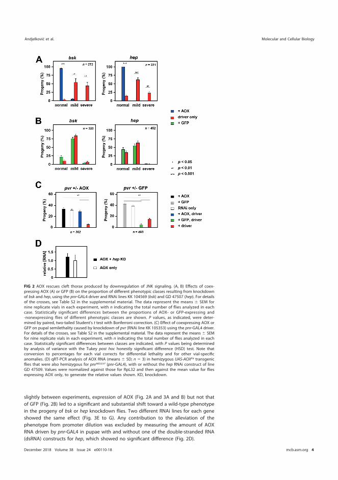
slightly between experiments, expression of AOX (Fig. 2A and 3A and B) but not thatof GFP (Fig. 2B) led to a significant and substantial shift toward a wild-type phenotypein the progeny of bsk or hep knockdown flies. Two different RNAi lines for each geneshowed the same effect (Fig. 3E to G). Any contribution to the alleviation of thephenotype from promoter dilution was excluded by measuring the amount of AOXRNA driven by pnr-GAL4 in pupae with and without one of the double-stranded RNA(dsRNA) constructs for hep, which showed no significant difference (Fig. 2D).
FIG 2 AOX rescues cleft thorax produced by downregulation of JNK signaling. (A, B) Effects of coex-pressing AOX (A) or GFP (B) on the proportion of different phenotypic classes resulting from knockdownof bsk and hep, using the pnr-GAL4 driver and RNAi lines KK 104569 (bsk) and GD 47507 (hep). For detailsof the crosses, see Table S2 in the supplemental material. The data represent the means � SEM fornine replicate vials in each experiment, with n indicating the total number of flies analyzed in eachcase. Statistically significant differences between the proportions of AOX- or GFP-expressing and-nonexpressing flies of different phenotypic classes are shown. P values, as indicated, were deter-mined by paired, two-tailed Student’s t test with Bonferroni correction. (C) Effect of coexpressing AOX orGFP on pupal semilethality caused by knockdown of pvr (RNAi line KK 105353) using the pnr-GAL4 driver.For details of the crosses, see Table S2 in the supplemental material. The data represent the means � SEMfor nine replicate vials in each experiment, with n indicating the total number of flies analyzed in eachcase. Statistically significant differences between classes are indicated, with P values being determinedby analysis of variance with the Tukey post hoc honestly significant difference (HSD) test. Note thatconversion to percentages for each vial corrects for differential lethality and for other vial-specificanomalies. (D) qRT-PCR analysis of AOX RNA (means � SD; n � 3) in hemizygous UAS-AOXF6 transgenicflies that were also hemizygous for pnrMD237 (pnr-GAL4), with or without the hep RNAi construct of lineGD 47509. Values were normalized against those for RpL32 and then against the mean value for fliesexpressing AOX only, to generate the relative values shown. KD, knockdown.
Andjelkovic et al. Molecular and Cellular Biology
December 2018 Volume 38 Issue 24 e00110-18 mcb.asm.org 4
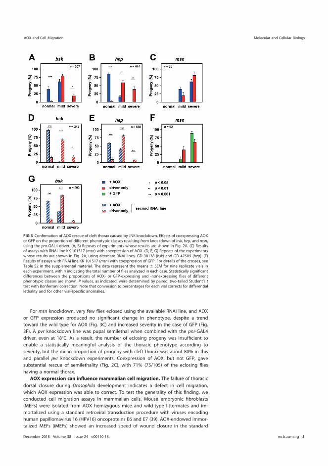
For msn knockdown, very few flies eclosed using the available RNAi line, and AOXor GFP expression produced no significant change in phenotype, despite a trendtoward the wild type for AOX (Fig. 3C) and increased severity in the case of GFP (Fig.3F). A pvr knockdown line was pupal semilethal when combined with the pnr-GAL4driver, even at 18°C. As a result, the number of eclosing progeny was insufficient toenable a statistically meaningful analysis of the thoracic phenotype according toseverity, but the mean proportion of progeny with cleft thorax was about 80% in thisand parallel pvr knockdown experiments. Coexpression of AOX, but not GFP, gavesubstantial rescue of semilethality (Fig. 2C), with 71% (75/105) of the eclosing flieshaving a normal thorax.
AOX expression can influence mammalian cell migration. The failure of thoracicdorsal closure during Drosophila development indicates a defect in cell migration,which AOX expression was able to correct. To test the generality of this finding, weconducted cell migration assays in mammalian cells. Mouse embryonic fibroblasts(MEFs) were isolated from AOX hemizygous mice and wild-type littermates and im-mortalized using a standard retroviral transduction procedure with viruses encodinghuman papillomavirus 16 (HPV16) oncoproteins E6 and E7 (39). AOX-endowed immor-talized MEFs (iMEFs) showed an increased speed of wound closure in the standard
FIG 3 Confirmation of AOX rescue of cleft thorax caused by JNK knockdown. Effects of coexpressing AOXor GFP on the proportion of different phenotypic classes resulting from knockdown of bsk, hep, and msn,using the pnr-GAL4 driver. (A, B) Repeats of experiments whose results are shown in Fig. 2A. (C) Resultsof assays with RNAi line KK 101517 (msn) with coexpression of AOX. (D, E, G) Repeats of the experimentswhose results are shown in Fig. 2A, using alternate RNAi lines, GD 38138 (bsk) and GD 47509 (hep). (F)Results of assays with RNAi line KK 101517 (msn) with coexpression of GFP. For details of the crosses, seeTable S2 in the supplemental material. The data represent the means � SEM for nine replicate vials ineach experiment, with n indicating the total number of flies analyzed in each case. Statistically significantdifferences between the proportions of AOX- or GFP-expressing and -nonexpressing flies of differentphenotypic classes are shown. P values, as indicated, were determined by paired, two-tailed Student’s ttest with Bonferroni correction. Note that conversion to percentages for each vial corrects for differentiallethality and for other vial-specific anomalies.
AOX and Cell Migration Molecular and Cellular Biology
December 2018 Volume 38 Issue 24 e00110-18 mcb.asm.org 5
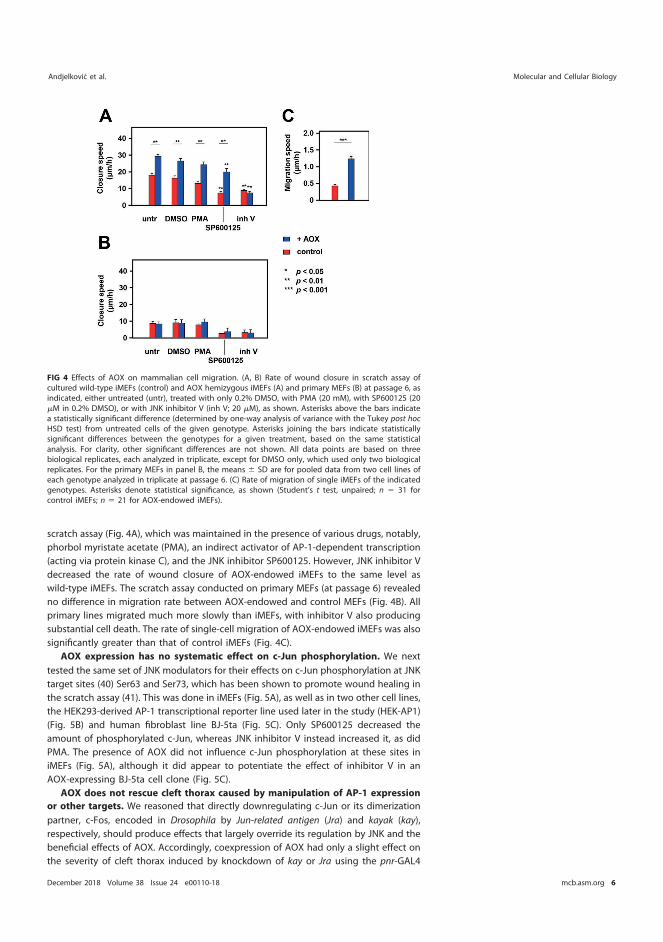
scratch assay (Fig. 4A), which was maintained in the presence of various drugs, notably,phorbol myristate acetate (PMA), an indirect activator of AP-1-dependent transcription(acting via protein kinase C), and the JNK inhibitor SP600125. However, JNK inhibitor Vdecreased the rate of wound closure of AOX-endowed iMEFs to the same level aswild-type iMEFs. The scratch assay conducted on primary MEFs (at passage 6) revealedno difference in migration rate between AOX-endowed and control MEFs (Fig. 4B). Allprimary lines migrated much more slowly than iMEFs, with inhibitor V also producingsubstantial cell death. The rate of single-cell migration of AOX-endowed iMEFs was alsosignificantly greater than that of control iMEFs (Fig. 4C).
AOX expression has no systematic effect on c-Jun phosphorylation. We nexttested the same set of JNK modulators for their effects on c-Jun phosphorylation at JNKtarget sites (40) Ser63 and Ser73, which has been shown to promote wound healing inthe scratch assay (41). This was done in iMEFs (Fig. 5A), as well as in two other cell lines,the HEK293-derived AP-1 transcriptional reporter line used later in the study (HEK-AP1)(Fig. 5B) and human fibroblast line BJ-5ta (Fig. 5C). Only SP600125 decreased theamount of phosphorylated c-Jun, whereas JNK inhibitor V instead increased it, as didPMA. The presence of AOX did not influence c-Jun phosphorylation at these sites iniMEFs (Fig. 5A), although it did appear to potentiate the effect of inhibitor V in anAOX-expressing BJ-5ta cell clone (Fig. 5C).
AOX does not rescue cleft thorax caused by manipulation of AP-1 expression
or other targets. We reasoned that directly downregulating c-Jun or its dimerizationpartner, c-Fos, encoded in Drosophila by Jun-related antigen (Jra) and kayak (kay),respectively, should produce effects that largely override its regulation by JNK and thebeneficial effects of AOX. Accordingly, coexpression of AOX had only a slight effect onthe severity of cleft thorax induced by knockdown of kay or Jra using the pnr-GAL4
FIG 4 Effects of AOX on mammalian cell migration. (A, B) Rate of wound closure in scratch assay ofcultured wild-type iMEFs (control) and AOX hemizygous iMEFs (A) and primary MEFs (B) at passage 6, asindicated, either untreated (untr), treated with only 0.2% DMSO, with PMA (20 mM), with SP600125 (20�M in 0.2% DMSO), or with JNK inhibitor V (inh V; 20 �M), as shown. Asterisks above the bars indicatea statistically significant difference (determined by one-way analysis of variance with the Tukey post hocHSD test) from untreated cells of the given genotype. Asterisks joining the bars indicate statisticallysignificant differences between the genotypes for a given treatment, based on the same statisticalanalysis. For clarity, other significant differences are not shown. All data points are based on threebiological replicates, each analyzed in triplicate, except for DMSO only, which used only two biologicalreplicates. For the primary MEFs in panel B, the means � SD are for pooled data from two cell lines ofeach genotype analyzed in triplicate at passage 6. (C) Rate of migration of single iMEFs of the indicatedgenotypes. Asterisks denote statistical significance, as shown (Student’s t test, unpaired; n � 31 forcontrol iMEFs; n � 21 for AOX-endowed iMEFs).
Andjelkovic et al. Molecular and Cellular Biology
December 2018 Volume 38 Issue 24 e00110-18 mcb.asm.org 6
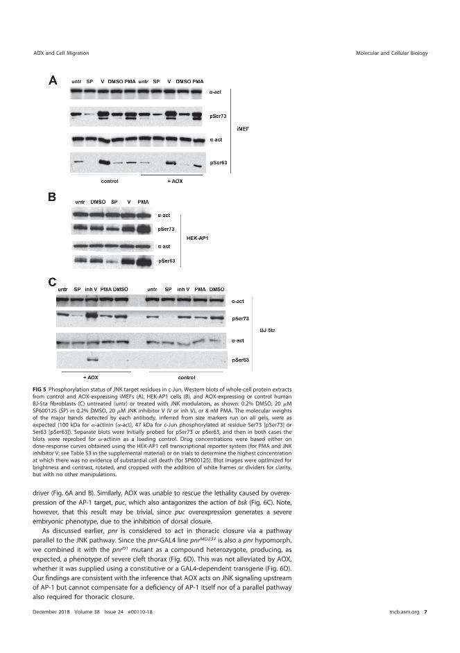
driver (Fig. 6A and B). Similarly, AOX was unable to rescue the lethality caused by overex-pression of the AP-1 target, puc, which also antagonizes the action of bsk (Fig. 6C). Note,however, that this result may be trivial, since puc overexpression generates a severeembryonic phenotype, due to the inhibition of dorsal closure.
As discussed earlier, pnr is considered to act in thoracic closure via a pathwayparallel to the JNK pathway. Since the pnr-GAL4 line pnrMD237 is also a pnr hypomorph,we combined it with the pnrD1 mutant as a compound heterozygote, producing, asexpected, a phenotype of severe cleft thorax (Fig. 6D). This was not alleviated by AOX,whether it was supplied using a constitutive or a GAL4-dependent transgene (Fig. 6D).Our findings are consistent with the inference that AOX acts on JNK signaling upstreamof AP-1 but cannot compensate for a deficiency of AP-1 itself nor of a parallel pathwayalso required for thoracic closure.
FIG 5 Phosphorylation status of JNK target residues in c-Jun. Western blots of whole-cell protein extractsfrom control and AOX-expressing iMEFs (A), HEK-AP1 cells (B), and AOX-expressing or control humanBJ-5ta fibroblasts (C) untreated (untr) or treated with JNK modulators, as shown: 0.2% DMSO, 20 �MSP600125 (SP) in 0.2% DMSO, 20 �M JNK inhibitor V (V or inh V), or 8 nM PMA. The molecular weightsof the major bands detected by each antibody, inferred from size markers run on all gels, were asexpected (100 kDa for �-actinin [�-act], 47 kDa for c-Jun phosphorylated at residue Ser73 [pSer73] orSer63 [pSer63]). Separate blots were initially probed for pSer73 or pSer63, and then in both cases theblots were reprobed for �-actinin as a loading control. Drug concentrations were based either ondose-response curves obtained using the HEK-AP1 cell transcriptional reporter system (for PMA and JNKinhibitor V; see Table S3 in the supplemental material) or on trials to determine the highest concentrationat which there was no evidence of substantial cell death (for SP600125). Blot images were optimized forbrightness and contrast, rotated, and cropped with the addition of white frames or dividers for clarity,but with no other manipulations.
AOX and Cell Migration Molecular and Cellular Biology
December 2018 Volume 38 Issue 24 e00110-18 mcb.asm.org 7
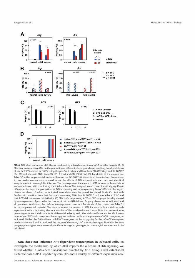
AOX does not influence AP1-dependent transcription in cultured cells. Toinvestigate the mechanism by which AOX impacts the outcome of JNK signaling, wetested whether it influences transcription directed by AP-1. Using a well-establishedluciferase-based AP-1 reporter system (42) and a variety of different expression con-
FIG 6 AOX does not rescue cleft thorax produced by altered expression of AP-1 or other targets. (A, B)Effects of coexpressing AOX on the proportion of different phenotypic classes resulting from knockdownof kay (at 25°C) and Jra (at 18°C), using the pnr-GAL4 driver and RNAi lines GD 6212 (kay) and KK 107997(Jra) (A) and alternate RNAi lines GD 19512 (kay) and GD 10835 (Jra) (B). For details of the crosses, seeTable S2 in the supplemental material. Because the GD 10835 (Jra) construct is carried on chromosomeX, two parallel crosses were required to test the effects of AOX expression in each sex, and statisticalanalysis was not meaningful in this case. The data represent the means � SEM for nine replicate vials ineach experiment, with n indicating the total number of flies analyzed in each case. Statistically significantdifferences between the proportions of AOX-expressing and -nonexpressing flies of different phenotypicclasses are shown. P values, as indicated, were determined by paired, two-tailed Student’s t test withBonferroni correction. Note that Jra knockdown using RNAi line KK 107997 (Jra) was lethal at 25°C andthat AOX did not rescue this lethality. (C) Effect of coexpressing AOX or GFP on pupal lethality causedby overexpression of puc under the control of the pnr-GAL4 driver. Progeny classes are as indicated, andall contained, in addition, the UAS-puc overexpression construct. For details of the crosses, see Table S2in the supplemental material. The data represent the means � SEM for nine replicate vials in eachexperiment, with n indicating the total number of flies analyzed in each case. Note that conversion topercentages for each vial corrects for differential lethality and other vial-specific anomalies. (D) Pheno-types of pnrMD237/pnrD1 compound heterozygotes with and without the presence of AOX transgenes, asindicated. Neither the GAL4-driven UAS-AOXF6 transgene nor homozygosity for the tub-AOX transgeneson chromosomes 2 and X produced the rescue of the strong cleft thorax phenotype. Note that becauseprogeny phenotypes were essentially uniform for a given genotype, no meaningful variances could becalculated.
Andjelkovic et al. Molecular and Cellular Biology
December 2018 Volume 38 Issue 24 e00110-18 mcb.asm.org 8
structs for AOX, we tested whether AOX expression in Drosophila S2 cells was able toalter AP-1-dependent transcription under different conditions of JNK pathway activa-tion. First, we compared the transcriptional readout in cells cotransfected with thereporter plasmids and with AOX cloned into the copper-inducible expression vectorpMT/V5-His B, with its natural stop codon, with that in cells transfected with the emptyvector. JNK pathway activation was achieved using the pUAST-Hepact plasmid, includedin all transfections in combination with pAct-Gal4, which promotes pUAST-Hepact
transcription by constitutive expression of Gal4. Transfection efficiency was controlledby the inclusion of a constitutively expressed plasmid encoding renilla luciferase, whichcan be experimentally distinguished from the firefly luciferase of the reporter construct.Finally, to measure background transcription independently of AP-1, the system in-cludes a mutated version of the reporter (which was used as an alternative in trans-fections), to which AP-1 does not bind.
Despite the complexity of this system, it gave clear-cut results. AOX produced nosignificant change in AP-1-dependent luciferase expression under both basal andJNK-activated conditions (Fig. 7A; Table S4). Next, we tested reporter cells cotransfectedwith a plasmid (pAC/AOX) (43) directing constitutive AOX expression under the controlof a �-actin promoter versus cells cotransfected with the empty vector. Again, AOXexpression had no effect on the transcriptional readout (Fig. 7B; Table S4). Using asystem in which JNK pathway activation and AOX induction were brought aboutsimultaneously by expression of the exogenous transcription factor Gal4, but this timeusing a control plasmid harboring a catalytically inactive, mutated AOX, we again foundno effect of AOX (Fig. 7C: see also the results of a parallel experiment in Table S4). AOXalso produced no significant difference in AP-1-dependent transcription in cells wherehep had been knocked down (Fig. 7D; Table S4).
We conducted a similar exercise in mammalian cells, using an HEK293 cell-derivedreporter cell line (here designated HEK-AP1), stably transduced with lentiviral con-structs expressing AOX or, as a control, the mutated, catalytically inactive variant(mutAOX). Successful transduction and cell cloning at limiting dilution were verified viathe fluorescence conferred by the cotransduced marker GFP, and AOX functionality wasverified by respirometry (Table S5). Although individual HEK-AP1 cell-derived clonesshowed a variable degree of AP-1-dependent transcriptional activity, AOX-expressingand control cell clones showed a similar susceptibility to the effects of the JNKantagonists SP600125 and inhibitor V (Fig. 7E). Surprisingly, SP600125 increased ratherthan decreased the transcriptional readout, despite the fact that it inhibited c-Junphosphorylation (Fig. 5B), although it did modestly suppress PMA-activated transcrip-tion in the reporter line (Fig. 7E).
Antimycin A differentially stimulates the migration of AOX-expressing cells. Togain insight into the intracellular process(es) underlying the enhanced migratorybehavior of AOX-expressing cells, we tested the effects of sublethal doses of variousmetabolic effectors on the relative rates of migration of AOX-expressing versus controliMEFs. In an initial experiment (Fig. 8A), we tested various oxidative phosphorylationinhibitors, antioxidants, and protease inhibitors in the wound-healing assay for adifferential effect on AOX-expressing cells. For further study, we selected three treat-ments that appeared to give a differential effect (antimycin A, oligomycin, and mito-quinone mesylate [MitoQ]), together with two that did not (rotenone and carbonylcyanide p-trifluoromethoxyphenylhydrazone [FCCP]), and measured wound closure infour independent experiments. Antimycin A had a significantly different effect on themigration of AOX-expressing MEFs versus wild-type MEFs (Fig. 8B), stimulating themigration of the former but suppressing that of the latter, whereas MitoQ, rotenone,oligomycin, and FCCP had no significant effects. To understand the implications ofthese findings for the mechanism by which AOX promotes cell migration, we checkedthe effects of AOX expression on respiration in the cell lines tested (Fig. 8C). AOX hadno significant effect on whole-cell respiration or on permeabilized cell respiration on cI-,cII-, and cIV-linked substrates. However, in the presence of antimycin A, it enabled
AOX and Cell Migration Molecular and Cellular Biology
December 2018 Volume 38 Issue 24 e00110-18 mcb.asm.org 9
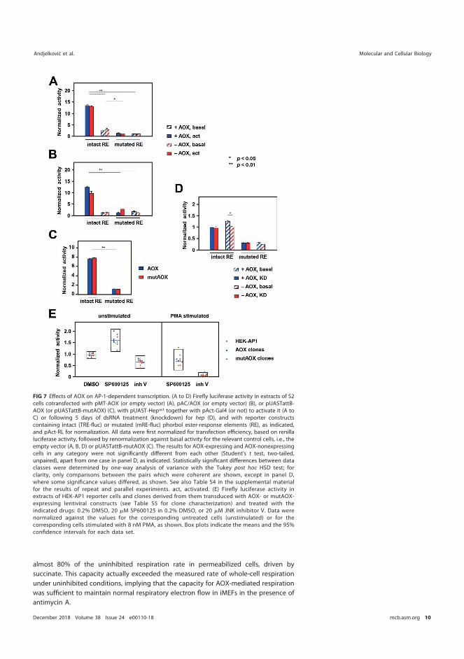
almost 80% of the uninhibited respiration rate in permeabilized cells, driven bysuccinate. This capacity actually exceeded the measured rate of whole-cell respirationunder uninhibited conditions, implying that the capacity for AOX-mediated respirationwas sufficient to maintain normal respiratory electron flow in iMEFs in the presence ofantimycin A.
FIG 7 Effects of AOX on AP-1-dependent transcription. (A to D) Firefly luciferase activity in extracts of S2cells cotransfected with pMT-AOX (or empty vector) (A), pAC/AOX (or empty vector) (B), or pUASTattB-AOX (or pUASTattB-mutAOX) (C), with pUAST-Hepact together with pAct-Gal4 (or not) to activate it (A toC) or following 5 days of dsRNA treatment (knockdown) for hep (D), and with reporter constructscontaining intact (TRE-fluc) or mutated (mRE-fluc) phorbol ester-response elements (RE), as indicated,and pAct-RL for normalization. All data were first normalized for transfection efficiency, based on renillaluciferase activity, followed by renormalization against basal activity for the relevant control cells, i.e., theempty vector (A, B, D) or pUASTattB-mutAOX (C). The results for AOX-expressing and AOX-nonexpressingcells in any category were not significantly different from each other (Student’s t test, two-tailed,unpaired), apart from one case in panel D, as indicated. Statistically significant differences between dataclasses were determined by one-way analysis of variance with the Tukey post hoc HSD test; forclarity, only comparisons between the pairs which were coherent are shown, except in panel D,where some significance values differed, as shown. See also Table S4 in the supplemental materialfor the results of repeat and parallel experiments. act, activated. (E) Firefly luciferase activity inextracts of HEK-AP1 reporter cells and clones derived from them transduced with AOX- or mutAOX-expressing lentiviral constructs (see Table S5 for clone characterization) and treated with theindicated drugs: 0.2% DMSO, 20 �M SP600125 in 0.2% DMSO, or 20 �M JNK inhibitor V. Data werenormalized against the values for the corresponding untreated cells (unstimulated) or for thecorresponding cells stimulated with 8 nM PMA, as shown. Box plots indicate the means and the 95%confidence intervals for each data set.
Andjelkovic et al. Molecular and Cellular Biology
December 2018 Volume 38 Issue 24 e00110-18 mcb.asm.org 10
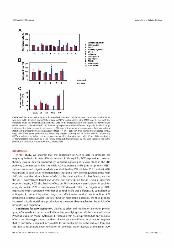
DISCUSSION
In this study, we showed that the expression of AOX is able to promote cellmigratory behavior in two different models: in Drosophila, AOX expression correctedthoracic closure defects produced by impaired signaling at several steps in the JNKpathway (summarized in Fig. 1A), while AOX-expressing iMEFs (but not primary MEFs)showed enhanced migration, which was abolished by JNK inhibitor V. In contrast, AOXwas unable to correct cell migration defects resulting from downregulation of the mainJNK substrate, the c-Jun subunit of AP-1, or by manipulation of other factors, such asthe AP-1 downstream target puc or the pnr transcription factor. Using a luciferasereporter system, AOX also had no effect on AP-1-dependent transcription in prolifer-ating Drosophila (S2) or mammalian (HEK293-derived) cells. The migration of AOX-expressing iMEFs compared with that of control iMEFs was differentially stimulated byantimycin A but not by other drugs that affect mitochondrial electron flow, ATPproduction, reactive oxygen species (ROS), or membrane potential. We thus proposeincreased mitochondrial heat production as the most likely mechanism by which AOXpromotes cell migration.
Conditions for AOX activation. Clearly, to affect cell motility or any other pheno-type, AOX needs to be enzymatically active, modifying the cellular metabolic state.Previous studies in model systems (13–19) found that AOX expression has only minimaleffects on phenotype under standard physiological conditions. Its activation requiresthat its substrate, ubiquinol, accumulate to substantial levels in the reduced form (44,45), due to respiratory chain inhibition or overload. Other aspects of metazoan AOX
FIG 8 Modulation of iMEF migration by metabolic inhibitors. (A, B) Relative rate of wound closure forwild-type iMEFs (control) and AOX-hemizygous iMEFs treated either with DMSO only (—) or with theindicated drugs (see Materials and Methods). Data are normalized against the closure rate for the givencell line treated only with DMSO. (A) Preliminary experiment with 9 different drugs. (B) For the 5 drugsindicated, the data represent the means � SD from 4 independent experiments. Asterisks indicatestatistically significant differences (Student’s t test, P � 0.01) between drug-treated and untreated (DMSOonly) cells of the given genotype. (C) Respiratory oxygen consumption of control and AOX-expressingiMEFs is indicated as follows: endo, endogenous whole-cell respiration; cI, cII, cIV, and AOX, respirationof permeabilized cells driven by cI-, cII-, or cIV-linked substrate mixes or by cII-linked substrate mix in thepresence of antimycin A (denoted AOX), respectively.
AOX and Cell Migration Molecular and Cellular Biology
December 2018 Volume 38 Issue 24 e00110-18 mcb.asm.org 11
regulation remain largely unknown. In plants, AOX is activated by pyruvate (44, 46),although this regulation has not been demonstrated in the metazoan enzyme (47).Nevertheless, AOX rescue of cleft thorax and promotion of iMEF motility imply that itcan become activated at the margin of a migrating cell sheet. Cell movement is anenergy-requiring process, and AMP-activated protein kinase (AMPK), one of the cell’skey regulators of energy homeostasis (48), is locally activated at the leading edge of amigrating cell sheet (49), where it promotes the movement of mitochondria into thissubcellular domain. This metabolic microenvironment may also favor AOX activation, ifthe extra demand exceeds the capacity of respiratory complex III. In addition to AMPK,the mitochondrial unfolded protein response (mtUPR), which has previously beenlinked with JNK signaling (50), may be elicited. Treatment with antimycin A, which wasshown here to differentially stimulate the migratory behavior of AOX-expressing iMEFs,should lead to the further activation of AOX, since blockade of cIII increases the ratioof ubiquinol to ubiquinone, diverting electron flow through AOX.
The proteins used for immortalization of MEFs, namely, the HPV16 E6 and E7oncoproteins, may also synergize with activated AOX. Among other effects (51), E7switches energy supply from respiration to glycolysis by activating pyruvate kinase (52).Switching to the high-capacity glycolytic pathway may underlie the increased migra-tory capacity of immortalized MEFs, as inferred previously for cancer cells (53, 54). AOXmay enhance this by increasing pyruvate clearance via the tricarboxylic acid cycle,promoting glycolytic flux. Similarly, E6 promotes glucose transport (55–57) and facili-tates the degradation of p53 (58), providing a possible link to AP-1 and JNK (59–62). E6also upregulates hypoxia-inducible transcription factor 1� (HIF-1�) (63) and mTORC1(64).
Is AP-1 the target of AOX? Reporter assays indicate that AP-1 and its transcriptionalactivity are not a direct target of AOX, although the Drosophila findings and the effectsof inhibitor V leave open a possible direct involvement of JNK, acting through othertargets.
The transcriptional reporter cell assays were conducted in proliferating cells, whereAP-1 may act differently than in a migrating cell sheet. It plays diverse physiologicalroles, and each of its subunits is encoded by a gene family (65). Although its canonicalmembers, c-Fos and c-Jun, drive cell proliferation (66), others are functionally diverseand are regulated by a host of mechanisms (67), including subunit composition (68),other MAP kinases (69), and mitochondrial ATP depletion (70).
The interpretation of experiments using JNK inhibitors rests upon assumptionsregarding their specificity, but the effects of SP600125 and JNK inhibitor V do not fitprevious assumptions regarding their mode of action. Inhibitor V was the only drugtested which blocked the migration-promoting effects of AOX in iMEFs, indicating thatit acts at a step different from that influenced by AOX. These drugs have previouslybeen studied almost exclusively in highly artificial in vitro systems (49, 71), taking littleaccount of their demonstrated activity against other kinases (71–73). The alternativeapproach of using genetic ablation of JNK to elucidate the AOX-responsive step cannoteasily be applied in mammalian cells, since JNK has diverse isoforms created byalternative splicing and is encoded by a multigene family, some of whose membershave undoubtedly been functionally redeployed during evolution (74).
AP-1 is subject to oxidative inactivation (65, 75), from which it is protected inDrosophila by the coactivator MBF1 (76), and the loss of MBF1 sensitizes animals to cleftthorax, if also exposed to H2O2. AOX decreases net ROS production from mitochondria(15, 77), which could compensate for the impaired upstream activation of AP-1, but, asdiscussed below, its effects on mammalian cell migration were not influenced by drugsthat alter ROS (MitoQ, N-acetyl cysteine; Fig. 8).
AOX and other signaling pathways. AOX may also act through other pathwaysindependently of or in conjunction with AP-1. For example, immortalized fibroblastsshow an enhanced induction of HIF-1� under hypoxia (78), while AOX also blunts thehypoxia response (79). In cancer cells, mitochondrial ROS activates many transcription
Andjelkovic et al. Molecular and Cellular Biology
December 2018 Volume 38 Issue 24 e00110-18 mcb.asm.org 12
factors (80), and interactions between AMPK and AP-1 have been reported in cardio-myocytes (81) and Jurkat T cells (82). Mitochondrial energy status also governs calciumtransport into mitochondria, which has major effects on cell migration (83).
Other transcription factors mediate mitochondrial responses to stress, notably, Nrf2(84), ATF4 (85), and other AP-1 paralogs. Although stressors that activate AP-1 via JNKhave only minimal effects on Nrf2- or ATF4-induced transcription (86), this does notexclude the possibility of the converse. JNK can be activated by mitochondrial ROS inischemia-reperfusion (87) or by the mtUPR (50). JNK itself has also been reported tointeract directly with mitochondria, triggering downstream effects (88).
AOX stimulates migratory behavior by a mechanism connected to metabolism.
Of the metabolic inhibitors tested, only antimycin A preferentially stimulated themigratory behavior of AOX-expressing cells compared with control cells in the wound-healing assay. This result provides a strong clue as to the mechanism of action of AOX.The ineffective treatments included agents that should increase mitochondrial mem-brane potential (oligomycin [89]) or decrease it (FCCP [90], rotenone [91]), othertreatments that inhibit mitochondrial ATP production (oligomycin, FCCP, and rotenone)or drugs that increase ROS production (oligomycin [92], rotenone [91, 93]) or dampenit (N-acetyl cysteine [94], MitoQ [95]) or that can have effects in either direction,depending on the dose (FCCP [96–98]). Agents that limit proteolytic turnover of cellularcomponents in lysosomes (chloroquine [99]; E64d [100]) or by caspases [carbobenzoxy-valyl-alanyl-aspartyl-(O-methyl)-fluoromethylketone (Z-VAD-FMK) (101)] also appearedto have no differential effect on the migration of AOX-expressing cells. Not only didthese other treatments fail to influence control and AOX-expressing cells differentially,but none of them produced substantial alterations to the rate of migration of controlcells.
Since AOX-expressing iMEFs continue to respire when treated with antimycin A,most metabolites should be only minimally affected. The biggest difference should bein the rate of mitochondrial ATP production, when respiratory complexes III and IV arereplaced by the non-proton-motive AOX. However, as indicated above, mitochondrialATP production as such cannot be the crucial factor determining the rate of migration.
In the presence of antimycin A, the energy released by AOX in catalyzing quinoloxidation is instead converted to heat. We earlier showed that AOX-driven respirationin human cells was able to maintain the same mitochondrial temperature with less thanhalf the amount of respiratory flux (102). In the iMEFs tested, the capacity for AOX-mediated respiration was sufficient to maintain respiratory electron flow in the pres-ence of antimycin A at the same rate as in untreated cells (Fig. 8C). This implies that thecells should be able to maintain normal redox homeostasis in the presence of the drugwith only a minimal metabolic disturbance, arising from the need to make more ATPthrough nonmitochondrial pathways. Maintenance of respiratory flux but in which fluxis driven through AOX rather than cIII implies a pronounced thermogenic effect. Thus,we propose increased mitochondrial heat production as the most likely mechanismwhereby AOX stimulates cell migration.
Fibroblast motility has long been known to be temperature dependent (103), andactin polymerization itself is highly affected by changes in temperature (104). Themigration of mesenchymal stem cells is stimulated by increased expression of heatshock protein 90 (105), which impinges on various signaling pathways, possibly un-derlying the different effects of JNK inhibitors with poor selectivity. Heat shock protein70 has also been shown to promote cell migration, by acting as a chaperone for thedelivery of proteins needed by migrating cells to the leading edge (106). Even atransient heat shock can promote cell migration in cancer cell lines (107), which may(108) or may not (107) depend on heat shock transcription factor 1 (HSF1).
Together, these data strongly suggest that AOX activation is able to promote cellmigration by raising the intracellular temperature, most likely in specific subcellularcompartments. The compensatory effects on JNK signaling are thus implied to beindirect, accounting also for the fact that AOX was able to correct cleft thorax in acompletely different model generated by deranged nuclear receptor signaling (8).
AOX and Cell Migration Molecular and Cellular Biology
December 2018 Volume 38 Issue 24 e00110-18 mcb.asm.org 13
Further validation of this hypothesis will require the development of quantitativelyreliable methods to measure the intracellular temperature in specific cell compartmentsin situ. It will require even more sophisticated technology to make such measurementsin vivo in the Drosophila pupa. Alternatively, if some other metabolic effect of AOX-driven respiration is responsible, we would need to wait for metabolomic technologiesto advance to the single-cell level (109) to identify plausible candidates, although novelin situ methods would be needed to take this to the subcellular level.
Developmental context of mitochondrial effects on cell migration. Insect meta-morphosis is fuelled by stored nutrients accumulated during larval growth, principally,lipid which is metabolized in mitochondria (110). A drop in the ATP level due tonutritional limitations should activate AMPK, so as to refocus resources onto ATPproduction. However, because all protein kinases depend on ATP as a substrate, ATPdeficiency should restrain other regulatory kinases and limit ATP-consuming develop-mental processes, such as cell migration. Assuming that JNK operates as one suchpathway, its inappropriate downregulation in the dorsal thoracic epithelium underconditions where the ATP supply is adequate could restrict cell migration while otherdevelopmental processes are energized normally, resulting in the specific failure ofmidline closure.
Rapid wound healing is also important for limiting pathogen invasion. Pathogensmay deplete the nutritional environment of a wound, potentially jeopardizing theprocesses that support tissue repair. Rapid and efficient wound closure in a nutritionallylimited environment may therefore depend on metabolic remodeling to supportmotility (111).
A full elucidation of the processes that link mitochondrial perturbations with cellmigration should be of considerable medical importance and might even enable thedesign of new and more effective treatments, e.g., for metastatic tumors, tissue injuries,and congenital midline closure defects. Confirmation of a role in these processes formitochondrial heat production may open up entirely new avenues for therapy.
MATERIALS AND METHODS
Drosophila strains and culture. The Drosophila strains used in the study and their sources aresummarized in Table S1 in the supplemental material. Flies were maintained in standard high-sugarmedium (15) on a 12-h light/12-h dark cycle at 25°C, except where indicated in the figure legends.Crosses were generally implemented in triplicate, with flies being tipped into new vials on threesuccessive days after mating.
Cell culture. Drosophila strain S2 cells were maintained as described previously (112). AOX-positiveand -negative mouse embryonic fibroblasts (MEFs) (113), sourced either from embryos transgenic for C.intestinalis AOX, inserted at the Rosa26 locus (18), or from their nontransgenic littermates, were studiedat passage 6. MEFs were immortalized (iMEFs) by retroviral transduction with HPV16 oncoproteins E6 andE7 (39) and maintained at 37°C in 5% CO2 in Dulbecco modified Eagle medium (DMEM; Sigma-Aldrich)supplemented with 20% (primary MEFs) or 10% (iMEFs) heat-inactivated fetal bovine serum (FBS;Sigma-Aldrich), 1% penicillin-streptomycin (Lonza), and 4 mM L-glutamine (Sigma-Aldrich). An AP-1reporter HEK293 recombinant cell line (JNK signaling pathway; BPS Bioscience), here abbreviatedHEK-AP1, was maintained at 37°C in 5% CO2 in minimal essential medium (HyClone) supplemented with10% heat-inactivated FBS (Sigma-Aldrich), 1% nonessential amino acids (HyClone), 1 mM sodiumpyruvate (Sigma-Aldrich), 1% penicillin-streptomycin (Lonza), and 400 �g/ml Geneticin (Gibco, LifeTechnologies). Geneticin was freshly distributed to each plate when cells were passaged. BJ-5ta humanfibroblasts (ATCC CRL-4001) were grown at 37°C in 5% CO2 in DMEM (Sigma-Aldrich) supplemented with10% heat-inactivated FBS (Sigma-Aldrich), 20% medium 199 (Sigma-Aldrich), 1% penicillin-streptomycin(Lonza), and 4 mM L-glutamine (Sigma-Aldrich). HEK-AP1 and BJ-5ta cells expressing C. intestinalis AOXor the mutated, catalytically inactive variant mutAOX (43) were obtained by pWPI-based lentiviraltransduction as described previously (114). Transduced cell populations were sorted according to GFPfluorescence using a BD FACSAria II cell sorter equipped with an 85-�m nozzle and operated at a sheathpressure of 45 lb/in2 and then cloned at limiting dilution. Clones were reverified for GFP fluorescence byfluorescence-activated cell sorting and for AOX functionality by respirometry, as described previously(43).
Wound-healing and single-cell migration assays. Samples of 90,000 cells were plated on 24-wellplates (CellStar; Greiner Bio-One) as technical triplicates (3 wells per sample). After 24 h, cells were treatedwith one of the following reagents: 0.2% dimethyl sulfoxide (DMSO; Hybri-Max; Sigma-Aldrich), 20 �MSP600125 (Sigma-Aldrich) in 0.2% DMSO, 20 �M JNK inhibitor V (Merck), or 20 nM phorbol myristateacetate (PMA; Sigma-Aldrich) in fresh medium. After 2 h, a linear scratch was made in the center of eachwell with a p10 (1- to 10-�l) pipette tip. The cells were then washed three times with medium to removedetached cells, and fresh medium containing the appropriate reagent was added to each well. For testing
Andjelkovic et al. Molecular and Cellular Biology
December 2018 Volume 38 Issue 24 e00110-18 mcb.asm.org 14
the effects of metabolic inhibitors in this assay, cells were seeded at 45,000 to 90,000 cells per well on24-well plates until a monolayer was formed (24 to 48 h), before the scratch was made. After rinsing oncewith normal medium, medium was replaced with 1 ml of fresh medium containing one of the followingdrugs: oligomycin (1 ng/ml), antimycin A (60 ng/ml), rotenone (150 nM), FCCP (10 �M), N-acetyl cysteine(5 mM), chloroquine (20 �M), E64d (10 �g/ml), MitoQ (250 nM; a kind gift of Mike Murphy, MRCMitochondrial Biology Unit, Cambridge, UK), valinomycin (10 �M), Z-VAD-FMK (20 �M), or just 4 �l DMSOas a solvent control. All chemicals were from Sigma-Aldrich, except where stated otherwise. To measuresingle-cell migration, samples of 15,000 cells were plated on 6-well plates (CellStar; Greiner Bio-One) astechnical triplicates (3 wells per sample). To minimize plate-specific effects, each plate contained bothAOX-positive and -negative iMEFs in all assays. Cells were imaged as described below.
Microscopy. Imaging of fly thoraxes used a Nikon digital DS-Fi1 high-definition color camera with aNikon SMZ 745T stereoscopic zoom microscope operated by NIS-Elements D (version 4.20) software. Forlive imaging of Drosophila embryos, eggs were collected from grape juice-agar plates after adult flieswere placed in a mating chamber for 1 h, covered by a dark box, dechorionated using double-sided tape(115), placed into 35-mm glass-bottom microwell dishes (MatTek Corporation) filled with Halocarbon Oil700 (Sigma-Aldrich), and imaged for GFP fluorescence using an Andor spinning disc confocal microscopeequipped with an Andor Neo 5.5 sCMOS vacuum-cooled camera at �20 magnification. The larvae werecleaned of adherent food using a soft paintbrush, dried on a precision wipe, placed on an ice-coldmicroscope slide (Menzel Gläser), and covered with a few drops of 50% ice-cold glycerol and then witha cover glass (thickness number, 11⁄2; Zeiss). Samples were placed in a �20°C freezer for 15 min toimmobilize the larvae. Live imaging used a Zeiss Axio Imager M2 upright microscope with a Zeiss ECPlan-Neofluar 5�/0.16, WD 18.5-mm air objective and ZEN software without the ApoTome function.Pupae were gently detached from the vial, placed with the dorsal side down into 35-mm glass-bottommicrowell dishes (MatTek Corporation), and imaged as described above for embryos. Wound closuretime-lapse images were taken with a ChipMan Technologies Cell-IQ observation incubator equipped witha Retiga EXi 1392 charge-coupled-device camera, using a Nikon CFI Plan Fluorescence DL objective(magnification, �10) until the wound closed (approximately 48 h) or for just 24 h when metabolicinhibitor drugs were applied. Images were taken every 30 min. The incubator environment was held ata temperature of 37°C and had an atmosphere of 5% CO2, 19% O2, and 76% N2. Wound-healing analysiswas performed using a Cell-IQ analyzer (version 4.3) by manually tracking the gap area, based on linesdrawn along the wounded area of an image taken every second hour. The speed of the collective motionof the cells was measured in the most linear part of the wound area over time. In the experiment usingmetabolic inhibitors, the most linear time interval for closure was also analyzed and applied across alltreatments and plates. To generate images for measuring single-cell migration, cells were transferred 24h after plating to a Nikon BioStation CT instrument equipped with a Nikon DS-1QM camera and imagedat �4 magnification. The environment in the incubator was held at a temperature of 37°C with a relativehumidity of 85% and a 5% CO2 atmosphere, as described above for the wound-healing assay. Imageswere taken every 6 min, and movies were made using CL Quant (version 3.10) software. Movement wasanalyzed using CellTracker image-processing software with semiautomated migration detection (116),ignoring cells that were dying or dividing.
qRT-PCR. RNA was extracted from S2 cells (117), and quantitative reverse transcription-PCR (qRT-PCR) was performed (16) as described previously, using RpL32 mRNA as an internal standard. Primers forRpL32 and AOX were those used previously (16), and those for hep mRNA were TTGGTTTCTTGGGGTCGATG and TGGACTCCAAGGCCAACACT, the sequences of both of which are shown 5= to 3=.
Transfections and luciferase reporter assays in S2 cells. Firefly/renilla luciferase dual-reporterassays in S2 cells used a cotransfection protocol (42), based on plasmids TRE-fluc, mRE-fluc, pAct-RL,pAct-Gal4, and pUAST-Hepact, kindly supplied by Dirk Bohmann, University of Rochester. Transfectionsused either TRE-fluc (a firefly luciferase reporter with the intact phorbol-ester response element) ormRE-fluc (with the mutated response element) plus pAct-RL (renilla luciferase for transfection normal-ization) with or without pAct-Gal4 together with pUAST-Hepact for activated transcription and with thefollowing plasmids or controls harboring AOX, as indicated in the figure legends: pMT-AOX (a kind giftof Filippo Scialó, Newcastle University), containing the C. intestinalis AOX-coding sequence recloned withits natural stop codon from the original vector, pCDNA5/FRT/TO (13), as an EcoRI fragment into thecopper-inducible vector pMT/V5-His B (Thermo Fisher Scientific); pAC/AOX and pAC/mutAOX (43, 112),containing, respectively, the wild-type C. intestinalis AOX-coding sequence and the mutated, catalyticallyinactive variant under the control of the constitutive actin-5C promoter plus the corresponding emptyvector pAc5.1/V5-His B (Invitrogen); and pUASTattB-AOX and pUASTattB-mutAOX (43), containing thesame transgenes cloned into the pUASTattB vector under the control of a Gal4-dependent promoter.Expression and induction of AOX were verified for each plasmid by transfection and Western blotting asdescribed previously (43), prior to use in luciferase reporter assays. Transfections used the FuGENE HDtransfection reagent (Promega), according to the manufacturer’s instructions, at a ratio of 3 �l FuGENEper �g of DNA. For each transfection, 3 � 105 cells/ml were plated on 6- or 12-well plates. To each wellwas added a transfection mix consisting of FuGENE, 1 �g of each plasmid to be used (per ml of culturemedium), and sterile water up to a final volume of 100 �l (6-well plates) or 50 �l (12-well plates).Transfections were carried out 1 h after plating (24 h in the case of pMT-AOX or the corresponding emptyvector, with addition of 500 �M CuSO4 after a further 24 h), and luciferase assays were performed 72 hafter transfection. In transfection mixtures to which mitochondrial inhibitors were added, 6 � 105 cells/mlwere plated on 6-well plates, and drugs were added 24 h after transfection at the concentrations shownin the figure legends. For luciferase reporter experiments in which hep expression was knocked down byRNAi, cells were also transfected with a dsRNA against the hep coding sequence, prepared by a
AOX and Cell Migration Molecular and Cellular Biology
December 2018 Volume 38 Issue 24 e00110-18 mcb.asm.org 15
two-round PCR-based procedure essentially as described previously (112), but using hep-specific primers(shown 5= to 3=) TGGAGGCAAAGCTCCAGGC and CGCGAACGAAGCAGCCAAGG for the first round andGAATTAATACGACTCACTATAGGGGAGACATCCGCCACCCACGCACCTTC and GAATTAATACGACTCACTATAGGGGAGATCCCATTGCCCAGGTCGCCCAG for the second round, followed by transcription using aMEGAscript T7 transcription kit (Life Technologies). Knockdown at the RNA level (to �85%) wasverified in transfected cells (112) by qRT-PCR. For combined dsRNA/reporter plasmid transfections, 1 �105 cells were plated per well in 24-well plates. After 30 min, cells were transfected with 300 ng of eachrelevant plasmid and 4 �g of hep-specific dsRNA per well in a total volume of 100 �l. A further 4 �g ofthe dsRNA was added 72 h later, and luciferase assays were conducted 110 h after the initial transfection.For luciferase assays, 75 �l of suspended cells from each well was transferred in triplicate to the wells ofa 96-well microplate (Lab Systems) and analyzed using a Dual-Glo luciferase assay system (Promega),according to the manufacturer’s protocol. Luminescence was measured using a Thermo LabsystemsLuminoskan Ascent plate reader.
Luciferase reporter assays in mammalian cells. Firefly luciferase reporter assays were carried outin HEK-AP1 cells and in AOX/mutAOX-expressing clones derived from them, as follows: 30,000 cells wereplated in technical duplicate (2 wells per sample) in luminometer-compatible Nunc MicroWell 96-wellplates with lids (Thermo Fisher Scientific). After 24 h, the medium was replaced with medium containingeither 0.2% DMSO, 20 �M SP600125 in 0.2% DMSO, 20 �M JNK inhibitor V, or no added drug. Cells wereincubated for 2 h at 37°C. For PMA treatment, a second replacement medium contained 8 nM PMA plus20 �M SP600125 in 0.2% DMSO, 20 �M JNK inhibitor V, or no other added drug, as appropriate, and thecells were incubated for a further 6 h. Luciferase assays were carried out using the Dual-Glo luciferaseassay system (Promega), according to the manufacturer’s protocol, and luminescence was measuredusing a PerkinElmer UV/visible plate reader.
Protein analysis by Western blotting. Batches of 300,000 MEFs or HEK-AP1 cells or 250,000 BJ-5tacells were plated on 6-well plates (CellStar; Greiner Bio-One). After 24 h, the medium was replaced withmedium containing either 0.2% DMSO, 20 �M SP600125 in 0.2% DMSO, 20 �M JNK inhibitor V, 8 nMPMA, or no added drug and the plate was incubated for 2 h (or 40 min, in the case of PMA). Cells werecarefully rinsed in ice-cold phosphate-buffered saline (PBS) and then scraped free on ice using a CytOnecell scraper (220 mm long, 11-mm blade) in 75 �l of resuspension buffer containing 100 mM NaCl, 10 mMTris-HCl, and 1 mM EDTA, pH 7.8, supplemented with cOmplete, Mini, EDTA-free protease inhibitor andphosphatase inhibitor cocktails (at the manufacturer’s recommended amount; Roche) and 1 mM phen-ylmethylsulfonyl fluoride. Protein concentrations were determined using the Bradford assay. After lysis bythe addition of an equal volume of SDS sample buffer (Laemmli 2� concentrate; Sigma-Aldrich), sampleswere heated for 5 min at 100°C and briefly centrifuged to remove particulates, and 20 �g of each extractwas loaded onto 18-well precast Any kD Criterion TGX Stain-Free protein gels (Bio-Rad), which were runand blotted as described previously (43). Blots were processed as described previously (15), but withblocking in 5% bovine serum albumin (BSA) in PBS-Tween for 1 h on a shaker and using the primaryantibody phospho-c-Jun (Ser 73) rabbit monoclonal no. 3270 (1:1,000; Cell Signaling Technology) orphospho-c-Jun (Ser63) II rabbit polyclonal 9261 (1:1,000; Cell Signaling Technology), with reprobingusing anti-�-actinin rabbit polyclonal C-20 (1:7,000; sc-7454-R; Santa Cruz Biotechnology). Secondaryantibody was peroxidase-labeled goat anti-rabbit IgG (1:10,000; PI-1000; Vector Laboratories). Thechemiluminescence of all blots was documented both with film and by using a Bio-Rad ChemiDocimager.
Respirometry. Whole-cell and permeabilized cell respiration was measured essentially as describedpreviously (118). iMEFs were seeded 24 h before the experiment and grown in DMEM containing 4.5g/liter glucose, 10% fetal bovine serum (Thermo Fisher Scientific), 2 mM GlutaMAX (Gibco), and 100 U/mlpenicillin plus 100 �g/ml streptomycin (Lonza). To activate cell respiration, the growth medium wasreplaced 1 h before the assay. Cells were detached with 0.05% trypsin and counted by trypan blueexclusion. Mitochondrial respiration in permeabilized cells was assayed using an Oroboros oxygraph-2Koxygraph (Oroboros, Innsbruck, Austria), with 2 � 106 iMEFs being directly suspended in the oxygraphchamber containing 2 ml of respiration buffer B (10 mM KH2PO4, 20 mM HEPES-KOH, 20 mM taurine, 0.5mM EGTA, 3 mM MgCl2, 1 mg/ml essentially fatty acid-free BSA, 60 mM potassium-lactobionate, 110 mMmannitol, 0.3 mM dithiothreitol, pH 7.1). After measuring endogenous whole-cell respiration, substratesand inhibitors were added in the following order: (i) digitonin (30 �g), to permeabilize the cells; (ii)sodium pyruvate (to 5 mM), sodium glutamate (to 5 mM), and sodium malate (to 2 mM) as a cI-linkedsubstrate mix, followed by ADP (to 2 mM); (iii) rotenone (to 150 nM) followed by succinate (to 10 mM)as a cII-linked substrate mix; (iv) antimycin A (to 30 ng/ml), to reveal AOX-mediated respiration; (v)n-propyl gallate (nPG; to 200 �M), to reveal any residual non-AOX-mediated oxygen consumption to besubtracted; (vi) N,N,N=,N=-tetramethyl-p-phenylenediamine (TMPD; to 1 mM) plus sodium L-ascorbate (to2 mM) as a cIV-linked substrate mix; and (vii) sodium azide (to 40 mM), to reveal any non-cIV-mediatedoxygen consumption to be subtracted. O2 consumption (in picomoles · second�1 · milliliter�1) wasnormalized to the amount of total proteins extracted from 1 � 106 cells and assayed by the Bradfordmethod (119). All chemicals were purchased from Sigma-Aldrich.
SUPPLEMENTAL MATERIAL
Supplemental material for this article may be found at https://doi.org/10.1128/MCB.00110-18.
SUPPLEMENTAL FILE 1, XLS file, 0.1 MB.SUPPLEMENTAL FILE 2, XLS file, 0.1 MB.
Andjelkovic et al. Molecular and Cellular Biology
December 2018 Volume 38 Issue 24 e00110-18 mcb.asm.org 16
SUPPLEMENTAL FILE 3, XLS file, 0.1 MB.SUPPLEMENTAL FILE 4, XLS file, 0.1 MB.SUPPLEMENTAL FILE 5, XLS file, 0.1 MB.
ACKNOWLEDGMENTS
This work was supported by the European Research Council (advanced grant 232738to H.T.J.), the Academy of Finland (Center of Excellence grant 272376 and AcademyProfessorship grant 283157 to H.T.J.), the Finnish Cultural Foundation (a grant from theVilho Rossin Fund to A.A.), the University of Tampere, the Tampere University HospitalMedical Research Fund, and the Sigrid Juselius Foundation.
We thank Tea Tuomela, Outi Kurronen, Merja Jokela, Sina Saari, and Samuli Har-tikainen for technical assistance; Marcos Oliveira for useful discussions; Dirk Bohmannfor the supply of reporter plasmids; Filippo Scialó for providing cells and plasmids; TroyFaithfull for critical reading of the manuscript; Maria Aatonen and Tiina Pessa-Morikawaand the Flow Cytometry Core Facility in the Department of Biosciences, University ofHelsinki, for assistance with flow cytometry; and Outi Paloheomo and Teemu Ihalainen(Tampere Imaging Facility, University of Tampere) and Mika Molin (Light MicroscopyUnit, Institute of Biotechnology, University of Helsinki) for help with microscopy.
REFERENCES
1. Pandya P, Orgaz JL, Sanz-Moreno V. 2017. Actomyosin contractility andcollective migration: may the force be with you. Curr Opin Cell Biol48:87–96. https://doi.org/10.1016/j.ceb.2017.06.006.
2. Hunter MV, Fernandez-Gonzalez R. 2017. Coordinating cell movementsin vivo: junctional and cytoskeletal dynamics lead the way. Curr OpinCell Biol 48:54–62. https://doi.org/10.1016/j.ceb.2017.05.005.
3. Yarrow JC, Perlman ZE, Westwood NJ, Mitchison TJ. 2004. A high-throughput cell migration assay using scratch wound healing, a com-parison of image-based readout methods. BMC Biotech 4:21. https://doi.org/10.1186/1472-6750-4-21.
4. Hayes P, Solon J. 2017. Drosophila dorsal closure: an orchestra of forcesto zip shut the embryo. Mech Dev 144:2–10. https://doi.org/10.1016/j.mod.2016.12.005.
5. Gorfinkiel N, Schamberg S, Blanchard GB. 2011. Integrative approachesto morphogenesis: lessons from dorsal closure. Genesis 49:522–533.https://doi.org/10.1002/dvg.20704.
6. Kockel L, Homsy JG, Bohmann D. 2001. Drosophila AP-1: lessons froman invertebrate. Oncogene 20:2347–2364. https://doi.org/10.1038/sj.onc.1204300.
7. Martín-Blanco E, Pastor-Pareja JC, García-Bellido A. 2000. JNK and de-capentaplegic signaling control adhesiveness and cytoskeleton dynam-ics during thorax closure in Drosophila. Proc Natl Acad Sci U S A97:7888–7893. https://doi.org/10.1073/pnas.97.14.7888.
8. Andjelkovic A, Kemppainen KK, Jacobs HT. 2016. Ligand-boundGeneSwitch causes developmental aberrations in Drosophila that arealleviated by the alternative oxidase. G3 (Bethesda) 6:2839–2846.https://doi.org/10.1534/g3.116.030882.
9. Abruzzese RV, Godin D, Burcin M, Mehta V, French M, Li Y, O’Malley BW,Nordstrom JL. 1999. Ligand-dependent regulation of plasmid-basedtransgene expression in vivo. Hum Gene Ther 10:1499–1507. https://doi.org/10.1089/10430349950017833.
10. McGuire SE, Roman G, Davis RL. 2004. Gene expression systems inDrosophila: a synthesis of time and space. Trends Genet 20:384–391.https://doi.org/10.1016/j.tig.2004.06.012.
11. Rogov AG, Sukhanova EI, Uralskaya LA, Aliverdieva DA, Zvyagilskaya RA.2014. Alternative oxidase: distribution, induction, properties, structure,regulation, and functions. Biochemistry (Mosc) 79:1615–1634. https://doi.org/10.1134/S0006297914130112.
12. Saha B, Borovskii G, Panda SK. 2016. Alternative oxidase and plantstress tolerance. Plant Signal Behav 11:e1256530. https://doi.org/10.1080/15592324.2016.1256530.
13. Hakkaart GA, Dassa EP, Jacobs HT, Rustin P. 2006. Allotopic expressionof a mitochondrial alternative oxidase confers cyanide resistance tohuman cell respiration. EMBO Rep 7:341–345. https://doi.org/10.1038/sj.embor.7400601.
14. Dassa EP, Dufour E, Gonçalves S, Paupe V, Hakkaart GA, Jacobs HT,
Rustin P. 2009. Expression of the alternative oxidase complementscytochrome c oxidase deficiency in human cells. EMBO Mol Med1:30–36. https://doi.org/10.1002/emmm.200900001.
15. Fernandez-Ayala DJ, Sanz Vartiainen AS, Kemppainen KK, Babusiak M,Mustalahti E, Costa R, Tuomela T, Zeviani M, Chung J, O’Dell KMO,Rustin P, Jacobs HT. 2009. Expression of the Ciona intestinalis alterna-tive oxidase (AOX) in Drosophila complements defects in mitochondrialoxidative phosphorylation. Cell Metab 9:449–460. https://doi.org/10.1016/j.cmet.2009.03.004.
16. Kemppainen KK, Rinne J, Sriram A, Lakanmaa M, Zeb A, Tuomela T,Popplestone A, Singh S, Sanz A, Rustin P, Jacobs HT. 2014. Expressionof alternative oxidase in Drosophila ameliorates diverse phenotypesdue to cytochrome oxidase deficiency. Hum Mol Genet 23:2078–2093.https://doi.org/10.1093/hmg/ddt601.
17. El-Khoury R, Dufour E, Rak M, Ramanantsoa N, Grandchamp N, Csaba Z,Duvillié B, Bénit P, Gallego J, Gressens P, Sarkis C, Jacobs HT, Rustin P.2013. Alternative oxidase expression in the mouse enables bypassingcytochrome c oxidase blockade and limits mitochondrial ROS overpro-duction. PLoS Genet 9:e1003182. https://doi.org/10.1371/journal.pgen.1003182.
18. Szibor M, Dhandapani PK, Dufour E, Holmström KM, Zhuang Y, SalwigI, Wittig I, Heidler J, Gizatullina Z, Gainutdinov T, German Mouse ClinicConsortium, Fuchs H, Gailus-Durner V, de Angelis MH, Nandania J,Velagapudi V, Wietelmann A, Rustin P, Gellerich FN, Jacobs HT, Braun T.2017. Broad AOX expression in a genetically tractable mouse modeldoes not disturb normal physiology. Dis Model Mech 10:163–171.https://doi.org/10.1242/dmm.027839.
19. El-Khoury R, Kaulio E, Lassila KA, Crowther DC, Jacobs HT, Rustin P.2016. Expression of the alternative oxidase mitigates beta-amyloidproduction and toxicity in model systems. Free Radic Biol Med 96:57–66. https://doi.org/10.1016/j.freeradbiomed.2016.04.006.
20. Henrich VC, Szekely AA, Kim SJ, Brown NE, Antoniewski C, Hayden MA,Lepesant JA, Gilbert LI. 1994. Expression and function of the ultras-piracle (usp) gene during development of Drosophila melanogaster. DevBiol 165:38–52. https://doi.org/10.1006/dbio.1994.1232.
21. Heitzler P, Haenlin M, Ramain P, Calleja M, Simpson P. 1996. A geneticanalysis of pannier, a gene necessary for viability of dorsal tissues andbristle positioning in Drosophila. Genetics 143:1271–1286.
22. Weston CR, Davis RJ. 2002. The JNK signal transduction pathway. CurrOpin Genet Dev 12:14–21. https://doi.org/10.1016/S0959-437X(01)00258-1.
23. Karin M, Gallagher E. 2005. From JNK to pay dirt: Jun kinases, theirbiochemistry, physiology and clinical importance. IUBMB Life 57:283–295. https://doi.org/10.1080/15216540500097111.
24. Grose R. 2003. Epithelial migration: open your eyes to c-Jun. Curr Biol13:R678–R680. https://doi.org/10.1016/S0960-9822(03)00607-9.
AOX and Cell Migration Molecular and Cellular Biology
December 2018 Volume 38 Issue 24 e00110-18 mcb.asm.org 17
25. Ozanne BW, Spence HJ, McGarry LC, Hennigan RF. 2007. Transcriptionfactors control invasion: AP-1 the first among equals. Oncogene 26:1–10. https://doi.org/10.1038/sj.onc.1209759.
26. Eferl R, Wagner EF. 2003. AP-1: a double-edged sword in tumorigenesis.Nat Rev Cancer 3:859–868. https://doi.org/10.1038/nrc1209.
27. Dhillon AS, Hagan S, Rath O. 2007. MAP kinase signaling pathwaysin cancer. Oncogene 26:3279–3290. https://doi.org/10.1038/sj.onc.1210421.
28. Ríos-Barrera LD, Riesgo-Escovar JR. 2013. Regulating cell morphogenesis:the Drosophila Jun N-terminal kinase pathway. Genesis 51:147–162.https://doi.org/10.1002/dvg.22354.
29. Ishimaru S, Ueda R, Hinohara Y, Ohtani M, Hanafusa H. 2004. PVRplays a critical role via JNK activation in thorax closure duringDrosophila metamorphosis. EMBO J 23:3984–3994. https://doi.org/10.1038/sj.emboj.7600417.
30. Agnes F, Suzanne M, Noselli S. 1999. The Drosophila JNK pathwaycontrols the morphogenesis of imaginal discs during metamorphosis.Development 126:5453–5462.
31. Zeitlinger J, Bohmann D. 1999. Thorax closure in Drosophila: involve-ment of Fos and the JNK pathway. Development 126:3947–3956.
32. Srivastava A, Dong Q. 2015. Regulation of a serine protease homologby the JNK pathway during thoracic development of Drosophila mela-nogaster. FEBS Open Bio 5:117–123. https://doi.org/10.1016/j.fob.2015.01.008.
33. Pastor-Pareja JC, Grawe F, Martin-Blanco E, Garcia-Bellido A. 2004.Invasive cell behavior during Drosophila imaginal disc eversion is me-diated by the JNK signaling cascade. Dev Cell 7:387–399. https://doi.org/10.1016/j.devcel.2004.07.022.
34. Srivastava A, Pastor-Pareja JC, Igaki T, Pagliarini R, Xu T. 2007. Basementmembrane remodeling is essential for Drosophila disc eversion andtumor invasion. Proc Natl Acad Sci U S A 104:2721–2726. https://doi.org/10.1073/pnas.0611666104.
35. Glise B, Noselli S. 1997. Coupling of Jun amino-terminal kinase andDecapentaplegic signaling pathways in Drosophila morphogenesis.Genes Dev 11:1738–1747. https://doi.org/10.1101/gad.11.13.1738.
36. Hou XS, Goldstein ES, Perrimon N. 1997. Drosophila Jun relays the Junamino-terminal kinase signal transduction pathway to the decapen-taplegic signal transduction pathway in regulating epithelial cell sheetmovement. Genes Dev 11:1728–1737. https://doi.org/10.1101/gad.11.13.1728.
37. Sato M, Saigo K. 2000. Involvement of pannier and u-shaped in regu-lation of decapentaplegic-dependent wingless expression in develop-ing Drosophila notum. Mech Dev 93:127–138. https://doi.org/10.1016/S0925-4773(00)00282-3.
38. Taya Y, O’Kane S, Ferguson MW. 1999. Pathogenesis of cleft palate inTGF-beta3 knockout mice. Development 126:3869–3879.
39. Lochmüller H, Johns T, Shoubridge EA. 1999. Expression of the E6 andE7 genes of human papillomavirus (HPV16) extends the life span ofhuman myoblasts. Exp Cell Res 248:186–193. https://doi.org/10.1006/excr.1999.4407.
40. Morton S, Davis RJ, McLaren A, Cohen P. 2003. A reinvestigation of themultisite phosphorylation of the transcription factor c-Jun. EMBO J22:3876–3886. https://doi.org/10.1093/emboj/cdg388.
41. Javelaud D, Laboureau J, Gabison E, Verrecchia F, Mauviel A. 2003.Disruption of basal JNK activity differentially affects key fibroblastfunctions important for wound healing. J Biol Chem 278:24624–24628.https://doi.org/10.1074/jbc.M301942200.
42. Chatterjee N, Bohmann D. 2012. A versatile �C31 based reportersystem for measuring AP-1 and Nrf2 signaling in Drosophila and intissue culture. PLoS One 7:e34063. https://doi.org/10.1371/journal.pone.0034063.
43. Andjelkovic A, Oliveira MT, Cannino G, Yalgin C, Dhandapani PK, DufourE, Rustin P, Szibor M, Jacobs HT. 2015. Diiron centre mutations in Cionaintestinalis alternative oxidase abolish enzymatic activity and preventrescue of cytochrome oxidase deficiency in flies. Sci Rep 5:18295.https://doi.org/10.1038/srep18295.
44. Hoefnagel MHN, Wiskich JT. 1998. Activation of the plant alternativeoxidase by high reduction levels of the Q-Pool and pyruvate. ArchBiochem Biophys 355:262–270. https://doi.org/10.1006/abbi.1998.0737.
45. Castro-Guerrero NA, Krab K, Moreno-Sanchez R. 2004. The alternativerespiratory pathway of Euglena mitochondria. J Bioenerg Biomembr36:459–469. https://doi.org/10.1023/B:JOBB.0000047328.82733.ef.
46. Umbach AL, Ng VS, Siedow JN. 2006. Regulation of plant alternative
oxidase activity: a tale of two cysteines. Biochim Biophys Acta 1757:135–142. https://doi.org/10.1016/j.bbabio.2005.12.005.
47. May B, Young L, Moore AL. 2017. Structural insights into the alternativeoxidases: are all oxidases made equal? Biochem Soc Trans 45:731–740.https://doi.org/10.1042/BST20160178.
48. Hardie DG. 2011. AMP-activated protein kinase: an energy sensor thatregulates all aspects of cell function. Genes Dev 25:1895–1908. https://doi.org/10.1101/gad.17420111.
49. Cunniff B, McKenzie AJ, Heintz NH, Howe AK. 2016. AMPK activityregulates trafficking of mitochondria to the leading edge during cellmigration and matrix invasion. Mol Biol Cell 27:2662–2674. https://doi.org/10.1091/mbc.e16-05-0286.
50. Pellegrino MW, Nargund AM, Haynes CM. 2013. Signaling the mito-chondrial unfolded protein response. Biochim Biophys Acta 1833:410–416. https://doi.org/10.1016/j.bbamcr.2012.02.019.
51. McLaughlin-Drubin ME, Münger K. 2009. The human papillomavirus E7oncoprotein. Virology 384:335–344. https://doi.org/10.1016/j.virol.2008.10.006.
52. Mazurek S, Zwerschke W, Jansen-Durr P, Eigenbrodt E. 2001. Effects ofthe human papilloma virus HPV-16 E7 oncoprotein on glycolysis andglutaminolysis: role of pyruvate kinase type M2 and the glycolytic-enzyme complex. Biochem J 356:247–256.
53. Yizhak K, Le Dévédec SE, Rogkoti VM, Baenke F, de Boer VC, Frezza C,Schulze A, van de Water B, Ruppin E. 2014. A computational study ofthe Warburg effect identifies metabolic targets inhibiting cancer mi-gration. Mol Syst Biol 10:744. https://doi.org/10.15252/msb.20134993.
54. Gaude E, Schmidt C, Gammage PA, Dugourd A, Blacker T, Chew SP,Saez-Rodriguez J, O’Neill JS, Szabadkai G, Minczuk M, Frezza C. 2018.NADH shuttling couples cytosolic reductive carboxylation of glutaminewith glycolysis in cells with mitochondrial dysfunction. Mol Cell 69:581–593. https://doi.org/10.1016/j.molcel.2018.01.034.
55. Vousden KH, Ryan KM. 2009. p53 and metabolism. Nat Rev Cancer9:691–700. https://doi.org/10.1038/nrc2715.
56. Matoba S, Kang JG, Patino WD, Wragg A, Boehm M, Gavrilova O, HurleyPJ, Bunz F, Hwang PM. 2006. p53 regulates mitochondrial respiration.Science 312:1650–1653. https://doi.org/10.1126/science.1126863.
57. Schwartzenberg-Bar-Yoseph F, Armoni M, Karnieli E. 2004. The tumorsuppressor p53 down-regulates glucose transporters GLUT1 andGLUT4 gene expression. Cancer Res 64:2627–2633. https://doi.org/10.1158/0008-5472.CAN-03-0846.
58. Scheffner M, Werness BA, Huibregtse JM, Levine AJ, Howley PM. 1990.The E6 oncoprotein encoded by human papillomavirus types 16 and 18promotes the degradation of p53. Cell 63:1129–1136. https://doi.org/10.1016/0092-8674(90)90409-8.
59. Schreiber M, Kolbus A, Piu F, Szabowski A, Möhle-Steinlein U, Tian J,Karin M, Angel P, Wagner EF. 1999. Control of cell cycle progression byc-Jun is p53 dependent. Genes Dev 13:607–619. https://doi.org/10.1101/gad.13.5.607.
60. Scherer SJ, Maier SM, Seifert M, Hanselmann RG, Zang KD, Muller-Hermelink HK, Angel P, Welter C, Schartl M. 2000. p53 and c-Junfunctionally synergize in the regulation of the DNA repair gene hMSH2in response to UV. J Biol Chem 275:37469–37473. https://doi.org/10.1074/jbc.M006990200.
61. Saha MN, Jiang H, Yang Y, Zhu X, Wang X, Schimmer AD, Qiu L, ChangH. 2012. Targeting p53 via JNK pathway: a novel role of RITA forapoptotic signaling in multiple myeloma. PLoS One 7:e30215. https://doi.org/10.1371/journal.pone.0030215.
62. Tu SP, Chi AL, Ai W, Takaishi S, Dubeykovskaya Z, Quante M, Fox JG,Wang TC. 2009. p53 inhibition of AP1-dependent TFF2 expressioninduces apoptosis and inhibits cell migration in gastric cancer cells. AmJ Physiol 297:G385–G396. https://doi.org/10.1152/ajpgi.90620.2008.
63. Guo Y, Meng X, Ma J, Zheng Y, Wang Q, Wang Y, Shang H. 2014. Humanpapillomavirus 16 E6 contributes HIF-1� induced Warburg effect byattenuating the VHL-HIF-1� interaction. Int J Mol Sci 15:7974–7986.https://doi.org/10.3390/ijms15057974.
64. Spangle JM, Münger K. 2010. The human papillomavirus type 16 E6oncoprotein activates mTORC1 signaling and increases protein synthe-sis. J Virol 84:9398–9407. https://doi.org/10.1128/JVI.00974-10.
65. Jochum W, Passegué E, Wagner EF. 2001. AP-1 in mouse developmentand tumorigenesis. Oncogene 20:2401–2412. https://doi.org/10.1038/sj.onc.1204389.
66. Angel P, Karin M. 1991. The role of Jun, Fos and the AP-1 complex incell-proliferation and transformation. Biochim Biophys Acta 1072:129–157.
Andjelkovic et al. Molecular and Cellular Biology
December 2018 Volume 38 Issue 24 e00110-18 mcb.asm.org 18
67. Vesely PW, Staber PB, Hoefler G, Kenner L. 2009. Translational regula-tion mechanisms of AP-1 proteins. Mutat Res 682:7–12. https://doi.org/10.1016/j.mrrev.2009.01.001.
68. Sheerin A, Thompson KS, Goyns MH. 2002. Altered composition of theAP-1 transcription factor in immortalized compared to normal prolif-erating cells. Cancer Lett 177:83–87. https://doi.org/10.1016/S0304-3835(01)00751-0.
69. Karin M, Liu Z-G, Zandi E. 1997. AP-1 function and regulation. Curr OpinCell Biol 9:240–246. https://doi.org/10.1016/S0955-0674(97)80068-3.
70. Li CH, Cheng YW, Liao PL, Yang YT, Kang JJ. 2010. Chloramphenicolcauses mitochondrial stress, decreases ATP biosynthesis, induces ma-trix metalloproteinase-13 expression, and solid-tumor cell invasion.Toxicol Sci 116:140–150. https://doi.org/10.1093/toxsci/kfq085.
71. Carboni S, Hiver A, Szyndralewiez C, Gaillard P, Gotteland JP, Vitte PA.2004. AS601245 (1,3-benzothiazol-2-yl (2-{[2-(3-pyridinyl) ethyl]amino}-4 pyrimidinyl) acetonitrile): a c-Jun NH2-terminal protein kinaseinhibitor with neuroprotective properties. J Pharmacol Exp Ther 310:25–32. https://doi.org/10.1124/jpet.103.064246.
72. Bain J, McLauchlan H, Elliott M, Cohen P. 2003. The specificities ofprotein kinase inhibitors: an update. Biochem J 371:199–204. https://doi.org/10.1042/bj20021535.
73. Tanemura S, Momose H, Shimizu N, Kitagawa D, Seo J, Yamasaki T,Nakagawa K, Kajiho H, Penninger Katada T, Nishina H. 2009. Blockageby SP600125 of Fc� receptor-induced degranulation and cytokine geneexpression in mast cells is mediated through inhibition of phosphati-dylinositol 3-kinase signaling pathway. J Biochem 145:345–354. https://doi.org/10.1093/jb/mvn172.
74. Gupta S, Barrett T, Whitmarsh AJ, Cavanagh J, Sluss HK, Dérijard B,Davis RJ. 1996. Selective interaction of JNK protein kinase isoforms withtranscription factors. EMBO J 15:2760–2770. https://doi.org/10.1002/j.1460-2075.1996.tb00636.x.
75. Abate C, Patel L, Rauscher FJ, Curran T. 1990. Redox regulation of Fosand Jun DNA-binding activity in vitro. Science 249:1157–1161. https://doi.org/10.1126/science.2118682.
76. Jindra M, Gaziova I, Uhlirova M, Okabe M, Hiromi Y, Hirose S. 2004.Coactivator MBF1 preserves the redox-dependent AP-1 activity duringoxidative stress in Drosophila. EMBO J 23:3538–3547. https://doi.org/10.1038/sj.emboj.7600356.
77. Sanz A, Fernández-Ayala DJ, Stefanatos RK, Jacobs HT. 2010. Mitochon-drial ROS production correlates with, but does not directly regulatelifespan in Drosophila. Aging (Albany NY) 2:200–223. https://doi.org/10.18632/aging.100137.
78. Poulios E, Trougakos IP, Gonos ES. 2006. Comparative effects of hypoxiaon normal and immortalized human diploid fibroblasts. Anticancer Res26:2165–2168.
79. Martínez-Reyes I, Diebold LP, Kong H, Schieber M, Huang H, Hensley CT,Mehta MM, Wang T, Santos JH, Woychik R, Dufour E, Spelbrink JN,Weinberg SE, Zhao Y, DeBerardinis R, Chandel NS. 2016. TCA cycle andmitochondrial membrane potential are necessary for diverse biologicalfunctions. Mol Cell 61:199–209. https://doi.org/10.1016/j.molcel.2015.12.002.
80. Verschoor ML, Wilson LA, Singh G. 2010. Mechanisms associated withmitochondrial-generated reactive oxygen species in cancer. Can JPhysiol Pharmacol 88:204–219. https://doi.org/10.1139/Y09-135.
81. Voelkl J, Alesutan I, Primessnig U, Feger M, Mia S, Jungmann A, CastorT, Viereck R, Stöckigt F, Borst O, Gawaz M, Schrickel JW, Metzler B, KatusHA, Müller OJ, Pieske B, Heinzel FR, Lang F. 2016. AMP-activated proteinkinase �1-sensitive activation of AP-1 in cardiomyocytes. J Mol CellCardiol 97:36–43. https://doi.org/10.1016/j.yjmcc.2016.04.009.
82. Jhun BS, Lee JY, Oh YT, Lee JH, Choe W, Baik HH, Kim SS, Yoon KS, HaJ, Kang I. 2006. Inhibition of AMP-activated protein kinase suppressesIL-2 expression through down-regulation of NF-AT and AP-1 activationin Jurkat T cells. Biochem Biophys Res Commun 351:986–992. https://doi.org/10.1016/j.bbrc.2006.10.138.
83. Paupe V, Prudent J. 2018. New insights into the role of mitochondrialcalcium homeostasis in cell migration. Biochem Biophys Res Commun500:75–86. https://doi.org/10.1016/j.bbrc.2017.05.039.
84. Hayes JD, Dinkova-Kostova AT. 2014. The Nrf2 regulatory networkprovides an interface between redox and intermediary metabolism.Trends Biochem Sci 39:199–218. https://doi.org/10.1016/j.tibs.2014.02.002.
85. Ameri K, Harris AL. 2008. Activating transcription factor 4. Int J BiochemCell Biol 40:14–21. https://doi.org/10.1016/j.biocel.2007.01.020.
86. Rössler OG, Thiel G. 2017. Specificity of stress-responsive transcription
factors Nrf2, ATF4, and AP-1. J Cell Biochem 118:127–140. https://doi.org/10.1002/jcb.25619.
87. Dougherty CJ, Kubasiak LA, Frazier DP, Li H, Xiong WC, Bishopric NH,Webster KA. 2004. Mitochondrial signals initiate the activation of c-JunN-terminal kinase (JNK) by hypoxia-reoxygenation. FASEB J 18:1060–1070. https://doi.org/10.1096/fj.04-1505com.
88. Xu J, Qin X, Cai X, Yang L, Xing Y, Li J, Zhang L, Tang Y, Liu J, Zhang X,Gao F. 2015. Mitochondrial JNK activation triggers autophagy andapoptosis and aggravates myocardial injury following ischemia/reperfusion. Biochim Biophys Acta 1852:262–270. https://doi.org/10.1016/j.bbadis.2014.05.012.
89. Rottenberg H, Wu SL. 1998. Quantitative assay by flow cytometry of themitochondrial membrane potential in intact cells. Biochim BiophysActa 1404:393–404. https://doi.org/10.1016/S0167-4889(98)00088-3.
90. Heytler PG. 1979. Uncouplers of oxidative phosphorylation. MethodsEnzymol 55:462–472. https://doi.org/10.1016/0076-6879(79)55060-5.
91. Barrientos A, Moraes CT. 1999. Titrating the effects of mitochondrialcomplex I impairment in the cell physiology. J Biol Chem 274:16188–16197. https://doi.org/10.1074/jbc.274.23.16188.
92. Liu Y, Schubert DR. 2009. The specificity of neuroprotection by antiox-idants. J Biomed Sci 16:98. https://doi.org/10.1186/1423-0127-16-98.
93. Yuyun X, Jinjun Q, Minfang X, Jing Q, Juan X, Rui M, Li Z, Jing G. 2012.Effects of low concentrations of rotenone upon mitohormesis in SH-SY5Y cells. Dose Response 11:270–280. https://doi.org/10.2203/dose-response.12-005.Gao.
94. Rushworth GF, Megson IL. 2014. Existing and potential therapeutic usesfor N-acetylcysteine: the need for conversion to intracellular glutathi-one for antioxidant benefits. Pharmacol Ther 141:150–159. https://doi.org/10.1016/j.pharmthera.2013.09.006.
95. Kelso GF, Porteous CM, Coulter CV, Hughes G, Porteous WK, Ledger-wood EC, Smith RA, Murphy MP. 2001. Selective targeting of a redox-active ubiquinone to mitochondria within cells: antioxidant and anti-apoptotic properties. J Biol Chem 276:4588–4596. https://doi.org/10.1074/jbc.M009093200.
96. Boveris A, Chance B. 1973. The mitochondrial generation of hydrogenperoxide. General properties and effect of hyperbaric oxygen. BiochemJ 134:707–716.
97. Brennan JP, Southworth R, Medina RA, Davidson SM, Duchen MR,Shattock MJ. 2006. Mitochondrial uncoupling, with low concentra-tion FCCP, induces ROS-dependent cardioprotection independentof KATP channel activation. Cardiovasc Res 72:313–321. https://doi.org/10.1016/j.cardiores.2006.07.019.
98. Aon MA, Cortassa S, O’Rourke B. 2010. Redox-optimized ROS balance:a unifying hypothesis. Biochim Biophys Acta 1797:865–877. https://doi.org/10.1016/j.bbabio.2010.02.016.
99. Seglen PO, Grinde BS, Solheim AE. 1979. Inhibition of the lysosomalpathway of protein degradation in isolated rat hepatocytes by ammo-nia, methylamine, chloroquine and leupeptin. Eur J Biochem 95:215–225. https://doi.org/10.1111/j.1432-1033.1979.tb12956.x.
100. Murray EJ, Grisanti MS, Bentley GV, Murray SS. 1997. E64d, amembrane-permeable cysteine protease inhibitor, attenuates the ef-fects of parathyroid hormone on osteoblasts in vitro. Metabolism 46:1090–1094. https://doi.org/10.1016/S0026-0495(97)90284-5.
101. Slee EA, Zhu H, Chow SC, MacFarlane M, Nicholson DW, Cohen GM.1996. Benzyloxycarbonyl-Val-Ala-Asp (OMe) fluoromethylketone (Z-VAD.FMK) inhibits apoptosis by blocking the processing of CPP32.Biochem J 315:21–24. https://doi.org/10.1042/bj3150021.
102. Chrétien D, Bénit P, Ha HH, Keipert S, El-Khoury R, Chang YT, JastrochM, Jacobs HT, Rustin P, Rak M. 2018. Mitochondria are physiologicallymaintained at close to 50 °C. PLoS Biol 16:e2003992. https://doi.org/10.1371/journal.pbio.2003992.
103. Parkinson WC. 1983. Motility of mouse fibroblasts in tissue culture.Biophys J 42:17–23. https://doi.org/10.1016/S0006-3495(83)84364-1.
104. Zimmerle CT, Frieden C. 1986. Effect of temperature on the mechanismof actin polymerization. Biochemistry 25:6432–6438. https://doi.org/10.1021/bi00369a014.
105. Gao F, Hu X, Xie X, Liu X, Wang J. 2015. Heat shock protein 90stimulates rat mesenchymal stem cell migration via PI3K/Akt andERK1/2 pathways. J Cell Biochem Biophys 71:481–489. https://doi.org/10.1007/s12013-014-0228-6.
106. Boroughs LK, Antonyak MA, Johnson JL, Cerione R. 2011. A unique rolefor heat shock protein 70 and its binding partner tissue transglutami-nase in cancer cell migration. J Biol Chem 286:37094–37107. https://doi.org/10.1074/jbc.M111.242438.
AOX and Cell Migration Molecular and Cellular Biology
December 2018 Volume 38 Issue 24 e00110-18 mcb.asm.org 19
107. Lang BJ, Nguyen L, Nguyen HC, Vieusseux JL, Chai RCC, ChristophiC, Fifis T, Kouspou MM, Price JT. 2012. Heat stress induces epithelialplasticity and cell migration independent of heat shock factor 1. CellStress Chaperones 17:765–778. https://doi.org/10.1007/s12192-012-0349-z.
108. O’Callaghan-Sunol C, Sherman MY. 2006. Heat shock transcriptionfactor (HSF1) plays a critical role in cell migration via maintaining MAPkinase signaling. Cell Cycle 5:1431–1437. https://doi.org/10.4161/cc.5.13.2915.
109. Emara S, Amer S, Ali A, Abouleila Y, Oga A, Masujima T. 2017. Single-cellmetabolomics, p 323–344. In Sussulini A (ed), Metabolomics: fromfundamentals to clinical applications. Springer, Cham, Switzerland.
110. Merkey AB, Wong CK, Hoshizaki DK, Gibbs AG. 2011. Energetics ofmetamorphosis in Drosophila melanogaster. J Insect Physiol 57:1437–1445. https://doi.org/10.1016/j.jinsphys.2011.07.013.
111. van Horssen R, Janssen E, Peters W, van de Pasch L, Lindert MM, vanDommelen MM, Linssen PC, Hagen TL, Fransen JA, Wieringa B. 2009.Modulation of cell motility by spatial repositioning of enzymatic ATP/ADP exchange capacity. J Biol Chem 284:1620–1627. https://doi.org/10.1074/jbc.M806974200.
112. Fukuoh A, Cannino G, Gerards M, Buckley S, Kazanciouglu S, Scialo F,Lihavainen E, Ribeiro A, Dufour E, Jacobs HT. 2014. Screen for mito-chondrial DNA copy number maintenance genes reveals essential rolefor ATP synthase. Mol Syst Biol 10:734. https://doi.org/10.15252/msb.20145117.
113. Abbondanzo SJ, Gadi I, Stewart CL. 1993. Derivation of embryonic stemcell lines. Methods Enzymol 225:803–823. https://doi.org/10.1016/0076-6879(93)25052-4.
114. Cannino G, El-Khoury R, Pirinen M, Hutz B, Rustin P, Jacobs HT,Dufour E. 2012. Glucose modulates respiratory complex I activity inresponse to acute mitochondrial dysfunction. J Biol Chem 287:38729–38740. https://doi.org/10.1074/jbc.M112.386060.
115. Reed BH, McMillan SC, Chaudhary R. 2009. The preparation of Drosoph-ila embryos for live-imaging using the hanging drop protocol. J Vis Exp25:1206. https://doi.org/10.3791/1206.
116. Piccinini F, Kiss A, Horvath P. 2016. CellTracker (not only) for dum-mies. Bioinformatics 32:955–957. https://doi.org/10.1093/bioinformatics/btv686.
117. Jõers P, Lewis SC, Fukuoh A, Parhiala M, Ellilä S, Holt IJ, Jacobs HT. 2013.Mitochondrial transcription terminator family members mTTF andmTerf5 have opposing roles in coordination of mtDNA synthesis. PLoSGenet 9:e1003800. https://doi.org/10.1371/journal.pgen.1003800.
118. Kuznetsov AV, Veksler V, Gellerich FN, Saks V, Margreiter R, Kunz WS.2008. Analysis of mitochondrial function in situ in permeabilized mus-cle fibers, tissues and cells. Nat Protoc 3:965–976. https://doi.org/10.1038/nprot.2008.61.
119. Bradford MM. 1976. A rapid and sensitive method for the quantita-tion of microgram quantities of protein utilizing the principle ofprotein-dye binding. Anal Biochem 72:248–254. https://doi.org/10.1016/0003-2697(76)90527-3.
Andjelkovic et al. Molecular and Cellular Biology
December 2018 Volume 38 Issue 24 e00110-18 mcb.asm.org 20

Expr
essi
on o
f the
Alte
rnat
ive
Oxi
dase
Influ
ence
s Jun
N-T
erm
inal
Kin
ase
Sign
alin
g an
d Ce
ll M
igra
tion
Ana
And
jelk
ovi
,
DOI:10
.112
8/MCB
.001
10-18
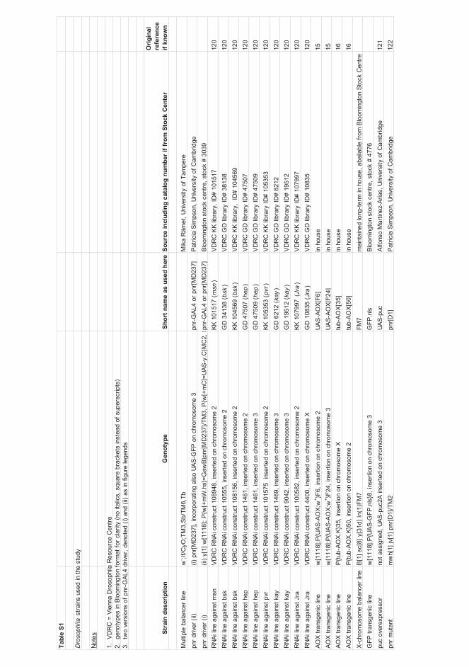
Dro
soph
ila
msn
bsk
bsk
hep
hep
pvr
kay kay
Jr
aJr
a
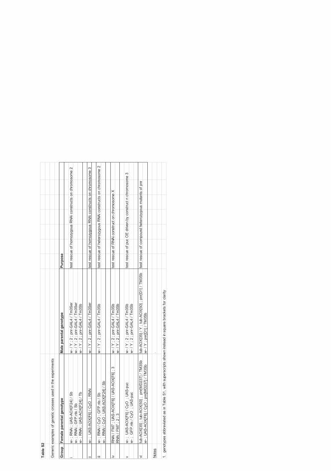
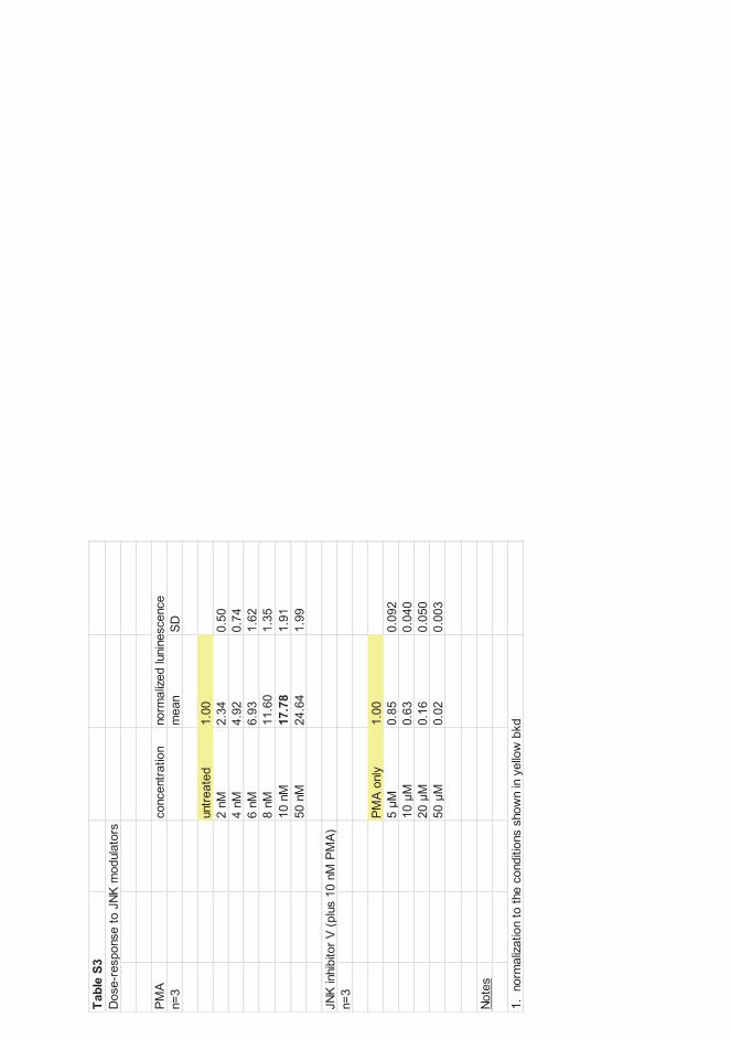
5 10
20
50

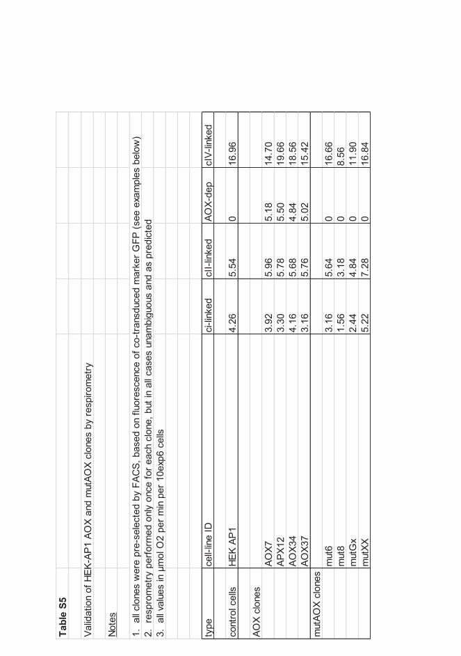
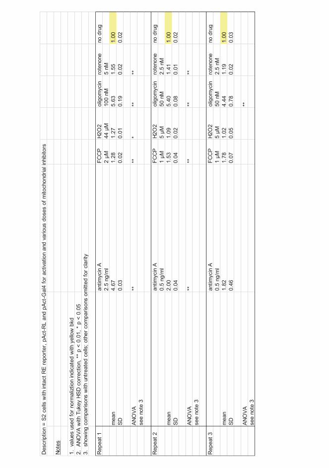
2 M
44
M
1 M
5 M
1 M
5 M


Related Documents