ORIGINAL ARTICLE Andrology Expression of a truncated form of KIT tyrosine kinase in human spermatozoa correlates with sperm DNA integrity Barbara Muciaccia 1 , Claudio Sette 2 , Maria Paola Paronetto 2 , Marco Barchi 2 , Simona Pensini 1 , Angela D’Agostino 1 , Loredana Gandini 3 , Raffaele Geremia 2 , Mario Stefanini 1 , and Pellegrino Rossi 2, * 1 Department of Histology and Medical Embryology, University of Rome ‘La Sapienza’, Rome, Italy 2 Department of Public Health and Cell Biology, Facolta’ di Medicina e Chirurgia, University of Rome ‘Tor Vergata’, via Montpellier 1, Rome 00133, Italy 3 Seminology Laboratory - Sperm Bank of the Department of Medical Physiopathology, University of Rome ‘La Sapienza’, Rome, Italy *Correspondence address. Tel: +39-06-72596272; Fax: +39-06-72596268; E-mail: [email protected] Submitted on April 16, 2010; resubmitted on May 27, 2010; accepted on June 4, 2010 background: TR-KIT, a truncated form of KIT (the KITL receptor), corresponding to the c-terminal half of the intracellular split tyro- sine kinase domain, is expressed during the haploid stages of mouse spermatogenesis, and is one of the candidate sperm factors possibly involved in egg activation at fertilization. methods: Immunocytochemistry of adult human testis, and studies of human semen samples from volunteer donors through immuno- fluorescence, confocal microscopy, flow cytometry, western blot and RT– PCR analyses were performed. results: We show that the TR-KIT is expressed during spermiogenesis in the human testis, and that it is maintained in human ejaculated spermatozoa. TR-KIT is localized both in the equatorial segment and in the sub-acrosomal region of the human sperm head. The equatorial localization of the TR-KIT persists after the spontaneous acrosome reaction. Cytometric analysis of several sperm samples from volunteer donors, showed variable degrees of the TR-KIT-specific immunolabeling, and a significant inverse correlation (Pearson’s coefficient, r ¼ 20.76, P , 0.0001, n ¼ 23) of the TR-KIT positivity with markers of sperm damage, i.e. DNA fragmentation, as revealed by terminal deoxynucleotidyl transferase-mediated deoxyuridine triphosphate-nick end labeling (TUNEL) analysis and the intense clusterin positivity. We also found less significant inverse correlation with altered head morphology (r ¼ 20.47, P , 0.05, n ¼ 23) and direct correlation with sperm forward motility parameters (r ¼ 0.59, P , 0.01, n ¼ 23). conclusions: The TR-KIT is present in the equatorial region of human spermatozoa, which is the first sperm component entering into the oocyte cytoplasm after fusion with the egg. This localization is consistent with the function previously proposed for this protein in mice. In addition, the TR-KIT represents a potential predictive parameter of human sperm quality. Key words: TR-KIT / human spermatozoa / equatorial segment / DNA damage / clusterin Introduction The KIT receptor tyrosine kinase (KITL receptor) plays an essential role in the mitotic stages of mouse spermatogenesis (Sette et al., 2000; Rossi et al., 2000, 2003; Pellegrini et al., 2008), but with the onset of meiosis its expression, at both the RNA and protein level ceases. After meiosis the TR-KIT, a truncated form of KIT corre- sponding to the c-terminal half of the intracellular split tyrosine kinase domain, is expressed beginning at the round spermatid stage (Sorrentino et al., 1991; Rossi et al., 1992). The mouse TR-KIT is originated by transcription of an alternative mRNA from a cryptic pro- moter in the 16th intron of the mouse KIT gene, which is active during spermiogenesis (Albanesi et al., 1996). TR-KIT localizes to the residual cytoplasm, midpiece and the post-acrosomal region of the mouse epi- dydimal spermatozoa (Albanesi et al., 1996; Sette et al., 1997). Micro- injection of the recombinant TR-KIT into metaphase II-arrested mouse oocytes triggers events of early embryogenesis, such as cortical granule exocytosis, completion of the second meiotic division, for- mation of pronuclei, and development up to the morula stage (Sette et al., 1997). The TR-KIT-mediated parthenogenetic egg activation & The Author 2010. Published by Oxford University Press on behalf of the European Society of Human Reproduction and Embryology. All rights reserved. For Permissions, please email: [email protected] Human Reproduction, Vol.25, No.9 pp. 2188– 2202, 2010 Advanced Access publication on July 3, 2010 doi:10.1093/humrep/deq168 at Universita' degli studi di Roma Foro Italico on September 24, 2014 http://humrep.oxfordjournals.org/ Downloaded from

Welcome message from author
This document is posted to help you gain knowledge. Please leave a comment to let me know what you think about it! Share it to your friends and learn new things together.
Transcript

ORIGINAL ARTICLE Andrology
Expression of a truncated form of KITtyrosine kinase in human spermatozoacorrelates with sperm DNA integrityBarbara Muciaccia1, Claudio Sette2, Maria Paola Paronetto2,Marco Barchi2, Simona Pensini1, Angela D’Agostino1,Loredana Gandini3, Raffaele Geremia2, Mario Stefanini 1,and Pellegrino Rossi2,*
1Department of Histology and Medical Embryology, University of Rome ‘La Sapienza’, Rome, Italy 2Department of Public Health and CellBiology, Facolta’ di Medicina e Chirurgia, University of Rome ‘Tor Vergata’, via Montpellier 1, Rome 00133, Italy 3Seminology Laboratory -Sperm Bank of the Department of Medical Physiopathology, University of Rome ‘La Sapienza’, Rome, Italy
*Correspondence address. Tel: +39-06-72596272; Fax: +39-06-72596268; E-mail: [email protected]
Submitted on April 16, 2010; resubmitted on May 27, 2010; accepted on June 4, 2010
background: TR-KIT, a truncated form of KIT (the KITL receptor), corresponding to the c-terminal half of the intracellular split tyro-sine kinase domain, is expressed during the haploid stages of mouse spermatogenesis, and is one of the candidate sperm factors possiblyinvolved in egg activation at fertilization.
methods: Immunocytochemistry of adult human testis, and studies of human semen samples from volunteer donors through immuno-fluorescence, confocal microscopy, flow cytometry, western blot and RT–PCR analyses were performed.
results: We show that the TR-KIT is expressed during spermiogenesis in the human testis, and that it is maintained in human ejaculatedspermatozoa. TR-KIT is localized both in the equatorial segment and in the sub-acrosomal region of the human sperm head. The equatoriallocalization of the TR-KIT persists after the spontaneous acrosome reaction. Cytometric analysis of several sperm samples from volunteerdonors, showed variable degrees of the TR-KIT-specific immunolabeling, and a significant inverse correlation (Pearson’s coefficient,r ¼ 20.76, P , 0.0001, n ¼ 23) of the TR-KIT positivity with markers of sperm damage, i.e. DNA fragmentation, as revealed by terminaldeoxynucleotidyl transferase-mediated deoxyuridine triphosphate-nick end labeling (TUNEL) analysis and the intense clusterin positivity. Wealso found less significant inverse correlation with altered head morphology (r ¼ 20.47, P , 0.05, n ¼ 23) and direct correlation with spermforward motility parameters (r ¼ 0.59, P , 0.01, n ¼ 23).
conclusions: The TR-KIT is present in the equatorial region of human spermatozoa, which is the first sperm component entering intothe oocyte cytoplasm after fusion with the egg. This localization is consistent with the function previously proposed for this protein in mice. Inaddition, the TR-KIT represents a potential predictive parameter of human sperm quality.
Key words: TR-KIT / human spermatozoa / equatorial segment / DNA damage / clusterin
IntroductionThe KIT receptor tyrosine kinase (KITL receptor) plays an essentialrole in the mitotic stages of mouse spermatogenesis (Sette et al.,2000; Rossi et al., 2000, 2003; Pellegrini et al., 2008), but with theonset of meiosis its expression, at both the RNA and protein levelceases. After meiosis the TR-KIT, a truncated form of KIT corre-sponding to the c-terminal half of the intracellular split tyrosinekinase domain, is expressed beginning at the round spermatid stage(Sorrentino et al., 1991; Rossi et al., 1992). The mouse TR-KIT is
originated by transcription of an alternative mRNA from a cryptic pro-moter in the 16th intron of the mouse KIT gene, which is active duringspermiogenesis (Albanesi et al., 1996). TR-KIT localizes to the residualcytoplasm, midpiece and the post-acrosomal region of the mouse epi-dydimal spermatozoa (Albanesi et al., 1996; Sette et al., 1997). Micro-injection of the recombinant TR-KIT into metaphase II-arrestedmouse oocytes triggers events of early embryogenesis, such as corticalgranule exocytosis, completion of the second meiotic division, for-mation of pronuclei, and development up to the morula stage (Setteet al., 1997). The TR-KIT-mediated parthenogenetic egg activation
& The Author 2010. Published by Oxford University Press on behalf of the European Society of Human Reproduction and Embryology. All rights reserved.For Permissions, please email: [email protected]
Human Reproduction, Vol.25, No.9 pp. 2188–2202, 2010
Advanced Access publication on July 3, 2010 doi:10.1093/humrep/deq168
at Universita' degli studi di R
oma Foro Italico on Septem
ber 24, 2014http://hum
rep.oxfordjournals.org/D
ownloaded from

requires its interaction with Fyn (a Src-like kinase) and the adaptorprotein Sam68, with the consequent activation of phospholipaseCg1 within the egg cytoplasm (Sette et al., 1998, 2002; Paronettoet al., 2003). Interestingly, Fyn and/or other egg-carried Src-likekinases have been shown to play a critical role in egg resumptionfrom meiosis II at fertilization and the subsequent zygotic developmentin mammals (Meng et al., 2006; McGinnis et al., 2007; Reut et al.,2007; Tomashov-Matar et al., 2008; Luo et al., 2009; Levi andShalgi, 2010).
Thus, the TR-KIT has been proposed as a sperm factor possiblyinvolved in sperm-mediated egg activation at fertilization (Rossiet al., 2003). However, it cannot be excluded that the TR-KIT,besides its eventual participation to the egg activation process at fer-tilization, plays additional roles during mouse spermatogenesis or inthe mature sperm cell function.
Restricted expression of KIT in the membrane of spermatogoniaand of TR-KIT in the cytoplasm of spermatids was also observed inthe rat seminiferous epithelium, both at the RNA and protein level(Prabhu et al., 2006). As in rodents, in the adult human testis full-length KIT is expressed in spermatogonia, as revealed by the use ofantibodies directed against the KIT extracellular domain (Nataliet al., 1992; Strohmeyer et al., 1995; Sandlow et al., 1996).However, no clear information is available about the possibleexpression of TR-KIT in post-meiotic stages in human testis. Immuno-cytochemical analysis using a polyclonal antibody directed against thehuman KIT c-terminus (Santa Cruz, C-19, cat. no. sc-168), whichrecognizes both full-length KIT and TR-KIT, showed positivity in sper-matogonia and in elongating spermatids of marmoset monkeys, butnot in meiotic spermatocytes, similarly to reported observations inrodents (von Schonfeldt, 1999). Using the same antibody, Feng et al.(2005) reported immunocytochemical reactivity at the level of theacrosome in the human sperm head, but, on the basis of westernblot analysis, they claimed that this positivity was due to the150 kDa full-length KIT. In a recent study, using another antibodydirected against the human KIT c-terminus (Dako, Anti-HumanCD117, cat. no. A4502), KIT positivity within the human testis wasconfined to spermatogonia and spermatids, whereas no staining wasfound in meiotic spermatocytes, nor in Sertoli cells, suggesting apattern of expression of the two alternative products of the KITgene analogous to that observed in rodents (Unni et al., 2009).Even more recently, by using a further polyclonal antibody directedagainst the c-terminus of human KIT (Abcam, cat. no. ab16832),KIT immunoreactivity in human spermatids was also evident (Heet al., 2010).
A truncated form of KIT analogous to mouse TR-KIT is aberrantlyexpressed in human cancer cell lines of various origin and in primaryprostate malignant tumors both at the RNA and protein level(Toyota et al., 1994; Takaoka et al., 1997; Paronetto et al., 2004).The aberrant transcript encoding human TR-KIT originates from acryptic promoter present within the 15th intron of the human KITgene (Toyota et al., 1994; Paronetto et al., 2004), and not from the16th intron, which appears to be the functional TR-KIT promoteronly in mice (Sakamoto et al., 2004). Interestingly, murine hemato-poietic stem cells and multipotent progenitors express the TR-KITat the RNA and protein level, so that a potential role for thisprotein in the self-renewal of these cells has been postulated (Zayaset al., 2008). The TR-KIT ability to activate early embryonic
development when microinjected into mouse eggs, its expression inhematopoietic stem cells, and its aberrant expression in humantumors, in which it activates Src-like kinase activity (Paronetto et al.,2004), suggest an important role of these intracellular alternative KITgene products in regulating developmental events involving growthfactor-independent cell proliferation.
Among assisted reproduction techniques (ART), the intracytoplas-mic sperm injection (ICSI) is overall considered a ‘safe’ technique,however, it bypasses the natural mechanisms of gamete selection. Inparticular, the injection of spermatozoa-carrying DNA damage mightbe dangerous in terms of potential malformations in the conceivedchild (Lewis, 2002). Moreover, DNA damage in the male germ line,mostly due to oxidative stress (Agarwal et al., 2008), has been associ-ated with male infertility, failed fertilization, impaired preimplantationdevelopment and poor pregnancy outcomes, whether the insemina-tion is natural or artificial (Irvine et al., 2000; Agarwal and Said,2003; Aitken et al., 2009; Barratt et al., 2010). A significant increasein DNA damage can also be found in sperm from infertile men classi-fied as normozoospermic on the basis of normal standard sperm par-ameters (Saleh et al., 2002). Thus, discovery of new sperm parameterspredictive of DNA integrity will have an important impact on thedevelopment of ARTs.
In the present paper, we have investigated the expression of theTR-KIT in human ejaculated spermatozoa from the seminal fluids ofvolunteer donors. We found that TR-KIT, but not full-length KIT, ispresent in the equatorial and sub-acrosomal region of the humansperm head. Moreover, our results show a significant positive corre-lation of the TR-KIT positivity with sperm DNA integrity, and wetherefore propose the TR-KIT as a new potential marker of humansemen quality.
Materials and Methods
Semen samplesSpecimens from 23 reputedly normozoospermic volunteer donorsattending the Seminology Laboratory - Sperm Bank of the Departmentof Medical Physiopathology, University of Rome ‘La Sapienza’, were col-lected by masturbation after 3–5 days of sexual abstinence, into sterileplastic jars. Material was utilized after having obtained written a consentfor the utilization of semen samples. Samples were allowed to liquefyfor 60 min at 378 C and were then evaluated according to WHOcriteria (World Health Organization, 1999). The variables takeninto consideration were: ejaculate volume (ml), sperm concentration(n × 106/ml), total sperm count (n × 106/ejaculate), forward motility(%) and morphology (% abnormal forms). None of the donors werefound to be oligospermic. Of the 23 donors, 20 were within thenormal standard parameters defined by WHO (normozoospermic),whereas the remaining 3 were borderline asthenoteratozoospermic.After removing seminal plasma by centrifugation, sperm pellets werewashed twice in 1× phosphate-buffered saline (PBS), counted anddivided into aliquots.
For swim-up purification of semen samples, 1 ml of each seminal samplewas diluted 1:2 with Hank’s medium and centrifuged for 10 min at 300 g.After centrifugation, the supernatant was discarded and an aliquot of0.5 ml of Hank’s was layered on the pellet. The spermatozoa wereallowed to migrate for 30 min at 378C. After migration, the supernatantwas gently aspirated and processed for further studies.
TR-KIT in human spermatozoa 2189
at Universita' degli studi di R
oma Foro Italico on Septem
ber 24, 2014http://hum
rep.oxfordjournals.org/D
ownloaded from

Immunohistochemistry andimmunocytochemistryHistological sections from two formalin-fixed paraffin-embedded humantesticular normal tissue fragments obtained from orchiectomy of testicularcancer patients (a gift from Dr Giovanni Bertalot, Ospedale di Leno,Brescia, Italy) were cut into 5-mm sections and mounted on polylysinecoated slides. Sections were dewaxed, rehydrated and processed in amicrowave for antigen retrieval in pH 6.0 citrate buffer. After quenchingof endogenous peroxidase and blocking of non-specific binding, sectionswere incubated for 1 h at room temperature (RT) with 1:200 polyclonalantibody directed against the human KIT c-terminus (Santa Cruz, C-19,cat. no. sc-168), which recognizes both full-length KIT and TR-KIT, exten-sively washed and then processed using the avidin–biotin peroxidasecomplex (ABC) procedure, according to the manufacturer’s protocol(UltraTek HRP Anti-Polyvalent kit, ScyTek Laboratories, USA), negativecontrols were performed using affinity-purified rabbit IgG or omittingthe primary antibody. Peroxidase activity was revealed using3,3-diaminobenzidine tetrahydrochloride (Roche, Italy), nuclei werequickly counterstained with Mayer’s hematoxylin. After washing, sectionswere permanently mounted and observed by light microscopy.
For immunocytochemical studies on human semen samples, we usedthe same UltraTek HRP Anti-Polyvalent kit and protocol describedabove. In particular before primary antibody incubation, ejaculated sper-matozoa, obtained after washing of sperm pellets and spotted into polyly-sine coated slides, were briefly treated with cold acetone (to fix andsimultaneously permeabilize cells) and air dried. Samples were mountedon slides and observed by light and phase-contrast microscopy. No stain-ing was evident with non-immune rabbit IgG, nor with the secondary anti-body alone. No nuclear counterstaining was performed.
Immunofluorescence analysis and confocalmicroscopyEjaculated spermatozoa, obtained after sperm pellet washing, werespotted into polylysine coated slides, fixed in cold acetone and air dried.After blocking for 2 h at RT with 1× PBS/5% bovine serum albumin(BSA 5%)/ normal goat serum (NGS) 10% [or 10% NGS plus 10%normal donkey serum (NDS) in double-immunostaining experiments],sperm cells were incubated overnight (O/N) at 48C with 1:200 C-19rabbit anti-human KIT antibody, extensively washed, incubated for 1 h atRT with 1:1000 secondary goat anti-rabbit Alexafluor 488-conjugated anti-body (Molecular Probes), washed, mounted and observed at fluorescencemicroscopy (Axioplan 2 Imaging system, Carl Zeiss, Germany). Negativecontrols were performed omitting primary antibodies and using theaffinity-purified rabbit IgG. Nuclei were stained using TOTO-3 dye(1:2000; Sigma Aldrich, Italy). In order to assess the specificity immuno-binding of C-19 polyclonal antibody to the human intracellular KITsperm antigen, before the immunostaining, primary antibody was pre-adsorbed with a 100-fold mass excess of the immunogenic cognatepeptide (Santa Cruz Biotechnology) and then used in paired controlslides to evaluate the efficiency of competition; this experiment wasrepeated three times on different semen samples.
In order to perform double-immunostaining for the TR-KIT and acrosin,incubation for 1h at RT with 1:500 mouse monoclonal anti-human acrosinantibody (Biosonda Corp., Chile) was performed after O/N C-19 anti KITincubation, whereas secondary antibodies, 1:1000 secondary Alexafluor488 goat anti-rabbit (Molecular Probes, Invitrogen) and 1:300 donkeyCy3-conjugated anti-mouse (Jackson ImmunoResearch, UK), were usedtogether for 1h at RT. Nuclei were stained using TOTO3 dyes (1:2000;Molecular Probes, Invitrogen). Labeled-sperm cells were observed witha Leica laser scanning microscope TCS SP2 equipped with three laser
lines. Each channel was acquired separately using specific laser lines toavoid a bleed-through of the fluorochromes. Photomicrographs wereacquired using LAS AF Leica Confocal Software (Leica, Germany).
Immunoblot analysisHuman spermatozoa from raw semen of normozoospermic donors andfrom patients recruited for fertility studies were resuspended in lysisbuffer (50 mM HEPES, pH7.5, 150 mM NaCl, 15 mM MgCl2, 15 mMEGTA, 250 mM NaVO4, 10 mM b-glycerophosphate, 1% Triton X100,0.1% SDS, supplemented with protease inhibitor cocktail) and sonicatedfor three cycles of 30 s on ice. The control recombinant mouse TR-KITprotein was prepared by protein extraction from transfected cell lines asdescribed previously (Albanesi et al., 1996; Sette et al., 1997). Lysateswere cleared by centrifugation at 10 000 g for 10 min. The supernatantfraction was collected, diluted in SDS-PAGE sample buffer and boiledfor 5 min. Samples were separated on 10% SDS-PAGE gels and transferredto polyvinylidene fluoride Immobilon-P membranes (Millipore) using asemidry blotting apparatus (BioRad). Western analysis was carried outas previously reported (Sette et al., 2002) using the C-19 anti-humanKIT (c-terminal) antibody (1:1000 dilution) O/N at 48C. Competitionwas performed by preincubating the antibody with an excess of the immu-nogenic peptide (100 × molar excess; 1:1 mass ratio) for 12 h beforedilution in the primary antibody solution for hybridization (Sette et al.,1997). Secondary anti-rabbit IgG conjugated to horseradish peroxidase(Amersham) were incubated with the membranes for 1 h at RT at a1:10 000 dilution in PBS containing 0.1% Tween 20. Immunostainedbands were detected by the chemiluminescent method (Santa CruzBiotech.). The human semen sample in which 100% of spermatozoalacked the acrosome was kindly provided by Dr Jan Tesarik (Center forReproductive Medicine, European Hospital, Rome, Italy).
For the RNA interference analysis in transfected cell lines, a 500-bpinverted repeat corresponding to part of the 5′ untranslated region andthe beginning of the mouse TR-KIT open reading frame (ORF) (Rossiet al., 1992) was subcloned in the pDECAP vector described by Shinagawaand Ishii (2003). HEK293 cells were co-transfected with a recombinantplasmid expressing the mouse TR-KIT ORF (Albanesi et al., 1996; Setteet al., 1997) and a second plasmid expressing an Myc epitope (tocontrol for transfection efficiency). Cells were also co-transfected eitherwith the pDECAP empty vector or with increasing amounts of the inter-fering plasmid constructs. Protein extracts from transfected cells were pro-cessed for immunoblot analysis. Western blots were probed with theC-19 anti-human-KIT-c-terminus antibody and an antibody directedagainst the Myc epitope.
RT–PCR analysisIn order to verify the presence of specific KIT mRNA molecules in spermcells following osmotic shock, the total RNA was extracted from 2 × 10⁶
purified ejaculated spermatozoa as previously described (Muciaccia et al.,2007) using high pure RNA isolation kit (Roche) according to the manu-facturer’s instructions. Purified RNA was treated for 15 min at 378Cwith 2 units of Deoxyribonuclease I, Amplification Grade (Invitrogen-Lifetechnologies) to prevent DNA genomic contamination. An aliquot oftotal RNA was reverse-transcribed, using Sensiscript Reverse Transcrip-tase (Qiagen), a highly sensitive enzyme recommended for very smallRNA amount (,50 ng) and oligo-dT as primer. Then, by PCR amplifica-tion, total RNA was tested for protamine-2 gene (PRM-2, specificallyexpressed only in haploid germ cells), using a primer set able to discrimi-nate between genomic DNA and sperm mRNA (Miller et al., 1994).
In order to amplify KIT mRNA and discriminate between different tran-scripts (full-length mRNA or truncated form), aliquots of cDNA were usedin amplification reactions using the following oligonucleotide primers
2190 Muciaccia et al.
at Universita' degli studi di R
oma Foro Italico on Septem
ber 24, 2014http://hum
rep.oxfordjournals.org/D
ownloaded from

spanning four contiguous human KIT gene exons (Accession number:nG_007456): e15 (forward) GTACTAATGAGTACATGGACATG; e16(forward) GCTCATACATAGAAAGAGATGTG; e17 (forward) TGTATTCACAGAGACTTGGCAGC; e18 (reverse) TGCTTTCAGGTGCCATCCACTTCAC.
Amplification conditions were: first denaturation at 948C for 3 min, then948C for 30 s, 658C for 45 s, 728C for 40 s for 50–80 cycles, in 50 ml finalvolume using 2 U of Taq DNA polymerase (Roche). A control reactiontube, in which reverse transcriptase enzyme was omitted, was alsoincluded to test possible DNA genomic contamination. Aliquots of PCRproduct (i.e. 1/5 of total reaction volume) were separated on 2%agarose gel and visualized by ethidium bromide staining.
Cytometric analysis of human sperm cellsPresence of intracellular TR-KIT and clusterin antigens on freshly isolatedsperm cells from presumably healthy donors was assessed by cytometricanalysis. Aliquots of 106 sperm cells/tube were briefly fixed on ice using2% paraformaldehyde, washed twice and then permeabilized using 0.1%sodium citrate/0.1% Triton X-100. After washing sperm cells were incu-bated for 30 min on ice with primary antibodies (1:200 in PBS/1% BSA,C-19 rabbit polyclonal antibody against the c-terminus of human KIT,#sc-168; H-330 polyclonal rabbit anti-human clusterin antibody,#sc-8354, both from Santa Cruz Biotechnology, Germany). The C-19 anti-body recognizes both the full-length KIT and the TR-KIT, whereas theanti-clusterin antibody can recognize either the acrosomal or the surfaceisoforms of clusterin. Sperm cells from the same samples were also incu-bated in a control tube with the same concentration of the correspondingaffinity-purified rabbit IgG (Sigma), as recommended by technical datasheets. Antibody-labeled sperm suspensions were washed twice with1 × PBS/1% BSA, incubated with phycoerythrin-conjugated secondaryantibody (Goat anti-rabbit-PE, by Jackson ImmunoResearch Laboratories,UK). In order to assess the presence of intracellular TR-KIT and to excludeits sperm surface expression, control tube reactions were also performedusing aliquots of unfixed and unpermeabilized sperm cells in which theanti-KIT antibody immunolabeling failed to detect any surface specificsignal (data not shown). The specificity of the sperm cells gate was verifiedby analysis of sperm DNA content using the DNA binding dye propidiumiodine. For double detection of both the TR-KIT antigen and the spermDNA fragmentation by the terminal deoxynucleotidyl transferase-mediated deoxyuridine triphosphate-nick end labeling (TUNEL) technique,aliquots of 1 × 10⁶ spermatozoa were treated by In Situ Cell Death Detec-tion Kit-Fluorescein (Roche, Italy), according to the manufacturer’s instruc-tions before the TR-KIT immunolabeling. Cells were then run through aflow cytometer (Epics XL Beckman Coulter, USA) using an Argon488 nm excitation laser and 5000 and 10 000 events were, respectively,acquired for each single and double reaction/tube. Results were expressedas the percentage of positive spermatozoa gated. In order to assess thespecificity immuno-binding of C-19 polyclonal antibody to human intra-cellular TR-KIT sperm antigen, before cytometric analysis, primary anti-body was pre-adsorbed with a 100-fold mass excess of theimmunogenic cognate peptide (Santa Cruz Biotechnology) and thenused in paired control tubes to evaluate the efficiency of competitionbinding reaction; the competition experiment was repeated four timeson different semen samples.
Statistical analysisData from morphological and cytometric analyses of semen samples wereutilized to build matrixes for the calculation of r-values (Pearson’s product-moment correlation coefficients); the significance (two-tailed probabilityvalues) of r coefficients were calculated on the basis of the correlationvalues and the sample sizes.
Results
Immunohistochemistry in normal adult testisUsing a polyclonal antibody directed against the c-terminus of humanKIT (Santa Cruz, C-19, sc-168), which recognizes both the full-lengthKIT and the TR-KIT, the KIT immunostaining was evident in the mem-brane of several spermatogonia in the basal layer and in interstitialLeydig cells, whereas Sertoli cells and spermatocytes inside the semi-niferous tubules were negative (Fig. 1A–C). Clear positivity was alsoevident in the haploid stages of spermatogenesis. KIT immunostainingwas evident in the cytoplasm of round (Fig. 1A and B) and elongatingspermatids (Fig.1C), sometimes with a distinct perinuclear distributionin the area of the developing acrosome (Fig. 1B). The presence of KITimmunostaining both in spermatogonia and in spermatids, but not inspermatocytes, is substantially similar to recent observations byother groups using two different polyclonal antibodies directedagainst the c-terminus of human KIT (Unni et al., 2009; He et al.,2010).
Analysis of ejaculated spermatozoaby immunocytochemistryPreparations of freshly ejaculated spermatozoa from normozoosper-mic subjects were fixed after permeabilization and probed with theC-19 anti-human KIT antibody, followed by secondary antibody con-jugated to peroxidase (Fig. 1D and E). Immunocytochemical analysisrevealed KIT staining in the anterior region of the sperm head, withmaximal intensity often observed in the equatorial segment(Fig. 1D), or isolated intense staining in the equatorial segmentalone (Fig. 1E). However, not all sperm heads resulted positive tothe KIT immunostaining (Fig. 1D). Notably, most of KIT-negative sper-matozoa appeared to have mis-shaped heads or other evidentanomalies extending to the midpiece (Fig. 1D).
Immunofluorescence and cytometric analysisof ejaculated spermatozoaA typical low magnification picture of fixed-permeabilized sperm prep-aration from a normozoospermic patient probed for immunofluores-cence with the anti-human KIT antibody (green-cyan signal), andcounterstained with TOTO-3 dye to identify sperm nuclei (bluesignal), is shown in Fig. 2A. Similar to immunocytochemistry obser-vations, KIT positivity was evident in most sperm heads, especiallyin the equatorial segment, but, again, not all spermatozoa appearedto be positive. Figure 2B shows that the fluorescent signal wasalmost completely abolished if the primary antibody had been pre-incubated with a 100-fold mass excess of the immunogenic peptide,demonstrating specificity of KIT immunostaining in the sperm head.
In order to quantify positivity of KIT immunostaining, we performedcytometric analysis. A typical example of fluorescence-activated cellsorting (FACS) analysis with a permeabilized sperm sample from anormozoospermic subject is shown in Fig. 2C. Specificity of the shift(red line) was confirmed by its abolishment after pre-incubation ofthe primary antibody with the immunogenic peptide (Fig. 2C, blueline). KIT positivity in spermatozoa varied in different subjects,ranging from 10 to 93%, with a median of 54% (Table I). No shiftwas obtained with the same antibody if sperm samples had not
TR-KIT in human spermatozoa 2191
at Universita' degli studi di R
oma Foro Italico on Septem
ber 24, 2014http://hum
rep.oxfordjournals.org/D
ownloaded from

been permeabilized (data not shown), indicating that the KIT positivityis due to intracellular sperm components.
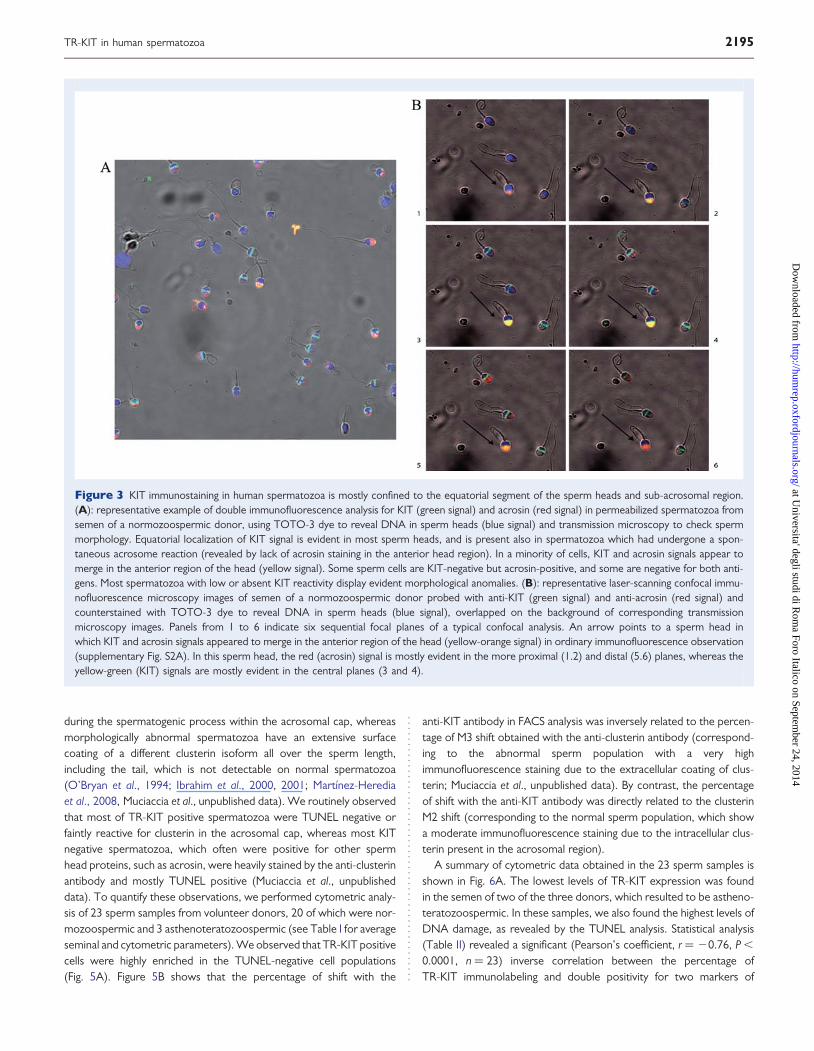
Subcellular localization of KITimmunostaining in human sperm headsTypical double immunofluorescence analysis performed with anti-KIT(green) and anti-acrosin (red) antibodies, confirmed that the KIT–positivity in human sperm was localized in the anterior region of thehead, with maximal intensity in the equatorial segment, behind theacrosome, or exclusively in the equatorial region (Fig. 3A; see alsoSupplementary Material, Fig. S2A). Notably, KIT equatorial stainingpersisted in acrosin-negative sperm heads, indicating that it is not
localized in the acrosome vesicle nor released during spontaneousacrosome reaction. In some sperm heads, KIT and acrosin signalsappeared to uniformly merge all over the anterior region of thehead (yellow signal), with no evident accumulation in the equatorialsegment. Some sperm heads were KIT-negative, but positive foracrosin staining, or negative for both signals. Careful morphologicalanalysis of KIT positivity in sperm samples from several normozoos-permic volunteer donors indicated that 50–80% of KIT-positive sper-matozoa showed maximal or isolated staining in the equatorialsegment, whereas the remaining percentage of sperm cells showedapparent merging of the KIT and acrosin signals in the anteriorregion of the head. When DNA counterstaining (blue signal inFig. 3A) was omitted, it was evident that also in spermatozoa
Figure 1 Immunohistochemical and immunocytochemical studies of human testis and spermatozoa with an anti-KIT (c-terminal) antibody. Local-ization of KIT immunostaining in histological sections of normal adult testis (A–C) and in permeabilized spermatozoa from semen of normozoosper-mic donors (D and E) using the C-19 rabbit polyclonal antibody against the c-terminus of human KIT. Staining was revealed with a secondary antibodyconjugated to peroxydase (hematoxylin counterstained). Light microscope images at lower (A and D) and higher (B, C and E) magnifications areshown. In (D and E), light microscope images were merged with phase contrast pictures. Arrows in (A–C) indicate positive staining in spermatogonia(SG), spermatids (ST) within the seminiferous tubules, and in interstitial Leydig cells (LC). Arrows in (D and E) indicate either absence of staining(negative) or staining in the equatorial and anterior head regions of spermatozoa.
2192 Muciaccia et al.
at Universita' degli studi di R
oma Foro Italico on Septem
ber 24, 2014http://hum
rep.oxfordjournals.org/D
ownloaded from

showing uniform positivity all over the anterior region of the head, theKIT signal was more heavily concentrated in the equatorial segment(Supplementary Material, Fig. S1).
To ascertain whether KIT positivity in the anterior region of thehead was due to co-localization with acrosin within the acrosomalmatrix, human spermatozoa were analyzed by laser-scanning confocalimmunofluorescence microscopy. As shown in the series of images inFig. 3B, representing six sequential focal planes of a typical confocalanalysis (supplementary Fig. S2B), proceeding from the proximal tothe distal plane, the red acrosin signal was the first to appear andthe last to disappear with respect to both the green (KIT) equatorialsignals and the yellow (KIT-acrosin merge) signal in the anterior regionof the head, which were closer to the plane of maximal blue staining(DNA). Thus, KIT positivity appears to be maximal in the sub-acrosomal region of the sperm heads, similar to the perinuclear stain-ing observed in developing spermatids (cfr. Fig. 1B,C).
Molecular identification of TR-KIT as the KITgene product normally expressed in humanspermatozoaIntracellular KIT immunostaining in the perinuclear region of haploidspermatids and in the sub-acrosomal-equatorial region of spermheads, suggests that, like in rodents, human post-meiotic KIT positivitymight be due to expression of the human equivalent of mouse TR-KIT.In order to clarify this point, we performed western blot analysis ofprotein extracts from freshly ejaculated human spermatozoa usingthe same antibody used for the morphological experiments. This anti-body is directed against the c-terminal portion shared by both the full-length KIT and TR-KIT. A typical immunoblot analysis is shown inFig. 4A. No signal was evident in the high molecular weight (MW)range of the blot, indicating that the 150 kDa full-length KIT is notexpressed in normal human spermatozoa. However, two lower
Figure 2 Immunofluorescence and cytometric analysis of permeabilized human spermatozoa with an anti-KIT (c-terminal) antibody. (A): immuno-fluorescence analysis of permeabilized spermatozoa from semen of a normozoospermic donor, using the C-19 rabbit polyclonal antibody against thec-terminus of human KIT (green/cyan signal) and TOTO-3 dye counterstaining of DNA in sperm heads (blue signal). KIT positivity is evident in theequatorial segment of most sperm nuclei. (B): same as in (A), except that the C-19 antibody had been previously incubated with an excess of the KITc-terminal immunogenic peptide. KIT staining is no longer evident in most of the sperm heads. This experiment was repeated with similar results onthree different sperm samples. (C): representative example of quantification of the percentage of KIT positivity in permeabilized human sperm samplesusing flow cytometry with the C-19 antibody. Specificity of the shift (red line) was confirmed by its abolishment after pre-incubation of the primaryantibody with the immunogenic peptide (blue line, which coincides with the black line obtained with non-immune IgGs). This experiment was repeatedwith similar results on four different sperm samples.
TR-KIT in human spermatozoa 2193
at Universita' degli studi di R
oma Foro Italico on Septem
ber 24, 2014http://hum
rep.oxfordjournals.org/D
ownloaded from
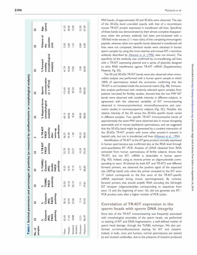
MW bands, of approximately 50 and 30 kDa were observed. The sizeof the 30 kDa band coincided exactly with that of a recombinantmouse TR-KIT protein expressed in transfected cell lines. Specificityof these bands was demonstrated by their almost complete disappear-ance when the primary antibody had been pre-incubated with a100-fold molar excess (1:1 mass ratio) of the competing immunogenicpeptide, whereas other non-specific bands detected in transfected celllines were not competed. Identical results were obtained in humansperm samples by using the cross-reactive anti-mouse-KIT-c-terminusantibody described by Albanesi et al. (1996; data not shown). Thespecificity of the antibody was confirmed by co-transfecting cell lineswith a TR-KIT expressing plasmid and a series of plasmids designedto elicit RNA interference against TR-KIT mRNA (SupplementaryMaterial, Fig. S3).
The 50 and 30 kDa TR-KIT bands were also observed when immu-noblot analysis was performed with a human sperm sample in which100% of spermatozoa lacked the acrosome, confirming that theTR-KIT is not localized inside the acrosomal matrix (Fig. 4B). Immuno-blot analysis performed with randomly selected sperm samples frompatients recruited for fertility studies, showed that the low MW KITbands were observed with variable intensity in different subjects, inagreement with the observed variability of KIT immunostainingobserved in immunocytochemical, immunofluorescence and cyto-metric studies in normozoospermic subjects (Fig. 4C). Notably, therelative intensity of the 50 versus the 30 kDa specific bands variedin different samples. Two specific TR-KIT immunoreactive bands ofapproximately the same MW were observed also in mouse elongatingspermatids and in mouse epididymis spermatozoa, and we suggestedthat the 50 kDa band might be generated by a covalent interaction ofthe 30 kDa TR-KIT protein with some other protein/s present inhaploid cells, but not in transfected cell lines (Albanesi et al., 1996).
Identification of TR-KIT as the KIT gene product normally expressedin human spermatozoa was confirmed also at the RNA level throughsemi-quantitative RT–PCR. Analysis of cDNA obtained from RNAextracted from human spermatozoa of fertile subjects shows thatTR-KIT, but not KIT, mRNA is detectable in human sperm(Fig. 4D). Indeed, using as reverse primer an oligonucleotide corre-sponding to exon 18 (shared by both KIT and TR-KIT) and differentforward primers, we observed the positive signal of the expectedsize (209 bp band) only when the primer annealed to the KIT exon17 (which corresponds to the first exon of the TR-KIT-specificmRNA expressed during mouse spermiogenesis). By contrast,forward primers that should amplify RNA encoding the full-lengthKIT receptor (oligonucleotides corresponding to sequences fromexon 15 and the beginning of exon 16), did not generate any RT–PCR product even after a higher number of PCR cycles.
Correlation of TR-KIT expression in thesperm heads with sperm DNA integritySince lack of the TR-KIT immunostaining was frequently associatedwith morphological anomalies of the sperm heads, we performedco-staining of KIT and DNA fragmentation, a well-defined marker ofsperm head damage, through the TUNEL technique. We also per-formed co-immunofluorescence staining for KIT and clusterin.Indeed, in bulls, rams and humans, normal spermatozoa are stainedby anti-clusterin antibodies, due to the presence of clusterin produced
....
....
....
....
....
....
....
....
....
....
....
....
....
....
....
....
....
....
....
....
....
....
....
....
....
....
....
....
....
....
....
....
....
....
....
....
....
....
....
....
....
....
....
....
....
....
....
....
....
....
....
....
....
....
....
....
....
....
....
....
....
....
..
Tab
leI
Sem
inal
para
met
ers
and
perc
enta
ges
ofpo
siti
vity
(as
mea
sure
dby
cyto
fluo
rim
etri
cor
mor
phol
ogic
alan
alys
is).
No.
ofsa
mpl
es22
2323
2323
323
2323
2323
% Clu
ster
in(M
2)FA
CS
posi
tivi
ty
% Clu
ster
in(M
3)FA
CS
posi
tivi
ty
% TU
NE
LFA
CS
posi
tivi
ty
%T
UN
EL
/C
lust
erin
(M3)
doub
leFA
CS
posi
tivi
ty
%T
R-K
ITFA
CS
posi
tivi
ty
%T
R-K
ITF
AC
Spo
siti
vity
afte
rsw
im-u
ppu
rifi
cati
on
%A
typi
calf
orm
s(m
orph
olog
ical
anal
ysis
)
Spe
rmco
ncen
trat
ion
(n3
106/m
l)
%Fo
rwar
dm
otili
ty(m
orph
olog
ical
anal
ysis
)
Vol
ume
ofth
eej
acul
ate
(ml)
Age
(yea
rs)
Mea
n63
.45
28.1
625
.03
19.2
552
.70
96.3
769
.13
115.
4341
.74
3.59
41.5
2
Med
ian
71.6
520
.95
18.1
810
.93
54.4
095
.90
68.0
010
0.00
45.0
03.
8038
.00
Stan
dard
devi
atio
n18
.88
22.0
620
.19
19.1
423
.27
1.46
8.26
83.2
414
.82
1.52
13.6
0
Min
imum
12.3
17.
147.
714.
7710
.29
95.2
058
.00
25.0
05.
000.
8022
.00
Max
imum
86.3
993
.43
79.8
168
.82
93.3
098
.00
88.0
040
0.00
60.0
06.
0067
.00
2194 Muciaccia et al.
at Universita' degli studi di R
oma Foro Italico on Septem
ber 24, 2014http://hum
rep.oxfordjournals.org/D
ownloaded from
during the spermatogenic process within the acrosomal cap, whereasmorphologically abnormal spermatozoa have an extensive surfacecoating of a different clusterin isoform all over the sperm length,including the tail, which is not detectable on normal spermatozoa(O’Bryan et al., 1994; Ibrahim et al., 2000, 2001; Martınez-Herediaet al., 2008, Muciaccia et al., unpublished data). We routinely observedthat most of TR-KIT positive spermatozoa were TUNEL negative orfaintly reactive for clusterin in the acrosomal cap, whereas most KITnegative spermatozoa, which often were positive for other spermhead proteins, such as acrosin, were heavily stained by the anti-clusterinantibody and mostly TUNEL positive (Muciaccia et al., unpublisheddata). To quantify these observations, we performed cytometric analy-sis of 23 sperm samples from volunteer donors, 20 of which were nor-mozoospermic and 3 asthenoteratozoospermic (see Table I for averageseminal and cytometric parameters). We observed that TR-KIT positivecells were highly enriched in the TUNEL-negative cell populations(Fig. 5A). Figure 5B shows that the percentage of shift with the
anti-KIT antibody in FACS analysis was inversely related to the percen-tage of M3 shift obtained with the anti-clusterin antibody (correspond-ing to the abnormal sperm population with a very highimmunofluorescence staining due to the extracellular coating of clus-terin; Muciaccia et al., unpublished data). By contrast, the percentageof shift with the anti-KIT antibody was directly related to the clusterinM2 shift (corresponding to the normal sperm population, which showa moderate immunofluorescence staining due to the intracellular clus-terin present in the acrosomal region).
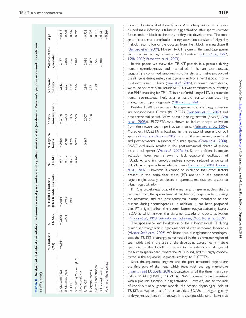
A summary of cytometric data obtained in the 23 sperm samples isshown in Fig. 6A. The lowest levels of TR-KIT expression was foundin the semen of two of the three donors, which resulted to be astheno-teratozoospermic. In these samples, we also found the highest levels ofDNA damage, as revealed by the TUNEL analysis. Statistical analysis(Table II) revealed a significant (Pearson’s coefficient, r ¼ 20.76, P ,
0.0001, n ¼ 23) inverse correlation between the percentage ofTR-KIT immunolabeling and double positivity for two markers of
Figure 3 KIT immunostaining in human spermatozoa is mostly confined to the equatorial segment of the sperm heads and sub-acrosomal region.(A): representative example of double immunofluorescence analysis for KIT (green signal) and acrosin (red signal) in permeabilized spermatozoa fromsemen of a normozoospermic donor, using TOTO-3 dye to reveal DNA in sperm heads (blue signal) and transmission microscopy to check spermmorphology. Equatorial localization of KIT signal is evident in most sperm heads, and is present also in spermatozoa which had undergone a spon-taneous acrosome reaction (revealed by lack of acrosin staining in the anterior head region). In a minority of cells, KIT and acrosin signals appear tomerge in the anterior region of the head (yellow signal). Some sperm cells are KIT-negative but acrosin-positive, and some are negative for both anti-gens. Most spermatozoa with low or absent KIT reactivity display evident morphological anomalies. (B): representative laser-scanning confocal immu-nofluorescence microscopy images of semen of a normozoospermic donor probed with anti-KIT (green signal) and anti-acrosin (red signal) andcounterstained with TOTO-3 dye to reveal DNA in sperm heads (blue signal), overlapped on the background of corresponding transmissionmicroscopy images. Panels from 1 to 6 indicate six sequential focal planes of a typical confocal analysis. An arrow points to a sperm head inwhich KIT and acrosin signals appeared to merge in the anterior region of the head (yellow-orange signal) in ordinary immunofluorescence observation(supplementary Fig. S2A). In this sperm head, the red (acrosin) signal is mostly evident in the more proximal (1.2) and distal (5.6) planes, whereas theyellow-green (KIT) signals are mostly evident in the central planes (3 and 4).
TR-KIT in human spermatozoa 2195
at Universita' degli studi di R
oma Foro Italico on Septem
ber 24, 2014http://hum
rep.oxfordjournals.org/D
ownloaded from

Figure 4 Human spermatozoa express TR-KIT, but not full-length KIT. (A): representative immunoblot analysis using the C-19 rabbit polyclonalantibody against the c-terminus of the KIT or the same antibody pre-incubated with an excess of the immunogenic peptides. Protein extractstested were (left to right) from mock- transfected HEK293 cells, from the same cells transfected with a vector expressing recombinant mouseTR-KIT, and from spermatozoa of two normozoospermic donors. High MW bands corresponding full-length KIT are absent, whereas two specific50 and 30 kDa TR-KIT bands are evident in both sperm samples. These bands, as well the recombinant 30 kDa TR-KIT band, are almost absentafter pre-incubation of C-19 with its cognate epitope. (B): immunoblot analysis of protein extracts from semen of a patient in which 100% of sper-matozoa lacked the acrosome. The specific TR-KIT bands are evident also in this sample, and high MW bands corresponding full-length KIT are absent.(C): immunoblot analysis using the C-19 antibody performed with eight randomly selected sperm samples from patients (indicated by numbers)recruited for fertility studies. The 50 and 30 kDa TR-KIT bands were observed with variable intensity in different subjects and, again, high MWbands corresponding full-length KIT are absent. Equivalent protein loading was checked by reversible Ponceau staining before probing blots withthe C-19 antibody. (D): a representative RT–PCR analysis of RNA extracted from spermatozoa of a normozoospermic donor, indicates thatRNA encoding for TR-KIT, but not for full-length KIT, is detectable in human spermatozoa as a remnant of transcription occurring during spermiogen-esis. The low MW bands present on the bottom of the lanes are due to the oligonucleotide primers used for amplification. No signals were obtainedwhen omitting RT before the amplification step of the reaction. This experiment was repeated on RNA extracted from sperm samples from threedifferent normozoospermic donors with similar results.
2196 Muciaccia et al.
at Universita' degli studi di R
oma Foro Italico on Septem
ber 24, 2014http://hum
rep.oxfordjournals.org/D
ownloaded from

Figure 5 TR-KIT expressing spermatozoa have a low level of DNA damage and an high percentage of TR-KIT positivity corresponds to low levels ofstaining with anti-clusterin antibodies. (A): representative double cytometric analysis for TR-KIT immuno-positivity and DNA fragmentation.on asperm sample from a normozoospermic donor. Permeabilized spermatozoa were treated by In Situ Cell Death Detection Kit-Fluorescein (Roche,Italy) before TR-KIT immunolabeling. with the C-19 antibody. Numbers within different gate quadrants refer to the percentage of: TR-KIT posi-tive/TUNEL negative (top left), TR-KIT negative/TUNEL negative (bottom left); TR-KIT positive/TUNEL positive (top right), and TR-KIT nega-tive/TUNEL positive cells (bottom right). TR-KIT positive cells appear to be highly enriched in the TUNEL-negative cell population. (B):representative contemporary cytometric analysis with anti-clusterin (left panels) and anti-KIT (right panels) antibodies of permeabilized spermsamples from two different donors, showing either high (top panels) or low (bottom panels) percentage of TR-KIT positivity (M1 peaks). In thesperm sample with high TR-KIT positivity (top panels), two separate peaks of clusterin positivity (M2 and M3) are evident: the M2 clusterin peakcorresponds to spermatozoa with moderate anti-clusterin staining in the acrosomal cap, whereas the M3 peak corresponds to spermatozoa withintense surface anti-clusterin staining (Muciaccia et al., unpublished data). In the sperm sample with low percentage of TR-KIT positivity (bottompanels), only the clusterin M3 peak is evident.
TR-KIT in human spermatozoa 2197
at Universita' degli studi di R
oma Foro Italico on Septem
ber 24, 2014http://hum
rep.oxfordjournals.org/D
ownloaded from

sperm damage, i.e. TUNEL positivity and intense clusterin immunola-beling (M3 peak). Conversely, these two negative parameters appearedto be in strong direct correlation to each other (r ¼ 0.96, P , 0.00001,n ¼ 23). In the semen samples from the same subjects, we also found aless significant direct correlation between TR-KIT positivity and spermforward motility parameters (r ¼ 0.59, P , 0.01, n ¼ 23), and inversecorrelation to the microscopically observed percentage of spermhead atypical forms (r ¼ 20.47, P , 0.05, n ¼ 23). A significantinverse correlation was also found between percentages of TR-KITimmunolabeling with increasing donors’ age (r ¼ 20.73, P , 0.0001,n ¼ 23), whereas no correlation was found between TR-KIT positivityand other semen parameters, such as sperm concentration (r ¼ 0.00)or volume of the ejaculate (r ¼ 20.05).
The most interesting data emerging from the FACS analysis is that theaverage percentage of TUNEL positivity in unselected sperm sampleswas 25.03+20.19 (median value: 18.18; n ¼ 23; Table I), whereasdouble positivity for both TUNEL and TR-KIT dropped to 7.69+
5.12 (median value: 6.81; n ¼ 17). Thus, percentages of TR-KIT immu-nolabeling of human spermatozoa correlates to sperm DNA integrity,suggesting that it might constitute a positive marker of semen qualityin human fertility studies. Further support to this hypothesis wasobtained by comparing cytometric analysis of three total unselectedsperm samples with that of the same samples after swim-up purification(see a representative example in Fig. 6B). An enrichment of TR-KITimmunolabeling was constantly observed after swim-up selections inall sperm samples tested, demonstrating that motile spermatozoa arean homogeneous population of TR-KIT immunoreactive cells (percen-tage of TR-KIT positivity in the three semen samples: 88.03+0.85 inthe raw semen, and 96.37+ 1.18 after swim-up selection).
DiscussionInfertile men are often characterized by a reduced sperm number,impaired sperm morphology, low sperm motility or, in many cases,
Figure 6 Correlation of TR-KIT presence in human sperm heads with semen quality. (A): graphic summary of cytometric data observed in spermsamples from 23 volunteer donors. Individual sperm samples shown on the X-axis were ordered according to increasing percentages of TR-KIT immu-nolabeling. In the same graph, we plotted the percentages values of cytometric double positivity for both TUNEL and intense clusterin immunolabeling(M3 peak); percentage of morphologically observed sperm atypical forms; percentages of forward (rectilinear) sperm motility. The inverse correlationbetween TR-KIT positivity and positivity for markers of sperm damage, i.e. double positivity for TUNEL and clusterin.(M3), is clear, whereas inversecorrelation with the levels of atypical forms and direct correlation with forward motility are less evident. (B): representative example of quantificationof the percentage of KIT immunolabeling in permeabilized human spermatozoa from a normozoospermic donor using flow cytometry with the C-19antibody, before (left panel) and after (right panel) swim-up purification of the semen sample.
2198 Muciaccia et al.
at Universita' degli studi di R
oma Foro Italico on Septem
ber 24, 2014http://hum
rep.oxfordjournals.org/D
ownloaded from
by a combination of all these factors. A less frequent cause of unex-plained male infertility is failure in egg activation after sperm–oocytefusion and/or block in the early embryonic development. The non-genomic paternal contribution to egg activation consists of triggeringmeiotic resumption of the oocytes from their block in metaphase II(Barroso et al., 2009). Mouse TR-KIT is one of the candidate spermfactors acting in egg activation at fertilization (Sette et al., 1997,1998, 2002; Paronetto et al., 2003).
In this paper, we show that TR-KIT protein is expressed duringhuman spermiogenesis and maintained in human spermatozoa,suggesting a conserved functional role for this alternative product ofthe KIT gene during male gametogenesis and/or at fertilization. In con-trast with previous claims (Feng et al., 2005), in human spermatozoa,we found no trace of full-length KIT. This was confirmed by our findingthat RNA encoding for TR-KIT, but not for full-length KIT, is present inhuman spermatozoa, likely as a remnant of transcription occurringduring human spermiogenesis (Miller et al., 1994).
Besides TR-KIT, other candidate sperm factors for egg activationare phospholipase C zeta (PLCZETA) (Saunders et al., 2002) andpost-acrosomal sheath WW domain-binding protein (PAWP) (Wuet al., 2007a). PLCZETA was shown to induce oocyte activationfrom the mouse sperm perinuclear matrix. (Fujimoto et al., 2004).Moreover, PLCZETA is localized in the equatorial segment of bullsperm (Yoon and Fissore, 2007), and in the acrosomal, equatorialand post-acrosomal segments of human sperm (Grasa et al., 2008).PAWP exclusively resides in the post-acrosomal sheath of guineapig and bull sperm (Wu et al., 2007a, b). Sperm deficient in oocyteactivation have been shown to lack equatorial localization ofPLCZETA, and immunoblot analysis showed reduced amounts ofPLCZETA in sperm from infertile men (Yoon et al., 2008; Heytenset al., 2009). However, it cannot be excluded that other factorspresent in the perinuclear theca (PT) and/or in the equatorialregion might equally be absent in spermatozoa that are unable totrigger egg activation.
PT (the cytoskeletal coat of the mammalian sperm nucleus that isremoved from the sperm head at fertilization) plays a role in joiningthe acrosome and the post-acrosomal plasma membrane to thenucleus during spermiogenesis. In addition, it has been proposedthat PT might harbor the sperm borne oocyte-activating factors(SOAFs), which trigger the signaling cascade of oocyte activation(Kimura et al., 1998; Sutovsky and Schatten, 2000; Ito et al., 2009).
The appearance and localization of the sub-acrosomal PT duringhuman spermiogenesis is tightly associated with acrosomal biogenesis(Alvarez Sedo et al., 2009). We found that, during human spermiogen-esis, the TR-KIT is strongly concentrated in the perinuclear region ofspermatids and in the area of the developing acrosome. In maturespermatozoa the TR-KIT is present in the sub-acrosomal layer ofthe human sperm head, where the PT is found, and it is highly concen-trated in the equatorial segment, similarly to PLCZETA.
Since the equatorial segment and the post-acrosomal regions arethe first part of the head which fuses with the egg membrane(Florman and Ducibella, 2006), localization of all the three main can-didate SOAFs (TR-KIT, PLCZETA, PAWP) seems to be consistentwith a possible function in egg activation. However, due to the lackof knock-out mice genetic models, the precise physiological role ofTR-KIT, as well as that of other candidate SOAFs, in triggering earlyembryogenesis remains unknown. It is also possible (and likely) that
....
....
....
....
....
....
....
....
....
....
....
....
....
....
....
....
....
....
....
....
....
....
....
....
....
....
....
....
....
....
....
....
....
....
....
....
....
....
....
....
....
....
....
....
....
....
....
....
....
....
....
....
....
....
....
....
....
....
....
....
....
....
..
Tab
leII
Ana
lysi
sof
stat
isti
calc
orre
lati
onbe
twee
nse
min
alpa
ram
eter
san
dcy
tofl
uori
met
ric
data
(r-v
alue
s5
Pea
rson
’spr
oduc
t-m
omen
tco
rrel
atio
nco
effi
cien
ts)
%C
lust
erin
(M3)
% TU
NE
L%
TU
NE
L/C
lust
erin
(M3)
Dou
ble
posi
tivi
ty% T
R-K
IT%
Aty
pica
lfo
rms
Spe
rmco
ncen
trat
ion
%Fo
rwar
dm
otili
tyV
olum
eof
the
ejac
ulat
eA
ge
%C
lust
erin
(M2)
20.
944
20.
898
20.
896
0.71
42
0.60
42
0.14
60.
682
0.19
72
0.81
9
%C
lust
erin
(M3)
0.96
40.
958
20.
719
0.78
42
0.07
42
0.85
12
0.03
80.
731
%T
UN
EL0.
981
20.
715
0.79
72
0.08
52
0.83
92
0.12
60.
742
%T
UN
EL+
Clu
ster
in(M
3)do
uble
posi
tivity
20.
763
0.72
92
0.08
52
0.78
62
0.07
60.
696
%T
R-K
IT2
0.46
90.
007
0.59
02
0.05
62
0.73
3
%A
typi
calf
orm
s2
0.36
02
0.92
12
0.06
20.
625
Sper
mco
ncen
trat
ion
0.38
82
0.57
60.
114
%Fo
rwar
dm
otili
ty–
0.01
82
0.64
0
Vol
ume
ofth
eej
acul
ate
20.
267
TR-KIT in human spermatozoa 2199
at Universita' degli studi di R
oma Foro Italico on Septem
ber 24, 2014http://hum
rep.oxfordjournals.org/D
ownloaded from

more than a single sperm factor is actually required for full egg acti-vation and progression of early embryonic development.
Regardless of the physiological role played by TR-KIT, however,another important result of our present study is the direct correlationbetween its presence in human sperm heads and sperm DNAintegrity.
TR-KIT expressing spermatozoa were highly enriched in theTUNEL- and clusterin-negative sperm cell populations, whereas sper-matozoa with evident morphological anomalies were frequentlyTR-KIT negative. The percentage of spermatozoa carrying TR-KITshowed a broad variation between different sperm samples fromunselected volunteer donors. However, almost 100% of the TR-KITpositivity was evident in sperm samples after swim-up selection.
The coincidence of TR-KIT absence and both DNA fragmentationand the high clusterin-positivity, does not appear as a mere conse-quence of general loss of other proteins in damaged or malformedspermatozoa, since these often retain an intact acrosome in theanterior region of the head, as revealed by the positive staining foracrosin. TR-KIT localization in the perinuclear region of the spermhead might imply an altered formation or damage of the PT as thebasis for the correlation between TR-KIT negativity and DNAfragmentation.
Since the TR-KIT expression appears to correlate particularly withsperm DNA integrity, we propose that TR-KIT could be a newmarker for evaluating human semen quality, particularly in spermsamples from subjects enrolled in ARTs such as in vitro fertilization(IVF) or ICSI.
Several reports showed no significant correlation between spermDNA damage and fertilization rates after ICSI (Bungum et al., 2004;Gandini et al., 2004; Greco et al., 2005; Li et al., 2006; Bakos et al.,2008), whereas other reports have shown a negative correlation(Lopes et al., 1998; Benchaib et al., 2003; Huang et al., 2005).These apparent discrepancies could be explained by the technicalnature of ICSI, in which morphologically normal sperm cells areselected for injection, thus increasing the chance of using a spermwith intact DNA, by the nature and extent of the DNA damage,and by the variation in DNA repair ability of microinjected oocytes.
Sperm DNA damage might cause arrest of embryonic development atstages in which expression of the paternal genome is required, howeverimpairment of fertilization has been observed even at the level of pronu-clei formation (reviewed by Barroso et al., 2009). Since the first steps ofembryonic development are mostly under the control of maternal tran-scripts, these studies suggest that sperm cells carrying DNA damagemight also have a lower intrinsic fertilizing capacity.
Our finding that the TR-KIT, a candidate SOAF is absent, or presentat lower levels, in DNA damaged spermatozoa, might contribute, atleast in part, to their low efficiency in triggering fertilization and/ornormal embryonic development after IVF or ICSI reported in somestudies.
In conclusion, we have shown that the TR-KIT is present in humanejaculated spermatozoa, where it is localized in the sub-acrosomal andequatorial segment. The equatorial localization of TR-KIT is compati-ble with its proposed function as a sperm factor at fertilization. More-over, we found a significant inverse correlation of TR-KIT positivitywith markers of sperm DNA damage. Thus, evaluation of theTR-KIT expression by cytometric analysis might be a predictive par-ameter of human semen quality, which could be analyzed in
hypofertile or infertile subjects. Since we show that high levels of clus-terin immunolabeling strongly correlate with DNA fragmentation, ourdata also suggest that FACS selection with anti-clusterin antibodies ofunpermeabilized sperm cells might be used to remove spermatozoawith high levels of DNA damage and low expression of TR-KIT(and, eventually, of other candidate SOAFs which co-localize inthe PT).
FundingWork was supported by PRIN 2007 grant to P.R. and M.S. (project200788TPYE_002), and by grants MFAG 4765 (AIRC) and “Rientrodei Cervelli” (MIUR) to M.B.
AcknowledgementsWe thank Jan Tesarik (Center for Reproductive Medicine, EuropeanHospital, Rome, Italy) and Giovanni Bertalot (Ospedale di Leno,Brescia, Italy) for the gift of human specimens, Annarita Di Saurofor help in recombinant DNA studies (RNA interference in transfectedcell lines), Fabrizio Padula and Stefania De Grossi for technical sup-ports in cytometric analysis and confocal microscopy, respectively.
Supplementary dataSupplementary data are available at http://humrep.oxfordjournals.org/.
ReferencesAgarwal A, Said TM. Role of sperm chromatin abnormalities and DNA
damage in male infertility. Hum Reprod Update 2003;9:331–345.Agarwal A, Makker K, Sharma R. Clinical relevance of oxidative stress in
male factor infertility: an update. Am J Reprod Immunol 2008;59:2–11.Aitken RJ, De Iuliis GN, McLachlan RI. Biological and clinical significance of
DNA damage in the male germ line. Int J Androl 2009;32:46–56.Albanesi C, Geremia R, Giorgio M, Dolci S, Sette C, Rossi P. A cell- and
developmental stage-specific promoter drives the expression of atruncated c-kit protein during mouse spermatid elongation.Development 1996;122:1291–1302.
Alvarez Sedo C, Oko R, Sutovsky P, Chemes H, Rawe VY. Biogenesis ofthe sperm head perinuclear theca during human spermiogenesis. FertilSteril 2009;92:1472–1473.
Bakos HW, Thompson JG, Feil D, Lane M. Sperm DNA damage isassociated with assisted reproductive technology pregnancy. Int JAndrol 2008;31:518–526.
Barratt CL, Aitken RJ, Bjorndahl L, Carrell DT, de Boer P, Kvist U,Lewis SE, Perreault SD, Perry MJ, Ramos L et al. Sperm DNA:organization, protection and vulnerability: from basic science to clinicalapplications–a position report. Hum Reprod 2010; Feb 6. [Epub aheadof print].
Barroso G, Valdespin C, Vega E, Kershenovich R, Avila R, Avendano C,Oehninger S. Developmental sperm contributions: fertilization andbeyond. Fertil Steril 2009;92:835–848.
Benchaib M, Braun V, Lornage J, Hadj S, Salle B, Lejeune H, Guerin JF.Sperm DNA fragmentation decreases the pregnancy rate in anassisted reproductive technique. Hum Reprod 2003;18:1023–1028.
Bungum M, Humaidan P, Spano M, Jepson K, Bungum L, Giwercman A.The predictive value of sperm chromatin structure assay (SCSA)
2200 Muciaccia et al.
at Universita' degli studi di R
oma Foro Italico on Septem
ber 24, 2014http://hum
rep.oxfordjournals.org/D
ownloaded from

parameters for the outcome of intrauterine insemination, IVF and ICSI.Hum Reprod 2004;19:1401–1408.
Feng HL, Sandlow JI, Zheng LJ. C-kit receptor and its possible function inhuman spermatozoa. Mol Reprod Dev 2005;70:103–110.
Florman HM, Ducibella T. Fertilization in mammals. In: Neill JD (ed). Knobiland Neill’s Physiology of Reproduction, 3rd edn. St. Louis, MO: ElsevierAcademic Press, 2006, 55–112.
Fujimoto S, Yoshida N, Fukui T, Amanai M, Isobe T, Itagaki C, Izumi T,Perry AC. Mammalian phospholipase Czeta induces oocyte activationfrom the sperm perinuclear matrix. Dev Biol 2004;274:370–383.
Gandini L, Lombardo F, Paoli D, Caruso F, Eleuteri P, Leter G,Ciriminna R, Culasso F, Dondero F, Lenzi A et al. Full-termpregnancies achieved with ICSI despite high levels of sperm chromatindamage. Hum Reprod 2004;19:1409–1417.
Grasa P, Coward K, Young C, Parrington J. The pattern of localization ofthe putative oocyte activation factor, phospholipase Czeta, inuncapacitated, capacitated, and ionophore-treated humanspermatozoa. Hum Reprod 2008;23:2513–2522.
Greco E, Romano S, Iacobelli M, Ferrero S, Baroni E, Minasi MG, Ubaldi F,Rienzi L, Tesarik J. ICSI in cases of sperm DNA damage: beneficialeffect of oral antioxidant treatment. Hum Reprod 2005;20:2590–2594.
He Z, Kokkinaki M, Jiang J, Dobrinski I, Dym M. Isolation, characterization,and culture of human spermatogonia. Biol Reprod 2010;82:363–372.
Heytens E, Parrington J, Coward K, Young C, Lambrecht S, Yoon SY,Fissore RA, Hamer R, Deane CM, Ruas M et al. Reduced amountsand abnormal forms of phospholipase C zeta (PLCzeta) inspermatozoa from infertile men. Hum Reprod 2009;24:2417–2428.
Huang CC, Lin DP, Tsao HM, Cheng TC, Liu CH, Lee MS. Sperm DNAfragmentation negatively correlates with velocity and fertilizationrates but might not affect pregnancy rates. Fertil Steril 2005;84:130–140.
Ibrahim NM, Gilbert GR, Loseth KJ, Crabo BG. Correlation betweenclusterin-positive spermatozoa determined by flow cytometry in bullsemen and fertility. J Androl 2000;21:887–894.
Ibrahim NM, Romano JE, Troedsson MH, Crabo BG. Effect of scrotalinsulation on clusterin-positive cells in ram semen and theirrelationship to semen quality. J Androl 2001;22:863–877.
Irvine DS, Twigg JP, Gordon EL, Fulton N, Milne PA, Aitken RJ. DNAintegrity in human spermatozoa: relationships with semen quality.J Androl 2000;21:33–44.
Ito C, Akutsu H, Yao R, Kyono K, Suzuki-Toyota F, Toyama Y,Maekawa M, Noda T, Toshimori K. Oocyte activation abilitycorrelates with head flatness and presence of perinuclear thecasubstance in human and mouse sperm. Hum Reprod 2009;24:2588–2595.
Kimura Y, Yanagimachi R, Kuretake S, Bortkiewicz H, Perry AC,Yanagimachi H. Analysis of mouse oocyte activation suggests theinvolvement of sperm perinuclear material. Biol Reprod 1998;58:1407–1415.
Levi M, Shalgi R. The role of Fyn kinase in the release from metaphase inmammalian oocytes. Mol Cell Endocrinol 2010;314:228–233.
Lewis SE. Importance of mitochondrial and nuclear sperm DNA in spermquality assessment and assisted reproduction outcome. Hum Fertil(Camb) 2002;5:102–109.
Li Z, Wang L, Cai J, Huang H. Correlation of sperm DNA damage with IVFand ICSI outcomes: a systematic review and meta-analysis. J AssistReprod Genet 2006;23:367–376.
Lopes S, Sun JG, Jurisicova A, Meriano J, Casper RF. Spermdeoxyribonucleic acid fragmentation is increased in poor-qualitysemen samples and correlates with failed fertilization inintracytoplasmic sperm injection. Fertil Steril 1998;69:528–532.
Luo J, McGinnis LK, Kinsey WH. Fyn kinase activity is required for normalorganization and functional polarity of the mouse oocyte cortex. MolReprod Dev 2009;76:819–831.
Martınez-Heredia J, de Mateo S, Vidal-Taboada JM, Ballesca JL, Oliva R.Identification of proteomic differences in asthenozoospermic spermsamples. Hum Reprod 2008;23:783–791.
McGinnis LK, Albertini DF, Kinsey WH. Localized activation of Src-familyprotein kinases in the mouse egg. Dev Biol 2007;306:241–254.
Meng L, Luo J, Li C, Kinsey WH. Role of Src homology 2 domain-mediatedPTK signaling in mouse zygotic development. Reproduction 2006;132:413–421.
Miller D, Tang PZ, Skinner C, Lilford R. Differential RNA fingerprinting as atool in the analysis of spermatozoal gene expression. Hum Reprod 1994;9:864–869.
Muciaccia B, Corallini S, Vicini E, Padula F, Gandini L, Liuzzi G, Lenzi A,Stefanini M. HIV-1 viral DNA is present in ejaculated abnormalspermatozoa of seropositive subjects. Hum Reprod 2007;22:2868–2878.
Natali PG, Nicotra MR, Sures I, Santoro E, Bigotti A, Ullrich A. Expressionof c-kit receptor in normal and transformed human non-lymphoidtissues. Cancer Res 1992;52:6139–6143.
O’Bryan MK, Murphy BF, Liu DY, Clarke GN, Baker HW. The use ofanticlusterin monoclonal antibodies for the combined assessment ofhuman sperm morphology and acrosome integrity. Hum Reprod 1994;9:1490–1496.
Paronetto MP, Venables JP, Elliott DJ, Geremia R, Rossi P, Sette C. TR-KITpromotes the formation of a multimolecular complex composed by Fyn,PLCg1 and Sam68. Oncogene 2003;22:8707–8715.
Paronetto MP, Farini D, Sammarco I, Maturo G, Vespasiani G, Geremia R,Rossi P, Sette C. Expression of a truncated form of the c-kit tyrosinekinase receptor and activation of Src kinase in human prostaticcancer. Am J Pathol 2004;164:1243–1251.
Pellegrini M, Filipponi D, Gori M, Barrios F, Lolicato F, Grimaldi P, Rossi P,Jannini EA, Geremia R, Dolci S. ATRA and KL promote differentiationtoward the meiotic program of male germ cells. Cell Cycle 2008;7:3878–3888.
Prabhu SM, Meistrich ML, McLaughlin EA, Roman SD, Warne S, Mendis S,Itman C, Loveland KL. Expression of c-Kit receptor mRNA and proteinin the developing, adult and irradiated rodent testis. Reproduction 2006;131:489–499.
Reut TM, Mattan L, Dafna T, Ruth KK, Ruth S. The role of Src familykinases in egg activation. Dev Biol 2007;312:77–89.
Rossi P, Marziali G, Albanesi C, Charlesworth A, Geremia R, Sorrentino V.A novel c-kit transcript, potentially encoding a truncated receptor,originates within a kit gene intron in mouse spermatids. Dev Biol1992;152:203–207.
Rossi P, Sette C, Dolci S, Geremia R. Role of c-kit in mammalianspermatogenesis. J Endocrinol Invest 2000;23:609–615.
Rossi P, Dolci S, Sette C, Geremia R. Molecular mechanisms utilized byalternative c-kit gene products in the control of spermatogonialproliferation and sperm-mediated egg activation. Andrologia 2003;35:71–78.
Sakamoto A, Yoneda A, Terada K, Namiki Y, Suzuki K, Mori T, Ueda J,Watanabe T. A functional truncated form of c-kit tyrosine kinase isproduced specifically in the testis of the mouse but not the rat, pig,or human. Biochem Genet 2004;42:441–451.
Saleh RA, Agarwal A, Nelson DR, Nada EA, El-Tonsy MH, Alvarez JG,Thomas AJ Jr, Sharma RK. Increased sperm nuclear DNA damage innormozoospermic infertile men: a prospective study. Fertil Steril 2002;78:313–318.
TR-KIT in human spermatozoa 2201
at Universita' degli studi di R
oma Foro Italico on Septem
ber 24, 2014http://hum
rep.oxfordjournals.org/D
ownloaded from

Sandlow JI, Feng HL, Cohen MB, Sandra A. Expression of c-KIT and itsligand, stem cell factor, in normal and subfertile human testiculartissue. J. Androl 1996;17:403–408.
Saunders CM, Larman MG, Parrington J, Cox LJ, Royse J, Blayney LM,Swann K, Lai FA. PLC zeta: a sperm-specific trigger of Ca(2+)oscillations in eggs and embryo development. Development 2002;129:3533–3544.
Sette C, Bevilacqua A, Bianchini A, Mangia F, Geremia R, Rossi P.Parthenogenetic activation of mouse eggs by microinjection of atruncated c-kit tyrosine kinase present in spermatozoa. Development1997;124:2267–2274.
Sette C, Bevilacqua A, Geremia R, Rossi P. Involvement of phospholipaseC g1 in mouse egg activation induced by a truncated form of the c-kittyrosine kinase present in spermatozoa. J Cell Biol 1998;142:1063–1074.
Sette C, Dolci S, Geremia R, Rossi P. Role of stem cell factor and ofalternative c-kit gene products in the establishment, maintenance andfunction of the germ cells. Int J Dev Biol 2000;44:599–608.
Sette C, Paronetto MP, Barchi M, Bevilacqua A, Geremia R, Rossi P.TR-KIT-induced resumption of the cell cycle in mouse eggs requiresactivation of a Src-like kinase. EMBO J 2002;21:5386–5395.
Shinagawa T, Ishii S. Generation of Ski-knockdown mice by expressing along double-strand RNA from an RNA polymerase II promoter.Genes Dev 2003;17:1340–1345.
Sorrentino V, Giorgi M, Geremia R, Besmer P, Rossi P. Expression of thec-kit protooncogene in the murine male germ cells. Oncogene 1991;6:149–151.
Strohmeyer T, Reese D, Press M, Ackermann R, Hartmann M, Slamon D.Expression of the c-kit proto-oncogene and its ligand stem cell factor(SCF) in normal and malignant human testicular tissue. J Urol 1995;153:511–515.
Sutovsky P, Schatten G. Paternal contributions to the mammalian zygote:fertilization after sperm-egg fusion. Int Rev Cytol 2000;195:1–65.
Takaoka A, Toyota M, Hinoda Y, Itoh F, Mita H, Kakiuchi H, Adachi M,Imai K. Expression and identification of aberrant c-kit transcripts inhuman cancer cells. Cancer Lett 1997;115:257–261.
Tomashov-Matar R, Levi M, Shalgi R. The involvement of Src family kinases(SFKs) in the events leading to resumption of meiosis. Mol Cell Endocrinol2008;282:56–62.
Toyota M, Hinoda Y, Itoh F, Takaoka A, Imai K, Yachi A. ComplementaryDNA cloning of truncated form of c-kit in human colon carcinoma cells.Cancer Res 1994;54:272–275.
Unni SK, Modi DN, Pathak SG, Dhabalia JV, Bhartiya D. Stage-specificlocalization and expression of c-kit in the adult human testis.J Histochem Cytochem 2009;57:861–869.
von Schonfeldt V, Krishnamurthy H, Foppiani L, Schlatt S. Magnetic cellsorting is a fast and effective method of enriching viablespermatogonia from Djungarian hamster, mouse, and marmosetmonkey testes. Biol Reprod 1999;61:582–589.
World Health Organization. WHO laboratory manual for the examinationof human semen and sperm-cervical mucus interaction. 4th edn,Cambridge, UK: Cambridge University Press, 1999.
Wu AT, Sutovsky P, Manandhar G, Xu W, Katayama M, Day BN, Park KW,Yi YJ, Xi YW, Prather RS et al. PAWP, a sperm-specific WWdomain-binding protein, promotes meiotic resumption andpronuclear development during fertilization. J Biol Chem 2007a;282:12164–12175.
Wu AT, Sutovsky P, Xu W, van der Spoel AC, Platt FM, Oko R. Thepost-acrosomal assembly of sperm head protein, PAWP, isindependent of acrosome formation and dependent on microtubularmanchette transport. Dev Biol 2007b;312:471–483.
Yoon SY, Fissore RA. Release of phospholipase C zeta and [Ca2 + ]ioscillation-inducing activity during mammalian fertilization. Reproduction2007;134:695–704.
Yoon SY, Jellerette T, Salicioni AM, Lee HC, Yoo MS, Coward K, Parrington J,Grow D, Cibelli JB, Visconti PE et al. Human sperm devoid of PLC, zeta 1fail to induce Ca(2+) release and are unable to initiate the first step ofembryo development. J Clin Invest 2008;118:3671–3681.
Zayas J, Spassov DS, Nachtman RG, Jurecic R. Murine hematopoieticstem cells and multipotent progenitors express truncatedintracellular form of c-kit receptor. Stem Cells Dev 2008;17:343–353.
2202 Muciaccia et al.
at Universita' degli studi di R
oma Foro Italico on Septem
ber 24, 2014http://hum
rep.oxfordjournals.org/D
ownloaded from
Related Documents