Expression and function of the chemokine receptor CCR7 in thyroid carcinomas Margarida Sancho 1 , Joaquim Miguel Vieira 1 , Cristina Casalou 2 , Marta Mesquita 3 , Teresa Pereira 3 , Branca Maria Cavaco 1 , Se ´rgio Dias 2,4 and Valeriano Leite 1,4,5 1 Molecular Endocrinology, Centro de Investigac ¸a ˜o de Patobiologica Molecular (CIPM), Instituto Portugue ˆs de Oncologia Francisco Gentil, Rua Professor Lima Basto, 1099-023 Lisboa, Portugal 2 Angiogenesis Group, Centro de Investigac ¸a ˜o de Patobiologica Molecular (CIPM), Instituto Portugue ˆs de Oncologia Francisco Gentil, Rua Professor Lima Basto, 1099-023 Lisboa, Portugal 3 Department of Pathology, Instituto Portugue ˆs de Oncologia Francisco Gentil, Lisboa, Portugal 4 Instituto de Medicina Molecular and 5 Faculdade de Cie ˆncias Me ´dicas da Universidade Nova de Lisboa, Lisboa, Portugal (Requests for offprints should be addressed to V Leite; Email: [email protected]) Abstract The chemokine receptor CCR7 plays a critical role in lymphocyte and dendritic cell trafficking into and within lymph nodes, the preferential metastatic site for papillary (PTC) and medullary (MTC) thyroid carcinomas. In order to determine a possible role for CCR7 in mediating the metastatic behaviour of thyroid carcinomas, we analysed its expression in normal and tumoral thyroid tissues of different histotypes and studied the in vitro effects of its activation by the CCR7 ligand, CCL21. Using real-time quantitative-PCR, we observed that CCR7 expression was higher in PTCs and MTCs than in follicular and poorly differentiated thyroid carcinomas. CCR7 expression was ninefold higher in classic compared with follicular variants of PTCs, and its expression in MTCs was significantly correlated with lymph node metastases. Immunohistochemical staining for CCR7 showed protein expression in neoplastic thyroid cells, with higher intensity in PTCs, MTCs and their lymph node metastases (LNMs). We further showed that CCL21 stimulation of a CCR7-expressing thyroid tumour cell line (TPC-1) promotes cell proliferation and migration, and the chemotactic effect of CCL21 in these cells involves actin polymerization, increased b1-integrin expression and increased matrix metalloproteinase secretion. Taken together, our results demonstrate that CCR7 activation on thyroid carcinoma cells by CCL21 – a chemokine abundantly expressed in lymph nodes – favours tissue invasion and cell proliferation, and therefore may promote thyroid carcinoma growth and LNM. Journal of Endocrinology (2006) 191, 229–238 Introduction Metastasis, the leading cause of death in cancer patients, is a complex, non-random and organ-specific process, which depends on the successful accomplishment of several sequential steps by the tumour cells, from the primary tumour to the secondary organs (Chambers et al. 2002). Two main theories have been proposed to explain the organ specificity seen for many tumours. According to the ‘seed and soil’ concept, different organs provide growth conditions optimized for specific cancers (Chambers et al. 2002, Fidler 2003); while the ‘homing theory’ states that different organs produce chemotactic factors, which preferentially attract specific types of metastatic tumour cells (Liotta 2001, Murphy 2001). Nevertheless, our knowledge of the molecular mechanisms underlying the metastatic process is still limited. Papillary (PTC) and medullary (MTC) thyroid carcinomas usually metastasize to the lymph nodes, whereas follicular (FTC) and poorly differentiated (PDTC) thyroid carcinomas, albeit originated from the same cell type as the PTCs, preferentially form bone, liver, lung and brain metastases (DeLellis et al. 2004). PTCs can be further subdivided into classic and follicular variants, which also exhibit different metastatic behaviours (classic PTCs have higher propensity to lymph node metastasis (LNM)). Therefore, thyroid carci- nomas provide a good model to study the mechanisms underlying the metastatic process. Chemokines are a family of small chemoattractant cytokines that mediate their effects by binding to G-protein-coupled receptors. Their main biological function is leukocyte activation and homing to specific anatomical sites (Zlotnik & Yoshie 2000). Because both metastasis and normal migration of leukocytes involve site-directed movement across vascular barriers, it was hypothesized that tumour cells may also use chemokine-mediated mechanisms during the metastatic process (Mu ¨ller et al. 2001). The chemokine receptor CCR7 is mainly involved in lymphocyte and dendritic cell trafficking into and within lymphoid tissues (Fo ¨rster et al. 1999, Zlotnik & Yoshie 2000, Horuk 2001). In fact, the chemokines CCL19 (ELC) and 229 Journal of Endocrinology (2006) 191, 229–238 DOI: 10.1677/joe.1.06688 0022–0795/06/0191–229 q 2006 Society for Endocrinology Printed in Great Britain Online version via http://www.endocrinology-journals.org

Welcome message from author
This document is posted to help you gain knowledge. Please leave a comment to let me know what you think about it! Share it to your friends and learn new things together.
Transcript
229
Expression and function of the che
mokine receptor CCR7 inthyroid carcinomasMargarida Sancho1, Joaquim Miguel Vieira1, Cristina Casalou2, Marta Mesquita3, Teresa Pereira3,
Branca Maria Cavaco1, Sergio Dias2,4 and Valeriano Leite1,4,5
1Molecular Endocrinology, Centro de Investigacao de Patobiologica Molecular (CIPM), Instituto Portugues de Oncologia Francisco Gentil, Rua Professor LimaBasto, 1099-023 Lisboa, Portugal
2Angiogenesis Group, Centro de Investigacao de Patobiologica Molecular (CIPM), Instituto Portugues de Oncologia Francisco Gentil, Rua Professor Lima Basto,1099-023 Lisboa, Portugal
3Department of Pathology, Instituto Portugues de Oncologia Francisco Gentil, Lisboa, Portugal4Instituto de Medicina Molecular and 5Faculdade de Ciencias Medicas da Universidade Nova de Lisboa, Lisboa, Portugal
(Requests for offprints should be addressed to V Leite; Email: [email protected])
Abstract
The chemokine receptor CCR7 plays a critical role in
lymphocyte and dendritic cell trafficking into and within
lymph nodes, the preferential metastatic site for papillary
(PTC) and medullary (MTC) thyroid carcinomas. In order to
determine a possible role for CCR7 in mediating the
metastatic behaviour of thyroid carcinomas, we analysed its
expression in normal and tumoral thyroid tissues of different
histotypes and studied the in vitro effects of its activation by the
CCR7 ligand,CCL21.Using real-time quantitative-PCR,we
observed that CCR7 expression was higher in PTCs and
MTCs than in follicular and poorly differentiated thyroid
carcinomas. CCR7 expression was ninefold higher in classic
comparedwith follicular variants of PTCs, and its expression in
MTCs was significantly correlated with lymph node
Journal of Endocrinology (2006) 191, 229–2380022–0795/06/0191–229 q 2006 Society for Endocrinology Printed in Great
metastases. Immunohistochemical staining for CCR7 showed
protein expression in neoplastic thyroid cells, with higher
intensity in PTCs, MTCs and their lymph node metastases
(LNMs). We further showed that CCL21 stimulation of a
CCR7-expressing thyroid tumour cell line (TPC-1) promotes
cell proliferation and migration, and the chemotactic effect of
CCL21 in these cells involves actin polymerization, increased
b1-integrin expression and increasedmatrixmetalloproteinase
secretion. Taken together, our results demonstrate that CCR7
activation on thyroid carcinoma cells by CCL21 – a
chemokine abundantly expressed in lymph nodes – favours
tissue invasion and cell proliferation, and therefore may
promote thyroid carcinoma growth and LNM.
Journal of Endocrinology (2006) 191, 229–238
Introduction
Metastasis, the leading cause of death in cancer patients, is a
complex, non-random and organ-specific process, which
depends on the successful accomplishment of several
sequential steps by the tumour cells, from the primary
tumour to the secondary organs (Chambers et al. 2002). Two
main theories have been proposed to explain the organ
specificity seen for many tumours. According to the ‘seed and
soil’ concept, different organs provide growth conditions
optimized for specific cancers (Chambers et al. 2002, Fidler
2003); while the ‘homing theory’ states that different organs
produce chemotactic factors, which preferentially attract
specific types of metastatic tumour cells (Liotta 2001, Murphy
2001). Nevertheless, our knowledge of the molecular
mechanisms underlying the metastatic process is still limited.
Papillary (PTC) and medullary (MTC) thyroid carcinomas
usually metastasize to the lymph nodes, whereas follicular
(FTC) and poorly differentiated (PDTC) thyroid carcinomas,
albeit originated from the same cell type as the PTCs,
preferentially form bone, liver, lung and brain metastases
(DeLellis et al. 2004). PTCs can be further subdivided into
classic and follicular variants, which also exhibit different
metastatic behaviours (classic PTCs have higher propensity to
lymph node metastasis (LNM)). Therefore, thyroid carci-
nomas provide a good model to study the mechanisms
underlying the metastatic process.
Chemokines are a family of small chemoattractant
cytokines that mediate their effects by binding to
G-protein-coupled receptors. Their main biological function
is leukocyte activation and homing to specific anatomical sites
(Zlotnik & Yoshie 2000). Because both metastasis and normal
migration of leukocytes involve site-directed movement
across vascular barriers, it was hypothesized that tumour
cells may also use chemokine-mediated mechanisms during
the metastatic process (Muller et al. 2001).
The chemokine receptor CCR7 is mainly involved in
lymphocyte and dendritic cell trafficking into and within
lymphoid tissues (Forster et al. 1999, Zlotnik & Yoshie 2000,
Horuk 2001). In fact, the chemokines CCL19 (ELC) and
DOI: 10.1677/joe.1.06688Britain Online version via http://www.endocrinology-journals.org
M SANCHO and others $ CCR7 in thyroid carcinomas230
CCL21 (SLC), the two CCR7 ligands, were reported to be
constitutively expressed in the lymph nodes, being essential
for the migration of lymphocytes to those tissues (Gunn et al.
1998, 1999, Ngo et al. 1998). CCR7 expression has also been
associated with LNM in gastric carcinoma (Mashino et al.
2002), oesophageal squamous cell carcinoma (Ding et al.
2003), non-small cell lung cancer (Takanami 2003), squamous
cell carcinoma of the head and neck (Wang et al. 2004) and
colorectal carcinoma (Gunther et al. 2005).
In order to clarify the role of CCR7 in thyroid carcinomas,
we analysed CCR7 expression in thyroid tumoral and normal
tissues, and observed the in vitro effects of CCL21 in the
proliferative, migratory and invasive characteristics of a
human thyroid carcinoma cell line.
Materials and Methods
Tissue samples and cell lines
Tissue samples were obtained from normal thyroid gland and
thyroid carcinomas of patientswhounderwent thyroidectomyat
our Institute between 2000 and 2003. After surgery, samples
were immediately frozen in liquid nitrogen or formalin-fixed
and embedded in paraffin. Haematoxylin- and eosin-stained
sections from each case were evaluated histologically to classify
the tumours according to the 1988 WHO histological
classification of thyroid tumours. For the present investigation,
46 samples were studied, including 19 papillary carcinomas (14
classic variants and five follicular variants), six follicular
carcinomas, nine medullary carcinomas, four poorly differ-
entiated carcinomas and eight normal tissues. Cases with
lymphocytic thyroiditis or significant lymphocytic infiltration
were excluded from this study, because lymphocytes express
both chemokines and their receptors.
RNAs from the following cell lines were kindly provided
by Dr Paula Soares: kat-10, B-CPAP, FB-2, K1 and TPC-1
(derived from PTCs), HTh74, 8505C, C643 and kat4
(from ATCs), kak-1 (from follicular adenoma) and XTC-1
(from Hurthle cell carcinoma). TPC-1 cell line, derived from
PTC, was cultured in RPMI medium (Gibco, Invitrogen,
Life Technologies, Inc.) supplemented with 10% heat-
inactivated fetal bovine serum (FBS), penicillin (100 U/ml),
streptomycin (100 mg/ml), amphotericin B (0$25 mg/ml) and
L-glutamine (2 mM) at 37 8C and in 5% CO2.
RNA extraction, reverse transcriptase (RT)-PCR and real-timequantitative-PCR analysis
Total RNA from tissue samples and cell lines was extracted and
purified with TRIzol reagent (Life Technologies, Inc.)
according to the manufacturer’s protocol. Total RNA
(1 mg) was reverse transcribed at 37 8C for 90 min, using oligo
(dT) primers and Superscript II reverse transcriptase (both from
Life Technologies). PCR was performed in 35 cycles of
amplification (95 8C for 1 min, 56 8C (CCR7) or 57 8C
(CCL19 and CCL21) for 1 min and 72 8C for 1 min) using
Journal of Endocrinology (2006) 191, 229–238
specific primers for CCR7 (forward, 50-CAGCCTTCCT-
GTGTGGTT-3 0; reverse, 5 0-AGGAACCAGGCTTTA-
AAGT-3 0; PCR product, 218 bp), CCL19 (forward,
50-ATGGCCCTGCTACTGGCC-30; reverse, 50-CAATGC-
TTGACTCGGACT-30; PCR product, 341 bp) and CCL21
(forward, 5 0-ATGGCTCAGTCACTGGCT-3 0; reverse,
50-GGCCCTTTAGGGGTCTGT-30; PCR product, 401 bp).
Lymphocytic thyroiditis cDNA was used as a positive control.
RNA integrity and cDNA synthesis efficiency were confirmed
by PCR amplification for the housekeeping gene phospho-
glycerate kinase-1 (PGK-1). PCR products were analysed by
electrophoresis in 2% agarose gel stained with ethidium bromide.
Real-time quantitative (RQ)-PCR was performed accor-
ding to themanufacturer’s instructions using TaqManUniversal
Master Mix (Applied Biosystems, Foster City, CA, USA), and
CCR7- and CD45-specific primers and probes designed by
Pre-Developed TaqMan Assay Reagents (Gene Expression
TaqMan assays; Applied Biosystems). The reaction was
performed in a 96-well reaction plate on an ABI-Prism
sequence detector (model 7900 HT; Applied Biosystems).
CCR7 expression in each sample was normalized relatively to
an endogenous control (GAPDH, primers and probe by assays-
on-demand; Applied Biosystems) and a calibrator (a pool of
normal thyroid tissues). Each sample was analysed in triplicate.
Immunohistochemical staining
Paraffin-embedded thyroid carcinoma sections were immuno-
histochemically stained for CCR7 using conventional horse-
radish peroxidase immunohistochemical staining methods.
Briefly, 2 mm sections were deparaffinized and treated with
0$6% H2O2 in methanol to inhibit endogenous peroxidase.
After microwave antigen retrieval, tissues were incubated with
10% normal goat serum (Dako X907; DAKOCorp., Golstrup,
Denmark) for 10 min and endogenous avidin and biotin were
blocked (Vector SP-2001, Vector Laboratories, Burlingame,
CA, USA). The tissue sections were incubated with 1:500
purifiedmouse anti-humanCCR7monoclonal antibody (2H4;
BD PharMingen, San Diego, CA, USA), overnight at 4 8C.
This was followed by sequential incubations with biotin-
conjugated secondary antibody, streptavidin-peroxidase and
3,30-diaminobenzidine (DAB; Dako K5001, Glostrup, Den-
mark) for visualization. The sections were counterstained with
Mayer’s haematoxylin. Negative controls included omission of
the primary antibody.
CCR7 cell surface expression analysis
For in vitro studies of CCR7 expression regulation, TPC-1
cells were cultured in 10% FBS, 1% FBS or serum-free RPMI.
After 72 h, the cells were fixed in a 4% paraformaldehyde
(PFA) solution for 10 min, incubated with 0$5 mg/ml anti-
CCR7 mouse monoclonal antibody (2H4; BD PharMingen)
for 30 min at 4 8C, and sequentially incubated with 1:500
FITC-conjugated goat anti-mouse IgM (Alexa-fluor 488;
Molecular Probes, Invitrogen, Barcelona, Spain) for 1 h. To
www.endocrinology-journals.org
CCR7 in thyroid carcinomas $ M SANCHO and others 231
determine the percentage of CCR7-positive cells, 5000 cells
were collected for each sample by flow cytometry (FACScan;
Becton, Dickinson and company, Franklin Lakes, NJ, USA).
Lymphocytes were used as positive control and negative
controls included omission of the primary antibody. The
percentages of CCR7-positive cells were normalized relative
to the normal growth conditions (RPMI/10% FBS). These
experiments were repeated three times.
Immunofluorescence staining
TPC-1 cells grown on glass coverslips until 70% confluent were
left untreated or stimulated with 200 nM CCL21 (6Ckine;
R&D Systems, Minneapolis, MN, USA) for 20 h. The cells
were then fixed in 4% PFA for 5 min, blocked in 5% normal
serum and incubated overnight at 4 8C with the primary
antibodies: mouse anti-human b1-integrin (P5D2) at 1:1000
dilution, rabbit anti-humanE-cadherin (H-108) at 1:75dilution
and goat anti-human fibronectin (N-20) at 1:100 dilution (all
from Santa Cruz Biotechnology, Heidelberg, Germany). The
cells were then incubated with conjugated secondary antibodies
(goat anti-mouse Alexa 488, goat anti-rabbit Alexa 594 and
donkey anti-goat 488, all from Molecular Probes) for 2 h at
room temperature. The samples were mounted in Vectashield
mounting medium (Vector-H-1000, Vector Laboratories) and
analysed by confocalmicroscopy. For negative controls, primary
or secondary antibodies were omitted. Sets of optical sections
with 0$5 mm intervals along the z-axis were obtained using a
True Confocal scanner microscope (Leica TCSISP2), with
63!1$4 oil objectives. Acquisition and image treatment were
performedwith theLSC software (Leica) andwith ImageJ 1.33u
software (USA).
Actin polymerization assay
TPC-1 cells, grown until 70% confluent in glass coverslips, were
incubated in the presence or absence of 200 nM CCL21 for
30 min and 1 h. For neutralizing experiments, CCR7 neutraliz-
ing antibody (5 mg/ml, monoclonal anti-human CCR7
antibody, R&D Systems) was added 1 h before stimulation
with CCL21. The cells were fixed in 4% PFA for 5 min,
permeabilized in 0$1% (v/v) Triton X-100 for 30 min, labelled
with 1 mg/ml FITC-phalloidin (Sigma-Aldrich) for 30 min, and
analysed by confocal microscopy as described before.
Gelatinolytic zymography
Supernatants from 4!105 viable TPC-1 cells were collected
after 12- and 24-h incubation in serum-free medium, with or
without CCL21 (200 nM), and metalloproteinase activity was
measured by gelatinolytic zymography, as previously described
(Leber & Balkwill 1997, Dias et al. 2000). Briefly, cell culture
supernatantswereprocessed throughSDS-PAGEgels containing
1%gelatin.Thegelswere subsequently incubated in2$5%Triton
X-100 for 1 h at room temperature, rinsed in distilled water and
placed in low-salt collagenase buffer (50 mM Tris (pH 7$6),
www.endocrinology-journals.org
0$2 MNaCl, 5 mMCaCl2 and 0$2%Brij-35) at 37 8C for 16 h.
Bands of gelatinolytic activity were visualized after staining the
gels with 10 ml of a 0$2% Coomassie blue solution and 190 ml
destain (distilledwater,methanol andglacial acetic acid, 6:3:1) for
30 min at room temperature. Image acquisition and densito-
metry analysis were performed with an Epson Perfection 1200
Photo scanner and with ImageJ 1.33u software.
Migration experiments
TPC-1 cells were resuspended in serum-free RPMI medium
and aliquots (100 ml) of 5!105 cells/mlwith orwithoutCCR7
neutralizing antibody (5 mg/ml, monoclonal anti-human
CCR7 antibody; R&D Systems) were added to 8 mm pore
Transwell inserts (Corning Incorporated Life Science, Corning,
NY, USA) placed into the wells of a 24-well plate. The lower
compartment contained serum-free RPMI medium with or
without CCL21 (42 nM). The migration was carried out at
37 8C and 5% CO2 for 12–20 h, and cells that had migrated to
the bottom of the well were counted (ten high-power
fields/well). Each experiment was repeated five times.
Cell proliferation assay
TPC-1 cells were cultured in 12-well plates at a cell density of
8!104 cells per well, in serum-free medium with or without
CCL21 (200 nM), and in the presence of a CCR7
neutralizing antibody (5 mg/ml, monoclonal anti-human
CCR7 antibody; R&D Systems) as a control condition.
After 24, 48 and 72 h, the number of viable cells was
determined by Trypan blue exclusion in a haemocytometer.
Each experimental condition was performed in duplicate and
the experiments were repeated three times.
Apoptosis analysis
TPC-1 cells were incubated in the presence or absence of
200 nM CCL21 for 48 h. The cells were washed with
incubation buffer (10 mM HEPES (pH 7$4), 10 mM NaCl,
5 mMCaCl2), and incubated for 30 min at room temperature
with 0$5 mg/ml propidium iodide (Sigma-Aldrich) and
annexin V-FITC (BD Biosciences, Pharmingen, CA, USA).
Five thousand cells were collected for each sample by flow
cytometry (FACScan; Becton-Dickinson).
Statistical analysis
Statistical significance of differences in CCR7 expression was
determined by Mann–Whitney test. Correlation between
CCR7 and CD45 expressions was analysed by Spearman’s
test. Statistical analysis of functional assays was performed by
one-way ANOVA or unpaired t-test as appropriate. Values are
given as averageGS.E.M. All statistical analyses were per-
formed using GraphPad Prism version 4.00 for Windows
(GraphPad Software, San Diego, CA, USA). P!0$05 were
considered significant.
Journal of Endocrinology (2006) 191, 229–238
M SANCHO and others $ CCR7 in thyroid carcinomas232
Results
CCR7 is expressed in thyroid carcinoma cell lines and humanthyroid tissues
In order to determine if thyroid carcinoma cell lines and
human thyroid tissues express CCR7 and CCL21, and if this
expression is dependent on the tumour histotype, we
performed RT- and RQ-PCR.
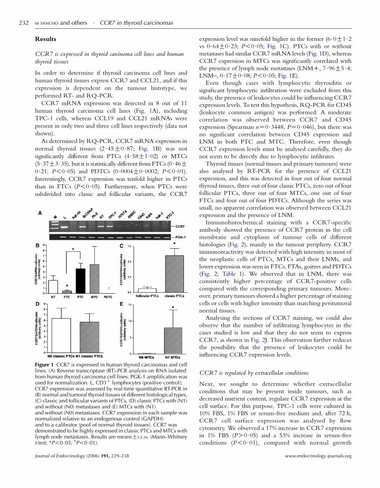
CCR7 mRNA expression was detected in 8 out of 11
human thyroid carcinoma cell lines (Fig. 1A), including
TPC-1 cells, whereas CCL19 and CCL21 mRNAs were
present in only two and three cell lines respectively (data not
shown).
As determined by RQ-PCR, CCR7 mRNA expression in
normal thyroid tissues (2$45G0$87; Fig. 1B) was not
significantly different from PTCs (4$58G1$02) or MTCs
(5$37G3$35), but it is statistically different from FTCs (0$46G0$21; P!0$05) and PDTCs (0$0004G0$0002; P!0$01).Interestingly, CCR7 expression was tenfold higher in PTCs
than in FTCs (P!0$05). Furthermore, when PTCs were
subdivided into classic and follicular variants, the CCR7
Figure 1 CCR7 is expressed in human thyroid carcinomas and celllines. (A) Reverse transcriptase (RT)-PCR analysis on RNA isolatedfrom human thyroid carcinoma cell lines. PGK-1 amplification wasused for normalization. L, CD3C lymphocytes (positive control);CCR7 expression was assessed by real-time quantitative RT-PCR in(B) normal and tumoral thyroid tissues of different histological types,(C) classic and follicular variants of PTCs, (D) classic PTCs with (N1)and without (N0) metastases and (E) MTCs with (N1)and without (N0) metastases. CCR7 expression in each sample wasnormalized relative to an endogenous control (GAPDH)and to a calibrator (pool of normal thyroid tissues). CCR7 wasdemonstrated to be highly expressed in classic PTCs and MTCs withlymph node metastases. Results are meansGS.E.M. (Mann–Whitneyt-test; *P!0$05; †P!0$01).
Journal of Endocrinology (2006) 191, 229–238
expression level was ninefold higher in the former (6$0G1$2vs 0$64G0$23; P!0$05; Fig. 1C). PTCs with or without
metastases had similar CCR7 mRNA levels (Fig. 1D), whereas
CCR7 expression in MTCs was significantly correlated with
the presence of lymph node metastases (LNMC, 7$96G5$4;LNM–, 0$17G0$08; P!0$05; Fig. 1E).
Even though cases with lymphocytic thyroiditis or
significant lymphocytic infiltration were excluded from this
study, the presence of leukocytes could be influencing CCR7
expression levels. To test this hypothesis, RQ-PCR for CD45
(leukocyte common antigen) was performed. A moderate
correlation was observed between CCR7 and CD45
expression (Spearman nZ0$3448, PZ0$046), but there wasno significant correlation between CD45 expression and
LNM in both PTC and MTC. Therefore, even though
CCR7 expression levels must be analysed carefully, they do
not seem to be directly due to lymphocytic infiltrates.
Thyroid tissues (normal tissues and primary tumours) were
also analysed by RT-PCR for the presence of CCL21
expression, and this was detected in four out of four normal
thyroid tissues, three out of four classic PTCs, zero out of four
follicular PTCs, three out of four MTCs, one out of four
FTCs and four out of four PDTCs. Although the series was
small, no apparent correlation was observed between CCL21
expression and the presence of LNM.
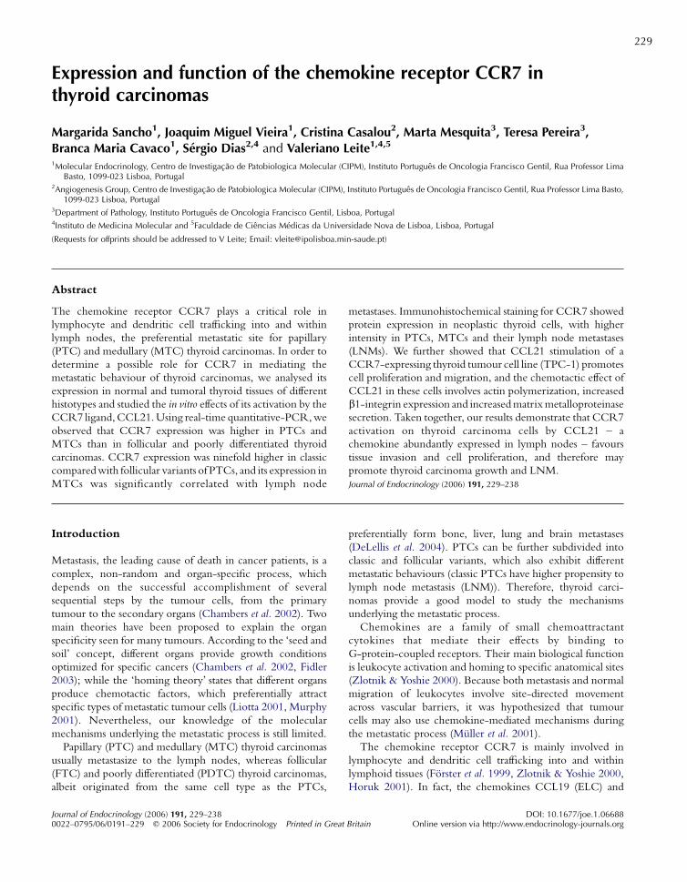
Immunohistochemical staining with a CCR7-specific
antibody showed the presence of CCR7 protein in the cell
membrane and cytoplasm of tumour cells of different
histologies (Fig. 2), mainly in the tumour periphery. CCR7
immunoreactivity was detected with high intensity in most of
the neoplastic cells of PTCs, MTCs and their LNMs, and
lower expression was seen in FTCs, FTAs, goitres and PDTCs
(Fig. 2; Table 1). We observed that in LNM, there was
consistently higher percentage of CCR7-positive cells
compared with the corresponding primary tumours. More-
over, primary tumours showed a higher percentage of staining
cells or cells with higher intensity than matching peritumoral
normal tissues.
Analysing the sections of CCR7 staining, we could also
observe that the number of infiltrating lymphocytes in the
cases studied is low and that they do not seem to express
CCR7, as shown in Fig. 2J. This observation further reduces
the possibility that the presence of leukocytes could be
influencing CCR7 expression levels.
CCR7 is regulated by extracellular conditions
Next, we sought to determine whether extracellular
conditions that may be present inside tumours, such as
decreased nutrient content, regulate CCR7 expression at the
cell surface. For this purpose, TPC-1 cells were cultured in
10% FBS, 1% FBS or serum-free medium and, after 72 h,
CCR7 cell surface expression was analysed by flow
cytometry. We observed a 17% increase in CCR7 expression
in 1% FBS (PO0$05) and a 53% increase in serum-free
conditions (P!0$01), compared with normal growth
www.endocrinology-journals.org
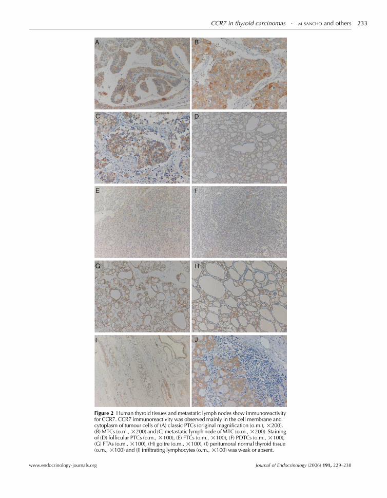
Figure 2 Human thyroid tissues and metastatic lymph nodes show immunoreactivityfor CCR7. CCR7 immunoreactivity was observed mainly in the cell membrane andcytoplasm of tumour cells of (A) classic PTCs (original magnification (o.m.), !200),(B) MTCs (o.m.,!200) and (C) metastatic lymph node of MTC (o.m.,!200). Stainingof (D) follicular PTCs (o.m., !100), (E) FTCs (o.m., !100), (F) PDTCs (o.m., !100),(G) FTAs (o.m., !100), (H) goitre (o.m., !100), (I) peritumoral normal thyroid tissue(o.m., !100) and (J) infiltrating lymphocytes (o.m., !100) was weak or absent.
CCR7 in thyroid carcinomas $ M SANCHO and others 233
www.endocrinology-journals.org Journal of Endocrinology (2006) 191, 229–238
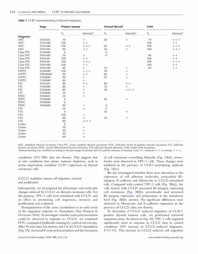
Table 1 CCR7 immunostaining of thyroid neoplasias
Stage Primary tumour Normal thyroid LNM
% Intensitya % Intensitya % Intensitya
DiagnosisMTC T2N1M1 30 C 20 C 70 C/CCMTC T3N1M0 100 CC 100 CCMTC T4N1M0 100 CCC 60 CC 100 CCCMTC T4N1M1 90 CC 10 C 100 CCCClass PTC T2N0M0 0 0Class PTC T4N1M0 10 C 0 90 CCClass PTC T4N1M1 100 CC 100 CCClass PTC T4N1M1 100 CCC 100 CCCClass PTC T4N1M0 100 CC 100 CCClass PTC T4N1M0 60 C 10 C 60 CCFVPTC T2N0M0 100 CCC 50 CFVPTC T2bN0M0 90 CC 60 CFVPTC T3N0M0 90 C 30 CFVPTC T3N0M0 80 CFTC T4N1M1 80 CC 80 CCFTC T3N0M0 80 C 10 CFTC T2N0M0 80 C 20 C/CCFTC T2N0M0 50 CPDTC T4N0M1 20 CPDTC T2N0M0 0 40 CPDTC T4N0M0 0PDTC T4N0M0 90 C 0FTA 50 C 0FTA 0 10 CFTA 100 CFTA 80 C 30 CFTA 80 C/CCGoiter 0Goiter 50 CGoiter 50 CGoiter 40 CGoiter 60 C
MTC, medullary thyroid carcinoma; Class PTC, classic papillary thyroid carcinoma; FVTC, follicular variant of papillary thyroid carcinoma; FTC, follicularthyroid carcinoma; PDTC, poorly differentiated thyroid carcinoma; FTA, follicular thyroid adenoma; LNM, lymph node metastases.aImmunostaining was classified according to the percentage of stained cells (%) and the intensity of staining: weak (C), moderate (CC) or strong (CCC).
M SANCHO and others $ CCR7 in thyroid carcinomas234
conditions (10% FBS; data not shown). This suggests that
in vitro conditions that mimic nutrient depletion, such as
serum deprivation, modulate CCR7 expression on thyroid
carcinoma cells.
CCL21 modulates tumour cell migration, invasionand proliferation
Subsequently, we investigated the phenotypic and molecular
changes induced by CCL21 on thyroid carcinoma cells. For
this purpose, TPC-1 cells were stimulated with CCL21, and
its effect in promoting cell migration, invasion and
proliferation was evaluated.
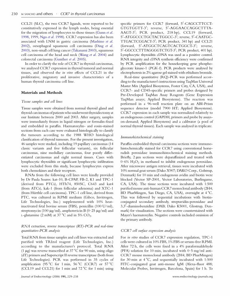
Reorganization of the actin cytoskeleton is an early event
in the migratory response to chemokines (Van Haastert &
Devreotes 2004). To investigate whether actin polymerization
could be observed in response to CCL21, we examined
FITC-conjugated phalloidin staining by confocal microscopy.
After 30 min (data not shown) and 1 h of CCL21 stimulation
(Fig. 3A), increased F-actin polymerization and the formation
Journal of Endocrinology (2006) 191, 229–238
of cell extensions resembling filopodia (Fig. 3A(b), arrow-
heads) were observed in TPC-1 cells. These changes were
inhibited in the presence of CCR7-neutralizing antibody
(Fig. 3A(c)).
We also investigated whether there were alterations in the
expression of cell adhesion molecules, particularly b1-integrin, E-cadherin and fibronectin in CCL21-stimulated
cells. Compared with control TPC-1 cells (Fig. 3B(a)), the
cells treated with CCL21 presented b1-integrin expressing
cell extensions (Fig. 3B(b); arrowheads) and increased
b1-integrin expression and polarization at the membrane
level (Fig. 3B(b); arrows). No significant differences were
observed in fibronectin and E-cadherin expression in the
presence of CCL21 (data not shown).
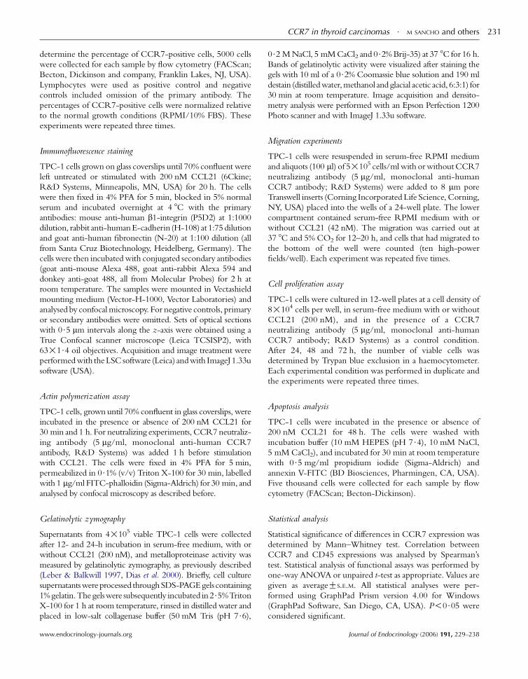
To determine if CCL21 induced migration of CCR7-
positive thyroid tumour cells, we performed transwell
migration assays. As shown in Fig. 4A, TPC-1 cells migrated
significantly more in response to CCL21 than in control
conditions (55% increase in CCL21-induced migration,
P!0$01). This increase in CCL21-induced cell migration
www.endocrinology-journals.org
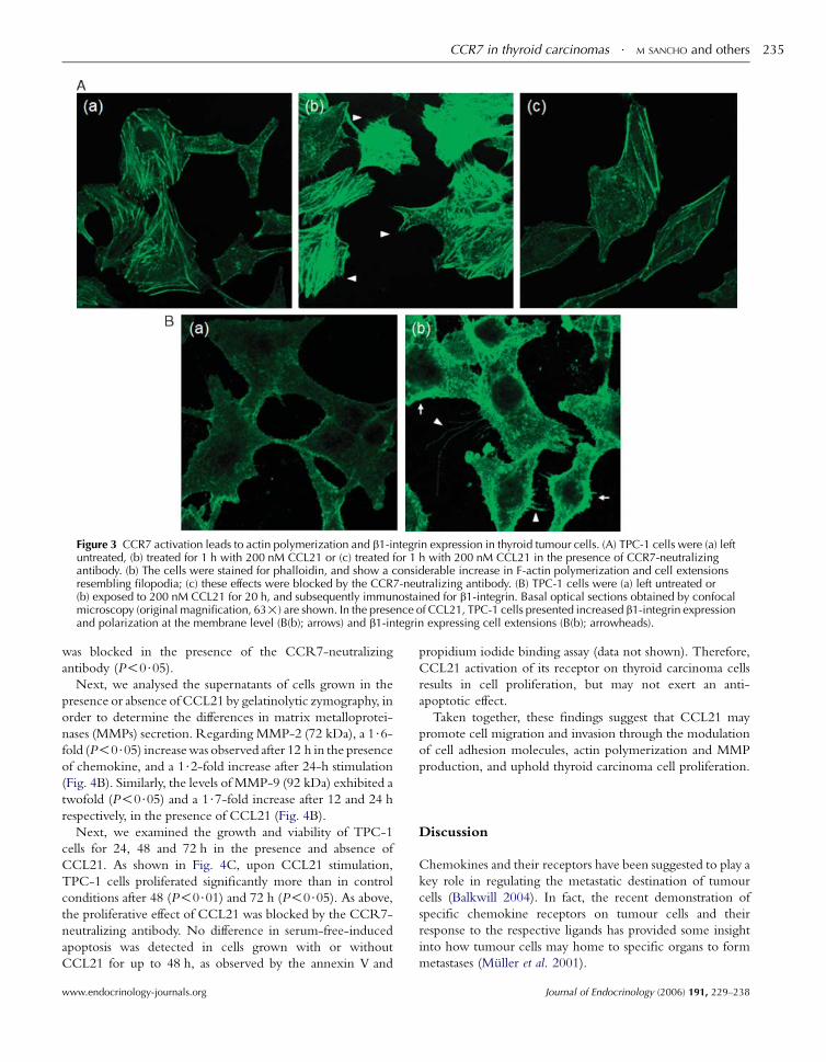
Figure 3 CCR7 activation leads to actin polymerization and b1-integrin expression in thyroid tumour cells. (A) TPC-1 cells were (a) leftuntreated, (b) treated for 1 h with 200 nM CCL21 or (c) treated for 1 h with 200 nM CCL21 in the presence of CCR7-neutralizingantibody. (b) The cells were stained for phalloidin, and show a considerable increase in F-actin polymerization and cell extensionsresembling filopodia; (c) these effects were blocked by the CCR7-neutralizing antibody. (B) TPC-1 cells were (a) left untreated or(b) exposed to 200 nM CCL21 for 20 h, and subsequently immunostained for b1-integrin. Basal optical sections obtained by confocalmicroscopy (original magnification, 63!) are shown. In the presence of CCL21, TPC-1 cells presented increased b1-integrin expressionand polarization at the membrane level (B(b); arrows) and b1-integrin expressing cell extensions (B(b); arrowheads).
CCR7 in thyroid carcinomas $ M SANCHO and others 235
was blocked in the presence of the CCR7-neutralizing
antibody (P!0$05).Next, we analysed the supernatants of cells grown in the
presence or absence of CCL21 by gelatinolytic zymography, in
order to determine the differences in matrix metalloprotei-
nases (MMPs) secretion. Regarding MMP-2 (72 kDa), a 1$6-fold (P!0$05) increasewas observed after 12 h in the presenceof chemokine, and a 1$2-fold increase after 24-h stimulation
(Fig. 4B). Similarly, the levels of MMP-9 (92 kDa) exhibited a
twofold (P!0$05) and a 1$7-fold increase after 12 and 24 h
respectively, in the presence of CCL21 (Fig. 4B).
Next, we examined the growth and viability of TPC-1
cells for 24, 48 and 72 h in the presence and absence of
CCL21. As shown in Fig. 4C, upon CCL21 stimulation,
TPC-1 cells proliferated significantly more than in control
conditions after 48 (P!0$01) and 72 h (P!0$05). As above,the proliferative effect of CCL21 was blocked by the CCR7-
neutralizing antibody. No difference in serum-free-induced
apoptosis was detected in cells grown with or without
CCL21 for up to 48 h, as observed by the annexin V and
www.endocrinology-journals.org
propidium iodide binding assay (data not shown). Therefore,
CCL21 activation of its receptor on thyroid carcinoma cells
results in cell proliferation, but may not exert an anti-
apoptotic effect.
Taken together, these findings suggest that CCL21 may
promote cell migration and invasion through the modulation
of cell adhesion molecules, actin polymerization and MMP
production, and uphold thyroid carcinoma cell proliferation.
Discussion
Chemokines and their receptors have been suggested to play a
key role in regulating the metastatic destination of tumour
cells (Balkwill 2004). In fact, the recent demonstration of
specific chemokine receptors on tumour cells and their
response to the respective ligands has provided some insight
into how tumour cells may home to specific organs to form
metastases (Muller et al. 2001).
Journal of Endocrinology (2006) 191, 229–238
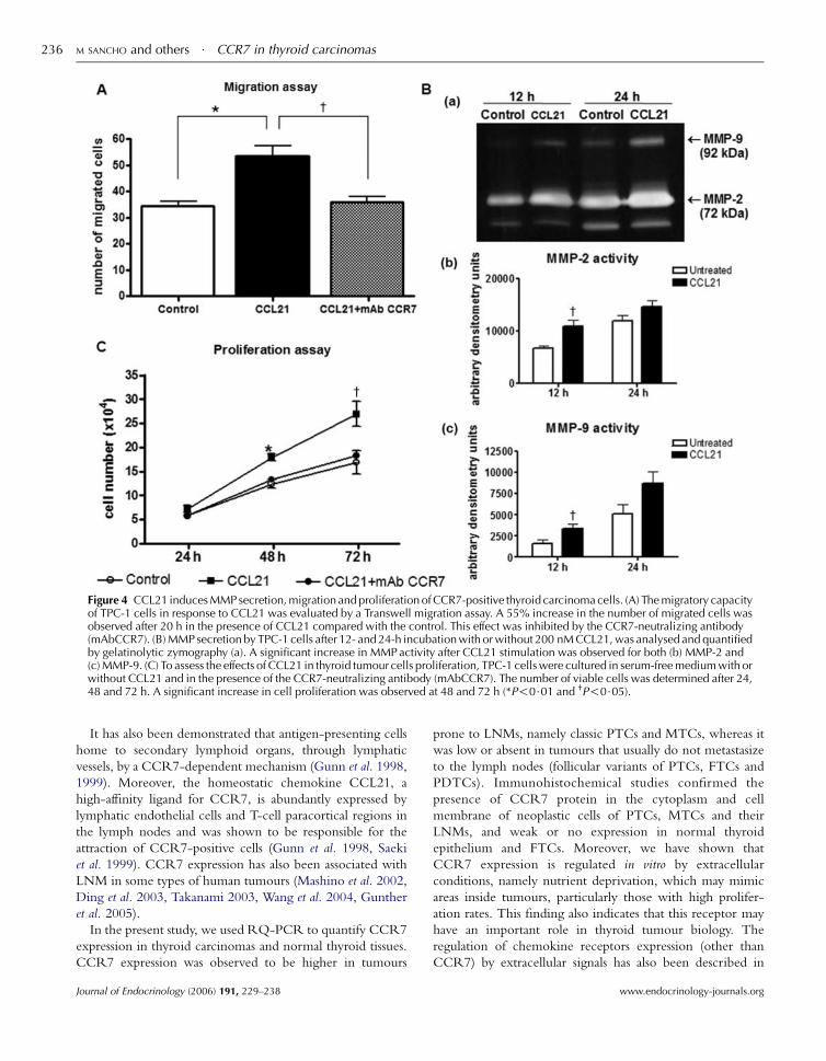
Figure 4 CCL21 induces MMP secretion, migration and proliferation of CCR7-positive thyroid carcinoma cells. (A) The migratory capacityof TPC-1 cells in response to CCL21 was evaluated by a Transwell migration assay. A 55% increase in the number of migrated cells wasobserved after 20 h in the presence of CCL21 compared with the control. This effect was inhibited by the CCR7-neutralizing antibody(mAbCCR7). (B) MMP secretion by TPC-1 cells after 12- and 24-h incubation with or without 200 nM CCL21, was analysed and quantifiedby gelatinolytic zymography (a). A significant increase in MMP activity after CCL21 stimulation was observed for both (b) MMP-2 and(c) MMP-9. (C) To assess the effects of CCL21 in thyroid tumour cells proliferation, TPC-1 cells were cultured in serum-free medium with orwithout CCL21 and in the presence of the CCR7-neutralizing antibody (mAbCCR7). The number of viable cells was determined after 24,48 and 72 h. A significant increase in cell proliferation was observed at 48 and 72 h (*P!0$01 and †P!0$05).
M SANCHO and others $ CCR7 in thyroid carcinomas236
It has also been demonstrated that antigen-presenting cells
home to secondary lymphoid organs, through lymphatic
vessels, by a CCR7-dependent mechanism (Gunn et al. 1998,
1999). Moreover, the homeostatic chemokine CCL21, a
high-affinity ligand for CCR7, is abundantly expressed by
lymphatic endothelial cells and T-cell paracortical regions in
the lymph nodes and was shown to be responsible for the
attraction of CCR7-positive cells (Gunn et al. 1998, Saeki
et al. 1999). CCR7 expression has also been associated with
LNM in some types of human tumours (Mashino et al. 2002,
Ding et al. 2003, Takanami 2003, Wang et al. 2004, Gunther
et al. 2005).
In the present study, we used RQ-PCR to quantify CCR7
expression in thyroid carcinomas and normal thyroid tissues.
CCR7 expression was observed to be higher in tumours
Journal of Endocrinology (2006) 191, 229–238
prone to LNMs, namely classic PTCs and MTCs, whereas it
was low or absent in tumours that usually do not metastasize
to the lymph nodes (follicular variants of PTCs, FTCs and
PDTCs). Immunohistochemical studies confirmed the
presence of CCR7 protein in the cytoplasm and cell
membrane of neoplastic cells of PTCs, MTCs and their
LNMs, and weak or no expression in normal thyroid
epithelium and FTCs. Moreover, we have shown that
CCR7 expression is regulated in vitro by extracellular
conditions, namely nutrient deprivation, which may mimic
areas inside tumours, particularly those with high prolifer-
ation rates. This finding also indicates that this receptor may
have an important role in thyroid tumour biology. The
regulation of chemokine receptors expression (other than
CCR7) by extracellular signals has also been described in
www.endocrinology-journals.org

CCR7 in thyroid carcinomas $ M SANCHO and others 237
response to other conditions, such as growth factors and
hypoxia (Scotton et al. 2001, Chen et al. 2005).
High levels of actin polymerization are required for the
formation of pseudopodia, which are needed for chemokine-
mediated cell migration and invasion into surrounding tissues
and efficient metastasis formation (Pokorna et al. 1994, Van
Haastert & Devreotes 2004). In order to determine cell
motility in stimulated cells, we performed phalloidin staining
and observed that actin polymerization was enhanced in
response to CCL21, with the formation of filopodia.
Furthermore, CCL21 stimulation increased b1-integrinexpression. Since b1-integrin has been suggested to play a
crucial role in mediating the adhesion and arrest of other cell
types at the endothelial vessels of lymph nodes (de la Rosa
et al. 2003), this change also supports a putative invasive
phenotype induced by CCR7 activation on thyroid
carcinoma cells. Other adhesion molecules may also be
involved in these processes.
The secretion and activation of proteolytic enzymes,
namely matrix metalloproteinases, are also believed to be
essential for tumour cell invasion through and across
extracellular barriers and, consequently, to form metastases
(Yu & Stamenkovic 2000, Egeblad & Werb 2002). TPC-1
cells stimulated with CCL21 showed an increase in MMP-2
and MMP-9 secretion, as analysed by gelatinolytic zymo-
graphy, which suggests a more invasive phenotype.
We have also shown that TPC-1 cells migrate in response
to CCL21 using the Transwell migration assay, thus
supporting the hypothesis that CCL21 concentrations in the
lymph nodes probably induce thyroid tumour cells migration
into these organs through a CCR7-mediated mechanism.
Therefore, we suggest that CCR7 activation by CCL21 may
promote thyroid carcinoma LNM via an increase in actin
polymerization, cell adhesion molecules modulation, cell
migration and MMPs secretion.
After tumour cells have reached a certain organ, their
ability to grow and effectively form metastases is dictated by
molecular interactions of the cells with the environment on
that specific organ. The effect of CCL21, which is highly
expressed in the lymph nodes, in cell proliferation, has
previously been reported in haematopoietic (Ploix et al.
2001) and mesangial cells (Banas et al. 2002), but it has
never been described in tumour cells. We examined the
effect of CCR7 activation and demonstrated that cell
proliferation is significantly increased after 48- and 72-h
stimulation with CCL21. This effect suggests that the
lymph nodes, where CCL21 is abundantly expressed,
provide conditions that favour the proliferation of metastatic
thyroid tumour cells, once migrated and arrested at these
secondary sites.
In conclusion, this study demonstrates that functional
CCR7 is expressed in specific subsets of thyroid carcinoma
cells, and that its ligand, CCL21, promotes thyroid tumour
cells migration, invasion and proliferation, leading to a more
invasive phenotype and contributing to lymph node-specific
metastatic growth. Taken together, these findings support the
www.endocrinology-journals.org
use of chemokine receptor antagonists (Robinson et al. 2003,
Howard & Galligan 2004) for the treatment of specific subsets
of thyroid tumours.
Acknowledgements
We gratefully acknowledge Prof. Marc Mareel for kindly
providing the TPC-1 cell line, Dr Paula Soares for cell lines
RNAs, Prof. Luıs Sobrinho for help with statistical analysis and
Dr Ana Luısa Catarino for histopathological classification. This
work was supported by Fundacao Astrazeneca. The authors
declare that there is no conflict of interest that would prejudice
the impartiality of this scientific work.
References
Balkwill F 2004 Cancer and the chemokine network. Nature Reviews. Cancer 4
540–550.
Banas B, Wornle M, Berger T, Nelson PJ, Cohen CD, Kretzler M, Pfirstinger J,
MackM, LippM, Grone HJ et al. 2002 Roles of SLC/CCL21 and CCR7 in
human kidney for mesangial proliferation, migration, apoptosis, and tissue
homeostasis. Journal of Immunology 168 4301–4307.
Chambers AF, Groom AC & MacDonald IC 2002 Dissemination and growth
of cancer cells in metastatic sites. Nature Reviews. Cancer 2 563–572.
Chen S, Tuttle DL, Oshier JT, Knot HJ, Streit WJ, Goodenow MM &
Harrison JK 2005 Transforming growth factor-beta1 increases CXCR4
expression, stromal-derived factor-1alpha-stimulated signalling and human
immunodeficiency virus-1 entry in human monocyte-derived macro-
phages. Immunology 114 565–574.
DeLellis RA, Lloyd RV, Heitz PU & Eng C (Eds). World Health Organization
Classification of Tumours. Pathology and Genetics of Tumours of Endocrine Organs.
Ch. 2, pp. 49–124. Lyon: IARC Press, 2004.
Dias S, Hattori K, Zhu Z, Heissig B, Choy M, Lane W, Wu Y, Chadburn A,
Hyjek E, Gill M et al. 2000 Autocrine stimulation of VEGFR-2 activates
human leukemic cell growth and migration. Journal of Clinical Investigation
106 511–521.
Ding Y, Shimada Y, Maeda M, Kawabe A, Kaganoi J, Komoto I, Hashimoto Y,
Miyake M, Hashida H & Imamura M 2003 Association of CC chemokine
receptor 7with lymphnodemetastasis of esophageal squamous cell carcinoma.
Clinical Cancer Research 9 3406–3412.
Egeblad M & Werb Z 2002 New functions for the matrix metalloproteinases
in cancer progression. Nature Reviews. Cancer 2 161–174.
Fidler IJ 2003 The pathogenesis of cancer metastasis: the ‘seed and soil’
hypothesis revisited. Nature Reviews. Cancer 3 453–458.
Forster R, Schubel A, Breitfeld D, Kremmer E, Renner-Muller I, Wolf E &
Lipp M 1999 CCR7 coordinates the primary immune response by
establishing functional microenvironments in secondary lymphoid organs.
Cell 99 23–33.
Gunn MD, Tangemann K, Tam C, Cyster JG, Rosen SD &Williams LT 1998
A chemokine expressed in lymphoid high endothelial venules promotes the
adhesion and chemotaxis of naive T lymphocytes. PNAS 95 7118–7122.
Gunn MD, Kyuwa S, Tam C, Kakiuchi T, Matsuzawa A, Williams LT &
Nakano H 1999 Mice lacking expression of secondary lymphoid organ
chemokine have defects in lymphocyte homing and dendritic cell
localization. Journal of Experimental Medicine 189 451–460.
Gunther K, Leier J, Henning G, Dimmler A, Weissbach R, Hohenberger W
& Forster R 2005 Prediction of lymph node metastasis in colorectal
carcinoma by expression of chemokine receptor CCR7. International
Journal of Cancer 116 726–733.
Horuk R 2001 Chemokine receptors. Cytokine and Growth Factor Reviews
2 313–335.
Journal of Endocrinology (2006) 191, 229–238
M SANCHO and others $ CCR7 in thyroid carcinomas238
Howard OM & Galligan CL 2004 An expanding appreciation of the role
chemokine receptors play in cancer progression. Current Pharmaceutical
Design 10 2377–2389.
Leber TM & Balkwill FR 1997 Zymography: a single-step staining method
for quantitation of proteolytic activity on substrate gels. Analytical
Biochemistry 249 24–28.
Liotta LA 2001 An attractive force in metastasis. Nature 410 24–25.
Mashino K, Sadanaga N, Yamaguchi H, Tanaka F, Ohta M, Shibuta K,
Inoue H & Mori M 2002 Expression of chemokine receptor CCR7 is
associated with lymph node metastasis of gastric cancer. Cancer Research 62
2937–2941.
Muller A, Homey B, Soto H, Ge N, Catron D, Buchanan ME, McClanahan T,
Murphy E, Yuan W, Wagner SN et al. 2001 Involvement of chemokine
receptors in breast cancer metastasis. Nature 410 50–56.
Murphy PM 2001 Chemokines and the molecular basis of cancer metastasis.
New England Journal of Medicine 345 833–835.
Ngo VN, Tang HL & Cyster JG 1998 Epstein-Barr virus-induced molecule 1
ligand chemokine is expressed by dendritic cells in lymphoid tissues and
strongly attracts naıve T cells and activated B cells. Journal of Experimental
Medicine 188 181–191.
Ploix C, Lo D&Carson MJ 2001 A ligand for the chemokine receptor CCR7
can influence the homeostatic proliferation of CD4 T cells and progression
of autoimmunity. Journal of Immunology 167 6724–6730.
Pokorna E, Jordan PW, O’Neill CH, Zicha D, Gilbert CS & Vesely P 1994
Actin cytoskeleton and motility in rat sarcoma cell populations with
different metastatic potential. Cell Motility and the Cytoskeleton 28 25–33.
Robinson SC, Scott KA, Wilson JL, Thompson RG, Proudfoot AE &
Balkwill FR 2003 A chemokine receptor antagonist inhibits experimental
breast tumour growth. Cancer Research 63 8360–8365.
de la Rosa G, Longo N, Rodriguez-Fernandez JL, Puig-Kroger A, Pineda A,
Corbi AL & Sanchez-Mateos P 2003 Migration of human blood dendritic
Journal of Endocrinology (2006) 191, 229–238
cells across endothelial cell monolayers: adhesion molecules and chemo-
kines involved in subset-specific transmigration. Journal of Leukocyte Biology
73 639–649.
Saeki H, Moore AM, Brown MJ & Hwang ST 1999 Cutting edge: secondary
lymphoid-tissue chemokine (SLC) and CC chemokine receptor 7 (CCR7)
participate in the emigration pathway of mature dendritic cells from the
skin to regional lymph nodes. Journal of Immunology 162 2472–2475.
Scotton C, Milliken D, Wilson J, Raju S & Balkwill F 2001 Analysis of CC
chemokine and chemokine receptor expression in solid ovarian tumours.
British Journal of Cancer 85 891–897.
Takanami I 2003 Overexpression of CCR7 mRNA in nonsmall cell lung
cancer: correlation with lymph node metastasis. International Journal of
Cancer 105 186–189.
Van Haastert PJ & Devreotes PN 2004 Chemotaxis: signalling the way
forward. Nature Reviews. Molecular Cell Biology 5 626–634.
Wang J, Xi L, Hunt JL, Gooding W, Whiteside TL, Chen Z, Godfrey TE &
Ferris RL 2004 Expression pattern of chemokine receptor 6 (CCR6) and
CCR7 in squamous cell carcinoma of the head and neck identifies a novel
metastatic phenotype. Cancer Research 64 1861–1866.
YuQ& Stamenkovic I 2000 Cell surface-localized matrix metalloproteinase-9
proteolytically activates TGF-b and promotes tumour invasion and
angiogenesis. Genes and Development 14 163–176.
Zlotnik A & Yoshie O 2000 Chemokines: a new classification system and their
role in immunity. Immunity 12 121–127.
Received in final form 21 June 2006Accepted 23 June 2006
www.endocrinology-journals.org
Related Documents


![chemokine/chemokine receptor pair ccL20/ccR6 in human ... · pancreas, stomach, prostate, testis, uterine cervix and skin[11]. The chemokine receptor CCR6 was originally described](https://static.cupdf.com/doc/110x72/5f9ac7b0798b75658905651c/chemokinechemokine-receptor-pair-ccl20ccr6-in-human-pancreas-stomach-prostate.jpg)