International Journal of Molecular Sciences Review Expression and Function of Host Defense Peptides at Inflammation Sites Suhanya V. Prasad, Krzysztof Fiedoruk , Tamara Daniluk, Ewelina Piktel and Robert Bucki * Department of Medical Microbiology and Nanobiomedical Engineering, Medical University of Bialystok, Mickiewicza 2c, Bialystok 15-222, Poland; [email protected] (S.V.P.); krzysztof.fi[email protected] (K.F.); [email protected] (T.D.); [email protected] (E.P.) * Correspondence: [email protected]; Tel.: +48-85-7485483 Received: 12 November 2019; Accepted: 19 December 2019; Published: 22 December 2019 Abstract: There is a growing interest in the complex role of host defense peptides (HDPs) in the pathophysiology of several immune-mediated inflammatory diseases. The physicochemical properties and selective interaction of HDPs with various receptors define their immunomodulatory effects. However, it is quite challenging to understand their function because some HDPs play opposing pro-inflammatory and anti-inflammatory roles, depending on their expression level within the site of inflammation. While it is known that HDPs maintain constitutive host protection against invading microorganisms, the inducible nature of HDPs in various cells and tissues is an important aspect of the molecular events of inflammation. This review outlines the biological functions and emerging roles of HDPs in different inflammatory conditions. We further discuss the current data on the clinical relevance of impaired HDPs expression in inflammation and selected diseases. Keywords: host defense peptides; human antimicrobial peptides; defensins; cathelicidins; inflammation; anti-inflammatory; pro-inflammatory 1. Introduction The human body is in a constant state of conflict with the unseen microbial world that threatens to disrupt the host cell function and colonize the body surfaces. The immune system has an arsenal of destructive mechanisms to neutralize the toxic effect of the microbial pathogens. It functions through two layers of defense systems: The innate system and the more intricate adaptive immune system, which closely communicate with each other [1]. Each of those systems form a complex network of immune cells, signaling molecules, and regulatory pathways. Inflammation is a reaction of the host immune system that acts to eliminate the source of inflammatory stimulus, ranging from pathogens to burn injuries [2]. Although microbial infections largely initiate the events of inflammation, we must note that inflammation is also a hallmark feature of various autoimmune, cancer, and systemic diseases [3]. Inflammation is a highly coordinated biochemical sequence of events that commences with the rapid migration of leukocytes to the site of infection, followed by adequate blood supply that transports different inflammatory mediators that control the course of the immune response [4]. However, while the initial events of inflammation are constructive and beneficial to the host, incompetent inflammatory resolution mechanisms, along with inefficient elimination of foreign bodies or pathogens and cellular debris, prompt the onset of chronic inflammation. Antimicrobial peptides (AMPs) such as defensins and cathelicidins represent a vital part of the human immune system due to their broad spectrum activity against pathogenic bacteria, fungi, protists, and enveloped viruses [5]. Furthermore, in recent years, a growing number of studies have recognized these peptides as potent immune modulators, implicated in multiple pro- and anti-inflammatory responses through (1) neutralization of bacterial toxins, (2) chemoattraction and activation of immune cells, (3) initiation of adaptive immunity, (4) neovascularization and wound healing, as well as (5) anti- or Int. J. Mol. Sci. 2020, 21, 104; doi:10.3390/ijms21010104 www.mdpi.com/journal/ijms

Welcome message from author
This document is posted to help you gain knowledge. Please leave a comment to let me know what you think about it! Share it to your friends and learn new things together.
Transcript
International Journal of
Molecular Sciences
Review
Expression and Function of Host Defense Peptides atInflammation Sites
Suhanya V. Prasad, Krzysztof Fiedoruk , Tamara Daniluk, Ewelina Piktel and Robert Bucki *
Department of Medical Microbiology and Nanobiomedical Engineering, Medical University of Bialystok,Mickiewicza 2c, Bialystok 15-222, Poland; [email protected] (S.V.P.); [email protected] (K.F.);[email protected] (T.D.); [email protected] (E.P.)* Correspondence: [email protected]; Tel.: +48-85-7485483
Received: 12 November 2019; Accepted: 19 December 2019; Published: 22 December 2019 �����������������
Abstract: There is a growing interest in the complex role of host defense peptides (HDPs) inthe pathophysiology of several immune-mediated inflammatory diseases. The physicochemicalproperties and selective interaction of HDPs with various receptors define their immunomodulatoryeffects. However, it is quite challenging to understand their function because some HDPs playopposing pro-inflammatory and anti-inflammatory roles, depending on their expression level withinthe site of inflammation. While it is known that HDPs maintain constitutive host protection againstinvading microorganisms, the inducible nature of HDPs in various cells and tissues is an importantaspect of the molecular events of inflammation. This review outlines the biological functions andemerging roles of HDPs in different inflammatory conditions. We further discuss the current data onthe clinical relevance of impaired HDPs expression in inflammation and selected diseases.
Keywords: host defense peptides; human antimicrobial peptides; defensins; cathelicidins; inflammation;anti-inflammatory; pro-inflammatory
1. Introduction
The human body is in a constant state of conflict with the unseen microbial world that threatensto disrupt the host cell function and colonize the body surfaces. The immune system has an arsenalof destructive mechanisms to neutralize the toxic effect of the microbial pathogens. It functionsthrough two layers of defense systems: The innate system and the more intricate adaptive immunesystem, which closely communicate with each other [1]. Each of those systems form a complexnetwork of immune cells, signaling molecules, and regulatory pathways. Inflammation is a reactionof the host immune system that acts to eliminate the source of inflammatory stimulus, ranging frompathogens to burn injuries [2]. Although microbial infections largely initiate the events of inflammation,we must note that inflammation is also a hallmark feature of various autoimmune, cancer, andsystemic diseases [3]. Inflammation is a highly coordinated biochemical sequence of events thatcommences with the rapid migration of leukocytes to the site of infection, followed by adequateblood supply that transports different inflammatory mediators that control the course of the immuneresponse [4]. However, while the initial events of inflammation are constructive and beneficial to thehost, incompetent inflammatory resolution mechanisms, along with inefficient elimination of foreignbodies or pathogens and cellular debris, prompt the onset of chronic inflammation.
Antimicrobial peptides (AMPs) such as defensins and cathelicidins represent a vital part of thehuman immune system due to their broad spectrum activity against pathogenic bacteria, fungi, protists,and enveloped viruses [5]. Furthermore, in recent years, a growing number of studies have recognizedthese peptides as potent immune modulators, implicated in multiple pro- and anti-inflammatoryresponses through (1) neutralization of bacterial toxins, (2) chemoattraction and activation of immunecells, (3) initiation of adaptive immunity, (4) neovascularization and wound healing, as well as (5) anti- or
Int. J. Mol. Sci. 2020, 21, 104; doi:10.3390/ijms21010104 www.mdpi.com/journal/ijms
Int. J. Mol. Sci. 2020, 21, 104 2 of 23
pro-tumor activity [6]. Actually, AMPs interact with innate and adaptive immune receptors, such as patternrecognition (PRRs) or chemokine receptors (CCRs), as well as inflammasomes and their complementsystems, creating a link between innate and adaptive immunity [7–9]. In addition, AMPs may regulatefundamental cellular processes, such as differentiation, proliferation, and programmed cell death, e.g.,by stimulating growth factor receptors or as complexes with the host nucleic acids [10,11], hence theyresemble cytokines and growth factors. Overall, these activities, by controlling inflammation and/oraccelerating repair process of the infected site, appear to support the direct microbicidal function of AMPsin resolving an infection. Therefore, the term host defense peptides (HDPs) was coined to encompasstheir pleiotropic nature, and association with both infectious as well as non-infectious inflammatoryresponses [12], although these two terms are used interchangeably. In the latter context, HDPs fit into thedefinition of “alarmins” or “danger signals”, i.e., various endogenous molecules collectively known asDAMPs (damage associated molecular patterns), which are released from damaged or dying cells andinitiate a diverse range of physiological and pathophysiological functions [13–15].
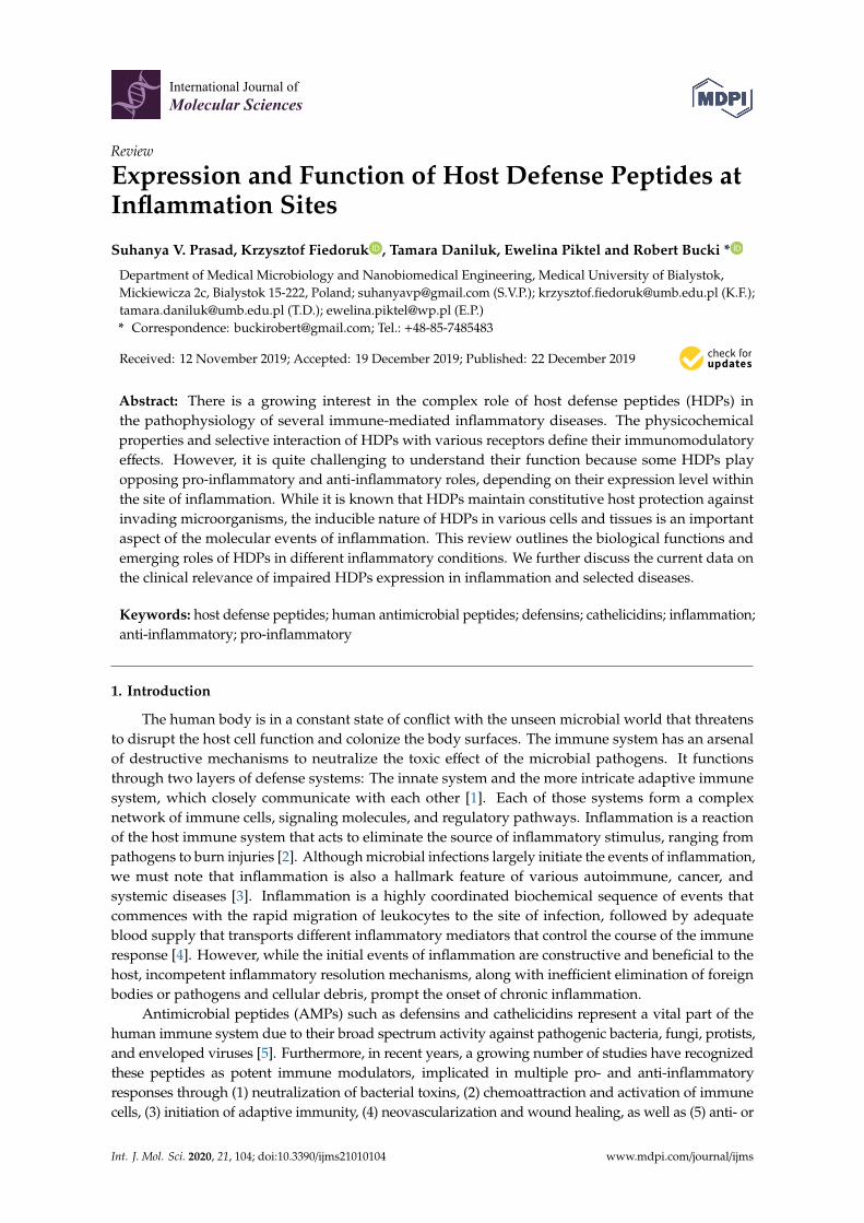
At present, HDPs are perceived as multifunctional agents that coordinate diverse immunesurveillance functions necessary to maintain homeostasis (Figure 1) [16]. However, if their productionis out of the physiological range, they may contribute to an undesirable inflammation in responseto local (e.g., periodontal, respiratory, intestinal, and skin) and systemic (e.g., sepsis) infections.They might also function as pathophysiological events of inflammatory diseases, cancers, and evenpsychiatric disorders (Table 1) [17–23]. In addition, HDPs have been indicated as potential biomarkersin numerous infectious and non-infectious diseases [24].
Int. J. Mol. Sci. 2019, 20, x FOR PEER REVIEW 2 of 25
inflammatory responses through (1) neutralization of bacterial toxins, (2) chemoattraction and activation of immune cells, (3) initiation of adaptive immunity, (4) neovascularization and wound healing, as well as (5) anti- or pro-tumor activity [6]. Actually, AMPs interact with innate and adaptive immune receptors, such as pattern recognition (PRRs) or chemokine receptors (CCRs), as well as inflammasomes and their complement systems, creating a link between innate and adaptive immunity [7–9]. In addition, AMPs may regulate fundamental cellular processes, such as differentiation, proliferation, and programmed cell death, e.g., by stimulating growth factor receptors or as complexes with the host nucleic acids [10,11], hence they resemble cytokines and growth factors. Overall, these activities, by controlling inflammation and/or accelerating repair process of the infected site, appear to support the direct microbicidal function of AMPs in resolving an infection. Therefore, the term host defense peptides (HDPs) was coined to encompass their pleiotropic nature, and association with both infectious as well as non-infectious inflammatory responses [12], although these two terms are used interchangeably. In the latter context, HDPs fit into the definition of “alarmins” or “danger signals”, i.e., various endogenous molecules collectively known as DAMPs (damage associated molecular patterns), which are released from damaged or dying cells and initiate a diverse range of physiological and pathophysiological functions [13–15].
At present, HDPs are perceived as multifunctional agents that coordinate diverse immune surveillance functions necessary to maintain homeostasis (Figure 1) [16]. However, if their production is out of the physiological range, they may contribute to an undesirable inflammation in response to local (e.g., periodontal, respiratory, intestinal, and skin) and systemic (e.g., sepsis) infections. They might also function as pathophysiological events of inflammatory diseases, cancers, and even psychiatric disorders (Table 1) [17–23]. In addition, HDPs have been indicated as potential biomarkers in numerous infectious and non-infectious diseases [24].
Figure 1. Illustration of the distinct role of host defense peptides at the sites of inflammation. Specific Toll-like receptors TLR-2, TLR-4, TLR-5, and TLR-6 are expressed on the plasma membrane of immune cells, non-immune cells, and intracellular compartments. TLR-7 and TLR-9 within endosomes participate in the host recognition of microbial cellular components and bind to host internal factors. The receptors, along with their adaptor proteins MyD88 and TRIF, can initiate the
Figure 1. Illustration of the distinct role of host defense peptides at the sites of inflammation.Specific Toll-like receptors TLR-2, TLR-4, TLR-5, and TLR-6 are expressed on the plasma membrane ofimmune cells, non-immune cells, and intracellular compartments. TLR-7 and TLR-9 within endosomesparticipate in the host recognition of microbial cellular components and bind to host internal factors.The receptors, along with their adaptor proteins MyD88 and TRIF, can initiate the inflammatorysignaling pathways. The host defense peptides promote innate immunity against various pathogensand maintain the immune system homeostasis. These peptides can also be induced in addition to theirconstitutive expression by the transcriptional modulatory factors NF-κB, AP-1, and intracellular releaseof cytokines and chemokines. They actively participate in coordinating the host immune signalingmechanisms during inflammation. Furthermore, HDPs can display both pro- and anti-inflammatoryproperties that may protect against the responses of inflammatory diseases. Abbreviations: AP-1,activator protein; NF-kB, nuclear factor kappa-light-chain enhancer of activated B cells; MAPK,mitogen-activated protein kinase; ERK1,2, extracellular signal-related kinases.
Int. J. Mol. Sci. 2020, 21, 104 3 of 23
That being said, expanding our knowledge regarding the molecular mechanisms behind expression,processing, and mutual interactions of HDPs with other immune system components is crucial to betterunderstand inflammation processes, and to develop new methods of anti-inflammatory treatment.Certainly, it is a challenging and long-term task, since a single antimicrobial peptide, e.g., humancathelicidin LL-37, may interact with dozens of proteins/receptors and subsequently engage hundredsof secondary effector proteins, as well as modify expression of >900 genes [6]. Considering this,the purpose of this review is to evaluate and summarize recent discoveries considering the functionalexpression and protective attributes of HDPs/AMPs in the acute inflammatory phase and the detrimentaleffects of their recruitment in chronic inflammation. Both in vitro and in vivo studies connecting theunderlying mechanisms governing the immunoregulatory role of these peptides in the inflammatorymicroenvironment will be discussed.
2. Overview of Human Antimicrobial Peptides
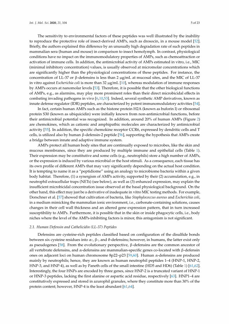
Antimicrobial peptides are widely distributed in all living organisms, representing ancient andprimary defense molecules, e.g., innate immune mechanisms conferred by the antimicrobial peptidesin insects usually devoid of adaptive responses [25]. Discovery of defensins in rabbit leucocytes,lactoferrin in cow milk, and lysozyme in human saliva are among the first reports of animal-originatedantimicrobial molecules, which paved the way for further identification and understanding of thephysiological function of other antimicrobial peptides and proteins [26]. At present, 2272 peptidesderived from animals, including ~130 of human origin (Figure 2), are recorded in the antimicrobialpeptide database (http://aps.unmc.edu/AP), a comprehensive source of naturally existing families ofantimicrobial peptides from all form of kingdoms of life [25].
Antimicrobial peptides and proteins contain a short chain of about 12–100 amino acids (Figure 2),and are classified according to their conformational structure (α, β, αβ, and non-αβ), amino acid motifs,and expression pattern [25,27]. For example, the major human AMPs, cathelicidin LL-37 and defensins,are characterized by α-helical and β-sheet structure, respectively. Furthermore, the latter are dividedinto α- and β-defensins based on the configuration of the disulfide bonds between six cysteine residues.AMPs are characterized by positive charge and substantial proportion (typically 50%) of hydrophobicresidues, thus they are also known as cationic antimicrobial peptides (CAPs). However, at present,some negatively charged peptides are also classified as AMPs, e.g., human β-defensin DEFB118,psoriasin, or α-synuclein (Figure 2). Nevertheless, this amphiphilic–cationic organization allowsthem to selectively associate, and in turn disrupt, highly negatively charged microbial membranes.Hence, it explains their broad spectrum of activity, encompassing all cellular pathogens and envelopedviruses. Additionally, the cationic nature of AMPs may possibly facilitate, via electrostatic forces,their interactions with diverse host receptors, which are behind the immunomodulatory potential ofthese peptides [28].
Certain AMPs, e.g., cathelicidins, are produced as inactive pro-peptides and must be proteolyticallyprocessed for activity. It is noteworthy that this may generate multiple length variants characterizedby diverse antimicrobial or immunomodulatory properties. Therefore, the presence of theappropriate proteases and their level is an important factor in regulating the function of the AMPs.Another important activity-related issue is that microbicidal action of AMPs is considerably suppressedby the physiological conditions present in some compartments of the body, including high salt,carbonate, lipoprotein, and polysaccharide concentrations [29–33].
Int. J. Mol. Sci. 2020, 21, 104 4 of 23
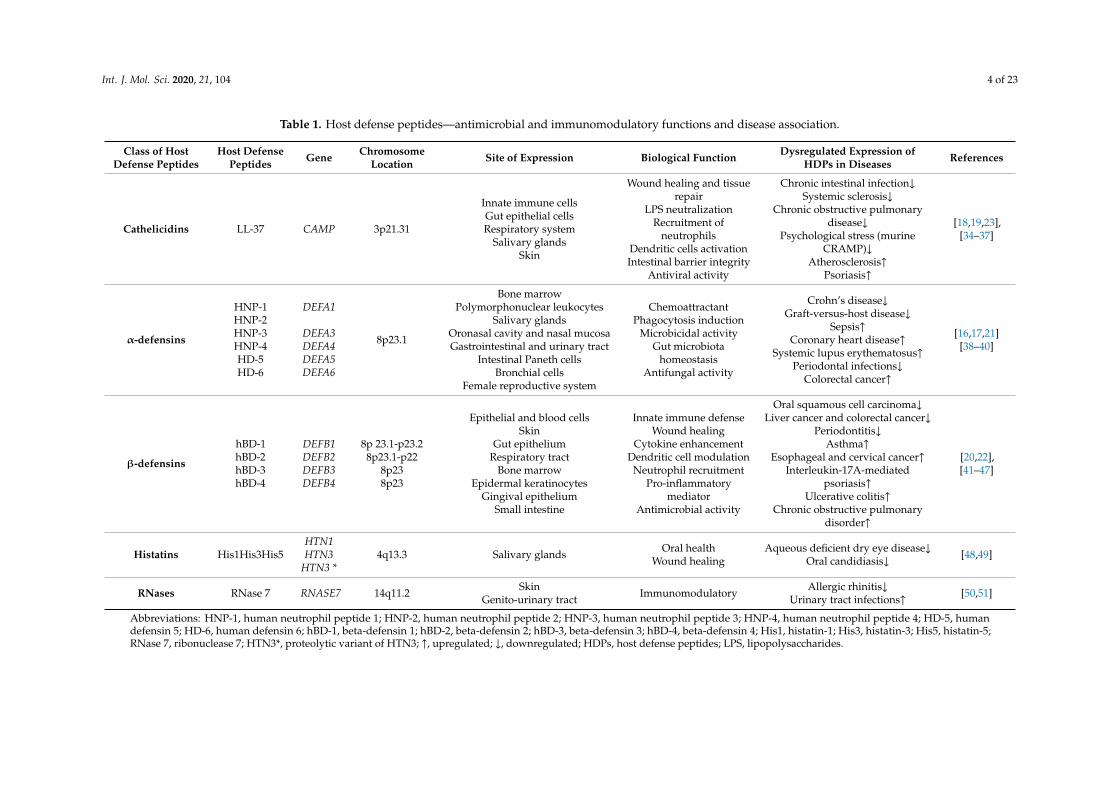
Table 1. Host defense peptides—antimicrobial and immunomodulatory functions and disease association.
Class of HostDefense Peptides
Host DefensePeptides Gene Chromosome
Location Site of Expression Biological Function Dysregulated Expression ofHDPs in Diseases References
Cathelicidins LL-37 CAMP 3p21.31
Innate immune cellsGut epithelial cellsRespiratory system
Salivary glandsSkin
Wound healing and tissuerepair
LPS neutralizationRecruitment of
neutrophilsDendritic cells activationIntestinal barrier integrity
Antiviral activity
Chronic intestinal infection↓Systemic sclerosis↓
Chronic obstructive pulmonarydisease↓
Psychological stress (murineCRAMP)↓
Atherosclerosis↑Psoriasis↑
[18,19,23],[34–37]
α-defensins
HNP-1HNP-2HNP-3HNP-4HD-5HD-6
DEFA1
DEFA3DEFA4DEFA5DEFA6
8p23.1
Bone marrowPolymorphonuclear leukocytes
Salivary glandsOronasal cavity and nasal mucosaGastrointestinal and urinary tract
Intestinal Paneth cellsBronchial cells
Female reproductive system
ChemoattractantPhagocytosis induction
Microbicidal activityGut microbiota
homeostasisAntifungal activity
Crohn’s disease↓Graft-versus-host disease↓
Sepsis↑Coronary heart disease↑
Systemic lupus erythematosus↑Periodontal infections↓
Colorectal cancer↑
[16,17,21][38–40]
β-defensins
hBD-1hBD-2hBD-3hBD-4
DEFB1DEFB2DEFB3DEFB4
8p 23.1-p23.28p23.1-p22
8p238p23
Epithelial and blood cellsSkin
Gut epitheliumRespiratory tract
Bone marrowEpidermal keratinocytes
Gingival epitheliumSmall intestine
Innate immune defenseWound healing
Cytokine enhancementDendritic cell modulation
Neutrophil recruitmentPro-inflammatory
mediatorAntimicrobial activity
Oral squamous cell carcinoma↓Liver cancer and colorectal cancer↓
Periodontitis↓Asthma↑
Esophageal and cervical cancer↑Interleukin-17A-mediated
psoriasis↑Ulcerative colitis↑
Chronic obstructive pulmonarydisorder↑
[20,22],[41–47]
Histatins His1His3His5HTN1HTN3
HTN3 *4q13.3 Salivary glands Oral health
Wound healingAqueous deficient dry eye disease↓
Oral candidiasis↓ [48,49]
RNases RNase 7 RNASE7 14q11.2 SkinGenito-urinary tract Immunomodulatory Allergic rhinitis↓
Urinary tract infections↑ [50,51]
Abbreviations: HNP-1, human neutrophil peptide 1; HNP-2, human neutrophil peptide 2; HNP-3, human neutrophil peptide 3; HNP-4, human neutrophil peptide 4; HD-5, humandefensin 5; HD-6, human defensin 6; hBD-1, beta-defensin 1; hBD-2, beta-defensin 2; hBD-3, beta-defensin 3; hBD-4, beta-defensin 4; His1, histatin-1; His3, histatin-3; His5, histatin-5;RNase 7, ribonuclease 7; HTN3*, proteolytic variant of HTN3; ↑, upregulated; ↓, downregulated; HDPs, host defense peptides; LPS, lipopolysaccharides.
Int. J. Mol. Sci. 2020, 21, 104 5 of 23
The sensitivity to environmental factors of these peptides was well illustrated by the inabilityto reproduce the protective role of insect-derived AMPs, such as drosocin, in a mouse model [52].Briefly, the authors explained this difference by an unusually high degradation rate of such peptides inmammalian sera (human and mouse) in comparison to insect hemolymph. In contrast, physiologicalconditions have no impact on the immunomodulatory properties of AMPs, such as chemoattraction oractivation of immune cells. In addition, the antimicrobial activity of AMPs estimated in vitro, i.e., MIC(minimal inhibitory concentration) values, is usually observed at micromolar concentrations whichare significantly higher than the physiological concentrations of these peptides. For instance, theconcentration of LL-37 or β-defensins is less than 2 µg/mL at mucosal sites, and the MIC of LL-37in vitro against Escherichia coli is more than 32 µg/mL [10], whereas modulation of immune responsesby AMPs occurs at nanomolar levels [53]. Therefore, it is possible that the other biological functionsof AMPs, e.g., as alarmins, may play more prominent roles than their direct microbicidal effects incombating invading pathogens in vivo [6,10,53]. Indeed, several synthetic AMP derivatives, known asinnate defense regulator (IDR) peptides, are characterized by potent immunomodulatory activities [54].
In fact, certain human AMPs such as the histone protein H2A (known as buforin I) or ribosomalprotein S30 (known as ubiquicidin) were initially known from non-antimicrobial functions, beforetheir antimicrobial potential was recognized. In addition, around 20% of human AMPs (Figure 2)are chemokines, which as cationic and amphipathic molecules are characterized by antimicrobialactivity [55]. In addition, the specific chemokine receptor CCR6, expressed by dendritic cells and Tcells, is utilized also by human β-defensin-2 peptide [56], supporting the hypothesis that AMPs createa bridge between innate and adaptive immune system.
AMPs protect all human body sites that are continually exposed to microbes, like the skin andmucous membranes, since they are produced by multiple immune and epithelial cells (Table 1).Their expression may be constitutive and some cells (e.g., neutrophils) store a high number of AMPs,or the expression is induced by various microbial or the host stimuli. As a consequence, each tissue hasits own profile of different AMPs that may vary significantly depending on the actual host condition.It is tempting to name it as a “peptidiome” using an analogy to microbiome bacteria within a givenbody habitat. Therefore, (1) a synergism of AMPs activity, supported by their (2) accumulation, e.g., inneutrophil extracellular traps (NETs) (see below), as well as (3) enhanced expression, may explain theinsufficient microbicidal concentration issue observed at the basal physiological background. On theother hand, this effect may just be a derivative of inadequate in vitro MIC testing methods. For example,Dorschner et al. [57] showed that cultivation of bacteria, like Staphylococcus aureus and Escherichia coli,in a medium mimicking the mammalian ionic environment, i.e., carbonate-containing solutions, causeschanges in their cell wall thickness and an altered gene expression pattern, that in turn increasedsusceptibility to AMPs. Furthermore, it is possible that in the skin or inside phagocytic cells, i.e., bodyniches where the level of the AMPs-inhibiting factors is minor, this antagonism is not significant.
2.1. Human Defensin and Cathelicidin (LL-37) Peptides
Defensins are cysteine-rich peptides classified based on configuration of the disulfide bondsbetween six cysteine residues into α-, β-, and θ-defensins; however, in humans, the latter exist onlyas pseudogenes [58]. From the evolutionary perspective, β-defensins are the common ancestor ofall vertebrate defensins, and α-defensins are mammalian-specific genes co-located with β-defensinones on adjacent loci on human chromosome 8p22–p23 [59,60]. Human α-defensins are producedmainly by neutrophils; hence, they are known as human neutrophil peptides 1–4 (HNP-1, HNP-2,HNP-3, and HNP-4), as well as by Paneth cells of the small intestine (HD5 and HD6) (Table 1) [61,62].Interestingly, the four HNPs are encoded by three genes, since HNP-2 is a truncated variant of HNP-1or HNP-3 peptides, lacking the first alanine or aspartic acid residue, respectively [63]. HNP1–4 areconstitutively expressed and stored in azurophil granules, where they constitute more than 30% of theprotein content; however, HNP-4 is the least abundant [61,64].
Int. J. Mol. Sci. 2020, 21, 104 6 of 23
Int. J. Mol. Sci. 2019, 20, x FOR PEER REVIEW 7 of 25
Figure 2. Comparison of human host defense peptides (n = 133) curated in antimicrobial peptide database (http://aps.unmc.edu/AP; accessed in September 2019). The dendrogram was built based on amino acid sequence alignment using MAFFT aligner (https://mafft.cbrc.jp) [65], visualized and annotated with Archaeopteryx [66] and iTOL [67], respectively. Net charge values of the peptides (at pH = 7.0) were estimated using Protein Calculator https://pepcalc.com/protein-calculator.php [68].
Figure 2. Comparison of human host defense peptides (n = 133) curated in antimicrobial peptidedatabase (http://aps.unmc.edu/AP; accessed in September 2019). The dendrogram was built basedon amino acid sequence alignment using MAFFT aligner (https://mafft.cbrc.jp) [65], visualized andannotated with Archaeopteryx [66] and iTOL [67], respectively. Net charge values of the peptides(at pH = 7.0) were estimated using Protein Calculator https://pepcalc.com/protein-calculator.php [68].
Int. J. Mol. Sci. 2020, 21, 104 7 of 23
In contrast, at least 17 β-defensins (hBDs) have been described, yet hBD1–hBD4 are the beststudied [69]. They are produced by various epithelial and mucosal cells, thus protecting body sitesdirectly exposed to microbes, such as respiratory, intestinal, and genitourinary tracts, as well as skin(Table 1), where their expression may be constitutive or inducible. For example, expression of thehBD-1 gene is essentially constitutive, whereas expression of the hBD2-4 genes is infection-related ortriggered by host-derived stimuli [70]. Remarkably, the microbicidal effect of hBD-3 peptide is notweakened in the presence of the physiological salt concentration found in mucus, which enables itto have a substantially strong anti-HIV effect [71]. In addition, ß-defensin genes (DEFB4, DEFB103,and DEFB104) have a high degree of copy-number variation (CNV), ranging from 2 to 12 copies perdiploid genome [72], which affects their expression level.
Cathelicidins were named based on a conserved cathelin-like domain connected with a C-terminalantimicrobial domain, and are produced mainly by leucocytes and epithelial cells [73,74]. In the humangenome, only one cathelicidin gene (CAMP) is present. Nevertheless, as the result of proteolyticcleavage by various proteases of its product, i.e., hCAP-18 (human cationic antimicrobial protein 18kDa), several cathelicidin peptide variants are generated (Figure 2). In detail, in the first step, hCAP-18is processed by protease 3 to the full-length active peptide LL-37 (leucine–leucine 37 aa), which in turn iscleaved into shorter variants by tissue-specific proteases. In the skin, serine proteases from the kallikreinfamily, SCTE (stratum corneum tryptic enzyme; kalikrein 5) and SCCE (stratum corneum chymotrypticenzyme; kalikrein 7), generate peptides KS30, KS22, LL29, and RK31 and KR20, respectively [75]. Infact, in the skin, LL-37 accounts for less than 20% of all cathelicidin variants. Interestingly, KS30, KS22,and LL29 are characterized by stronger antimicrobial activity, but lack of chemotactic properties. Onthe other hand, RK31 and KR20 peptides possess weak antibacterial but strong antifungal activity.Recently, also the TLN-58 variant, possibly generated by neutrophil elastase (ELA2), has been foundin the skin palmoplantar pustulosis (PPP) vesicles [76]. Furthermore, since hCAP-18 is present insemen, a longer, by an additional alanine residue, peptide ALL-38 is produced as the result of action ofprostate-derived protease, gastricsin, under acid vaginal pH conditions.
2.2. Other Host Antimicrobial Peptides
Besides the classical antimicrobial peptides, there is an array of small proteins regulatingimmunomodulatory and antimicrobial functions against a broad range of pathogens. For instance,histatin, lysozyme, hepcidin, thrombocidin-1, neuropeptide α-MSH, RNase 7, RNase 5, and dermcidinare inherently expressed in specific tissues and cells (Table 1). Briefly, histatins 1, 3, and 5 belong to afamily of salivary peptides that help to maintain the human oral mucosa, along with the β-defensins.An elevated expression of histatin 5 is detected in the saliva of children with a high level of dentalcavities harboring specific bacterial species, such as Streptococcus mutans, S. sanguinis, S. mitis, as well asLactobacillus rhamnosus in the oral environment [77,78]. In contrast, RNAse 7 is abundantly found withinspecialized uroepithelial cells in bladder lining, ureters, and kidneys, protecting the urinary systemfrom invading microbes. This peptide exhibits a significant role in maintaining a bacteria-free bladder,as it inhibits the microbial activity of various drug-resistant microbes, including Klebsiella pneumoniae,Pseudomonas aeruginosa, and vancomycin-resistant Enterococcus faecium [79]. Another importantantimicrobial peptide synthesized in the liver is hepcidin. While its primary function involvesmaintenance of iron absorption and transport, hepcidin also exhibits strong antimicrobial activity.During inflammatory conditions, hepcidin mRNA expression is highly stimulated by the cytokinesIL-6, IL-1α and IL-1β, which modulates host response [80].
3. Role of Host Defense Peptides in Inflammation
Over the years, our view on antimicrobial peptides (AMPs) has evolved from just endogenousantibiotics into multifunctional agents (HDPs), which execute their antimicrobial tasks at the sametime as participating in a pro-inflammatory response and, if required, mediating its suppression.Currently, HDPs are perceived as factors contributing either to efficient clearance of infections or
Int. J. Mol. Sci. 2020, 21, 104 8 of 23
resolution of the infected sites. To illustrate, these peptides not only attract immune cells, e.g.,neutrophils, but also by blocking apoptosis prolong their lifespan, and in turn phagocytic functions [81].On the other hand, HDPs may function as a “molecular brake” on macrophage-driven inflammation tomaximize eradication of pathogens with minimal adverse effects on surrounding tissues [82].
Furthermore, HDPs are essential for proper host–microbiota interactions. In this context,HDPs serve as a buffer, maintaining immune homeostasis via neutralization of pro-inflammatoryMAMPs, e.g., lipopolysaccharides (LPS) and lipoteichoic acid (LTA), constantly released by microbiota,as well as a factor shaping its composition, hence protecting from dysbiosis [83]. For example,LL-37 inhibits the expression of specific pro-inflammatory genes up-regulated by NF-κB in the presenceof LPS, unlike to LPS-induced genes which antagonize inflammation and certain chemokine genesclassically considered pro-inflammatory [84]. On the other hand, the microbiota are a key factor instimulating production of HDPs, as supported in a classical experiment by Mangoni et al. [85], showingthat the presence of HDPs in frog skin (Rana esculenta) is microbiota-dependent, and frogs living in asterile, i.e., the microbiota-free, environment do not synthesize antimicrobial peptides.
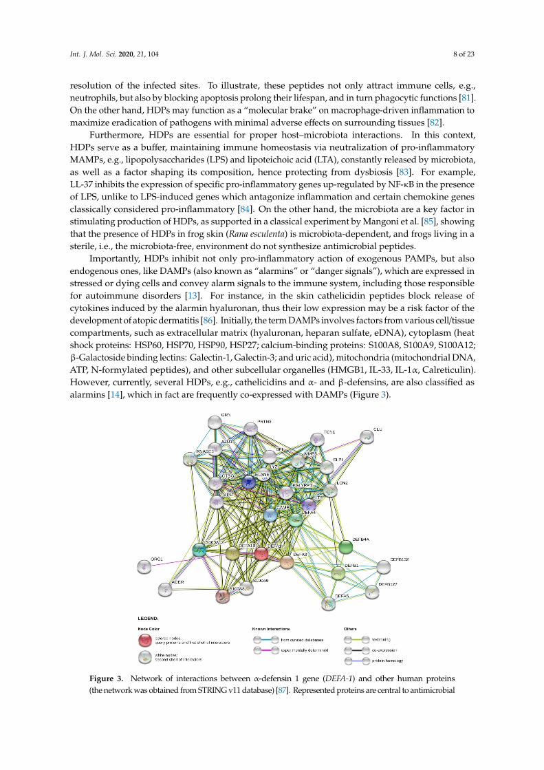
Importantly, HDPs inhibit not only pro-inflammatory action of exogenous PAMPs, but alsoendogenous ones, like DAMPs (also known as “alarmins” or “danger signals”), which are expressed instressed or dying cells and convey alarm signals to the immune system, including those responsiblefor autoimmune disorders [13]. For instance, in the skin cathelicidin peptides block release ofcytokines induced by the alarmin hyaluronan, thus their low expression may be a risk factor of thedevelopment of atopic dermatitis [86]. Initially, the term DAMPs involves factors from various cell/tissuecompartments, such as extracellular matrix (hyaluronan, heparan sulfate, eDNA), cytoplasm (heatshock proteins: HSP60, HSP70, HSP90, HSP27; calcium-binding proteins: S100A8, S100A9, S100A12;β-Galactoside binding lectins: Galectin-1, Galectin-3; and uric acid), mitochondria (mitochondrial DNA,ATP, N-formylated peptides), and other subcellular organelles (HMGB1, IL-33, IL-1α, Calreticulin).However, currently, several HDPs, e.g., cathelicidins and α- and β-defensins, are also classified asalarmins [14], which in fact are frequently co-expressed with DAMPs (Figure 3).Int. J. Mol. Sci. 2019, 20, x FOR PEER REVIEW 10 of 25
Figure 3. Network of interactions between α-defensin 1 gene (DEFA-1) and other human proteins (the network was obtained from STRING v11 database) [87]. Represented proteins are central to antimicrobial and immunomodulatory activities. Abbreviations: AGER, advanced glycosylation end-product-specific receptor; BPI, bactericidal permeability increasing protein; CAMP, cathelicidin antimicrobial peptide; CLU, clusterin; CTSG, cathepsin; DEFA3, defensin alpha 3; DEFA4, defensin alpha 4; DEFA5, defensin alpha 5; DEFA1B, defensin alpha 1B; DEFB4A, defensin beta 4A; DEFB1, defensin beta 1; DEFB132, defensin beta 132; DEFB127, defensin beta 127; ELANE, neutrophil elastase; GRN, granulin precursor; LCN2, lipocalin 2; LTF, lactotransferrin; LYZ, lysozyme; MPO, myeloperoxidase; MMP8, matrix metallopeptidase 8; ORC1, origin recognition complex subunit 1; PGLYRP1, peptidoglycan recognition protein 1; PRTN3, proteinase 3; RNASE3, ribonuclease A family member 3; SLP I, secretory leukocyte peptidase inhibitor; S100A8, S100 calcium binding protein A8; S100A9, S100 calcium binding protein A9; S100A12, S100 calcium binding protein A12; TCN1, transcobalamin 1.
Indeed, a link between HDPs and multiple autoinflammatory diseases such as skin disease (atopic dermatitis, psoriasis, rosacea) or microbiota-related ones, e.g., IBD (Crohn’s disease, colitis ulcerosa), acne vulgaris, and periodontitis, has been established by several studies (see below). An enhancement of Th17 response by HDPs may serve as an example. Briefly, HDPs efficiently attract Th17 (T helper 17 cells), which in turn secrete pro-inflammatory cytokines, IL-17A, IL-17F, IL-21, and IL-22, responsible for mounting mucosal defense against pathogenic microbes in the respiratory or intestinal tract. For instance, IL-17A and IL-22 work synergistically to induce certain β-defensins hBD-1, hBD-3, and hBD-4 in both human and primary mouse gastric epithelial cells (GEC) and
Figure 3. Network of interactions between α-defensin 1 gene (DEFA-1) and other human proteins(the network was obtained from STRING v11 database) [87]. Represented proteins are central to antimicrobial
Int. J. Mol. Sci. 2020, 21, 104 9 of 23
and immunomodulatory activities. Abbreviations: AGER, advanced glycosylation end-product-specificreceptor; BPI, bactericidal permeability increasing protein; CAMP, cathelicidin antimicrobial peptide;CLU, clusterin; CTSG, cathepsin; DEFA3, defensin alpha 3; DEFA4, defensin alpha 4; DEFA5, defensinalpha 5; DEFA1B, defensin alpha 1B; DEFB4A, defensin beta 4A; DEFB1, defensin beta 1; DEFB132,defensin beta 132; DEFB127, defensin beta 127; ELANE, neutrophil elastase; GRN, granulin precursor;LCN2, lipocalin 2; LTF, lactotransferrin; LYZ, lysozyme; MPO, myeloperoxidase; MMP8, matrixmetallopeptidase 8; ORC1, origin recognition complex subunit 1; PGLYRP1, peptidoglycan recognitionprotein 1; PRTN3, proteinase 3; RNASE3, ribonuclease A family member 3; SLP I, secretory leukocytepeptidase inhibitor; S100A8, S100 calcium binding protein A8; S100A9, S100 calcium binding proteinA9; S100A12, S100 calcium binding protein A12; TCN1, transcobalamin 1.
Indeed, a link between HDPs and multiple autoinflammatory diseases such as skin disease(atopic dermatitis, psoriasis, rosacea) or microbiota-related ones, e.g., IBD (Crohn’s disease,colitis ulcerosa), acne vulgaris, and periodontitis, has been established by several studies (see below).An enhancement of Th17 response by HDPs may serve as an example. Briefly, HDPs efficiently attractTh17 (T helper 17 cells), which in turn secrete pro-inflammatory cytokines, IL-17A, IL-17F, IL-21,and IL-22, responsible for mounting mucosal defense against pathogenic microbes in the respiratory orintestinal tract. For instance, IL-17A and IL-22 work synergistically to induce certain β-defensins hBD-1,hBD-3, and hBD-4 in both human and primary mouse gastric epithelial cells (GEC) and gastroidsco-cultured with Helicobacter pylori [88]. On the other hand, an elevated level of Th17 cells has beenconnected with various autoimmune diseases, such as systemic lupus erythematosus, rheumatoidarthritis, or psoriasis [89].
Also, genetically-mediated deficiency/excess of HDPs, gene sequence polymorphisms, as well asdisturbed expression may be a risk factor in inflammatory diseases. For instance, Hollox et al. [90]showed a significant association between higher genomic copy numbers for β-defensin genes,ranging from 2 to 7 copies, and the relative risk of developing psoriasis. Likewise, a lower thehBD-2 gene copy number in the β-defensin locus predisposes to colonic Crohn’s disease [91].Recently, experimental evidence has highlighted the genetic association between the clinical phenotypeof sepsis and DEFA-1/DEFA-3 copy number. Transgenic mice models were engineered to produce ahigh gene copy number of DEFA-1/DEFA-3, which manipulated the outcome of sepsis progression [92].The consequential effect was compared to the low gene copy number wild-type mice models, in thatthe former showed chronic inflammation, endothelial cell damage, vascular leakage, severe organinjury, and mortality. Thus, treatment of patients with sepsis can be challenging due to the underlyingindividual genetic associations. However, further research is needed to obtain conclusive data.In addition, single nucleotide polymorphisms (SNPs) of the hBD-1 gene was connected with thepathogenesis of inflammatory bowel diseases and chronic gastritis [93], as well as oral diseases [94].
In general, expression of HDPs is enhanced during infection or inflammation through transcriptionfactors initialized by pro-inflammatory cytokines or signaling pathways associated with activationof PRRs, e.g., Toll-like receptors (TLRs). For instance, promoter regions of α- and β-defensin genescontain binding sites for major cellular transcription factors, notably nuclear factor κB (NF-κB) andactivator protein 1 (AP-1) (Figure 1). It should be noted that NF-κB also plays a crucial role in thepathogenesis of Crohn’s disease, along with many other pro-inflammatory molecules that modulatethe hBD-2 expression [95], as well as in triggering its production (and IL-6) in severe sepsis [96].Moreover, the expression of genes encoding LL-37 (and hBD-2) is modulated by vitamin D3 viabinding with specific DNA sequences in their promoters, the so-called vitamin D response elements(VDRE) [97]. Additionally, a recent in silico analysis identified a wide range of transcription factorswhich possibly bind and modulate the gene transcription of many antimicrobial peptides and proteins,such as LL-37, RNAse1, CCL18, CXCL14, and HTN1 [98].
In line with this, it has been established that DNA methylation of the CpG sites in the 5′
flanking region of the hBD-1 gene contributes to its deficiency in patients with atopic dermatitis [99].Furthermore, point mutations in the promoter region of hBD-1 explain a cancer-specific loss of thispeptide in 90% and 82% of renal cell carcinomas and prostate [100]. Thus, hBD-1 was suggested as a
Int. J. Mol. Sci. 2020, 21, 104 10 of 23
potential tumor suppressor gene for urological cancers. Also, in oral squamous cell carcinoma (OSCC),hBD-1 appears to have anti-tumor properties, while hBD-2 and hBD-3 might be proto-oncogenes [101].
Nonetheless, the relation between HDPs and inflammation is not always straightforward,and either their deficiency or overproduction, as well as a balance between pro- or anti-inflammatoryeffects, may contribute to the pathological inflammatory response. For instance, in atopic dermatitis(AD), despite severe skin inflammation, the expression level of major skin HDPs, dermcidin, LL-37,hBD-2, and hBD-3, is not increased, hence patients with AD are more prone to skin infections andhave altered skin colonization patterns. By contrast, in psoriasis, expression of LL-37, hBD-2, andhBD-3 is elevated, hence skin infection is rare. Nevertheless, LL-37 and hBDs are considered as a majordriving force of inflammation in psoriasis by mechanisms involving increased production of IFN-αand activation of pDCs, respectively [102]. In addition, these peptides stimulating degranulationof mast cells and increasing production of the pruritogenic cytokine IL-31 may escalate itching(pruritus) manifestation [103]. However, it has been recently observed that LL-37 may also act asan anti-inflammatory agent by blocking the release of inflammatory cytokine IL-1β, depending onits concentration. Interestingly, this observation possibly explains the mechanism underlying theparadoxical effectiveness of vitamin D3, i.e., inducer of LL-37 expression, in treatment of psoriasis [11].Similarly, hBD-3 may inhibit inflammation by inducing expression of anti-inflammatory cytokineIL-37 in keratinocytes [104]. An elevated level of cathelicidin is also observed in other inflammatoryskin conditions, namely rosacea and palmoplantar pustulosis, but instead of the native form of LL-37,its proteolytically cleaved variants drive the inflammation [76,105].
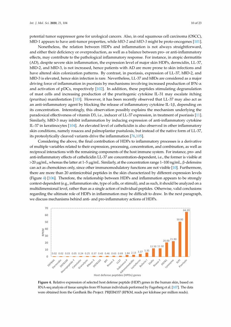
Considering the above, the final contribution of HDPs to inflammatory processes is a derivativeof multiple variables related to their expression, processing, concentration, and combination, as well asreciprocal interactions with the remaining components of the host immune system. For instance, pro- andanti-inflammatory effects of cathelicidin LL-37 are concentration-dependent, i.e., the former is visible at>20 µg/mL, whereas the latter at 1–5 µg/mL. Similarly, at the concentration range 1–100 ng/mL, β-defensinscan act as chemokines only, since other immunomodulatory functions are not visible [10]. Furthermore,there are more than 20 antimicrobial peptides in the skin characterized by different expression levels(Figure 4) [106]. Therefore, the relationship between HDPs and inflammation appears to be stronglycontext-dependent (e.g., inflammation site, type of cells, or stimuli), and as such, it should be analyzed on amultidimensional level, rather than as a single action of individual peptides. Otherwise, valid conclusionsregarding the ultimate role of HDPs in inflammation may be difficult to draw. In the next paragraph,we discuss mechanisms behind anti- and pro-inflammatory actions of HDPs.
Int. J. Mol. Sci. 2019, 20, x FOR PEER REVIEW 12 of 25
inflammatory skin conditions, namely rosacea and palmoplantar pustulosis, but instead of the native form of LL-37, its proteolytically cleaved variants drive the inflammation [76,105].
Considering the above, the final contribution of HDPs to inflammatory processes is a derivative of multiple variables related to their expression, processing, concentration, and combination, as well as reciprocal interactions with the remaining components of the host immune system. For instance, pro- and anti-inflammatory effects of cathelicidin LL-37 are concentration-dependent, i.e., the former is visible at >20 μg/mL, whereas the latter at 1–5 μg/mL. Similarly, at the concentration range 1–100 ng/mL, β-defensins can act as chemokines only, since other immunomodulatory functions are not visible [10]. Furthermore, there are more than 20 antimicrobial peptides in the skin characterized by different expression levels (Figure 4) [106]. Therefore, the relationship between HDPs and inflammation appears to be strongly context-dependent (e.g., inflammation site, type of cells, or stimuli), and as such, it should be analyzed on a multidimensional level, rather than as a single action of individual peptides. Otherwise, valid conclusions regarding the ultimate role of HDPs in inflammation may be difficult to draw. In the next paragraph, we discuss mechanisms behind anti- and pro-inflammatory actions of HDPs.
Figure 4. Relative expression of selected host defense peptide (HDP) genes in the human skin, based on RNA-seq analysis of tissue samples from 95 human individuals performed by Fagerberg et al. [107]. The data were obtained from the GenBank Bio Project: PRJEB4337 (RPKM, reads per kilobase per million reads).
4. Molecular Mechanisms of Anti- and Pro-Inflammatory Action of HDPs
The immunomodulatory potential of HDPs is strictly connected with their ability to recruit and activate immune and non-immune cells, as well as a direct or indirect impact on their fate, including maturation, differentiation, degranulation, or apoptosis [10]. This is mediated through interaction with a wide range of membrane-bound and intracellular receptors, followed by stimulation of their downstream signaling pathways. So far, HDPs have been recognized to interact with the following receptors: (1) Pattern recognition receptors (PRRs), (2) purinergic G-protein coupled receptors (formyl peptide receptor like-1), (3) P2X7 receptor, (4) MRGPRX2, (5) chemokine receptors (commonly known CCR2, CCR6), (6) epidermal growth factor receptors (tyrosine kinases), (7) integrin family receptors (macrophage-1 antigen), nucleotide oligomerization domain (NOD) proteins, and NODlike receptors (NLRs), and their number is still growing [108,109]. Hence, HDPs modulate immune responses using the same receptors as MAMPs/PAMPs and DAMPs [110,111]. To illustrate, β-defensins attract cells by interaction mainly with CCR2 and CCR6 receptors, and exert their “alarmin” activity, e.g., induction of cytokine production, via TLRs, EGFR, GPCR, and MrgX2 ones; however, both activities may overlap in one receptor.
0.02 0.02 0.03 0.05 0.24 0.25 0.37 0.43 0.46 0.63 0.69 1.84 2.13 3.37 5.3511.50 11.50
20.00 21.90
50.00
64.20
0
10
20
30
40
50
60
70
RPKM
Host defense peptides (HPDs) genes
Figure 4. Relative expression of selected host defense peptide (HDP) genes in the human skin, based onRNA-seq analysis of tissue samples from 95 human individuals performed by Fagerberg et al. [107]. The datawere obtained from the GenBank Bio Project: PRJEB4337 (RPKM, reads per kilobase per million reads).
Int. J. Mol. Sci. 2020, 21, 104 11 of 23
4. Molecular Mechanisms of Anti- and Pro-Inflammatory Action of HDPs
The immunomodulatory potential of HDPs is strictly connected with their ability to recruitand activate immune and non-immune cells, as well as a direct or indirect impact on their fate,including maturation, differentiation, degranulation, or apoptosis [10]. This is mediated throughinteraction with a wide range of membrane-bound and intracellular receptors, followed by stimulationof their downstream signaling pathways. So far, HDPs have been recognized to interact with thefollowing receptors: (1) Pattern recognition receptors (PRRs), (2) purinergic G-protein coupled receptors(formyl peptide receptor like-1), (3) P2X7 receptor, (4) MRGPRX2, (5) chemokine receptors (commonlyknown CCR2, CCR6), (6) epidermal growth factor receptors (tyrosine kinases), (7) integrin familyreceptors (macrophage-1 antigen), nucleotide oligomerization domain (NOD) proteins, and NODlikereceptors (NLRs), and their number is still growing [108,109]. Hence, HDPs modulate immuneresponses using the same receptors as MAMPs/PAMPs and DAMPs [110,111]. To illustrate, β-defensinsattract cells by interaction mainly with CCR2 and CCR6 receptors, and exert their “alarmin” activity,e.g., induction of cytokine production, via TLRs, EGFR, GPCR, and MrgX2 ones; however, bothactivities may overlap in one receptor.
It should be noted that TLR receptors may be the root cause of certain HDP-associateddiseases. For instance, in individuals with rosacea, a higher expression of TLR-2 sensitizes thefacial skin to microbes or environmental stimuli. Under these conditions, enhanced expressionof kallikrein-5 proteinase is observed in keratinocytes, and ultimately affects production ofcathelicidin peptides, which drives inflammation and abnormal growth of blood vessels [105].Moreover, the tumor-suppressing effect of hBD-1 is associated with its ability to modulate epidermalgrowth factor and human epidermal growth factor receptor 2 (EGFR/HER2)-associated signalingpathways [112]. Finally, hBD-3, through deactivation of TLR-4 and TLR-2, may reduce the adverseimmune reaction initialized by NF-κB in response to LPS [113].
Furthermore, β-defensins and cathelicidins, in the same manner as PAMPs (e.g., LPS) andDAMPs (e.g., heat shock antigens Hsp60 and Hsp70), are ligands of Toll-like receptor 4 (TLR-4).However, the resulting outcome of the receptor’s stimulation may be different for these molecules.For instance, unlike LPS, hBD-3 does not induce production of IL-10, which is an importantanti-inflammatory cytokine, e.g., via suppressing function of antigen-presenting cells (APCs),suggesting that hBD-3 can shift the immune response toward pro-inflammatory direction [114].Similarly, hBD-2 via TLR-4 leads to maturation of dendritic cells (DCs), which consequently exhibitTh1-polarized responses, such as the production of pro-inflammatory cytokines IL-12, IL-1α, IL-1β,and IL-6, which may possibly counter suppressive action of microbial factors by generating morerobust host inflammatory and Th1 responses [115]. In contrast, cathelicidin is considered as aninhibitor of TLR-4, and thus can antagonize with other TLR-4 ligands released during skin injury, e.g.,hyaluronan [10].
Therefore, HPDs joining properties of MAMPs/PAMPs and DAMPs may operate as centralnodes in a network that coordinates immune response to infections as well as non-infectious insults.For instance, it has been shown that synergistic action of MAMPs/PAMPs and DAMPs is necessaryfor synthesis and subsequent secretion of pro-inflammatory cytokine IL-1β [13]. It is important tonote that a lack of IL-1β results in high susceptibility to infections, but its overproduction causesuncontrolled inflammation and tissue damage via T cell-mediated autoinflammatory response [116].Indeed, overproduction of IL-1β was observed in patients with inflammatory bowel disease, andhas been connected with deficiency of α-defensins that serve as regulators of IL-1β maturation [117].As aforementioned, also in psoriasis, LL-37 may act as an inhibitor of the IL-1β release in keratinocytesby blocking activation of the cytosolic DNA-sensing signaling AIM2, i.e., cytosolic receptor for dsDNA.Hence, cytoplasmic DNA appears to contribute to the pathogenesis of psoriasis via activation of IL-1βin keratinocytes by AIM2-mediated inflammasomes [11].
In this context, it is relevant to mention the relationship of HDPs and self-nucleic acids, and itsimpact on inflammation. Under normal homeostatic conditions, the host-derived nucleic acids released
Int. J. Mol. Sci. 2020, 21, 104 12 of 23
from damaged and dying cells do not mediate inflammatory responses because of the systematicregulation and physiological location of nucleic acid sensing TLR7/9. However, several studieshave shown that HDPs may disturb immune tolerance to self-nucleic acids, and in turn significantlyenhance cell responses—in particular, plasmacytoid dendritic cells (pDCs) [118]. In fact, in the skin,this mechanism has been identified as an important initiator of psoriasis development, where LL-37 anddefensins are able to condense self-DNA into particles, which are internalized by pDCs, inducing robustIFN-α response via activation of the TLR-9 signaling pathway [119,120]. This enhances production oflarge amounts of type I IFN, leading to the functional activation of myeloid dendritic cells (mDCs),monocytes, NK cells, keratinocytes, as well as Th1/Th17 differentiation, which further increase thepro-inflammatory, e.g., IFN-γ, IL-22, and IL-17, cytokine expression [120,121]. In line with this, a novelmechanism of nucleic acid recognition by LL-37 utilizing cell surface RNA scavenger receptors (SRs)has been described [122], which results is enhanced clathrin-dependent endocytosis, facilitating theoverproduction of inflammatory cytokines and chemokines. Recently also RNase7 was found to utilizeplasmacytoid dendritic cell (pDC) TLR-9 signaling mode of IFN-α activation even more strongly thanLL-37, emphasizing its crucial role in autoimmune inflammatory skin diseases [123]. Interestingly,other antimicrobial peptides expressed in the skin, such as psoriasin, elafin, or hBD-1, lack the ability ofinteracting with the host nucleic acids, which may be related to their lower net charge (Figure 2) [120].
Another interesting consequence of interactions between HDPs and the host nucleic acids is anovel wound healing mechanism, where LL-37 may alter wound repair by modifying the responses todsRNAs released as a result of skin injury. In detail, LL-37 enhances endosomal uptake of non-codingdouble stranded RNA in TLR-3-mediated mechanism, that results in activation of several importantwound repair growth factors, including fibroblast growth factor (FGF2), and heparin binding EGF-likegrowth factor (HBEGF) from the dermal keratinocytes and fibroblasts [124]. Inhibition of LL-37/dsRNArelation may contribute to the development of hyperproliferation-based diseases, like psoriasis,whereas its augmentation can lead to increased wound regeneration in pathological conditions ofabnormal wound repair (e.g., diabetic ulcers).
Finally, LL-37 actively participates in neutrophil extracellular trap (NET) formation via disruption ofthe nuclear membrane and promotes their stability [125]. NETs are structures composed of decondensedchromatin and multiple enzymes (elastase, myeloperoxidase, gelatinase, etc.) and proteins, includingantimicrobial ones. Thus, NETs act as a mechanical barrier that entraps and subsequently reducesspreading of pathogens and/or their toxic products into the host tissues, where the antimicrobial activityof HPDs is boosted by their accumulation and combination [126]. Moreover, Stephan et al. have shownthat complexes of LL-37/DNA formed inside human macrophages may participate in defense againstintracellular bacteria, e.g., mycobacteria [127]. Accordingly, a recent study investigated the therapeuticpotential of LL-37 in modulating macrophage-mediated excessive inflammatory responses. It was foundthat LL-37 reduced the severity of tuberculosis by rapidly enhancing the anti-inflammatory cytokineTGF-β, IL-10, and prostaglandin E from the infected macrophages [128]. However, further studiesregarding the exogenous effect of LL-37 in severe pulmonary tuberculosis are warranted. Interestingly,administration of vitamin D3 or another potent inducer of LL-37, i.e., 4-phenyl butyrate (PBA), may bean alternative treatment method of tuberculosis [129].
5. Deregulations of HDPs Expression in Selected Diseases
5.1. Periodontal Diseases
An imbalanced unhealthy oral microbiota ushers the entry of various cariogenic, periodontalmicrobes which engenders oral biofilm formation and periodontal diseases such as gingivitis andperiodontitis. The oral epithelial tissues, mainly the gingival epithelium, play a significant role inresisting the colonization of unfavorable oral pathogens. These tissues readily secrete beta-defensinpeptides, as well as histatins, which are the major host defense proteins of the saliva that maintainhomeostasis of oral microbiota [130]. A significant correlation is observed between elevated levels
Int. J. Mol. Sci. 2020, 21, 104 13 of 23
of hBD-2, hBD-4, and HNP4 in the oral mucosal epithelial cells of both adults and children with thedevelopment of dental caries. They are considered as important clinical biomarkers of periodontaldiseases and dental caries. It has been shown that there is a declined expression of beta-defensin1 mRNA gene in the inflamed gingival tissues and periodontal structures. Conversely, chroniccases of periodontitis manifest an elevated expression of the hBD-1 gene [131,132]. The severity ofperiodontal diseases and dental plaques grows, along with a heightened expression of hBD-2 andhistatin-5. Higher activity of pro-inflammatory cytokines modulates the progression of the infection,which further stimulates the production of the defensin peptides through various transcriptionfactors [133]. Recently, 89 patients were monitored according to their periodontal status in relation toother clinical parameters [134]. This study identified an increase in the salivary production of hBD-2,triggered by inflammatory processes and pathogen derived metabolites that can be considered as apossible diagnostic biomarker s of periodontal diseases.
Healing of periodontal lesions is initiated by various growth factors, pro-inflammatory mediators,and antimicrobial peptides accumulating at the infected site. In detail, a complex network of highlyspecialized growth factors, namely, insulin-like growth factor (IGF1, IGF2), transforming growthfactor (TGF-α, TGF-β), epidermal growth factor, and platelet-derived growth factor, coordinates thereparative process by rapid differentiation of keratinocytes and fibroblasts [135]. These growth factorsalso assist the wound healing mechanism by influencing the gene expression pattern of antimicrobialpeptides that typically participate in the epithelial cell proliferation, migration, and inhibition ofcolonizing microbial pathogens at the site of injury. While it is established that wounding influencesthe expression of HDPs, not all of them function the same way. Recent reports have highlighted thedistinct immune responses triggered within the wounded gingival epithelial cells (GECs) and gingivalfibroblasts (HGFs) upon treatment with IGF1 and TGF-α. These growth factors enabled efficient woundclosure and differently modulated the expression of hBD-2, CCL20, IL-1, and IL-8. The findings indicatethat hBD-2 was exclusively enhanced in the gingival epithelial cells measured at set time points of 6 hand 24 h post-wounding, particularly in those cells associated with the keratinocyte differentiationmarker involucrin. Additionally, hBD-2 along with CCL20, IL-1, and IL-8 control the invasion ofbacterial microbes and impact the neutrophil defense mechanisms [136]. Contrarily, the woundedgingival fibroblasts (HGFs) witnessed a substantially low expression pattern of hBD-2 and CCL20,with or without growth factor treatment, that was suggested as a mechanism protecting fibroblastovergrowth into the epithelial wound [136]. fibroblast overgrowth into the epithelial wound.
5.2. Inflammatory Lung Diseases
Cystic fibrosis (CF) is a life-limiting disease characterized by recurrent respiratory infectionsand inflammation, connected to altered composition and volume of the airway surface liquid (ASL).For instance, a reduced bicarbonate HCO3
- secretion resulting in a decrease of airway surface pH(average 6.8–7.5) was observed. Interestingly, it was also found that the acidic pH weakened the actionof LL-37 and hBD-3 against invasive Staphylococcus aureus and Pseudomonas aeruginosa infections byaffecting their structural net charge. Therefore, it could be suggested that a similar mechanism ofacidic pH-reduced antimicrobial activity may occur in other inflammatory conditions taking place incerebral spinal fluid, peritoneal fluid, and pleural fluid. Additionally, it has been noted that high ionicstrength (Na+, K+, Cl+) may impair the antimicrobial activity of hBD-2, lysozyme, and lactoferrin [137].Moreover, CF patients suffer from viscous sputum that accumulates and obstructs their airways.The thick mucus is characterized by heterogenous complex aggregates of DNA and F-actin filamentsderived from leukocytes that have encountered necrotic death. Thus, the antimicrobial function ofLL-37, lysozyme, lactoferrin, and hBD-3 released in the respiratory airways is substantially hindered asthey stabilize DNA/F-actin bundles. Additionally, neutralization of the immune function of neutrophilprotease and IL-8 take place during DNA/F-actin bundles formation [138,139]. It is also worthwhile tounderline that abundant secretion of cysteine cathepsins from the macrophages hinders the functionalexpression of hBD-2.
Int. J. Mol. Sci. 2020, 21, 104 14 of 23
Chronic obstructive pulmonary disease (COPD), bronchitis, and asthma are all characterized byinflammation that develops as a consequence of pro-inflammatory mediator secretion. Immune cellsdistributed throughout the lungs are responsible for sudden exacerbations associated with theproduction of cytokines, oxidative stress, and protease secretion, including caspases, neutrophilelastase, and matrix metalloproteinases. One study reports the enhanced expression of hBD-2 in thedistal airway epithelial cells of COPD patients, but a rather diminished expression of hBD-2 in thecentral airways, despite the exaggerated expression of TLR-4 receptors [140]. This distinct variationwas found to be in correlation with exposure to cigarette smoking. While it is evident that everycell in the body requires ATP for its biological function of energy production and retention, little isknown about its possible involvement in the immune system response to bacterial infection andinflammation. In a P. aeruginosa-infected rat model, ATP administration led to rapid stimulationof hBD-2 production. The mechanism of ATP action involved NADPH family of oxidases (DUOX1) via ion channel receptors P2X, P2Y activation, and regulation of multiple signaling pathwaysERK1/2 and NF-κB [141]. The released defensin peptide was found to control the inflammatoryprocesses underlying the acute infection of pneumonia by suppression of TNF-α and IL-6. Furthermore,another study detected the potent ability of the IL-17 family of cytokines in the induction mechanismof the hBD-2 gene. Typically, most of the immune cells, including T helper cells, macrophages,dendritic cells, and natural killer cells, secrete IL-17 family of cytokines. These cytokines act inconcordance with the tumor necrosis factor and IL-1 to promote the induction of other inflammatorymediators production, which individually or collectively can stimulate the secretion of beta-defensinsvia the activation of various signaling pathways [142,143]. For example, the stimulatory functions ofIL-17 in the airway epithelial cells promoting transcription of the hBD-2 gene through the action ofJAK and NF-κB signaling have been reported [144]. Moreover, IL-17 has an impact on other cytokinessuch as IL-1α, IL-β, IL-6, IL-7, and TNF-α, which contribute to the production of hBD-2. On theother hand, the alveolar macrophages and dendritic cells consistently maintain the release of IL-22.According to a recent study, in which alveolar epithelial cells (A549) were screened for the abundantdisplay of IL-22 receptors and subjected to treatment with different doses of IL-22, an increase of hBD-2mRNA transcript synthesis via the STAT3 pathway was observed [145]. Thus, this study revealed anew immunomodulatory role of IL-22 in stimulation of the lung defensins in response to exposureto pathogenic bacteria and viruses. Interestingly, hBD-1 has also emerged as a clinical biomarker ofCOPD and other inflammatory lung diseases, such as asthma [146]. However, in this case, an alteredexpression of hBD-1 may be aggravated by gene copy number variations.
5.3. Inflammatory Bowel Diseases
The human defensins 5 and 6 (HD5, HD6) are particularly important in preserving the homeostaticequilibrium of the enteric mucosa layer, exhibiting different effects against the essential inducers of theirsecretion, i.e., various products of the Gram-positive and Gram-negative bacteria [147]. To illustrate,there is a remarkable reduction in the expression levels of HD5 and HD6 by the Paneth cells ininflammatory bowel conditions such as Crohn’s disease. This shift in expression could be attributedto the cause by genetic changes in the NOD2 receptor [148]. Furthermore, a recent study suggeststhe possibility of using other HDPs, such as the level of fecal HNP, as a non-invasive biomarker ofintestinal inflammation in patients suffering from colitis ulcerosa [149].
The human beta-defensins are also naturally expressed in the epithelial cells of the gastric mucosaand extensively participate in host defense against Helicobacter pylori colonization, a bacterium presentin a high proportion (~80%) of people throughout the world [150]. Multilevel signaling pathwayspromote the molecular mechanism of induction of beta-defensins in response to the initial stagesof H. pylori infection. In addition, it has been shown that the phosphorylation of a serine residueof EGFR may modulate the release of hBD-3 [151], revealingan underlying interdependent relationbetween the stimulated transforming growth factor β-activated kinase-1 (TAK1), p38α pathway,
Int. J. Mol. Sci. 2020, 21, 104 15 of 23
and phosphorylation of EGFR receptor in the amplified release of hBD-3 in the gastric mucosa involvedin H. pylori infection.
6. Conclusion
The multifunctional host defense peptides provide a link between innate and adaptive immunityagainst different microorganisms and contribute to inflammation of infected sites. Depending on the celltype and extracellular environment, some of these peptides exert contrasting functions, wherein theypromote or suppress inflammatory processes. A strongly compromised action of host defense peptidesagainst intruders and delayed resolution of inflammatory mediators underlies the development ofinflammation in different diseases. Some of these peptides may serve as potential clinical biomarkersfor a wide range of inflammatory diseases. Evidently, antimicrobial regulation is crucial to limitthe exacerbation of inflammatory signaling molecules. While various factors govern the release ofHDPs, any dysregulation can favor an imbalanced feedback mechanism between the host-inducedanti-inflammatory and pro-inflammatory processes. In summary, a deeper understanding of thediverse functional roles of HDPs in the body’s physiological response to inflammation and disease iscrucial and represents the first approach to develop new therapeutic strategies based on HDPs aimedat resolving the progression of inflammatory diseases and strengthening the host barrier defenses.
Author Contributions: Conceptualization, writing, and original draft preparation, S.V.P.; conceptualization,writing, figure preparation, and review and editing, K.F.; writing and figure preparation, T.D.; conceptualizationand writing, E.P.; conceptualization and review and editing, R.B. All authors have read and agreed to the publishedversion of the manuscript.
Funding: This work was financially supported by the National Science Center, Poland under Grant:UMO-2018/30/M/NZ6/00502 (to RB). This work was conducted within a project which received funding from theEuropean Union’s Horizon 2020 research and innovation programme under the Marie Skłodowska-Curie grantagreement No. 754432 and the Polish Ministry of Science and Higher Education, from financial resources forscience in 2018–2023 granted for the implementation of an international co-financed project.
Conflicts of Interest: The authors declare no conflict of interest. The funders had no role in the design of the study,in the collection, analyses, or interpretation of data; in the writing of the manuscript; or in the decision to publish.
Abbreviations
AMPs Antimicrobial peptidesATP Adenosine triphosphateBM Bone morphogenetic proteinCAMP Cathelicidin antimicrobial peptideCCR2 C–C chemokine receptor type 2CCR6 C–C chemokine receptor type 6CNV Copy number variationCOPD Chronic obstructive pulmonary diseaseDAMPs Damage-associated molecular patternsDC Dendritic cellDUOX1 Dual oxidase 1EGFR Epidermal growth factor receptorELA2 Neutrophil elastase 2FGF2 Fibroblast growth factorGEC Gingival epithelial cellHBEGF Heparin binding EGF like growth factorHER2 Human epidermal growth factor receptor 2HGFs Human gingival fibroblastsHMGB1 High mobility group box 1IDR Innate defense regulator
Int. J. Mol. Sci. 2020, 21, 104 16 of 23
IGF Insulin like growth factorIL-1α Interleukin-1αIL-33 Interleukin-33JAK Janus KinaseLPS LipopolysaccharidesLTA Lipoteichoic acidMAPK Mitogen-activated protein kinaseMIC Minimum inhibitory concentrationMIP–3 Macrophage inflammatory protein-3 alphaMRGPRX2 Mas-related G-protein coupled receptor member X2NETs Neutrophil extracellular trapsNF-κB Nuclear factor kappa-light-chain-enhancer of activated B cellsNOD2 Nucleotide-binding oligomerization domainOSCC Oral squamous cell carcinomaP2X7 Purinoceptor 7SNP Single nucleotide polymorphismSTAT Signal transducer and activator of transcription
References
1. Mills, C.D.; Ley, K.; Buchmann, K.; Canton, J. Sequential Immune Responses: The Weapons of Immunity.J. Innate Immun. 2015, 7, 443–449. [CrossRef] [PubMed]
2. Chen, L.; Deng, H.; Cui, H.; Fang, J.; Zuo, Z.; Deng, J.; Li, Y.; Wang, X.; Zhao, L. Inflammatory responses andinflammation-associated diseases in organs. Oncotarget 2018, 9, 7204–7218. [CrossRef] [PubMed]
3. Libby, P. Inflammatory mechanisms: The molecular basis of inflammation and disease. Nutr. Rev.2007, 65, S140–S146. [CrossRef]
4. Muller, W.A. Getting leukocytes to the site of inflammation. Vet. Pathol. 2013, 50, 7–22. [CrossRef] [PubMed]5. Gupta, S.; Bhatia, G.; Sharma, A.; Saxena, S. Host defense peptides: An insight into the antimicrobial world.
J. Oral. Maxillofac. Pathol. 2018, 22, 239–244. [CrossRef]6. Hancock, R.E.; Haney, E.F.; Gill, E.E. The immunology of host defence peptides: Beyond antimicrobial
activity. Nat. Rev. Immunol. 2016, 16, 321–334. [CrossRef]7. Kopp, Z.A.; Jain, U.; Van Limbergen, J.; Stadnyk, A.W. Do antimicrobial peptides and complement collaborate
in the intestinal mucosa? Front. Immunol. 2015, 6, 17. [CrossRef]8. Bhat, S.; Song, Y.H.; Lawyer, C.; Milner, S.M. Modulation of the complement system by human beta-defensin
2. J. Burns Wounds 2007, 5, e10.9. Lee, E.Y.; Lee, M.W.; Wong, G.C.L. Modulation of toll-like receptor signaling by antimicrobial peptides.
Semin. Cell Dev. Biol. 2019, 88, 173–184. [CrossRef]10. Lai, Y.; Gallo, R.L. AMPed up immunity: How antimicrobial peptides have multiple roles in immune defense.
Trends Immunol. 2009, 30, 131–141. [CrossRef]11. Dombrowski, Y.; Schauber, J. Cathelicidin LL-37: A defense molecule with a potential role in psoriasis
pathogenesis. Exp. Dermatol. 2012, 21, 327–330. [CrossRef] [PubMed]12. Haney, E.F.; Straus, S.K.; Hancock, R.E.W. Reassessing the Host Defense Peptide Landscape. Front. Chem.
2019, 7, 43. [CrossRef] [PubMed]13. Saïd-Sadier, N.; Ojcius, D.M. Alarmins, inflammasomes and immunity. Biomed. J. 2012, 35, 437–449.
[CrossRef] [PubMed]14. Pouwels, S.D.; Heijink, I.H.; ten Hacken, N.H.; Vandenabeele, P.; Krysko, D.V.; Nawijn, M.C.; van
Oosterhout, A.J. DAMPs activating innate and adaptive immune responses in COPD. Mucosal. Immunol.2014, 7, 215–226. [CrossRef] [PubMed]
15. Roh, J.S.; Sohn, D.H. Damage-Associated Molecular Patterns in Inflammatory Diseases. Immune. Netw.2018, 18, e27. [CrossRef]
16. Fruitwala, S.; El-Naccache, D.W.; Chang, T.L. Multifaceted immune functions of human defensins andunderlying mechanisms. Semin. Cell Dev. Biol. 2019, 88, 163–172. [CrossRef]
Int. J. Mol. Sci. 2020, 21, 104 17 of 23
17. Jourdain, M.L.; Pierrard, L.; Kanagaratnam, L.; Velard, F.; Sergheraert, J.; Lefèvre, B.; Gangloff, S.C.;Braux, J. Antimicrobial peptide gene expression in periodontitis patients: A pilot study. J. Clin. Periodontol.2018, 45, 524–537. [CrossRef]
18. Uysal, P.; Simsek, G.; Durmus, S.; Sozer, V.; Aksan, H.; Yurt, S.; Cuhadaroglu, C.; Kosar, F.; Gelisgen, R.;Uzun, H. Evaluation of plasma antimicrobial peptide LL-37 and nuclear factor-kappaB levels in stablechronic obstructive pulmonary disease. Int. J. Chron Obstruct. Pulmon. Dis 2019, 14, 321–330. [CrossRef]
19. Muniz, L.R.; Knosp, C.; Yeretssian, G. Intestinal antimicrobial peptides during homeostasis, infection, anddisease. Front. Immunol. 2012, 3, 310. [CrossRef]
20. Kolbinger, F.; Loesche, C.; Valentin, M.A.; Jiang, X.; Cheng, Y.; Jarvis, P.; Peters, T.; Calonder, C.; Bruin, G.;Polus, F.; et al. β-Defensin 2 is a responsive biomarker of IL-17A-driven skin pathology in patients withpsoriasis. J. Allergy Clin. Immunol. 2017, 139, 923–932. [CrossRef]
21. Xie, G.H.; Chen, Q.X.; Cheng, B.L.; Fang, X.M. Defensins and sepsis. Biomed. Res. Int. 2014, 2014, 180109.[CrossRef] [PubMed]
22. Joly, S.; Compton, L.M.; Pujol, C.; Kurago, Z.B.; Guthmiller, J.M. Loss of human beta-defensin 1, 2, and 3expression in oral squamous cell carcinoma. Oral Microbiol. Immunol. 2009, 24, 353–360. [CrossRef] [PubMed]
23. Aberg, K.M.; Radek, K.A.; Choi, E.H.; Kim, D.K.; Demerjian, M.; Hupe, M.; Kerbleski, J.; Gallo, R.L.;Ganz, T.; Mauro, T.; et al. Psychological stress downregulates epidermal antimicrobial peptide expressionand increases severity of cutaneous infections in mice. J. Clin. Invest. 2007, 117, 3339–3349. [CrossRef][PubMed]
24. Silva, O.N.; Porto, W.F.; Ribeiro, S.M.; Batista, I.; Franco, O.L. Host-defense peptides and their potential useas biomarkers in human diseases. Drug Discov. Today 2018, 23, 1666–1671. [CrossRef]
25. Wang, G.; Li, X.; Wang, Z. APD3: The antimicrobial peptide database as a tool for research and education.Nucleic Acids Res. 2016, 44, D1087–D1093. [CrossRef]
26. Pasupuleti, M.; Schmidtchen, A.; Malmsten, M. Antimicrobial peptides: Key components of the innateimmune system. Crit. Rev. Biotechnol. 2012, 32, 143–171. [CrossRef]
27. Nakatsuji, T.; Gallo, R.L. Antimicrobial peptides: Old molecules with new ideas. J. Invest. Dermatol.2012, 132, 887–895. [CrossRef]
28. Semple, F.; Dorin, J.R. beta-Defensins: Multifunctional modulators of infection, inflammation and more?J. Innate Immun. 2012, 4, 337–348. [CrossRef]
29. Sieprawska-Lupa, M.; Mydel, P.; Krawczyk, K.; Wójcik, K.; Puklo, M.; Lupa, B.; Suder, P.; Silberring, J.;Reed, M.; Pohl, J.; et al. Degradation of human antimicrobial peptide LL-37 by Staphylococcus aureus-derivedproteinases. Antimicrob. Agents Chemother. 2004, 48, 4673–4679. [CrossRef]
30. Baranska-Rybak, W.; Sonesson, A.; Nowicki, R.; Schmidtchen, A. Glycosaminoglycans inhibit the antibacterialactivity of LL-37 in biological fluids. J. Antimicrob. Chemother. 2006, 57, 260–265. [CrossRef]
31. Wang, G. Human antimicrobial peptides and proteins. Pharmaceuticals 2014, 7, 545–594. [CrossRef] [PubMed]32. Tomita, T.; Hitomi, S.; Nagase, T.; Matsui, H.; Matsuse, T.; Kimura, S.; Ouchi, Y. Effect of ions on antibacterial
activity of human beta defensin 2. Microbiol. Immunol. 2000, 44, 749–754. [CrossRef] [PubMed]33. Goldman, M.J.; Anderson, G.M.; Stolzenberg, E.D.; Kari, U.P.; Zasloff, M.; Wilson, J.M. Human beta-defensin-1
is a salt-sensitive antibiotic in lung that is inactivated in cystic fibrosis. Cell 1997, 88, 553–560. [CrossRef]34. Sun, L.; Wang, W.; Xiao, W.; Yang, H. The Roles of Cathelicidin LL-37 in Inflammatory Bowel Disease.
Inflamm. Bowel. Dis. 2016, 22, 1986–1991. [CrossRef] [PubMed]35. Dürr, U.H.; Sudheendra, U.S.; Ramamoorthy, A. LL-37, the only human member of the cathelicidin family of
antimicrobial peptides. Biochim. Biophys. Acta 2006, 1758, 1408–1425. [CrossRef] [PubMed]36. Scott, M.G.; Davidson, D.J.; Gold, M.R.; Bowdish, D.; Hancock, R.E. The human antimicrobial peptide LL-37
is a multifunctional modulator of innate immune responses. J. Immunol. 2002, 169, 3883–3891. [CrossRef]37. Edfeldt, K.; Agerberth, B.; Rottenberg, M.E.; Gudmundsson, G.H.; Wang, X.B.; Mandal, K.; Xu, Q.; Yan, Z.Q.
Involvement of the antimicrobial peptide LL-37 in human atherosclerosis. Arterioscler. Thromb. Vasc. Biol.2006, 26, 1551–1557. [CrossRef]
38. Cerrillo, E.; Moret, I.; Iborra, M.; Ramos, D.; Busó, E.; Tortosa, L.; Sáez-González, E.; Nos, P.; Beltrán, B.Alpha-defensins (α-Defs) in Crohn’s disease: Decrease of ileal α-Def 5 via permanent methylation andincrease in plasma α-Def 1-3 concentrations offering biomarker utility. Clin. Exp. Immunol. 2018, 192, 120–128.[CrossRef]
Int. J. Mol. Sci. 2020, 21, 104 18 of 23
39. Sthoeger, Z.M.; Bezalel, S.; Chapnik, N.; Asher, I.; Froy, O. High alpha-defensin levels in patients withsystemic lupus erythematosus. Immunology 2009, 127, 116–122. [CrossRef]
40. Lisitsyn, N.A.; Bukurova, Y.A.; Nikitina, I.G.; Krasnov, G.S.; Sykulev, Y.; Beresten, S.F. Enteric alpha defensinsin norm and pathology. Ann. Clin. Microbiol. Antimicrob. 2012, 11, 1. [CrossRef]
41. Bonamy, C.; Sechet, E.; Amiot, A.; Alam, A.; Mourez, M.; Fraisse, L.; Sansonetti, P.J.; Sperandio, B. Expressionof the human antimicrobial peptideβ-defensin-1 is repressed by the EGFR-ERK-MYC axis in colonic epithelialcells. Sci. Rep. 2018, 8, 18043. [CrossRef] [PubMed]
42. Ling, Y.M.; Chen, J.Y.; Guo, L.; Wang, C.Y.; Tan, W.T.; Wen, Q.; Zhang, S.D.; Deng, G.H.; Lin, Y.; Kwok, H.F.β-defensin 1 expression in HCV infected liver/liver cancer: An important role in protecting HCV progressionand liver cancer development. Sci. Rep. 2017, 7, 13404. [CrossRef] [PubMed]
43. Dilek, F.; Emin, Ö.; Gültepe, B.; Yazıcı, M.; Çakır, E.; Gedik, A.H. Evaluation of nasal fluid β-defensin 2 levelsin children with allergic rhinitis. Turk. Pediatri. Ars. 2017, 52, 79–84. [CrossRef] [PubMed]
44. Andresen, E.; Günther, G.; Bullwinkel, J.; Lange, C.; Heine, H. Increased expression of beta-defensin 1(DEFB1) in chronic obstructive pulmonary disease. PLoS ONE 2011, 6, e21898. [CrossRef] [PubMed]
45. Shi, N.; Jin, F.; Zhang, X.; Clinton, S.K.; Pan, Z.; Chen, T. Overexpression of human β-defensin 2 promotesgrowth and invasion during esophageal carcinogenesis. Oncotarget 2014, 5, 11333–11344. [CrossRef][PubMed]
46. Xu, D.; Zhang, B.; Liao, C.; Zhang, W.; Wang, W.; Chang, Y.; Shao, Y. Human beta-defensin 3 contributesto the carcinogenesis of cervical cancer via activation of NF-κB signaling. Oncotarget 2016, 7, 75902–75913.[CrossRef]
47. Abe, S.; Miura, K.; Kinoshita, A.; Mishima, H.; Miura, S.; Yamasaki, K.; Hasegawa, Y.; Higashijima, A.;Jo, O.; Sasaki, K.; et al. Copy number variation of the antimicrobial-gene, defensin beta 4, is associated withsusceptibility to cervical cancer. J. Hum. Genet. 2013, 58, 250–253. [CrossRef]
48. Kalmodia, S.; Son, K.N.; Cao, D.; Lee, B.S.; Surenkhuu, B.; Shah, D.; Ali, M.; Balasubramaniam, A.; Jain, S.;Aakalu, V.K. Presence of Histatin-1 in Human Tears and Association with Aqueous Deficient Dry EyeDiagnosis: A Preliminary Study. Sci. Rep. 2019, 9, 10304. [CrossRef]
49. Khan, S.A.; Fidel, P.L.; Thunayyan, A.A.; Varlotta, S.; Meiller, T.F.; Jabra-Rizk, M.A. Impaired Histatin-5Levels and Salivary Antimicrobial Activity against. J. AIDS Clin. Res. 2013, 4. [CrossRef]
50. Bogefors, J.; Kvarnhammar, A.M.; Millrud, C.R.; Georén, S.K.; Cardell, L.O. LEAP-2, LL-37 and RNase7in tonsillar tissue: Downregulated expression in seasonal allergic rhinitis. Pathog. Dis. 2014, 72, 55–60.[CrossRef]
51. Spencer, J.D.; Schwaderer, A.L.; Wang, H.; Bartz, J.; Kline, J.; Eichler, T.; DeSouza, K.R.; Sims-Lucas, S.;Baker, P.; Hains, D.S. Ribonuclease 7, an antimicrobial peptide upregulated during infection, contributes tomicrobial defense of the human urinary tract. Kidney Int. 2013, 83, 615–625. [CrossRef] [PubMed]
52. Hoffmann, R.; Bulet, P.; Urge, L.; Otvos, L., Jr. Range of activity and metabolic stability of syntheticantibacterial glycopeptides from insects. Biochim. Biophys. Acta 1999, 1426, 459–467. [CrossRef]
53. Weinberg, A.; Jin, G.; Sieg, S.; McCormick, T.S. The yin and yang of human Beta-defensins in health anddisease. Front. Immunol. 2012, 3, 294. [CrossRef] [PubMed]
54. Haney, E.F.; Hancock, R.E. Peptide design for antimicrobial and immunomodulatory applications. Biopolymers2013, 100, 572–583. [CrossRef]
55. Yang, D.; Chen, Q.; Hoover, D.M.; Staley, P.; Tucker, K.D.; Lubkowski, J.; Oppenheim, J.J. Many chemokinesincluding CCL20/MIP-3alpha display antimicrobial activity. J. Leukoc. Biol. 2003, 74, 448–455. [CrossRef]
56. Vongsa, R.A.; Zimmerman, N.P.; Dwinell, M.B. CCR6 regulation of the actin cytoskeleton orchestrateshuman beta defensin-2- and CCL20-mediated restitution of colonic epithelial cells. J. Biol. Chem.2009, 284, 10034–10045. [CrossRef]
57. Dorschner, R.A.; Lopez-Garcia, B.; Peschel, A.; Kraus, D.; Morikawa, K.; Nizet, V.; Gallo, R.L. The mammalianionic environment dictates microbial susceptibility to antimicrobial defense peptides. FASEB J. 2006, 20, 35–42.[CrossRef]
58. Venkataraman, N.; Cole, A.L.; Ruchala, P.; Waring, A.J.; Lehrer, R.I.; Stuchlik, O.; Pohl, J.; Cole, A.M.Reawakening retrocyclins: Ancestral human defensins active against HIV-1. PLoS Biol. 2009, 7, e95.[CrossRef]
Int. J. Mol. Sci. 2020, 21, 104 19 of 23
59. Liu, L.; Zhao, C.; Heng, H.H.; Ganz, T. The human beta-defensin-1 and alpha-defensins are encoded byadjacent genes: Two peptide families with differing disulfide topology share a common ancestry. Genomics1997, 43, 316–320. [CrossRef]
60. Schneider, J.J.; Unholzer, A.; Schaller, M.; Schäfer-Korting, M.; Korting, H.C. Human defensins. J. Mol. Med.2005, 83, 587–595. [CrossRef]
61. Ganz, T.; Selsted, M.E.; Szklarek, D.; Harwig, S.S.; Daher, K.; Bainton, D.F.; Lehrer, R.I. Defensins. Naturalpeptide antibiotics of human neutrophils. J. Clin. Invest. 1985, 76, 1427–1435. [CrossRef] [PubMed]
62. Bevins, C.L. Paneth cell defensins: Key effector molecules of innate immunity. Biochem. Soc. Trans.2006, 34, 263–266. [CrossRef] [PubMed]
63. Lehrer, R.I.; Lichtenstein, A.K.; Ganz, T. Defensins: Antimicrobial and cytotoxic peptides of mammalian cells.Annu. Rev. Immunol. 1993, 11, 105–128. [CrossRef] [PubMed]
64. Faurschou, M.; Kamp, S.; Cowland, J.B.; Udby, L.; Johnsen, A.H.; Calafat, J.; Winther, H.; Borregaard, N.Prodefensins are matrix proteins of specific granules in human neutrophils. J. Leukoc. Biol. 2005, 78, 785–793.[CrossRef] [PubMed]
65. Katoh, K.; Rozewicki, J.; Yamada, K.D. MAFFT online service: Multiple sequence alignment, interactivesequence choice and visualization. Brief. Bioinform. 2019, 20, 1160–1166. [CrossRef]
66. Czech, L.; Huerta-Cepas, J.; Stamatakis, A. A Critical Review on the Use of Support Values in Tree Viewersand Bioinformatics Toolkits. Mol. Biol. Evol. 2017, 34, 1535–1542. [CrossRef]
67. Letunic, I.; Bork, P. Interactive tree of life (iTOL) v3: An online tool for the display and annotation ofphylogenetic and other trees. Nucleic Acids Res. 2016, 44, W242–W245. [CrossRef]
68. Lear, S.; Cobb, S.L. Pep-Calc.com: A set of web utilities for the calculation of peptide and peptoid propertiesand automatic mass spectral peak assignment. J. Comput. Aided Mol. Des. 2016, 30, 271–277. [CrossRef]
69. Suarez-Carmona, M.; Hubert, P.; Delvenne, P.; Herfs, M. Defensins: “Simple” antimicrobial peptides orbroad-spectrum molecules? Cytokine Growth Factor Rev. 2015, 26, 361–370. [CrossRef]
70. Ganz, T. Defensins: Antimicrobial peptides of innate immunity. Nat. Rev. Immunol 2003, 3, 710–720.[CrossRef]
71. Dhople, V.; Krukemeyer, A.; Ramamoorthy, A. The human beta-defensin-3, an antibacterial peptide withmultiple biological functions. Biochim. Biophys. Acta 2006, 1758, 1499–1512. [CrossRef] [PubMed]
72. Hollox, E.J.; Armour, J.A.; Barber, J.C. Extensive normal copy number variation of a beta-defensinantimicrobial-gene cluster. Am. J. Hum. Genet. 2003, 73, 591–600. [CrossRef] [PubMed]
73. Bucki, R.; Leszczynska, K.; Namiot, A.; Sokolowski, W. Cathelicidin LL-37: A multitask antimicrobial peptide.Arch. Immunol Ther. Exp. 2010, 58, 15–25. [CrossRef] [PubMed]
74. Kosciuczuk, E.M.; Lisowski, P.; Jarczak, J.; Strzałkowska, N.; Józwik, A.; Horbanczuk, J.; Krzyzewski, J.;Zwierzchowski, L.; Bagnicka, E. Cathelicidins: Family of antimicrobial peptides. A review. Mol. Biol. Rep.2012, 39, 10957–10970. [CrossRef] [PubMed]
75. Murakami, M.; Lopez-Garcia, B.; Braff, M.; Dorschner, R.A.; Gallo, R.L. Postsecretory processing generatesmultiple cathelicidins for enhanced topical antimicrobial defense. J. Immunol. 2004, 172, 3070–3077. [CrossRef]
76. Murakami, M.; Kameda, K.; Tsumoto, H.; Tsuda, T.; Masuda, K.; Utsunomiya, R.; Mori, H.; Miura, Y.;Sayama, K. TLN-58, an Additional hCAP18 Processing Form, Found in the Lesion Vesicle of PalmoplantarPustulosis in the Skin. J. Invest. Dermatol. 2017, 137, 322–331. [CrossRef]
77. Gornowicz, A.; Tokajuk, G.; Bielawska, A.; Maciorkowska, E.; Jabłonski, R.; Wójcicka, A.; Bielawski, K.The assessment of sIgA, histatin-5, and lactoperoxidase levels in saliva of adolescents with dental caries.Med. Sci. Monit. 2014, 20, 1095–1100. [CrossRef]
78. Güncü, G.N.; Yilmaz, D.; Könönen, E.; Gürsoy, U.K. Salivary Antimicrobial Peptides in Early Detection ofPeriodontitis. Front. Cell Infect. Microbiol. 2015, 5, 99. [CrossRef]
79. Spencer, J.D.; Schwaderer, A.L.; Dirosario, J.D.; McHugh, K.M.; McGillivary, G.; Justice, S.S.; Carpenter, A.R.;Baker, P.B.; Harder, J.; Hains, D.S. Ribonuclease 7 is a potent antimicrobial peptide within the human urinarytract. Kidney Int. 2011, 80, 174–180. [CrossRef]
80. Lee, P.; Peng, H.; Gelbart, T.; Wang, L.; Beutler, E. Regulation of hepcidin transcription by interleukin-1 andinterleukin-6. Proc. Natl Acad Sci. USA 2005, 102, 1906–1910. [CrossRef]
81. Nagaoka, I.; Suzuki, K.; Niyonsaba, F.; Tamura, H.; Hirata, M. Modulation of neutrophil apoptosis byantimicrobial peptides. ISRN Microbiol. 2012, 2012, 345791. [CrossRef] [PubMed]
Int. J. Mol. Sci. 2020, 21, 104 20 of 23
82. Brook, M.; Tomlinson, G.H.; Miles, K.; Smith, R.W.; Rossi, A.G.; Hiemstra, P.S.; van ‘t Wout, E.F.; Dean, J.L.;Gray, N.K.; Lu, W.; et al. Neutrophil-derived alpha defensins control inflammation by inhibiting macrophagemRNA translation. Proc. Natl. Acad. Sci. USA 2016, 113, 4350–4355. [CrossRef] [PubMed]
83. Meade, K.G.; O’Farrelly, C. beta-Defensins: Farming the Microbiome for Homeostasis and Health.Front. Immunol. 2018, 9, 3072. [CrossRef] [PubMed]
84. Agier, J.; Efenberger, M.; Brzezinska-Blaszczyk, E. Cathelicidin impact on inflammatory cells. Cent. Eur. J.Immunol. 2015, 40, 225–235. [CrossRef] [PubMed]
85. Mangoni, M.L.; Miele, R.; Renda, T.G.; Barra, D.; Simmaco, M. The synthesis of antimicrobial peptides in theskin of Rana esculenta is stimulated by microorganisms. FASEB J. 2001, 15, 1431–1432. [CrossRef] [PubMed]
86. Morioka, Y.; Yamasaki, K.; Leung, D.; Gallo, R.L. Cathelicidin antimicrobial peptides inhibithyaluronan-induced cytokine release and modulate chronic allergic dermatitis. J. Immunol.2008, 181, 3915–3922. [CrossRef]
87. Szklarczyk, D.; Gable, A.L.; Lyon, D.; Junge, A.; Wyder, S.; Huerta-Cepas, J.; Simonovic, M.; Doncheva, N.T.;Morris, J.H.; Bork, P.; et al. STRING v11: Protein-protein association networks with increasedcoverage, supporting functional discovery in genome-wide experimental datasets. Nucleic Acids Res.2019, 47, D607–D613. [CrossRef]
88. Dixon, B.R.; Radin, J.N.; Piazuelo, M.B.; Contreras, D.C.; Algood, H.M. IL-17a and IL-22 Induce Expressionof Antimicrobials in Gastrointestinal Epithelial Cells and May Contribute to Epithelial Cell Defense againstHelicobacter pylori. PLoS ONE 2016, 11, e0148514. [CrossRef]
89. Zambrano-Zaragoza, J.F.; Romo-Martinez, E.J.; Duran-Avelar Mde, J.; Garcia-Magallanes, N.;Vibanco-Perez, N. Th17 cells in autoimmune and infectious diseases. Int. J. Inflam. 2014, 2014, 651503.[CrossRef]
90. Hollox, E.J.; Huffmeier, U.; Zeeuwen, P.L.; Palla, R.; Lascorz, J.; Rodijk-Olthuis, D.; van de Kerkhof, P.C.;Traupe, H.; de Jongh, G.; den Heijer, M.; et al. Psoriasis is associated with increased beta-defensin genomiccopy number. Nat. Genet. 2008, 40, 23–25. [CrossRef]
91. Fellermann, K.; Stange, D.E.; Schaeffeler, E.; Schmalzl, H.; Wehkamp, J.; Bevins, C.L.; Reinisch, W.; Teml, A.;Schwab, M.; Lichter, P.; et al. A chromosome 8 gene-cluster polymorphism with low human beta-defensin2 gene copy number predisposes to Crohn disease of the colon. Am. J. Hum. Genet. 2006, 79, 439–448.[CrossRef] [PubMed]
92. Chen, Q.; Yang, Y.; Hou, J.; Shu, Q.; Yin, Y.; Fu, W.; Han, F.; Hou, T.; Zeng, C.; Nemeth, E.; et al. Increasedgene copy number of DEFA1/DEFA3 worsens sepsis by inducing endothelial pyroptosis. Proc. Natl. Acad Sci.USA 2019, 116, 3161–3170. [CrossRef] [PubMed]
93. Huang, Y.P.; Wang, T.Y.; Wang, W.; Sun, H.Z. Association between Genetic Polymorphisms in DEFB1 andSusceptibility to Digestive Diseases. Med. Sci. Monit. 2015, 21, 2240–2250. [CrossRef] [PubMed]
94. Polesello, V.; Zupin, L.; Di Lenarda, R.; Biasotto, M.; Ottaviani, G.; Gobbo, M.; Cecco, L.; Alberi, G.; Pozzato, G.;Crovella, S.; et al. Impact of DEFB1 gene regulatory polymorphisms on hBD-1 salivary concentration.Arch. Oral Biol. 2015, 60, 1054–1058. [CrossRef] [PubMed]
95. Atreya, I.; Atreya, R.; Neurath, M.F. NF-kappaB in inflammatory bowel disease. J. Intern. Med.2008, 263, 591–596. [CrossRef]
96. Mookherjee, N.; Rehaume, L.M.; Hancock, R.E. Cathelicidins and functional analogues as antisepsis molecules.Expert Opin. Ther. Targets 2007, 11, 993–1004. [CrossRef]
97. Wang, T.T.; Nestel, F.P.; Bourdeau, V.; Nagai, Y.; Wang, Q.; Liao, J.; Tavera-Mendoza, L.; Lin, R.; Hanrahan, J.W.;Mader, S.; et al. Cutting edge: 1,25-dihydroxyvitamin D3 is a direct inducer of antimicrobial peptide geneexpression. J. Immunol. 2004, 173, 2909–2912. [CrossRef]
98. Mandal, S.M.; Manna, S.; Mondal, S.; Ghosh, A.K.; Chakraborty, R. Transcriptional regulation of humandefense peptides: A new direction in infection control. Biol. Chem. 2018, 399, 1277–1284. [CrossRef]
99. Noh, Y.H.; Lee, J.; Seo, S.J.; Myung, S.C. Promoter DNA methylation contributes to human β -defensin-1deficiency in atopic dermatitis. Anim. Cells Syst. 2018, 22, 172–177. [CrossRef]
100. Sun, C.Q.; Arnold, R.; Fernandez-Golarz, C.; Parrish, A.B.; Almekinder, T.; He, J.; Ho, S.M.; Svoboda, P.;Pohl, J.; Marshall, F.F.; et al. Human beta-defensin-1, a potential chromosome 8p tumor suppressor: Control oftranscription and induction of apoptosis in renal cell carcinoma. Cancer Res. 2006, 66, 8542–8549. [CrossRef]
Int. J. Mol. Sci. 2020, 21, 104 21 of 23
101. Winter, J.; Pantelis, A.; Reich, R.; Martini, M.; Kraus, D.; Jepsen, S.; Allam, J.P.; Novak, N.; Wenghoefer, M.Human beta-defensin-1, -2, and -3 exhibit opposite effects on oral squamous cell carcinoma cell proliferation.Cancer Invest. 2011, 29, 196–201. [CrossRef] [PubMed]
102. Takahashi, T.; Gallo, R.L. The Critical and Multifunctional Roles of Antimicrobial Peptides in Dermatology.Dermatol. Clin. 2017, 35, 39–50. [CrossRef] [PubMed]
103. Niyonsaba, F.; Ushio, H.; Hara, M.; Yokoi, H.; Tominaga, M.; Takamori, K.; Kajiwara, N.; Saito, H.; Nagaoka, I.;Ogawa, H.; et al. Antimicrobial peptides human beta-defensins and cathelicidin LL-37 induce the secretionof a pruritogenic cytokine IL-31 by human mast cells. J. Immunol. 2010, 184, 3526–3534. [CrossRef] [PubMed]
104. Smithrithee, R.; Niyonsaba, F.; Kiatsurayanon, C.; Ushio, H.; Ikeda, S.; Okumura, K.; Ogawa, H. Humanβ-defensin-3 increases the expression of interleukin-37 through CCR6 in human keratinocytes. J. Dermatol.Sci. 2015, 77, 46–53. [CrossRef]
105. Yamasaki, K.; Gallo, R.L. Rosacea as a disease of cathelicidins and skin innate immunity. J. Investig. Dermatol.Symp. Proc. 2011, 15, 12–15. [CrossRef]
106. Schauber, J.; Gallo, R.L. Antimicrobial peptides and the skin immune defense system. J. Allergy Clin. Immunol.2008, 122, 261–266. [CrossRef]
107. Fagerberg, L.; Hallström, B.M.; Oksvold, P.; Kampf, C.; Djureinovic, D.; Odeberg, J.; Habuka, M.;Tahmasebpoor, S.; Danielsson, A.; Edlund, K.; et al. Analysis of the human tissue-specific expressionby genome-wide integration of transcriptomics and antibody-based proteomics. Mol. Cell Proteomics2014, 13, 397–406. [CrossRef]
108. Suresh, R.; Mosser, D.M. Pattern recognition receptors in innate immunity, host defense, andimmunopathology. Adv. Physiol. Educ. 2013, 37, 284–291. [CrossRef]
109. Skovbakke, S.L.; Holdfeldt, A.; Forsman, H.; Bylund, J.; Franzyk, H. The Role of Formyl Peptide Receptorsfor Immunomodulatory Activities of Antimicrobial Peptides and Peptidomimetics. Curr. Pharm. Des.2018, 24, 1100–1120. [CrossRef]
110. Mogensen, T.H. Pathogen recognition and inflammatory signaling in innate immune defenses. Clin. Microbiol.Rev. 2009, 22, 240–273. [CrossRef]
111. Schaefer, L. Complexity of danger: The diverse nature of damage-associated molecular patterns. J. Biol.Chem. 2014, 289, 35237–35245. [CrossRef] [PubMed]
112. Sun, C.Q.; Arnold, R.S.; Hsieh, C.L.; Dorin, J.R.; Lian, F.; Li, Z.; Petros, J.A. Discovery and mechanisms ofhost defense to oncogenesis: Targeting the beta-defensin-1 peptide as a natural tumor inhibitor. Cancer Biol.Ther. 2019, 20, 774–786. [CrossRef] [PubMed]
113. Cui, D.; Lyu, J.; Li, H.; Lei, L.; Bian, T.; Li, L.; Yan, F. Human β-defensin 3 inhibits periodontitis developmentby suppressing inflammatory responses in macrophages. Mol. Immunol. 2017, 91, 65–74. [CrossRef][PubMed]
114. Funderburg, N.; Lederman, M.M.; Feng, Z.; Drage, M.G.; Jadlowsky, J.; Harding, C.V.; Weinberg, A.; Sieg, S.F.Human -defensin-3 activates professional antigen-presenting cells via Toll-like receptors 1 and 2. Proc. Natl.Acad. Sci. USA 2007, 104, 18631–18635. [CrossRef] [PubMed]
115. Biragyn, A.; Ruffini, P.A.; Leifer, C.A.; Klyushnenkova, E.; Shakhov, A.; Chertov, O.; Shirakawa, A.K.;Farber, J.M.; Segal, D.M.; Oppenheim, J.J.; et al. Toll-like receptor 4-dependent activation of dendritic cells bybeta-defensin 2. Science 2002, 298, 1025–1029. [CrossRef] [PubMed]
116. Carta, S.; Semino, C.; Sitia, R.; Rubartelli, A. Dysregulated IL-1β Secretion in Autoinflammatory Diseases: AMatter of Stress? Front. Immunol. 2017, 8, 345. [CrossRef] [PubMed]
117. Shi, J.; Aono, S.; Lu, W.; Ouellette, A.J.; Hu, X.; Ji, Y.; Wang, L.; Lenz, S.; van Ginkel, F.W.; Liles, M.;et al. A novel role for defensins in intestinal homeostasis: Regulation of IL-1beta secretion. J. Immunol.2007, 179, 1245–1253. [CrossRef]
118. Tewary, P.; de la Rosa, G.; Sharma, N.; Rodriguez, L.G.; Tarasov, S.G.; Howard, O.M.; Shirota, H.; Steinhagen, F.;Klinman, D.M.; Yang, D.; et al. β-Defensin 2 and 3 promote the uptake of self or CpG DNA, enhance IFN-αproduction by human plasmacytoid dendritic cells, and promote inflammation. J. Immunol. 2013, 191, 865–874.[CrossRef]
119. Lande, R.; Gregorio, J.; Facchinetti, V.; Chatterjee, B.; Wang, Y.H.; Homey, B.; Cao, W.; Wang, Y.H.; Su, B.;Nestle, F.O.; et al. Plasmacytoid dendritic cells sense self-DNA coupled with antimicrobial peptide. Nature2007, 449, 564–569. [CrossRef]
Int. J. Mol. Sci. 2020, 21, 104 22 of 23
120. Lande, R.; Chamilos, G.; Ganguly, D.; Demaria, O.; Frasca, L.; Durr, S.; Conrad, C.; Schroder, J.; Gilliet, M.Cationic antimicrobial peptides in psoriatic skin cooperate to break innate tolerance to self-DNA. Eur. J.Immunol. 2015, 45, 203–213. [CrossRef]
121. McAleer, J.P.; Kolls, J.K. Mechanisms controlling Th17 cytokine expression and host defense. J. Leukoc. Biol.2011, 90, 263–270. [CrossRef] [PubMed]
122. Takahashi, T.; Kulkarni, N.N.; Lee, E.Y.; Zhang, L.J.; Wong, G.C.L.; Gallo, R.L. Cathelicidin promotesinflammation by enabling binding of self-RNA to cell surface scavenger receptors. Sci. Rep. 2018, 8, 4032.[CrossRef] [PubMed]
123. Kopfnagel, V.; Wagenknecht, S.; Harder, J.; Hofmann, K.; Kleine, M.; Buch, A.; Sodeik, B.; Werfel, T. RNase 7Strongly Promotes TLR9-Mediated DNA Sensing by Human Plasmacytoid Dendritic Cells. J. Invest. Dermatol.2018, 138, 872–881. [CrossRef] [PubMed]
124. Adase, C.A.; Borkowski, A.W.; Zhang, L.J.; Williams, M.R.; Sato, E.; Sanford, J.A.; Gallo, R.L. Non-codingDouble-stranded RNA and Antimicrobial Peptide LL-37 Induce Growth Factor Expression from Keratinocytesand Endothelial Cells. J. Biol. Chem. 2016, 291, 11635–11646. [CrossRef] [PubMed]
125. Neumann, A.; Vollger, L.; Berends, E.T.; Molhoek, E.M.; Stapels, D.A.; Midon, M.; Friaes, A.; Pingoud, A.;Rooijakkers, S.H.; Gallo, R.L.; et al. Novel role of the antimicrobial peptide LL-37 in the protection ofneutrophil extracellular traps against degradation by bacterial nucleases. J. Innate Immun. 2014, 6, 860–868.[CrossRef]
126. Delgado-Rizo, V.; Martinez-Guzman, M.A.; Iniguez-Gutierrez, L.; Garcia-Orozco, A.; Alvarado-Navarro, A.;Fafutis-Morris, M. Neutrophil Extracellular Traps and Its Implications in Inflammation: An Overview.Front. Immunol. 2017, 8, 81. [CrossRef]
127. Stephan, A.; Batinica, M.; Steiger, J.; Hartmann, P.; Zaucke, F.; Bloch, W.; Fabri, M. LL37:DNAcomplexes provide antimicrobial activity against intracellular bacteria in human macrophages. Immunology2016, 148, 420–432. [CrossRef]
128. Torres-Juarez, F.; Cardenas-Vargas, A.; Montoya-Rosales, A.; González-Curiel, I.; Garcia-Hernandez, M.H.;Enciso-Moreno, J.A.; Hancock, R.E.; Rivas-Santiago, B. LL-37 immunomodulatory activity duringMycobacterium tuberculosis infection in macrophages. Infect. Immun. 2015, 83, 4495–4503. [CrossRef]
129. Mily, A.; Rekha, R.S.; Kamal, S.M.; Arifuzzaman, A.S.; Rahim, Z.; Khan, L.; Haq, M.A.; Zaman, K.;Bergman, P.; Brighenti, S.; et al. Significant Effects of Oral Phenylbutyrate and Vitamin D3 AdjunctiveTherapy in Pulmonary Tuberculosis: A Randomized Controlled Trial. PLoS ONE 2015, 10, e0138340.[CrossRef]
130. Khurshid, Z.; Naseem, M.; Sheikh, Z.; Najeeb, S.; Shahab, S.; Zafar, M.S. Oral antimicrobial peptides: Typesand role in the oral cavity. Saudi. Pharm. J. 2016, 24, 515–524. [CrossRef]
131. Gursoy, U.K.; Könönen, E. Understanding the roles of gingival beta-defensins. J. Oral Microbiol. 2012, 4.[CrossRef] [PubMed]
132. Costa, L.C.M.; Soldati, K.R.; Fonseca, D.C.; Costa, J.E.; Abreu, M.H.N.G.; Costa, F.O.; Zandim-Barcelos, D.L.;Cota, L.O.M. Gingival crevicular fluid levels of human beta-defensin 1 in individuals with and withoutchronic periodontitis. J. Periodontal Res. 2018, 53, 736–742. [CrossRef] [PubMed]
133. Jurczak, A.; Koscielniak, D.; Papiez, M.; Vyhouskaya, P.; Krzysciak, W. A study on β-defensin-2 and histatin-5as a diagnostic marker of early childhood caries progression. Biol. Res. 2015, 48, 61. [CrossRef] [PubMed]
134. Pereira, A.L.; Franco, G.C.; Cortelli, S.C.; Aquino, D.R.; Costa, F.O.; Raslan, S.A.; Cortelli, J.R. Influence ofperiodontal status and periodontopathogens on levels of oral human β-defensin-2 in saliva. J. Periodontol.2013, 84, 1445–1453. [CrossRef] [PubMed]
135. Werner, S.; Grose, R. Regulation of wound healing by growth factors and cytokines. Physiol. Rev.2003, 83, 835–870. [CrossRef]
136. Dommisch, H.; Winter, J.; Götz, W.; Miesen, J.; Klein, A.; Hierse, L.; Deschner, J.; Jäger, A.; Eberhard, J.;Jepsen, S. Effect of growth factors on antimicrobial peptides and pro-inflammatory mediators during woundhealing. Clin. Oral. Investig. 2015, 19, 209–220. [CrossRef]
137. Abou Alaiwa, M.H.; Reznikov, L.R.; Gansemer, N.D.; Sheets, K.A.; Horswill, A.R.; Stoltz, D.A.; Zabner, J.;Welsh, M.J. pH modulates the activity and synergism of the airway surface liquid antimicrobials β-defensin-3and LL-37. Proc. Natl. Acad Sci. USA 2014, 111, 18703–18708. [CrossRef]
138. Weiner, D.J.; Bucki, R.; Janmey, P.A. The antimicrobial activity of the cathelicidin LL37 is inhibited by F-actinbundles and restored by gelsolin. Am. J. Respir. Cell Mol. Biol. 2003, 28, 738–745. [CrossRef]
Int. J. Mol. Sci. 2020, 21, 104 23 of 23
139. Bucki, R.; Byfield, F.J.; Janmey, P.A. Release of the antimicrobial peptide LL-37 from DNA/F-actin bundles incystic fibrosis sputum. Eur. Respir. J. 2007, 29, 624–632. [CrossRef]
140. Liao, Z.; Dong, J.; Hu, X.; Wang, T.; Wan, C.; Li, X.; Li, L.; Guo, L.; Xu, D.; Wen, F. Enhanced expression ofhuman β-defensin 2 in peripheral lungs of patients with chronic obstructive pulmonary disease. Peptides2012, 38, 350–356. [CrossRef]
141. Dalcin, D.; Ulanova, M. The Role of Human Beta-Defensin-2 in Pseudomonas aeruginosa Pulmonary Infectionin Cystic Fibrosis Patients. Infect. Dis. Ther. 2013, 2, 159–166. [CrossRef] [PubMed]
142. Archer, N.K.; Adappa, N.D.; Palmer, J.N.; Cohen, N.A.; Harro, J.M.; Lee, S.K.; Miller, L.S.; Shirtliff, M.E.Interleukin-17A (IL-17A) and IL-17F Are Critical for Antimicrobial Peptide Production and Clearance ofStaphylococcus aureus Nasal Colonization. Infect. Immun. 2016, 84, 3575–3583. [CrossRef] [PubMed]
143. Altieri, A.; Piyadasa, H.; Recksiedler, B.; Spicer, V.; Mookherjee, N. Cytokines IL-17, TNF and IFN-γ Alterthe Expression of Antimicrobial Peptides and Proteins Disparately: A Targeted Proteomics Analysis usingSOMAscan Technology. Vaccines 2018, 6. [CrossRef] [PubMed]
144. Kusagaya, H.; Fujisawa, T.; Yamanaka, K.; Mori, K.; Hashimoto, D.; Enomoto, N.; Inui, N.; Nakamura, Y.;Wu, R.; Maekawa, M.; et al. Toll-like receptor-mediated airway IL-17C enhances epithelial host defense in anautocrine/paracrine manner. Am. J. Respir Cell Mol. Biol. 2014, 50, 30–39. [CrossRef]
145. Li, A.; Gan, Y.; Wang, R.; Liu, Y.; Ma, T.; Huang, M.; Cui, X. IL-22 Up-Regulates β-Defensin-2 Expression inHuman Alveolar Epithelium via STAT3 but Not NF-κB Signaling Pathway. Inflammation 2015, 38, 1191–1200.[CrossRef]
146. Baines, K.J.; Wright, T.K.; Simpson, J.L.; McDonald, V.M.; Wood, L.G.; Parsons, K.S.; Wark, P.A.; Gibson, P.G.Airway β-Defensin-1 Protein Is Elevated in COPD and Severe Asthma. Mediators Inflamm. 2015, 2015, 407271.[CrossRef]
147. Nakamura, K.; Sakuragi, N.; Takakuwa, A.; Ayabe, T. Paneth cell α-defensins and enteric microbiota inhealth and disease. Biosci. Microbiota Food Health 2016, 35, 57–67. [CrossRef]
148. Wehkamp, J.; Salzman, N.H.; Porter, E.; Nuding, S.; Weichenthal, M.; Petras, R.E.; Shen, B.; Schaeffeler, E.;Schwab, M.; Linzmeier, R.; et al. Reduced Paneth cell alpha-defensins in ileal Crohn’s disease. Proc. Natl.Acad. Sci. USA. 2005, 102, 18129–18134. [CrossRef]
149. Kanmura, S.; Hamamoto, H.; Morinaga, Y.; Oda, K.; Fujita, T.; Arima, S.; Nasu, Y.; Sasaki, F.; Hashimoto, S.;Taguchi, H.; et al. Fecal Human Neutrophil Peptide Levels Correlate with Intestinal Inflammation inUlcerative Colitis. Digestion 2016, 93, 300–308. [CrossRef]
150. Pero, R.; Coretti, L.; Nigro, E.; Lembo, F.; Laneri, S.; Lombardo, B.; Daniele, A.; Scudiero, O. β-Defensins inthe Fight against Helicobacter pylori. Molecules 2017, 22, 424. [CrossRef]
151. Muhammad, J.S.; Zaidi, S.F.; Zhou, Y.; Sakurai, H.; Sugiyama, T. Novel epidermal growth factor receptorpathway mediates release of human β-defensin 3 from Helicobacter pylori-infected gastric epithelial cells.Pathog. Dis. 2016, 74. [CrossRef]
© 2019 by the authors. Licensee MDPI, Basel, Switzerland. This article is an open accessarticle distributed under the terms and conditions of the Creative Commons Attribution(CC BY) license (http://creativecommons.org/licenses/by/4.0/).
Related Documents