i Expression and Analysis of Ricin A Chain in Saccharoymces cerevisiae by MARIANNE MICHELLE BARICEVIC A Dissertation submitted to the Graduate School-New Brunswick Rutgers, The State University of New Jersey in partial fulfillment of the requirements for the degree of Doctor of Philosophy Graduate Program in Microbiology and Molecular Genetics written under the direction of Nilgun Tumer and approved by ________________________ ________________________ ________________________ ________________________ New Brunswick, New Jersey [May, 2008]

Welcome message from author
This document is posted to help you gain knowledge. Please leave a comment to let me know what you think about it! Share it to your friends and learn new things together.
Transcript
i
Expression and Analysis of Ricin A Chain in Saccharoymces cerevisiae
by
MARIANNE MICHELLE BARICEVIC
A Dissertation submitted to the
Graduate School-New Brunswick
Rutgers, The State University of New Jersey
in partial fulfillment of the requirements
for the degree of
Doctor of Philosophy
Graduate Program in Microbiology and Molecular Genetics
written under the direction of
Nilgun Tumer
and approved by
________________________
________________________
________________________
________________________
New Brunswick, New Jersey
[May, 2008]

ii
ABSTRACT OF THE DISSERTATION
Expression and Analysis of Ricin A Chain in Saccharoymces cerevisiae
By MARIANNE MICHELLE BARICEVIC
Dissertation Director:
Nilgun Tumer
Ricin is a ribosome inactivating protein (RIP) isolated from ricinus communis, the castor
bean plant. RIPs catalytically depurinate an adenine residue from the highly conserved
sarcin/ricin loop in the large ribosomal RNA subunit, rendering the ribosome unable to
translate protein. Due to its potential use as a bioweapon, understanding how ricin gains
access to and depurinates ribosomes is of high importance. There is currently no
approved vaccine or treatment for ricin intoxication. Learning the residues that are
critical for ricin toxicity and enzymatic activity may help to generate a potential vaccine
for ricin exposure. Here, I describe an analysis of ricin A chain (RTA), the enzymatic
subunit of ricin, in Saccharomyces cerevisiae. The results provide evidence that ricin
cytotoxicity is not necessarily a result of ribosome depurination and translation inhibition,
ricin utilizes components of the ER Association Degradation (ERAD) pathway to reach
the cytosol from the ER and the C-terminus of RTA is essential for enzymatic activity
and protein translocation across the ER membrane.

iii
ACKNOWLEDGEMENTS
I would like to express my sincere gratitude to the people who have helped make
this thesis possible. First, I would like to thank my adviser and mentor, Dr. Nilgun
Tumer, who was willing to train me as an undergraduate and helped propagate my
interest in molecular genetics. She has been an inspirational role model and has taught
me that commitment and focus will take you far in the world of scientific research. I
would also like to thank past and present members of the Tumer lab. Rong Di has been a
wealth of knowledge and support. She has helped me throughout my graduate career
technically, analytically and emotionally. Andrew Tortora has been extremely
instrumental with technical help, and Xiao-Ping Li was essential for the generation and
analysis of the random mutations discussed in Chapter 2. I would also like to
acknowledge Dr. Katalin Hudak, who taught me many of the assays used in this thesis.
Dr. Wendie Cohick has provided support and suggestions for my thesis and career path,
and the opportunity to collaborate with her and her lab members has helped to expand my
scientific knowledge and understanding.
I must thank Dr. Kathleen Scott and Susan Coletta who provided me with NSF
funding for 3 years as a GK-12 fellow. The opportunity they gave me showed me that
science education can be just as rewarding as scientific research. I am also especially
grateful for my thesis committee, who were honest and helpful with their suggestions and
constant support.
Finally, I would like to thank my friends, family and especially my parents, who
have supported me through the bad and good times during my graduate career. Without
their constant encouragement and love, this thesis would not be complete.

iv
Table of Contents
Abstract……………………..…………………………………………………………….ii
Acknowledgements………………………………………………………………………iii
Table of Contents…………………………………………………………………………iv
List of Tables……………………………………………………………………………...v
List of Figures………………..…………………………………………………...………vi
Chapter 1: Introduction……………………………………………………………………1
Chapter 2: Ribosome depurination is not sufficient for ricin-mediated cell death in
Saccharomyces cerevisiae……………………………….………....................................18
Chapter 3: Ricin A chain utilizes the ERAD machinery to reach the cytosol…………...36
Chapter 4: Mutations in the C-terminal hydrophobic stretch of ricin A chain inhibit
retro-translocation without reducing the catalytic activity…………………………........57
Chapter 5: Conclusions…………………………………………………………………..78
Materials and Methods…………………………………………………………………...83
References………………………………………………………………………………..87
Curriculum Vitae……………………………………………………………………...…93

v
List of Tables
Table 2.1: Characterization of pre-RTA mutants obtained by random mutagenesis…….21
Table 4.1: Characterizations of pre-RTA and RTA point mutations…………………….60
Table 4.2: The location of the C-terminal pre-RTA mutations………………………….61
Table 4.3: The location of the C-terminal RTA mutations………………………………65

vi
List of Figures
Figure 1.1: Linear schematic of the three types of RIPs…………………………………..2
Figure1.2: The backbone structures of RTA and PAP……………………………………3
Figure 1.3: The 28S ribosome indicating the location of the sarcin/ricin loop (SRL),
ribosomal protein L3 (L3) and elongation factor G (EFG)………………………………..7
Figure 1.4: A. The site of depurination in the SRL, and B. the structural model of the
active site of PAP and RTA with adenine…………………………………………….…..8
Figure 1.5: Yeast cells expressing RTA are not viable…………………………………..15
Figure 1.6: Analysis of rRNA depurination of RTA and pre-RTA……………………...15
Figure 1.7: 0.1 µg of RTA is sufficient to depurinate animal ribosomes………………..16
Figure 2.1: The pre-RTA mutants are expressed in yeast………………………………..20
Figure 2.2: Viability of cells expressing pre-RTA and the mutant forms of RTA………24
Figure 2.3: Ribosome Depurination in yeast expressing pre-RTA and the mutant forms
in vivo……………………………………………………………………………………26
Figure 2.4: Ribosome depurination by pre-RTA and mutants in vitro…………………..27
Figure 2.5: Three-dimensional structure of mature RTA showing the positions of the
point mutations and the α-helices and β-sheets that contain these mutations……………31
Figure 3.1: The Sec61-32 and Sec61-41 yeast mutants reduce the cytotoxicity of pre-
RTA……………………………………………………………………………………....39
Figure 3.2: Sec61-32 and Sec61-41 yeast expressing pre-RTA are viable compared to
wildtype yeast……………………………………………………………………………40
Figure 3.3: Pre-RTA is stabilized in Sec61-32 and Sec61-41…………………………..41
Figure 3.4: RTA is not stabilized in Sec61-32 and Sec61-42…………………………...42
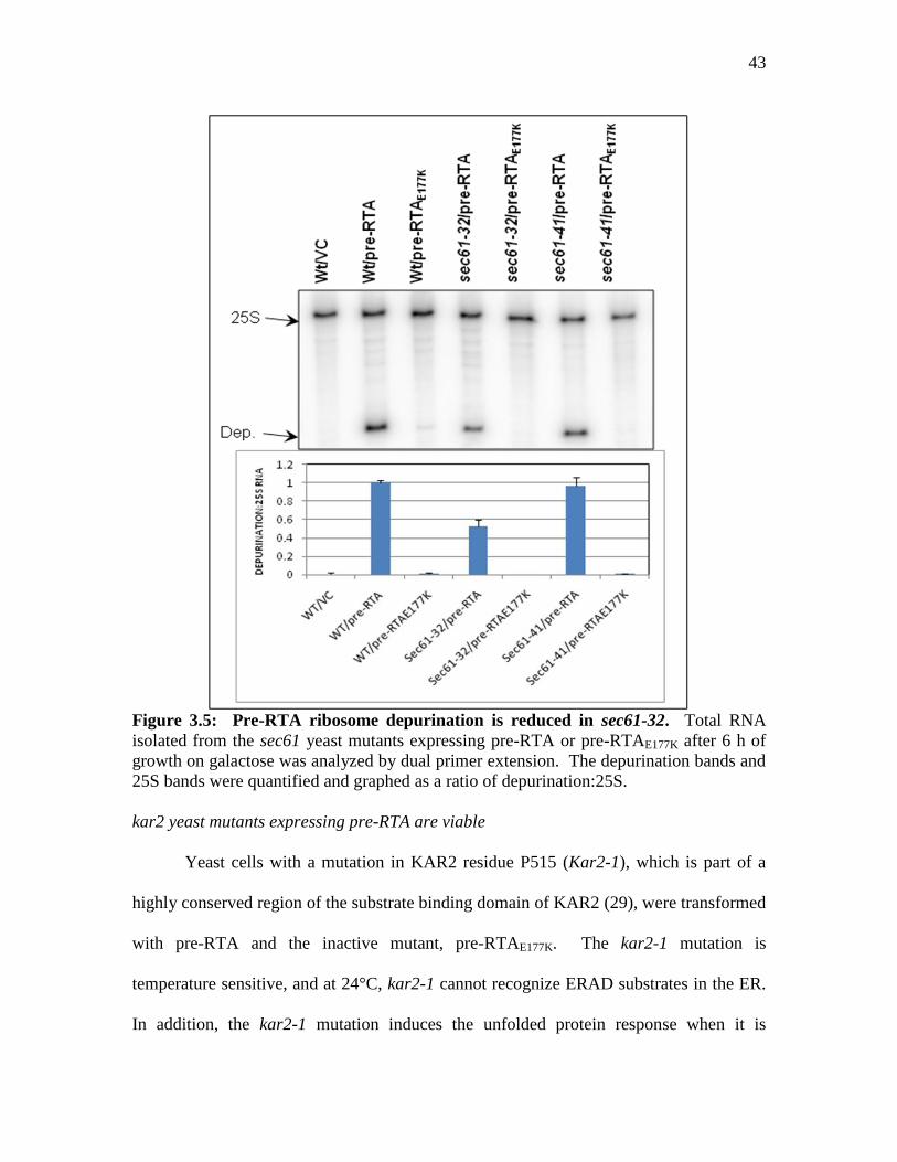
Figure 3.5: Pre-RTA ribosome depurination is reduced in Sec61-32…………………...43
Figure 3.6: Kar-2 yeast mutants are more viable than wildtype yeast when expressing
pre-RTA………………………………………………………………………………….44

vii
Figure 3.7: The expression pattern of pre-RTA and pre-RTAE177K is altered in Kar-2
yeast mutants……………………………………………………………………………..45
Figure 3.8: Pre-RTA can still depurinate Kar2-1 ribosomes……………………………46
Figure 3.9: Pre-RTA is stabilized in the Ubc7 mutant…………………………………..47
Figure 3.10: RAD23 yeast mutants expressing pre-RTA are viable……………………48
Figure 3.11: Pre-RTA expression is reduced in the RAD23 mutant……………………49
Figure 3.12: Pre-RTA depurination is reduced in RAD23 yeast mutants………………50
Figure 3.13: Pre-RTA is still toxic in the proteasome mutant…………………………..51
Figure 3.14: Pre-RTAE177K is stabilized in the proteasome mutant……………………...51
Figure 4.1: Structural comparison and sequence alignment of Shiga-toxin 2 (STX2),
Pokeweed Antiviral Protein (PAP) and Ricin A chain (RTA)…………………………..58
Figure 4.2: Viability analysis of pre-RTA C-terminal deletion and point mutations…...61
Figure 4.3: Immunoblot analysis of pre-RTA expressiona……………………………...62
Figure 4.4 Ribosome depurination in yeast expressing pre-RTA and the mutant forms..63
Figure 4.5: Pre-RTA mutants that don’t depurinate ribosomes can translate protein…..64
Figure 4.6: Viability analysis of mature RTA C-terminal deletion and point mutations..66
Figure 4.7: Immunoblot analysis of mature RTA expression…………………………...66
Figure 4.8: Ribosome depurination in yeast expressing RTA and the mutant forms…...67
Figure 4.9: Translation inhibition of RTA and the mutant forms……………………….67
Figure 4.10: A structural comparison of the C-terminal residues of RTA……………...69
Figure 4.11: A direct comparison of the deletion mutants in pre-RTA and RTA………72
Figure 4.12: A direct comparison of the point mutants in pre-RTA and RTA………….74
Figure 5.1: Model for ricin induced cell death………………………………………….82


1
CHAPTER 1: INTRODUCTION
HISTORY OF RICIN
Ricin has been recognized as a toxin since the 6th
century B.C., when castor beans
were used in Greek and Egyptian medicine. But it wasn’t until the late 1800’s when a
doctoral student in Estonia actually used the term ricin to describe the active protein that
caused the agglutination of erythrocytes treated with extracts from ricinus communis
seeds (1). Soon after, Paul Ehrlich used ricin and a related protein, abrin from the rosary
pea plant, to establish the fundamentals of immunology (1). He showed that injecting
small amounts of ricin into mice conferred immunity to the toxins. However, the
mechanism of ricin toxicity, structure and cellular entry was paid little attention until the
1970s. Sjur Olsnes was the first person to recognize the subunit composition of ricin and
its enzymatic action on ribosomes (1). In 1986, Yaeta Endo was able to show that the
target of ricin is the highly conserved stem-loop structure of the large ribosomal RNA
called the α-sarcin-ricin loop (SRL), and that ricin depurinates the SRL (2). Soon after,
Jon Robertus solved the crystal structure of ricin and suggested a mechanism for catalysis
(3). This mechanism consists of the enzymatic A-chain of ricin depurinating a specific
adenine residue from the large ribosomal subunit, rendering the ribosome unable to
translate proteins. Because of this activity, ricin belongs to a specific class of proteins
called Ribosome Inactivating Proteins (RIPs), which includes proteins from plants, such
as pokeweed antiviral protein (PAP) and bacteria, such as Shiga–like toxin (Stx) from E.
coli and Shiga toxin from Shigella.
2
RTA STRUCTURE
Ricin is a type II RIP, which means it is a heterodimer consisting of an enzymatic
A chain disulfide bound to a lectin binding B chain. Together, the A and B chains are
considered to be the ricin holotoxin. Other type II RIPs are Shiga toxin and Shiga-like
toxin isolated from Shigella dysenteriae and Escherichia coli, respectively, and abrin
from Abrus precatorius. Most type I RIPs are single chain enzymatic proteins, such as
PAP and saporin. These single chain proteins are similar to the A chain of a type II RIP,
and it has been suggested that the type II RIP evolved from a fusion between a type I RIP
and a lectin-binding protein (4). Because type I RIPs do not have a B-chain to bind to
cells and allow for optimal cellular uptake, they are considerably less toxic than type II
RIPs. The type III RIPs, such as those purified from maize and barley, are generated as a
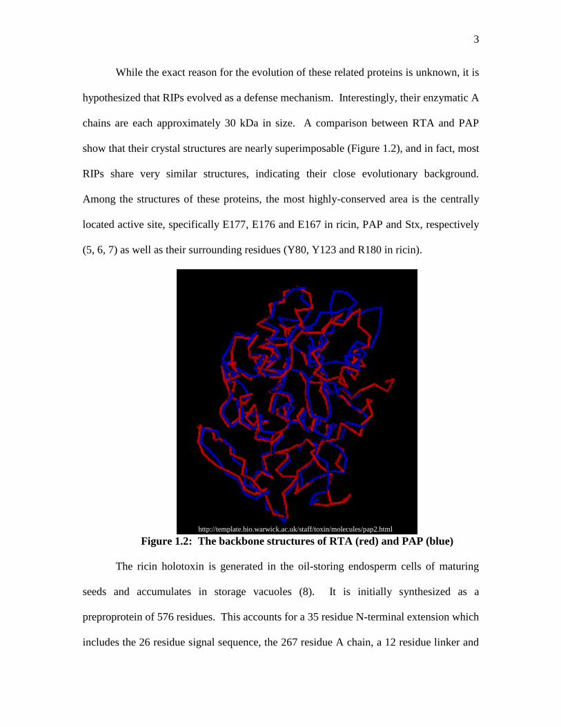
single chain protein, but must undergo proteolytic processing to become active. Figure
1.1 shows a schematic depicting the linear structures of the three types of RIPs.
Figure 1.1: Linear schematic of the three types of RIPs
3
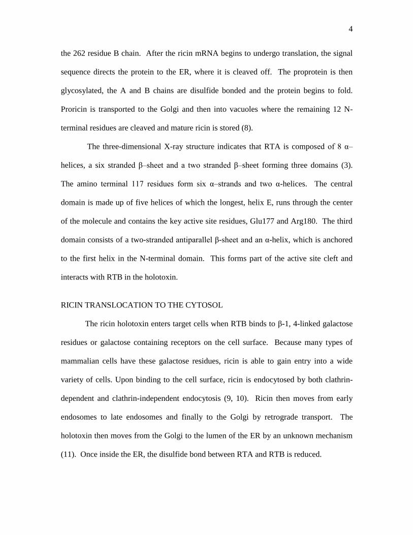
While the exact reason for the evolution of these related proteins is unknown, it is
hypothesized that RIPs evolved as a defense mechanism. Interestingly, their enzymatic A
chains are each approximately 30 kDa in size. A comparison between RTA and PAP
show that their crystal structures are nearly superimposable (Figure 1.2), and in fact, most
RIPs share very similar structures, indicating their close evolutionary background.
Among the structures of these proteins, the most highly-conserved area is the centrally
located active site, specifically E177, E176 and E167 in ricin, PAP and Stx, respectively
(5, 6, 7) as well as their surrounding residues (Y80, Y123 and R180 in ricin).
Figure 1.2: The backbone structures of RTA (red) and PAP (blue)
The ricin holotoxin is generated in the oil-storing endosperm cells of maturing
seeds and accumulates in storage vacuoles (8). It is initially synthesized as a
preproprotein of 576 residues. This accounts for a 35 residue N-terminal extension which
includes the 26 residue signal sequence, the 267 residue A chain, a 12 residue linker and
http://template.bio.warwick.ac.uk/staff/toxin/molecules/pap2.html
4
the 262 residue B chain. After the ricin mRNA begins to undergo translation, the signal
sequence directs the protein to the ER, where it is cleaved off. The proprotein is then
glycosylated, the A and B chains are disulfide bonded and the protein begins to fold.
Proricin is transported to the Golgi and then into vacuoles where the remaining 12 N-
terminal residues are cleaved and mature ricin is stored (8).
The three-dimensional X-ray structure indicates that RTA is composed of 8 α–
helices, a six stranded β–sheet and a two stranded β–sheet forming three domains (3).
The amino terminal 117 residues form six α–strands and two α-helices. The central
domain is made up of five helices of which the longest, helix E, runs through the center
of the molecule and contains the key active site residues, Glu177 and Arg180. The third
domain consists of a two-stranded antiparallel β-sheet and an α-helix, which is anchored
to the first helix in the N-terminal domain. This forms part of the active site cleft and
interacts with RTB in the holotoxin.
RICIN TRANSLOCATION TO THE CYTOSOL
The ricin holotoxin enters target cells when RTB binds to β-1, 4-linked galactose
residues or galactose containing receptors on the cell surface. Because many types of
mammalian cells have these galactose residues, ricin is able to gain entry into a wide
variety of cells. Upon binding to the cell surface, ricin is endocytosed by both clathrin-
dependent and clathrin-independent endocytosis (9, 10). Ricin then moves from early
endosomes to late endosomes and finally to the Golgi by retrograde transport. The
holotoxin then moves from the Golgi to the lumen of the ER by an unknown mechanism
(11). Once inside the ER, the disulfide bond between RTA and RTB is reduced.
5
Once RTA and RTB are separated, it is hypothesized that RTA is exported to the
cytosol by hijacking a quality control mechanism called the ER associated degradation
(ERAD) pathway (12). ERAD occurs when proteins are recognized as being misfolded
or non-native in the lumen of the ER. The aberrant protein is exported from the ER to the
cytosol where it undergoes ubiquitination and proteolytic degradation. The main protein
exporting channel in ERAD is the transmembrane Sec61 translocon, which consists of
the three proteins, SEC61, SSS1 and SBH1 (13). Together, these proteins form a channel
that allows co-translational protein import from active ribosomes, as well as protein
export into the cytosol. Sec61 is also responsible for retrotranslocation of misfolded
proteins out of the ER into the cytosol where they undergo degradation via the ubiquitin-
proteasome system (14).
In the ER lumen, resident chaperones assist with proper folding of proteins. A
prolonged association of a chaperone with an unfolded protein may target the protein for
ERAD (15). The Hsp70 chaperone, KAR2, is one of the primary chaperones responsible
for recognizing misfolded proteins and guiding them to the translocon (16). KAR2 may
also act as a luminal “gate” to the Sec61 translocon and will only open when ERAD
substrates are being exported. If KAR2 spends too much time attempting to fold an ER
protein, the protein will be sent to undergo further modifications, such as the breaking of
disulfide bonds and trimming of mannose residues, before being sent through the
translocon.
There are several possible paths that an ERAD substrate can follow once it is
translocated through the Sec61 channel. One pathway involves the ubiquitination of
substrates during export by ubiquitin conjugating enzymes, such as UBC6 or UBC7 (16),
6
which is subsequently followed by degradation in the proteasome. However, ubiquitin
conjugating enzymes attach ubiquitin residues to lysine residues of target proteins.
Interestingly, RTA only has two lysine residues. It has been reported that even though
RTA may be recognized as an ERAD substrate, its low number of lysine residues (2
lysines), may render it capable of avoiding ubiquitination, thereby avoiding proteolysis in
the proteasome (17).
In addition to ubiquitin tagging, ERAD substrates must be structurally modified in
order to undergo proteasomal degradation. To fit into the proteasome, sugar residues
which can result from glycosylation in the ER, must be cleaved. The cytosolic protein,
PNG1 is a deglycosylating enzyme. Normal ubiquitinated substrates are recognized by
another protein, RAD23, which has a ubiquitin-like domain and is also capable of
binding to PNG1 (18). RAD23 recognizes ubiquitinated substrates, and directs them to
Png1, where they are deglycosylated. RAD23 might then bring the deglycosylated
substrate to the proteasome for degradation (18).
There is also evidence for another pathway for ERAD substrate export which
involves the direct association of the proteasome with the Sec61 translocon (19). This
direct association may actually allow the proteasome to aid in the extraction of the
misfolded protein of the ER, and to undergo direct proteasomal degradation, without the
help of any other chaperones or factors.
RIBOSOME DEPURINATION
Once RTA reaches the cytosol, it enzymatically inactivates the large 28S subunit
of active ribosomes. Endo et al discovered the affected molecular site by all RIPs while
working with ricin in 1988 (2). They found that RIPs remove the first adenine from a
7
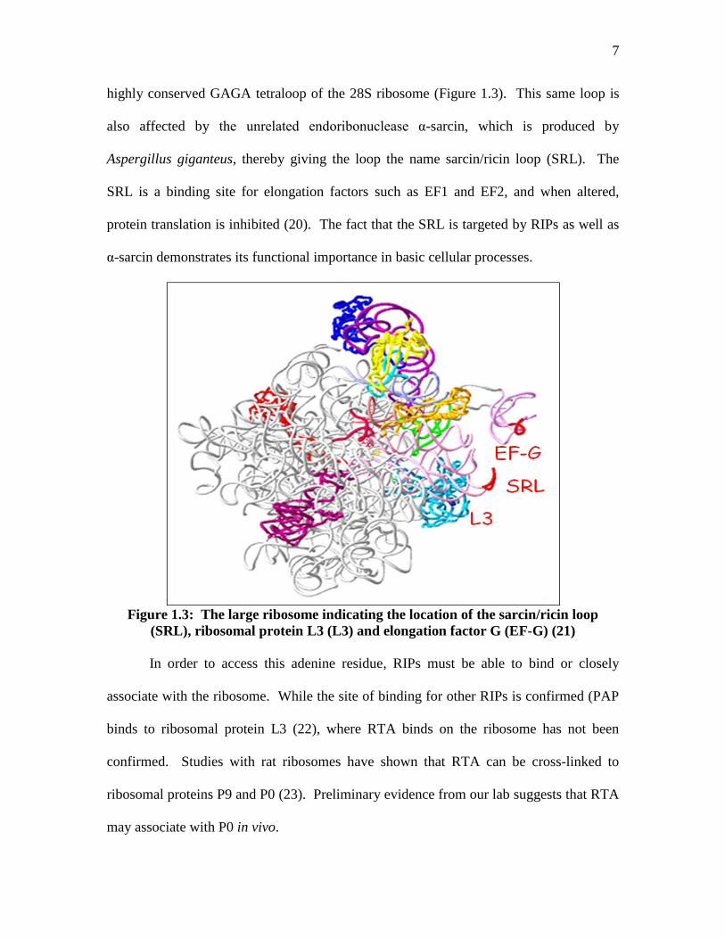
highly conserved GAGA tetraloop of the 28S ribosome (Figure 1.3). This same loop is
also affected by the unrelated endoribonuclease α-sarcin, which is produced by
Aspergillus giganteus, thereby giving the loop the name sarcin/ricin loop (SRL). The
SRL is a binding site for elongation factors such as EF1 and EF2, and when altered,
protein translation is inhibited (20). The fact that the SRL is targeted by RIPs as well as
α-sarcin demonstrates its functional importance in basic cellular processes.
Figure 1.3: The large ribosome indicating the location of the sarcin/ricin loop
(SRL), ribosomal protein L3 (L3) and elongation factor G (EF-G) (21)
In order to access this adenine residue, RIPs must be able to bind or closely
associate with the ribosome. While the site of binding for other RIPs is confirmed (PAP
binds to ribosomal protein L3 (22), where RTA binds on the ribosome has not been
confirmed. Studies with rat ribosomes have shown that RTA can be cross-linked to
ribosomal proteins P9 and P0 (23). Preliminary evidence from our lab suggests that RTA
may associate with P0 in vivo.

8
Once RTA is associated with the rRNA, it enzymatically attacks the adenine
residue by specifically cleaving the N-glycosidic bond between the adenine and the sugar
residue (Figure 1.4A) (2). This depurination event occurs at the active site in RTA, and is
conserved among all of the RIPs (2). It is expected that two tyrosine residues Tyr-80 and
Tyr-123 sandwich the adenine ring of the rRNA target. Once the adenine is held in
position, Arg-180 protonates the N-3 atom of the adenine, while Glu-177 stabilizes a
positive oxocarbonium transition state and interacts with the ribose (Figure 1.4B) (24).
Figure 1.4: A. The site of depurination in the SRL, and B. the structural model of
the active site of PAP and RTA with adenine (Parikh B, thesis 2004).
The ability of RIPs to inhibit protein synthesis was recognized in 1972 even
before ribosome depurination was discovered as the underlying mechanism (1). Initially,
the 60S ribosomal subunit was determined to be affected by ricin. Later, the binding site

9
for EF2 was specifically identified as being blocked by ricin activity (20), rendering the
ribosome unable to partake in the elongation step of protein translation. Later studies by
Sperti et al. also showed that the translocation step was specifically affected due to the
inhibition of GTP hydrolysis and GTPase activity of EF2 (20).
RICIN CYTOTOXICITY
While it seems logical that RIP toxicity is a result of ribosome depurination, there
is evidence to support that toxicity and depurination can be separated (25, 26). There are
possible alternative mechanisms leading to cytotoxicity in ricin, namely the inhibition of
the unfolded protein response (UPR) and ribotoxic stress.
UPR is a response mechanism that occurs when there are too many unfolded
proteins in the ER. As a result of the UPR, the folding capacity of the cell is increased,
the amount of new proteins translocated to the ER lumen is reduced and
retrotranslocation out of the ER and degradation of ER-localized proteins is increased
(27). Thus, UPR is closely linked with ER associated degradation (ERAD).
Generally, when a cell undergoes stress, the normal protein maturation process
will be affected, leading to an increase in the levels of unfolded proteins. To assist with
protein folding in yeast, KAR2 is recruited to help bind and fold these misfolded
substrates (29). KAR2 is also necessary for other cellular processes, such as gating of the
Sec61 channel. Cellular stress is indicated when the level of free KAR2 is low, which
slows down protein translation initiation to prevent the further accumulation of more
unfolded proteins (30). In addition, the recruitment of KAR2 to unfolded proteins may
initiate the UPR pathway. KAR2 has been shown to associate with the ER luminal
sensor domain of Ire1, which is a trans-membrane kinase that also has a transmembrane

10
domain, a cytoplasmic Ser/Thr kinase domain and an RNase L-like nuclease domain (29).
Ire1 recognizes the accumulation of unfolded proteins, possibly as a result of dissociation
of KAR2, and begins induction of the UPR. This entails Ire1 dimerization and activation,
and cleavage of the transcription factor HAC1 mRNA. Spliced HAC1 mRNA binds to
the unfolded protein response elements (UPRE) found in UPR-responsive genes, and
helps to enhance production of their proteins. One of these genes is KAR2, which is
upregulated to aid in proper folding of misfolded substrates. It has recently been reported
that RTA prevents splicing of HAC1 mRNA, which results in the inhibition of UPR (28).
This inhibition of the UPR may be responsible for ricin-induced cytotoxicity.
Ribotoxic stress is a pathway induced by events of cellular stress and is marked
by activation of cJun NH2
-terminal kinases (JNKs) (31). Activation of JNK leads to a
cascade of various kinase pathways that either lead to cell repair and recovery, or to
apoptosis. Exposure to anisomycin, an antibiotic that inhibits the eukaryotic peptidyl
transferase reaction in protein translation, is known to activate JNKs (31). While
inhibition of protein synthesis via anisomycin would logically be considered an event of
cellular stress, and thus an activator of ribotoxic stress, it has been shown that JNK
activation via anisomycin occurs even when protein synthesis is only partially affected.
This indicates that the ability of anisomycin to cause ribotoxic stress does not rely
entirely on its ability to inhibit protein synthesis (31). Because both RTA and
anisomycin damage the ribosome and inhibit translation, it is possible that, like
anisomycin, RTA induces ribotoxic stress.
Interestingly, IREI can also activate JNKs during the UPR (32). In addition,
PERK, a transmembrane protein kinase in the ER, is activated by events that occur during

11
ER stress, such as accumulation of misfolded proteins. PERK also phosphorylates eIF2α,
and when PERK is overexpressed in cells in vitro, it can inhibit protein translation (33),
which could lead to ribotoxic stress. Therefore, ricin toxicity may result from a
combination of UPR inhibition, ribotoxic stress and ribosome depurination.
RICIN POISONING
Exposure to ricin can occur via inhalation, ingestion or injection of the powdered,
crystallized or liquid forms of the toxin or, in the case of ingestion, consumption of the
castor beans (34). The amount of ricin that results in toxicity and the resulting symptoms
depends on the route of exposure. However, the epithelial cells of both the
gastrointestinal tract and the respiratory pathway are the primary targets of ricin
intoxication (34). The damage inflicted by RTA on the epithelial cells often results in a
clinical manifestation called vascular leak syndrome (VLS), which is characterized by
hypoalbuminemia and edema (35). VLS does not result from the inhibition of protein
synthesis of RTA, as VLS is observed several hours before protein synthesis inhibition
(35). Instead, in vitro evidence has shown that VLS activates apoptosis via caspase 3
(36), suggesting that death via ricin intoxication may be due to complications from the
VLS activity, and not ribosome depurination.
While inhalation and injection of ricin are considerably more lethal than
ingestion, the threat of ricin contamination to food, water and air supplies are most likely.
Reports have shown that ingestion of as little as one half of a castor bean resulted in
symptoms, and as few as two beans resulted in death (37). Symptoms begin within 4-10
hours of ingestion and are usually nonspecific, such as diarrhea, abdominal pain and
heartburn. 4-36 hours after ingestion more severe symptoms, such as liver dysfunction or

12
low blood pressure may occur, possibly resulting in death (37). Consumption of large
quantities of charcoal and cleansing of the GI tract with a cathartic may help to eliminate
ricin toxicity after ingestion (38).
The dissemination of aerosolized ricin is probably the most likely way for a terror
attack to occur. Ricin intoxication via inhalation is dependent on the size of the particles.
The smaller the particle, the more likely it will be inhaled and lodged deep into the
respiratory system (39). The lethal dose for mice for particles less than 5 µm in size was
determined to be 3-5 µg/kg of body weight. Animal studies in which ricin was inhaled
showed that inflammation and necrosis of cells in the airway and lungs occurred as a
result of ricin toxicity. While there are no current reports of ricin intoxication via
inhalation, past studies have indicated that symptoms occur 4-8 hours after inhalation of
ricin particles (34). It is expected that symptoms of ricin intoxication can occur up to 24
hours after inhalation, and that the primary cause of death is respiratory failure (34, 40).
The injection of ricin is the least likely method for a large scale terror attack, and
there are no current reports of ricin intoxication via injection. However, the lethal dose
for mice that were exposed to ricin parenterally was 0.7-2 µL/kg of body weight (41).
The symptoms of ricin intoxication occur 10-12 hours after injection and include fever,
nausea and abdominal pain. Tissue damage at the site of injection is sometimes present
(41, 42) and multisystem organ failure may eventually occur (34, 43).
13
RICIN TREATMENT
Development of a ricin vaccine would need to maintain the toxin’s active site, and
the active site of ricin is responsible for the damage to ribosomes and subsequent cell
death. Therefore, the generation of mutant forms of ricin that have a functional active
site, but do not result in cell death are ideal for vaccine development. While there is
currently no approved ricin vaccine, there are and have been some possible candidates.
Inactivated, formaldehyde treated ricin (toxoid) has been successfully used as
both an intranasal (44) and orally administered (45) vaccine in animal studies to prevent
ricin intoxication. However, there is a threat of ricin toxoid reverting back to the active
form. Deglycosylated RTA has also been administered in low concentrations to initiate
an antibody response in mice. Although successful production of neutralizing antibodies
relies on a mucosal adjuvant, which sometimes results in nasal inflammation, clinical
trials are currently undergoing investigation (46).
Passive immunity against ricin intoxication was achieved in mouse studies using
anti-ricin monoclonal IgG (47), but the use of a murine antibody to neutralize ricin in
humans will elicit a negative immune response. More recently, a chimeric ricin antibody
was generated by coupling murine antigen-binding domains to human constant domains
(48). This antibody shows promise for use with clinical studies.
Most recently, RiVax, a recombinant RTA vaccine is under development. RiVax
contains the point mutations Y80A, which affects substrate binding and V76M in RTA.
These mutations are proposed to affect both vascular leak syndrome and the ribotoxic
stress response (49), but not the depurination activity of RTA. Results from animal
studies show that administration of a concentration of 1-10 µg of RiVax via gavage or

14
aerosol was able to prevent ricin-induced tissue damage and death from a challenge of
10X the lethal dose (LD50) of ricin (49). Human trials were carried out in which
volunteers were vaccinated with varied concentrations of RiVax, and anti-RTA
antibodies and ricin-neutralizing antibodies were assayed (50). The main conclusion of
the study was that RiVax would protect humans against injection of 0.3-3.0 mg of ricin.
Further studies need to be conducted to determine the use of RiVax to prevent
intoxication of ricin inhalation or ingestion (50).
SUMMARY OF WORK TO BE PRESENTED
In order to understand the mechanisms of RTA toxicity, a yeast Saccharomyces
cerevisiae system was implemented and optimized because this system has been
successfully used to study mechanisms of cytotoxicity of another RIP, PAP. Although
the ultimate goal of ricin research is often to understand how to prevent and treat ricin
intoxication in humans, yeast is an excellent model to use to analyze the mechanisms of
cytotoxicity of ricin since the target of ricin, the SRL, is highly conserved between yeast,
plants and mammalian cells and because yeast can easily be genetically manipulated.
Yeast are eukaryotic and preliminary data from our lab and the laboratory of our
collaborators has shown that ricin behaves similarly in both mammalian and yeast cells.
Yeast also utilize the ERAD pathway, suggesting that yeast are an ideal model for
investigating the translocation of ricin through target cells. RTA is toxic to yeast cells
(Figure 1.5), and both yeast and animal ribosomes are depurinated by RTA (Figure 1.6A
and 1.7). The ERAD pathway in yeast has been characterized fairly well, and there is a
library of yeast ERAD mutants available for analysis. Therefore, the expression of RTA

15
in yeast may provide information for mechanisms of RTA-induced toxicity in
mammalian cells.
Figure 1.5: Yeast cells expressing RTA are not viable. Yeast cells expressing RTA,
the active site mutant, RTAE177K, or the empty vector were induced for 10 hours on SD-
Leu galactose media and were then plated as serial dilutions onto SD-Leu glucose plates.
Figure 1.6: Analysis of rRNA depurination of RTA and pre-RTA. A. Schematic
representation of dual-oligo primer extension assay. Two different end-labeled primers
16
(Depurination primer and 25S control primer) were annealed to rRNA and reverse-
transcribed. The resulting fragments represent the extension products that have stopped
prematurely at the depurination site and extension products that have stopped at the 5’
end of the 25S rRNA. B. The primer extension products for RTA and pre-RTA
representing the extent of depurination and the amount of total rRNA present at the
indicated times post induction (hours). C. The extent of depurination shown in B was
quantified by calculating the ratio of the depurination fragment to the 25S control
fragment and was expressed as a percentage.
Figure 1.7: 0.1 µg/mL of RTA is sufficient to depurinate animal ribosomes. Bovine
mammary epithelial cells were grown to confluence in 6-well plates. Prior to treatment,
cells were serum deprived for 16 hrs. Cells were treated with 0.1µg/ml or 1 µg/ml RTA
for 2, 4, 6, 8 and 10 hrs. Total RNA was isolated using Trizol (Invitrogen) and analyzed
by primer extension as described above using a depurination primer and a 28S control
primer. As a vehicle control, cells were treated with the same amount of glycerol as in
the 1 mg/ml treatment.
In the following chapters I will discuss new information regarding possible
mechanisms for the cytotoxicity of RTA. In chapter two I will discuss the results of
random mutagenesis of RTA, which demonstrate that RTA toxicity is not a direct result
17
of ribosome depurination and translation inhibition. Chapter three discusses how RTA
uses the ERAD machinery to reach the cytosol, without undergoing complete
proteosomal degradation. In the final chapter, I will discuss the results from site-directed
mutagenesis of the C-terminal end of RTA, and how this may provide information
regarding the driving force of RTA’s utilization of the ERAD pathway. In addition to
gaining a better understanding of RTA, there are several references and suggestions
regarding the mechanism of other RIPs, namely PAP and Shiga-like toxin from E. coli,
which will enhance general knowledge on the medical uses and potential treatments for
exposure to these toxins.
18
CHAPTER 2: Ribosome depurination is not sufficient for ricin-mediated cell death
in Saccharomyces cerevisiae
INTRODUCTION
Understanding the residues that are critical for RTA toxicity is essential for the
generation of ricin vaccines or antidotes. The search for additional critical residues has
previously been carried out by expressing mutated RTA in E. coli, followed by the
subsequent analysis of in vitro enzymatic activity. Other researchers have passaged the
plasmid containing mature RTA through an E. coli mutator strain and identified five
different mutations in residues at the active site cleft (5), including Glu177, Trp211,
Gly212 and Ser215, which are located on the same helix, and Ile252, which is located
close to the C-terminal end (5). Recently, another large scale mutagenesis screen was
conducted using error-prone PCR of mature RTA (51). Several new point mutations with
reduced toxicity were identified. While these studies identified several residues essential
for enzymatic activity, the correlation between ribosome depurination and cytotoxicity
had not been addressed. Here, we conducted a large-scale mutagenesis study of pre-RTA
in the yeast, Saccharomyces cerevisiae, and isolated RTA alleles based on their inability
to kill yeast cells. The nontoxic RTA alleles were characterized with respect to their
ability to depurinate ribosomes, inhibit translation and cause cell death. Several alleles
depurinated ribosomes and inhibited total translation to the same level as the wild type
RTA, but did not cause cell death. These results demonstrated that ribosome
depurination does not account entirely for the cytotoxicity of ricin.
19
RESULTS
Random Mutagenesis
The full length cDNA corresponding to pre-RTA, which consists of a 35 residue
N-terminal extension and the 267 residue coding sequence was cloned into the yeast
expression vector downstream of the GAL1 promoter, mutagenized using hydroxylamine
and transformed into yeast. Cells were plated on media containing glucose and replica
plated on galactose containing plates. Out of a total of 15,000 transformants screened,
128 (0.82%) were able to grow on galactose containing media, indicating resistance of
yeast to the mutated toxin. Immunoblot analysis showed that RTA expression was
detected in 87 (68%) out of 128 colonies. Of the 87 colonies that showed detectable
RTA expression, 37 expressed protein at the same molecular weight as wild type RTA,
and 50 expressed smaller variants of RTA. All 87 plasmids isolated were retransformed
into yeast to confirm that the resistance was due to the plasmid. Nucleotide sequence
analysis identified a total of 35 different mutations, which led to the loss of cytotoxicity
(Table 2.1). A majority of the mutations were isolated multiple times from different
plates, indicating that the mutagenesis screen was saturated. The mutants were divided
into three groups: Group I contains 16 different mutations with a premature termination
codon, resulting in a truncated form of the protein. Group II contained 9 different
frameshift mutations. In this group, the N-termini of the proteins were the same as
preRTA, but the C-termini were different depending on the position of the frameshift
mutation. Additional amino acids added to the C-termini before the stop codon are
indicated in Table 2.1. Group III consisted of 14 different point mutations, which
20
resulted in single amino acid changes in the protein. Only two mutants in this group
contained double point mutations. To determine which mutation was necessary for the
loss of cytotoxicity, single mutations were generated by site-directed mutagenesis.
Expression of RTA containing the single mutations corresponding to each double
mutation was toxic to yeast, indicating that the two different point mutations are required
simultaneously for the loss of cytotoxicity.
Table 2.1 shows the number of occurrence of the base pair changes, including the
silent mutations. As expected for hydroxylamine mutagenesis, C/G to T/A transitions
accounted for 80% of the total base pair changes. The frequency of other base pair
changes was relatively low. The frequency of the deletions or additions was around 12%.
Due to the high frequency of C to T changes, 11 out of 14 glutamines encoded by CAA/G
in pre-RTA were changed to stop codons (TAA/G) resulting in premature termination.
Random mutagenesis work was done by Xiao Ping Li.
21
Table 2.1: Characterization of pre-RTA mutants obtained by random mutagenesis
Protein change
No. of
occurrences Cytotoxicity
Depurination
(% of wt)
Translation
(% of vector)
Doubling
time (h)
pre-RTA Yes 100 35 18
Vector control No 2 100 6.3
Group I
Q19 stop 1 No 5 ND ND
Q55 stop 2 No 1 ND ND
Q112 stop 2 No 1 ND ND
Q128 stop 3 No 1 ND ND
G140 stop 1 No 1 ND ND
S149 stop 1 No 2 ND ND
Q160 stop 3 No 12.3 ND ND
Q173 stop 3 No 4.3 ND ND
S176 stop 2 No 4.5 ND ND
Q182 stop 2 No 3.4 ND ND
W211 stop 4 No 3.7 ND ND
Q219 stop 2 No 7.9 ND ND
Q223 stop 3 No 4.3 ND ND
Q231 stop 3 No 9.6 60 9
Q233 stop 6 No 6.7 59 8.7
L248 stop 2 No 15.8 58 7
Group II
T77P + 4a 1 No 0.4 ND ND
Y84T + 48 1 No 5.6 ND ND
F92S + 40 1 No 2.4 ND ND
R114D + 18 1 No 2.8 ND ND
P202L + 1 2 No 2.3 ND ND
R213D + 31 1 No 2.8 ND ND
R213D +31c 1 No 3.2 ND ND
S215F + 6 1 No 5.5 ND ND
P250L + 1 1 No 5.1 ND ND
Group III
G83D 6 No 41 62 12
G140R 2 No 5 93 9.1
A147P 3 No 33 69 10
E177K 3 No 5.6 73 9.8
Δ I 184 1 No 8.2 69 9
E208K 2 No 29 58 10
G212E 9 No 19 88 6.9
S215F 2 No 110 32 15
P95L-E145K 1 No 115 41 10
P95L (by PCR) Yes 149 34 26
E145K (by PCR) Yes 108 27 18
P250L-A253V 1 No 5.2 100 7.7
P250L (by PCR) Yes 158 31 20
A253V (by PCR) Yes 175 30 24

22
aThe numbers after the mutations indicate the numbers of amino acids added to the C termini before a stop codon is
generated. bND, not determined. cThe amino acids added to the C terminus are different from those added for the mutation listed immediately above
(26).
Wild type pre-RTA and the nontoxic mutants are expressed in yeast
Immunoblot analysis using polyclonal antibodies against RTA was used to
examine protein expression in each mutant 6 h post-induction. As shown in Figure 2.1,
mature RTA standard from Ricinus communis (Sigma) contained two bands, possibly due
to different levels of glycosylation. Protein isolated from yeast harboring the pre-RTA
plasmid contained two bands that co-migrated with the mature form of RTA (Figure 2.1
first two lanes), indicating that pre-RTA synthesized in yeast is processed the same way
as RTA in plants.
Immunoblot analysis indicated that all 39 mutants that contained the premature
termination codons (Figure 2.1A), the frameshift mutations (Figure 2.1B) or the point
mutations (Figures 2.1C and 2.1D) expressed detectable levels of RTA. The blot was
reprobed with antibody against dolichol-phosphate mannose synthase (Dpm1p) as a
loading control. The mutant proteins migrated on SDS-PAGE according to their size,
except for the double mutant, P250L-A253V, which contained larger and smaller bands,
indicating possible effects on protein aggregation and breakdown. Analysis of the single
mutations corresponding to this double mutant indicated that P250L mutation contributed
to the observed effects. In general, yeast cells carrying the non-toxic mutations expressed
higher levels of RTA than cells carrying wild type or toxic forms (P95L) of pre-RTA.
These results demonstrated that the loss of cytotoxicity of the mutant alleles was not due
to the loss of protein expression. Viability assays were done by Xiao Ping Li.

23
Figure 2.1: The pre-RTA mutants are expressed in yeast. Membrane fractions (15
μg) isolated from cells expressing pre-RTA or mutants containing a premature
termination codon (A), a frameshift mutation (B), a single point mutation (C), or a double
point mutation (D) were separated on a 12% SDS-polyacrylamide gel and probed with
polyclonal anti-RTA (1:3,000). The RTA standard (1.5 ng) was purified RTA. The blots
were stripped and probed with the ER membrane marker Dpm1p as a loading control
(work done by Xiao Ping Li, 26).
Pre-RTA mutants are not toxic to yeast cells
Irreversible growth inhibition was examined by conducting viability assays. As
shown in Figure 2.2, cells were plated on glucose plates after induction on galactose for
the indicated times. The top panel shows the yeast cells harboring either the wild type
pre-RTA plasmid or the empty vector. Upon induction in yeast, the wild type RTA
reduced the viability of cells by almost 3 logs at 12 h. In contrast, the nontoxic RTA
mutants exhibited minimal loss of viability at 10 hours post induction (Figure 2.2). All
nontoxic mutants analyzed exhibited similar viability as the cells harboring the empty

24
vector. Only L248* in group I and P250L+S in group II, are shown because they had the
shortest deletion at their C-termini. The two double mutants, P95L-E145K and P250L-
A253V were nontoxic and did not reduce viability. However, the single mutations
corresponding to each double mutation (P95L, E145K and P250L, A253V) were toxic
(Table 2.1 and Figure 2.2) and reduced the viability of yeast cells. Protein expression
analysis was done by Xiao Ping Li.
Figure 2.2: Viability of cells expressing pre-RTA and the mutant forms of RTA.
Yeast cells were first grown in SD-Leu medium supplemented with 2% glucose to an
optical density at 600 nm of 0.3 and then transferred to SD-Leu supplemented with 2%
galactose. At the indicated hours postinduction on SD-Leu medium containing galactose
(left), serial dilutions were spotted on SD-Leu plates supplemented with 2% glucose. The
top two panels show the cell viability up to 12 h in cells expressing the wild-type pre-
RTA or harboring the empty vector (work done by Xiao Ping Li, 26).
Nontoxic RTA mutants depurinate rRNA
To determine if the reduced toxicity of the pre-RTA mutants was due to reduced
depurination of ribosomes, total RNA was isolated from each mutant and depurination of
the rRNA was examined by dual primer extension at 6 h post induction. As shown in
Figure 2.3, ribosomes were depurinated in cells expressing pre-RTA. Proteins that were
truncated at their C-terminal end, Q231*, Q233*, and L248* showed a very weak
depurination band, indicating that these mutants retained a low level of depurination
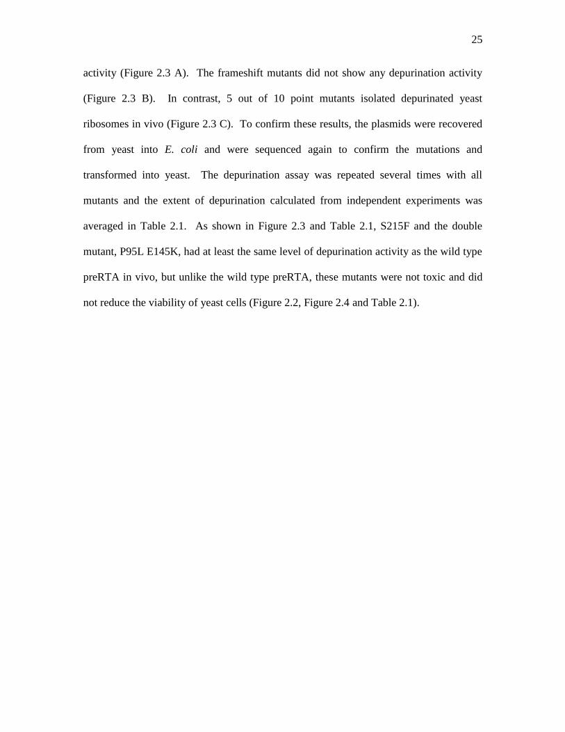
25
activity (Figure 2.3 A). The frameshift mutants did not show any depurination activity
(Figure 2.3 B). In contrast, 5 out of 10 point mutants isolated depurinated yeast
ribosomes in vivo (Figure 2.3 C). To confirm these results, the plasmids were recovered
from yeast into E. coli and were sequenced again to confirm the mutations and
transformed into yeast. The depurination assay was repeated several times with all
mutants and the extent of depurination calculated from independent experiments was
averaged in Table 2.1. As shown in Figure 2.3 and Table 2.1, S215F and the double
mutant, P95L E145K, had at least the same level of depurination activity as the wild type
preRTA in vivo, but unlike the wild type preRTA, these mutants were not toxic and did
not reduce the viability of yeast cells (Figure 2.2, Figure 2.4 and Table 2.1).
26
Figure 2.3: Ribosome depurination in yeast expressing pre-RTA and the mutant
forms in vivo. Total RNA isolated after 6 h of growth on galactose was analyzed by dual
primer extension. Primer extension analysis of the mutants with a change corresponding
to a premature termination codon (A), a frameshift mutation (B), or a point mutation (C)
is shown.
To determine if the mutant proteins were enzymatically active in vitro, we
extracted the mutant proteins from yeast and examined ribosome depurination after
treating purified yeast ribosomes with the wild type and the mutant proteins in vitro. As

27
shown in Figure 2.4, the wild type pre-RTA extracted from yeast depurinated yeast
ribosomes in vitro. The S215F and the double mutant, P95L-E145K, depurinated yeast
ribosomes in vitro, while P250L-A253V was not able to depurinate ribosomes in vitro.
The in vivo depurination results were the same as those obtained with proteins isolated
from yeast in vitro and demonstrated that S215F and P95L-E145K are catalytically
active, while P250L-A253V is not.
Figure 2.4: Ribosome depurination by pre-RTA and mutants in vitro. (A) Total
protein extracted from the cytosolic fraction of 10 ml of yeast cells expressing pre-RTA
or the mutants was analyzed on a 12% SDS-polyacrylamide gel and probed with
polyclonal anti-RTA (1:3,000). The first lane is purified RTA standard (10 ng). (B)
Ribosomes isolated from yeast cells were treated with either wild-type pre-RTA or the
S215F, P95L-E145K, and P250L-A253V mutants extracted from the cytosolic fractions
of yeast cells in vitro, and the extents of depurination were determined by dual primer
extension analysis. The first lane corresponds to the untreated ribosomes, and the second
lane corresponds to primer extension analysis with protein extracted from cells harboring
the empty vector. (C) The extents of ribosome depurination were quantified using a
PhosphorImager from three independent depurination experiments with the wild-type and
mutant proteins extracted from yeast in vitro.

28
Ribosome depurination results in translation inhibition
To determine if ribosome depurination correlated with translation inhibition, we
examined total translation in cells expressing pre-RTA by [35
S]-methionine incorporation.
As shown in Table 2.1, total translation was reduced to 35% in cells expressing the wild
type pre-RTA compared to total translation in cells harboring the empty vector. The wild
type pre-RTA did not inhibit translation completely, indicating that some translation still
occurs in the presence of RTA. Total translation was not inhibited in yeast expressing the
mutants which did not depurinate ribosomes. In contrast, total translation was inhibited
in S215F and in the double mutant, P95L E145K, which depurinated ribosomes (Table
2.1). These results demonstrated that translation inhibition correlated well with the extent
of depurination, indicating that depurinated ribosomes are unable to translate protein.
DISCUSSION
Here, we conducted large-scale mutagenesis of pre-RTA in the yeast,
Saccharomyces cerevisiae, and isolated nontoxic RTA mutants on the basis of their
inability to kill yeast cells. The pre-RTA instead of the mature RTA was used for
mutagenesis to isolate mutants defective in intracellular trafficking, protein folding,
stability and interaction with ribosomes. In a recent study using PCR-based mutagenesis
of the mature RTA gene, 80% of the changes observed were T to C and A to G
transitions (51). In contrast, 80% of the changes observed in our study using chemical
mutagenesis were either C to T or G to A transitions. The PCR-based mutagenesis and
the chemical mutagenesis complement one another and generate a wide array of useful
mutations. However, they each have their own limitations. It is difficult to generate only
single mutations in the in the PCR-based method. Multiple mutations are often obtained

29
and single mutations must then be generated to identify the mutation responsible for the
phenotype. In contrast, only 2 of the 35 mutants generated in our study using
hydroxylamine contained double mutations (Table 2.1).
The mutants isolated here were first screened for the loss of cytotoxicity and then
by protein expression. Mutants that survived when RTA was induced were characterized
for expression, and only those that expressed detectable levels of RTA were further
characterized by nucleotide sequence analysis. The sequencing data correlated very well
with the molecular weight of each protein. The RTA-specific antibody generated using
the mature RTA as an antigen was able to recognize very small RTA peptides, including
an 18 amino acid N-terminal peptide with a molecular weight of 5.8 kDa (Q19 stop) (data
not shown). Immunoblot analysis indicated that the nontoxic mutant forms of RTA were
expressed at higher levels than the wild type or the toxic forms of RTA (Figure 2.1).
Of the nine frameshift mutations isolated, seven of them were caused by a single
base pair deletion and two of them had two base pair deletions (Table 2.1). These nine
frameshift mutations were isolated only once. The twenty-five mutations with stop
codons or single amino acid changes were caused by single base pair changes. Most of
these mutations were isolated more than twice, and some were isolated nine times from
different plates, indicating that the mutation screen was saturated. Furthermore, eleven
out of the fourteen glutamines in pre-RTA were changed to stop codons, providing
further evidence that the mutagenesis screen was saturated. Mutations were not isolated
in three glutamines, Gln5, Gln98 and Gln266. If Gln5 were changed to a stop codon, the
resulting four amino acid peptide would not have been detected by immunoblot analysis.
If Gln266 were changed to a stop codon, RTA would be toxic (52) and would not be
30
isolated by our screen. Therefore, the only mutation we did not isolate is Gln98 changed
to a stop codon.
The first 26 amino acids of the 35 amino acid N-terminal extension of pre-RTA
represent the signal sequence. Mature RTA does not have the signal sequence, and
therefore, it does not enter the ER. Despite this, mature RTA is toxic to yeast, as shown
in figure 1.5. Hydroxylamine treatment did not result in any mutations in the N-terminal
extension of pre-RTA, suggesting that these mutations did not affect the toxicity of RTA.
Even if a mutation in the N-terminal extension had occurred, it might disrupt the ability
of preRTA to translocate into the ER without affecting its cytotoxicity, since expression
of the mature RTA is toxic to yeast (51). Similarly, mutations were not recovered at
Asn10 and Asn236, which are glycosylated in the mature RTA. These results provided
further evidence that glycosylation does not affect the toxicity of RTA (53).
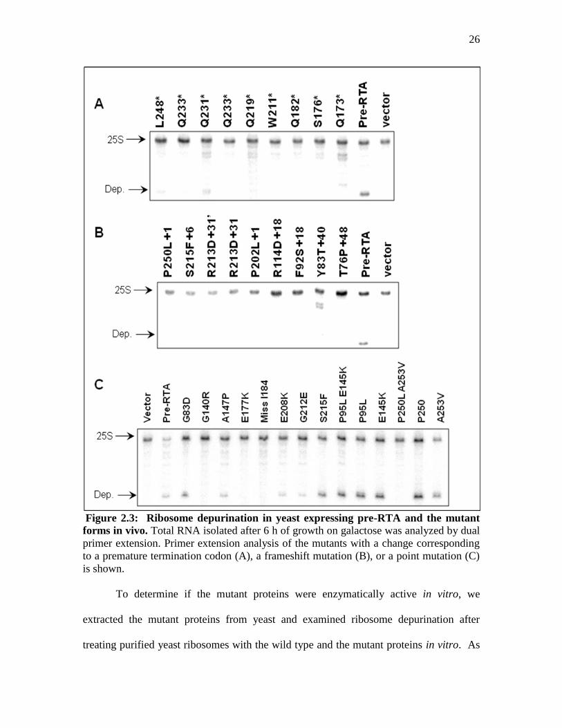
The results from three separate random mutagenesis studies and several
systematic deletion experiments, indicate that there are five regions important for the
function of RTA: β strand D, α helix D, E, G-H, and a hydrogen-bonded turn and β
strand region (Ile249 to Val256) close to the C-terminal end of the protein (Figure 2.5)
(51, 5, 52, 54). The α helix E contains the active site residues, Glu177 and Arg180. The
E177K mutation was isolated several times in different studies (51, 5). Mutations in
Arg180, such as R180G (51) and Ile184 (ΔI184) (54) at the beginning of helix F
disrupted the enzymatic activity of RTA in vitro, emphasizing the critical nature of this
region (Table 2.1). In our study, deletion of Ile184 led to loss of cytotoxicity and a
significant reduction in ribosome depurination activity of RTA in vivo (Table 2.1).

31
Ile184 may be critical for enzymatic activity, since it contacts Phe181 and methylene
carbons of Glu177, stabilizing the active center (54).
Figure 2.5: Three-dimensional structure of mature RTA showing the positions of
the point mutations and the α-helices and β-sheets that contain these mutations. Coordinates of the crystal structure from the Protein Data Bank 1J1M were used in
conjunction with the Protein Explorer software to create this figure. The point mutations
are shown in blue. The active site mutation is shown in black. The double mutations are
shown in green and cyan.
The alpha helices G to H have been the target of many different mutations,
including those at Leu207, Glu208, Trp211, Gly212, Leu214 and Ser215 (55, 56, 54).
Mutations in this region did not eliminate the depurination activity completely but they
reduced the cytotoxicity. A mutation at Glu208 (E208K) reduced, but did not completely

32
eliminate the depurination activity of RTA (Table 2.1). Previous studies have shown that
Glu208, which is at the bottom of the active site cleft can substitute for Glu177 in the
E177A mutant (55). The E208D mutant with no change at position 177 had in vitro
enzymatic activity equal to the wild type protein (54), indicating that Glu208 by itself
does not play a major role in depurination.
The point mutation at Ser215 (S215F) in helix H, did not affect ribosome
depurination in vivo, but significantly decreased the cytotoxicity of RTA (Table 2.1 and
Figure 2.3). This mutant was enzymatically active in vitro (Figure 2.4). Previous studies
showed that Ser215 can be deleted from RTA without complete loss of activity (56).
Since S215F mutation led to loss of cytotoxicity without affecting ribosome depurination,
the role of Ser215 in cytotoxicity can be separated from ribosome depurination. A point
mutation in Gly212 in helix H (G212E) significantly reduced the depurination activity in
vivo. Deletion of Gly212 led to loss of enzymatic activity of RTA in vitro (54). These
results indicated that Gly212 in helix H is critical for ribosome depurination.
The α helix D crosses helix E in the middle (Figure 2.5). Each of the amino acids
in helix D could be deleted, provided that the deletion does not disrupt the amphipathicity
of the helix (57). Deletion of Ala147 in helix D abolished the activity of RTA in vitro,
since the hydrophobic surface of helix D protects the helix E from solvent, further
stabilizing the active center (54). The point mutation A147P reduced the depurination
activity of RTA and led to loss of its cytotoxicity (Table 2.1). The A147P mutation likely
disrupted the structure of helix D in the middle, destabilizing the active site. The point
mutation at Gly140 (G140R), which is located at the beginning of helix D, resulted in the
loss of both cytotoxicity and depurination (Table 2.1 and Figure 2.3). However, deletion
33
of this glycine did not affect the activity of RTA in vitro (54). These results indicated
that the structure of RTA might be affected more when Gly140 is changed to an arginine
than when it was deleted.
Mutation G83D (NT1031), which is in β strand D, eliminated the cytotoxicity of
RTA in yeast cells and reduced its depurination activity (Figure 2.3). However, the
G83D mutation did not completely eliminate the depurination activity of RTA in vivo.
Since Gly83 is relatively distant from the active site, it is unlikely that Gly83 participates
in the catalysis. Previous studies indicated that RTA lost its depurination activity when
Gly83 was deleted (52, 54). These results suggested that β strand D might be important
for the interaction of RTA with the ribosome, such that a mutation in this residue may
affect binding of RTA to the ribosome. A point mutation in the corresponding Gly in
pokeweed antiviral protein (PAP) (G75D) led to loss of depurination in vivo (25) and
affected binding of PAP to ribosomes (58). In contrast to PAP, Gly83 in ricin is not
sufficient for ribosome binding, since the G83D mutant retains some depurination
activity in vivo.
The final important region is close to the C-terminal end of RTA. Stop codon
mutations demonstrated that deleting 20 (L248 stop) amino acids from the C-terminal end
of pre-RTA eliminated its cytotoxicity in yeast. The last frame shift mutation, P250L+S,
which deleted 17 amino acids from the C-terminus and changed Pro250 to Leu,
eliminated the depurination activity (Table I). Deletions from R258 to P262 or P263 to
F267 did not affect cytotoxicity (52). However, mutations upstream of Arg258, at
Ile252, Leu254 and Val256 eliminated the cytotoxicity of RTA (51, 5). The single
mutations at Pro250 (P250L) and at Ala253 (A253V) had little effect on the cytotoxicity
34
of RTA or its ability to depurinate ribosomes. However, when they were combined
(P250L-A253V), both cytotoxicity and ribosome depurination were eliminated. These
results indicated that the C-terminal region of RTA is critical for ribosome depurination
and cytotoxicity.
Sequence alignment analysis between RTA, a type II RIP, with mature PAP, a
type I RIP, demonstrated only 30% identity. Many mutations which led to loss of
cytotoxicity were in the residues which were conserved between RTA and PAP,
indicating that these residues were critical for RIP activity. A majority of residues which
are invariant among RIPs play an important role in the depurination reaction. The rest
contribute to overall structure of the enzyme or may be critical for intracellular
trafficking. Ricin has to enter the cytosol to depurinate ribosomes. Some bacterial toxins
form pores in membranes. Ricin does not form pores in membranes, but may enter the
cytosol from the ER using the Sec61 protein translocon (see Chapter 3). We have
previously demonstrated that the C-terminal sequence of RTA is fairly homomlogous
with the C-terminal sequence of PAP, which is critical for its transport into the cytosol
(59). Since the point mutations in P250L-A253V correspond to this region, the double
mutant may be unable to retrotranslocate from the ER into the cytosol. Further evidence
for this is provided by the accumulation of larger forms of the protein in the ER in this
mutant (Figure 2.1).
Although single mutations at Pro95 (P95L) and Glu145 (E145K) did not reduce
cytotoxicity, the double mutant, P95L-E145K, was not toxic to yeast cells. The double
mutant depurinated ribosomes at wild type levels (Table 2.1 and Figure 2.3), indicating
that Pro95 and Glu145 were critical for cytotoxicity, but not for ribosome depurination
35
activity. These results and previous mutagenesis studies indicated that in some cases two
amino acids must be changed simultaneously to eliminate the cytotoxicity of RTA (5).
In these mutants, the first amino acid was usually located in a β strand region and the
second amino acid was located in a α-helix region (L62-L129, L74-L139/T159/R193 (5)
and P95-E145 in this study). The X-ray crystal structure indicated that the mutated
residues in different regions of RTA do not interact with each other (Figure 2.5). Further
studies will address the role of this mutant in the cytotoxicity of RTA.
Different methods for random mutagenesis have been used to isolate nontoxic
RTA mutants in yeast cells (51, 5). Systematic deletion analysis has also been used to
identify amino acids critical for the activity of RTA (54). We present the cytotoxicity and
depurination data together and provide the first evidence that cytotoxicity of RTA is not
entirely due to ribosome depurination.

36
CHAPTER 3: Ricin A chain utilizes the ERAD machinery to reach the cytosol
INTRODUCTION
The ricin holotoxin consists of a lectin binding B-chain and a ribosome
depurinating A-chain. Upon binding to N-acetyl-galactosamine residues on target cells,
ricin is taken up by endocytosis and transported to the Golgi complex. Ricin then
undergoes retrograde transport to the ER where the disulfide bond between the A and B
chains is reduced. RTA unfolds when the A and B chains separate allowing it to pass
through the ER membrane to reach the cytosol. It must then refold to become active and
depurinate ribosomes (11).
In order to reach the cytosol from the ER, RTA may utilize the Endoplasmic
Reticulum-Associated Degradation (ERAD) pathway. The ERAD pathway is a quality
control system that ensures that native proteins have been folded into their proper
conformation before leaving the ER. If a protein is detected as misfolded or incomplete,
it is removed from the ER and destroyed via the cytoplasmic ubiquitin-proteasome
pathway (60). There are many proteins involved in the transport of RTA via the ERAD
and proteasome pathway.
The Sec61translocon, which includes the proteins SEC61, SSS1 and SBH1 in
yeast, is a protein conducting channel in the ER membrane (13). Sec61 is the primary
export channel for ERAD and as such, is used to transport misfolded proteins from the
ER to the tightly-associated cytosolic proteasomes where the misfolded proteins are
degraded. In addition to protein export, SEC61 allows for co-translational protein import
into the ER. Proteins destined to reach the ER must have an N-terminal signal which is
recognized by the signal recognition particle (SRP) (30). Once this signal sequence is
37
translated, the actively translating ribosome must line up with the Sec61 channel (19) so
that the protein may enter the Sec61 channel while it is unfolded.
The formation and function of the Sec61 complex in yeast relies on SEC63 (61),
which is another integral membrane protein. The Sec63 complex is required for post-
translational import of proteins into the ER and does not require the SRP. SEC63
contains a DNAJ-like domain that anchors an Hsp70 chaperone, KAR2, to the
translocation channel (62). KAR2 is responsible for recognizing unfolded proteins in the
ER and helping to fold them properly, and is also expected to help facilitate ERAD in
yeast. KAR2 is also essential for the formation of a lumenal seal during protein import
into the Sec61 channel. Together, SEC63 and KAR2 allow post-translational
translocation of proteins into the ER lumen (62), while SEC63, KAR2 and SEC61 are all
necessary (61) for co-translational translocation.
Proteins that are destined to be degraded by the proteasome are ubiquitinated in
the cytosol. Ubiquitins are attached to lysine residues of proteasome substrates via
ubiquitin-conjugating enzymes (ubc’s). Previous studies with ubc deletion mutants
ubc6Δ, ubc7Δ and the double deletion mutant ubc6Δ/ubc7Δ demonstrated that the
absence of these enzymes retards the ability of the cell to degrade proteasomal substrates
(63). Reports have demonstrated, however, that ricin is not ubiquitinated due to the low
number of lysine residues (17).
Before the ERAD substrate can be sent to the proteasome, modifications often
occur. One of these modifications is deglycosylation. Long carbohydrate chains are too
bulky for the proteasome (64) and are often removed in the cytosol with an N-glycanase
called PNG1. PNG1 recognizes and cleaves glycosyl chains. However, in order to get

38
the glycosylated substrate to PNG1, RAD23 is needed. RAD23 has an N-terminal
ubiquitin-like domain that is recognized by the proteasome. It also has a ubiquitin
recognizing domain, and is capable of associating with PNG1. It is suspected that
RAD23 recognizes and binds to ubiquitin residues of proteasome substrates, and then
associates with PNG1, which subsequently cleaves the glycosyl groups. RAD23 then
helps to facilitate the transfer of the ubiquitinated substrate to the proteasome via its
ubiquitin-like domain.
If the pathway from the ER to the proteasome breaks down at any point, there is
an opportunity for the ERAD substrate to reach the cytosol in its native and possibly
active form. In this study, pre-RTA and the active site mutant pre-RTAE177K were
transformed into yeast with mutations in various components of the ERAD pathway.
There is evidence suggesting that RTA uses Sec61 to enter the cytosol (11). However,
there has been little evidence to support that RTA utilizes other proteins involved in
ERAD. An initial screen was conducted in many ERAD mutants and those mutants that
demonstrated either resistance to RTA or increased toxicity of RTA were analyzed
further, specifically sec61, kar2, rad23Δ, ubc7Δ and pre1-1, pre2-2. The stabilization or
decrease in ricin toxicity that was observed in theses mutants indicates that RTA does use
the ERAD pathway to reach the cytosol.
RESULTS
The cytotoxicity of pre-RTA is reduced in sec61 mutants
To determine if the Sec61 translocon is necessary for RTA cytotoxicity, yeast
with a mutation in the ER luminal regions of the third and fourth transmembrane helices
of SEC61 were transformed with pre-RTA or pre-RTAE177K, or the mature RTA and

39
RTAE177K (Figure 3.1 B) (65). These yeast mutants are temperature sensitive and when
grown at the restrictive temperature, the ability of SEC61 to export proteins from the ER
to the cytosol is inhibited, while the import function is not. Pre-RTA or RTA were
cloned into a vector with a V5 epitope tag (pYES) in order to detect expression. After
transformation, yeast colonies were streaked on both SD-Leu –Ura media containing
glucose and SD-Leu –Ura media containing galactose (Figure 3.1 B). Sec61-32 and
sec61-41 were able to survive better than wildtype yeast when transformed with pre-
RTA, although sec61-32 cells seemed more resistant to pre-RTA than sec61-41. These
results show that the effect of pre-RTA cytotoxicity was reduced in these Sec61 mutants,
and that a properly functioning Sec61 translocon is necessary for the cytotoxicity of pre-
RTA. In addition, viability assays were conducted as previously described, and the
sec61-32 and sec61-41 yeast cells expressing pre-RTA were more viable than wildtype
cells expressing pre-RTA (Figure 3.2).
Figure 3.1: The sec61-32 and sec61-41 yeast mutants reduce the cytotoxicity of pre-
RTA. A. The sec61-32 and sec61-41 mutations are located on the lumenal side of
transmembrane domains 3 and 4, and impair protein export from the ER to the cytosol
(65). B. Yeast sec61-32 and sec61-41 mutants expressing pre-RTA were streaked onto
SD-Leu plates containing galactose.

40
Figure 3.2: sec61-32 and sec61-41 yeast expressing pre-RTA are viable compared to
wildtype yeast. Yeast cells expressing pre-RTA or pre-RTAE177K were induced for 10
and 24 hours on SD-Leu galactose media and were then plated as serial dilutions onto
SD-Leu glucose plates.
Pre-RTA is stabilized in sec61 mutants
In order to confirm that the reduction in toxicity of the Sec61 mutants was not due
to a reduction in pre-RTA expression, immunoblot analysis was conducted. The
transformed yeast cells were grown in SD-Leu –Ura liquid media containing glucose
until they reached a cell density of approximately OD600 0.3. The cells were then
transferred to SD-Leu –Ura containing galactose media. Aliquots of the cells were taken
at 4, 6, 10 and 24 hours post-induction. The cells were lysed using a low salt buffer and
the cellular components were fractionated into membrane and cytosolic proteins. The

41
membrane proteins were then run on a 15% SDS-PAGE gel, transferred to nitrocellulose
membranes and probed with α-V5 antibodies.
The amount of pre-RTA and pre-RTAE177K in the ER membrane fraction of
wildtype yeast cells is destabilized over time, and at 24 hours post-induction there is very
little protein associated with the ER (Figure 3.3). In contrast, in the Sec61 mutant strains,
pre-RTA and pre-RTAE177K are stabilized even at 24 hours post-induction. This indicates
that pre-RTA is accumulating in the ER of the sec61 yeast mutants.
Figure 3.3: Pre-RTA is stabilized in sec61-32 and sec61-42. Membrane fractions (15
μg) isolated from Sec61 mutant yeast cells expressing pre-RTA or pre-RTAE177K at 4, 6,
10 and 24 hours post-induction were separated on a 12% SDS-polyacrylamide gel and
probed with anti-V5 antibody. The blots were stripped and probed with the ER membrane
marker Dpm1p as a loading control.
To further confirm the use of SEC61 by pre-RTA and pre-RTAE177K, mature RTA
and RTAE177K were transformed into the same yeast strains. RTA and RTAE177K do not
have the ER signal sequence and are not translocated into the ER lumen. RTA was just
as toxic in the sec61 mutants as in the wildtype yeast cells (data not shown) and there was
no accumulation of protein associated with the ER fraction. However, even though these
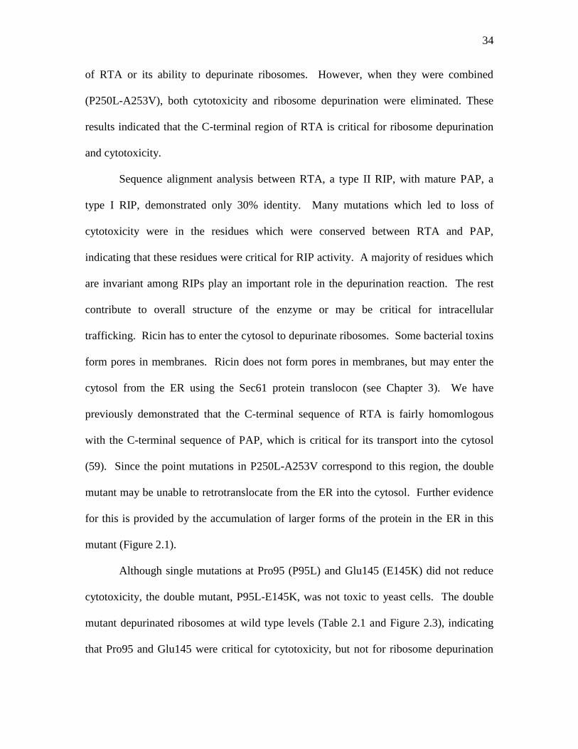
42
mature proteins are not entering the ER, they are still associated with the ER, as indicated
by the detection of RTA and RTAE177K with the ER fraction of cell lysates (Figure 3.4).
Figure 3.4: RTA is not stabilized in sec61-32. Membrane fractions (15 μg) isolated
from Sec61 mutant yeast cells expressing RTA or RTAE177K at 4, 6, 10 and 14 hours post-
induction were separated on a 12% SDS-polyacrylamide gel and probed with anti-V5
antibody. The blots were stripped and probed with the ER membrane marker Dpm1p as a
loading control.
Pre-RTA ribosome depurination is reduced in sec61-32
Because the sec61 mutants exhibited reduced cytotoxicity but high levels of pre-
RTA expression, it was essential to determine if the protein was still active in these yeast
mutants. Yeast cells were grown as described above and induced on galactose containing
media for 6 hours. Total RNA was isolated from these yeast cells and subsequently
subjected to a dual primer extension assay to determine if the protein is still depurinating
yeast ribosomes.
As seen in Figure 3.5, pre-RTA is capable of depurinating ribosomes of both
sec61 yeast mutants. However, the level of pre-RTA depurination is slightly reduced in
sec61-32 cells. The reduction in depurination in Sec61-32 cells is probably not due to
lack of activity of pre-RTA. Instead, it is most likely a result of pre-RTA getting retained
in the ER, and not accessing ribosomes in the cytosol. This may explain why sec61-32
expressing pre-RTA grows slightly better on galactose than sec61-41 (Figure 3.1).
43
Figure 3.5: Pre-RTA ribosome depurination is reduced in sec61-32. Total RNA
isolated from the sec61 yeast mutants expressing pre-RTA or pre-RTAE177K after 6 h of
growth on galactose was analyzed by dual primer extension. The depurination bands and
25S bands were quantified and graphed as a ratio of depurination:25S.
kar2 yeast mutants expressing pre-RTA are viable
Yeast cells with a mutation in KAR2 residue P515 (Kar2-1), which is part of a
highly conserved region of the substrate binding domain of KAR2 (29), were transformed
with pre-RTA and the inactive mutant, pre-RTAE177K. The kar2-1 mutation is
temperature sensitive, and at 24°C, kar2-1 cannot recognize ERAD substrates in the ER.
In addition, the kar2-1 mutation induces the unfolded protein response when it is

44
expressed (29). The kar2-1 yeast transformants were streaked on SD -Ura media
containing glucose and SD -Ura media containing galactose. Kar2-1 mutants were fairly
resistant to pre-RTA expression and were able to grow on galactose containing media.
To confirm that pre-RTA is not as toxic to kar2-1 cells as wildtype cells, a
viability assay was performed. Transformed wildtype and kar2-1 cells were grown as
indicated above in liquid media. After 4 and 10 hours of growth on galactose, the cells
were collected and spotted as a serial dilution onto SD-Ura plates containing glucose.
After two days of growth at the permissive temperature, the plates were analyzed. Pre-
RTA in the kar2-1 yeast cells did not affect cell viability as much as in wildtype yeast
cells (Figure 3.6), indicating that Kar2-1 is necessary for pre-RTA cytotoxicity.
Figure 3.6: kar-2-1 yeast mutants are more viable than wildtype yeast when
expressing pre-RTA. After 4 and 10 hours of growth on SD-Leu medium containing
galactose, serial dilutions were spotted on SD-Leu plates supplemented with 2% glucose.
Pre-RTA is recognized as a substrate of KAR2
kar2-1 yeast cells transformed with pre-RTA and pre-RTAE177K were grown as
described above and were induced on galactose containing media after 4, 6, 10 and 14
hours. Cells were lysed and the membrane fraction was run on a 15% SDS-PAGE gel
and probed with α-V5 antibody. Figure 3.7 shows that there are several bands migrating

45
slightly higher than the RTA standard, indicating the different levels of gylcosylation of
pre-RTA. Compared to the expression of pre-RTA in wildtype yeast cells, expression in
kar2-1 mutant cells indicates an altered level of glycosylation. Because glycosylation
occurs in the ER, the amount or extent of gylcosylation of pre-RTA can be used as an
indication of how long the protein remained in the ER. Compared to pre-RTA expression
in the wildtype cells, the kar2-1 cells show a reduction in the glycosylated forms of pre-
RTA. This suggests that the kar2-1 mutation is affecting the amount of time that pre-
RTA is retained in the ER. One of the functions of wildtype KAR2 is to bind to unfolded
proteins and help to stabilize and refold them. However, because the kar2-1 mutants are
defective in their substrate binding ability, pre-RTA is most likely not recognized by
kar2-1, and is therefore sent through the Sec61 translocon quicker than it would in
wildtype cells.
Figure 3.7: The expression pattern of pre-RTA and pre-RTAE177K is altered in kar2-
1 yeast mutants. Membrane fractions (15 μg) isolated from wildtype and kar2-1 mutant
yeast cells expressing pre-RTA or pre-RTAE177K at 4, 6, 10 and 14 hours post-induction
were separated on a 15% SDS-polyacrylamide gel and probed with anti-V5 antibody. The
blots were stripped and probed with the ER membrane marker Dpm1p as a loading
control.
Pre-RTA can still depurinate yeast ribosomes in kar2-1 mutants
To determine if the kar2-1 mutation affects the enzymatic activity of pre-RTA or
the complete ability of pre-RTA to reach the cytosol, primer extension analysis was used.
The dual primer extension assay was performed on total RNA of wildtype and kar2-1

46
yeast cells expressing pre-RTA as described above. In both wildtype and kar2-1 mutants,
depurination occurred (Figure 3.8). This indicates that pre-RTA is still able to eventually
enter the cytosol in kar2-1 mutant cells, and that there is no effect of depurination activity
in these yeast mutants.
Figure 3.8: Pre-RTA can still depurinate kar2-1 ribosomes. Total RNA isolated from
wildtype and kar2-1 yeast mutants expressing pre-RTA after 6 h of growth on galactose
was analyzed by dual primer extension. The depurination bands and 25S bands were
quantified and graphed as a ratio of depurination:25S.
kar2-113 has no effect on pre-RTA
Pre-RTA and pre-RTAE177K were also transformed into another kar2 mutant,
kar2-113. This yeast mutant is deficient in the ATPase activity of KAR2 (29), which
blocks the dissociation of KAR2 from IRE1, a UPR activator. In contrast to the kar2-1
mutant, the kar2-113 cells were still resistant to pre-RTA. The expression pattern and
depurination were all similar to that of wildtype cells, indicating that the KAR2 ATPase
domain is not essential for pre-RTA toxicity (data not shown).
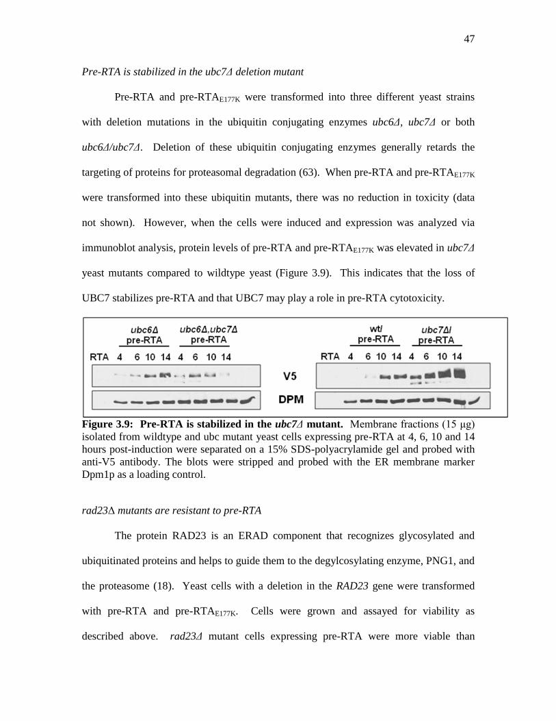
47
Pre-RTA is stabilized in the ubc7Δ deletion mutant
Pre-RTA and pre-RTAE177K were transformed into three different yeast strains
with deletion mutations in the ubiquitin conjugating enzymes ubc6Δ, ubc7Δ or both
ubc6Δ/ubc7Δ. Deletion of these ubiquitin conjugating enzymes generally retards the
targeting of proteins for proteasomal degradation (63). When pre-RTA and pre-RTAE177K
were transformed into these ubiquitin mutants, there was no reduction in toxicity (data
not shown). However, when the cells were induced and expression was analyzed via
immunoblot analysis, protein levels of pre-RTA and pre-RTAE177K was elevated in ubc7Δ
yeast mutants compared to wildtype yeast (Figure 3.9). This indicates that the loss of
UBC7 stabilizes pre-RTA and that UBC7 may play a role in pre-RTA cytotoxicity.
Figure 3.9: Pre-RTA is stabilized in the ubc7Δ mutant. Membrane fractions (15 μg)
isolated from wildtype and ubc mutant yeast cells expressing pre-RTA at 4, 6, 10 and 14
hours post-induction were separated on a 15% SDS-polyacrylamide gel and probed with
anti-V5 antibody. The blots were stripped and probed with the ER membrane marker
Dpm1p as a loading control.
rad23Δ mutants are resistant to pre-RTA
The protein RAD23 is an ERAD component that recognizes glycosylated and
ubiquitinated proteins and helps to guide them to the degylcosylating enzyme, PNG1, and
the proteasome (18). Yeast cells with a deletion in the RAD23 gene were transformed
with pre-RTA and pre-RTAE177K. Cells were grown and assayed for viability as
described above. rad23Δ mutant cells expressing pre-RTA were more viable than

48
wildtype cells (Figure 3.10), suggesting that pre-RTA toxicity relies on a function of
RAD23.
Figure 3.10: rad23Δ yeast mutants expressing pre-RTA are viable. After 6 and 10
hours of growth on SD-Leu medium containing galactose, serial dilutions of wildtype and
rad23Δ mutant yeast cells expressing pre-RTA, pre-RTAE177K or the empty vector control
were spotted on SD-Leu plates supplemented with 2% glucose.
Pre-RTA expression is reduced by the deletion of rad23Δ
Pre-RTA expression was analyzed via a 15% SDS-PAGE and immunoblot
probing as described above. Compared to wildtype cells, rad23Δ mutant cells expressed
pre-RTA to a lower extent (Figure 3.11). This may indicate that RAD23 is one of the
proteins that play a role in the stability of RTA in the cytosol. It is possible that the
reduction in pre-RTA cytotoxicity (Figure 3.10) is due to a reduction in total pre-RTA
accumulation. Although RAD23 seems essential for pre-RTA toxicity, as shown with the
viability assay, it is possible that there are additional proteins that aid in PNG1 mediated
deglycosylation. Also, because there is a destabilization of pre-RTA in the rad23Δ
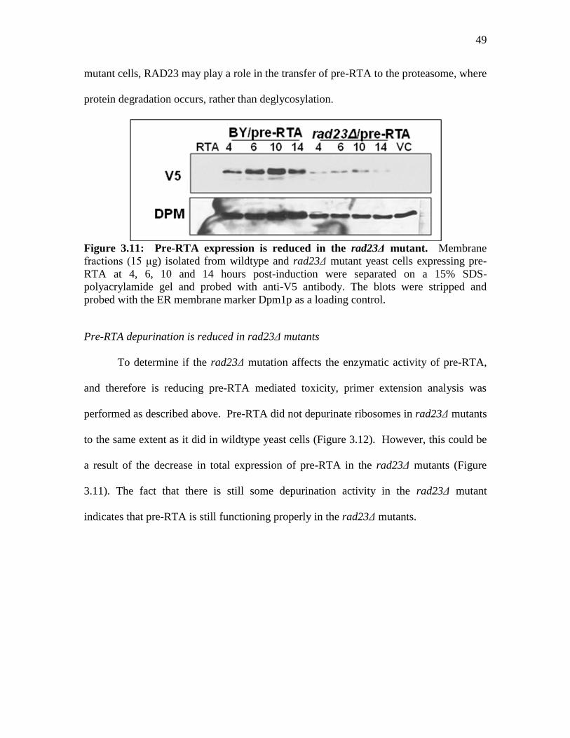
49
mutant cells, RAD23 may play a role in the transfer of pre-RTA to the proteasome, where
protein degradation occurs, rather than deglycosylation.
Figure 3.11: Pre-RTA expression is reduced in the rad23Δ mutant. Membrane
fractions (15 μg) isolated from wildtype and rad23Δ mutant yeast cells expressing pre-
RTA at 4, 6, 10 and 14 hours post-induction were separated on a 15% SDS-
polyacrylamide gel and probed with anti-V5 antibody. The blots were stripped and
probed with the ER membrane marker Dpm1p as a loading control.
Pre-RTA depurination is reduced in rad23Δ mutants
To determine if the rad23Δ mutation affects the enzymatic activity of pre-RTA,
and therefore is reducing pre-RTA mediated toxicity, primer extension analysis was
performed as described above. Pre-RTA did not depurinate ribosomes in rad23Δ mutants
to the same extent as it did in wildtype yeast cells (Figure 3.12). However, this could be
a result of the decrease in total expression of pre-RTA in the rad23Δ mutants (Figure
3.11). The fact that there is still some depurination activity in the rad23Δ mutant
indicates that pre-RTA is still functioning properly in the rad23Δ mutants.

50
Figure 3.12: Pre-RTA depurination is reduced in rad23Δ yeast mutants. Total RNA
isolated from wildtype and rad23Δ yeast mutants expressing pre-RTA after 6 h of growth
on galactose was analyzed by dual primer extension. The depurination bands and 25S
bands were quantified and graphed as a ratio of depurination:25S.
pre-RTAE177K is stabilized in Proteasome Mutants
The yeast mutant pre1-1, pre2-2 is defective in the chymotryptic-like activity of
the 19S subunit of the proteasome (66). The initial phenotype that was observed when
the double mutation was isolated was an accumulation of ubiquitinated substrates in the
cytosol, due to the fact that these substrates were no longer degraded by the mutated
proteasome. pre1-1, pre2-2 cells were transformed with pre-RTA and pre-RTAE177K.
RTA toxicity was similar in both wildtype cells and pre1-1, pre2-2 cells as confirmed by
lack of growth on galactose media (Figure 3.13). Because expression of pre-RTA in the
pre1-1, pre2-2 cells was extremely toxic and the cells grew poorly, pre-RTAE177K was
used to examine protein stability in the proteasome mutants. Wildtype and pre1-1, pre2-
2 cells transformed with pre-RTAE177K were grown and aliquoted at 4, 6, 10 and 14 hours
post-induction as described above. Protein expression was analyzed by SDS-PAGE and
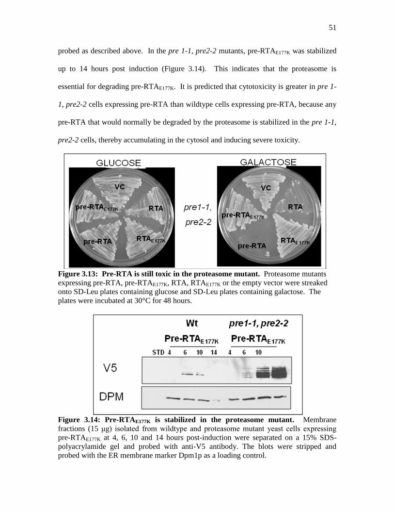
51
probed as described above. In the pre 1-1, pre2-2 mutants, pre-RTAE177K was stabilized
up to 14 hours post induction (Figure 3.14). This indicates that the proteasome is
essential for degrading pre-RTAE177K. It is predicted that cytotoxicity is greater in pre 1-
1, pre2-2 cells expressing pre-RTA than wildtype cells expressing pre-RTA, because any
pre-RTA that would normally be degraded by the proteasome is stabilized in the pre 1-1,
pre2-2 cells, thereby accumulating in the cytosol and inducing severe toxicity.
Figure 3.13: Pre-RTA is still toxic in the proteasome mutant. Proteasome mutants
expressing pre-RTA, pre-RTAE177K, RTA, RTAE177K or the empty vector were streaked
onto SD-Leu plates containing glucose and SD-Leu plates containing galactose. The
plates were incubated at 30°C for 48 hours.
Figure 3.14: Pre-RTAE177K is stabilized in the proteasome mutant. Membrane
fractions (15 μg) isolated from wildtype and proteasome mutant yeast cells expressing
pre-RTAE177K at 4, 6, 10 and 14 hours post-induction were separated on a 15% SDS-
polyacrylamide gel and probed with anti-V5 antibody. The blots were stripped and
probed with the ER membrane marker Dpm1p as a loading control.

52
DISCUSSION
The results presented here provide evidence that pre-RTA uses the endoplasmic
reticulum associated degradation pathway to reach the cytosol in yeast. Several studies
have been published that support the use of ERAD by RTA. My work provided further
support for this and identified the components of the ERAD pathway critical for RTA
transport.
Olsnes (11) first found that RTA was associated with Sec61 by co-
immunoprecipitation assays. He showed that Sec61 pulled down glycosylated, but not
unglycosylated RTA. This indicates that RTA associates with SEC61 in the ER, where
glycosylation occurs. Another study (67) analyzed RTA in yeast and showed that an
inactive form of RTA with a Kar2 ER signal sequence, RTAE177D, was stabilized in the
sec61 mutants. However, they did not analyze wildtype RTA in these sec61 mutants.
In this study, we suggest that Sec61 is necessary for pre-RTA toxicity. By
eliminating the export ability of Sec61 with the temperature sensitive mutants sec61-32
and sec61-42, accumulation of pre-RTA and pre-RTAE177K is increased in the ER
fraction. Accordingly, the translocation of pre-RTA from the ER to the cytosol is
probably affected, at least in sec61-32. While detection of pre-RTA and even the non-
toxic pre-RTAE177K in the cytosolic fraction of cell lysates is difficult, one way to
determine if pre-RTA is even reaching the cytosol is to examine ribosome depurination.
In the sec61-32 cells, pre-RTA ribosome depurination is reduced in wildtype cells,
indicating that pre-RTA is not reaching the cytosol as efficiently in that mutant.
Further evidence for the use of SEC61 by pre-RTA is shown by the lack of effect
that the sec61 mutants had on RTA and RTAE177K. These mature proteins are in the same

53
yeast expression vector as pre-RTA and pre-RTAE177K, but do not have the ER signal
sequence. Although these mature proteins associate with the ER, as indicated by the
accumulation of protein in the ER fraction of cell lysates, they are not glycosylated,
indicating that they don’t enter the ER lumen. As expected, toxicity of RTA in the sec61
mutants was the same as in wildtype cells, and there was no accumulation of RTA or
RTAE177K.
The functions of KAR2 are of particular interest when studying RTA due to the
fact that KAR2 has many roles as an ER resident chaperone. One of the functions of
KAR2 is helping to fold misfolded or slowly folding proteins in the ER lumen (29). As a
result of this chaperone activity, there are several possible events to follow. One possible
outcome is that KAR2 will correctly stabilize and fold the protein, allowing it to continue
on its route. Another possibility is that KAR2 will assist with the folding of the protein,
but will take an extended amount of time. When there is a large amount of KAR2
attempting to properly fold a substrate in the ER, the cell is signaled to slow down the
translation of more of these proteins, and overall protein translation is reduced. In
addition, when KAR2 is recruited to these misfolded substrates, the Unfolded Protein
Response (UPR) is activated (27). When the UPR is activated, the protein folding
capabilities of the cell are maximized to allow the cell to begin functioning properly
again.
There is little published evidence that indicates that RTA directly uses KAR2.
Here, we show that a mutation in the substrate binding domain of KAR2 reduced the
toxicity of and changed the amount of glycosylation of pre-RTA. It is possible that a
function of kar2-1 in pre-RTA translocation may be to help stabilize and fold the protein,
54
and that this assistance is necessary for sufficient amounts of pre-RTA to reach the
cytosol to induce cytotoxicity. Although the ability to depurinate yeast ribosomes in both
wildtype and kar2-1 mutants was the same, it is important to realize that depurination and
cytotoxicity do not always correlate in pre-RTA, as demonstrated in Chapter 2.
In addition to the kar2-1 mutant cells, kar2-113 cells were transformed with pre-
RTA. kar2-113 cells are mutated in the ATPase binding domain, and are expected to be
defective in KAR2-mediated import into the ER (29). As previously described, protein
import into the ER can occur co-translationally or post-translationally. KAR2 and
SEC63 are two necessary factors for the post-translational translocation of proteins into
the ER, and it is expected that KAR2 acts as a luminal gate to allow entry through the
Sec61 translocon. When pre-RTA expression was induced in the kar2-113 cells, there
was no reduction of cytotoxicity. In addition, the protein expression pattern of pre-RTA
in these cells was the same as in wildtype cells. Pre-RTA was also able to depurinate to
the same extent in kar2-113 cells as in wildtype cells, indicating that the mutation is not
affecting the enzymatic ability of pre-RTA. These results demonstrate that KAR2 is
either not essential for pre-RTA import into the ER, or that pre-RTA is translocated into
the ER co-translationally and does not require the use of KAR2 for entry into the ER.
When rad23Δ deletion mutants were transformed with pre-RTA, cells were more
viable than the pre-RTA transformed wildtype cells. However, pre-RTA was not
stabilized and did not accumulate in the rad23Δ cells. Previous studies by Kim (18) used
RTA as a substrate to better understand the interaction between RAD23 and PNG1.
RAD23 recognizes and binds to ubiquitin residues of proteasomal substrates. RAD23
also binds PNG1, which deglycosylates proteasome substrates prior to delivery to the
55
proteasome. A model was devised which shows RAD23 binding to the ubiquitin residues
of a glycosylated proteasome substrate (RTA) and then binding to PNG1. This complex
then allows PNG1 access to deglycosylate RTA. RTA is subsequently transported to the
proteasome via the ubiquitin-like domain of RAD23 that binds to the proteasome (18).
Of course, this model of the dependency of RTA on RAD23 is based on the fact
that RTA is ubiquitinated as a result of being an ERAD substrate. However, previous
studies (17) that looked at the ubiquitination of RTA suggested that RTA is not
ubiquitinated. Ubiquitin residues are attached to lysine residues of proteasome
substrates. RTA only has two lysine residues, and it has been postulated that this low
lysine content allows RTA to escape from the ER via ERAD, but then evade degradation
in the proteasome because it does not get heavily ubiquitinated (17). In order to test this
hypothesis, the group expressed RTA in yeast mutants defective in ubiquitin conjugating
enzymes. They found that when the ability of the cell to attach ubiquitin residues was
abolished, there was little effect on the toxicity or protein stabilization of RTA. In
another study, several residues in RTA were changed to lysines (68). The additional
lysine residues appeared to have a reduction in cytotoxicity and an increase in
proteasomal degradation. The authors therefore concluded that RTA does not get
ubiquitinated and is therefore able to avoid degradation by the proteasome. However, in
this study, we have found that deletion of the ubiquitin conjugating enzyme, UBC7,
greatly increased the stability of pre-RTA and pre-RTAE177K expression, suggesting that
RTA may get ubiquitinated to some degree.
Many groups have studied the degradation of RTA via the proteasome, which is
the final destination for ubiquitinated ERAD substrates. In this study, pre-RTA was so
56
toxic to the proteasome mutant cells and the wildtype cells that it was difficult to analyze
the effect of the proteasome mutant on pre-RTA. Therefore, pre-RTAE177K was
transformed into the proteasome mutant cells and analyzed. As expected, pre-RTAE177K
was stabilized in the proteasome mutant, indicating that the proteasome does normally
degrade a significant amount of RTA. As the proteasome is the endpoint for ERAD
substrates, it supports the theory that RTA does normally utilize the ERAD pathway to
some degree.
Taken together, the data presented here suggests that RTA exits the ER via the
SEC61 translocon and utilizes the components of ERAD to reach the cytosol.
Interestingly, these data indicate that RTA is both ubiquitinated and targeted to the
proteasome, which should ultimately lead to its degradation. However, there must be at
least a portion of RTA that escapes this degradation to access and depurinate ribosomes.
The fact that the deletion mutants ubc7Δ, but not ubc6Δ or the double mutant
ubc6Δ,ubc7Δ affected stability of pre-RTA may indicate that RTA is ubiquitinated by
UBC7, but not UBC6. This may suggest that the total amount of RTA that is targeted to
the proteasome is less than other proteasome substrates that normally get ubiquitinated by
both UBC6 and UBC7.
57
CHAPTER 4: Mutations in the C-terminal hydrophobic stretch of ricin A chain
inhibit retro-translocation without reducing the catalytic activity
INTRODUCTION
There is a strong similarity among several regions of the sequences of RIPs, and
protein structures of several of them allow for near perfect superimposable images
(Figure 4.1). One region of high sequence homology is the C-terminal domain (Figure
4.1). For several RIPs, namely Stx and PAP, there have been studies suggesting that the
C-terminus is necessary for protein translocation in the cell, primarily by containing a
hydrophobic membrane spanning domain (69, 70). These hydrophobic domains have
been suggested to act as membrane insertion extensions that can target the RIP to specific
membranes in the target cell. Studies in shiga toxin demonstrated that disruption of this
hydrophobic domain via point or truncation mutations resulted in an inactive protein (71).
Interestingly, there is a common critical residue in both PAP (N253) and Shiga-toxin
(N241) that, when deleted along with the remaining downstream residues, results in a
loss of cytotoxicity and depurination ability (69, 70). In order to determine if the C-
terminus of ricin is also essential for cytotoxicity, various truncation and point mutations
were generated and analyzed in both mature RTA (RTA) and RTA with its N-terminal
signal sequence (pre-RTA). The results suggest that ricin, like shiga-toxin and PAP,
relies on the C-terminal residues for membrane translocation and enzymatic activity.

58
Figure 4.1: Structural comparison and sequence alignment of Shiga-toxin 2 (STX2),
Pokeweed Antiviral Protein (PAP) and Ricin A chain (RTA). Protein Explorer
(www.proteinexplorer.org) was used to generate the structural models using the PDB IDs
1R4P (STX2), 1PAG (PAP) and 1IFT (RTA), The green tail represents the C-terminal
residues that are essential for protein toxicity.
RESULTS
The C-terminal residues of pre-RTA are essential for cytotoxicity
Stop codon mutations (*) were generated in residues I249, P250, I251, I252,
A253, L254, M255, V256 and Y257 in order to truncate the C-terminus of pre-RTA
(Table 4.2). Deletion of Y257 and the downstream residues did not affect the ability of
pre-RTA to kill yeast cells (Figure 4.2). However, the deletion of V256 and the residues
downstream resulted in a non-toxic pre-RTA mutant, suggesting that residue V256 and
the downstream residues are essential for pre-RTA cytotoxicity. Deletion of further
upstream residues M255, L254, A253, I252, I251, P250 and I249 also resulted in non-
toxic pre-RTA variants. To determine if particular amino acids or the entire C-terminus
is critical for pre-RTA mediated cytotoxicity, site-directed point mutations were also
generated in these residues in order to disrupt the hydrophobicity of the C-terminus. All
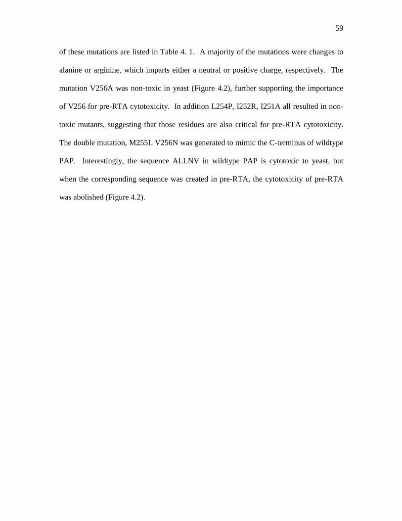
59
of these mutations are listed in Table 4. 1. A majority of the mutations were changes to
alanine or arginine, which imparts either a neutral or positive charge, respectively. The
mutation V256A was non-toxic in yeast (Figure 4.2), further supporting the importance
of V256 for pre-RTA cytotoxicity. In addition L254P, I252R, I251A all resulted in non-
toxic mutants, suggesting that those residues are also critical for pre-RTA cytotoxicity.
The double mutation, M255L V256N was generated to mimic the C-terminus of wildtype
PAP. Interestingly, the sequence ALLNV in wildtype PAP is cytotoxic to yeast, but
when the corresponding sequence was created in pre-RTA, the cytotoxicity of pre-RTA
was abolished (Figure 4.2).
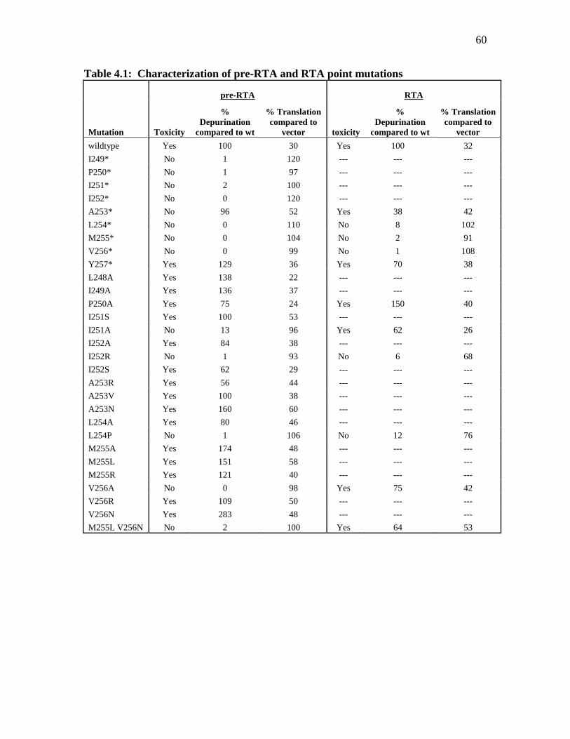
60
Table 4.1: Characterization of pre-RTA and RTA point mutations
pre-RTA RTA
Mutation Toxicity
%
Depurination
compared to wt
% Translation
compared to
vector toxicity
%
Depurination
compared to wt
% Translation
compared to
vector
wildtype Yes 100 30 Yes 100 32
I249* No 1 120 --- --- ---
P250* No 1 97 --- --- ---
I251* No 2 100 --- --- ---
I252* No 0 120 --- --- ---
A253* No 96 52 Yes 38 42
L254* No 0 110 No 8 102
M255* No 0 104 No 2 91
V256* No 0 99 No 1 108
Y257* Yes 129 36 Yes 70 38
L248A Yes 138 22 --- --- ---
I249A Yes 136 37 --- --- ---
P250A Yes 75 24 Yes 150 40
I251S Yes 100 53 --- --- ---
I251A No 13 96 Yes 62 26
I252A Yes 84 38 --- --- ---
I252R No 1 93 No 6 68
I252S Yes 62 29 --- --- ---
A253R Yes 56 44 --- --- ---
A253V Yes 100 38 --- --- ---
A253N Yes 160 60 --- --- ---
L254A Yes 80 46 --- --- ---
L254P No 1 106 No 12 76
M255A Yes 174 48 --- --- ---
M255L Yes 151 58 --- --- ---
M255R Yes 121 40 --- --- ---
V256A No 0 98 Yes 75 42
V256R Yes 109 50 --- --- ---
V256N Yes 283 48 --- --- ---
M255L V256N No 2 100 Yes 64 53
61
Table 4.2: The location of the C-terminal pre-RTA mutations. Non-toxic mutants are
boxed in red.

62
Figure 4.2: Viability analysis of pre-RTA C-terminal deletion and point mutations.
Yeast cells were grown on SD-Leu containing 2% glucose to an A600 of 0.3 and then
transferred to SD-Leu medium containing 2% galactose to induce pre-RTA expression. A
serial dilution of cells was plated on SD-Leu plates containing 2% glucose for 10 h post-
induction. Plates were incubated at 30°C for approximately 48 h.
The expression pattern of the pre-RTA mutants is different in non-toxic mutants
To confirm the presence of pre-RTA protein expression, 12% PAGE and
immunoblot analysis was conducted. All of the mutants express pre-RTA (Figure 4.3) to
varying extents. The non-toxic C-terminal deletion mutants generate a protein that has
multiple lower molecular weight bands, suggesting that perhaps this protein is getting
destabilized or degraded in the cell. In addition, the non-toxic point mutations generate
higher levels of pre-RTA protein, probably due to the fact that the cells are not being
killed by the toxin.
Figure 4.3: Immunoblot analysis of pre-RTA expression. Membrane fractions (15
μg) isolated from cells expressing pre-RTA or mutants were separated on a 12% SDS-
polyacrylamide gel and probed with polyclonal anti-RTA (1:5,000). The blots were
stripped and probed with the ER membrane marker Dpm1p as a loading control.
The C-terminus is essential for pre-RTA depurination in yeast
To determine if the cytotoxicity of the pre-RTA mutants correlated with ribosome
depurination in yeast, total RNA was isolated from yeast expressing all of the C-terminal
63
deletion and point mutations. The RNA was subsequently subjected to dual primer
extension analysis. As expected, all of the toxic pre-RTA mutants were able to
depurinate yeast ribosomes, while all of the non-toxic pre-RTA mutants were not (Figure
4.4). Interestingly, A253* was nontoxic in yeast, but was able to depurinate yeast
ribosomes, although to a lesser extent than wildtype pre-RTA.
Figure 4.4 Ribosome depurination in yeast expressing pre-RTA and the mutant
forms. Total RNA isolated after 6 h of growth on galactose was analyzed by dual primer
extension analysis using two different end-labeled primers: the depurination primer
(Dep), which was used to measure the extent of depurination, and the 25S rRNA primer
(25S), which was used to measure the total amount of 25S rRNA. Primer extension
analysis of cells harboring the empty vector is shown as a control.
Pre-RTA mutants that are non-toxic and not depurinating do not inhibit total translation
in yeast
Ribosome depurination by pre-RTA results in inhibition of protein translation.
Therefore, it is expected that the pre-RTA mutants that don’t depurinate yeast ribosomes

64
should not inhibit translation. To confirm the ability of the pre-RTA mutants to inhibit
total translation in yeast, [35
S]-methionine incorporation was assayed. As expected, the
pre-RTA mutants that do not depurinate yeast ribosomes do not inhibit protein synthesis
(Figure 4.5).
Figure 4.5: Pre-RTA mutants that don’t depurinate ribosomes can translate
protein. Yeast cells were grown to an A600 of 0.3 in SD-Leu-Met containing 2%
glucose. Cells were then resuspended in SD-Leu-Met containing 2% galactose for 6 h to
induce the expression of either wild-type pre-RTA or the mutant forms. At time zero,
[35
]-methionine was added to induced cells. After 30 min, 400 μl of yeast cells was
removed for growth measurements, and additional aliquots of 400 μl were assayed for
methionine incorporation in duplicate as previously described. The cpm was normalized
to the A600 reading, and rates of translation were determined as cpm/A600/minute. Final
results were displayed as percentages of total translation in yeast harboring the empty
vector.
Mutations affecting the cytotoxicity of pre-RTA do not affect the toxicity of RTA
In order to determine if the C-terminus of pre-RTA plays a role in protein
transport or localization in yeast, the same mutations that impaired the ability of pre-RTA
to kill yeast cells were generated in mature RTA (Table 4.3) and assayed for cytotoxicity
(Figure 4.6), expression (Figure 4.7), depurination (Figure 4.8) and translation inhibition
(Figure 4.9). These mutations were: Y257*, V256*, M255*, L254*, A253*, V256A,
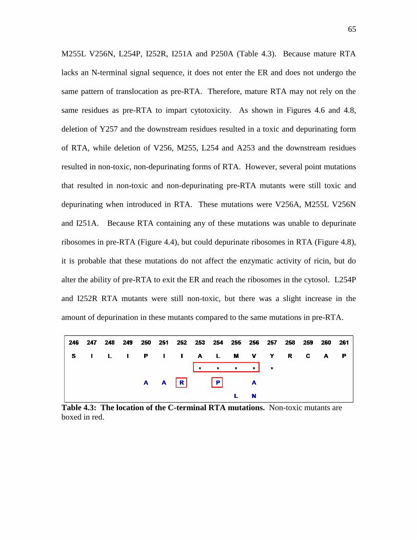
65
M255L V256N, L254P, I252R, I251A and P250A (Table 4.3). Because mature RTA
lacks an N-terminal signal sequence, it does not enter the ER and does not undergo the
same pattern of translocation as pre-RTA. Therefore, mature RTA may not rely on the
same residues as pre-RTA to impart cytotoxicity. As shown in Figures 4.6 and 4.8,
deletion of Y257 and the downstream residues resulted in a toxic and depurinating form
of RTA, while deletion of V256, M255, L254 and A253 and the downstream residues
resulted in non-toxic, non-depurinating forms of RTA. However, several point mutations
that resulted in non-toxic and non-depurinating pre-RTA mutants were still toxic and
depurinating when introduced in RTA. These mutations were V256A, M255L V256N
and I251A. Because RTA containing any of these mutations was unable to depurinate
ribosomes in pre-RTA (Figure 4.4), but could depurinate ribosomes in RTA (Figure 4.8),
it is probable that these mutations do not affect the enzymatic activity of ricin, but do
alter the ability of pre-RTA to exit the ER and reach the ribosomes in the cytosol. L254P
and I252R RTA mutants were still non-toxic, but there was a slight increase in the
amount of depurination in these mutants compared to the same mutations in pre-RTA.
Table 4.3: The location of the C-terminal RTA mutations. Non-toxic mutants are
boxed in red.
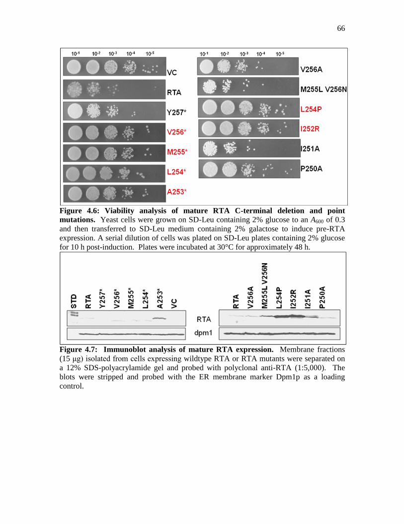
66
Figure 4.6: Viability analysis of mature RTA C-terminal deletion and point
mutations. Yeast cells were grown on SD-Leu containing 2% glucose to an A600 of 0.3
and then transferred to SD-Leu medium containing 2% galactose to induce pre-RTA
expression. A serial dilution of cells was plated on SD-Leu plates containing 2% glucose
for 10 h post-induction. Plates were incubated at 30°C for approximately 48 h.
Figure 4.7: Immunoblot analysis of mature RTA expression. Membrane fractions
(15 μg) isolated from cells expressing wildtype RTA or RTA mutants were separated on
a 12% SDS-polyacrylamide gel and probed with polyclonal anti-RTA (1:5,000). The
blots were stripped and probed with the ER membrane marker Dpm1p as a loading
control.

67
Figure 4.8: Ribosome depurination in yeast expressing RTA and the mutant forms.
Total RNA isolated after 6 h of growth on galactose was analyzed by dual primer
extension analysis using two different end-labeled primers: the depurination primer
(Dep), which was used to measure the extent of depurination, and the 25S rRNA primer
(25S), which was used to measure the total amount of 25S rRNA. Primer extension
analysis of cells harboring the empty vector is shown as a control.
Figure 4.9: Translation inhibition of RTA and the mutant forms. Yeast cells were
grown to an A600 of 0.3 in SD-Leu-Met containing 2% glucose. Cells were then
resuspended in SD-Leu-Met containing 2% galactose for 6 h to induce the expression of
either wild-type pre-RTA or the mutant forms. At time zero, [35
S]-methionine was added
to induced cells. After 30 min, 400 μl of yeast cells was removed for growth
measurements, and additional aliquots of 400 μl were assayed for methionine
incorporation in duplicate as previously described. The cpm was normalized to the A600
reading, and rates of translation were determined as cpm/A600/minute. Final results were
displayed as percentages of total translation in yeast harboring the empty vector.
68
DISCUSSION
The C-termini of the RIPs shiga toxin and PAP are necessary for cytotoxicity (69,
70). Protein prediction software, TMpred indicates that there is a strong putative
transmembrane domain from pre-RTA residues 245-257. Studies have shown that this
domain in ricin may be used for toxin localization in target cells (73). After binding to
the target cell, the ricin holotoxin is endocytized and transported to the trans-Golgi
network. It then undergoes retrograde transport and enters the endoplasmic reticulum.
After separating from RTB, RTA is transported into the cytosol, perhaps by taking
advantage of the ER-Associated Degradation (ERAD) pathway and the Sec61 translocon
as discussed earlier. Upon entering the cytosol, RTA must bind to and depurinate
ribosomes. The crystal structure has been solved for ricin (3). In the holotoxin, the C-
terminal residues discussed in this paper are buried between RTB and RTA. After
separation from RTB, the C-terminal residues of RTA are exposed and resemble a tail or
extension (Figure 4.11). Since it contains a hydrophobic motif, this tail may serve to aid
in localization of RTA by inserting into a target membrane. To better understand the
function of this C-terminal domain in ricin, we generated deletion mutants and point
mutations that either truncated the C-terminal residues of ricin or changed the
composition of the hydrophobic domain (Table 4.2).

69
Figure 4.10: A structural comparison of the C-terminal residues of RTA. Protein
Explorer (www.proteinexplorer.org) was used to generate the structural models of 1IFT
(RTA). The C-terminal deleted residues are highlighted in green and demonstrate the
increase in proximity to the active site of RTA as the C-terminus is deleted up to residue
A253.
Initially, the deletion and point mutations were generated in the C terminus of pre-
RTA. In pre-RTA, deletion of the C-terminus up to and includingY257 maintained
toxicity (Figure 4.2). Protein expression showed two bands migrating at the same size as
the purified RTA standard, indicating that the protein was able to enter the ER and get
glycosylated (Figure 4.3). In addition, the protein was able to exit the ER and enter the
cytosol, as indicated by its wildtype levels of ribosome depurination (Figure 4.3). Y257*
was also able to inhibit total translation in yeast to the same extent as wildtype pre-RTA
(Figure 4.4). However, deleting only one more residue upstream of Y257, V256,
abolished all wildtype activities, including cytotoxicity, depurination and translation
70
inhibition. Accordingly, as adjacent upstream deletions were generated, almost all of the
generated mutants were nontoxic and non-depurinating.
One of the deletion mutants, A253*, behaved differently from the other non-toxic
mutants. While A253* was non-toxic, it was still able to depurinate yeast ribosomes,
though to a lesser extent than that of wildtype pre-RTA. In addition, A253* was still able
to inhibit total translation in yeast to similar levels as wildtype pre-RTA. Protein
structure analysis using Protein Explorer indicates that A253 is one of the closest C-
terminal residues to the active site residue of pre-RTA, residue E177 (Figure 4.11). It is
possible that when the C-terminus is deleted, the remaining residues might fold
improperly, blocking access of the active site to ribosomes. However, deletion of all of
the residues up to and including A253 removes the entire C-terminal tail (Figure 4.10).
This may open the active site cleft again, allowing it to access ribosomes. When the
residues upstream of A253 are sequentially deleted (I252*, I251*, P250* and I249*),
cytotoxicity and depurination activity are again abolished (Figures 4.2 and 4.3). It is
possible that these upstream deletions from A253 may cause the structure of the active
site cleft to become blocked again.
In addition to truncation mutations, several point mutations resulted in the loss of
cytotoxicity of pre-RTA (Figure 4.2). The majority of the residues were changed to
alanine to impart a neutral charge, as well as asparagine to impart a positive charge. By
generating the point mutants, it was expected that the hydrophobic motif of the C-
terminus of pre-RTA would be disrupted, thereby abolishing any effect the C-terminus
may have for ricin translocation. In addition, we generated a double mutation to change
the residues of pre-RTA to be identical to the critical residues for PAP cytotoxicity. This

71
indicates that while the C-terminus of RIPs is essential for cytotoxicity, the specific
sequence of each may be different to perpetuate this toxicity. In pre-RTA, I251A, I252R,
L254P, V256A and the double mutant M255L V256N were all non-toxic, non-
depurinating mutations, indicating that they are necessary for pre-RTA toxicity and
ribosome depurination in yeast.
The results of both the truncation mutants and point mutations in pre-RTA
suggested two possibilities for the way that the C-terminus is used for ricin toxicity. One
possibility is that the C-terminus is necessary for the enzymatic function of ricin. This
hypothesis is supported by the fact that the C-terminal residues discussed here lie in fairly
close proximity to the active site of RTA (Figure 4.11), especially I251, I252 and A253.
Another possible function of the C-terminus of ricin could be for the promotion of
translocation of the toxin out of the ER into the cytosol. To further determine the role of
the C-terminus in ricin, several of the non-toxic pre-RTA mutations were generated in
mature RTA and analyzed in yeast. Mature RTA does not have a signal sequence and
does not enter the ER. Therefore, mutations that affect the cytotoxicity by inhibiting the
translocation of pre-RTA out of the ER should not have an effect on RTA.
The deletion mutants Y257*, V256*, M255*, L254* and A253* were generated
in RTA and the protein expression, toxicity, depurination and translation inhibition of
each was analyzed (Figures 4.7, 4.8, 4.9, 4.10). Figure 4.11 shows a summary of these
mutations in pre-RTA and RTA. Again, the Y257* mutant was toxic and caused
depurination while the V256* mutant was non-toxic and non-depurinating. The upstream
mutations, M255* and L254* behaved similarly in RTA as in pre-RTA. A253* also
behaved the same in RTA as in pre-RTA. The fact that both pre-RTA and RTA A253* is
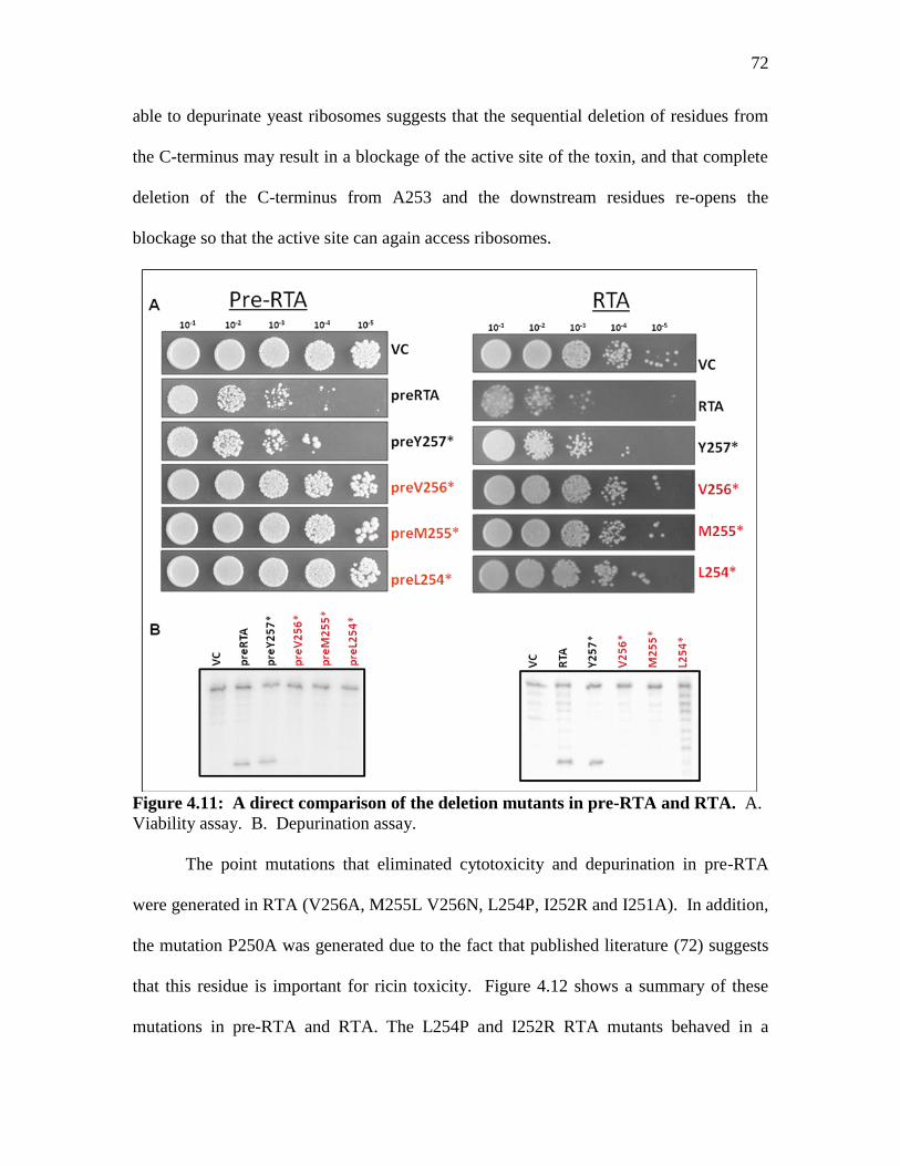
72
able to depurinate yeast ribosomes suggests that the sequential deletion of residues from
the C-terminus may result in a blockage of the active site of the toxin, and that complete
deletion of the C-terminus from A253 and the downstream residues re-opens the
blockage so that the active site can again access ribosomes.
Figure 4.11: A direct comparison of the deletion mutants in pre-RTA and RTA. A.
Viability assay. B. Depurination assay.
The point mutations that eliminated cytotoxicity and depurination in pre-RTA
were generated in RTA (V256A, M255L V256N, L254P, I252R and I251A). In addition,
the mutation P250A was generated due to the fact that published literature (72) suggests
that this residue is important for ricin toxicity. Figure 4.12 shows a summary of these
mutations in pre-RTA and RTA. The L254P and I252R RTA mutants behaved in a
73
similar way as in pre-RTA. Both were non-toxic and not depurinating. P250A was toxic
and depurinating in both pre-RTA and RTA, suggesting that P250 is not an essential
residue for ricin toxicity or depurination. However, the mutations V256A, M255L
V256N and I251A were still toxic and able to depurinate yeast ribosomes when generated
in RTA. To further confirm the enzymatic activity of these mutants, translation
inhibition of all of the RTA mutants was performed. All of the mutants that did not
depurinate yeast ribosomes did not inhibit protein translation, while those mutants that
were still able to depurinate yeast ribosomes were still able to inhibit protein synthesis
(Figure 4.10).

74
Figure 4.12: A direct comparison of the point mutants in pre-RTA and RTA. A.
Viability assay. B. Depurination assay.
Because V256A, M255L V256N and I251A mutations in pre-RTA are unable to
depurinate but do depurinate when introduced in RTA, these mutations probably do not
inhibit enzymatic activity of pre-RTA or RTA, but do inhibit the translocation of pre-
RTA from the ER into the cytosol. The C-terminal residues of ricin form a putative
transmembrane domain, which could possibly be used as a translocation signal for entry

75
into the cytosol from the ER (73). The point mutations V256A, M255L V256N and
I251A might disrupt this motif, thereby inhibiting the ability of the protein to translocate
out of the ER to the cytosol.
Similar studies conducted in yeast expressing mature STX1 or STX1 that is
targeted to enter the ER membrane (pre-STX1) yielded slightly different results (71).
Leu 240 in STX1 corresponds to RTA V256 when the sequence is aligned. The
truncation of pre-STX1 up to and including Leu240 (pre-STX1 L240*) resulted in a non-
toxic form of the protein. However, when the same truncation was made in mature STX1
which does not go into the ER, the protein regained toxicity. Those results suggest that
deleting L240 and the downstream residues from STX1 does not affect its enzymatic
activity, but does inhibit retrotranslocation (71). To determine if the C-terminal
sequence or only L240 is critical for retrotranslocation, mutations that changed pre-STX1
L240 to alanine, arginine, asparagine or aspartic acid were generated. None of the
mutations significantly reduced the toxicity, indicating that the entire C-terminal
sequence, and not just L240, is responsible for the translocation of preSTX1 from the ER
to cytosol. In contrast, the truncation up to V256 in both RTA and pre-RTA resulted in
non-toxic and non-depurinating proteins. These results indicate that the entire C-terminal
sequence from V256 to F267 is essential for enzymatic activity, but not necessarily
retrotranslocation (71).
Like RTA, PAP, a type I RIP, must also traverse an internal cellular membrane t o
gain access to ribosomes (74). It is thought that this membrane is the ER. Studies in
yeast have indicated that the C-teminus of PAP is essential for its translocation into the
cytosol (69). RTA V256 corresponds to N253 in PAP. When PAP N253 was changed to

76
a stop codon (N253*), the protein lost toxicity and was unable to depurinate yeast
ribosomes. Immunoblots of cellular fractionation showed that the accumulation of PAP
in the cytosol was dramatically reduced in N253*. Further upstream truncation mutants
were generated in PAP, and when the C-terminal residues were deleted up to and
including A250, which corresponds to residue L254 in RTA, PAP was still non-toxic and
the protein was no longer able to reach the cytosol at all. These results indicate that A250
and the downstream residues are important for translocation of PAP into the cytosol (69).
Point mutations that changed N253 to alanine (N253A) and arginine (N253R) also
reduced the level of cytotoxicity of PAP and the ability to depurinate yeast ribosomes,
but did not affect the ability of the protein to translocate into the cytosol. These results
indicate that N253 is critical for enzymatic activity of the protein, but N253 alone is not
critical for protein translocation. Interestingly, pre-RTA V256A resulted in a non-toxic
non-depurinating protein, while pre-RTA V256R was still toxic and able to depurinate
yeast ribosomes. Although arginine is a basic residue that should disrupt the
hydrophobicity and alanine is a hydrophobic residue that should maintain the
hydrophobicity of the C-terminus, it is possible that the larger size of arginine maintains
an interaction with the active site, while the smaller alanine cannot.
While RIPs generally depurinate ribosomes in the same way, the critical residues
that carry out the necessary steps for ribosome depurination seem to vary. However, in
the three RIPs discussed here, the C-terminus plays a lead role in either protein
translocation into the cytosol, enzymatic activity or both. The results of this study show
that the C-terminal sequence V256-F267 in RTA is necessary for maintaining the
enzymatic activity of the protein. In addition, we demonstrate that the specific residues

77
V256, V256 and M255 together and I251 are critical for RTA retrotranslocation from the
ER into the cytosol.
78
CHAPTER 5: Conclusions
The threat of ricin exposure as a result of bioterrorism is a major concern as there
is currently no approved vaccine or treatment available for ricin intoxication. Because
wildtype ricin is highly toxic to humans, nontoxic recombinant vaccines need to be
developed based on a detailed understanding of the structure and molecular mechanism
of action of ricin. Ricin is toxic to yeast, and we have expressed RTA in yeast to
examine the residues that are critical for translocation into the cytosol and enzymatic
activity, as well as the mechanism of entry into the cytosol. Three separate analyses of
pre-RTA in yeast were conducted and a summary of each of these studies is discussed
below.
After the ricin holotoxin is endocytosed by the target cell, only 5% is transported
to the Golgi. It then undergoes retrograde transport to the ER, and finally to the cytosol,
where it can depurinate ribosomes (11). To target the most critical step of ricin
translocation, which is the exit out of the ER and into the cytosol, we have cloned RTA
with the N-terminal signal sequence (pre-RTA) into yeast so that the protein will
translocate from the ER to the cytosol as the holotoxin does.
RTA depurinates the SRL of the large ribosomal subunit, which results in a halt in
the translocation step of protein translation. While it seems that this inhibition of protein
translation would result in death of the target cell, little is actually known about how
ricin-induced cell death occurs. To investigate the relationship between ribosome
depurination and the cytotoxicity of ricin, we conducted large-scale mutagenesis of pre-
RTA in yeast and isolated nontoxic RTA mutants on the basis of their inability to kill
yeast cells. A total of 35 different mutations were generated. These mutations were
79
classified as either an insertion of a premature stop codon, insertion of an amino acid
resulting in a frameshift in the reading frame, or single or double point mutations. All of
the pre-RTA mutants were expressed in yeast and the ability to kill yeast cells, depurinate
yeast ribosomes and inhibit translation in yeast was analyzed. Several of the non-toxic
point mutations were still able to depurinate yeast ribosomes and inhibit protein
translation. This was the first report to provide evidence that ribosome depurination and
translation inhibition by RTA do not lead directly to cell death. Because these mutations
result in an enzymatically active toxin but do not kill cells, they show promise for use as
potential ricin vaccines.
Upon binding to the surface of target cells, the ricin holotoxin is endocytized and
transported to the Golgi complex. It then moves to the ER where RTA and RTB are
separated. During this time, RTA must undergo at least partial unfolding so that it can
pass through the ER membrane to enter the cytosol. To determine if RTA utilizes
components of the ERAD pathway to exit the ER, yeast strains with mutations in ERAD
proteins were transformed with pre-RTA or pre-RTAE177K. The proteins involved in
ERAD that had an effect on pre-RTA were SEC61, which is a protein conducting channel
in the ER membrane, KAR2, which is a protein folding chaperone, UBC7, a ubiquitin-
conjugating enzyme that tags proteins for proteasomal degradation, RAD23, which helps
to transfer ubiquinated substrates to the proteasome and the proteasome, which degrades
proteins. When pre-RTA was expressed in sec61 mutants defective in protein export, it
was stabilized with the membrane fraction of fractionated cell lysates. This indicates that
pre-RTA needs a properly functioning Sec61 translocon to exit the ER. Cytotoxicity and
depurination induced by pre-RTA were not affected when expressed in yeast cells with a

80
mutation in KAR2. However, the expression pattern of pre-RTA in kar2-1 yeast was
altered, indicating that KAR2 plays a role in the stabilization or export of pre-RTA from
the ER. Pre-RTA was stabilized in yeast cells that had a deletion in the UBC7 gene,
indicating that UBC7 may be necessary for the degradation of pre-RTA in the
proteasome. In rad23Δ deletion mutants, cytotoxicity and depurination of pre-RTA was
reduced, most likely because of a decrease in total protein expression. This suggests that
RAD23 is necessary for RTA toxicity and depurination. In yeast cells with a mutation in
the proteasome, pre-RTAE177K was stabilized, and pre-RTA transformants were extremely
toxic. This indicates that a portion of pre-RTA reaches the cytosol and is degraded by the
proteasome. Taken together, the results of the SEC61, KAR2, UBC7, RAD23 and
proteasome mutants provide strong evidence that pre-RTA uses the ERAD machinery to
reach the cytosol.
The C terminus of RTA contains a putative transmembrane domain which could
mediate toxin translocation across the ER membrane into the cytosol where it can reach
ribosomes. The C-terminal residues of RTA are also in very close proximity to the active
site (E177) and could potentially affect its depurination activity. To determine if the C
terminal sequence is essential for RTA cytotoxicity, depurination or membrane
translocation, a series of deletion and point mutations were generated in pre-RTA and
RTA and the proteins were expressed in yeast. For both pre-RTA and RTA, deletion of
Y257 and the downstream residues resulted in a toxic, depurinating protein, while
deletion of V256 and the downstream residues abolished both cytotoxicity and
depurination. This indicates that V256 and the downstream residues are essential for the
enzymatic activity of RTA. When point mutations V256A, M255L V256N and I252A
81
were introduced in pre-RTA, the resulting proteins were non-toxic and non-depurinating
in pre-RTA. However, the same mutations in RTA resulted in proteins that were toxic
and depurinated. This suggests that these mutations do not affect the enzymatic activity,
but do inhibit the ability of pre-RTA to translocate from the ER into the cytosol. These
studies indicate that the C-terminal sequence of RTA is important for its enzymatic
activity, and that there are specific residues that are critical for translocation of the toxin
across the ER membrane into the cytosol.
SUMMARY
In summary, the results presented here suggest that 1) depurination and translation
inhibition of RTA does not always result in cytotoxicity and cell death, 2) RTA utilizes
several components of the ERAD pathway to exit the ER and 3) residues in the C-
terminus of RTA are essential for enzymatic activity and protein translocation. These
results have identified specific residues of RTA as well as cellular proteins and
components that can potentially be used as targets for the generation of treatments of
ricin intoxication. RTA mutants that are able to depurinate ribosomes but are not toxic to
yeast, such as S215F and P95L E145K have a functional active site, but are non-toxic to
yeast cells. These two mutants may prove to be good candidates for vaccine
development as the active site will still initiate an immune response, but the toxin itself
may not induce cell death in the host organism.
The rad23Δ deletion mutant inhibited both the expression and depurination levels
of pre-RTA in yeast. If RAD23 can be inhibited in humans with little or no deleterious
effects, it could be used as a potential target to prevent ricin intoxication. Alternatively,
when the proteasome cannot function properly, pre-RTAE177K is stabilized to very high
82
levels. Perhaps an increase in the level of proteasome expression could help to degrade
excess RTA in the event of ricin exposure.
Because ribosome depurination does not account entirely for the cytotoxicity of
RTA, there must be another mechanism employed by ricin to induce cell death. A recent
publication has shown that RTA inhibits the UPR (28). The UPR increases the
transcription of many genes that express ERAD proteins to aid in the translocation of
misfolded proteins out of the ER and, ultimately, to the proteasome. If the UPR is not
regulated, cell death will eventually occur (75), as the cell will become overwhelmed by
the high concentration of misfolded proteins. It is possible that the inactivation of the
UPR by RTA is necessary for RTA to avoid degradation via ERAD. A model could
therefore be generated in which the cytotoxicity caused by RTA is a combination of both
inhibition of the UPR and translation inhibition as a result of ribosome depurination
(Figure 5.1).
Figure 5.1: Model for ricin induced cell death. Normal misfolded proteins activate the
UPR, which leads to upregulation of ERAD components and protein degradation in the
proteasome. RTA inhibits the UPR, thereby limiting ERAD and allowing at least some
RTA to escape proteasomal degradation and reach the cytosol.
83
MATERIALS AND METHODS
Yeast expression vector
The pre-RTA cDNA was constructed in our laboratory as previously described (26). The
pre-RTA and RTA cDNA was cloned into the yeast expression vector YEp351
downstream of the galactose-inducible GAL1 promoter (58) or into the pYES vector. The
pre-RTA plasmid was transformed into Saccharomyces cerevisiae strain W303 (MATa
ade2-1 trp1-1 ura3-1 leu2-3,112 his3-11,15 can1-100 [from B. Thomas, Columbia
University, New York, NY]), and transformants were selected on SD-Leu medium
containing 2% glucose.
Analysis of pre-RTA or RTA expression
Yeast cells harboring the pre-RTA or RTA plasmids were grown on selective media
containing 2% glucose to an A600 of 0.3. Cells were pelleted at 2,000 × g for 5 min,
resuspended in selective medium containing 2% galactose, and grown for 6 h to induce
RTA expression. For immunoblot analysis, ER membrane fractions were isolated as
previously described (74). The membrane fraction was dissolved in sodium dodecyl
sulfate (SDS) buffer and heated at 37°C for 10 min before loading onto a 12% SDS-
polyacrylamide gel. The blots were probed using polyclonal anti-RTA antibodies
(1:5,000) produced in rabbits (Covance Research Products, Denver, PA) or monoclonal
V5 antibodies. The blots were then stripped for 30 min with 8 M guanidine hydrochloride
and reprobed with antibody to dolichol-phosphate mannose synthase (Dpm1p; Invitrogen,

84
Carlsbad, CA) (1:4,000). Purified RTA standard was obtained from Sigma Aldrich (St.
Louis, MO).
Cell viability analysis
Yeast cells expressing pre-RTA or RTA were grown on selective media containing 2%
glucose to an A600 of 0.3 and then transferred to selective medium containing 2%
galactose to induce pre-RTA expression. A serial dilution of cells was plated onto plates
containing 2% glucose at the indicated times post induction. Plates were incubated at
30°C for approximately 48 h.
rRNA depurination assay
Dual primer extension analysis was conducted to quantify rRNA depurination. 2 μg of
total yeast RNA from cells expressing pre-RTA or RTA was hybridized with 106 cpm of
end-labeled depurination primer (5′-AGCGGATGGTGCTTCGCGGCAATG-3′). The
second primer hybridized upstream of the depurination site close to the 5′ end of the 25S
rRNA. To quantify the extent of depurination, the target RNA was initially hybridized in
the presence of excess amounts (700 pmol) of the two [γ-32
P]ATP-end-labeled negative-
strand primers. The depurination primer described above annealed 73 nucleotides (nt) 3′
of the depurination site (A3137) on the 25S rRNA. The 25S control primer (5′-
TTCACTCGCCGTTACTAAGG-3′) annealed 100 nt 3′ of the 25S rRNA 5′ end. To
allow for accurate quantification, the labeled 25S control primer was diluted 1:4 with
unlabeled 25S control primer. Superscript II reverse transcriptase was used to extend the
primer product. Extension products for the control and depurination fragments (100 nt

85
and 73 nt, respectively) were separated on a 7 M urea-5% polyacrylamide denaturing gel
and visualized and quantified on a PhosphorImager (Molecular Dynamics, Sunnyvale,
CA). The amount of total yeast RNA used was previously determined to be in the linear
range of detection.
In vivo [35
S]methionine incorporation
Translation inhibition was measured by in vivo [35
S]methionine incorporation. Yeast
cells were grown to an A600 of 0.3 in selective –Met media containing 2% glucose. Cells
were then resuspended in selective -Met media containing 2% galactose for 6 h to induce
the expression of pre-RTA or RTA. At time zero, [35
S]methionine was added to induced
cells. After 30 min, 400 μl of yeast cells was removed for growth measurements, and
additional aliquots of 400 μl were assayed for methionine incorporation in duplicate. The
cpm was normalized to the A600 reading, and rates of translation were determined as
cpm/A600/minute. Final results were displayed as percentages of total translation in yeast
harboring the empty vector.
Extraction of proteins from yeast and in vitro depurination assay
Yeast cells (50 ml) containing pre-RTA were induced on galactose for 6 h. Cells were
resuspended in 1× low-salt buffer (20 mM HEPES-KOH, pH 7.6, 100 mM potassium
acetate, 5 mM magnesium acetate, 1 mM EDTA, 2 mM dithiothreitol, and 0.1 mM
phenylmethylsulfonyl fluoride) and lysed using glass beads. Samples were centrifuged
briefly to remove cell debris and glass beads. The supernatant was transferred to a new
tube and centrifuged at 100,000 × g for 30 min to remove cell membranes and ribosomes.
The resulting supernatant (100 μl) was collected. Yeast ribosomes (15 μl) were incubated
86
with RTA protein extracted from yeast (10 μl) in 10× RIP buffer (600 mM KCl, 100 mM
Tris-HCl, pH 7.4, and 100 mM MgCl2) at 30°C for 30 min. One hundred microliters of
2× extraction buffer (240 mM NaCl, 50 mM Tris-HCl, pH 8.8, 20 mM EDTA, and 2%
SDS) was added, and rRNA was extracted with phenol:chloroform and precipitated with
ethanol. The rRNA was analyzed using the dual primer extension assay as described
above.
Site-directed mutagenesis
The Quikchange Site-Directed Mutagenesis Kit (Stratagene) was used to generate the
point mutations in pre-RTA or RTA according to manufacturer instructions.
Structural Analysis of RTA structure
Coordinates from the crystal structures from the Protein Data Bank of PAP (1PAG),
STX2 (14RP) and RTA (1IFT) were analyzed using Protein Explorer software
(Proteinexplorer.org).

87
References
1. Olsnes S. The history of ricin, abrin and related toxins. Toxicon. 2004 Sep
15;44(4):361-70.
2. Endo Y, Tsurugi K. Mechanism of action of ricin and related toxic lectins on
eukaryotic ribosomes. Nucleic Acids Symp Ser. 1986;(17)(17):187-90.
3. Mlsna D, Monzingo AF, Katzin BJ, Ernst S, Robertus JD. Structure of recombinant
ricin A chain at 2.3 A. Protein Sci. 1993 Mar;2(3):429-35.
4. Van Damme EJ, Roy S, Barre A, Rouge P, Van Leuven F, Peumans WJ. The major
elderberry (sambucus nigra) fruit protein is a lectin derived from a truncated type 2
ribosome-inactivating protein. Plant J. 1997 Dec;12(6):1251-60.
5. Frankel A, Schlossman D, Welsh P, Hertler A, Withers D, Johnston S. Selection and
characterization of ricin toxin A-chain mutations in saccharomyces cerevisiae. Mol Cell
Biol. 1989 Feb;9(2):415-20.
6. Hur Y, Hwang DJ, Zoubenko O, Coetzer C, Uckun FM, Tumer NE. Isolation and
characterization of pokeweed antiviral protein mutations in saccharomyces cerevisiae:
Identification of residues important for toxicity. Proc Natl Acad Sci U S A. 1995 Aug
29;92(18):8448-52.
7. Hovde CJ, Calderwood SB, Mekalanos JJ, Collier RJ. Evidence that glutamic acid 167
is an active-site residue of shiga-like toxin I. Proc Natl Acad Sci U S A. 1988
Apr;85(8):2568-72.
8. Lord JM, Roberts LM, Robertus JD. Ricin: Structure, mode of action, and some
current applications. FASEB J. 1994 Feb;8(2):201-8.
9. van Deurs B, Pedersen LR, Sundan A, Olsnes S, Sandvig K. Receptor-mediated
endocytosis of a ricin-colloidal gold conjugate in vero cells. intracellular routing to
vacuolar and tubulo-vesicular portions of the endosomal system. Exp Cell Res. 1985
Aug;159(2):287-304.
10. Moya M, Dautry-Varsat A, Goud B, Louvard D, Boquet P. Inhibition of coated pit
formation in Hep2 cells blocks the cytotoxicity of diphtheria toxin but not that of ricin
toxin. J Cell Biol. 1985 Aug;101(2):548-59.
11. Wesche J, Rapak A, Olsnes S. Dependence of ricin toxicity on translocation of the
toxin A-chain from the endoplasmic reticulum to the cytosol. J Biol Chem. 1999 Nov
26;274(48):34443-9.
12. Lord JM, Roberts LM, Lencer WI. Entry of protein toxins into mammalian cells by
crossing the endoplasmic reticulum membrane: Co-opting basic mechanisms of
88
endoplasmic reticulum-associated degradation. Curr Top Microbiol Immunol.
2005;300:149-68.
13. Zhou M, Schekman R. The engagement of Sec61p in the ER dislocation process. Mol
Cell. 1999 Dec;4(6):925-34.
14. Pilon M, Schekman R, Romisch K. Sec61p mediates export of a misfolded secretory
protein from the endoplasmic reticulum to the cytosol for degradation. EMBO J. 1997
Aug 1;16(15):4540-8.
15. van Laar T, van der Eb AJ, Terleth C. Mif1: A missing link between the unfolded
protein response pathway and ER-associated protein degradation? Curr Protein Pept Sci.
2001 Jun;2(2):169-90.
16. Romisch K. Endoplasmic reticulum-associated degradation. Annu Rev Cell Dev Biol.
2005;21:435-56.
17. Deeks ED, Cook JP, Day PJ, Smith DC, Roberts LM, Lord JM. The low lysine
content of ricin A chain reduces the risk of proteolytic degradation after translocation
from the endoplasmic reticulum to the cytosol. Biochemistry. 2002 Mar 12;41(10):3405-
13.
18. Kim I, Ahn J, Liu C, Tanabe K, Apodaca J, Suzuki T, et al. The Png1-Rad23 complex
regulates glycoprotein turnover. J Cell Biol. 2006 Jan 16;172(2):211-9.
19. Ng W, Sergeyenko T, Zeng N, Brown JD, Romisch K. Characterization of the
proteasome interaction with the Sec61 channel in the endoplasmic reticulum. J Cell Sci.
2007 Feb 15;120(Pt 4):682-91.
20. Brigotti M, Rambelli F, Zamboni M, Montanaro L, Sperti S. Effect of alpha-sarcin
and ribosome-inactivating proteins on the interaction of elongation factors with
ribosomes. Biochem J. 1989 Feb 1;257(3):723-7.
21. Ban N, Nissen P, Hansen J, Moore PB, Steitz TA. The complete atomic structure of
the large ribosomal subunit at 2.4 A resolution. Science. 2000 Aug 11;289(5481):905-20.
22. Hudak KA, Dinman JD, Tumer NE. Pokeweed antiviral protein accesses ribosomes
by binding to L3. J Biol Chem. 1999 Feb 5;274(6):3859-64.
23. Vater CA, Bartle LM, Leszyk JD, Lambert JM, Goldmacher VS. Ricin A chain can
be chemically cross-linked to the mammalian ribosomal proteins L9 and L10e. J Biol
Chem. 1995 May 26;270(21):12933-40.
24. Kim Y, Robertus JD. Analysis of several key active site residues of ricin A chain by
mutagenesis and X-ray crystallography. Protein Eng. 1992 Dec;5(8):775-9.
25. Hudak KA, Parikh BA, Di R, Baricevic M, Santana M, Seskar M, et al. Generation of
pokeweed antiviral protein mutations in saccharomyces cerevisiae: Evidence that
89
ribosome depurination is not sufficient for cytotoxicity. Nucleic Acids Res. 2004 Aug
10;32(14):4244-56.
26. Li XP, Baricevic M, Saidasan H, Tumer NE. Ribosome depurination is not sufficient
for ricin-mediated cell death in saccharomyces cerevisiae. Infect Immun. 2007
Jan;75(1):417-28.
27. Shen X, Zhang K, Kaufman RJ. The unfolded protein response--a stress signaling
pathway of the endoplasmic reticulum. J Chem Neuroanat. 2004 Sep;28(1-2):79-92.
28. Parikh BA, Tortora A, Li XP, Tumer NE. Ricin inhibits activation of the unfolded
protein response by preventing splicing of the HAC1 mRNA. J Biol Chem. 2008 Jan 7.
29. Kimata Y, Kimata YI, Shimizu Y, Abe H, Farcasanu IC, Takeuchi M, et al. Genetic
evidence for a role of BiP/Kar2 that regulates Ire1 in response to accumulation of
unfolded proteins. Mol Biol Cell. 2003 Jun;14(6):2559-69.
30. Alder NN, Shen Y, Brodsky JL, Hendershot LM, Johnson AE. The molecular
mechanisms underlying BiP-mediated gating of the Sec61 translocon of the endoplasmic
reticulum. J Cell Biol. 2005 Jan 31;168(3):389-99.
31. Iordanov MS, Pribnow D, Magun JL, Dinh TH, Pearson JA, Chen SL, et al.
Ribotoxic stress response: Activation of the stress-activated protein kinase JNK1 by
inhibitors of the peptidyl transferase reaction and by sequence-specific RNA damage to
the alpha-sarcin/ricin loop in the 28S rRNA. Mol Cell Biol. 1997 Jun;17(6):3373-81.
32. Urano F, Bertolotti A, Ron D. IRE1 and efferent signaling from the endoplasmic
reticulum. J Cell Sci. 2000 Nov;113 Pt 21:3697-702.
33. Harding HP, Zhang Y, Bertolotti A, Zeng H, Ron D. Perk is essential for translational
regulation and cell survival during the unfolded protein response. Mol Cell. 2000
May;5(5):897-904.
34. Audi J, Belson M, Patel M, Schier J, Osterloh J. Ricin poisoning: A comprehensive
review. JAMA. 2005 Nov 9;294(18):2342-51.
35. Soler-Rodriguez AM, Ghetie MA, Oppenheimer-Marks N, Uhr JW, Vitetta ES. Ricin
A-chain and ricin A-chain immunotoxins rapidly damage human endothelial cells:
Implications for vascular leak syndrome. Exp Cell Res. 1993 Jun;206(2):227-34.
36. Baluna R, Coleman E, Jones C, Ghetie V, Vitetta ES. The effect of a monoclonal
antibody coupled to ricin A chain-derived peptides on endothelial cells in vitro: Insights
into toxin-mediated vascular damage. Exp Cell Res. 2000 Aug 1;258(2):417-24.
37. Challoner KR, McCarron MM. Castor bean intoxication. Ann Emerg Med. 1990
Oct;19(10):1177-83.

90
38. Bigalke H, Rummel A. Medical aspects of toxin weapons. Toxicology. 2005 Oct
30;214(3):210-20.
39. Griffiths GD, Lindsay CD, Allenby AC, Bailey SC, Scawin JW, Rice P, et al.
Protection against inhalation toxicity of ricin and abrin by immunisation. Hum Exp
Toxicol. 1995 Feb;14(2):155-64.
40. Roy CJ, Hale M, Hartings JM, Pitt L, Duniho S. Impact of inhalation exposure
modality and particle size on the respiratory deposition of ricin in BALB/c mice. Inhal
Toxicol. 2003 May;15(6):619-38.
41. Fodstad O, Johannessen JV, Schjerven L, Phil A. Toxicity of abrin and ricin in mice
and dogs. J Toxicol Environ Health. 1979(5):1073-84.
42. Godal A, Fodstad O, Ingebrigtsen K, Pihl A. Pharmacological studies of ricin in mice
and humans. Cancer Chemother Pharmacol. 1984;13(3):157-63.
43. Fine DR, Shepherd HA, Griffiths GD, Green M. Sub-lethal poisoning by self-
injection with ricin. Med Sci Law. 1992 Jan;32(1):70-2.
44. Griffiths GD, Bailey SC, Hambrook JL, Keyte MP. Local and systemic responses
against ricin toxin promoted by toxoid or peptide vaccines alone or in liposomal
formulations. Vaccine. 1998 Mar;16(5):530-5.
45. Kende M, Yan C, Hewetson J, Frick MA, Rill WL, Tammariello R. Oral
immunization of mice with ricin toxoid vaccine encapsulated in polymeric microspheres
against aerosol challenge. Vaccine. 2002 Feb 22;20(11-12):1681-91.
46. Kende M, Del Giudice G, Rivera N, Hewetson J. Enhancement of intranasal
vaccination in mice with deglycosylated chain A ricin by LTR72, a novel mucosal
adjuvant. Vaccine. 2006 Mar 15;24(12):2213-21.
47. Lemley PV, Wright DC. Mice are actively immunized after passive monoclonal
antibody prophylaxis and ricin toxin challenge. Immunology. 1992 Jul;76(3):511-3.
48. Wang Y, Guo L, Zhao K, Chen J, Feng J, Sun Y, et al. Novel chimeric anti-ricin
antibody C4C13 with neutralizing activity against ricin toxicity. Biotechnol Lett. 2007
Dec;29(12):1811-6.
49. Smallshaw JE, Richardson JA, Vitetta ES. RiVax, a recombinant ricin subunit
vaccine, protects mice against ricin delivered by gavage or aerosol. Vaccine. 2007 Oct
16;25(42):7459-69.
50. Vitetta ES, Smallshaw JE, Coleman E, Jafri H, Foster C, Munford R, et al. A pilot
clinical trial of a recombinant ricin vaccine in normal humans. Proc Natl Acad Sci U S A.
2006 Feb 14;103(7):2268-73.
91
51. Allen SC, Byron A, Lord JM, Davey J, Roberts LM, Ladds G. Utilisation of the
budding yeast saccharomyces cerevisiae for the generation and isolation of non-lethal
ricin A chain variants. Yeast. 2005 Dec;22(16):1287-97.
52. Kitaoka Y. Involvement of the amino acids outside the active-site cleft in the catalysis
of ricin A chain. Eur J Biochem. 1998 Oct 1;257(1):255-62.
53. O'Hare M, Roberts LM, Thorpe PE, Watson GJ, Prior B, Lord JM. Expression of
ricin A chain in escherichia coli. FEBS Lett. 1987 May 25;216(1):73-8.
54. Munishkin A, Wool IG. Systematic deletion analysis of ricin A-chain function. single
amino acid deletions. J Biol Chem. 1995 Dec 22;270(51):30581-7.
55. Frankel A, Welsh P, Richardson J, Robertus JD. Role of arginine 180 and glutamic
acid 177 of ricin toxin A chain in enzymatic inactivation of ribosomes. Mol Cell Biol.
1990 Dec;10(12):6257-63.
56. Morris KN, Wool IG. Determination by systematic deletion of the amino acids
essential for catalysis by ricin A chain. Proc Natl Acad Sci U S A. 1992 Jun
1;89(11):4869-73.
57. Morris KN, Wool IG. Analysis of the contribution of an amphiphilic alpha-helix to
the structure and to the function of ricin A chain. Proc Natl Acad Sci U S A. 1994 Aug
2;91(16):7530-3.
58. Hudak KA, Hammell AB, Yasenchak J, Tumer NE, Dinman JD. A C-terminal
deletion mutant of pokeweed antiviral protein inhibits programmed +1 ribosomal
frameshifting and Ty1 retrotransposition without depurinating the sarcin/ricin loop of
rRNA. Virology. 2001 Jan 5;279(1):292-301.
59. Baykal U, Tumer NE. The C-terminus of pokeweed antiviral protein has distinct roles
in transport to the cytosol, ribosome depurination and cytotoxicity. Plant J. 2007
Mar;49(6):995-1007.
60. Meusser B, Hirsch C, Jarosch E, Sommer T. ERAD: The long road to destruction.
Nat Cell Biol. 2005 Aug;7(8):766-72.
61. Nishikawa SI, Fewell SW, Kato Y, Brodsky JL, Endo T. Molecular chaperones in the
yeast endoplasmic reticulum maintain the solubility of proteins for retrotranslocation and
degradation. J Cell Biol. 2001 May 28;153(5):1061-70.
62. Romisch K. Surfing the Sec61 channel: Bidirectional protein translocation across the
ER membrane. J Cell Sci. 1999 Dec;112:4185-91.
63. Lenk U, Yu H, Walter J, Gelman MS, Hartmann E, Kopito RR, et al. A role for
mammalian Ubc6 homologues in ER-associated protein degradation. J Cell Sci. 2002 Jul
15;115(Pt 14):3007-14.
92
64. Hirsch C, Jarosch E, Sommer T, Wolf DH. Endoplasmic reticulum-associated protein
degradation--one model fits all? Biochim Biophys Acta. 2004 Nov 29;1695(1-3):215-23.
65. Wilkinson BM, Critchley AJ, Stirling CJ. Determination of the transmembrane
topology of yeast Sec61p, an essential component of the endoplasmic reticulum
translocation complex. J Biol Chem. 1996 Oct 11;271(41):25590-7.
66. Heinemeyer W, Gruhler A, Mohrle V, Mahe Y, Wolf DH. PRE2, highly homologous
to the human major histocompatibility complex-linked RING10 gene, codes for a yeast
proteasome subunit necessary for chrymotryptic activity and degradation of ubiquitinated
proteins. J Biol Chem. 1993 Mar 5;268(7):5115-20.
67. Simpson JC, Roberts LM, Romisch K, Davey J, Wolf DH, Lord JM. Ricin A chain
utilises the endoplasmic reticulum-associated protein degradation pathway to enter the
cytosol of yeast. FEBS Lett. 1999 Oct 1;459(1):80-4.
68. Lord JM, Deeks E, Marsden CJ, Moore K, Pateman C, Smith DC, et al. Retrograde
transport of toxins across the endoplasmic reticulum membrane. Biochem Soc Trans.
2003 Dec;31(Pt 6):1260-2.
69. Baykal U, Tumer NE. The C-terminus of pokeweed antiviral protein has distinct roles
in transport to the cytosol, ribosome depurination and cytotoxicity. Plant J. 2007
Mar;49(6):995-1007.
70. Suhan ML, Hovde CJ. Disruption of an internal membrane-spanning region in shiga
toxin 1 reduces cytotoxicity. Infect Immun. 1998 Nov;66(11):5252-9.
71. LaPointe P, Wei X, Gariepy J. A role for the protease-sensitive loop region of shiga-
like toxin 1 in the retrotranslocation of it’s a domain from the endoplasmic reticulem
lumen. JBC. 2005 June 17:280(24):23310-23318.
72. Simpson JC, Lord JM, Roberts LM. Point mutations in the hydrophobic C-terminal
region of ricin A chain indicate that Pro250 plays a key role in membrane translocation.
Eur J Biochem. 1995 Sep 1;232(2):458-63.
73. Chaddock JA, Roberts LM, Jungnickel B, Lord JM. A hydrophobic region of ricin A
chain which may have a role in membrane translocation can function as an efficient
noncleaved signal peptide. Biochem Biophys Res Commun. 1995 Dec 5;217(1):68-73.
74. Parikh BA, Baykal U, Di R, Tumer NE. Evidence for retro-translocation of pokeweed
antiviral protein from endoplasmic reticulum into cytosol and separation of its activity on
ribosomes from its activity on capped RNA. Biochemistry. 2005 Feb 22;44(7):2478-90.
75. Lin JH, Li H, Yasumura D, Cohen HR, Zhang C, Panning B, et al. IRE1 signaling
affects cell fate during the unfolded protein response. Science. 2007 Nov
9;318(5852):944-9.

93
Curriculum Vitae
Education
Graduate School, Rutgers University, New Brunswick, NJ 2002-2008
Ph.D. candidate, Microbiology and Molecular Genetics
Rutgers College, Rutgers University, New Brunswick, NJ 1998-2002
B.S. Genetics
Research Positions
Graduate Student 2002-2008
Rutgers University, New Brunswick, NJ
Undergraduate Research Assistant 2000-2002
Rutgers University, New Brunswick, NJ
Clinical Research Documenter 1997-1998
Novartis Pharmaceuticals, East Hanover, NJ
Teaching Experience
Teaching Assistant-Rutgers University fall 2007
Instruct laboratory exercises in Methods in Recombination DNA Technology
(5 contact hours/week)
Douglass Project Graduate Fellow /Advisor 2007-2008
Provide Douglass Residential College students with general advising in STEM
fields at Rutgers University.
Douglass Summer Institute Instructor 2007
Taught a variety of science lessons for high school girls, including
dissections, forensics and the cardiovascular system.
Douglass Weekend Academy 2007
Instructed high school girls on the methods of DNA isolation and forensics.
NSF GK-12 Fellow 2004-2007
Developed and implemented hands-on enrichment lessons for earth, life and
physical sciences, and mathematics, pre-algebra and algebra at the middle
school level. Also served as an instructor for middle school students aboard
the Rutgers Science Explorer, a mobile science laboratory serving the state of
NJ. Trained other graduate student fellows to present activities on the Rutgers
Science Explorer .

94
Mad Science Presenter 1998-1999
Presented science experiments for local boy-scout troops.
Publications
Baricevic, M and Tumer, NE. 2007. The C-terminus of ricin A chain is essential for
cytotoxicity. Northeast Biodefense Center Fourth Annual Retreat. West Point, New
York.
Baricevic M, Li XP, Saidasan H and Tumer NE. 2007. Ribosome depurination is not
sufficient for ricin mediated cell death. Infect Immun. 75(1):417-28
Baricevic M, Li XP, Parikh B, Saidasan H and Tumer NE. 2006. Ribosome
depurination and translation inhibition are not sufficient for ricin mediated cell
death. Northeast Biodefense Center Third Annual Retreat, program p 83. Lake
George, New York.
Cohick W, Jetzt A, Cheng J, Leibowitz B, Baricevic M and Tumer N. 2006. Molecular
mechanisms of ricin-induced apoptosis in mammalian cells. Northeast Biodefense
Center Third Annual Retreat, program p 83. Lake George, New York.
Baricevic M and Tumer NE. Analysis of the cytotoxicity of ricin A chain in yeast.
2005 American Society for Virology 24th Annual Meeting, program p211. The
Pennsylvania State University, University Park, Pennsylvania.
Hudak KA, Parikh BA, Di R, Baricevic M, Santana M, Seskar M and Tumer NE. 2004.
Generation of pokeweed antiviral protein mutations in Saccharomyces cerevisiae:
evidence that ribosome depurination is not sufficient for cytotoxicity. Nucleic Acids
Research. 32(14):4244-56.
Related Documents