Open Access Available online http://breast-cancer-research.com/content/7/6/R998 R998 Vol 7 No 6 Research article Expression analysis of candidate breast tumour suppressor genes on chromosome 16q Tom van Wezel 1 , Marcel Lombaerts 1 , Eddy H van Roon 1 , Katja Philippo 1 , Hans J Baelde 1 , Karoly Szuhai 2 , Cees J Cornelisse 1 and Anne-Marie Cleton-Jansen 1 1 Department of Pathology, Leiden University Medical Center, Albiniusdreef 2, 2333ZA Leiden, The Netherlands 2 Department of Human and Clinical Genetics, Leiden University Medical Center, Albiniusdreef 2, 2333ZA Leiden, The Netherlands Corresponding author: Tom van Wezel, [email protected] Received: 12 Jul 2005 Revisions requested: 25 Aug 2005 Revisions received: 8 Sep 2005 Accepted: 26 Sep 2005 Published: 18 Oct 2005 Breast Cancer Research 2005, 7:R998-R1004 (DOI 10.1186/bcr1337) This article is online at: http://breast-cancer-research.com/content/7/6/R998 © 2005 van Wezel et al.; licensee BioMed Central Ltd. This is an Open Access article distributed under the terms of the Creative Commons Attribution License (http://creativecommons.org/licenses/by/ 2.0 ), which permits unrestricted use, distribution, and reproduction in any medium, provided the original work is properly cited. Abstract Introduction Chromosome arm 16q is the second most frequent target of loss of heterozygosity in breast cancer and is, therefore, a candidate to contain one or more classic tumour suppressor genes (TSGs). E-cadherin at 16q22 was identified as a TSG in lobular breast cancer, but TSGs in ductal breast cancer remain elusive. Several genes have been suggested as potential candidates (e.g. CBFA2T3, CTCF and WWOX) but no inactivating mutations could be identified in these genes and they thus fail to fit the classic two-hit model for a TSG. With the completion of the human transcriptome, new candidate genes can be distinguished. Besides mutational inactivation, a TSG could, at least in a subset of the tumours, be transcriptionally suppressed or even inactivated. Studying candidate genes for expression and somatic mutations could thus identify the TSGs. Methods Possible candidates CBFA2T3, TERF2 and TERF2IP, FBXL8 and LRRC29 and FANCA were studied for insertion and deletion mutations and for expression differences using quantitative RT-PCR in a panel of tumour cell lines and primary tumours with and without loss of 16q. Results None of the genes showed mutations or obvious expression differences. FANCA expression increased with tumour grade. Conclusion Apparently, the underlying genetics at chromosome 16q are complex or the TSGs remain to be identified. Multiple mechanisms, such as mutations, promoter hypermethylation or haploinsufficiency, might lead to the inactivation of a TSG. Introduction The long arm of chromosome 16 is a frequent target for loss of heterozygosity (LOH) in sporadic breast cancer [1]. Detailed mapping of LOH revealed at least two frequently deleted genomic regions on chromosome 16q22.1 and 16q24.3 that could harbour classical tumour suppressor genes (TSGs) [2,3]. Mutation analysis identified the homophilic epithelial cell adhesion gene CDH1 encoding E- cadherin, located at 16q22.1, as a TSG, but only in the histo- logical subset of lobular breast cancer and not in the more fre- quent ductal breast cancer [4]. Thus, the TSGs in ductal breast cancer remain elusive. To identify these TSGs, many genes have already been screened and excluded as candi- dates [5-8]. Although some studies have suggested other genes as potential candidates (e.g. the transcriptional co- repressor CBFA2T3 (MTG16) [9], the zinc finger transcription factor CTCF [10] or the oxidoreductase WWOX [11]), these genes fail to fit the classic two-hit model for a TSG because no inactivating mutations could be identified in the retained copy of them. Apparently the underlying genetics at chromo- some 16q is more complex than originally conceived or the TSGs remain to be identified [12]. Multiple mechanisms, such as mutations, promoter hypermethylation or haploinsufficiency might lead to the inactivation of a TSG [12]. Regardless of the mechanism, however, it can be expected that the TSG will, at least in a subset of the tumours, be transcriptionally sup- pressed or even inactivated. Thus, studying candidate genes for expression and somatic mutations could identify the TSGs. Another problem that has hampered the identification of TSGs is the nature of the smallest region of overlap (SRO) deter- mined by LOH mapping. Indeed, the selection of candidate LOH = loss of heterozygosity; qPCR = quantitative reverse transcriptase polymerase chain reaction; RFVI = relative fold variability index; SRO = small- est region of overlap; TSG = tumour suppressor gene.

Welcome message from author
This document is posted to help you gain knowledge. Please leave a comment to let me know what you think about it! Share it to your friends and learn new things together.
Transcript

Available online http://breast-cancer-research.com/content/7/6/R998
Open AccessVol 7 No 6Research articleExpression analysis of candidate breast tumour suppressor genes on chromosome 16qTom van Wezel1, Marcel Lombaerts1, Eddy H van Roon1, Katja Philippo1, Hans J Baelde1, Karoly Szuhai2, Cees J Cornelisse1 and Anne-Marie Cleton-Jansen1
1Department of Pathology, Leiden University Medical Center, Albiniusdreef 2, 2333ZA Leiden, The Netherlands2Department of Human and Clinical Genetics, Leiden University Medical Center, Albiniusdreef 2, 2333ZA Leiden, The Netherlands
Corresponding author: Tom van Wezel, [email protected]
Received: 12 Jul 2005 Revisions requested: 25 Aug 2005 Revisions received: 8 Sep 2005 Accepted: 26 Sep 2005 Published: 18 Oct 2005
Breast Cancer Research 2005, 7:R998-R1004 (DOI 10.1186/bcr1337)This article is online at: http://breast-cancer-research.com/content/7/6/R998© 2005 van Wezel et al.; licensee BioMed Central Ltd. This is an Open Access article distributed under the terms of the Creative Commons Attribution License (http://creativecommons.org/licenses/by/2.0), which permits unrestricted use, distribution, and reproduction in any medium, provided the original work is properly cited.
Abstract
Introduction Chromosome arm 16q is the second mostfrequent target of loss of heterozygosity in breast cancer and is,therefore, a candidate to contain one or more classic tumoursuppressor genes (TSGs). E-cadherin at 16q22 was identifiedas a TSG in lobular breast cancer, but TSGs in ductal breastcancer remain elusive. Several genes have been suggested aspotential candidates (e.g. CBFA2T3, CTCF and WWOX) butno inactivating mutations could be identified in these genes andthey thus fail to fit the classic two-hit model for a TSG. With thecompletion of the human transcriptome, new candidate genescan be distinguished. Besides mutational inactivation, a TSGcould, at least in a subset of the tumours, be transcriptionallysuppressed or even inactivated. Studying candidate genes forexpression and somatic mutations could thus identify the TSGs.
Methods Possible candidates CBFA2T3, TERF2 andTERF2IP, FBXL8 and LRRC29 and FANCA were studied forinsertion and deletion mutations and for expression differencesusing quantitative RT-PCR in a panel of tumour cell lines andprimary tumours with and without loss of 16q.
Results None of the genes showed mutations or obviousexpression differences. FANCA expression increased withtumour grade.
Conclusion Apparently, the underlying genetics at chromosome16q are complex or the TSGs remain to be identified. Multiplemechanisms, such as mutations, promoter hypermethylation orhaploinsufficiency, might lead to the inactivation of a TSG.
IntroductionThe long arm of chromosome 16 is a frequent target for lossof heterozygosity (LOH) in sporadic breast cancer [1].Detailed mapping of LOH revealed at least two frequentlydeleted genomic regions on chromosome 16q22.1 and16q24.3 that could harbour classical tumour suppressorgenes (TSGs) [2,3]. Mutation analysis identified thehomophilic epithelial cell adhesion gene CDH1 encoding E-cadherin, located at 16q22.1, as a TSG, but only in the histo-logical subset of lobular breast cancer and not in the more fre-quent ductal breast cancer [4]. Thus, the TSGs in ductalbreast cancer remain elusive. To identify these TSGs, manygenes have already been screened and excluded as candi-dates [5-8]. Although some studies have suggested othergenes as potential candidates (e.g. the transcriptional co-repressor CBFA2T3 (MTG16) [9], the zinc finger transcription
factor CTCF [10] or the oxidoreductase WWOX [11]), thesegenes fail to fit the classic two-hit model for a TSG becauseno inactivating mutations could be identified in the retainedcopy of them. Apparently the underlying genetics at chromo-some 16q is more complex than originally conceived or theTSGs remain to be identified [12]. Multiple mechanisms, suchas mutations, promoter hypermethylation or haploinsufficiencymight lead to the inactivation of a TSG [12]. Regardless of themechanism, however, it can be expected that the TSG will, atleast in a subset of the tumours, be transcriptionally sup-pressed or even inactivated. Thus, studying candidate genesfor expression and somatic mutations could identify the TSGs.
Another problem that has hampered the identification of TSGsis the nature of the smallest region of overlap (SRO) deter-mined by LOH mapping. Indeed, the selection of candidate
R998LOH = loss of heterozygosity; qPCR = quantitative reverse transcriptase polymerase chain reaction; RFVI = relative fold variability index; SRO = small-est region of overlap; TSG = tumour suppressor gene.

Breast Cancer Research Vol 7 No 6 van Wezel et al.
R999
tumour suppressor genes in previous studies is driven by theexact location of a gene in the SRO. Unfortunately, consensuson SROs is low and based on just a few tumours. The LOHevents in these tumours could be based on non-specificgenetic aberrations or even false-positive/negative LOH-call-ing. In this study, we have selected genes that are located notin the smallest region, but in the most common region, whichis much larger. The selection of these candidate genes is notdriven by their location but based on the function of the genesthat fit that of a tumour suppressor gene or the involvement ofthese genes or their homologs in breast or other cancers. Thestudy was not restricted to mutational inactivation butfocussed on possible transcriptional down regulation.
With the completion of the human genome and gene maps[13,14], other likely candidate genes on chromosome 16qhave appeared. Here we describe the gene-expression analy-sis of new candidate genes in breast tumours.
Two interacting genes, TERF2 and TERF2IP, which areinvolved in telomere maintenance, reside on 16q22.1 and16q23.1, respectively. These genes are interesting candi-dates because, together with several other factors, they formthe TERF2 complex that is primarily involved in telomere main-tenance [15]. TERF2 protects human telomeres from end-to-end fusions [16] and TERF2IP has a role in the regulation oftelomere length distribution [17]. Decreased expression ofTERF2 was reported in leukaemia and in gastric cancer[18,19], fitting a TSG function.
Two F-box proteins, FBXL8 and LRRC29, are located on chro-mosome 16q22.1. F-box proteins determine substrate specif-icity of the SCF complexes in ubiquitin-proteasomeproteolysis. Uncontrolled degradation of proteins may underliethe development and progression of malignancies; a deletionof the hCdc4 F-box protein was found in breast cancer [20-22]. As two F-box proteins are located on 16q, these might bepotential TSG candidates.
Recently, the Fanconi anaemia complex was connected tobreast cancer; BRCA1 directly interacts with the Fanconipathway, of which BRCA2 was recently identified as one com-ponent [23,24]. Although we previously excluded FANCA,located on 16q24, as a classic TSG by mutation analysis [5],other mechanisms could lead to inactivation of FANCA. Wetherefore included FANCA in the expression analysis to detectpossible loss of expression in breast tumours.
We used quantitative reverse transcriptase PCR (qPCR) toperform expression studies on these genes in a panel of breastcancer cell lines and primary breast tumours with defined LOHstatus at chromosome 16q [3,25,26]. For the proper normali-zation of the expression levels in qPCR studies, we selectednew control genes. These genes were selected from expres-
Table 1
Mammary cell lines and tumours
LOH 16q status
Mammary cell lines
BT20 LOH 16q24
BT474 Retention
BT483 Retention
BT549 LOH 16q
CAMA LOH 16q
Du4475 Retention
HBL100 Retention
HS578t LOH 16q22
MCF10A -
MCF10F -
MCF12A -
MCF7 Retention
MDA-MB-134 LOH 16q
MDA-MB-157 LOH 16q24
MDA-MB-175 Retention
MDA-MB-231 LOH 16q
MDA-MB-330 Retention
MDA-MB-361 Retention
MDA-MB-435 Retention
MDA-MB-453 LOH 16q24
MDA-MB-468 LOH 16q
MPE600 LOH 16q
OCUBF LOH 16q
SKBR3 LOH 16q
SKBR5 LOH 16q
SUM185 LOH 16q
Sum44PE LOH 16q
SUM52 LOH 16q
T47d LOH 16q
ZR75 LOH 16q
Primary breast tissues numbers
2 Normal breast
11 Retention
15 LOH 16q
9 LOH 16q21-ter
3 LOH 16q22
2 LOH 16q24
LOH, loss of heterozygosity.

Available online http://breast-cancer-research.com/content/7/6/R998
R1000
sion data generated by microarray experiments by picking themost stably expressed genes from these experiments.
Materials and methodsMaterial and RNA isolationCell lines, listed in Table 1, were obtained from ATCC, exceptfor the MPE600 (provided by Dr F Waldman, California PacificMedical Center), SKBr5 (provided by Dr E Stockert, Sloan-Kettering Institute) and Sum44PE and Sum185PE (providedby Dr SP Ethier). OCUB-F was obtained from the Riken GeneBank. All cell lines were grown in RPMI culture medium(Gibco-BRL, Grand Island,. NY, USA) with 5 mM glutamine/10% fetal calf serum at 37°C under 5% CO2, and harvestedat 70% to 80% confluence for RNA isolation. LOH and phys-ical status on chromosome 16 for these cell lines was pub-lished previously [25,27].
From a series of fresh frozen breast tumours, tested for LOHon the long arm of chromosome 16 as described previously[3], we selected a representative panel of tumours with differ-ent LOH status at chromosome 16q and with at least 50%tumour cells on examination of a hematoxylin and eosin-stained section by a pathologist. The series consists of 189patients operated on between 1986 and 1993 in three Dutchhospitals [3]. Patient material was obtained on approval oflocal medical ethics committees. RNA from cell lines andsnap-frozen tumours was isolated using TRIZOL (Invitrogen,Breda, The Netherlands) and subsequently purified with Qia-gen RNeasy columns combined with the RNase-free DNasekit (Qiagen Sciences, Germantown, MD, USA) according tothe manufacturer's instructions. cDNA was made using AMVreverse transcriptase (Roche Diagnostics, Basel,Switzerland).
Quantitative reverse transcriptase PCRqPCR primers were designed in Primer Express (Applied Bio-systems Applied Biosystems, Foster City, CA, USA) and prim-ers for fragment analysis were designed using the primer3program [28]. qPCR reactions were performed on an iCycler(Biorad, Hercules, CA, USA) using the SybrGreen qPCRcore-kit (Eurogentec, Seraing, Belgium). Cycle conditionswere: 10 minutes at 94°C followed by 40 cycles of 10 s at94°C and 1 minute at 60°C. Cycle threshold extraction wasperformed using the iCycler IQ software (version 3, Biorad).
The primers used were: CPSF6, 5'-AAGATTGCCTTCAT-GGAATTGAG-3', 5'-TCGTGATCTACTATGGTCCCTCTCT-3'; CYPA, 5'-TCATCTGCACTGCCAAGACTG-3', 5'-CAT-GCCTTCTTTCACTTTGCC-3'; FANCA, 5'-TTAATACCTCG-GTGCCCGAA-3', 5'-AGTCCCCACGATCAGCCA-3';LRRC29, 5'-CCTGCACGCCTGCCC-3', 5'-TGCAGT-CAGCTCATAGAGCAGACACTGGA-3'; HNRPM, 5'-GAG-GCCATGCTCCTGGG-3', 5'-TTTAGCATCTTCCATGTGAAATCG-3'; CBFA2T3, 5'-ACATCTGGAGGAAGGCTGAAGAG-3', 5'-GCTCCATCTT-
GGCACGCT-3'; PFKP, 5'-ACCCCTTCGGCATTCGAC-3',5'-AGCAAGGCGATGACTGCC-3'; TERF2IP, 5'-AAGCT-CAAGCGGAAGGCG-3', 5'-TCTGGAGTTCTCTTATTCT-GTGGTTC-3'; TAF1C, 5'-GACCGCACCGGAGTGAAG-3',5'-AACGAAAAAGCAACAGACCACA-3'; and TERF2, 5'-GGTACGGGGACTTCAGACAG-3', 5'-CGCGACA-GACACTGCATAAC-3'. For all PCRs, a standard curve wasgenerated using five 1:5 dilutions of pooled cDNA from normalbreast epithelial cell lines (MCF10A, MCF10F, MCF12). Rela-tive concentrations of mRNA for each gene were calculatedfrom the standard curve. After qPCR, dissociation curves weremade to check the quality of the reaction. Reactions with morethan one peak in the dissociation curve were discarded. Usingthe GeNorm applet, stably expressed control genes for nor-malization were selected; the three most stable expressed
Figure 1
Quantitative reverse transcriptase PCR expression analysis in breast cancer cell lines and tumoursQuantitative reverse transcriptase PCR expression analysis in breast cancer cell lines and tumours. Expression analysis in (a) breast cancer cell lines and (b) breast tumours stratified according to their loss of het-erozygosity (LOH) status on the long arm of chromosome 16.

Breast Cancer Research Vol 7 No 6 van Wezel et al.
R1001
genes were used to calculate normalization factors for eachcell line or tumour cDNA [29]. For normalization, the highestexpression values for each gene were set to 1 and subse-quently divided by the normalization factor generated by theGeNorm applet.
Alternatively, we calculated the rlative fold variability index(RFVI) for each gene, as described [30]. The baseline RFVIswere calculated for the control genes CPSF6, HNRPM, TBPand CYPA. These ranged between 11 and 42, reflectingexperimental or population variations.
Fragment analysisStandard fragment analysis on genomic DNA was performedusing fluorescent-labelled primers (Isogen Life Science, IJssel-stein, The Netherlands) on an ABI377 and analysed usingGeneScan and Genotyper software (Applied Biosystems).Sequencing was performed at the sequence core of the Lei-den Genome Technology Center. Four genes were screenedto detect small insertions and deletions. Products rangingfrom 200 to 500 base pairs were generated to screen theexons of TERF2IP, TERF2, LRRC29 and FBXL8 (4, 10, 4 and6 products, respectively). Fluorescent fragment analysisdetects most if not all insertions and deletions due to the sizedifferences and detection of mutations varies from 60% to88% [31,32].
Results and discussionQuantitative reverse transcriptase PCR expression analysisRNA expression of the candidate TSGs at the frequentlydeleted long arm of chromosome 16 (FANCA, TERF2IP,TERF2, FBXL8, LRRC29 and CBFA2T3 [9]) was studiedusing qPCR. Expression of the genes in normal breast tissueis a prerequisite for a function as a TSG. We therefore firsttested the expression by qPCR in three normal breast cell linesand two normal breast tissues. FANCA, TERF2IP, TERF2,LRRC29 and CBFA2T3 were expressed in breast tissue. ForFBXL8, however, we failed to show any expression in normalbreast, breast cell lines or breast tumours using different com-binations of RT-PCR primers or northern blot analysis (notshown). We therefore excluded FBXL8 from any further anal-ysis and as a candidate TSG. Expression analysis of the geneswas studied using real-time qPCR. Relative expression levelsof the genes for each sample were calculated from a standardcurve from the pooled normal breast cell lines, MCF10A,MCF10F and MCF12A.
Control genes for normalizationFor accurate normalization of qPCR data, multiple stablyexpressed control genes are required [29] because expres-sion variations in a single control gene could have significantimpact on the relative expression levels of genes under study.Several control genes are widely used for normalization, suchas HPRT (encoding hypoxanthine phosphoribosyl transferase
1), GAPDH (encoding glyceraldehyde-3-phosphate dehydro-genase), TBP (encoding TATA box binding protein) andPBGD (encoding porphobilinogen deaminase orhydroxymethylbilane synthase) [29]. The use of CYPA (encod-ing cyclophilin A or peptidylprolyl isomerase A) has also beenreported for normalization of breast cancer cell lines usingqPCR [30].
For each qPCR experiment, the optimal set of controls needsto be tested and the most used controls are not necessarilythe best controls. To identify additional control genes specifi-cally suitable for breast cancer cell lines and tumours, weselected possible control genes from gene-expression micro-arrays hybridised with breast cancer cell lines (Lombaerts,M.,van Wezel,T., Philippo,K., Dierssen,J.W.F., Zimmerman,R.M.E.,Oosting,J., van Eijk,R., Eilers,P.H., Van De Water,B., Cor-nelisse,C.J., and Cleton-Jansen,A.M.manuscript submitted).From these cDNA arrays, the most stably expressed possiblecontrol genes with the least expression variation in multiplebreast cancer cell lines were selected. These were CPSF6(encoding cleavage and polyadenylation specific factor 6),HNRPM (encoding heterogeneous nuclear ribonucleoproteinM), PFKP (encoding the phosphofructokinase PFKP) andTAF1C (encoding TBP-associated factor, RNA polymeraseIC). CPSF6, HNRPM, PFKP and TAF1C, together withHPRT, GAPDH, TBP, PBGD and CYPA, were compared forexpression stability in the panel of breast cancer cell lines.Four colon tumour cell lines LS411, LS180, SW480 and
Table 2
Stability factor M for cell lines and tumours
Rank Control gene M
Cell lines
1 CPSF6 0.58
1 CYPA 0.58
2 TBP 0.83
3 HNRPM 1.08
4 PBGD 1.18
5 TAF1C 1.27
6 HPRT 1.47
7 GAPD 1.75
8 PFKP 1.96
Tumours
1 CPSF6 0.38
1 HNRPM 0.38
2 TBP 0.54
3 CYPA 0.65
4 HPRT 0.69
Available online http://breast-cancer-research.com/content/7/6/R998
R1002
SW837 were also included. The most stable control genes fornormalization of the relative concentrations of mRNA wereidentified using the GeNorm software [29]. In the panel ofbreast cancer cell lines, the most stably expressed genes wereTBP, CYPA and CPSF6 (Fig. 1), and in the colon cancer celllines, CPSF6, HNRPM and TBP appeared the most stablyexpressed genes (data not shown). For the breast tumours, wetested only the genes HPRT, CYPA, TBP, CPSF6 andHNRPM as the availability of tumour RNA is limited. GeNormcalculates the stability factor 'M' of all control genes by com-paring the variation in expression for all genes. A low M-valuerepresents low variation in expression. Table 2 lists the stabilityfactors for all control genes tested. CPSF6, CYPA and TBPwere the most stable and were used in subsequent experi-ments. The commonly used control genes HPRT and GAPDHare much more variable and less suitable as control genes inbreast tumours.
The identification of control genes for the normalization ofqPCR experiments proved useful as both CPSF6 andHNPRM were very stably expressed in both breast cancer celllines and tumours. In general, the control genes were morestable in the tumours than in the cell lines (for CPSF6, M =0.58 for the cell lines and M = 0.38 in tumours). This is possi-bly due to multiple cell types that are present in a tumour(tumour and stromal cells and infiltrating lymphocytes)whereas cell lines usually are monocultures.
Expression study in cell lines and tumoursWe used a panel of three normal breast cell lines (MCF10A,MCF10F and MCF12A), and 27 breast cancer cell lines(Table 1), 1 with LOH q22, 3 with LOH q24, 9 with retentionof 16q and 14 with loss of 16q [25,27]. Furthermore, wetested a panel of two normal breast samples and 40 breasttumours with loss of (part of) the q-arm or with retention of 16q(Table 1). Expression values were normalized by a factor cal-culated from the three most stable control genes for the celllines and tumours, (Table 2). Additionally, we calculated theRFVI for each gene between the samples with the highest andlowest expression, using the base-line expression value ofeach gene in normal breast tissue or cell line, as describedpreviously [30]. The baseline RFVI for the control genesCPSF6, HNRPM, TBP and CYPA ranged between 11 and42, reflecting experimental or population variations.
As FBXL8 expression could not be detected, this gene wasnot included in further analysis. LRRC29 was tested first in thecell lines (Fig. 1a). This showed no difference between celllines with and without LOH, and also the RFVI index forLRRC29 was below the baseline (Fig. 2). These data excludeLRRC29 as a candidate gene and LRRC29 expression was,therefore, not studied in the tumours.
Both TERF2IP and TERF2 show somewhat reduced levels ofexpression in cell lines with LOH of chromosome arm 16q
when compared with those with 16q retention (Fig. 1a); how-ever, these differences are not significant. Both genes weresubsequently tested in the tumour panel; again, slightlyreduced expression was detected in samples with LOH (Fig1b). However, the expression was also reduced in tumourswith loss of 16q24 alone whereas TERF2IP and TERF2 arelocated on 16q22. The expression appears to be lower in alltumours compared to normal tissue, regardless of LOH oreven grade (data not shown). Also, RFVI levels for TERF2IPand TERF2 are 38 and 41, respectively, which are in the samerange as the housekeeping control genes (i.e. below the
Figure 2
Relative fold variability index (RFVI) of the analysed candidate genes in breast tumours (hatched bars) and breast cancer cell lines (grey bars)Relative fold variability index (RFVI) of the analysed candidate genes in breast tumours (hatched bars) and breast cancer cell lines (grey bars). Only FANCA and CBFA2T3 in cell lines display levels above the baseline.
Figure 3
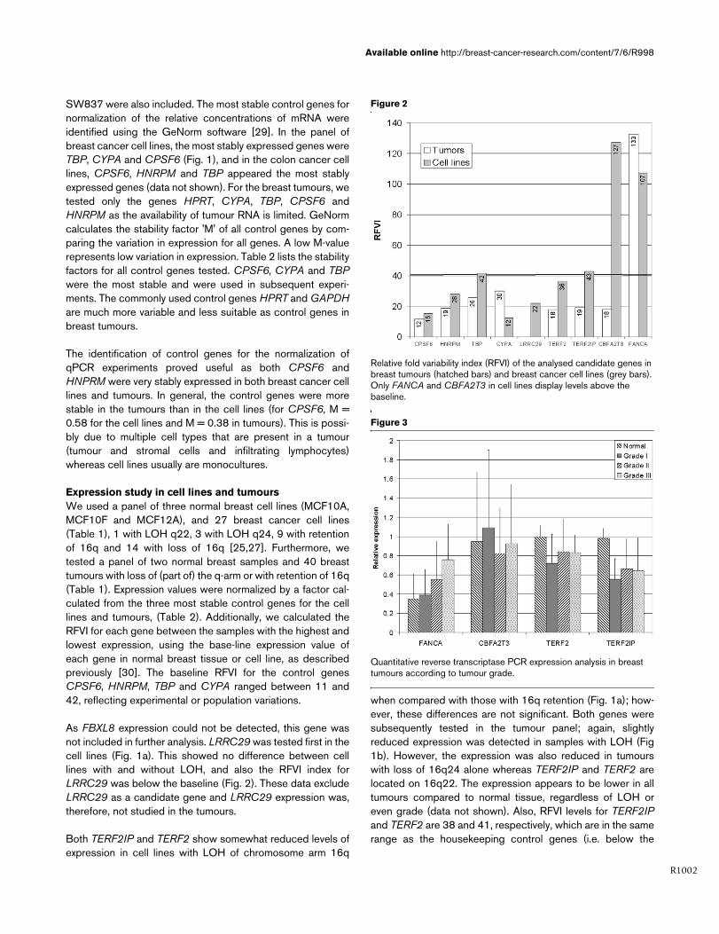
Quantitative reverse transcriptase PCR expression analysis in breast tumours according to tumour gradeQuantitative reverse transcriptase PCR expression analysis in breast tumours according to tumour grade.

Breast Cancer Research Vol 7 No 6 van Wezel et al.
R1003
baseline level). Based on their gene expression variation,therefore, neither genes are likely to be candidate TSGs.
CBFA2T3 encodes a translocation partner of AML1 in mye-loid leukaemia and a transcription repressor. Based on itslocation on chromosomal band 16q24.3, high variation inexpression in breast cancer cell lines, loss of protein expres-sion in breast tumours and in vitro growth inhibition of breastcancer cell lines, CBFA2T3 has been proposed as a candi-date TSG [9,30]. We therefore tested this gene in our panelof cell lines and tumours to confirm this finding. We found alarge expression variation for CBFA2T3 in cell lines, resultingin a high RFVI value of 122 (Fig. 2), which, however, was notassociated with LOH at 16q (Fig. 1a). In primary tumours,there was a tendency for higher CBFA2T3 expression intumours with LOH of 16q24 (Fig. 1b), where this gene islocated, and the RFVI value was below the baseline. Thesedata do not support the candidacy of CBFA2T3 as a TSG on16q.
FANCA is involved in DNA repair and located at 16q24.3. Itsexpression in tumours is higher than in normal tissue and,remarkably, expression increases with tumour grade (Fig. 3).There is no association between LOH at 16q and tumourgrade, but there is a difference in the mechanism of LOH whencomparing low- and high-grade breast cancers. Whereas low-grade tumours show preferential physical loss of 16q, high-grade tumours show mitotic recombination [33]. This couldindicate that FANCA is involved only in well differentiatedbreast cancer. In cell lines, FANCA levels are slightly reducedin tumours with LOH. Interestingly, in tumours with loss of thecomplete q-arm, FANCA expression is lower than in thosewith retention of 16q. In those with loss of q21-ter, however,expression is higher than in tumours with retention, suggestingagain that the mechanism of 16q LOH may be associated withthe targeted TSG.
Fragment analysisTERF2IP, TERF2, FBXL8 and LRRC29 were screened usingfragment analysis for (small) genomic deletions or insertions intheir exons in 21 breast cancer cell lines and 32 breasttumours. CBFA2T3 and FANCA were previously screened formutations, without identifying any inactivating or somatic muta-tions [5,9]. Although TSGs are, in many cases, inactivatedthrough deletions and insertions, no mutations were found.
ConclusionWe studied CBFA2T3, FANCA, FBXL8, LRRC29, TERF2and TERF2IP, six potential breast cancer TSG candidategenes located on the long arm of chromosome 16, which isinvolved in LOH in more than 50% of breast cancer cases.These genes were studied using qPCR to detect possibletranscriptional down-regulation in a representative panel ofbreast cancer cell lines and primary tumours with well definedpatterns of LOH at chromosome 16q. For reliable qPCR, two
new, stable control genes for normalization of qPCR experi-ments, HNRPM and CPSF6, were identified. We did notdetect any significant difference in expression of the candidategenes related to the LOH status of tumours and cell lines.Mutation analysis of these genes did not reveal inactivating,tumour specific alterations. Therefore, these genes are unlikelyto be candidates for the classic tumour suppressor gene onchromosome 16q. The identification of the underlying tumoursuppressor genes and their mechanisms of inactivationremains a difficult task. New insights into neoplastic transfor-mation indicate that somatic tumour genetics are far morecomplex than originally conceived, involving multiple non-clas-sic TSGs with individual small effects [34,35]. Successfulidentification of these genes requires an integrated genomicapproach, combining the analysis of LOH, copy numberchanges and expression studies.
Competing interestsThe authors declare that they have no competing interests.
Authors' contributionsTVW drafted the manuscript, performed qPCR and mutationanalysis, designed and coordinated the study and performedthe analysis. ML participated in study design, performedqPCR. EHVR performed qPCR and analysis and isolatedRNA, KP performed qPCR and RNA isolation HJB helpeddesign the qPCR study, KS helped design the qPCR study,CJC participated in design, coordination of the study andAMCJ conceived of the study and participated in design, coor-dination of the study and helped to draft the manuscript. Allauthors read and approved the final manuscript.
AcknowledgementsGrant support: Dutch cancer society RUL 2000–2205. We thank Domi-nique Bongaerts, Anna Roukens and Sandra van Eijgen for experimental support.
References1. Cleton-Jansen AM, Moerland EW, Kuipers-Dijkshoorn NJ, Callen
DF, Sutherland GR, Hansen B, Devilee P, Cornelisse CJ: At leasttwo different regions are involved in allelic imbalance on chro-mosome arm 16q in breast cancer. Genes ChromosomesCancer 1994, 9:101-107.
2. Knudson AG Jr: Genetic predisposition to cancer. CancerDetect Prev 1984, 7:1-8.
3. Cleton-Jansen AM, Callen DF, Seshadri R, Goldup S, Mccallum B,Crawford J, Powell JA, Settasatian C, van Beerendonk H, MoerlandEW, et al.: Loss of heterozygosity mapping at chromosomearm 16q in 712 breast tumors reveals factors that influencedelineation of candidate regions. Cancer Res 2001,61:1171-1177.
4. Berx G, Cleton-Jansen AM, Nollet F, De Leeuw WJ, d van V, Cor-nelisse C, Van Roy F: E-cadherin is a tumour/invasion suppres-sor gene mutated in human lobular breast cancers. EMBO J1995, 14:6107-6115.
5. Cleton-Jansen AM, Moerland EW, Pronk JC, van Berkel CG, Apos-tolou S, Crawford J, Savoia A, Auerbach AD, Mathew CG, CallenDF, et al.: Mutation analysis of the Fanconi anaemia A gene inbreast tumours with loss of heterozygosity at 16q24.3. Br JCancer 1999, 79:1049-1052.
6. Crawford J, Ianzano L, Savino M, Whitmore S, Cleton-Jansen AM,Settasatian C, d'Apolito M, Seshadri R, Pronk JC, Auerbach AD, etal.: The PISSLRE gene:structure, exon skipping, and exclusion
http://www.ncbi.nlm.nih.gov/entrez/query.fcgi?cmd=Retrieve&db=PubMed&dopt=Abstract&list_uids=7513539
http://www.ncbi.nlm.nih.gov/entrez/query.fcgi?cmd=Retrieve&db=PubMed&dopt=Abstract&list_uids=7513539
http://www.ncbi.nlm.nih.gov/entrez/query.fcgi?cmd=Retrieve&db=PubMed&dopt=Abstract&list_uids=7513539
http://www.ncbi.nlm.nih.gov/entrez/query.fcgi?cmd=Retrieve&db=PubMed&dopt=Abstract&list_uids=6367981
http://www.ncbi.nlm.nih.gov/entrez/query.fcgi?cmd=Retrieve&db=PubMed&dopt=Abstract&list_uids=8557030

Available online http://breast-cancer-research.com/content/7/6/R998
R1004
as tumor suppressor in breast cancer. Genomics 1999,56:90-97.
7. Moerland E, Breuning MH, Cornelisse CJ, Cleton-Jansen AM:Exclusion of BBC1 and CMAR as candidate breast tumour-suppressor genes. Br J Cancer 1997, 76:1550-1553.
8. Whitmore SA, Settasatian C, Crawford J, Lower KM, Mccallum B,Seshadri R, Cornelisse CJ, Moerland EW, Cleton-Jansen AM, Tip-ping AJ, et al.: Characterization and screening for mutations ofthe growth arrest- specific 11 (GAS11) and C16orf3 genes at16q24.3 in breast cancer. Genomics 1998, 52:325-331.
9. Kochetkova M, McKenzie OL, Bais AJ, Martin JM, Secker GA, Ses-hadri R, Powell JA, Hinze SJ, Gardner AE, Spendlove HE, et al.:CBFA2T3 (MTG16) is a putative breast tumor suppressor genefrom the breast cancer loss of heterozygosity region at16q24.3. Cancer Res 2002, 62:4599-4604.
10. Filippova GN, Qi CF, Ulmer JE, Moore JM, Ward MD, Hu YJ, Louki-nov DI, Pugacheva EM, Klenova EM, Grundy PE, et al.: Tumor-associated zinc finger mutations in the CTCF transcription fac-tor selectively alter tts DNA-binding specificity. Cancer Res2002, 62:48-52.
11. Watanabe A, Hippo Y, Taniguchi H, Iwanari H, Yashiro M,Hirakawa K, Kodama T, Aburatani H: An opposing view onWWOX protein function as a tumor suppressor. Cancer Res2003, 63:8629-8633.
12. Devilee P, Cleton-Jansen AM, Cornelisse CJ: Ever sinceKnudson. Trends Genet 2001, 17:569-573.
13. Lander ES, Linton LM, Birren B, Nusbaum C, Zody MC, Baldwin J,Devon K, Dewar K, Doyle M, FitzHugh W, et al.: Initial sequencingand analysis of the human genome. Nature 2001,409:860-921.
14. Venter JC, Adams MD, Myers EW, Li PW, Mural RJ, Sutton GG,Smith HO, Yandell M, Evans CA, Holt RA, et al.: The sequence ofthe human genome. Science 2001, 291:1304-1351.
15. Ye JZ, Donigian JR, Van Overbeek M, Loayza D, Luo Y, KrutchinskyAN, Chait BT, de Lange T: TIN2 binds TRF1 and TRF2 simulta-neously and stabilizes the TRF2 complex on telomeres. J BiolChem 2004, 279:47264-47271.
16. van Steensel B, Smogorzewska A, de Lange T: TRF2 protectshuman telomeres from end-to-end fusions. Cell 1998,92:401-413.
17. Li B, de Lange T: Rap1 affects the length and heterogeneity ofhuman telomeres. Mol Biol Cell 2003, 14:5060-5068.
18. Yamada K, Yagihashi A, Yamada M, Asanuma K, Moriai R, Koba-yashi D, Tsuji N, Watanabe N: Decreased gene expression fortelomeric-repeat binding factors and TIN2 in malignant hemat-opoietic cells. Anticancer Res 2002, 22:1315-1320.
19. Yamada M, Tsuji N, Nakamura M, Moriai R, Kobayashi D, YagihashiA, Watanabe N: Down-regulation of TRF1, TRF2 and TIN2genes is important to maintain telomeric DNA for gastriccancers. Anticancer Res 2002, 22:3303-3307.
20. Calhoun ES, Jones JB, Ashfaq R, Adsay V, Baker SJ, Valentine V,Hempen PM, Hilgers W, Yeo CJ, Hruban RH, et al.: BRAF andFBXW7 (CDC4, FBW7, AGO, SEL10) mutations in distinct sub-sets of pancreatic cancer:potential therapeutic targets. Am JPathol 2003, 163:1255-1260.
21. Mao JH, Perez-losada J, Wu D, DelRosario R, Tsunematsu R,Nakayama K, Brown K, Bryson S, Balmain A: Fbxw7/Cdc4 is ap53-dependent, haploinsufficient tumour suppressor gene.Nature 2004, 432:775-779.
22. Strohmaier H, Spruck CH, Kaiser P, Won KA, Sangfelt O, Reed SI:Human F-box protein hCdc4 targets cyclin E for proteolysisand is mutated in a breast cancer cell line. Nature 2001,413:316-322.
23. Howlett NG, Taniguchi T, Olson S, Cox B, Waisfisz Q, Die-Smulders C, Persky N, Grompe M, Joenje H, Pals G, et al.: Bial-lelic Inactivation of BRCA2 in Fanconi Anemia. Science 2002,297:606-609.
24. D'Andrea AD, Grompe M: The Fanconi anaemia/BRCApathway. Nat Rev Cancer 2003, 3:23-34.
25. Callen DF, Crawford J, Derwas C, Cleton-Jansen AM, CornelisseCJ, Baker E: Defining regions of loss of heterozygosity of 16qin breast cancer cell lines. Cancer Genet Cytogenet 2002,133:76-82.
26. Harkes IC, Elstrodt F, Dinjens WN, Molier M, Klijn JG, Berns EM,Schutte M: Allelotype of 28 human breast cancer cell lines andxenografts. Br J Cancer 2003, 89:2289-2292.
27. van de Wetering ML, Barker N, Harkes IC, van der Heyden M, DijkNJ, Hollestelle A, Klijn JG, Clevers H, Schutte M: Mutant E-cad-herin breast cancer cells do not display constitutive Wntsignaling. Cancer Res 2001, 61:278-284.
28. Rozen S, Skaletsky H: Primer3 on the WWW for general usersand for biologist programmers. Methods Mol Biol 2000,132:365-386.
29. Vandesompele J, De Preter K, Pattyn F, Poppe B, Van Roy N, DePaepe A, Speleman F: Accurate normalization of real-timequantitative RT-PCR data by geometric averaging of multipleinternal control genes. Genome Biol 2002,3:research0034.1-research0034.11.
30. Powell JA, Gardner AE, Bais AJ, Hinze SJ, Baker E, Whitmore S,Crawford J, Kochetkova M, Spendlove HE, Doggett NA, et al.:Sequencing, transcript identification, and quantitative geneexpression profiling in the breast cancer loss of heterozygos-ity region 16q24.3 reveal three potential tumor-suppressorgenes. Genomics 2002, 80:303-310.
31. Ganguly A, Rock MJ, Prockop DJ: Conformation-sensitive gelelectrophoresis for rapid detection of single-base differencesin double-stranded PCR products and DNA fragments:evi-dence for solvent-induced bends in DNA heteroduplexes.Proc Natl Acad Sci U S A 1993, 90:10325-10329.
32. Eng C, Brody LC, Wagner TM, Devilee P, Vijg J, Szabo C, TavtigianSV, Nathanson KL, Ostrander E, Frank TS: Interpreting epidemi-ological research:blinded comparison of methods used toestimate the prevalence of inherited mutations in BRCA1. JMed Genet 2001, 38:824-833.
33. Cleton-Jansen AM, Buerger H, Haar N, Philippo K, van de VijverMJ, Boecker W, Smit VT, Cornelisse CJ: Different mechanismsof chromosome 16 loss of heterozygosity in well- versuspoorly differentiated ductal breast cancer. Genes Chromo-somes Cancer 2004, 41:109-116.
34. Fabarius A, Willer A, Yerganian G, Hehlmann R, Duesberg P: Spe-cific aneusomies in Chinese hamster cells at different stagesof neoplastic transformation, initiated by nitrosomethylurea.Proc Natl Acad Sci U S A 2002, 99:6778-6783.
35. Rubin H: Degrees and kinds of selection in spontaneous neo-plastic transformation:An operational analysis. Proc Natl AcadSci USA 2005, 102:9276-9281.
http://www.ncbi.nlm.nih.gov/entrez/query.fcgi?cmd=Retrieve&db=PubMed&dopt=Abstract&list_uids=9413939
http://www.ncbi.nlm.nih.gov/entrez/query.fcgi?cmd=Retrieve&db=PubMed&dopt=Abstract&list_uids=9413939
http://www.ncbi.nlm.nih.gov/entrez/query.fcgi?cmd=Retrieve&db=PubMed&dopt=Abstract&list_uids=9413939
http://www.ncbi.nlm.nih.gov/entrez/query.fcgi?cmd=Retrieve&db=PubMed&dopt=Abstract&list_uids=9790751
http://www.ncbi.nlm.nih.gov/entrez/query.fcgi?cmd=Retrieve&db=PubMed&dopt=Abstract&list_uids=9790751
http://www.ncbi.nlm.nih.gov/entrez/query.fcgi?cmd=Retrieve&db=PubMed&dopt=Abstract&list_uids=9790751
http://www.ncbi.nlm.nih.gov/entrez/query.fcgi?cmd=Retrieve&db=PubMed&dopt=Abstract&list_uids=9476899
http://www.ncbi.nlm.nih.gov/entrez/query.fcgi?cmd=Retrieve&db=PubMed&dopt=Abstract&list_uids=9476899
http://www.ncbi.nlm.nih.gov/entrez/query.fcgi?cmd=Retrieve&db=PubMed&dopt=Abstract&list_uids=8234293
Related Documents