This journal is © the Owner Societies 2014 Phys. Chem. Chem. Phys. Cite this: DOI: 10.1039/c4cp05095a Exploring the binding mechanisms of MIF to CXCR2 using theoretical approaches† Lei Xu, a Youyong Li, b Dan Li, a Peng Xu, c Sheng Tian, b Huiyong Sun, b Hui Liu a and Tingjun Hou* a Macrophage migration inhibitory factor (MIF) is a multi-functional protein that acts as a cytokine and as an enzyme. Recently, MIF was identified as a non-canonical ligand of G protein-coupled chemokine receptor CXCR2 with low nanomolar affinity in leukocyte arrest and chemotaxis, but the precise knowledge of the molecular determinants of the MIF–CXCR2 interface has remained unknown. Therefore, we employed homology modeling, protein–protein docking, molecular dynamics (MD) simulations, Molecular Mechanics/Generalized Born Surface Area (MM/GBSA) binding free energy calculations and MM/GBSA binding free energy decomposition to obtain insights into the molecular recognition of MIF with CXCR2. The predicted binding pattern of MIF–CXCR2 is in good agreement with the experimental data and sheds light on the functional role of important MIF–CXCR2 interface residues in association with binding and signaling. According to our predictions, the R11A/D44A double mutations of MIF exhibit a pronounced defect in the binding affinity of MIF to CXCR2, resulting in large conformational changes. The potential two-site binding model for the MIF–CXCR2 recognition was proposed: initialized primarily by the non-polar interactions including the van der Waals and hydrophobic interactions, the N-terminal region of CXCR2 contacts the N-like loop and b-sheet of MIF (site 1), and then the ECL2 and ECL3 regions of CXCR2 form strong interactions with the pseudo-(E)LR motif and C-terminus of MIF, which induces the molecular thermodynamic motion of TMs for signal transduction (site 2). This study will extend our understanding to the binding mechanisms of MIF to CXCR2 and provide useful information for the rational design of potent inhibitors selectively targeting the MIF–CXCR2 interactions. Introduction Macrophage migration inhibitory factor (MIF) is a proinflamma- tory cytokine that regulates both the innate and adaptive immune responses. 1 MIF can also activate the mitogen- activated protein kinase (MAPK) cascade and counter-act the inhibitory effects of glucocorticoids within the immune system. 2 Growing evidence has highlighted the importance of MIF in tumor growth, such as control of cell proliferation and promotion of angiogenesis. 3 In addition to its physiological and pathological activities, MIF is regarded as a D-dopachrome tautomerase, 4 a phenylpyruvate tautomerase 5 and a thiol–protein oxidoreductase. 6 Accordingly, MIF is recognized as an important therapeutic target for inflammatory diseases and tumors. Cytokines usually activate signal-transduction pathways, gene transcription and the expression of downstream effector a College of Pharmaceutical Sciences, Zhejiang University, Hangzhou, Zhejiang 310058, China. E-mail: [email protected], [email protected]; Tel: +86-571-88208412 b Institute of Functional Nano & Soft Materials (FUNSOM), Soochow University, Suzhou, Jiangsu 215123, China c Department of Orthopedics, Second Military Medical University affiliated Changzheng Hospital, Shanghai, 200003, China † Electronic supplementary information (ESI) available: Table S1 Binding free energy contributions of the key binding-site residues of CXCR2 predicted by the binding energy decomposition (kcal mol 1 ). Table S2 Binding free energy contributions of the key binding-site residues of MIF predicted by the binding energy decomposition (kcal mol 1 ). Table S3 Binding free energy contributions of the key binding-site residues of CXCR2 in the mutated complex predicted by the binding energy decomposition (kcal mol 1 ). Fig. S1 Sequence alignment of CXCR2 (residues 1–339) and CXCR4 (PDB ID: 3ODU) used for homology modeling. Fig. S2 MD simulation box of the MIF–CXCR2 complex, the lipid and water molecules. There are 125 121 atoms in the simulation box (Model I was chosen to represent). Fig. S3 The Ramachandran plot of CXCR2 constructed by homology modeling. Fig. S4 Comparison of the complex structure predicted by the protein–protein docking (ribbon colored in gray) and the conformation after the last 20 ns MD trajectory (ribbon colored in green). The arrow indicates the movement of TM1, TM4 and TM5. Fig. S5 Schematic depiction of the major interactions of the averaged structure of MIF–CXCR2 over the last 10 ns MD trajectory (generated by the LIGPLOT program). (a) and (b) represent residues 1 to 57 and 58 to 114 of MIF respectively. Fig. S6 Schematic depiction of the major interactions of the averaged structure of the R11A/D44A mutant of MIF with CXCR2 over the last 10 ns MD trajectory (generated by the LIGPLOT program). (a) and (b) represent residues 1 to 57 and 58 to 114 of MIF respectively. See DOI: 10.1039/c4cp05095a Received 4th November 2014, Accepted 11th December 2014 DOI: 10.1039/c4cp05095a www.rsc.org/pccp PCCP PAPER Published on 11 December 2014. Downloaded by Soochow University China on 27/12/2014 04:15:53. View Article Online View Journal

Welcome message from author
This document is posted to help you gain knowledge. Please leave a comment to let me know what you think about it! Share it to your friends and learn new things together.
Transcript
This journal is© the Owner Societies 2014 Phys. Chem. Chem. Phys.
Cite this:DOI: 10.1039/c4cp05095a
Exploring the binding mechanisms of MIF toCXCR2 using theoretical approaches†
Lei Xu,a Youyong Li,b Dan Li,a Peng Xu,c Sheng Tian,b Huiyong Sun,b Hui Liua andTingjun Hou*a
Macrophage migration inhibitory factor (MIF) is a multi-functional protein that acts as a cytokine and as
an enzyme. Recently, MIF was identified as a non-canonical ligand of G protein-coupled chemokine
receptor CXCR2 with low nanomolar affinity in leukocyte arrest and chemotaxis, but the precise
knowledge of the molecular determinants of the MIF–CXCR2 interface has remained unknown. Therefore,
we employed homology modeling, protein–protein docking, molecular dynamics (MD) simulations,
Molecular Mechanics/Generalized Born Surface Area (MM/GBSA) binding free energy calculations and
MM/GBSA binding free energy decomposition to obtain insights into the molecular recognition of MIF with
CXCR2. The predicted binding pattern of MIF–CXCR2 is in good agreement with the experimental
data and sheds light on the functional role of important MIF–CXCR2 interface residues in association with
binding and signaling. According to our predictions, the R11A/D44A double mutations of MIF exhibit a
pronounced defect in the binding affinity of MIF to CXCR2, resulting in large conformational changes. The
potential two-site binding model for the MIF–CXCR2 recognition was proposed: initialized primarily by the
non-polar interactions including the van der Waals and hydrophobic interactions, the N-terminal region of
CXCR2 contacts the N-like loop and b-sheet of MIF (site 1), and then the ECL2 and ECL3 regions of
CXCR2 form strong interactions with the pseudo-(E)LR motif and C-terminus of MIF, which induces the
molecular thermodynamic motion of TMs for signal transduction (site 2). This study will extend our
understanding to the binding mechanisms of MIF to CXCR2 and provide useful information for the rational
design of potent inhibitors selectively targeting the MIF–CXCR2 interactions.
Introduction
Macrophage migration inhibitory factor (MIF) is a proinflamma-tory cytokine that regulates both the innate and adaptiveimmune responses.1 MIF can also activate the mitogen-activated protein kinase (MAPK) cascade and counter-act theinhibitory effects of glucocorticoids within the immune system.2
Growing evidence has highlighted the importance of MIF in
tumor growth, such as control of cell proliferation and promotionof angiogenesis.3 In addition to its physiological and pathologicalactivities, MIF is regarded as a D-dopachrome tautomerase,4 aphenylpyruvate tautomerase5 and a thiol–protein oxidoreductase.6
Accordingly, MIF is recognized as an important therapeutic targetfor inflammatory diseases and tumors.
Cytokines usually activate signal-transduction pathways,gene transcription and the expression of downstream effector
a College of Pharmaceutical Sciences, Zhejiang University, Hangzhou, Zhejiang 310058, China. E-mail: [email protected], [email protected];
Tel: +86-571-88208412b Institute of Functional Nano & Soft Materials (FUNSOM), Soochow University, Suzhou, Jiangsu 215123, Chinac Department of Orthopedics, Second Military Medical University affiliated Changzheng Hospital, Shanghai, 200003, China
† Electronic supplementary information (ESI) available: Table S1 Binding free energy contributions of the key binding-site residues of CXCR2 predicted by the bindingenergy decomposition (kcal mol�1). Table S2 Binding free energy contributions of the key binding-site residues of MIF predicted by the binding energy decomposition(kcal mol�1). Table S3 Binding free energy contributions of the key binding-site residues of CXCR2 in the mutated complex predicted by the binding energydecomposition (kcal mol�1). Fig. S1 Sequence alignment of CXCR2 (residues 1–339) and CXCR4 (PDB ID: 3ODU) used for homology modeling. Fig. S2 MD simulationbox of the MIF–CXCR2 complex, the lipid and water molecules. There are 125 121 atoms in the simulation box (Model I was chosen to represent). Fig. S3 TheRamachandran plot of CXCR2 constructed by homology modeling. Fig. S4 Comparison of the complex structure predicted by the protein–protein docking (ribboncolored in gray) and the conformation after the last 20 ns MD trajectory (ribbon colored in green). The arrow indicates the movement of TM1, TM4 and TM5. Fig. S5Schematic depiction of the major interactions of the averaged structure of MIF–CXCR2 over the last 10 ns MD trajectory (generated by the LIGPLOT program). (a) and(b) represent residues 1 to 57 and 58 to 114 of MIF respectively. Fig. S6 Schematic depiction of the major interactions of the averaged structure of the R11A/D44A mutantof MIF with CXCR2 over the last 10 ns MD trajectory (generated by the LIGPLOT program). (a) and (b) represent residues 1 to 57 and 58 to 114 of MIF respectively.See DOI: 10.1039/c4cp05095a
Received 4th November 2014,Accepted 11th December 2014
DOI: 10.1039/c4cp05095a
www.rsc.org/pccp
PCCP
PAPER
Publ
ishe
d on
11
Dec
embe
r 20
14. D
ownl
oade
d by
Soo
chow
Uni
vers
ity C
hina
on
27/1
2/20
14 0
4:15
:53.
View Article OnlineView Journal

Phys. Chem. Chem. Phys. This journal is© the Owner Societies 2014
molecules by binding to cognate receptors. By binding to theextracellular domain of CD74, MIF activates the extracellular-regulated mitogen-activated protein (ERK-MAP) kinase and isassociated into a signaling complex with CD44 and Src-tyrosinekinases.7,8 Bernhagen et al. reported that MIF is a noncognateligand of the CXC chemokine receptors CXCR2 and CXCR4,and is instrumental in inflammatory leukocyte recruitment inatherosclerosis, targeting monocytes and neutrophils throughCXCR2 and T cells through CXCR4. MIF can bind to CXCR2with low nanomolar affinity and induce CXCR2-mediatedleukocyte arrest and chemotaxis.9,10 Chemokine receptors belongto the rhodopsin-like family of G-protein-coupled receptors(GPCRs) and are termed on the basis of the correspondingbinding chemokines, such as CR, CCR, CXCR and CX3CR.11
There are two subgroups of the CXC chemokines, termed asthe ELR+ and ELR� chemokines. Mutagenesis or alanine scan-ning experiments demonstrated that substitution of the ELRresidues at the N terminus of the ELR+ CXC chemokines, suchas CXCL18, leads to a dramatic loss of CXCR2 binding affinity.12
Recently the emerging group of ‘chemokine-like function’ (CLF)includes chemotactic polypeptides such as human b-defensin(HBD), which cannot be classified into known chemokine sub-families owing to the lack of the prototypical N-terminal cysteinemotif but exhibit structural or functional features and can signalthrough chemokine receptors, for instance, CCR6.13,14
MIF belongs to the class of CLF chemokines, and plays a keyrole in the progression of atherosclerosis, including leukocyterecruitment and arrest.15 In 2008, Weber et al. revealed that MIFharbored a pseudo-(E)LR motif (Asp-44-X-Arg-11) constitutedby non-adjacent residues in neighboring loops with identicalparallel spacing of the canonical ELR motif.16 Although MIFdoes not share any significant sequence homology with CXCL8,the 3D structure of the MIF monomer exhibits structural homo-logy with the CXCL8 dimer. In 2011, Kraemer et al. identified anN-like loop (sequence stretch 46–55) of MIF, which appears tomirror the N loop of CXCL8 (Fig. 1), by applying a variety ofbiochemical, biophysical and functional assays associated withatherosclerotic leukocyte recruitment processes.17
Although the emerging importance of the MIF–CXCR2 inter-actions in the inflammatory pathogenesis of atherosclerosishas attracted extensive attention, the molecular details of theprotein–protein interaction interface between MIF and CXCR2have remained unknown. Based on the above experimentaldata, we employed a comprehensive set of computational toolsto explore the MIF–CXCR2 interaction in the current study.First, homology modeling was carried out to construct the 3Dmodel of CXCR2 based on the crystal structure of CXCR4.18
Then, the ZDOCK approach was employed to predict thepotential binding modes of the MIF monomer in complex withCXCR2. The best complex structure in each cluster and theR11A/D44A-MIF double mutant were subjected to 20 ns MDsimulations and the binding affinity between MIF and CXCR2was estimated using the Molecular Mechanics/GeneralizedBorn Surface Area (MM/GBSA) approach. At last, MM/GBSAbinding energy decomposition analysis was employed to highlightthe key residues responsible for the MIF–CXCR2 interactions.
We expect that this study can provide useful information intackling the binding mechanisms of MIF to CXCR2.
Materials and methodsHomology modeling
The 3D model of the human CXCR2 was constructed based onthe structure of CXCR4 by using the modeller method19 imple-mented in Discovery Studio (DS) molecular simulation package(version 2.5).20 The N-terminal 26 residues (PDB entry: 3ODU)of the CXCR4 crystal structure do not have interpretable density
Fig. 1 Structure and surface models of CXCL8 and MIF monomer. (a) TheELR motif located in the N-terminus and N-loop (orange) of CXCL8; (b) theelectrostatic potential map of CXCL8 calculated by Delphi; (c) the pseudo-(E)LR motif and N-like loop (green) of MIF monomer; (d) the electrostaticpotential map of MIF monomer. The equipotential contours at �1 kT e�1
and +1 kT e�1 are shown. Red indicates �1 kT e�1 and blue +1 kT e�1;(e) trimeric structure of MIF shown in surface mode. N-like loop regionsand pseudo-(E)LR motifs of the three monomers are shown in purple andblue respectively.
Paper PCCP
Publ
ishe
d on
11
Dec
embe
r 20
14. D
ownl
oade
d by
Soo
chow
Uni
vers
ity C
hina
on
27/1
2/20
14 0
4:15
:53.
View Article Online
This journal is© the Owner Societies 2014 Phys. Chem. Chem. Phys.
and are presumed to be disordered.18 So we modeled themissing N-terminal part of CXCR4 based on the NMR data ofthe CXCR4 N-terminus in complex with CXCL12 (PDB entry:2K04)21 using DS 2.5 as we did in our recent study to model thestructure of CXCR4 in complex with CXCL12.22 The amino acidsequence of human CXCR2 was retrieved from the Uniprotdatabase (accession number: P25025), and the sequences werealigned (Fig. S1, ESI†) based on the sequence analysis ofchemokine receptor families.23,24 Then, the homology modelof CXCR2 was constructed based on the 3D structure of CXCR4by using a modeller in DS 2.5.20 The conformations ofN-terminus and extracellular loops (ECLs) were constrained bytwo disulfide bonds, one linking Cys39 at the N-terminal partwith Cys286, and the other connecting Cys119 with Cys196 ofECL2, as indicated in the recent crystal structures of chemokinereceptors.18,25 The final model was minimized by 10 000 cyclesusing the CHARMM force field and implicit solvent model(a generalized Born model with molecular volume) in DS 2.5.20
Protein–protein docking
The crystal structure of MIF was retrieved from the protein databank (PDB entry: 1LJT26), and chain A was kept and the othertwo chains were eliminated. The ZDOCK approach27 imple-mented in DS 2.5 was employed to predict the structure of theMIF monomer in complex with CXCR2. ZDOCK shows good
performance in the CAPRI challenge in the prediction ofprotein–protein complexes,27 by adopting pairwise shapecomplementarity, desolvation and electrostatics as scoringfunctions optimized using fast Fourier transform.28 ZDOCKcan exhaustively search all rotational and translational spacefor a ligand protein with respect to its receptor protein.29 Here,five important residues (Arg11, Asp44 and Leu46 for MIF, andTyr23 and Glu198 for CXCR2) were constrained to be inside thebinding interface according to the experimental data,16,17 andall the atoms of these residues are within the distance cutoff of10 Å to any atom of the other protein. The ZRANK re-rankingmethod was then employed to re-evaluate the docked posespredicted using the ZDOCK calculations.30 RDOCK was used tooptimize and score the docked poses based on the CHARMMforce field.29 At last, 148 docked poses were structurally classi-fied into three clusters, and the representative structure of eachcluster was chosen for the following MD simulations.
Molecular dynamics (MD) simulations
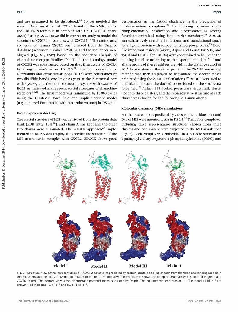
For the best complex predicted by ZDOCK, the residues R11 andD44 of MIF were mutated to Ala in DS 2.5.20 Then, four complexes,including three representative structures chosen from threeclusters and one mutant were subjected to the MD simulations(Fig. 2). Each complex was embedded in a periodic structure of1-palmytoyl-2-oleoyl-sn-glycero-3-phosphatidylcholine (POPC), and
Fig. 2 Structural view of the representative MIF–CXCR2 complexes predicted by protein–protein docking chosen from the three best binding models inthree clusters and the R11A/D44A double mutant of Model I. The top view in each column shows the complex structure (MIF is colored in green andCXCR2 in red). The bottom view is the electrostatic potential maps calculated by Delphi. The equipotential contours at �1 kT e�1 and +1 kT e�1 areshown. Red indicates �1 kT e�1 and blue +1 kT e�1.
PCCP Paper
Publ
ishe
d on
11
Dec
embe
r 20
14. D
ownl
oade
d by
Soo
chow
Uni
vers
ity C
hina
on
27/1
2/20
14 0
4:15
:53.
View Article Online

Phys. Chem. Chem. Phys. This journal is© the Owner Societies 2014
the lipid molecules within 5 Å of the complex were eliminated inVMD.31 Each system was then inserted into a water box (TIP3water model)32 and the waters within 5 Å of the lipid and proteinwere removed. Two Cl� were placed in the grids with the strongestpositive coulombic potentials in order to neutralize the system.The whole system includes the MIF monomer, CXCR2, lipidmolecules, water molecules and counterions, with 125 121,124 886, 124 985 and 125 105 atoms per periodic cell for fourmodels, respectively (Fig. S2, ESI†), and the box size is B110 Å �110 Å� 110 Å. The Amber ff99SB force field was employed for thecomplex.33 The lipid parameters were established by Lipidbook,34
a public database of force field parameters with special emphasison lipids based on the general AMBER force field (gaff).35 Theparticle mesh Ewald (PME) algorithm was employed to handle thelong-range electrostatics.36 The whole system was first equili-brated for 1 ns with the protein fixed under constant temperature(310 K) and constant pressure (1 atm). Then, another 1 ns equili-bration was carried out without any restraint. Starting from the lastframe of the equilibration, 20 ns simulations were carried out. TheMD simulations were performed by using the NAMD 2.9.37 TheSHAKE procedure was employed to constrain all bonds involvinghydrogen atoms,38 and the time step was set to 2 fs. Coordinatetrajectories were saved every 10 ps in the process of MD runs.
MM/GBSA binding free energy calculations
To determine the most stable MIF–CXCR2 complex predicted byprotein–protein docking, the binding free energy (DGbind) for eachcomplex was estimated using the MM/GBSA methodology basedon the snapshots retrieved from the single MD trajectory of eachcomplex, which is much faster and requires less sampling incomparison with the separate-trajectory protocol.39–45 In MM/GBSA, DGbind was evaluated through the following equation:46
DGbind = Gcomplex � Gprotien � Gligand = DH + DGsolvation � TDS
= DEMM + DGGB + DGSA � TDS (1)
where DEMM is the gas-phase interaction energy between MIFand CXCR2, including the electrostatic and van der Waalsenergies; DGGB and DGSA are the polar and non-polar contribu-tions of the desolvation free energy, respectively; �TDS is thechange of the conformational entropy upon ligand binding,47
which was calculated by the normal-mode analysis (NMA)based on the 20 snapshots evenly extracted from the last10 ns of each MD trajectory. The modified GB model (igb = 2)developed by Onufriev and coworkers was employed to estimatethe electrostatic desolvation energy.48 The exterior and solutedielectric constants were set to 80 and 4, respectively. Thenonpolar part of desolvation was estimated by the solventaccessible surface area (SASA) with the Laboratoire de Chimiedes Polymres Organiques (LCPO) model:49 DGSA = 0.0072 �DSASA. In total, 1000 snapshots extracted from the last 10 nsMD trajectory were used to calculate all energy terms.
MM/GBSA binding energy decomposition
In order to obtain the contribution of each residue to thebinding free energy, MM/GBSA was employed to decompose
the total binding free energy into the four contributions fromindividual residue–residue pairs,50–53 including the van derWaals contribution (DEvdw), the electrostatic contribution(DEele), the polar desolvation contribution (DGGB) and thenon-polar desolvation contribution (DGSA), as shown in eqn (2):
DGresidue–residue = DGvdw + DGele + DGsolvation
= DGvdw + DGele + DGGB + DGSA (2)
The GB model with the GB parameters developed by Onufrievet al.48 was used to calculate the electrostatic desolvation energy(DGGB). The exterior dielectric and solute dielectric constantswere set to 80 and 4, respectively. The non-polar component ofdesolvation (DGSA) was estimated by SASA through the ICOSAtechnique.53 The 1000 snapshots extracted from the last 10 nsMD trajectory were used to estimate the energy terms shownin eqn (2).
Dynamic cross-correlation map (DCCM)
In order to evaluate the dynamic correlations between differentprotein domains, DCCM analysis was employed to compare thecorrelation differences for CXCR2.20,54 The correlation matrixwas evaluated across all Ca atoms of Model 1 and its R11A/D44A-double mutant. The correlation coefficient Sij betweentwo atoms i and j over the course of the simulation trajectory isdefined as:
Sij ¼Dri � Drj� �
ffiffiffiffiffiffiffiffiffiffiffiffiffiffiffiffiffiffiffiffiffiffiffiffiffiffiffiffiffiffiffiffiffiffiffiffiffiffiffiffiffiDri � Drii Drj � Drj
� ��q (3)
where displacement vectors Dri or Drj are the instantaneousfluctuations of the position of ith or jth atom with respect to itsmean position, and the h. . .i represents trajectory averages.Positively correlated residues move in the same direction, i.e.Sij = 1, while anticorrelated residues move in the oppositedirection, i.e. Sij = �1.
Results and discussionConstruction of the 3D structural model of CXCR2
The structures of chemokine receptors have a few peculiaritiesthat need careful consideration in comparison with most otherGPCRs. For example, chemokine receptors have an unorthodoxT2.56�P2.58 sequence motif for which a unique conformation isanticipated. The 3D structure of CXCR2 was constructed basedon the recently elucidated crystal structure of its family member,CXCR4, in complex with a small molecule (PDB entry: 3ODU18).The sequence identity and similarity between CXCR2 and CXCR4are 32.7% and 55.9%, respectively. Among the ten modelsgenerated by modeller, the model with the lowest probabilitydensity function (PDF) was chosen and further refined byenergy minimization. The quality of the refined model wasthen assessed with respect to the conformation of the peptidebackbone and the packing environment. The Ramachandranplot suggests that the Phi/Psi angles of most residues are withinthe allowed ranges, and the percentage of the residues withPhi/Psi angles in the most favorable ranges is around 90%,
Paper PCCP
Publ
ishe
d on
11
Dec
embe
r 20
14. D
ownl
oade
d by
Soo
chow
Uni
vers
ity C
hina
on
27/1
2/20
14 0
4:15
:53.
View Article Online

This journal is© the Owner Societies 2014 Phys. Chem. Chem. Phys.
similar to that of the template structure (Fig. S3, ESI†). In short,the quality of the Ramachandran plot was satisfactory for themodel, and our model is reliable for further docking and MDsimulations.
MIF–CXCR2 complex structures predicted by protein–proteindocking
Although MIF does not share sequence homology with CXCL8,they exhibit apparent similar architectural homology. Theresidues Arg11 and Asp44 of MIF are located in neighboringloops in a parallel and adjacent position in 3D space to form apseudo-(E)LR motif, similar to the classical ELR+ motif ofCXCL8. The N-like loop, which is positioned between the b2and b3 strands of MIF and spans the residues 47–56, alsoresembles the N loop of CXCL8. The sequence lengths of the N-like loop of MIF and the N loop of CXCL8 are identical (10 aa),and they share leucine (or isoleucine) and proline residues atthe start and end, respectively, but their sequences only showlimited similarity. Similar to CXCL8, MIF also contains severalpolarized or charged residues in the interface, though the MIFN-like loop does not possess any basic residues. Overall, thedistribution of the electrostatic potentials of the supposedreceptor binding interface of MIF is somewhat similar to thatof CXCL8. Surface topology analysis of the MIF trimer structureshows that the N-like loop is readily solvent exposed andtherefore associated with the receptor interactions of the trimericMIF. In fact, the N-like loop regions of the trimeric MIF seemto form radius arm-like extensions around an adhesive centercomposed of the pseudo-(E)LR motif and they may be responsiblefor the elaborated potential interactions with a receptor (Fig. 1).
Three representative binding structures chosen from thethree clusters predicted by protein–protein docking are shownin Fig. 2. Model I chosen from cluster 1 has a docking score of�29.16, while Model II and Model III have the scores of1.73 and �0.17, respectively. Mutant is the R11A/D44A-MIFdouble mutant of Model I. Electrostatic potential (EP) surfaceanalysis was employed to investigate electrostatic complemen-tarity for the binding interface of a MIF–CXCR2 complex. TheEPs were predicted by Delphi. Based on the results of the EPsurface analysis, the N-terminal residues and extracellularloops of CXCR2 show strongly negative potentials, primarilycontributed from Glu2, Asp3, Glu7, Asp9, Glu12, Asp13, Glu18,Asp19, Asp35, Glu198, Asp199, Asp274, Glu284, Glu287 andAsp293, while MIF shows obviously positive potentials, mainlycontributed from the positively charged side chains of severalbasic residues (Arg11, Lys32, His40, His62 and Lys66). There-fore, the electrostatic complementarity of the MIF surface-exposed residues and the CXCR2 N terminus and extracellularloops are important for the interaction of MIF–CXCR2, thoughit may not be as crucial as CXCR2 with its canonical ligandCXCL8 with more basic residues.
MD simulations and MM/GBSA calculations
Three different binding structures predicted by protein–proteindocking, as well as the R11A/D44A-MIF double mutant ofModel I, were subjected for further MD simulations in explicit
aqueous solution to identify the potential near-native structureof MIF with CXCR2. The RMSD values for the Ca atoms of MIFwith CXCR2 during the production phase relative to the startingstructures are shown in Fig. 3. The averaged Ca RMSDs for fourmodels are 4.15, 6.04, 4.80 and 6.32 Å, respectively. Model Iexhibits less fluctuation in comparison with Model II andModel III, and reaches equilibrium after B10 ns. However,the conformation of mutant changes dramatically with respectto the initial structure, indicating that the substitution of theseresidues has a very unfavorable impact on the binding ofMIF to CXCR2, which is in agreement with the experimentalevidence.16 A detailed analysis of the main chain root meansquare fluctuation (RMSF) versus the residue number of CXCR2is shown in Fig. 4. The RMSF plot indicates that TM3, ECL2 andECL3 show relatively smaller fluctuations due to the majorinteraction with MIF, whereas TM5 and TM6 show relativelylarger fluctuations. The region around the residues Val187 andGln280 are more rigid due to the direct interaction with MIF.
Fig. 3 RMSD of the backbone Ca atoms of the three representativemodels and R11A/D44A double mutant of Model I with respect to the firstsnapshot as a function of time.
Fig. 4 RMSF of backbone atoms versus residue number of CXCR2.
PCCP Paper
Publ
ishe
d on
11
Dec
embe
r 20
14. D
ownl
oade
d by
Soo
chow
Uni
vers
ity C
hina
on
27/1
2/20
14 0
4:15
:53.
View Article Online

Phys. Chem. Chem. Phys. This journal is© the Owner Societies 2014
The MM/GBSA method was employed to estimate the absolutebinding free energies for the four models. As shown in Table 1,Model I, with the lowest energy (DGpred = �85.64 kcal mol�1), ismore stable than the others. Compared with Model I, mutantshows a much higher energy (DGpred = �46.76 kcal mol�1),suggesting that the R11A/D44A double mutations severely impairthe binding of MIF to CXCR2. According to the data shown inTable 1, the rank of the binding free energies for Model I, ModelII and Model III is consistent with that of the protein–proteindocking scores. Table 1 shows that the van der Waals andelectrostatic terms are the primary favorable components forthe MIF–CXCR2 interaction, especially the former. The non-polar solvation term, which corresponds to the burial of SASAupon binding, contribute slightly favorably, whereas the polarsolvation term obviously opposes the binding.
Conformational change of MIF–CXCR2 during MD simulations
According to the conformational change of the MIF–CXCR2complex shown in Fig. 5, CXCR2 has a higher fluctuation due tothe higher dynamic feature of the CXCR2 N-terminus. Theaverage Ca RMSDs for MIF, CXCR2, complex and the CXCR2N-terminal regions are 3.91, 5.96, 5.00, and 9.68 Å, respectively.As shown in Fig. 6, compared with the other TMs, TM1, TM4and TM5 exhibit more fluctuations. The intracellular part ofTM1 moves to TM7 for B6 Å. TM4 undergoes a clockwise
rotation, and its intracellular part moves inward by B5 Å. Theintracellular part of TM5 moves to TM6 for B5 Å (Fig. S4, ESI†).The averaged Ca RMSDs for TM1, TM4 and TM5 are 4.0, 3.8 and3.5 Å, respectively. TM2, TM3, TM6 and TM7 can also undergomovement, and the average Ca RMSDs of them are 2.3, 2.9,1.8 and 2.8 Å, respectively.
Then the averaged conformation of Model I generated fromthe last 10 ns MD trajectory was analyzed. According to the energycomponents of the binding free energies in Table 1, the electro-static interaction is important for the binding of MIF withCXCR2. Thus, the important hydrogen-bonding interactions withthe key residues on the binding interface of MIF–CXCR2 over theMD simulations are illustrated in Table 2. Asp44 in the pseudo-(E)LR motif of MIF forms a H-bond with Gln280 of the CXCR2ECL3 domain (15.11% occupancy). The side chain of Gln45 formsa stable H-bond with Glu284 (32.63% occupancy). Leu46 in theN-like loop of MIF can also form a stable H-bond with Gln283 ofthe CXCR2 ECL2 domain (50.32% occupancy), which are in goodagreement with the previous mutational analyses.16,17 Cys39,which is located in the CXCR2 N-terminus and forms a disulfidebond with Cys286 at the tip of the helix VII, forms a stableH-bond with Asn6 (31.05% occupancy). The acidic residuesAsn105 and Asn110 of the C-terminal region of MIF, which wasdemonstrated to be important in modulating the stabilization ofthe tertiary structure and the enzymatic activity of MIF,55 canform H-bond interactions with Asn191 and Val187 of ECL2(16.89% and 21.00% occupancies). Compared with the polar
Table 1 The predicted binding free energies and the individual energy components for the studied systems (kcal mol�1)
System DEvdwa DEele
b DGSAc DGGB
d TDSe DEenthalpyf DGpred
g,h
Model I �142.62 � 0.12h �80.75 � 0.52 �21.4 � 0.07 96.55 � 0.38 �62.40 � 0.79 �148.04 � 0.35 �85.64 � 0.35Model II �89.56 � 1.54 �46.53 � 2.38 �13.39 � 1.33 58.11 � 1.34 �54.82 � 3.04 �91.37 � 3.76 �36.55 � 3.76Model III �113.34 � 1.07 �138.74 � 3.14 �20.38 � 0.47 147.2 � 1.88 �56.27 � 2.88 �125.22 � 4.26 �68.95 � 4.26Mutant �88.62 � 2.05 �47.39 � 5.01 �14.47 � 1.54 58.86 � 3.79 �44.86 � 6.92 �91.62 � 4.08 �46.76 � 4.08
a van der Waals energy. b Electrostatic energy. c Electrostatic contribution to solvation. d Non-polar contribution to solvation. e Entropic con-tribution. f Binding free energy in the absence of entropic contribution. g Binding free energy. h Standard deviations based on two blocks (block 1:10–15 ns, block 2: 16–20 ns).
Fig. 5 RMSD of the backbone Ca atoms of the ligand (MIF), receptor(CXCR2), complex (MIF–CXCR2) and the N-terminus of CXCR2 withrespect to the first snapshot as a function of time (Model I is chosen torepresent).
Fig. 6 RMSD of the backbone Ca atoms of CXCR2 TMs with respect tothe first snapshot as a function of time.
Paper PCCP
Publ
ishe
d on
11
Dec
embe
r 20
14. D
ownl
oade
d by
Soo
chow
Uni
vers
ity C
hina
on
27/1
2/20
14 0
4:15
:53.
View Article Online
This journal is© the Owner Societies 2014 Phys. Chem. Chem. Phys.
interactions between MIF and CXCR2, the nonpolar components,especially the van der Waals term, determine the differences ofthe binding affinities of the three models. The favorable nonpolarMIF–CXCR2 interactions can be explained by the structuralfeature of the hydrophobic residues on the protein–proteininteraction interface. The lipophilicity distribution of the averagedstructure for the MIF monomer with CXCR2 is shown in Fig. S5(ESI†). Some hydrophobic residues of the CXCR2 N-terminalregion, such as Leu29, Pro30, Pro31, Phe32, Leu33, Leu34, Ala36,Ala37 and Pro38, are exposed outside to interact with the hydro-phobic region of the N-like loop and the b4 strand of the MIFmonomer (Leu46, Met47, Ala48, Phe49, Pro55, Pro91, Val94 andIle96). The Leu46 hydrophobic pocket constituted by the residuesArg11, Val14, Phe18, Leu19, Val39, His40, Val41, Val42 and Pro43has been demonstrated to be important in stabilizing the con-formation of MIF in solution, and the substitution of Leu46 canperturb the secondary and tertiary structure of the protein but do
not influence the oligomerization state of MIF.56 In addition, thehydrophobic residues of the CXCR2 ECL2 region, such as Val187and Val192, can form favorable van der Waals interactions withthe highly conserved (495%) C-terminal region of MIF acrossdifferent species,55 such as Met101, Ala103, Ala104, Val106, Gly107and Trp108.
Identification of the ‘hot spot’ residues responsible for MIF–CXCR2 interactions and their plausible binding mode
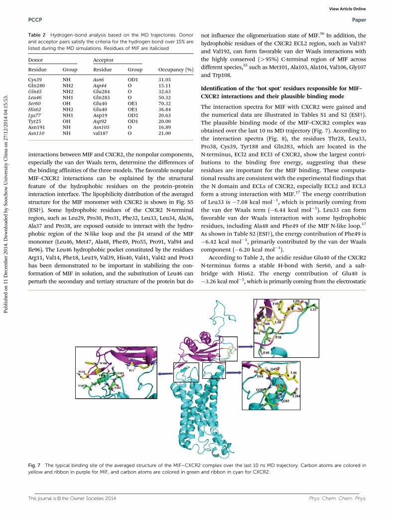
The interaction spectra for MIF with CXCR2 were gained andthe numerical data are illustrated in Tables S1 and S2 (ESI†).The plausible binding mode of the MIF–CXCR2 complex wasobtained over the last 10 ns MD trajectory (Fig. 7). According tothe interaction spectra (Fig. 8), the residues Thr28, Leu33,Pro38, Cys39, Tyr188 and Gln283, which are located in theN-terminus, ECl2 and ECl3 of CXCR2, show the largest contri-butions to the binding free energy, suggesting that theseresidues are important for the MIF binding. These computa-tional results are consistent with the experimental findings thatthe N domain and ECLs of CXCR2, especially ECL2 and ECL3form a strong interaction with MIF.17 The energy contributionof Leu33 is �7.08 kcal mol�1, which is primarily coming fromthe van der Waals term (�6.44 kcal mol�1). Leu33 can formfavorable van der Waals interaction with some hydrophobicresidues, including Ala48 and Phe49 of the MIF N-like loop.17
As shown in Table S2 (ESI†), the energy contribution of Phe49 is�6.42 kcal mol�1, primarily contributed by the van der Waalscomponent (�6.20 kcal mol�1).
According to Table 2, the acidic residue Glu40 of the CXCR2N-terminus forms a stable H-bond with Ser60, and a salt-bridge with His62. The energy contribution of Glu40 is�3.26 kcal mol�1, which is primarily coming from the electrostatic
Table 2 Hydrogen-bond analysis based on the MD trajectories. Donorand acceptor pairs satisfy the criteria for the hydrogen bond over 15% arelisted during the MD simulations. Residues of MIF are italicised
Donor Acceptor
Occupancy (%)Residue Group Residue Group
Cys39 NH Asn6 OD1 31.05Gln280 NH2 Asp44 O 15.11Gln45 NH2 Glu284 O 32.63Leu46 NH1 Gln283 O 50.32Ser60 OH Glu40 OE1 70.32His62 NH2 Glu40 OE1 36.84Lys77 NH1 Asp19 OD2 20.63Tyr25 OH Asp92 OD1 20.00Asn191 NH Asn105 O 16.89Asn110 NH val187 O 21.00
Fig. 7 The typical binding site of the averaged structure of the MIF–CXCR2 complex over the last 10 ns MD trajectory. Carbon atoms are colored inyellow and ribbon in purple for MIF, and carbon atoms are colored in green and ribbon in cyan for CXCR2.
PCCP Paper
Publ
ishe
d on
11
Dec
embe
r 20
14. D
ownl
oade
d by
Soo
chow
Uni
vers
ity C
hina
on
27/1
2/20
14 0
4:15
:53.
View Article Online

Phys. Chem. Chem. Phys. This journal is© the Owner Societies 2014
term (�14.88 kcal mol�1), though it can be tremendously counter-acted by the polar desolvation term. As shown in Table 2, thebackbone of Val187 of ECL2 forms a H-bond with Asn110.The electrostatic term of the Val187 is �1.64 kcal mol�1, and theenergy contribution of Asn110 is �16.4 kcal mol�1, which isprimarily contributed by the electrostatic term (�39.58 kcal mol�1).Fig. 7 shows that Tyr188 of ECL2 can form aryl–aryl interaction withTrp108 in a T-shaped geometry. The energy contribution of Tyr188is �10.14 kcal mol�1, which is mainly coming from the van derWaals term (�9.82 kcal mol�1). Table 2 shows that the acid residueGln280 in ECL3 can form a H-bond with Asp44 of the MIF pseudo-(E)LR motif, which is critical for the MIF binding to ECL2 and ECL3based on the peptide array analysis.17 The energy contribution of
Gln280 is �2.74 kcal mol�1, which is mainly contributed from theelectrostatic term (�2.3 kcal mol�1), while the energy contributionof Asp44 is �2.6 kcal mol�1, primarily coming from the electro-static term (�4.38 kcal mol�1) as well. The backbone of Gln283 alsoforms a stable H-bond with Leu46 of the MIF N-like loop. Theenergy contribution of Gln283 is �6.48 kcal mol�1, and theelectrostatic term is �2.94 kcal mol�1. In addition, the side chainof Gln283 can form hydrophobic interactions with Val42, Pro43and Leu46, and its van der Waals contribution is�5.55 kcal mol�1.
The Leu46 hydrophobic pocket plays a key role in stabilizingthe conformation of MIF, and Arg11 is an important componentof the Leu46 hydrophobic site and forms a sort of a ‘‘cap’’ to thepocket.56 The energy contribution of Leu46 is �8.58 kcal mol�1,
Fig. 8 Residue–residue interaction spectra for (a) CXCR2, (b) MIF, (c) the polar energy (DEele + DGGB) for CXCR2, (d) the polar energy (DEele + DGGB) forMIF, (e) the non-polar energy (DEvdw + DGSA) for CXCR2, and (f) the non-polar energy (DEvdw + DGSA) for MIF.
Paper PCCP
Publ
ishe
d on
11
Dec
embe
r 20
14. D
ownl
oade
d by
Soo
chow
Uni
vers
ity C
hina
on
27/1
2/20
14 0
4:15
:53.
View Article Online
This journal is© the Owner Societies 2014 Phys. Chem. Chem. Phys.
which is mainly coming from the nonpolar interaction energyterm (�8.74 kcal mol�1). The backbone of Gln284 forms a stableH-bond with Gln45, and the electrostatic energy term of Gln284is �3.66 kcal mol�1.
Comparison of the structures of CXCR2 with the wild-type MIFand R11A/D44A-MIF double mutant
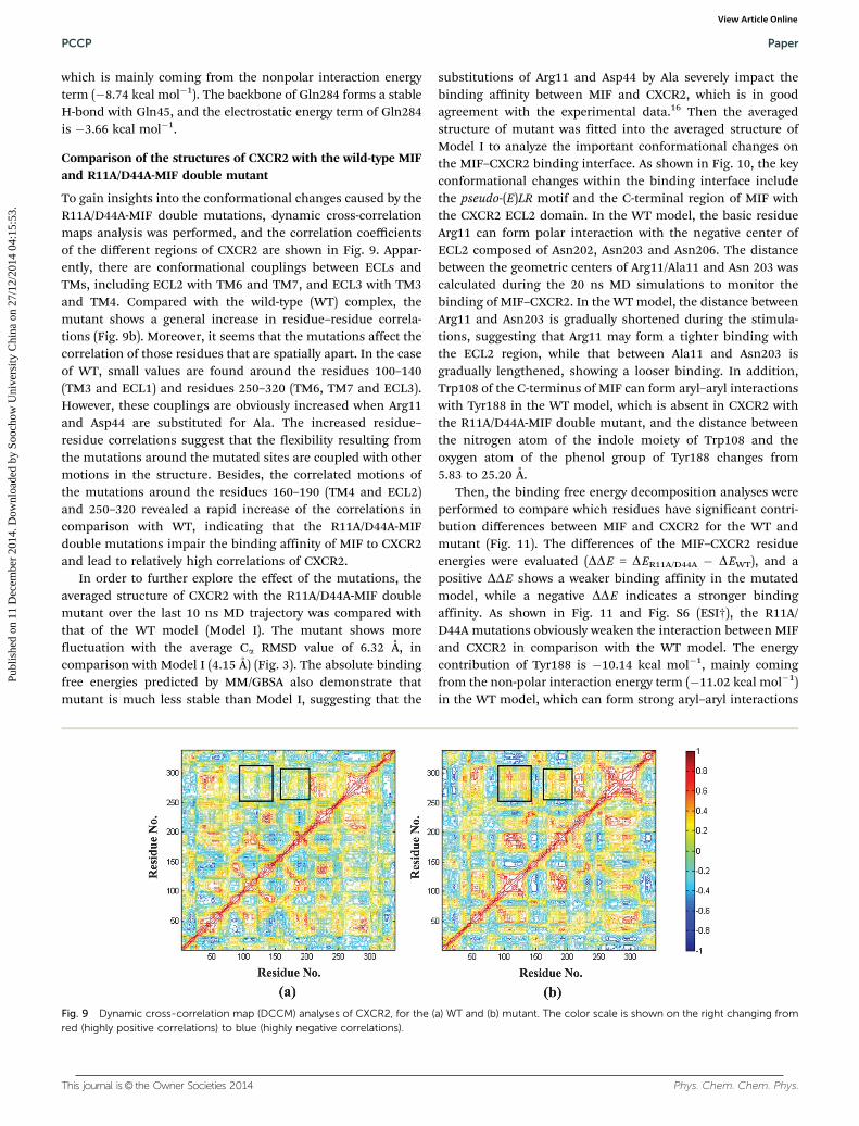
To gain insights into the conformational changes caused by theR11A/D44A-MIF double mutations, dynamic cross-correlationmaps analysis was performed, and the correlation coefficientsof the different regions of CXCR2 are shown in Fig. 9. Appar-ently, there are conformational couplings between ECLs andTMs, including ECL2 with TM6 and TM7, and ECL3 with TM3and TM4. Compared with the wild-type (WT) complex, themutant shows a general increase in residue–residue correla-tions (Fig. 9b). Moreover, it seems that the mutations affect thecorrelation of those residues that are spatially apart. In the caseof WT, small values are found around the residues 100–140(TM3 and ECL1) and residues 250–320 (TM6, TM7 and ECL3).However, these couplings are obviously increased when Arg11and Asp44 are substituted for Ala. The increased residue–residue correlations suggest that the flexibility resulting fromthe mutations around the mutated sites are coupled with othermotions in the structure. Besides, the correlated motions ofthe mutations around the residues 160–190 (TM4 and ECL2)and 250–320 revealed a rapid increase of the correlations incomparison with WT, indicating that the R11A/D44A-MIFdouble mutations impair the binding affinity of MIF to CXCR2and lead to relatively high correlations of CXCR2.
In order to further explore the effect of the mutations, theaveraged structure of CXCR2 with the R11A/D44A-MIF doublemutant over the last 10 ns MD trajectory was compared withthat of the WT model (Model I). The mutant shows morefluctuation with the average Ca RMSD value of 6.32 Å, incomparison with Model I (4.15 Å) (Fig. 3). The absolute bindingfree energies predicted by MM/GBSA also demonstrate thatmutant is much less stable than Model I, suggesting that the
substitutions of Arg11 and Asp44 by Ala severely impact thebinding affinity between MIF and CXCR2, which is in goodagreement with the experimental data.16 Then the averagedstructure of mutant was fitted into the averaged structure ofModel I to analyze the important conformational changes onthe MIF–CXCR2 binding interface. As shown in Fig. 10, the keyconformational changes within the binding interface includethe pseudo-(E)LR motif and the C-terminal region of MIF withthe CXCR2 ECL2 domain. In the WT model, the basic residueArg11 can form polar interaction with the negative center ofECL2 composed of Asn202, Asn203 and Asn206. The distancebetween the geometric centers of Arg11/Ala11 and Asn 203 wascalculated during the 20 ns MD simulations to monitor thebinding of MIF–CXCR2. In the WT model, the distance betweenArg11 and Asn203 is gradually shortened during the stimula-tions, suggesting that Arg11 may form a tighter binding withthe ECL2 region, while that between Ala11 and Asn203 isgradually lengthened, showing a looser binding. In addition,Trp108 of the C-terminus of MIF can form aryl–aryl interactionswith Tyr188 in the WT model, which is absent in CXCR2 withthe R11A/D44A-MIF double mutant, and the distance betweenthe nitrogen atom of the indole moiety of Trp108 and theoxygen atom of the phenol group of Tyr188 changes from5.83 to 25.20 Å.
Then, the binding free energy decomposition analyses wereperformed to compare which residues have significant contri-bution differences between MIF and CXCR2 for the WT andmutant (Fig. 11). The differences of the MIF–CXCR2 residueenergies were evaluated (DDE = DER11A/D44A � DEWT), and apositive DDE shows a weaker binding affinity in the mutatedmodel, while a negative DDE indicates a stronger bindingaffinity. As shown in Fig. 11 and Fig. S6 (ESI†), the R11A/D44A mutations obviously weaken the interaction between MIFand CXCR2 in comparison with the WT model. The energycontribution of Tyr188 is �10.14 kcal mol�1, mainly comingfrom the non-polar interaction energy term (�11.02 kcal mol�1)in the WT model, which can form strong aryl–aryl interactions
Fig. 9 Dynamic cross-correlation map (DCCM) analyses of CXCR2, for the (a) WT and (b) mutant. The color scale is shown on the right changing fromred (highly positive correlations) to blue (highly negative correlations).
PCCP Paper
Publ
ishe
d on
11
Dec
embe
r 20
14. D
ownl
oade
d by
Soo
chow
Uni
vers
ity C
hina
on
27/1
2/20
14 0
4:15
:53.
View Article Online

Phys. Chem. Chem. Phys. This journal is© the Owner Societies 2014
with Trp108 and hydrophobic interaction with Val106 of theMIF C-terminus, whereas the energy contribution of Tyr188 isreduced to�0.82 kcal mol�1 in the mutant (Table S3, ESI†). Theenergy contribution of Ser189 is �5.58 kcal mol�1 in the WTmodel, which is primarily from the non-polar interactionenergy term (�6.14 kcal mol�1), and it can form hydrophobicinteraction with Trp108, whereas the energy contribution ofSer189 is reduced to �0.92 kcal mol�1 in the mutant.
Binding mechanism of MIF with CXCR2
Although the chemokine receptor can be activated by variouschemokine ligands with a range of specificities and affinities, ageneral two-site mechanism of the ligand–receptor interactionhas been recognized, including the interactions between the
receptor N-domain and the ligand N-terminal loop residues(site I), and between the receptor extracellular loop and theligand N-terminal residues (site II). The site 1 binding motif isproposed to contribute differently to binding affinity andreceptor selectivity, while site 2 is responsible for controllingbinding affinity and receptor activation. The interactionbetween CXCR2 and CXCL8 is also involved in the two-sitebinding mechanism, including the N-terminus and N loop,extracellular loop and N-terminus, respectively, especially theELR motif.
As represented in the dynamic and energetic analyses men-tioned above, the putative binding mechanism of MIF withCXCR2 was proposed at the molecular level: primarily initia-lized by the non-polar interactions including the van der Waalsand hydrophobic interactions, the N-terminal region of CXCR2makes the first contact with the N-like loop and b-sheet of MIF(site 1); owing to the steric complementarity and electrostaticattraction, the ECL2 and ECL3 regions of CXCR2 form stronginteractions with the pseudo-(E)LR motif and C-terminus ofMIF, which induces the molecular thermodynamic motion ofTMs for signal transduction (site 2). Some hydrophobic resi-dues of the CXCR2 N-terminal region, including Leu29, Pro30,Pro31, Phe32, Leu33, Leu34, Ala36, Ala37 and Pro38, can formstrong interactions with the hydrophobic region of the N-likeloop and b4 strand of MIF (Leu46, Met47, Ala48, Phe49, Pro55,Pro91, Val94 and Ile96). The acidic residues of the CXCR2N-terminus, including Asp19 and Glu40, can also form polarinteraction with Lys77, Ser60 and His62 over the MD simula-tions. All these interactions result in the movement of MIFtowards CXCR2, triggering the 2nd step interaction among theprotein–protein binding interface. The hydrophobic residues ofECL2, such as Val187, Tyr188 and Val192, can form favorablevan der Waals interactions with the C-terminal region of MIF,such as Met101, Ala103, Ala104, Val106 and Trp108. The basicresidue Arg11 of the MIF pseudo-(E)LR motif can form strongpolar interaction with the negative center of ECL2 composed of
Fig. 10 Comparison the difference between the WT (Model I) and R11A/D44A double mutant of Model I. (a) The distance between the Arg11/Ala11 of MIFand Gln203 of the CXCR2 ECL2 region during the MD simulations; (b) the superimposition of the averaged structures of Model I and R11A/D44A mutantof Model I. Carbon atoms and ribbon of MIF are colored in green in WT and cyan in mutant; carbon atoms and ribbon of CXCR2 are colored in purple andgreen in WT, and carbon atoms and ribbon of CXCR2 are colored in yellow and cyan in mutant.
Fig. 11 Differences in the contribution of the residues to the binding freeenergy between the WT (Model I) and R11A/D44A double mutant of Model I(DDE = DER11A/D44A � DEWT). A positive DDE represents a weaker bindingaffinity in the mutated protein, while a negative DDE represents a strongerbinding affinity.
Paper PCCP
Publ
ishe
d on
11
Dec
embe
r 20
14. D
ownl
oade
d by
Soo
chow
Uni
vers
ity C
hina
on
27/1
2/20
14 0
4:15
:53.
View Article Online

This journal is© the Owner Societies 2014 Phys. Chem. Chem. Phys.
Asn202, Asn203 and Asn206. Tyr188 can form strong aryl–arylinteractions with Trp108 and hydrophobic interaction withVal106 of the MIF C-terminus. The acidic residue Asp44 canform a stable H-bond with Gln280 of ECL3. The conformationalchange of the CXCR2 extracellular region induces the molecularthermodynamic motion of TMs, especially TM5 and TM6.
Conclusion
In the present study, a comprehensive set of computationaltools, including homology modeling, protein–protein docking,MD simulations, MM/GBSA binding free energy calculationsand MM/GBSA binding free energy decomposition, wereemployed to obtain insights into the molecular recognitionof MIF with CXCR2. The plausible near-native structure ofMIF–CXCR2 was obtained and the ‘hot-spot’ residues importantwithin the protein–protein binding interface were highlighted,which is in good agreement with the experimental data. Dynamicand energetic analyses demonstrated that the mutation of thepseudo-(E)LR motif of MIF severely compromise its binding toCXCR2. Moreover, the potential binding mechanism between MIFand CXCR2 was proposed, and these structural determinants maypave the way in the rational design of potent agents to target theproinflammatory MIF–CXCR2 interaction selectively by pinpointingthe ’hot-spot’ residues within the binding interface.
Acknowledgements
This study was supported by the National Science Foundationof China (21173156), the National Basic Research Program ofChina (973 program, 2012CB932600). Support from the PriorityAcademic Program Development of Jiangsu Higher EducationInstitutes (PAPD) and the Grant from Jiangsu Science andTechnology Commission (BY2011131) are also appreciated.
References
1 T. Calandra and T. Roger, Macrophage migration inhibitoryfactor: A regulator of innate immunity, Nat. Rev. Immunol.,2003, 3, 791–800.
2 E. F. Morand, M. Leech and B. Jurgen, MIF: a new cytokinelink between rheumatoid arthritis and atherosclerosis,Nat. Rev. Drug Discovery, 2006, 5, 399–410.
3 T. Hagemann, S. C. Robinson, R. G. Thompson, K. Charles,H. Kulbe and F. R. Balkwill, Ovarian cancer cell-derivedmigration inhibitory factor enhances tumor growth,progression, and angiogenesis, Mol. Cancer Ther., 2007, 6,1993–2002.
4 H. Sugimoto, M. Taniguchi, A. Nakagawa, I. Tanaka,M. Suzuki and J. Nishihira, Crystal structure of humanD-dopachrome tautomerase, a homologue of macrophagemigration inhibitory factor, at 1.54 angstrom resolution,Biochemistry, 1999, 38, 3268–3279.
5 E. Rosengren, P. Aman, S. Thelin, C. Hansson, S. Ahlfors,P. Bjork, L. Jacobsson and H. Rorsman, The macrophage
migration inhibitory factor MIF is a phenylpyruvate tauto-merase, FEBS Lett., 1997, 417, 85–88.
6 R. Kleemann, A. Kapurniotu, R. W. Frank, A. Gessner,R. Mischke, O. Flieger, S. Juttner, H. Brunner andJ. Bernhagen, Disulfide analysis reveals a role for macro-phage migration inhibitory factor (MIF) as thiol-proteinoxidoreductase, J. Mol. Biol., 1998, 280, 85–102.
7 L. Leng, C. N. Metz, Y. Fang, J. Xu, S. Donnelly, J. Baugh,T. Delohery, Y. B. Chen, R. A. Mitchell and R. Bucala, MIFsignal transduction initiated by binding to CD74, J. Exp.Med., 2003, 197, 1467–1476.
8 X. R. Shi, L. Leng, T. Wang, W. K. Wang, X. Du, J. Li,C. McDonald, Z. Chen, J. W. Murphy, E. Lolis, P. Noble,W. Knudson and R. Bucala, CD44 is the signaling compo-nent of the macrophage migration inhibitory factor-CD74receptor complex, Immunity, 2006, 25, 595–606.
9 J. Bernhagen, R. Krohn, H. Lue, J. L. Gregory, A. Zernecke,R. R. Koenen, M. Dewor, I. Georgiev, A. Schober, L. Leng,T. Kooistra, G. Fingerle-Rowson, P. Ghezzi, R. Kleemann,S. R. McColl, R. Bucala, M. J. Hickey and C. Weber, MIF is anoncognate ligand of CXC chemokine receptors in inflam-matory and atherogenic cell recruitment, Nat. Med., 2007,13, 587–596.
10 S. P. Fricker and M. Metz, Chemokine receptor modeling:an interdisciplinary approach to drug design, Future Med.Chem., 2014, 6, 91–114.
11 M. Veenstra and R. M. Ransohoff, Chemokine receptorCXCR2: Physiology regulator and neuroinflammationcontroller?, J. Neuroimmunol., 2012, 246, 1–9.
12 C. A. Hebert, R. V. Vitangcol and J. B. Baker, ScanningMutagenesis of Interleukin-8 Identifies a Cluster of Resi-dues Required for Receptor-Binding, J. Biol. Chem., 1991,266, 18989–18994.
13 B. Degryse and M. de Virgilio, The nuclear protein HMGB1,a new kind of chemokine?, FEBS Lett., 2003, 553, 11–17.
14 D. Yang, O. Chertov, N. Bykovskaia, Q. Chen, M. J. Buffo,J. Shogan, M. Anderson, J. M. Schroder, J. M. Wang, O. M. Z.Howard and J. J. Oppenheim, beta-defensins: Linkinginnate and adaptive immunity through dendritic and T cellCCR6, Science, 1999, 286, 525–528.
15 H. Noels, J. Bernhagen and C. Weber, Macrophage MigrationInhibitory Factor: A Noncanonical Chemokine Important inAtherosclerosis, Trends Cardiovasc. Med., 2009, 19, 76–86.
16 C. Weber, S. Kraemer, M. Drechsler, H. Q. Lue, R. R.Koenen, A. Kapurniotu, A. Zernecke and J. Bernhagen,Structural determinants of MIF functions in CXCR2-mediated inflammatory and atherogenic leukocyte recruit-ment, Proc. Natl. Acad. Sci. U. S. A., 2008, 105, 16278–16283.
17 S. Kraemer, H. Q. Lue, A. Zernecke, A. Kapurniotu,E. Andreetto, R. Frank, B. Lennartz, C. Weber andJ. Bernhagen, MIF-chemokine receptor interactions inatherogenesis are dependent on an N-loop-based 2-sitebinding mechanism, FASEB J., 2011, 25, 894–906.
18 B. L. Wu, E. Y. T. Chien, C. D. Mol, G. Fenalti, W. Liu,V. Katritch, R. Abagyan, A. Brooun, P. Wells, F. C. Bi, D. J.Hamel, P. Kuhn, T. M. Handel, V. Cherezov and R. C. Stevens,
PCCP Paper
Publ
ishe
d on
11
Dec
embe
r 20
14. D
ownl
oade
d by
Soo
chow
Uni
vers
ity C
hina
on
27/1
2/20
14 0
4:15
:53.
View Article Online

Phys. Chem. Chem. Phys. This journal is© the Owner Societies 2014
Structures of the CXCR4 Chemokine GPCR with Small-Molecule and Cyclic Peptide Antagonists, Science, 2010,330, 1066–1071.
19 A. Sali, L. Potterton, F. Yuan, H. Vanvlijmen and M. Karplus,Evaluation of Comparative Protein Modeling by Modeler,Proteins, 1995, 23, 318–326.
20 T. Ichiye and M. Karplus, Collective Motions in Proteins - aCovariance Analysis of Atomic Fluctuations in Molecular-Dynamics and Normal Mode Simulations, Proteins: Struct.,Funct., Genet., 1991, 11, 205–217.
21 C. T. Veldkamp, C. Seibert, F. C. Peterson, N. B. De la Cruz,J. C. Haugner, H. Basnet, T. P. Sakmar and B. F. Volkman,Structural Basis of CXCR4 Sulfotyrosine Recognition by theChemokine SDF-1/CXCL12, Sci. Signaling., 2008, 1, ra4.
22 L. Xu, Y. Y. Li, H. Y. Sun, D. Li and T. J. Hou, Structural basisof the interactions between CXCR4 and CXCL12/SDF-1revealed by theoretical approaches, Mol. BioSyst., 2013, 9,2107–2117.
23 D. Rajasekaran, C. Keeler, M. A. Syed, M. C. Jones, J. K.Harrison, D. Q. Wu, V. Bhandari, M. E. Hodsdon andE. J. Lolis, A Model of GAG/MIP-2/CXCR2 Interfaces andIts Functional Effects, Biochemistry, 2012, 51, 5642–5654.
24 P. de Kruijf, H. D. Lim, L. Roumen, V. A. Renjaan, J. Q. Zhao,M. L. Webb, D. S. Auld, J. Wijkmans, G. J. R. Zaman, M. J.Smit, C. de Graaf and R. Leurs, Identification of a NovelAllosteric Binding Site in the CXCR2 Chemokine Receptor,Mol. Pharmacol., 2011, 80, 1108–1118.
25 Q. Tan, Y. Zhu, J. Li, Z. Chen, G. W. Han, I. Kufareva, T. Li,L. Ma, G. Fenalti, J. Li, W. Zhang, X. Xie, H. Yang, H. Jiang,V. Cherezov, H. Liu, R. C. Stevens, Q. Zhao and B. Wu,Structure of the CCR5 Chemokine Receptor-HIV EntryInhibitor Maraviroc Complex, Science, 2013, 341, 1387–1390.
26 J. B. Lubetsky, A. Dios, J. L. Han, B. Aljabari, B. Ruzsicska,R. Mitchell, E. Lolis and Y. Al-Abed, The tautomerase activesite of macrophage migration inhibitory factor is a potentialtarget for discovery of novel anti-inflammatory agents,J. Biol. Chem., 2002, 277, 24976–24982.
27 K. Wiehe, B. Pierce, J. Mintseris, W. W. Tong, R. Anderson,R. Chen and Z. Weng, ZDOCK and RDOCK performance inCAPRI rounds 3, 4, and 5, Proteins, 2005, 60, 207–213.
28 R. Chen and Z. P. Weng, Docking unbound proteins usingshape complementarity, desolvation, and electrostatics,Proteins, 2002, 47, 281–294.
29 R. Chen, L. Li and Z. P. Weng, ZDOCK: An initial-stageprotein-docking algorithm, Proteins, 2003, 52, 80–87.
30 B. Pierce and Z. P. Weng, ZRANK: Reranking protein dock-ing predictions with an optimized energy function, Proteins,2007, 67, 1078–1086.
31 J. Hsin, A. Arkhipov, Y. Yin, J. E. Stone and K. Schulten,Using VMD: an introductory tutorial, Curr. Protocol Bioin-form., 2008, ch. 5, unit 5.7.
32 W. L. Jorgensen, J. Chandrasekhar, J. D. Madura, R. W. Impeyand M. L. Klein, Comparison of Simple Potential Functions forSimulating Liquid Water, J. Chem. Phys., 1983, 79, 926–935.
33 V. Hornak, R. Abel, A. Okur, B. Strockbine, A. Roitberg andC. Simmerling, Comparison of multiple amber force fields
and development of improved protein backbone para-meters, Proteins, 2006, 65, 712–725.
34 J. Domanski, P. J. Stansfeld, M. S. P. Sansom andO. Beckstein, Lipidbook: A Public Repository for Force-FieldParameters Used in Membrane Simulations, J. Membr. Biol.,2010, 236, 255–258.
35 J. M. Wang, R. M. Wolf, J. W. Caldwell, P. A. Kollman andD. A. Case, Development and testing of a general amberforce field, J. Comput. Chem., 2004, 25, 1157–1174.
36 T. Darden, D. York and L. Pedersen, Particle Mesh Ewald -an N.Log(N) Method for Ewald Sums in Large Systems,J. Chem. Phys., 1993, 98, 10089–10092.
37 J. C. Phillips, R. Braun, W. Wang, J. Gumbart, E. Tajkhorshid,E. Villa, C. Chipot, R. D. Skeel, L. Kale and K. Schulten,Scalable molecular dynamics with NAMD, J. Comput. Chem.,2005, 26, 1781–1802.
38 J. P. Ryckaert, G. Ciccotti and H. J. C. Berendsen, Numericalintegration of the cartesian equations of motion of asystem with constraints: molecular dynamics of n-alkanes,J. Comput. Phys., 1977, 23, 327–341.
39 T. Hou, J. Wang, Y. Li and W. Wang, Assessing the Perfor-mance of the MM/PBSA and MM/GBSA Methods. 1. TheAccuracy of Binding Free Energy Calculations Based onMolecular Dynamics Simulations, J. Chem. Inf. Model., 2011,51, 69–82.
40 L. Xu, Y. Li, L. Li, S. Zhou and T. Hou, Understandingmicroscopic binding of macrophage migration inhibitoryfactor with phenolic hydrazones by molecular docking,molecular dynamics simulations and free energy calcula-tions, Mol. BioSyst., 2012, 8, 2260–2273.
41 T. Hou and R. Yu, Molecular dynamics and free energystudies on the wild-type and double mutant HIV-1 proteasecomplexed with amprenavir and two amprenavir-relatedinhibitors: Mechanism for binding and drug resistance,J. Med. Chem., 2007, 50, 1177–1188.
42 L. Xu, H. Sun, Y. Li, J. Wang and T. Hou, Assessing thePerformance of MM/PBSA and MM/GBSA Methods. 3. TheImpact of Force Fields and Ligand Charge Models, J. Phys.Chem. B, 2013, 117, 8408–8421.
43 H. Sun, Y. Li, S. Tian, L. Xu and T. Hou, Assessing thePerformance of MM/PBSA and MM/GBSA Methods. 4.Accuracies of MM/PBSA and MM/GBSA Methodologies Eval-uated by Various Simulation Protocols using PDBbind DataSet, Phys. Chem. Chem. Phys., 2014, 16, 16719–16729.
44 H. Liu and X. Yao, Molecular Basis of the Interaction for anEssential Subunit PA-PB1 in Influenza Virus RNA Poly-merase: Insights from Molecular Dynamics Simulationand Free Energy Calculation, Mol. Pharmaceutics, 2009, 7,75–85.
45 Q. Xue, J. L. Zhang, Q. C. Zheng, Y. L. Cui, L. Chen,W. T. Chu and H. X. Zhang, Exploring the Molecular Basisof dsRNA Recognition by Mss116p Using MolecularDynamics Simulations and Free-Energy Calculations, Lang-muir, 2013, 29, 11135–11144.
46 P. A. Kollman, I. Massova, C. Reyes, B. Kuhn, S. H. Huo,L. Chong, M. Lee, T. Lee, Y. Duan, W. Wang, O. Donini,
Paper PCCP
Publ
ishe
d on
11
Dec
embe
r 20
14. D
ownl
oade
d by
Soo
chow
Uni
vers
ity C
hina
on
27/1
2/20
14 0
4:15
:53.
View Article Online

This journal is© the Owner Societies 2014 Phys. Chem. Chem. Phys.
P. Cieplak, J. Srinivasan, D. A. Case and T. E. Cheatham,Calculating structures and free energies of complex molecules:Combining molecular mechanics and continuum models,Acc. Chem. Res., 2000, 33, 889–897.
47 J. Wang, T. Hou and X. Xu, Recent Advances in Free EnergyCalculations with a Combination of Molecular Mechanicsand Continuum Models, Curr. Comput.-Aided Drug Des.,2006, 2, 287–306.
48 A. Onufriev, D. Bashford and D. A. Case, Exploring proteinnative states and large-scale conformational changes with amodified generalized born model, Proteins, 2004, 55, 383–394.
49 J. Weiser, P. S. Shenkin and W. C. Still, Approximate atomicsurfaces from linear combinations of pairwise overlaps(LCPO), J. Comput. Chem., 1999, 20, 217–230.
50 T. Hou, W. Zhang, D. A. Case and W. Wang, Characteriza-tion of domain-peptide interaction interface: A case studyon the amphiphysin-1 SH3 domain, J. Mol. Biol., 2008, 376,1201–1214.
51 T. Hou, Y. Li and W. Wang, Prediction of peptides bindingto the PKA RII alpha subunit using a hierarchical strategy,Bioinformatics, 2011, 27, 1814–1821.
52 L. Li, Y. Y. Li, L. L. Zhang and T. J. Hou, Theoretical Studieson the Susceptibility of Oseltamivir against Variants of 2009
A/H1N1 Influenza Neuraminidase, J. Chem. Inf. Model.,2012, 52, 2715–2729.
53 H. Gohlke, C. Kiel and D. A. Case, Insights into protein-protein binding by binding free energy calculation and freeenergy decomposition for the Ras-Raf and Ras-RaIGDScomplexes, J. Mol. Biol., 2003, 330, 891–913.
54 W. Xue, Y. Yang, X. Wang, H. Liu and X. Yao, ComputationalStudy on the Inhibitor Binding Mode and Allosteric RegulationMechanism in Hepatitis C Virus NS3/4A Protein, PLoS One,2014, 9, e87077.
55 F. El-Turk, M. Cascella, H. Ouertatani-Sakouhi, R. L.Narayanan, L. Leng, R. Bucala, M. Zweckstetter,U. Rothlisberger and H. A. Lashuel, The conformationalflexibility of the carboxy terminal residues 105-114 is a keymodulator of the catalytic activity and stability of macro-phage migration inhibitory factor, Biochemistry, 2008, 47,10740–10756.
56 F. El-Turk, B. Fauvet, A. Ashrafi, H. Ouertatani-Sakouhi,M. K. Cho, M. Neri, M. Cascella, U. Rothlisberger,F. Pojer, M. Zweckstetter and H. Lashuel, Characterizationof Molecular Determinants of the Conformational Stabilityof Macrophage Migration Inhibitory Factor: Leucine 46Hydrophobic Pocket, PLoS One, 2012, 7, e45024.
PCCP Paper
Publ
ishe
d on
11
Dec
embe
r 20
14. D
ownl
oade
d by
Soo
chow
Uni
vers
ity C
hina
on
27/1
2/20
14 0
4:15
:53.
View Article Online
Related Documents