BS(integrated) BIOCHEMISTRY Email ID:[email protected] Biochemistry Sixth Edition By Khair Ullah Chapter 3: Exploring Proteins and Proteomes

Exploring Proteins and Proteomes. Stryer,CHAPTER 3 ppt
Jul 16, 2015
Welcome message from author
This document is posted to help you gain knowledge. Please leave a comment to let me know what you think about it! Share it to your friends and learn new things together.
Transcript

BS(integrated) BIOCHEMISTRYEmail ID:[email protected]
BiochemistrySixth Edition
By Khair Ullah
Chapter 3:Exploring Proteins and Proteomes

Methods in Protein Chemistry
These are methods used in isolation, purification, detection, degradation, analysis and synthesis of proteins.
As one would expect, most of these involve aqueous media and require a knowledge of pH, pKas, and charge on a peptide at various pH values.
Proteome: defines the compete functional information about a group of proteins thatwork together as a functional unit.

Assay of an Enzyme
In this reaction, the increase in absorbance of NADH at 340 nm is used to follow the formation of pyruvate
This is an oxidation-reduction reaction
Enzyme Activity
Total activity in soution use Unit of enzyme activity: µmol substrate/min or mol S/sec = katal
To follow during purification use Specific activity: µmol substrate/min-mg E
or mol S/sec-kg E = katal/kg E
To compare different enzymes use Molecular activity also called turn-over number (TON): µmol substrate/min-µmol E
or mol S/sec-mol E = katal/mol E

Centrifugation
Separation of a cell homogenate.

Solubility of ProteinsSalting in: When proteins are placed in an aqueous solution, the only ionic species in solution are the other protein molecules. Water, although polar, is only slightly ionized so the proteins tend to aggregate based on ionic interactions that form between themselves. The interactions between protein molecules are more favorable than interactions between water and a protein.
At low salt concentration (NaCl), other ionic species are now present to compete with the ionic protein:protein interactions. As a result, the ionic interactions between proteins break up and the proteins dissolve. Both the small ions (from NaCl) and the proteins are solvated by water.

Solubility of ProteinsSalting out: At high salt concentration (typically with (NH4)2SO4 or Na2SO4), water molecules are more strongly attracted to these small ions (especially multivalent ions) than to the large protein molecules. The proteins are left then to seek whatever favorable interactions exist and these are the protein:protein associations which result in aggregation and precipitation.
Isoelectric precipitation: At the pI there is zero net charge on a protein. At a pH away from the pI, each protein molecule bears an identical charge (either + or - depending on the pH) resulting in repulsion between molecules. At the pI, no repulsion occurs, and the proteins will aggregate and precipitate.
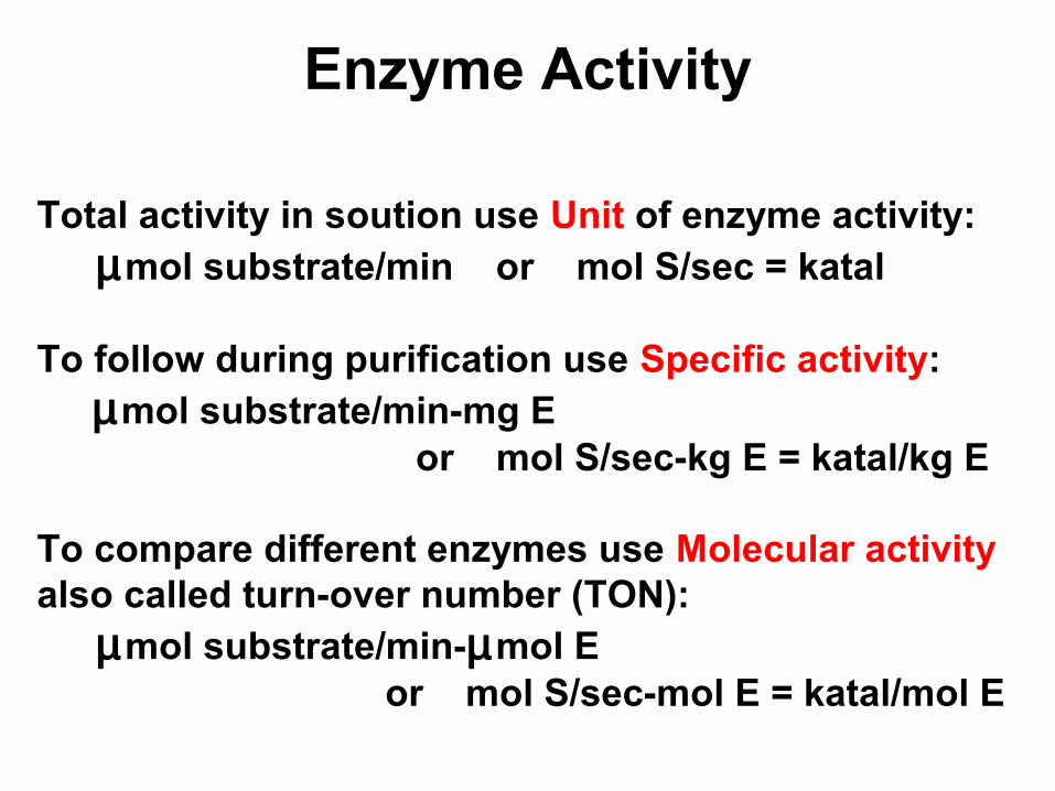
Determining the isoelectric point (pI)
Isoelectric point: The pI is the pH at which there is zero net charge on a molecule. Look at Asp.
The zero net charge form is a part of the first two ionizations. Therefore, the maximum amount of this is present at a pH of (2.09 + 3.86)/2 = 2.98 = pI.
HOOC-CH2-CH-COOH
NH3+
HOOC-CH2-CH-COO-NH3
+
+ H+ 2.09
HOOC-CH2-CH-COO-NH3
+
-OOC-CH2-CH-COO-NH3
+
-OOC-CH2-CH-COO-NH3
+
-OOC-CH2-CH-COO-NH2
+ H+ 3.86
+ H+ 9.82

Dialysis
Separation ofvery large from very small molecules is based on anattempt to equilibrateconcentration.
Osmotic pressure
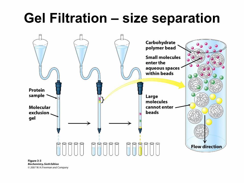
Gel Filtration – size separation

Affinity Chromatography

HPLC(up to 5000 psi)
Proteins can be detected from absorbance of the peptide bonds in the uv at 220 nm.
However, it is morecommonly doneat 280 nm as seenearlier.
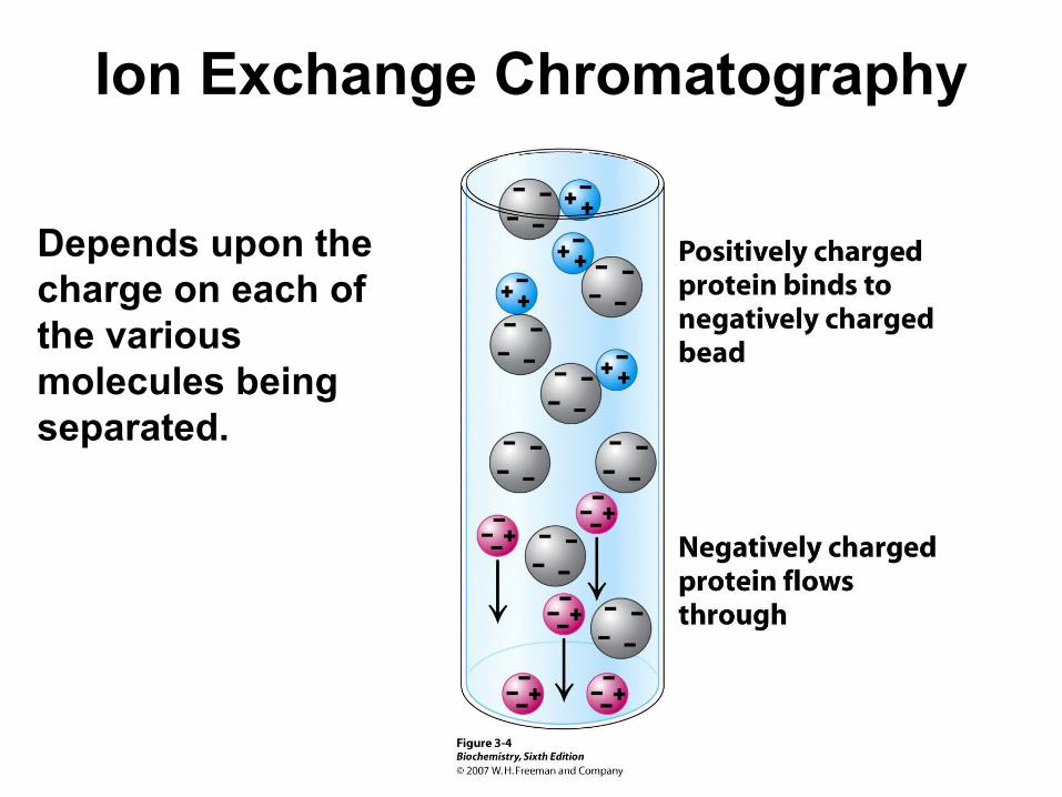
Ion Exchange Chromatography
Depends upon the charge on each of the various molecules being separated.

Ion Exchange Resins

Determining the Charge on a Peptide
Determing the charge on a peptide involves a knowledge of ionic equilibria, pKas and the ionic forms present at a given pH.
In a peptide, the amino terminus, the carboxy terminus and the ionizable side chains may be charged at a given pH.
The sum of these charges gives the net peptide charge.

Electrophoresis
Separation of molecules by electrophoresis depends upon:
1. the strength of the electric field (voltage), 2. the charge on each molecule,3. the frictional coefficient of movement
through the solid support which in turn depends on the radius or mass of the molecule.
So, essentially electrophoresis separates based on a charge/mass ratio.

Electrophoresis
A classical electrophoresis apparatus.
V
buffer buffer
solid support for sample
cooling plate

Sodium Dodecyl Sulfate
SDS is an anionic detergent that binds uniformly along a protein chain. About one SDS binds for every two amino acid residues. Thus all proteins bear the same charge/mass ratio and separation by electrophoresis will be based on mass alone.

SDS Gel Electrophoresis


Proteins after Staining
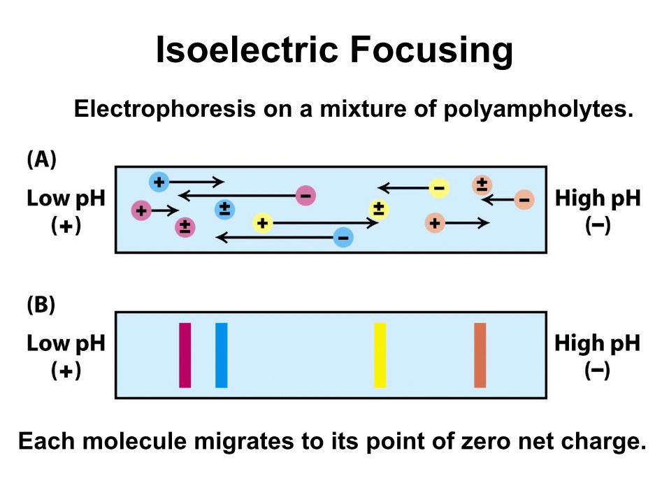
Isoelectric Focusing
Electrophoresis on a mixture of polyampholytes.
Each molecule migrates to its point of zero net charge.

SDS PAGE after IEF


Sequential Purification Steps

SDS electrophoresis

UltracentrifugationThe S value is a measure of the rate of sedimentation,(a sedimentation coefficient) and is not linear with MWbecause of molecular shape.
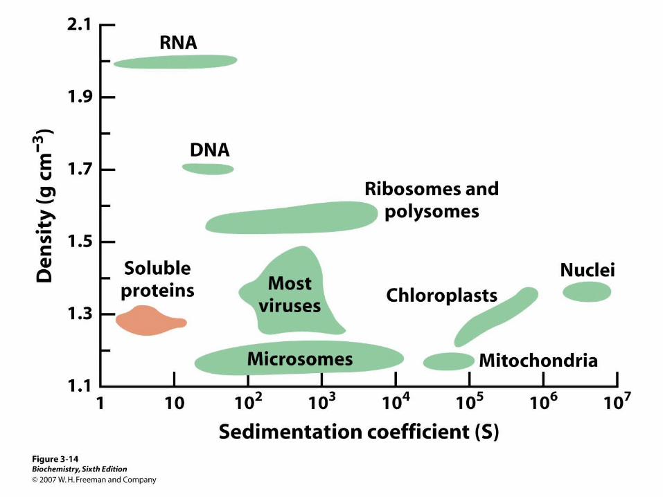

In a sucrose or cesium chloride gradienta molecule migrates thebuoyant density equal toits own.


Amino Acid AnalysisDetermines amino acid composition of a protein.A protein is hydrolyzed in 6N HCl, 24 hrs at 100oC.Separation of AA by ion exchange chromatography.

Detecting Amino Acids
Classical reagent for amino acids. Reaction requires 2-5 min at 100oC and givesnanomole level detection.
Ruhemann’s Purple570 nm
OH
O
O
OHNH2-CH-COOH
CH3
+
O
O
O
O
NCO2CH3
CHO+ +2
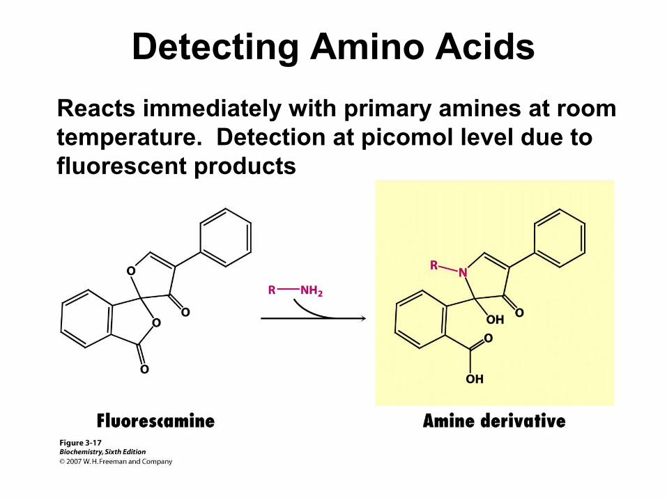
Detecting Amino Acids
Reacts immediately with primary amines at roomtemperature. Detection at picomol level due to fluorescent products

Automated Sequencing
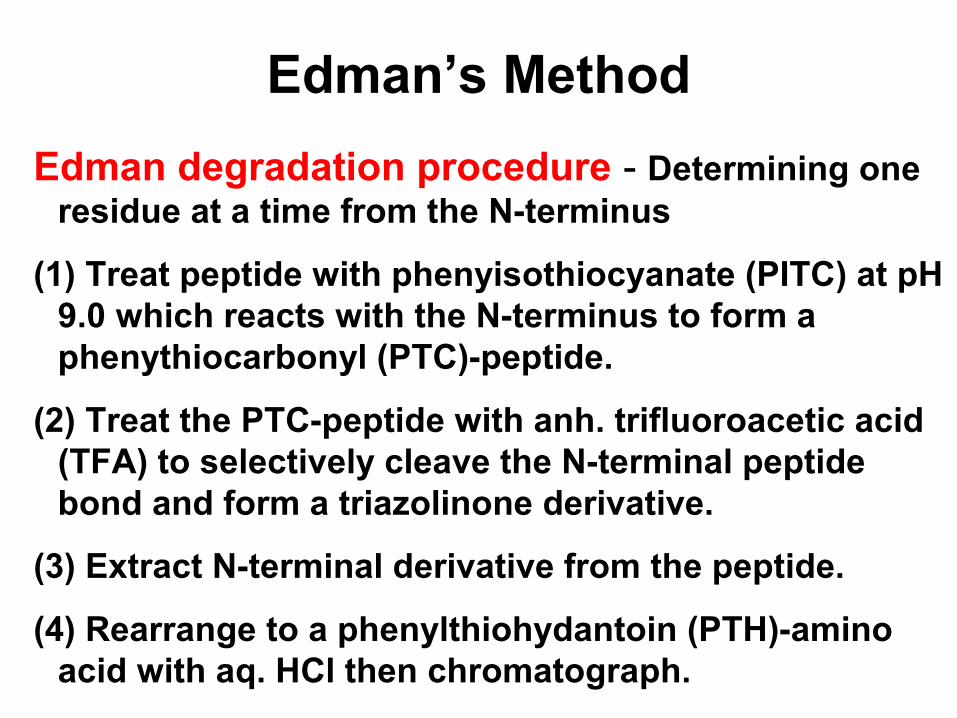
Edman’s Method
Edman degradation procedure - Determining one residue at a time from the N-terminus
(1) Treat peptide with phenyisothiocyanate (PITC) at pH 9.0 which reacts with the N-terminus to form a phenythiocarbonyl (PTC)-peptide.
(2) Treat the PTC-peptide with anh. trifluoroacetic acid (TFA) to selectively cleave the N-terminal peptide bond and form a triazolinone derivative.
(3) Extract N-terminal derivative from the peptide.
(4) Rearrange to a phenylthiohydantoin (PTH)-amino acid with aq. HCl then chromatograph.

Edman’sMethod
Phenylthio-hydantoin(PTH)
PITC

Edman’s Method
N=C=S
phenylthiocarbamyl-peptide
+ NH2 - CH - C - N - CH -C
O
H O
CH2-OH
CH3
NH - C -
S
NH - CH - C - N - CH -C
O
H O
CH2-OH
CH3
S
N
O
CH3NH -+
N
N
O
CH3S
NH2 - CH - C
O
CH2-OH
thiazolinone derivativephenylthiohydantoin derivative

Separation of PTH-AAs by HPLC

N-Terminal Reagents
DNFB - Sanger’s reagent (dinitrofluorobenzene)
DANSYL choride (dimethylaminonaphthalenesulfonyl chloride)
NO2
NO2
F
DNFB
SO2Cl
N(CH3)2
DANSYL-Cl
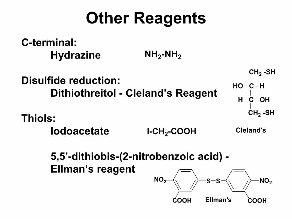
Other ReagentsC-terminal:
Hydrazine
Disulfide reduction:Dithiothreitol - Cleland’s Reagent
Thiols:Iodoacetate
5,5’-dithiobis-(2-nitrobenzoic acid) - Ellman’s reagent
NH2-NH2
HO C H
H C OH
CH2 -SH
CH2 -SH
Cleland's
NO2 S S
COOH
NO2
COOHEllman's
I-CH2-COOH

Protein Cleavage
Protein sequencing is most manageable with small polypeptides.
Therefore, in order to sequence a large protein, it must be cleaved into smaller pieces.
Cleavage is conducted using either chemical or enzymatic methods.
The pieces must be separated and purified before sequencing.

Chemical and Enzymatic Cleavage

CNBr Cleavage at Met

CNBr Cleavage
at Met

Enzymatic cleavage by Trypsin

An Example, Peptide overlap

Dithiothreitol Reduction of –S-S-

Iodoacetate reaction with -SH

Performic acid oxidation of –S-S-

Sequencing using DNA
Edman’s degradation has been a tremendous asset in protein sequencing, however, for larger proteins recombinant DNA technology is now being used.

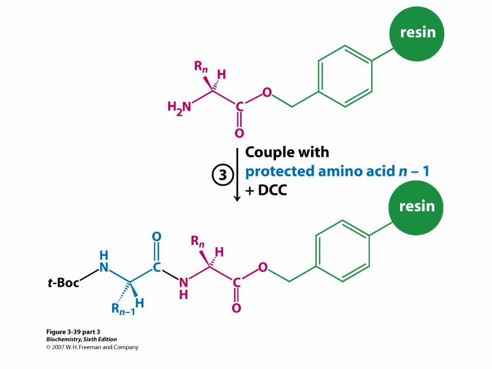
Merrifield Solid-Phase SynthesisMerrifield and coworkers at Rockefeller Institute devised a revolutionary solid phase method of protein synthesis in 1963. The proved that this method was effective for larger proteins by synthesizing ribonuclease, an enzyme with 124 amino acid residues.
A chloromethylated polystyrene polymer (resin) was used as the solid support.
Dicyclohexylcarbodiimide was used as an amino acid activator and a water scavenger in the condensation reaction.
Merrifield received a Nobel Prize in 1984 for this work.


Other Methodologies
Immunochemistry: (omit as this is in section 3.3)ELISA = Enzyme-linked immunosorbent assay
Western blottingFluorescense microscopy
Mass Spectrometry: (section 3.5)MALDI = matrix-assisted laser desorption- ionization TOF = time of flight
X-ray Crystallography: (section 3.6)
Nuclear Magnetic Resonance: (section 3.6) NOESY = nuclear Overhauser enhancement spectroscopy.

• Amino acid sequence data provide a basis for preparing antibodies specific for
a protein of interest.
• Amino acid sequence are valuable for making DNA probes that are specific for
the genes encoding the corresponding proteins.
• The nucleotide sequence of DNA (gene) directly reveals the entire amino acid
sequence of the protein encoded by the gene.
• However, DNA sequence can not disclose the information regarding post-
translational modification.
Practical Usage of Amino Acid and DNA Sequences

Antibody
• Antibody (immunoglobulin) is a protein synthesized by an animal in response to
the presence of a foreign substance (antigen).
• Antibodies have specific and high affinity against antigens.
• Proteins, polysaccharides and nucleic acids can be effective antigens.
• Epitope : a specific group or cluster (portion) of antigen to stimulate the
synthesis of an antibody and recognized by a specific antibody (antigenic
determinant)
• Hapten : a small molecule containing epitope attached to a carrier

Antibody (continued)
• Each antibody producing cell synthesizes only one type of
antibody recognizing a single kind of epitope.
• The proliferation of a given antibody producing cell is
stimulated by the binding of its designated antigen to the
cell surface receptor of the antibody producing cell .
• Periodic injections of an antigen into the host animal can
raise the antibodies specifically recognizing the injected
foreign substance.
• Blood withdrawn from the immunized host animal
centrifugation separation of blood cells (pellet) and
serum (supernatant) anti-serum
• Anti-serum contains multiple kinds of antibodies each
recognizing a different surface feature of the same
antigen.
• This heterogenic antibodies are called as polyclonal
antibodies.
• This heterogeneity can complicate the use of these
antibodies.
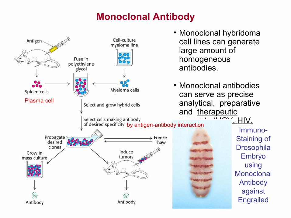
Monoclonal Antibody
• Monoclonal hybridoma cell lines can generate large amount of homogeneous antibodies.
• Monoclonal antibodies can serve as precise analytical, preparative and therapeutic reagents.(HCV, HIV, herceptin) Immuno-
Staining ofDrosophila
Embryousing
MonoclonalAntibodyagainst
Engrailed
Plasma cell
by antigen-antibody interaction

Monoclonal antibody drugs?
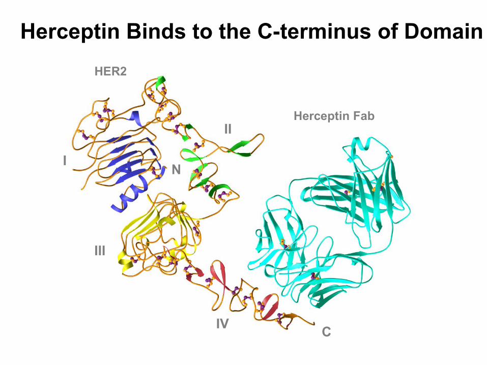
Herceptin Binds to the C-terminus of Domain IV
Herceptin Fab
I
III
II
IV
N
C
HER2
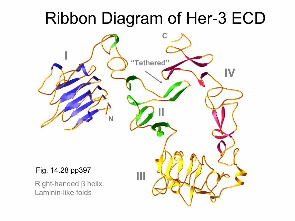
Ribbon Diagram of Her-3 ECD
N
C
“Tethered”I
II
III
IV
Right-handed β helixLaminin-like folds
Fig. 14.28 pp397

Surface representations of EGFR and HER2 in Antibody-Bound Conformations
Herceptin

ELISA (Enzyme-Linked Immuno-Sorbent Assay)
Antibody detection, anti-HIV antibody
Antigen detection
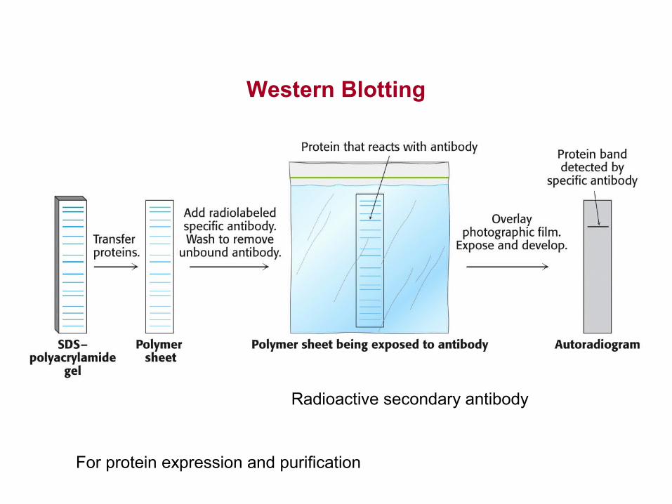
Western Blotting
Radioactive secondary antibody
For protein expression and purification

Immuno-FluorescenceMicroscopy
Actin Filament Stainingusing α-actin antibody
Immuno-ElectronMicroscopy
Detection of a channel proteinfrom the synaptic vesicles
using antibodies tagged withelectron-dense markers such as gold
or ferritin(Resolution better than 10 nm)
Fluorescence-labeled antibodies(resolution 200nm)
ex) Glucocorticoid receptor
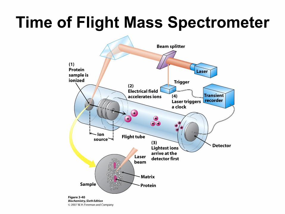
Time of Flight Mass Spectrometer

Nuclear Magnetic Resonance

End of Chapter 3
Thank You
Related Documents