Experimental comparison and cross-validation of Affymetrix HT plate and cartridge array gene expression platforms Normand E. Allaire ⁎, Leila E. Rieder, Jadwiga Bienkowska, John P. Carulli Biogen Idec, Inc., 14 Cambridge Center, Cambridge, MA 02142, USA abstract article info Article history: Received 23 January 2008 Accepted 16 June 2008 Available online 17 September 2008 Keywords: High throughput Gene expression The successful use of gene expression microarrays in basic research studies has spawned interest in the use of this technology for clinical trial and population-based studies, but cost, complexity of sample processing and tracking, and limitations of sample throughput have restricted their use for these very large-scale investigations. The Affymetrix GeneChip Plate Array System addresses these concerns and could facilitate larger studies if the data prove to be comparable to industry-standard cartridge arrays. Here we present a comparative evaluation of performance between Affymetrix GeneChip Human 133A cartridge and plate arrays with an emphasis on the assessment of systematic variation and its impact on log ratio data. This study utilized two standardized control RNAs on four independent lots of plate and cartridge arrays. We found that HT plate arrays showed improved specificity and were more reproducible over a wide intensity range, but cartridge arrays exhibit better sensitivity. Not surprisingly, artifactual changes due to positional effects were detectable on plate arrays, but were generally small in number and magnitude and in practice may be removed using standard fold-change and p-value thresholds. Overall, log ratio data between cartridges and plate arrays were remarkably concordant. We conclude that HT arrays offer significant improvements over cartridge arrays for large-scale studies. © 2008 Elsevier Inc. All rights reserved. The successful use of gene expression microarrays in basic research studies has spawned interest in the use of this technology for clinical trial and population-based studies [1–3], but cost [4], complexity of sample processing and tracking [5], and limitations of sample throughput [6] have restricted their use for these very large-scale investigations. RNA expression profiling using oligonucleotide arrays has been an industry standard for many years, although the technology has been continuously evolving. Historically, a major focus has been to increase element density with the goal of enabling the interrogation of entire complex genomes on a single array. Now that whole-genome expression arrays are common, the center of attention has shifted from increasing transcript representation to improving sample throughput and reducing processing costs. The most recent advances in density have allowed for assay miniaturiza- tion and organization of arrays into the standard format of a 96-well microtiter plate. This format change has enabled the use of automated liquid-handling instruments for laborious hybridization, washing, and staining steps and modified high-content plate scanners for scanning steps. Automation of processing steps will significantly reduce labor by streamlining and simplifying sample processing and tracking to allow for expression profiling of larger sample sets than are currently practical with cartridge-or slide-based microarrays. We conservatively estimate hands-on labor costs can be reduced by 75%. In addition to reduced labor cost, we have realized a savings of 50% on arrays due to manufacturing savings as a result of assay miniaturization. Although the potential cost savings with this technology are impressive, to be useful in practice data from this new plate-based technology must be concordant with its industry-standard cartridge counterpart. Both HG-U133A plates and their cartridge counterparts are fabricated by in situ synthesis of 25-mer oligonucleotides [3,7], contain the same genome content, and use the same probe-set strategies to measure gene expression levels. Although it is true that both formats share these key design and fabrication elements, there are several differences that could impact array data. The most notable difference is the reduction in feature size from 11 to 8 μm on plate compared to cartridge arrays, which reduces cell surface area by 47.1%. Second, HT plate arrays are designed with an open-flow cell and cartridge arrays use an enclosed system. The advantage of this open design is that it allows for automation of hybridization, washing, staining, and scanning steps, which should reduce processing variability. However, this architecture makes the HT arrays susceptible to contamination by dust and particles from the laboratory environ- ment and arrays can be easily damaged if touched. Additionally, the open configuration requires that plate hybridizations occur under static conditions. Alternatively, the cartridge arrays' enclosed-flow cell is protected from external contact but must be washed and stained via independently temperature-controlled fluidics modules, which could introduce greater processing variability. The closed-flow cell of the cartridge system allows for nonstatic hybridizations, which may Genomics 92 (2008) 359–365 ⁎ Corresponding author. Fax: +1 617 679 3200. E-mail address: [email protected] (N.E. Allaire). 0888-7543/$ – see front matter © 2008 Elsevier Inc. All rights reserved. doi:10.1016/j.ygeno.2008.06.010 Contents lists available at ScienceDirect Genomics journal homepage: www.elsevier.com/locate/ygeno

Welcome message from author
This document is posted to help you gain knowledge. Please leave a comment to let me know what you think about it! Share it to your friends and learn new things together.
Transcript
Genomics 92 (2008) 359–365
Contents lists available at ScienceDirect
Genomics
j ourna l homepage: www.e lsev ie r.com/ locate /ygeno
Experimental comparison and cross-validation of Affymetrix HT plate and cartridgearray gene expression platforms
Normand E. Allaire⁎, Leila E. Rieder, Jadwiga Bienkowska, John P. CarulliBiogen Idec, Inc., 14 Cambridge Center, Cambridge, MA 02142, USA
⁎ Corresponding author. Fax: +1 617 679 3200.E-mail address: [email protected] (N.E. A
0888-7543/$ – see front matter © 2008 Elsevier Inc. Aldoi:10.1016/j.ygeno.2008.06.010
a b s t r a c t
a r t i c l e i n f oArticle history:
The successful use of gene e Received 23 January 2008Accepted 16 June 2008Available online 17 September 2008Keywords:High throughputGene expression
xpression microarrays in basic research studies has spawned interest in the use ofthis technology for clinical trial and population-based studies, but cost, complexity of sample processing andtracking, and limitations of sample throughput have restricted their use for these very large-scaleinvestigations. The Affymetrix GeneChip Plate Array System addresses these concerns and could facilitatelarger studies if the data prove to be comparable to industry-standard cartridge arrays. Here we present acomparative evaluation of performance between Affymetrix GeneChip Human 133A cartridge and platearrays with an emphasis on the assessment of systematic variation and its impact on log ratio data. This studyutilized two standardized control RNAs on four independent lots of plate and cartridge arrays. We found thatHT plate arrays showed improved specificity and were more reproducible over a wide intensity range, butcartridge arrays exhibit better sensitivity. Not surprisingly, artifactual changes due to positional effects weredetectable on plate arrays, but were generally small in number and magnitude and in practice may beremoved using standard fold-change and p-value thresholds. Overall, log ratio data between cartridges andplate arrays were remarkably concordant. We conclude that HT arrays offer significant improvements overcartridge arrays for large-scale studies.
© 2008 Elsevier Inc. All rights reserved.
The successful use of gene expression microarrays in basic research
studies has spawned interest in the use of this technology for clinicaltrial and population-based studies [1–3], but cost [4], complexity ofsample processing and tracking [5], and limitations of samplethroughput [6] have restricted their use for these very large-scaleinvestigations. RNA expression profiling using oligonucleotide arrayshas been an industry standard for many years, although thetechnology has been continuously evolving. Historically, a majorfocus has been to increase element density with the goal of enablingthe interrogation of entire complex genomes on a single array. Nowthat whole-genome expression arrays are common, the center ofattention has shifted from increasing transcript representation toimproving sample throughput and reducing processing costs. Themost recent advances in density have allowed for assay miniaturiza-tion and organization of arrays into the standard format of a 96-wellmicrotiter plate. This format change has enabled the use of automatedliquid-handling instruments for laborious hybridization, washing, andstaining steps and modified high-content plate scanners for scanningsteps. Automation of processing steps will significantly reduce laborby streamlining and simplifying sample processing and tracking toallow for expression profiling of larger sample sets than are currentlypractical with cartridge-or slide-basedmicroarrays.We conservativelyestimate hands-on labor costs can be reduced by 75%. In addition tollaire).
l rights reserved.
reduced labor cost, we have realized a savings of 50% on arrays due tomanufacturing savings as a result of assay miniaturization. Althoughthe potential cost savings with this technology are impressive, to beuseful in practice data from this new plate-based technology must beconcordant with its industry-standard cartridge counterpart.
Both HG-U133A plates and their cartridge counterparts arefabricated by in situ synthesis of 25-mer oligonucleotides [3,7],contain the same genome content, and use the same probe-setstrategies to measure gene expression levels. Although it is true thatboth formats share these key design and fabrication elements, thereare several differences that could impact array data. The most notabledifference is the reduction in feature size from 11 to 8 μm on platecompared to cartridge arrays, which reduces cell surface area by 47.1%.Second, HT plate arrays are designed with an open-flow cell andcartridge arrays use an enclosed system. The advantage of this opendesign is that it allows for automation of hybridization, washing,staining, and scanning steps, which should reduce processingvariability. However, this architecturemakes the HT arrays susceptibleto contamination by dust and particles from the laboratory environ-ment and arrays can be easily damaged if touched. Additionally, theopen configuration requires that plate hybridizations occur understatic conditions. Alternatively, the cartridge arrays' enclosed-flow cellis protected from external contact but must be washed and stained viaindependently temperature-controlled fluidics modules, which couldintroduce greater processing variability. The closed-flow cell of thecartridge system allows for nonstatic hybridizations, which may

Fig. 1. Schematic representation of the experimental design. See Materials and methods for details. Key features are that bulk labeling and fragmentation were completed usingstandardized RNA with spiked controls and then hybridized to either cartridge or plate arrays.
360 N.E. Allaire et al. / Genomics 92 (2008) 359–365
improve scan uniformity. Third, scanning technology differs signifi-cantly between HT plate and cartridge arrays. The cartridge arraysystem produces a single image generated by excitation with a solid-state laser and captured with a confocal scanner. The HT arrayplatform uses a charged couple device (CCD) camera to collect 48 low-and high-exposure images and then assembles them into a singleimage for each array. A final potential source of variation that is uniqueto plate arrays is intraplate positional effects. These effects areattributed to differences in evaporation and heat transfer due to thepositions of individual arrays within the plate.
This paper details results from a series of benchmarking experi-ments comparing the Affymetrix HG-U133A_2 cartridge and its HTplate array counterpart. To our knowledge this is the first independentassessment of this new HT plate array platform. We utilized twohighly characterized reference RNAs used by the Micro-Array QualityConsortium (MAQC) [8,14–17] as our test samples (Fig. 1). We addedpoly(A) controls to the input test RNA and hybridization control spikesto the pooled postlabeled and fragmented target cRNA, thus allowingan assessment of hybridization variance independent of prehybridiza-tion factors. By assessing four lots of plate and cartridge arrays wewere able to obtain a snapshot of the manufacturing variance of arrayproduction. Finally, using QPCR data that were generated from thesereference RNAs allowed for a microarray-independent assessment offold-change accuracy.
Results
Global quality metrics
Scan quality on a global level was assessed using the percentageof probe sets scored present (%P) as calculated by Microarray Suite5.0. These detection calls, and associated p values, are determinedbased on nonparametric rank tests [21] and are based onempirically determined default detection call settings as recom-mended by the manufacturer for both cartridge and HT plate arrays.We grouped scans by sample type for all cartridges or an equivalentnumber of randomly selected plate arrays (n=24) (Fig. 2A). We firstnoted that range of %P for replicate hybridizations on HT platearrays appeared smaller and the median higher than those oncartridge arrays. We then tested each sample grouping (humanbrain reference RNA (HBRR) or universal human reference RNA(UHRR)) for differences in %P (Student's t test). We noticed asignificant increase in %P for HT plate compared to cartridge scansfor HBRR but not UHRR (p=0.04 and 0.98, respectively), indicating
improved performance based on sample type. We also observed atrend in the percentage of coefficient of variation (%CV) of the percent-age of present calls: HBRR_CartNUHRR_CartNUHRR_PlateNHBRR_Plate(%CV=3.50, 2.47, 1.93, 1.71, respectively). Additionally, we observed anapproximate twofold increase in the number of probe sets that werecalled marginal with cartridge versus HT plate arrays (2.5 and 1.2%,respectively, data not shown).
To investigate global array performance further we evaluated twokey metrics, background level (Fig. 2B) and Raw Q (defined as thepixel-to-pixel variation of the background probe cells; Fig. 2C) forcartridge and HT plate arrays (n=16) as indicators of total processnoise and scanner noise, respectively. Background values for HT arrayswere an average of 1.38 times higher (n=16, p=1.18×10⁻¹⁸) than forcartridge arrays for both UHRR and HBRR (HT array HBRR, UHRR,cartridge HBRR, UHRR: 78.85, 78.59, 58.24, 56.24, respectively).Additionally, we observed an average 1.75-fold increase (n=16,p=2.09×10⁻²³) in scanner noise for both HBRR and UHRR using theCCD camera-based HT array scanner compared to the solid-statelaser/PMT of the cartridge scanner (HT array HBRR, UHRR, cartridgeHBRR, UHRR: 3.18, 2.94, 1.80, 1.69, respectively).
Interlot and intralot variation
Lot variance due to manufacturing differences can be a significantsource of variation for microarray technologies [9–11]. To assess thisvariation we compared replicate HBRR hybridizations (n=4) fromcartridge and HT plate array manufacturing lots (n=4). HT plate andcartridge arrays were GCRMA normalized by lot. Interlot variation (thevariation within and between array lots) was quantitatively deter-mined by calculating the %CV of the log2 intensity for each probe setacross replicate hybridizations for each lot (Figs. 3A and 3B). It isimmediately apparent that HT arrays show improved interlot repro-ducibility as indicated by the lower variation among array curvesrelative to cartridge curves. Additionally, HT plate arrays exhibitimproved intralot consistency as indicated by smaller variance withineach lot curve. The resultant %CV curves for both cartridge and platearrays display a characteristic shape that begins high, climbs to a peak,and then gradually declines to a minimum value. It is not surprisingthat the log intensity at the %CV maximum correlates with thebackground signal for each cartridge or plate array lot. Furthermore, isclear from these graphs that the background signals of the HT platearray lots are more consistent but also higher than for cartridge arraylots. This indicates improved lot consistency of plate arrays but alsolower sensitivity than cartridge arrays.
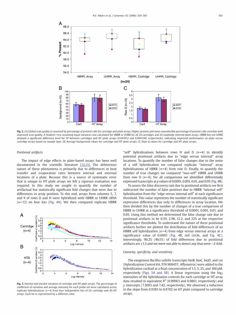
Fig. 2. (A) Global scan quality as assessed by percentage of present calls for cartridge and plate arrays. Higher present andmore reproducible percentage of present calls correlate withimproved scan quality. A Student t test assuming equal variances was calculated for HBRR or UHRR for all 24 cartridges and 24 randomly selected plate arrays. HBRR but not UHRRattained a significant difference level for %P between cartridges and HT plate arrays (0.043912 and 0.9841149, respectively), indicating improved performance on plate versuscartridge arrays based on sample type. (B) Average background values for cartridge and HT plate arrays. (C) Raw Q values for cartridge and HT plate arrays.
361N.E. Allaire et al. / Genomics 92 (2008) 359–365
Positional artifacts
The impact of edge effects in plate-based assays has been welldocumented in the scientific literature [12,13]. The deleteriousnature of these phenomena is primarily due to differences in heattransfer and evaporation rates between internal and externallocations of a plate. Because this is a source of systematic errorthat is unique to HT plate arrays we felt a rigorous evaluation wasrequired. In this study we sought to quantify the number ofartifactual but statistically significant fold changes that were due todifferences in array position. To this end, arrays from columns 5, 7,and 9 of rows D and H were hybridized with HBRR or UHRR cRNA(n=12) on four lots (Fig. 4A). We then compared replicate HBRR
Fig. 3. Interlot and intralot variation of cartridge and HT plate arrays. The percentage ofcoefficient of variation and average intensity for each probe set were calculated acrossreplicate hybridizations (n=4) from four independent lots of (A) cartridge and (B) HTarrays. Each lot is represented by a different color.
“self” hybridizations between rows H and D (n=4) to identifypotential positional artifacts due to “edge versus internal” arraylocations. To quantify the number of false changes due to the noiseof a self hybridization we compared replicate “internal” arrayhybridizations of HBRR (n=4) from row D. Finally, to quantify thenumber of true changes we compared “non-self” HBRR and UHRRfrom row D (n=4). For all comparisons we identified differentiallyexpressed transcripts atp values of 0.0001, 0.001, 0.01, and 0.05 (Fig. 4B).
To assess the false discovery rate due to positional artifacts we firstsubtracted the number of false positives due to HBRR “internal self”hybridization from the “edge versus internal self” at each significancethreshold. This value represents the number of statistically significantexpression differences due only to differences in array location. Wethen divided this by the number of changes of a true comparison ofHBRR to UHRR at a significance threshold of 0.0001, 0.001, 0.01, and0.05. Using this method we determined the false change rate due topositional artifacts to be 0.59, 2.98, 12.2, and 22% at the respectivesignificance thresholds. To understand the nature of these positionalartifacts further we plotted the distribution of fold differences of anHBRR self hybridization (n=4) from edge versus internal arrays at asignificance value of 0.0001 (Fig. 4B, red circle, and Fig. 4C).Interestingly, 90.2% (46/51) of fold differences due to positionalartifacts are ≤1.5 andwewere not able to detect any that were N2-fold.
Linearity, specificity, and sensitivity
The exogenous Bacillus subtilis transcripts bioB, bioC, bioD, and cre(Hybridization Control Kit, P/N 900457; Affymetrix) were added to thehybridization cocktail at a final concentration of 1.5, 5, 25, and 100 pM,respectively (Figs. 5A and 5B). A linear regression using the log2intensities of the hybridization controls for each cartridge or HT arraydata resulted in equivalent R2 (0.99963 and 0.9861, respectively) andy intercepts (7.3093 and 7.42, respectively). We observed a reductionin the slope from 0.9383 to 0.8702 on HT plate compared to cartridgearrays.
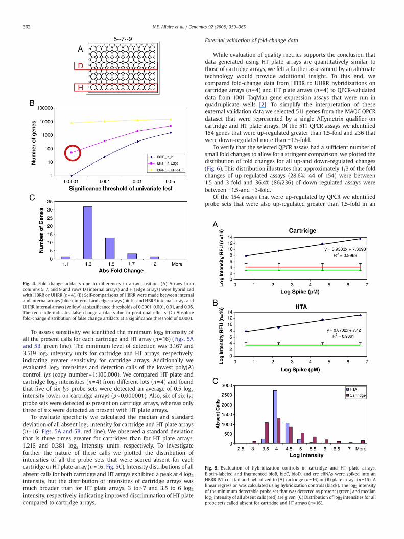
Fig. 4. Fold-change artifacts due to differences in array position. (A) Arrays fromcolumns 5, 7, and 9 and rows D (internal arrays) and H (edge arrays) were hybridizedwith HBRR or UHRR (n=4). (B) Self-comparisons of HBRR were made between internaland internal arrays (blue), internal and edge arrays (pink), and HBRR internal arrays andUHRR internal arrays (yellow) at significance thresholds of 0.0001, 0.001, 0.01, and 0.05.The red circle indicates false change artifacts due to positional effects. (C) Absolutefold-change distribution of false change artifacts at a significance threshold of 0.0001.
Fig. 5. Evaluation of hybridization controls in cartridge and HT plate arrays.Biotin-labeled and fragmented bioB, bioC, bioD, and cre cRNAs were spiked into anHBRR IVT cocktail and hybridized to (A) cartridge (n=16) or (B) plate arrays (n=16). Alinear regression was calculated using hybridization controls (black). The log2 intensityof the minimum detectable probe set that was detected as present (green) and medianlog2 intensity of all absent calls (red) are given. (C) Distribution of log2 intensities for allprobe sets called absent for cartridge and HT arrays (n=16).
362 N.E. Allaire et al. / Genomics 92 (2008) 359–365
To assess sensitivity we identified the minimum log2 intensity ofall the present calls for each cartridge and HT array (n=16) (Figs. 5Aand 5B, green line). The minimum level of detection was 3.167 and3.519 log2 intensity units for cartridge and HT arrays, respectively,indicating greater sensitivity for cartridge arrays. Additionally weevaluated log2 intensities and detection calls of the lowest poly(A)control, lys (copy number=1:100,000). We compared HT plate andcartridge log2 intensities (n=4) from different lots (n=4) and foundthat five of six lys probe sets were detected an average of 0.5 log2intensity lower on cartridge arrays (pb0.000001). Also, six of six lysprobe sets were detected as present on cartridge arrays, whereas onlythree of six were detected as present with HT plate arrays.
To evaluate specificity we calculated the median and standarddeviation of all absent log2 intensity for cartridge and HT plate arrays(n=16; Figs. 5A and 5B, red line). We observed a standard deviationthat is three times greater for cartridges than for HT plate arrays,1.216 and 0.381 log2 intensity units, respectively. To investigatefurther the nature of these calls we plotted the distribution ofintensities of all the probe sets that were scored absent for eachcartridge or HT plate array (n=16; Fig. 5C). Intensity distributions of allabsent calls for both cartridge and HT arrays exhibited a peak at 4 log2intensity, but the distribution of intensities of cartridge arrays wasmuch broader than for HT plate arrays, 3 toN7 and 3.5 to 6 log2intensity, respectively, indicating improved discrimination of HT platecompared to cartridge arrays.
External validation of fold-change data
While evaluation of quality metrics supports the conclusion thatdata generated using HT plate arrays are quantitatively similar tothose of cartridge arrays, we felt a further assessment by an alternatetechnology would provide additional insight. To this end, wecompared fold-change data from HBRR to UHRR hybridizations oncartridge arrays (n=4) and HT plate arrays (n=4) to QPCR-validateddata from 1001 TaqMan gene expression assays that were run inquadruplicate wells [2]. To simplify the interpretation of theseexternal validation data we selected 511 genes from the MAQC QPCRdataset that were represented by a single Affymetrix qualifier oncartridge and HT plate arrays. Of the 511 QPCR assays we identified154 genes that were up-regulated greater than 1.5-fold and 236 thatwere down-regulated more than −1.5-fold.
To verify that the selected QPCR assays had a sufficient number ofsmall fold changes to allow for a stringent comparison, we plotted thedistribution of fold changes for all up-and down-regulated changes(Fig. 6). This distribution illustrates that approximately 1/3 of the foldchanges of up-regulated assays (28.6%; 44 of 154) were between1.5-and 3-fold and 36.4% (86/236) of down-regulated assays werebetween −1.5-and −3-fold.
Of the 154 assays that were up-regulated by QPCR we identifiedprobe sets that were also up-regulated greater than 1.5-fold in an

Fig. 6. Fold-change distribution of MAQC QPCR validated genes. 154 up-regulated≥1.5-fold and 236 down-regulated ≥−1.5-fold genes that were represented by a singleAffymetrix qualifier on HG U133A_2 and HT-HG U133A were selected from the MAQCQPCR dataset.
Fig. 8. Accuracy of HT plate and cartridge arrays by MAQC QPCR. 154 up-regulated and236 down-regulated QPCR-validated log ratios were compared to (A) HT plate array logratios or (B) cartridge array log ratios.
363N.E. Allaire et al. / Genomics 92 (2008) 359–365
HBRR versus UHRR comparison for cartridge or HT plate arrays (n=4;Fig. 7A). Of the 154 up-regulated genes detected using QPCR, 141 and143 were also detected using HT plate arrays and cartridge arrays,respectively, and 136 were detected by all three methods. Sevendifferences were detected as up-regulated by both HT plate arrays andQPCR, but not cartridges, and five differences were detected asup-regulated by both cartridges and QPCR, but not HT plate arrays. Ofthe five HT array-specific up-regulated changes we classified two as“less reliable” because they were less than 2-fold or had HBRR orUHRR values with a QPCR quality value of “low expression” and/or“high standard deviation” or “not expressed.” By the same reasoningwe classified five of the seven cartridge-specific changes asless reliable.
By QPCR we identified 236 down-regulated assays that were alsodown-regulated by more than −1.5 in an HBRR versus UHRRcomparison for cartridge and HT plate arrays (n=4; Fig. 7B). Of the236 down-regulated assays detected using QPCR, 216 and 218 werealso detected using HT plate arrays and cartridge arrays, respectively,and 208 were detected by all three methods. Eight probe sets weredetected as down-regulated by both HT plate arrays and QPCR, but notcartridges, and 10 probe sets were detected as down-regulated byboth cartridges and QPCR, but not HT plate arrays. All of the 8HTA-specific or 10 cartridge-specific changes were classified as lessreliable because they were less than 2-fold or had HBRR or UHRRvalues with a QPCR quality value of low expression and/or highstandard deviation or not expressed.
HT plate (n=4) and cartridge array (n=4) ratios (HBRR to UHRR)were each compared to all 154 up-regulated and 218 down-regulated
Fig. 7. Venn diagram of HT and cartridge arrays and 154 up-regulated and 236down-regulated QPCR-validated fold changes. Gray values were classified as “lessreliable” because they were less than 2-fold or had HBRR or UHRR values with a QPCRquality value of “low expression” and/or “high standard deviation” or “not expressed.”
MAQCQPCR ratios (Figs. 8A and 8B). A linear regression of the HT plateand cartridge arrays to the MAC QPCR log ratio data produced R2
values of 0.8288 and 0.8363, respectively. Additionally, we observed acomplete correlation of directionality between HT plate or cartridgearray ratios and MAQC QPCR ratios. We observed a “shelf effect” forQPCR log ratios greater than 5 for both HT plate and cartridge arrays.To understand the nature of this effect we plotted the log2 intensitiesof HBRR and UHRR against these ratios (data not shown). This analysisrevealed that the observed shelf effect was due to inaccurateestimation of UHRR log intensities that were close to the noise,resulting in inflation of the HT plate and cartridge array log ratios.
Discussion
Our goal was to assess the Affymetrix HT plate array as acompanion technology to the industry-standard cartridge arraysthat are used for expression profiling in the Biogen Idec TranscriptProfiling core facility. To determine if HT plate arrays are technicallyequivalent to cartridge arrays and to understand the limitations of thistechnology we designed our experiments to address several keyquestions. First, we sought to understand how global qualityindicators like percentage of present calls, average background, andRaw Q were affected using HT plate versus cartridge arrays. Second,we wanted to evaluate manufacturing variance by assessing lotreproducibility for both cartridge and plate arrays. Third, we wantedto assess the impact of edge effects on fold-change data for HT platearrays. Fourth, we chose to compare linearity, specificity, andsensitivity of HT plate versus cartridge arrays. Finally, we wanted toassess accuracy of fold change for plate and cartridge arrays comparedto an externally validated QPCR dataset.
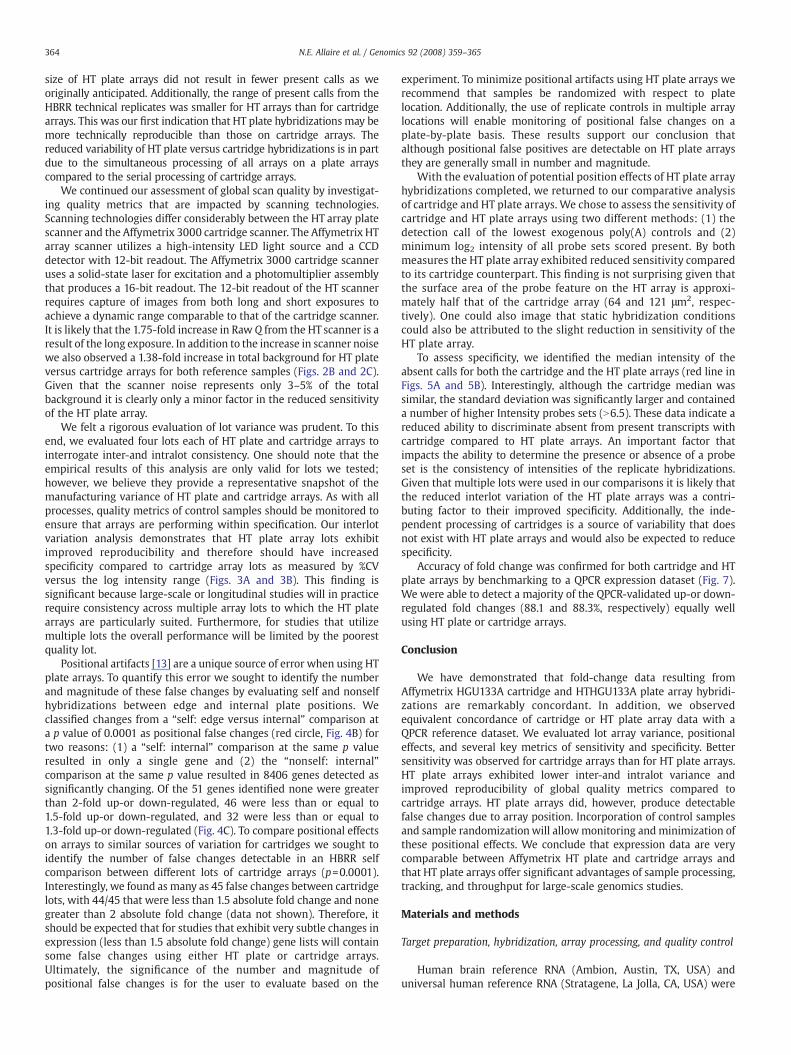
We began our analysis by comparing global quality metrics for bothcartridge andHT plate arrays. In practice the primary qualitymetric thatis used to assess individual scan quality is the percentage of probes thatscored present [18–20]. The percentage of transcripts that score presentis a robust global indicator of scan quality because all processing-relatedvariables impact this single metric. Interestingly, the reduced feature
364 N.E. Allaire et al. / Genomics 92 (2008) 359–365
size of HT plate arrays did not result in fewer present calls as weoriginally anticipated. Additionally, the range of present calls from theHBRR technical replicates was smaller for HT arrays than for cartridgearrays. This was our first indication that HT plate hybridizations may bemore technically reproducible than those on cartridge arrays. Thereduced variability of HT plate versus cartridge hybridizations is in partdue to the simultaneous processing of all arrays on a plate arrayscompared to the serial processing of cartridge arrays.
We continued our assessment of global scan quality by investigat-ing quality metrics that are impacted by scanning technologies.Scanning technologies differ considerably between the HT array platescanner and the Affymetrix 3000 cartridge scanner. The Affymetrix HTarray scanner utilizes a high-intensity LED light source and a CCDdetector with 12-bit readout. The Affymetrix 3000 cartridge scanneruses a solid-state laser for excitation and a photomultiplier assemblythat produces a 16-bit readout. The 12-bit readout of the HT scannerrequires capture of images from both long and short exposures toachieve a dynamic range comparable to that of the cartridge scanner.It is likely that the 1.75-fold increase in Raw Q from the HT scanner is aresult of the long exposure. In addition to the increase in scanner noisewe also observed a 1.38-fold increase in total background for HT plateversus cartridge arrays for both reference samples (Figs. 2B and 2C).Given that the scanner noise represents only 3–5% of the totalbackground it is clearly only a minor factor in the reduced sensitivityof the HT plate array.
We felt a rigorous evaluation of lot variance was prudent. To thisend, we evaluated four lots each of HT plate and cartridge arrays tointerrogate inter-and intralot consistency. One should note that theempirical results of this analysis are only valid for lots we tested;however, we believe they provide a representative snapshot of themanufacturing variance of HT plate and cartridge arrays. As with allprocesses, quality metrics of control samples should be monitored toensure that arrays are performing within specification. Our interlotvariation analysis demonstrates that HT plate array lots exhibitimproved reproducibility and therefore should have increasedspecificity compared to cartridge array lots as measured by %CVversus the log intensity range (Figs. 3A and 3B). This finding issignificant because large-scale or longitudinal studies will in practicerequire consistency across multiple array lots to which the HT platearrays are particularly suited. Furthermore, for studies that utilizemultiple lots the overall performance will be limited by the poorestquality lot.
Positional artifacts [13] are a unique source of error when using HTplate arrays. To quantify this error we sought to identify the numberand magnitude of these false changes by evaluating self and nonselfhybridizations between edge and internal plate positions. Weclassified changes from a “self: edge versus internal” comparison ata p value of 0.0001 as positional false changes (red circle, Fig. 4B) fortwo reasons: (1) a “self: internal” comparison at the same p valueresulted in only a single gene and (2) the “nonself: internal”comparison at the same p value resulted in 8406 genes detected assignificantly changing. Of the 51 genes identified none were greaterthan 2-fold up-or down-regulated, 46 were less than or equal to1.5-fold up-or down-regulated, and 32 were less than or equal to1.3-fold up-or down-regulated (Fig. 4C). To compare positional effectson arrays to similar sources of variation for cartridges we sought toidentify the number of false changes detectable in an HBRR selfcomparison between different lots of cartridge arrays (p=0.0001).Interestingly, we found as many as 45 false changes between cartridgelots, with 44/45 that were less than 1.5 absolute fold change and nonegreater than 2 absolute fold change (data not shown). Therefore, itshould be expected that for studies that exhibit very subtle changes inexpression (less than 1.5 absolute fold change) gene lists will containsome false changes using either HT plate or cartridge arrays.Ultimately, the significance of the number and magnitude ofpositional false changes is for the user to evaluate based on the
experiment. To minimize positional artifacts using HT plate arrays werecommend that samples be randomized with respect to platelocation. Additionally, the use of replicate controls in multiple arraylocations will enable monitoring of positional false changes on aplate-by-plate basis. These results support our conclusion thatalthough positional false positives are detectable on HT plate arraysthey are generally small in number and magnitude.
With the evaluation of potential position effects of HT plate arrayhybridizations completed, we returned to our comparative analysisof cartridge and HT plate arrays. We chose to assess the sensitivity ofcartridge and HT plate arrays using two different methods: (1) thedetection call of the lowest exogenous poly(A) controls and (2)minimum log2 intensity of all probe sets scored present. By bothmeasures the HT plate array exhibited reduced sensitivity comparedto its cartridge counterpart. This finding is not surprising given thatthe surface area of the probe feature on the HT array is approxi-mately half that of the cartridge array (64 and 121 μm2, respec-tively). One could also image that static hybridization conditionscould also be attributed to the slight reduction in sensitivity of theHT plate array.
To assess specificity, we identified the median intensity of theabsent calls for both the cartridge and the HT plate arrays (red line inFigs. 5A and 5B). Interestingly, although the cartridge median wassimilar, the standard deviation was significantly larger and containeda number of higher Intensity probes sets (N6.5). These data indicate areduced ability to discriminate absent from present transcripts withcartridge compared to HT plate arrays. An important factor thatimpacts the ability to determine the presence or absence of a probeset is the consistency of intensities of the replicate hybridizations.Given that multiple lots were used in our comparisons it is likely thatthe reduced interlot variation of the HT plate arrays was a contri-buting factor to their improved specificity. Additionally, the inde-pendent processing of cartridges is a source of variability that doesnot exist with HT plate arrays and would also be expected to reducespecificity.
Accuracy of fold change was confirmed for both cartridge and HTplate arrays by benchmarking to a QPCR expression dataset (Fig. 7).We were able to detect a majority of the QPCR-validated up-or down-regulated fold changes (88.1 and 88.3%, respectively) equally wellusing HT plate or cartridge arrays.
Conclusion
We have demonstrated that fold-change data resulting fromAffymetrix HGU133A cartridge and HTHGU133A plate array hybridi-zations are remarkably concordant. In addition, we observedequivalent concordance of cartridge or HT plate array data with aQPCR reference dataset. We evaluated lot array variance, positionaleffects, and several key metrics of sensitivity and specificity. Bettersensitivity was observed for cartridge arrays than for HT plate arrays.HT plate arrays exhibited lower inter-and intralot variance andimproved reproducibility of global quality metrics compared tocartridge arrays. HT plate arrays did, however, produce detectablefalse changes due to array position. Incorporation of control samplesand sample randomizationwill allowmonitoring andminimization ofthese positional effects. We conclude that expression data are verycomparable between Affymetrix HT plate and cartridge arrays andthat HT plate arrays offer significant advantages of sample processing,tracking, and throughput for large-scale genomics studies.
Materials and methods
Target preparation, hybridization, array processing, and quality control
Human brain reference RNA (Ambion, Austin, TX, USA) anduniversal human reference RNA (Stratagene, La Jolla, CA, USA) were
365N.E. Allaire et al. / Genomics 92 (2008) 359–365
used for this evaluation. The bacterial poly(A) control spikes lys, phe,thr, and dap (Affymetrix) were added to HBRR and UHRR to achieve afinal copy number ratio of 1:100,000, 1:50,000, 1:25,000, and 1:6667,respectively. The Affymetrix automated Target Preparation protocol(TP_0001) was used to prepare 48 wells of labeled and unfragmentedcRNA for both test samples according to the GeneChip ExpressionAnalysis Technical Manual for Cartridge Arrays Using the GeneChip ArrayStation (P/N 702064, Affymetrix). Labeled, unfragmented cRNA yieldswere calculated for each set of 48 wells and high-quality replicateswere then pooled and redistributed to a 96-well plate for manualfragmentation (data not shown). Fragmented cRNA test samples wererepooled to achieve uniformity and then split into two aliquots andadded to a hybridization cocktail containing the hybridization controlsBioB, BioC, BioD, and cre (P/N 900458, Affymetrix) for HT plates orcartridge arrays.
Four different lots of 24-array HT HG-U133A plates werehybridized, washed, and stained using the Affymetrix automatedprotocol (HYB_0001 and WS_0001) according to the GeneChipExpression Analysis Technical Manual for HT Plate Arrays Using theGeneChip Array Station (P/N 702063, Affymetrix). HBRR and UHRRlabeled cRNAwas hybridized to 12 arrays from each lot for a total of 96arrays. Plate arrays were scanned using a GeneChip HT array platescanner version 1.0 (Affymetrix) to acquire raw image files (.dat).Probe cell intensities (.cel), probe set present/marginal/absent calls(.chp), and global quality metrics (.rpt) files for each scanned imagewere generated using the GCOS software statistical algorithm version1.0 (Affymetrix). Global quality metrics were imported into Spotfire(Spotfire, Palo Alto, CA, USA) for visualization.
Six arrays from four different lots of HG-U133A 2.0 cartridges weremanually hybridized with an HBRR or UHRR hybridization cocktailaccording to the GeneChip Expression Analysis Technical Manual forCartridge Arrays Using the GeneChip Array Station (P/N 702064,Affymetrix) for a total of 48 arrays. Washing and staining werecompleted using a Fluidics Station 450 according to the GeneChipExpression Analysis Technical Manual for Cartridge Arrays Using theGeneChip Array Station (P/N 702064, Affymetrix). Cartridge arrayswere scanned on a GeneChip Scanner 3000 6G (Affymetrix) to acquireraw image files (.dat). Probe cell intensities (.cel), present/marginal/absent calls (.chp), and global array quality metrics (.rpt) files weregenerated for each scanned image using GCOS software statisticalalgorithm version 1.0 (Affymetrix). Global quality metrics wereimported into Spotfire (Spotfire) for visualization.
Statistical analysis
Analyses were performed in the R statistical language using BRBArrayTools version 3.6.3 developed by Richard Simon and Amy PengLam (http://linus.nci.nih.gov/BRB-ArrayTools.html). Quantile normal-
ization and probe set summarizations were computed independentlyfor plate or cartridge arrays using the GCRMA procedure as imple-mented in BRB ArrayTools. Microsoft Excel was employed for standarddeviations, coefficient of variation, and t-test calculations.
Acknowledgments
We thank Dr. Matvey Lukashev (Immunobiology Research, BiogenIdec) for invaluable discussion of this study.We also thank Dr. SuzanneSzak and Dr. Huo Li (Computational Biology, Biogen Idec) for excellenttechnical help.
References
[1] J.C. Fuscoe, W. Tong, L. Shi, QA/QC issues to aid regulatory acceptance ofmicroarray gene expression data, Environ. Mol. Mutagen. 48 (2007) 349–353.
[2] G. Gibson, Microarrays in ecology and evolution: a preview, Mol. Ecol. 11 (2002)17–24.
[3] E. Strauss, Arrays of hope, Cell 127 (2006) 657–659.[4] H. Chen, J. Li, Nanotechnology: moving from microarrays toward nanoarrays,
Methods Mol. Biol. 381 (2007) 411–436.[5] P. Honore, et al., MicroArray Facility: a laboratory information management
system with extended support for nylon based technologies, BMC Genomics 7(2006) 240.
[6] T. Morrison, et al., Nanoliter high throughput quantitative PCR, Nucleic Acids Res.34 (2006) e123.
[7] D.J. Lockhart, et al., Expression monitoring by hybridization to high-densityoligonucleotide arrays, Nat. Biotechnol. 14 (1996) 1675–1680.
[8] L. Shi, et al., The MicroArray Quality Control (MAQC) project shows inter-andintraplatform reproducibility of gene expression measurements, Nat. Biotechnol.24 (2006) 1151–1161.
[9] M. Held, K. Gase, I.T. Baldwin, Microarrays in ecological research: a case study of acDNA microarray for plant–herbivore interaction, BMC Ecol. 4 (2004) 13.
[10] M.J. Hessner, et al., Utilization of a labeled tracking oligonucleotide forvisualization and quality control of spotted 70-mer arrays, BMC Genomics 5(2004) 12.
[11] M. Lee, et al., Performance characteristics of 65-mer oligonucleotide microarrays,Anal. Biochem. 368 (2007) 70–78.
[12] B.K. Lundholt, K.M. Scudder, L. Pagliaro, A simple technique for reducing edgeeffect in cell-based assays, J. Biomol. Screen 8 (2003) 566–570.
[13] D.G. Oliver, et al., Thermal gradients in microtitration plates: effects on enzyme-linked immunoassay, J. Immunol. Methods 42 (1981) 195–201.
[14] R.D. Canales, et al., Evaluation of DNA microarray results with quantitative geneexpression platforms, Nat. Biotechnol. 24 (2006) 1115–1122.
[15] J. Chen, et al., Reproducibility of microarray data: a further analysis of microarrayquality control (MAQC) data, BMC Bioinformatics 8 (2007) 412.
[16] T.A. Patterson, et al., Performance comparison of one-color and two-colorplatforms within the MicroArray Quality Control (MAQC) project, Nat. Biotechnol.24 (2006) 1140–1150.
[17] W. Tong, et al., Evaluation of external RNA controls for the assessment ofmicroarray performance, Nat. Biotechnol. 24 (2006) 1132–1139.
[18] M. Bakay, et al., A web-accessible complete transcriptome of normal human andDMD muscle, Neuromuscul. Disord. 12 (Suppl. 1) (2002) S125–S141.
[19] M. Schinke-Braun, J.A. Couget, Expression profiling using Affymetrix GeneChipprobe arrays, Methods Mol. Biol. 366 (2007) 13–40.
[20] K. Uno, H.R. Ueda, Microarrays: quality control and hybridization protocol,Methods Mol. Biol. 362 (2007) 225–243.
[21] Affymetrix, Fine Tuning Your Data Analysis. 2005: 9. Affymetrix, Santa Clara, CA.
Related Documents