15 Experimental Autoimmune Myocarditis: Role of Renin Angiotensin System Kenichi Watanabe 1 , Somasundaram Arumugam 1 , Rajarajan A. Thandavarayan 1,2 and Makoto Kodama 3 1 Department of Clinical Pharmacology, Faculty of Pharmaceutical Sciences, Niigata University of Pharmacy and Applied Life Sciences, Niigata, 2 Department of Functional and Analytical Food Sciences, Niigata University of Pharmacy and Applied Life Sciences, Niigata, 3 First Department of Medicine, Niigata University Graduate School of Medical and Dental Sciences, Niigata, Japan 1. Introduction Myocarditis is defined as myocardial inflammation allied with edema, cellular infiltration, apoptosis and necrosis of cardiomyocytes (Rosenstein et al., 2000). It is likely to be a complex disease and its etiology has been associated with various infections, systemic diseases, drugs, and toxins. Among them, a wide array of organisms, including viral, bacterial, rickettsial, fungal, and parasitic organisms have been implicated as causative agents (Feldman & McNamara, 2000). There are two types of myocarditis viz, lymphocytic and giant cell myocarditis. Acute myocarditis must be considered in patients who present with recent onset of cardiac failure or arrhythmia, though the onset of clinical cardiac symptoms may be vague in many patients. Fulminant myocarditis is a distinct entity characterized by the sudden onset of severe congestive heart failure or cardiogenic shock, usually following a flu-like illness. Giant cell myocarditis is a rare, frequently fatal disorder of unknown origin characterized by the presence of giant cell inflammatory infiltrate in the myocardium with widespread necrosis and degeneration of myocardial fibers (Batra & Lewis, 2001). It may be associated with various systemic autoimmune diseases (Kuhl & Schultheiss, 2010). Heart reactive autoantibodies are found in a high percentage of patients with myocarditis. Identified autoantigens include the β1 adrenoreceptor adenine dinucleotide translocator, branched-chain keto acid dehydrogenase, cardiac myosin, sarcolemmal and myolemmal proteins, connective tissue, and extracellular matrix proteins including laminin. Antigenic mimicry between the dominant self molecules and the infectious agents also contributes to the disease process (Caforio et al., 1997; Neumann et al., 1990; Maisch, 1989). 2. Experimental autoimmune myocarditis (EAM) The EAM model has been extensively used as a disease model of human myocarditis (Kodama et al., 1990). Experimental data revealed several similarities between this model

Welcome message from author
This document is posted to help you gain knowledge. Please leave a comment to let me know what you think about it! Share it to your friends and learn new things together.
Transcript
15
Experimental Autoimmune Myocarditis: Role of Renin Angiotensin System
Kenichi Watanabe1, Somasundaram Arumugam1, Rajarajan A. Thandavarayan1,2 and Makoto Kodama3
1Department of Clinical Pharmacology, Faculty of Pharmaceutical Sciences, Niigata University of Pharmacy and Applied Life Sciences, Niigata,
2Department of Functional and Analytical Food Sciences, Niigata University of Pharmacy and Applied Life Sciences, Niigata,
3First Department of Medicine, Niigata University Graduate School of Medical and Dental Sciences, Niigata,
Japan
1. Introduction Myocarditis is defined as myocardial inflammation allied with edema, cellular infiltration, apoptosis and necrosis of cardiomyocytes (Rosenstein et al., 2000). It is likely to be a complex disease and its etiology has been associated with various infections, systemic diseases, drugs, and toxins. Among them, a wide array of organisms, including viral, bacterial, rickettsial, fungal, and parasitic organisms have been implicated as causative agents (Feldman & McNamara, 2000). There are two types of myocarditis viz, lymphocytic and giant cell myocarditis. Acute myocarditis must be considered in patients who present with recent onset of cardiac failure or arrhythmia, though the onset of clinical cardiac symptoms may be vague in many patients. Fulminant myocarditis is a distinct entity characterized by the sudden onset of severe congestive heart failure or cardiogenic shock, usually following a flu-like illness. Giant cell myocarditis is a rare, frequently fatal disorder of unknown origin characterized by the presence of giant cell inflammatory infiltrate in the myocardium with widespread necrosis and degeneration of myocardial fibers (Batra & Lewis, 2001). It may be associated with various systemic autoimmune diseases (Kuhl & Schultheiss, 2010). Heart reactive autoantibodies are found in a high percentage of patients with myocarditis. Identified autoantigens include the β1 adrenoreceptor adenine dinucleotide translocator, branched-chain keto acid dehydrogenase, cardiac myosin, sarcolemmal and myolemmal proteins, connective tissue, and extracellular matrix proteins including laminin. Antigenic mimicry between the dominant self molecules and the infectious agents also contributes to the disease process (Caforio et al., 1997; Neumann et al., 1990; Maisch, 1989).
2. Experimental autoimmune myocarditis (EAM) The EAM model has been extensively used as a disease model of human myocarditis (Kodama et al., 1990). Experimental data revealed several similarities between this model
Myocarditis
310
and the original disease in human. The current EAM model will provide the opportunity for further fundamental research into myocarditis. EAM is similar to the giant cell myocardits and is more likely to progress into dilated cardiomayopathy (DCM) (Kodama et al., 1990). EAM can be induced by injection of cardiac myosin with Freund’s adjuvant in to the footpads of Lewis rats. The immunized rats become ill and immobile at day 14 and then their activity gradually recovers beginning in the fourth week. The diseased rats show severe myocardial damage with inflammatory cell infiltration. Rats with EAM that survive the acute phase develop postmyocarditis DCM after 4 months or more (Watanabe et al., 2001). Rat EAM is characterized by its high morbidity and mortality (Kamal et al., 2010). Pericardial effusions, cardiac enlargement and discoloration of the cardiac surface are the macroscopic findings of EAM (Kodama et al., 1992). These findings are common in the autopsy of the patients with myocarditis and EAM Lewis rats but rarely reported in other experimental animal models of myocarditis.
2.1 Pathogenesis of EAM In the rat model of EAM, cardiac myosin is one of the major inflammation inducing agents used commonly. It is composed of two heavy chains of about 2000 amino acids and four light chains. This protein acts as antigen and stimulates the inflammatory reactions in the rat especially targeting the myocardium. In the rat immune system, T cells recognize 10-20 amino acid residues, and B cells recognize 5-10 amino acid peptides as antigens. Amino acid residues 1539-1555 of the rat cardiac myosin α-chain would be the myocarditogenic epitope of EAM (Pummerer et al., 1996). Direct sub fragment analysis revealed that actually several myocarditogenic epitopes existed on cardiac myosin. The most effective epitope is present on the residues 1070-1165 of the porcine cardiac myosin β-chain (Inomata et al., 1995). Antigen-specific breakdown of self-tolerance due to the molecular mimicry of myocarditogenic epitopes initiates the autoimmune reaction (Jones et al., 1997). This is followed by the stimulation and proliferation of myocarditogenic T cells. Activated T cells secrete many cytokines, chemokines and other mediators, which recruit and activate other inflammatory cells. The inflammatory mediators damage the myocardium and interfere with the cardiac function (Smith & Allen, 1992; Goren et al., 1998; Ishiyama et al., 1998). Administration of porcine cardiac myosin with complete Freund’s adjuvant, which comprises of inactivated and dried Mycobacterium tuberculosis leads to antigen presentation of cardiac myosin-specific T cells in the peripheral lymphatic organs. Freund’s adjuvant, an immunopotentiator, plays an important role in the cell-mediated immunity leads to the breakdown of self-tolerance by activation of antigen presenting cells, enhancement of the expression of major histocompatibility molecules as well as increases in vascular permeability. T cells produced de novo in the bone marrow play a major role in the pathogenesis of the autoimmune myocarditis (Bergelson et al., 1997). Release of cardiac myosin from the damaged heart leads to further activation of specific T cells. Autoantibodies developed against the myosin bind to the injured heart and destroy them (Fedoseyeva et al., 2002).
2.2 Role of inflammation in myocarditis Cytokines play important roles in the pathogenesis of myocarditis (Ding et al., 2010; Yuan et al., 2010; Huang et al., 2009; Ingkanisorn et al., 2006). Interleukin (IL) -2 mRNA appears in

Experimental Autoimmune Myocarditis: Role of Renin Angiotensin System
311
the EAM hearts at the onset of the disease (Okura et al., 1998). Subsequently, mRNA of IL-1β, interferon γ, and tumor necrosis factor (TNF) α increases (Maeda et al., 2005). In the recovery phase IL-10 appears in the heart. IL1, IL2, IL6 and TNFα are involved in the impairment of cardiac contractility, IL1 and IL6 may induce hypertrophy of myocytes whereas IL1 and TNFα may play a role in the development of myocardial fibrosis. Th2 cytokine IL-10 plays a protective role in the development of EAM (Watanabe et al., 2001). IL10 possesses immunomodulatory properties involving the inhibition of macrophage function and the production of proinflammatory cytokines (Nishio et al., 1999). In rat EAM, infiltration of the inflammatory cells into the myocardium may be mediated by monocyte chemoattractant protein-1 (MCP-1) or other chemokines as MCP-1 mRNA is strongly expressed in the heart coincident with the onset of the disease and persists until the recovery phase (Goser et al., 2005). Nitric oxide may also play a role in the development of autoimmune myocarditis, by exacerbating inflammatory responses to cardiac infections (Kittleson et al., 2005). Angiotensin (Ang) II, the principal effector peptide of the renin–angiotensin system (RAS), has been reported to induce immune and inflammatory response in various cardiac disease conditions, including atherosclerosis, hypertension, left ventricular hypertrophy, myocardial infarction, heart failure and myocarditis (Ferrario & Strawn, 2006; Schmieder et al., 2007).
3. Renin angiotensin system (RAS) The RAS is a central element of the physiological and pathological responses of the cardiovascular system. Its primary effector hormone, Ang II, not only intercedes immediate physiological effects of vasoconstriction and blood pressure regulation, but is also implicated in inflammation, endothelial dysfunction, hypertension and heart failure (Opie & Sack, 2001). Many of the cellular effects of Ang II appear to be mediated by ROS generated by NAD(P)H oxidase (Koumallos et al., 2011). Two subtypes of Ang II receptors have been defined on the basis of their differential pharmacological and biochemical properties: Ang II type 1 receptors (AT1), which are involved in most of the well-known physiological effects of Ang II, and Ang II type 2 receptors (AT2), which have a less well-defined role but appear capable of counterbalancing some of the effects of AT1 stimulation. AT1 transactivates growth pathways and mediates major Ang II effects such as vasoconstriction, increased cardiac contractility, renal tubular sodium reabsorption, cell proliferation, vascular and cardiac hypertrophy, inflammatory responses, and oxidative stress. AT2 is believed to induce essentially opposite effects, including vasodilation, antigrowth and antihypertrophic effects, and to play a significant role in blood pressure (BP) regulation (Oudit & Penninger, 2011; Horiuchi et al., 1999; Matsubara, 1998; Siragy, 2000; Carey et al., 2001). The discovery of Ang (1–7), an endogenous peptide which opposes the pressor, proliferative, profibrotic, and prothrombotic actions mediated by Ang II has contributed to the realization that the RAS is composed of two opposing arms: the pressor arm constituted by the enzyme angiotensin-converting enzyme (ACE), Ang II as the product, and the AT1 receptor as the main protein mediating the biological actions of Ang II; the second arm is composed of the monocarboxypeptidase ACE2, Ang (1–7) produced through hydrolysis of Ang II, and the Mas receptor as the protein conveying the vasodilator, antiproliferative, antifibrotic, and antithrombotic effects of Ang (1–7) (Petty et al., 2009; Ferrario, 2011).
Myocarditis
312
Hypertrophy of cardiac myocytes is an adaptive response in the damaged heart. Initially, hypertrophy acts as a compensatory mechanism to preserve cardiac function, but when sustained, it becomes a major risk factor for congestive heart failure and sudden cardiac death. Until recently, most in vitro and in vivo studies of the roles of AT1 and AT2 indicated that AT1 mediates the growth promoting, fibrotic, and hypertrophic effects of Ang II on cardiovascular tissues and that AT2 exerts counterbalancing suppressant effects (Gao & Zucker, 2011). Evidence has been provided that the circulating and local RAS promote the development of myocardial fibrosis in hypertensive heart disease and chronic heart failure where both Ang II and aldosterone stimulate collagen synthesis in a dose-dependent manner while Ang II additionally suppresses the activity of matrix metalloproteinase 1, the key enzyme of interstitial collagen degradation, that synergistically leads to progressive collagen accumulation within the myocardial interstitium (Lijnen & Petrov, 2003). Therefore, the physiological role of RAS on the development of myocardial fibrosis could be established. In addition to its role in the regulation of arterial pressure, Ang II is known to mediate effects on cell growth and apoptosis and to have pro-oxidative and proinflammatory effects. Apoptosis can be induced in cardiomyocytes by a variety of factors and pathways, a number of findings suggest that the effectors of the RAS can be critically involved in cardiomyocyte apoptosis (Fabris et al., 2011; Guleria et al., 2011; Yamada et al., 1996). Peroxisome proliferator activated receptors (PPARs), members of the superfamily of ligand regulated transcription factors, are expressed in the cardiovascular system and control diverse vascular functions by mediating appropriate changes to gene expression. PPARα and PPARγ modulate the RAS by transcriptional control of renin, angiotensinogen, ACE and AT1 (Takeyama et al., 2000; Lansang et al., 2006).
4. Angiotensin receptor blockers (ARBs) ARBs preferentially block AT1 and leave AT2 unopposed. Long-term administration of ARBs results in a several-fold increase in plasma Ang and thus a possible overstimulation of AT2. It is generally accepted that the effects of stimulation of AT2 on the cardiovascular system are beneficial and that no harm would result from increased activation of these receptors; indeed, activation of AT2 is believed to contribute to the benefits of blocking AT1 (Levy, 2004). Various ARBs were screened for their role in the treatment of acute myocarditis and are found to have significant activity against acute myocarditis. ARBs prevent progression of systolic heart failure, thereby reducing cardiac morbidity and mortality (Lindholm et al., 2002; Cohn & Tognoni, 2001). They also reduce myocardial damage during myocarditis. The major cardiovascular actions of Ang II have been reported to be mediated by the AT1, and AT1 antagonists are therapeutically effective for the treatment of patients with heart failure by reducing cytokines and oxidative stress through their anti-inflammatory effects. Thus, the blockade of AT1 is an important way to interrupt the RAS (Sukumaran et al., 2010). Recently, an AT1 antagonist has been shown to ameliorate EAM by the suppression of myocardial damage and inflammatory events in the myocardium in addition to hemodynamic modifications, and it has been reported to inhibit nitric oxide (NO) production in macrophages and IL 1 production. ARB treatment decreased myocardial fibrosis and its marker molecules (i.e. RNA expression of TGF-1 and collagen-III), and improved the survival rate and cardiac function in rats with DCM after myocarditis in a

Experimental Autoimmune Myocarditis: Role of Renin Angiotensin System
313
dose dependent manner. Treatment with oral ARB improved both systolic and diastolic functions, increased neurohormonal parameter, such as plasma Ang II, and ameliorated myocardial remodeling and its marker molecules (Sukumaran et al., 2010, 2011a, 2011b; Shirai et al., 2005).
4.1 ARB and oxidative stress EAM rats also suffer from various stresses including reactive oxygen species (ROS) mediated oxidative stress. There are several evidences for the adverse cardiac effects triggered by redox cycling of ROS, generated in part by an NADPH oxidase dependent pathway. Reports also add the role of Ang II in triggering the oxidative stress in which increase in the levels of NADPH oxidase subunits like gp91phox, NOX4, p22phox, p40phox, p47phox, p67phox, rac1 and 3-Nitrotyrosine in rat EAM. ARBs can block the myocardial oxidative stress in EAM evidenced by the decreased levels of these markers (Sukumaran et al., 2011b; Seko, 2006; Singh et al., 2008).
4.2 ARB and hemodynamics Central venous pressure (CVP) and left ventricular end diastolic pressure (LVEDP) were significantly higher and mean blood pressure (MBP), LVP and +dP/dt were significantly lower in EAM rats indicating systolic and diastolic dysfunction. CVP and LVEDP were significantly decreased in the ARB treated EAM rats. Myocardial contractility parameters including intraventricular pressure change were improved in EAM rats treated with ARB. Echocardiographic analysis also showed the improvement of cardiac remodeling with ARB treatment evidenced by decreased LVDd and LVDs and increased fractional shortening and ejection fraction (Sukumaran et al., 2010; 2011a; 2011b; Shirai et al., 2005; Tsutsui et al., 2007).
4.3 ARB and cardiomyocyte apoptosis Inappropriate apoptosis contributes to the pathogenesis of a number of cardiac diseases and is recognized as an important factor in cardiovascular remodeling. AT1 mediated cardiomyocyte apoptosis is due to the pathologic involvement of RAS where ARB can block the actions of Ang II on AT1 thereby preventing the cellular apoptosis in the myocardium. There were several reports indicating the myocardial apoptosis in the EAM rats and ARBs can effectively prevent it due to their action on RAS. For instance, ARB can block the endoplamic reticulum stress and caspase12 activation in the EAM rats thus prevents cardiomyocyte apoptosis (Singh et al., 2008; Tsutsui et al., 2007; Ye et al., 2010; Matsusaka et al., 2006) (Figure 1).
4.4 ARB and inflammation in EAM AT1 antagonists are reported to suppress cytokine production and the transcription of cytokine genes in vitro and in vivo (Matsubara, 1998; Siragy, 2000; Carey et al., 2001). ARBs can decrease the expression of IFN-gamma (interferon-gamma), FasL (Fas ligand), iNOS (inducible nitric oxide synthase) and PFP (pore-forming protein) in myocardial tissue, indicating suppression of the activation of infiltrating killer lymphocytes (Seko, 2006). ARB administration downregulates Th1 cytokines (IFN-gamma and IL-2) while upregulating Th2 cytokines (IL-4 and IL-10). Thus, studies of RAS antagonists in inflammatory diseases suggested that Ang II was involved in immune and inflammatory responses and ARBs are useful candidates in preventing the inflammation associated with those disorders. In our lab
Myocarditis
314
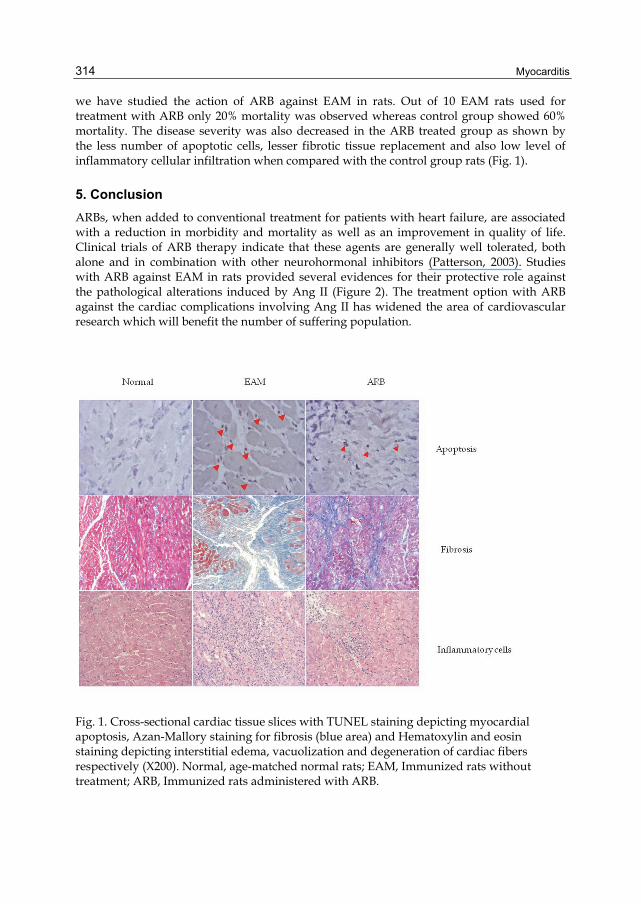
we have studied the action of ARB against EAM in rats. Out of 10 EAM rats used for treatment with ARB only 20% mortality was observed whereas control group showed 60% mortality. The disease severity was also decreased in the ARB treated group as shown by the less number of apoptotic cells, lesser fibrotic tissue replacement and also low level of inflammatory cellular infiltration when compared with the control group rats (Fig. 1).
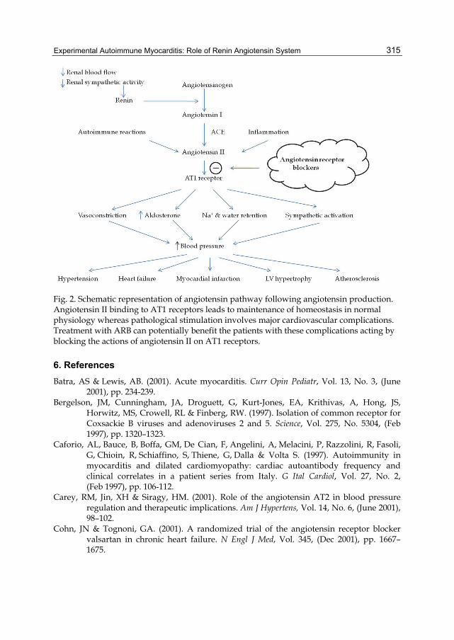
5. Conclusion ARBs, when added to conventional treatment for patients with heart failure, are associated with a reduction in morbidity and mortality as well as an improvement in quality of life. Clinical trials of ARB therapy indicate that these agents are generally well tolerated, both alone and in combination with other neurohormonal inhibitors (Patterson, 2003). Studies with ARB against EAM in rats provided several evidences for their protective role against the pathological alterations induced by Ang II (Figure 2). The treatment option with ARB against the cardiac complications involving Ang II has widened the area of cardiovascular research which will benefit the number of suffering population.
Fig. 1. Cross-sectional cardiac tissue slices with TUNEL staining depicting myocardial apoptosis, Azan-Mallory staining for fibrosis (blue area) and Hematoxylin and eosin staining depicting interstitial edema, vacuolization and degeneration of cardiac fibers respectively (X200). Normal, age-matched normal rats; EAM, Immunized rats without treatment; ARB, Immunized rats administered with ARB.
Experimental Autoimmune Myocarditis: Role of Renin Angiotensin System
315
Fig. 2. Schematic representation of angiotensin pathway following angiotensin production. Angiotensin II binding to AT1 receptors leads to maintenance of homeostasis in normal physiology whereas pathological stimulation involves major cardiovascular complications. Treatment with ARB can potentially benefit the patients with these complications acting by blocking the actions of angiotensin II on AT1 receptors.
6. References Batra, AS & Lewis, AB. (2001). Acute myocarditis. Curr Opin Pediatr, Vol. 13, No. 3, (June
2001), pp. 234-239. Bergelson, JM, Cunningham, JA, Droguett, G, Kurt-Jones, EA, Krithivas, A, Hong, JS,
Horwitz, MS, Crowell, RL & Finberg, RW. (1997). Isolation of common receptor for Coxsackie B viruses and adenoviruses 2 and 5. Science, Vol. 275, No. 5304, (Feb 1997), pp. 1320–1323.
Caforio, AL, Bauce, B, Boffa, GM, De Cian, F, Angelini, A, Melacini, P, Razzolini, R, Fasoli, G, Chioin, R, Schiaffino, S, Thiene, G, Dalla & Volta S. (1997). Autoimmunity in myocarditis and dilated cardiomyopathy: cardiac autoantibody frequency and clinical correlates in a patient series from Italy. G Ital Cardiol, Vol. 27, No. 2, (Feb 1997), pp. 106-112.
Carey, RM, Jin, XH & Siragy, HM. (2001). Role of the angiotensin AT2 in blood pressure regulation and therapeutic implications. Am J Hypertens, Vol. 14, No. 6, (June 2001), 98–102.
Cohn, JN & Tognoni, GA. (2001). A randomized trial of the angiotensin receptor blocker valsartan in chronic heart failure. N Engl J Med, Vol. 345, (Dec 2001), pp. 1667– 1675.
Myocarditis
316
Ding, L, Hanawa, H, Ota, Y, Hasegawa, G, Hao, K, Asami, F, Watanabe, R, Yoshida, T, Toba, K, Yoshida, K, Ogura, M, Kodama, M & Aizawa, Y. (2010). Lipocalin-2/neutrophil gelatinase-B associated lipocalin is strongly induced in hearts of rats with autoimmune myocarditis and in human myocarditis. Circ J, Vol 74, No. 3, (Mar 2010), pp. 523-530.
Ersoy, E, Kus, CN, Sener, U, Coker, I & Zorlu, Y. (2005). The effects of interferon-beta on interleukin-10 in multiple sclerosis patients. Eur J Neurol, Vol. 12, No. 3, (Mar 2005), pp. 208-211.
Fabris, B, Candido, R, Bortoletto, M, Toffoli, B, Bernardi, S, Stebel, M, Bardelli,M, Zentilin, L, Giacca, M & Carretta, R. (2011). Stimulation of cardiac apoptosis in ovariectomized hypertensive rats: potential role of the renin-angiotensin system. J Hypertens. Vol. 29, No. 2, (Feb 2011), pp. 273-281.
Fedoseyeva, EV, Kishimoto, K, Rolls, HK, Illigens, BM, Dong, VM, Valujskikh, A, Heeger, PS, Sayegh, MH & Benichou G. (2002). Modulation of tissue-specific immune response to cardiac myosin can prolong survival of allogeneic heart transplants. J Immunol, Vol. 169, No. 3, (Aug 2002), pp. 1168-1174.
Feldman, AM & McNamara, D. (2000). Myocarditis. N Engl J Med, Vol. 343, No. 19, (Nov 2000), pp. 1388–1398.
Ferrario, CM & Strawn, WB. (2006). Role of the renin-angiotensin-aldosterone system and proinflammatory mediators in cardiovascular disease. Am J Cardiol, Vol. 98, No. 1, (July 2006), pp. 121-128.
Ferrario, CM. (2011). ACE2: more of Ang-(1-7) or less Ang II? Curr Opin Nephrol Hypertens, Vol. 20, No. 1, (Jan 2011), pp. 1-6.
Gao, L & Zucker, IH. (2011). AT2 receptor signaling and sympathetic regulation. Curr Opin Pharmacol. Vol. 11, No. 2, (Apr 2011), pp. 124-130.
Goren, N, Leiros, CP, Sterin-Borda, L & Borda, E. (1998). Nitric oxide synthase in experimental autoimmune myocarditis dysfunction. J Mol Cell Cardiol, Vol. 30, No. 11, (Nov 1998), pp. 2467-2474.
Goser, S, Ottl, R, Brodner, A, Dengler, TJ, Torzewski, J, Egashira, K, Rose, NR, Katus, HA & Kaya, Z. (2005). Critical Role for Monocyte Chemoattractant Protein-1 and Macrophage Inflammatory Protein-1α in Induction of Experimental Autoimmune Myocarditis and Effective Anti–Monocyte Chemoattractant Protein-1 Gene Therapy. Circulation, Vol. 112, No. 22, (Nov 2005), pp. 3400-3407.
Guleria, RS, Choudhary, R, Tanaka, T, Baker, KM & Pan, J. (2011). Retinoic acid receptor-mediated signaling protects cardiomyocytes from hyperglycemia induced apoptosis: role of the renin-angiotensin system. J Cell Physiol. Vol. 226, No. 5, (May 2011), 1292-1307.
Horiuchi, M, Akishita, M & Dzau, VJ. (1999). Recent progress in angiotensin II type 2 receptor research in the cardiovascular system. Hypertension, Vol. 33, No. 2, (Feb 1999), pp. 613–621.
Huang, CH, Vallejo, JG, Kollias, G & Mann, DL. (2009). Role of the innate immune system in acute viral myocarditis. Basic Res Cardiol, Vol. 104, No. 3, (May 2009), pp. 228-237.
Ingkanisorn, WP, Paterson, DI, Calvo, KR, Rosing, DR, Schwartzentruber, DJ, Fuisz, AR & Arai AE. (2006). Cardiac magnetic resonance appearance of myocarditis caused by high dose IL-2: similarities to community-acquired myocarditis. J Cardiovasc Magn Reson, Vol. 8, No. 2, (2006), pp. 353-360.

Experimental Autoimmune Myocarditis: Role of Renin Angiotensin System
317
Inomata, T, Hanawa, H, Miyanishi, T, Yajima, E, Nakayama, S, Maita, T, Kodama, M, Izumi, T, Shibata, A & Abo, T. (1995). Localization of porcine cardiac myosin epitopes that induce experimental autoimmune myocarditis. Circ Res, Vol. 76, No. 5, (May 1995), pp. 726-33.
Ishiyama, S, Hiroe, M, Nishikawa, T, Shimojo, T, Abe, S, Fujisaki, H, Ito, H, Yamakawa, K, Kobayashi, N, Kasajima, T & Marumo, F. (1998). The Fas/Fas ligand system is involved in the pathogenesis of autoimmune myocarditis in rats. J. Immunol., Vol. 161, No. 9, (Nov 1998), pp. 4695-4701.
Jones, DE, Palmer, JM, Leon, MP, Yeaman, SJ, Bassendine, MF & Diamon, AG. (1997). T cell responses to tuberculin purified protein derivative in primary biliary cirrhosis: evidence for defective T cell function. Gut, Vol.40, No. 2, (Feb 1997), pp. 277-283.
Kamal, FA, Watanabe, K, Ma, M, Abe, Y, ElBarbary, R, Kodama, M & Aizawa, Y. (2010). A novel phenylpyridazinone, T-3999, reduces the progression of autoimmune myocarditis to dilated cardiomyopathy. Heart and vessels, Vol. 26, No. 1, (Jan 2010), pp. 81-90.
Kittleson, MM, Lowenstein, CJ, & Hare, JM. (2005). Novel Pathogenetic Mechanisms in Myocarditis: Nitric Oxide Signaling. Heart Failure Clinics, Vol. 1, No. 3, (Oct 2005), pp. 345-361.
Kodama, M, Matsumoto, Y & Fujiwara, M. (1992). In vivo lymphocyte-mediated myocardial injuries demonstrated by adoptive transfer of experimental autoimmune myocarditis. Circulation, Vol. 85, No. 5, (May 1992), pp. 1918-1926.
Kodama, M, Matsumoto, Y, Fujiwara, M, Masani, F, Izumi, T & Shibata, A. (1990). A novel experimental model of giant cell myocarditis induced in rats by immunization with cardiac myosin fraction. Clin. Immunol. Immunopathol, Vol. 57, No. 2, (Nov 1990), pp. 250–262.
Koumallos, N, Nteliopoulos, G, Paschalis, A, Dimarakis, I & Yonan, N. (2011). Therapeutic Interventions to Renin-Angiotensin-Aldosterone System, and Vascular Redox State. Recent Pat Cardiovasc Drug Discov. 2011 May 20.
Kuhl, U & Schultheiss, HP. (2010). Myocarditis in children. Heart Fail Clin, Vol. 6, No. 4, (Oct 2010) pp. 483-496.
Lansang, MC, Coletti, C, Ahmed, S, Gordon, MS & Hollenberg, NK. (2006). Effects of the PPAR-gamma agonist rosiglitazone on renal haemodynamics and the renin-angiotensin system in diabetes. Journal of the Renin-Angiotensin-Aldosterone System, Vol. 7, No. 3, (Sep 2006), pp. 175–180.
Levy, BI. (2004). Can Angiotensin II Type 2 Receptors Have Deleterious Effects in Cardiovascular Disease?: Implications for Therapeutic Blockade of the Renin–Angiotensin System. Circulation, Vol. 109, No. 1, (Jan 2004), pp. 8-13.
Lijnen, PJ & Petrov, VV. (2003). Role of intracardiac renin-angiotensin-aldosterone system in extracellular matrix remodeling. Methods Find Exp Clin Pharmacol. Vol. 25, No. 7, (Sep 2003), pp. 541-564.
Lindholm, LH, Ibsen, H, Dahlof, B, Devereux, RB, Beevers, G, de Faire, U, Fyhrquist, F, Julius, S, Kjeldsen, SE, Kristiansson, K, Lederballe-Pedersen, O, Nieminen, MS, Omvik, P, Oparil, S, Wedel, H, Aurup, P, Edelman, J & Snapinn, S. (2002). LIFE Study Group. Cardiovascular morbidity and mortality in patients with diabetes in the Losartan Intervention For Endpoint reduction in hypertension study (LIFE) a
Myocarditis
318
randomised trial against atenolol. Lancet, Vol. 359, No. 9311, (Mar 2002), pp. 1004–1010.
Maeda, K, Shioi, T, Kosugi, R, Yoshida, Y, Takahashi, K, Machida, Y & Izumi, T. (2005). Rapamycin ameliorates experimental autoimmune myocarditis. Int Heart J, Vol. 46, No. 3, (May 2005), pp. 513-530.
Maisch, B. (1989). Autoreactivity to the cardiac myocyte, connective tissue and the extracellular matrix in heart disease and postcardiac injury. Springer seminars in Immunopathology, Vol. 11, No. 4, (1989), pp. 369-395.
Matsubara, H. (1998). Pathophysiological role of angiotensin II type 2 receptor in cardiovascular and renal diseases. Circ Res, Vol. 83, No. 12, (Dec 1998), pp. 1182–1191.
Matsusaka, H, Kinugawa, S, Ide, T, Matsushima, S, Shiomi, T, Kubota, T, Sunagawa, K & Tsutsui, H. (2006). Angiotensin II type 1 receptor blocker attenuates exacerbated left ventricular remodeling and failure in diabetes-associated myocardial infarction. J Cardiovasc Pharmacol, Vol. 48, No. 3, (Sep 2006), pp. 95-102.
Neumann, DA, Burek, CL, Baughman, KL, Rose, NR & Herskowitz, A. (1990). Circulating heart-reactive antibodies in patients with myocarditis or cardiomyopathy. J Am Coll Cardiol, Vol. 16, No. 6, (Nov 1990), pp. 839-846.
Nishio, R, Matsumori, A, Shioi, T, Ishida, H & Sasayama, S. (1999). Treatment of Experimental Viral Myocarditis With Interleukin-10. Circulation, Vol. 100, No. 10, (Sep 1999), pp. 1102-1108.
Okura, Y, Yamamoto, T, Goto, S, Inomata, T, Hirono, S, Hanawa, H, Feng, L, Wilson, CB, Kihara, I, Izumi, T, Shibata, A, Aizawa, Y, Seki, S & Abo, T. (1997). Characterization of cytokine and iNOS mRNA expression in situ during the course of experimental autoimmune myocarditis in rats. J Mol Cell Cardiol, Vol. 29, No. 2, (Feb 1997), pp. 491–502.
Okura, Y, Takeda, Honda, S, Hanawa, H, Watanabe, H, Kodama, M, Izumi, T, Aizawa, Y, Seki & S, Abo, T. (1998). Recombinant Murine Interleukin-12 Facilitates Induction of Cardiac Myosin–Specific Type 1 Helper T Cells in Rats. Circulation Research, Vol. 82, No. 10, (Jun 1998), pp. 1035-1042.
Opie, LH & Sack, MN. (2001). Enhanced angiotensin II activity in heart failure: reevaluation of the counterregulatory hypothesis of receptor subtypes. Circ Res, Vol. 88, No. 7, (Apr 2001), pp. 654–658.
Oudit, GY & Penninger, JM. (2011). Recombinant Human Angiotensin-Converting Enzyme 2 as a New Renin-Angiotensin System Peptidase for Heart Failure Therapy. Curr Heart Fail Rep. 2011 May 26.
Patterson, JH. (2003). Angiotensin II receptor blockers in heart failure. Pharmacotherapy, Vol. 23, No. 2, (Feb 2003), pp. 173-182.
Petty, WJ, Miller, AA, McCoy, TP, Gallagher, PE, Tallant, EA & Torti, FM. (2009). Phase I and pharmacokinetic study of angiotensin-(1-7), an endogenous antiangiogenic hormone. Clin Cancer Res, Vol. 15, No. 23, (Dec 2009), pp. 7398-7404.
Pummerer, CL, Luze, K, Grässl, G, Bachmaier, K, Offner, F, Burrell, SK, Lenz, DM, Zamborelli, TJ, Penninger, JM & Neu, N. (1996). Identification of cardiac myosin peptides capable of inducing autoimmune myocarditis in BALB/c mice. J Clin Invest, Vol. 97, No. 9, (May 1996), pp. 2057–2062.

Experimental Autoimmune Myocarditis: Role of Renin Angiotensin System
319
Rosenstein, ED, Zucker, MJ & Kramer, N. (2000). Giant cell myocarditis: most fatal of autoimmune diseases. Semin Arthritis Rheum,Vol. 30, No. 1, (Aug 2000), pp. 1–16.
Schmieder, RE, Hilgers, KF, Schlaich, MP & Schmidt, BM. (2007). Renin-angiotensin system and cardiovascular risk. Lancet, Vol. 369, No. 9568, (Apr 2007), pp. 1208-1219.
Seko, Y. (2006). Effect of the angiotensin II receptor blocker olmesartan on the development of murine acute myocarditis caused by coxsackievirus B3. Clin Sci (Lond), Vol. 110, No. 3, (Mar 2006), pp. 379-386.
Shirai, K, Watanabe, K, Ma, M, Wahed, MI, Inoue, M, Saito, Y, Suresh, PS, Kashimura, T, Tachikawa, H, Kodama, M & Aizawa, Y. (2005). Effects of angiotensin-II receptor blocker candesartan cilexetil in rats with dilated cardiomyopathy. Mol Cell Biochem, Vol. 269, No. 1-2, (Jan 2005), pp. 137-142.
Singh, VP, Le, B, Khode, R, Baker, KM & Kumar, R. (2008). Intracellular angiotensin II production in diabetic rats is correlated with cardiomyocyte apoptosis, oxidative stress, and cardiac fibrosis. Diabetes, Vol. 57, No. 12, (Dec 2008), pp. 3297-306.
Siragy, HM. (2000). The role of the AT2 in hypertension. Am J Hypertens, Vol. 13, No. 5, (May 2000), 62–67.
Smith, SC & Allen, PM. (1992). Expression of myosin-class II major histocompatibility complexes in the normal myocardium occurs before induction of autoimmune myocarditis. Proc. Natl. Acad. Sci. U. S. A., Vol. 89, No. 19, (Oct 1992), pp. 9131-9135.
Sukumaran, V, Watanabe, K, Veeraveedu, PT, Gurusamy, N, Ma, M, Thandavarayan, RA, Lakshmanan, AP, Yamaguchi, K, Suzuki, K & Kodama, M. (2011). Olmesartan, an AT(1) Antagonist, Attenuates Oxidative Stress, Endoplasmic Reticulum Stress and Cardiac Inflammatory Mediators in Rats with Heart Failure Induced by Experimental Autoimmune Myocarditis. Int J Biol Sci, Vol. 7, No. 2, (Feb 2011), pp. 154-167.
Sukumaran, V, Watanabe, K, Veeraveedu, PT, Ma, M, Gurusamy, N, Rajavel, V, Suzuki, K, Yamaguchi, K, Kodama, M & Aizawa, Y. (2011). Telmisartan ameliorates experimental autoimmune myocarditis associated with inhibition of inflammation and oxidative stress. Eur J Pharmacol, Vol. 652, No. 1-3, (Feb 2011), pp. 126-135.
Sukumaran, V, Watanabe, K, Veeraveedu, PT, Thandavarayan, RA, Gurusamy, N, Ma, M, Yamaguchi, K, Suzuki, K, Kodama, M & Aizawa, Y. (2010). Beneficial effects of olmesartan, an angiotensin II receptor type 1 antagonist, in rats with dilated cardiomyopathy. Exp Biol Med (Maywood), Vol. 235, No. 11, (Nov 2010), pp. 1338-1346.
Takeyama, K, Kodera, Y, Suzawa, M & Kato, S. Peroxisome proliferator-activated receptor(PPAR)--structure, function, tissue distribution, gene expression. Nippon Rinsho, Vol. 58, No. 2, (Feb 2000), pp. 357-363.
Tsutsui, H, Matsushima, S, Kinugawa, S, Ide, T, Inoue, N, Ohta, Y, Yokota, T, Hamaguchi, S & Sunagawa, K. (2007). Angiotensin II type 1 receptor blocker attenuates myocardial remodeling and preserves diastolic function in diabetic heart. Hypertens Res, Vol. 30, No. 5, (May 2007), pp. 439-449.
Watanabe, K, Nakazawa, M, Fuse, K, Hanawa, H, Kodama, M, Aizawa, Y, Ohnuki, T, Gejyo, F, Maruyama, H, Miyazaki, J. (2001). Protection against autoimmune myocarditis by gene transfer of interleukin-10 by electroporation. Circulation, Vol 104, No. 10, (Sep 2001), pp. 1098 -1100.

Myocarditis
320
Yamada, T, Horiuchi, M, Dzau, VJ. (1996). Angiotensin II type 2 receptor mediates programmed cell death. Proc Nat Acad Sci U S A, Vol. 93, No. 1, (Jan 1996), pp. 156–160.
Ye, Y, Keyes, KT, Zhang, CF, Perez-Polo, JR, Lin, Y & Birnbaum, Y. (2010). Additive effect of TAK-491, a new angiotensin receptor blocker, and pioglitazone, in reducing myocardial infarct size. Cardiovasc Drugs Ther, Vol. 24, No. 2, (Apr 2010), pp. 107-120.
Yuan, J, Cao, AL, Yu, M, Lin, QW, Yu, X, Zhang, JH, Wang, M, Guo, HP & Liao, YH. (2010). Th17 cells facilitate the humoral immune response in patients with acute viral myocarditis. J Clin Immunol, Vol. 30, No. 2, (Mar 2010), pp. 226-234.
Related Documents