, 20140094, published 18 June 2014 281 2014 Proc. R. Soc. B M. J. Sweet, A. Croquer and J. C. Bythell Acropora cervicornis coral Caribbean pathogens of white band disease in the endangered Experimental antibiotic treatment identifies potential Supplementary data tml http://rspb.royalsocietypublishing.org/content/suppl/2014/06/17/rspb.2014.0094.DC1.h "Data Supplement" References http://rspb.royalsocietypublishing.org/content/281/1788/20140094.full.html#ref-list-1 This article cites 38 articles, 7 of which can be accessed free This article is free to access Email alerting service here right-hand corner of the article or click Receive free email alerts when new articles cite this article - sign up in the box at the top http://rspb.royalsocietypublishing.org/subscriptions go to: Proc. R. Soc. B To subscribe to on June 18, 2014 rspb.royalsocietypublishing.org Downloaded from on June 18, 2014 rspb.royalsocietypublishing.org Downloaded from

Welcome message from author
This document is posted to help you gain knowledge. Please leave a comment to let me know what you think about it! Share it to your friends and learn new things together.
Transcript

, 20140094, published 18 June 2014281 2014 Proc. R. Soc. B M. J. Sweet, A. Croquer and J. C. Bythell
Acropora cervicorniscoral Caribbeanpathogens of white band disease in the endangered
Experimental antibiotic treatment identifies potential
Supplementary data
tml http://rspb.royalsocietypublishing.org/content/suppl/2014/06/17/rspb.2014.0094.DC1.h
"Data Supplement"
Referenceshttp://rspb.royalsocietypublishing.org/content/281/1788/20140094.full.html#ref-list-1
This article cites 38 articles, 7 of which can be accessed free
This article is free to access
Email alerting service hereright-hand corner of the article or click Receive free email alerts when new articles cite this article - sign up in the box at the top
http://rspb.royalsocietypublishing.org/subscriptions go to: Proc. R. Soc. BTo subscribe to
on June 18, 2014rspb.royalsocietypublishing.orgDownloaded from on June 18, 2014rspb.royalsocietypublishing.orgDownloaded from
on June 18, 2014rspb.royalsocietypublishing.orgDownloaded from
rspb.royalsocietypublishing.org
ResearchCite this article: Sweet MJ, Croquer A,
Bythell JC. 2014 Experimental antibiotic
treatment identifies potential pathogens of
white band disease in the endangered
Caribbean coral Acropora cervicornis.
Proc. R. Soc. B 281: 20140094.
http://dx.doi.org/10.1098/rspb.2014.0094
Received: 17 January 2014
Accepted: 20 May 2014
Subject Areas:microbiology, molecular biology,
health and disease and epidemiology
Keywords:coral, disease, treatment, antibiotics,
Philaster, Vibrio
Author for correspondence:M. J. Sweet
e-mail: [email protected]
Electronic supplementary material is available
at http://dx.doi.org/10.1098/rspb.2014.0094 or
via http://rspb.royalsocietypublishing.org.
& 2014 The Authors. Published by the Royal Society under the terms of the Creative Commons AttributionLicense http://creativecommons.org/licenses/by/3.0/, which permits unrestricted use, provided the originalauthor and source are credited.
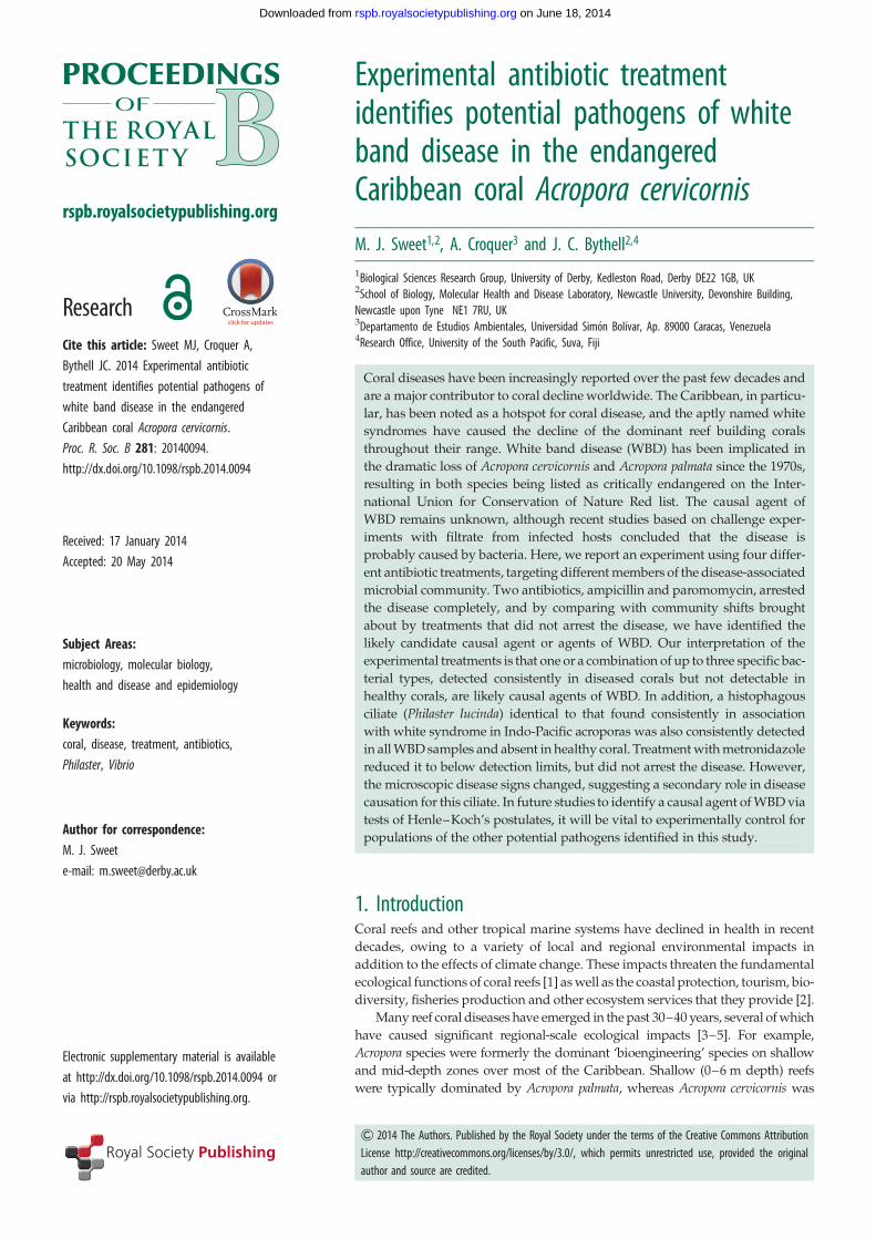
Experimental antibiotic treatmentidentifies potential pathogens of whiteband disease in the endangeredCaribbean coral Acropora cervicornis
M. J. Sweet1,2, A. Croquer3 and J. C. Bythell2,4
1Biological Sciences Research Group, University of Derby, Kedleston Road, Derby DE22 1GB, UK2School of Biology, Molecular Health and Disease Laboratory, Newcastle University, Devonshire Building,Newcastle upon Tyne NE1 7RU, UK3Departamento de Estudios Ambientales, Universidad Simon Bolıvar, Ap. 89000 Caracas, Venezuela4Research Office, University of the South Pacific, Suva, Fiji
Coral diseases have been increasingly reported over the past few decades and
are a major contributor to coral decline worldwide. The Caribbean, in particu-
lar, has been noted as a hotspot for coral disease, and the aptly named white
syndromes have caused the decline of the dominant reef building corals
throughout their range. White band disease (WBD) has been implicated in
the dramatic loss of Acropora cervicornis and Acropora palmata since the 1970s,
resulting in both species being listed as critically endangered on the Inter-
national Union for Conservation of Nature Red list. The causal agent of
WBD remains unknown, although recent studies based on challenge exper-
iments with filtrate from infected hosts concluded that the disease is
probably caused by bacteria. Here, we report an experiment using four differ-
ent antibiotic treatments, targeting different members of the disease-associated
microbial community. Two antibiotics, ampicillin and paromomycin, arrested
the disease completely, and by comparing with community shifts brought
about by treatments that did not arrest the disease, we have identified the
likely candidate causal agent or agents of WBD. Our interpretation of the
experimental treatments is that one or a combination of up to three specific bac-
terial types, detected consistently in diseased corals but not detectable in
healthy corals, are likely causal agents of WBD. In addition, a histophagous
ciliate (Philaster lucinda) identical to that found consistently in association
with white syndrome in Indo-Pacific acroporas was also consistently detected
in all WBD samples and absent in healthy coral. Treatment with metronidazole
reduced it to below detection limits, but did not arrest the disease. However,
the microscopic disease signs changed, suggesting a secondary role in disease
causation for this ciliate. In future studies to identify a causal agent of WBD via
tests of Henle–Koch’s postulates, it will be vital to experimentally control for
populations of the other potential pathogens identified in this study.
1. IntroductionCoral reefs and other tropical marine systems have declined in health in recent
decades, owing to a variety of local and regional environmental impacts in
addition to the effects of climate change. These impacts threaten the fundamental
ecological functions of coral reefs [1] as well as the coastal protection, tourism, bio-
diversity, fisheries production and other ecosystem services that they provide [2].
Many reef coral diseases have emerged in the past 30–40 years, several of which
have caused significant regional-scale ecological impacts [3–5]. For example,
Acropora species were formerly the dominant ‘bioengineering’ species on shallow
and mid-depth zones over most of the Caribbean. Shallow (0–6 m depth) reefs
were typically dominated by Acropora palmata, whereas Acropora cervicornis was
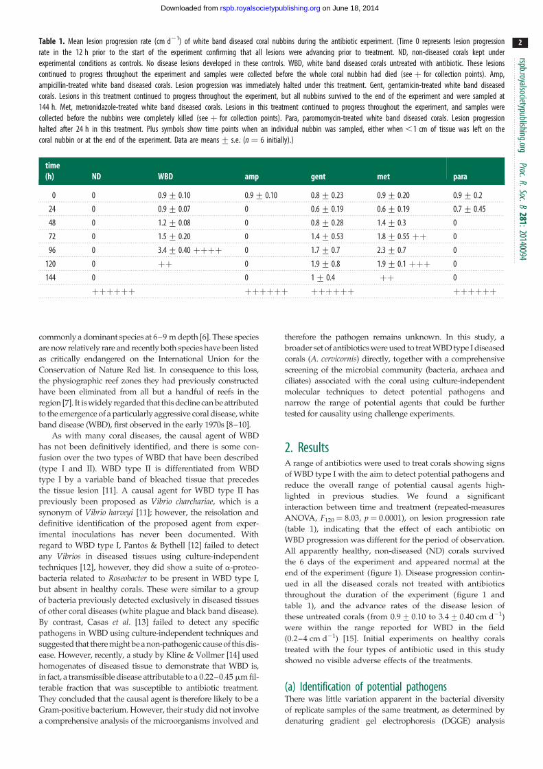
Table 1. Mean lesion progression rate (cm d21) of white band diseased coral nubbins during the antibiotic experiment. (Time 0 represents lesion progressionrate in the 12 h prior to the start of the experiment confirming that all lesions were advancing prior to treatment. ND, non-diseased corals kept underexperimental conditions as controls. No disease lesions developed in these controls. WBD, white band diseased corals untreated with antibiotic. These lesionscontinued to progress throughout the experiment and samples were collected before the whole coral nubbin had died (see þ for collection points). Amp,ampicillin-treated white band diseased corals. Lesion progression was immediately halted under this treatment. Gent, gentamicin-treated white band diseasedcorals. Lesions in this treatment continued to progress throughout the experiment, but all nubbins survived to the end of the experiment and were sampled at144 h. Met, metronidazole-treated white band diseased corals. Lesions in this treatment continued to progress throughout the experiment, and samples werecollected before the nubbins were completely killed (see þ for collection points). Para, paromomycin-treated white band diseased corals. Lesion progressionhalted after 24 h in this treatment. Plus symbols show time points when an individual nubbin was sampled, either when ,1 cm of tissue was left on thecoral nubbin or at the end of the experiment. Data are means+ s.e. (n ¼ 6 initially).)
time(h) ND WBD amp gent met para
0 0 0.9+ 0.10 0.9+ 0.10 0.8+ 0.23 0.9+ 0.20 0.9+ 0.2
24 0 0.9+ 0.07 0 0.6+ 0.19 0.6+ 0.19 0.7+ 0.45
48 0 1.2+ 0.08 0 0.8+ 0.28 1.4+ 0.3 0
72 0 1.5+ 0.20 0 1.4+ 0.53 1.8+ 0.55 þþ 0
96 0 3.4+ 0.40 þþþþ 0 1.7+ 0.7 2.3+ 0.7 0
120 0 þþ 0 1.9+ 0.8 1.9+ 0.1 þþþ 0
144 0 0 1+ 0.4 þþ 0
þþþþþþ þþþþþþ þþþþþþ þþþþþþ
rspb.royalsocietypublishing.orgProc.R.Soc.B
281:20140094
2
on June 18, 2014rspb.royalsocietypublishing.orgDownloaded from
commonly a dominant species at 6–9 m depth [6]. These species
are now relatively rare and recently both species have been listed
as critically endangered on the International Union for the
Conservation of Nature Red list. In consequence to this loss,
the physiographic reef zones they had previously constructed
have been eliminated from all but a handful of reefs in the
region [7]. It is widely regarded that this decline can be attributed
to the emergence of a particularly aggressive coral disease, white
band disease (WBD), first observed in the early 1970s [8–10].
As with many coral diseases, the causal agent of WBD
has not been definitively identified, and there is some con-
fusion over the two types of WBD that have been described
(type I and II). WBD type II is differentiated from WBD
type I by a variable band of bleached tissue that precedes
the tissue lesion [11]. A causal agent for WBD type II has
previously been proposed as Vibrio charchariae, which is a
synonym of Vibrio harveyi [11]; however, the reisolation and
definitive identification of the proposed agent from exper-
imental inoculations has never been documented. With
regard to WBD type I, Pantos & Bythell [12] failed to detect
any Vibrios in diseased tissues using culture-independent
techniques [12], however, they did show a suite of a-proteo-
bacteria related to Roseobacter to be present in WBD type I,
but absent in healthy corals. These were similar to a group
of bacteria previously detected exclusively in diseased tissues
of other coral diseases (white plague and black band disease).
By contrast, Casas et al. [13] failed to detect any specific
pathogens in WBD using culture-independent techniques and
suggested that there might be a non-pathogenic cause of this dis-
ease. However, recently, a study by Kline & Vollmer [14] used
homogenates of diseased tissue to demonstrate that WBD is,
in fact, a transmissible disease attributable to a 0.22–0.45 mm fil-
terable fraction that was susceptible to antibiotic treatment.
They concluded that the causal agent is therefore likely to be a
Gram-positive bacterium. However, their study did not involve
a comprehensive analysis of the microorganisms involved and
therefore the pathogen remains unknown. In this study, a
broader set of antibiotics were used to treat WBD type I diseased
corals (A. cervicornis) directly, together with a comprehensive
screening of the microbial community (bacteria, archaea and
ciliates) associated with the coral using culture-independent
molecular techniques to detect potential pathogens and
narrow the range of potential agents that could be further
tested for causality using challenge experiments.
2. ResultsA range of antibiotics were used to treat corals showing signs
of WBD type I with the aim to detect potential pathogens and
reduce the overall range of potential causal agents high-
lighted in previous studies. We found a significant
interaction between time and treatment (repeated-measures
ANOVA, F120 ¼ 8.03, p ¼ 0.0001), on lesion progression rate
(table 1), indicating that the effect of each antibiotic on
WBD progression was different for the period of observation.
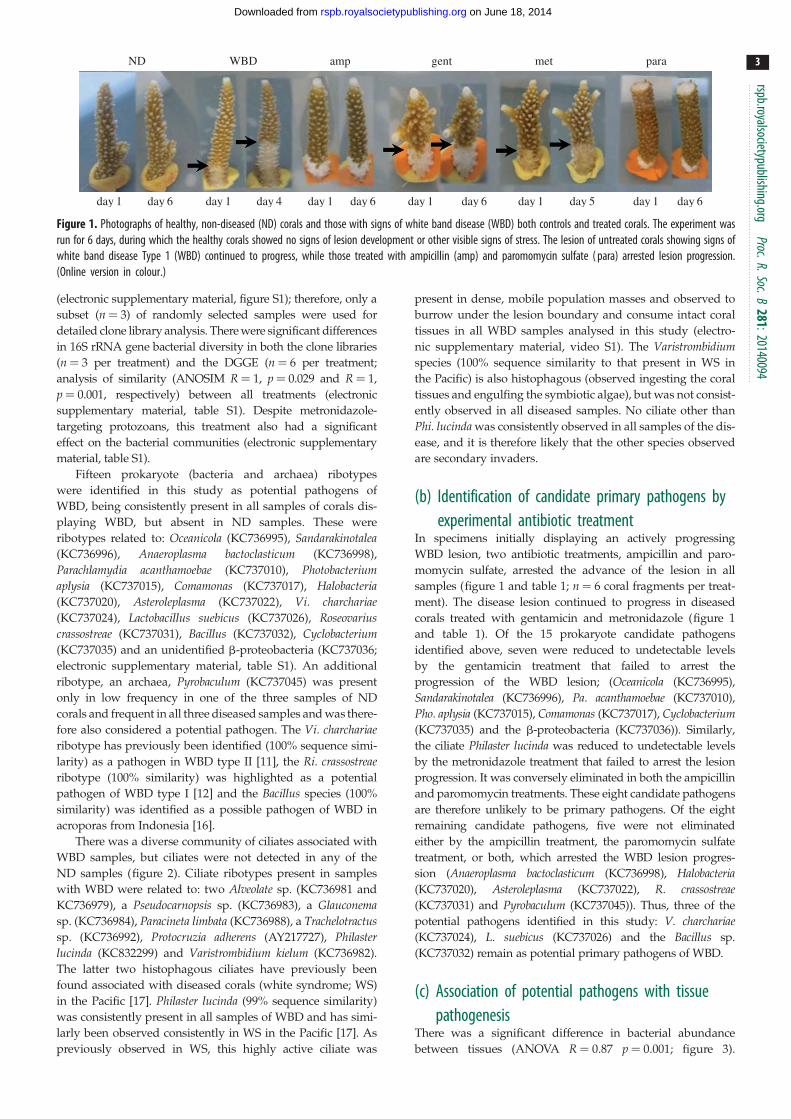
All apparently healthy, non-diseased (ND) corals survived
the 6 days of the experiment and appeared normal at the
end of the experiment (figure 1). Disease progression contin-
ued in all the diseased corals not treated with antibiotics
throughout the duration of the experiment (figure 1 and
table 1), and the advance rates of the disease lesion of
these untreated corals (from 0.9+ 0.10 to 3.4+ 0.40 cm d21)
were within the range reported for WBD in the field
(0.2–4 cm d21) [15]. Initial experiments on healthy corals
treated with the four types of antibiotic used in this study
showed no visible adverse effects of the treatments.
(a) Identification of potential pathogensThere was little variation apparent in the bacterial diversity
of replicate samples of the same treatment, as determined by
denaturing gradient gel electrophoresis (DGGE) analysis
day 1
ND WBD amp gent met para
day 6 day 1 day 4 day 1 day 1day 6 day 6 day 1day 1 day 5 day 6
Figure 1. Photographs of healthy, non-diseased (ND) corals and those with signs of white band disease (WBD) both controls and treated corals. The experiment wasrun for 6 days, during which the healthy corals showed no signs of lesion development or other visible signs of stress. The lesion of untreated corals showing signs ofwhite band disease Type 1 (WBD) continued to progress, while those treated with ampicillin (amp) and paromomycin sulfate ( para) arrested lesion progression.(Online version in colour.)
rspb.royalsocietypublishing.orgProc.R.Soc.B
281:20140094
3
on June 18, 2014rspb.royalsocietypublishing.orgDownloaded from
(electronic supplementary material, figure S1); therefore, only a
subset (n ¼ 3) of randomly selected samples were used for
detailed clone library analysis. There were significant differences
in 16S rRNA gene bacterial diversity in both the clone libraries
(n ¼ 3 per treatment) and the DGGE (n ¼ 6 per treatment;
analysis of similarity (ANOSIM R ¼ 1, p ¼ 0.029 and R ¼ 1,
p ¼ 0.001, respectively) between all treatments (electronic
supplementary material, table S1). Despite metronidazole-
targeting protozoans, this treatment also had a significant
effect on the bacterial communities (electronic supplementary
material, table S1).
Fifteen prokaryote (bacteria and archaea) ribotypes
were identified in this study as potential pathogens of
WBD, being consistently present in all samples of corals dis-
playing WBD, but absent in ND samples. These were
ribotypes related to: Oceanicola (KC736995), Sandarakinotalea(KC736996), Anaeroplasma bactoclasticum (KC736998),
Parachlamydia acanthamoebae (KC737010), Photobacteriumaplysia (KC737015), Comamonas (KC737017), Halobacteria(KC737020), Asteroleplasma (KC737022), Vi. charchariae(KC737024), Lactobacillus suebicus (KC737026), Roseovariuscrassostreae (KC737031), Bacillus (KC737032), Cyclobacterium(KC737035) and an unidentified b-proteobacteria (KC737036;
electronic supplementary material, table S1). An additional
ribotype, an archaea, Pyrobaculum (KC737045) was present
only in low frequency in one of the three samples of ND
corals and frequent in all three diseased samples and was there-
fore also considered a potential pathogen. The Vi. charchariaeribotype has previously been identified (100% sequence simi-
larity) as a pathogen in WBD type II [11], the Ri. crassostreaeribotype (100% similarity) was highlighted as a potential
pathogen of WBD type I [12] and the Bacillus species (100%
similarity) was identified as a possible pathogen of WBD in
acroporas from Indonesia [16].
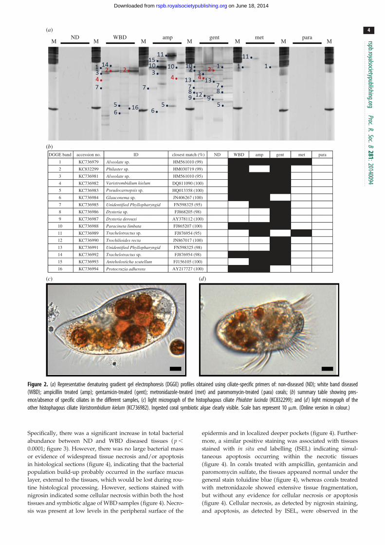
There was a diverse community of ciliates associated with
WBD samples, but ciliates were not detected in any of the
ND samples (figure 2). Ciliate ribotypes present in samples
with WBD were related to: two Alveolate sp. (KC736981 and
KC736979), a Pseudocarnopsis sp. (KC736983), a Glauconemasp. (KC736984), Paracineta limbata (KC736988), a Trachelotractussp. (KC736992), Protocruzia adherens (AY217727), Philasterlucinda (KC832299) and Varistrombidium kielum (KC736982).
The latter two histophagous ciliates have previously been
found associated with diseased corals (white syndrome; WS)
in the Pacific [17]. Philaster lucinda (99% sequence similarity)
was consistently present in all samples of WBD and has simi-
larly been observed consistently in WS in the Pacific [17]. As
previously observed in WS, this highly active ciliate was
present in dense, mobile population masses and observed to
burrow under the lesion boundary and consume intact coral
tissues in all WBD samples analysed in this study (electro-
nic supplementary material, video S1). The Varistrombidiumspecies (100% sequence similarity to that present in WS in
the Pacific) is also histophagous (observed ingesting the coral
tissues and engulfing the symbiotic algae), but was not consist-
ently observed in all diseased samples. No ciliate other than
Phi. lucinda was consistently observed in all samples of the dis-
ease, and it is therefore likely that the other species observed
are secondary invaders.
(b) Identification of candidate primary pathogens byexperimental antibiotic treatment
In specimens initially displaying an actively progressing
WBD lesion, two antibiotic treatments, ampicillin and paro-
momycin sulfate, arrested the advance of the lesion in all
samples (figure 1 and table 1; n ¼ 6 coral fragments per treat-
ment). The disease lesion continued to progress in diseased
corals treated with gentamicin and metronidazole (figure 1
and table 1). Of the 15 prokaryote candidate pathogens
identified above, seven were reduced to undetectable levels
by the gentamicin treatment that failed to arrest the
progression of the WBD lesion; (Oceanicola (KC736995),
Sandarakinotalea (KC736996), Pa. acanthamoebae (KC737010),
Pho. aplysia (KC737015), Comamonas (KC737017), Cyclobacterium(KC737035) and the b-proteobacteria (KC737036)). Similarly,
the ciliate Philaster lucinda was reduced to undetectable levels
by the metronidazole treatment that failed to arrest the lesion
progression. It was conversely eliminated in both the ampicillin
and paromomycin treatments. These eight candidate pathogens
are therefore unlikely to be primary pathogens. Of the eight
remaining candidate pathogens, five were not eliminated
either by the ampicillin treatment, the paromomycin sulfate
treatment, or both, which arrested the WBD lesion progres-
sion (Anaeroplasma bactoclasticum (KC736998), Halobacteria(KC737020), Asteroleplasma (KC737022), R. crassostreae(KC737031) and Pyrobaculum (KC737045)). Thus, three of the
potential pathogens identified in this study: V. charchariae(KC737024), L. suebicus (KC737026) and the Bacillus sp.
(KC737032) remain as potential primary pathogens of WBD.
(c) Association of potential pathogens with tissuepathogenesis
There was a significant difference in bacterial abundance
between tissues (ANOVA R ¼ 0.87 p ¼ 0.001; figure 3).
(a)
(b)
(c) (d)
ND WBDM M M M M MM
amp gent met para
DGGE band
1 Alveolate sp. HM561010 (99)
Philaster sp. HM030719 (99)
Alveolate sp. HM561010 (95)
Varistrombidium kielum DQ811090 (100)
Pseudocarnopsis sp. HQ013358 (100)
Glauconema sp. JN406267 (100)
Unidentified Phyllopharyngid
Unidentified Phyllopharyngid
FN598325 (95)
Dysteria sp.
Dysteria derouxi
FJ868205 (98)
AY378112 (100)
Paracineta limbata
Trachelotractus sp.
Trachelotractus sp.
Anteholosticha scutellum
Protocruzia adherens
Trochilioides recta
FJ865207 (100)
FJ876954 (95)
JN867017 (100)
FN598325 (98)
FJ876954 (98)
FJ156105 (100)
KC736979
KC832299
KC736981
KC736982
KC736983
KC736984
KC736985
KC736986
KC736987
KC736988
KC736989
KC736990
KC736991
KC736992
KC736993
KC736994 AY217727 (100)
2
3
4
5
6
7
8
9
10
11
12
13
14
15
16
accession no. ID ND WBD amp gent met paraclosest match (%)
Figure 2. (a) Representative denaturing gradient gel electrophoresis (DGGE) profiles obtained using ciliate-specific primers of: non-diseased (ND); white band diseased(WBD); ampicillin treated (amp); gentamicin-treated (gent); metronidazole-treated (met) and paromomycin-treated ( para) corals; (b) summary table showing pres-ence/absence of specific ciliates in the different samples, (c) light micrograph of the histophagous ciliate Phialster lucinda (KC832299); and (d ) light micrograph of theother histophagous ciliate Varistrombidium kielum (KC736982). Ingested coral symbiotic algae clearly visible. Scale bars represent 10 mm. (Online version in colour.)
rspb.royalsocietypublishing.orgProc.R.Soc.B
281:20140094
4
on June 18, 2014rspb.royalsocietypublishing.orgDownloaded from
Specifically, there was a significant increase in total bacterial
abundance between ND and WBD diseased tissues ( p ,
0.0001; figure 3). However, there was no large bacterial mass
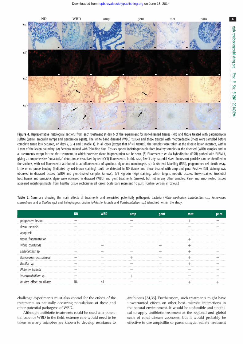
or evidence of widespread tissue necrosis and/or apoptosis
in histological sections (figure 4), indicating that the bacterial
population build-up probably occurred in the surface mucus
layer, external to the tissues, which would be lost during rou-
tine histological processing. However, sections stained with
nigrosin indicated some cellular necrosis within both the host
tissues and symbiotic algae of WBD samples (figure 4). Necro-
sis was present at low levels in the peripheral surface of the
epidermis and in localized deeper pockets (figure 4). Further-
more, a similar positive staining was associated with tissues
stained with in situ end labelling (ISEL) indicating simul-
taneous apoptosis occurring within the necrotic tissues
(figure 4). In corals treated with ampicillin, gentamicin and
paromomycin sulfate, the tissues appeared normal under the
general stain toluidine blue (figure 4), whereas corals treated
with metronidazole showed extensive tissue fragmentation,
but without any evidence for cellular necrosis or apoptosis
(figure 4). Cellular necrosis, as detected by nigrosin staining,
and apoptosis, as detected by ISEL, were observed in the
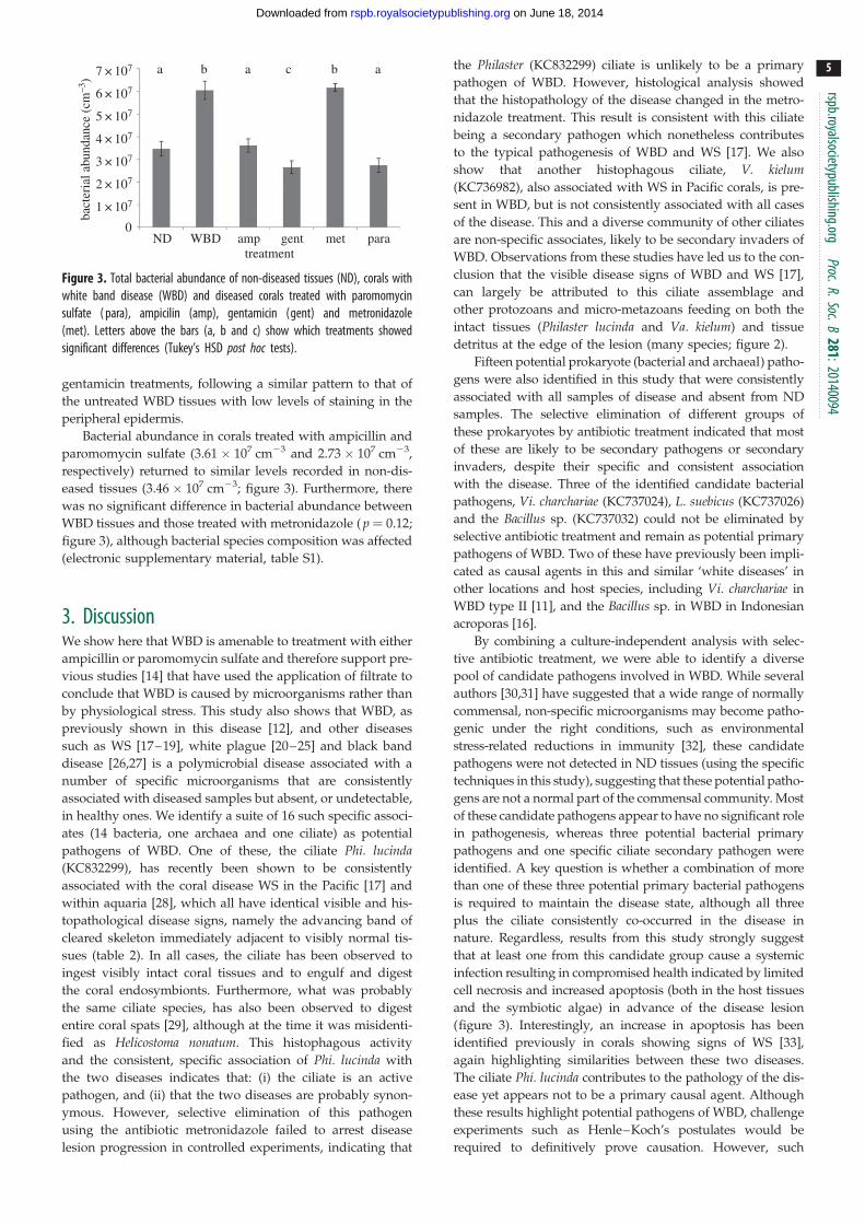
ND
a b a c b a
0
1 × 10⁷
bact
eria
l abu
ndan
ce (
cm–3
)
2 × 10⁷
3 × 10⁷
4 × 10⁷
5 × 10⁷
6 × 10⁷
7 × 10⁷
WBD amp genttreatment
met para
Figure 3. Total bacterial abundance of non-diseased tissues (ND), corals withwhite band disease (WBD) and diseased corals treated with paromomycinsulfate ( para), ampicilin (amp), gentamicin (gent) and metronidazole(met). Letters above the bars (a, b and c) show which treatments showedsignificant differences (Tukey’s HSD post hoc tests).
rspb.royalsocietypublishing.orgProc.R.Soc.B
281:20140094
5
on June 18, 2014rspb.royalsocietypublishing.orgDownloaded from
gentamicin treatments, following a similar pattern to that of
the untreated WBD tissues with low levels of staining in the
peripheral epidermis.
Bacterial abundance in corals treated with ampicillin and
paromomycin sulfate (3.61 � 107 cm23 and 2.73 � 107 cm23,
respectively) returned to similar levels recorded in non-dis-
eased tissues (3.46 � 107 cm23; figure 3). Furthermore, there
was no significant difference in bacterial abundance between
WBD tissues and those treated with metronidazole ( p ¼ 0.12;
figure 3), although bacterial species composition was affected
(electronic supplementary material, table S1).
3. DiscussionWe show here that WBD is amenable to treatment with either
ampicillin or paromomycin sulfate and therefore support pre-
vious studies [14] that have used the application of filtrate to
conclude that WBD is caused by microorganisms rather than
by physiological stress. This study also shows that WBD, as
previously shown in this disease [12], and other diseases
such as WS [17–19], white plague [20–25] and black band
disease [26,27] is a polymicrobial disease associated with a
number of specific microorganisms that are consistently
associated with diseased samples but absent, or undetectable,
in healthy ones. We identify a suite of 16 such specific associ-
ates (14 bacteria, one archaea and one ciliate) as potential
pathogens of WBD. One of these, the ciliate Phi. lucinda(KC832299), has recently been shown to be consistently
associated with the coral disease WS in the Pacific [17] and
within aquaria [28], which all have identical visible and his-
topathological disease signs, namely the advancing band of
cleared skeleton immediately adjacent to visibly normal tis-
sues (table 2). In all cases, the ciliate has been observed to
ingest visibly intact coral tissues and to engulf and digest
the coral endosymbionts. Furthermore, what was probably
the same ciliate species, has also been observed to digest
entire coral spats [29], although at the time it was misidenti-
fied as Helicostoma nonatum. This histophagous activity
and the consistent, specific association of Phi. lucinda with
the two diseases indicates that: (i) the ciliate is an active
pathogen, and (ii) that the two diseases are probably synon-
ymous. However, selective elimination of this pathogen
using the antibiotic metronidazole failed to arrest disease
lesion progression in controlled experiments, indicating that
the Philaster (KC832299) ciliate is unlikely to be a primary
pathogen of WBD. However, histological analysis showed
that the histopathology of the disease changed in the metro-
nidazole treatment. This result is consistent with this ciliate
being a secondary pathogen which nonetheless contributes
to the typical pathogenesis of WBD and WS [17]. We also
show that another histophagous ciliate, V. kielum(KC736982), also associated with WS in Pacific corals, is pre-
sent in WBD, but is not consistently associated with all cases
of the disease. This and a diverse community of other ciliates
are non-specific associates, likely to be secondary invaders of
WBD. Observations from these studies have led us to the con-
clusion that the visible disease signs of WBD and WS [17],
can largely be attributed to this ciliate assemblage and
other protozoans and micro-metazoans feeding on both the
intact tissues (Philaster lucinda and Va. kielum) and tissue
detritus at the edge of the lesion (many species; figure 2).
Fifteen potential prokaryote (bacterial and archaeal) patho-
gens were also identified in this study that were consistently
associated with all samples of disease and absent from ND
samples. The selective elimination of different groups of
these prokaryotes by antibiotic treatment indicated that most
of these are likely to be secondary pathogens or secondary
invaders, despite their specific and consistent association
with the disease. Three of the identified candidate bacterial
pathogens, Vi. charchariae (KC737024), L. suebicus (KC737026)
and the Bacillus sp. (KC737032) could not be eliminated by
selective antibiotic treatment and remain as potential primary
pathogens of WBD. Two of these have previously been impli-
cated as causal agents in this and similar ‘white diseases’ in
other locations and host species, including Vi. charchariae in
WBD type II [11], and the Bacillus sp. in WBD in Indonesian
acroporas [16].
By combining a culture-independent analysis with selec-
tive antibiotic treatment, we were able to identify a diverse
pool of candidate pathogens involved in WBD. While several
authors [30,31] have suggested that a wide range of normally
commensal, non-specific microorganisms may become patho-
genic under the right conditions, such as environmental
stress-related reductions in immunity [32], these candidate
pathogens were not detected in ND tissues (using the specific
techniques in this study), suggesting that these potential patho-
gens are not a normal part of the commensal community. Most
of these candidate pathogens appear to have no significant role
in pathogenesis, whereas three potential bacterial primary
pathogens and one specific ciliate secondary pathogen were
identified. A key question is whether a combination of more
than one of these three potential primary bacterial pathogens
is required to maintain the disease state, although all three
plus the ciliate consistently co-occurred in the disease in
nature. Regardless, results from this study strongly suggest
that at least one from this candidate group cause a systemic
infection resulting in compromised health indicated by limited
cell necrosis and increased apoptosis (both in the host tissues
and the symbiotic algae) in advance of the disease lesion
(figure 3). Interestingly, an increase in apoptosis has been
identified previously in corals showing signs of WS [33],
again highlighting similarities between these two diseases.
The ciliate Phi. lucinda contributes to the pathology of the dis-
ease yet appears not to be a primary causal agent. Although
these results highlight potential pathogens of WBD, challenge
experiments such as Henle–Koch’s postulates would be
required to definitively prove causation. However, such
ND
(a)
(b)
(c)
(d)
WBD amp gent met para
Figure 4. Representative histological sections from each treatment at day 6 of the experiment for non-diseased tissues (ND) and those treated with paromomycinsulfate ( para), ampicilin (amp) and gentamicin (gent). The white band diseased (WBD) tissues and those treated with metronidazole (met) were sampled beforecomplete tissue loss occurred, on days 2, 3, 4 and 5 (table 1). In all cases (except that of ND tissues), the samples were taken at the disease lesion interface, within1 mm of the lesion boundary. (a) Sections stained with Toluidine blue. Tissues appear indistinguishable from healthy samples in the diseased (WBD) samples and inall treatments except for the Met treatment, in which extensive tissue fragmentation can be seen. (b) Fluorescence in situ hybridization (FISH) probed with EUBMIX,giving a comprehensive ‘eubacterial’ detection as visualized by red (CY3) fluorescence. In this case, few if any bacterial-sized fluorescent particles can be identified inthe sections, with red fluorescence attributed to autofluorescence of symbiotic algae and nematocysts. (c) In situ end labelling (ISEL), programmed cell death assay.Little or no probe binding (indicated by red-brown staining) could be detected in ND tissues and those treated with amp and para. Positive ISEL staining wasobserved in diseased tissues (WBD) and gent-treated samples (arrows). (d ) Nigrosin (Nig) staining, which targets necrotic tissues. Brown-stained (necrotic)host tissues and symbiotic algae were observed in diseased (WBD) and gent treatments (arrows), but not in any other samples. Para- and amp-treated tissuesappeared indistinguishable from healthy tissue sections in all cases. Scale bars represent 10 mm. (Online version in colour.)
Table 2. Summary showing the main effects of treatments and associated potentially pathogenic bacteria (Vibrio carchariae, Lactobacillus sp., Roseovariuscrassostreae and a Bacillus sp.) and histophagous ciliates (Philaster lucinda and Varistrombidium sp.) identified within the study.
ND WBD amp gent met para
progressive lesion 2 þ 2 þ þ 2
tissue necrosis 2 þ 2 þ 2 2
apoptosis 2 þ 2 þ 2 2
tissue fragmentation 2 2 2 2 þ 2
Vibrio carchariae 2 þ 2 þ þ 2
Lactobacillus sp. 2 þ 2 þ þ 2
Roseovarius crassostreae 2 þ þ þ þ 2
Bacillus sp. 2 þ 2 þ þ 2
Philaster lucinda 2 þ 2 þ 2 2
Varistrombidium sp. 2 þ þ þ 2 2
in vitro effect on ciliates NA NA 2 2 þ þ
rspb.royalsocietypublishing.orgProc.R.Soc.B
281:20140094
6
on June 18, 2014rspb.royalsocietypublishing.orgDownloaded from
challenge experiments must also control for the effects of the
treatments on naturally occurring populations of these and
other potential pathogens of WBD.
Although antibiotic treatments could be used as a poten-
tial cure for WBD in the field, extreme care would need to be
taken as many microbes are known to develop resistance to
antibiotics [34,35]. Furthermore, such treatments might have
unwarranted effects on other host–microbe interactions in
the natural environment. It would be unfeasible and unethi-
cal to apply antibiotic treatment at the regional and global
scale of coral disease zoonoses, but it would probably be
effective to use ampicillin or paromomycin sulfate treatment
r
7
on June 18, 2014rspb.royalsocietypublishing.orgDownloaded from
in specific circumstances where collateral effects could be
minimized, for example in aquarium treatments.
spb.royalsocietypublishing.orgProc.R.Soc.B
281:20140094
4. MethodsAcropora cervicornis showing signs of WBD (type I) were monitored
to show signs of progression. Only those with advancing lesions
were used in the experiment. N ¼ 2, 5 cm2 coral nubbins showing
signs of WBD (type I) were placed in three individual aquaria per
treatment. Therefore, n ¼ 6 replicate coral nubbins were used for
each treatment. All corals were maintained in the aquarium for
24 h prior to the start of the experiment to allow for acclimatization
and to confirm lesion progression in diseased nubbins. Four types
of antibiotics were used in treatments to determine their effects on
the diseased corals; ampicillin, gentamicin, metronidazole and par-
omomycin sulfate. 100 mg ml21 was used for all four antibiotics
after preliminary laboratory trials on both bacteria and healthy
corals. The antibiotics were added directly into tanks filled with
3 l of seawater collected from the original location of the corals.
Repeat dosage was dissolved in 1.5 l of seawater every 12 h, and
half the water in the experimental tanks was replaced with the
new water. n ¼ 6 corals were used per treatment (n ¼ 2 per tank,
three tanks). One set with WBD were left untreated in the tanks
and sampled before all the tissue had been lost. Healthy corals
were also collected and held in the aquaria for the duration of the
experiment to address any tank effects on the health of the corals.
Prior to the onset of the main experiment, healthy corals were
also treated with the antibiotics at the same dose rates used
within the experiment to ensure that the antibiotics were having
no adverse effect on the corals. All these treatments survived to
the end of the experiment with no visual appearance of tissue
deterioration or discoloration.
Ampicillin belongs to the penicillin group of beta-lactam
antibiotics, it is able to penetrate Gram-positive and some Gram-
negative bacteria. It acts as a competitive inhibitor of the enzyme
transpeptidase, which is needed by bacteria to make their cell
walls. Inhibition of cell wall synthesis ultimately leads to cell lysis.
Gentamicin targets mainly Gram-negative bacterium and inhibits
protein synthesis. Metronidazole is a nitroimidazole antibiotic used
particularly for anaerobic bacteria (particularly from the genus clos-
tridium and bacteroides) and protozoa. Once metronidazole is taken
up by the microorganisms, it is non-enzymatically reduced by react-
ing with reduced ferredoxin, which is generated by pyruvate
oxidoreductase. Many of the reduced nitroso intermediates will
form sulfinamides and thioether linkages with cysteine-bearing
enzymes, thereby deactivating these critical enzymes. Paromomycin
sulfate is a protein synthesis inhibitor and binds directly to the 16S
rRNA. It is a broad spectrum antibiotic which is soluble in water.
Rates of application of ampicillin, gentamicin and metronida-
zole were measured twice daily at 10.00 and 16.00 for 6 days until
the end of the experiment. Paromomycin sulfate was applied
only twice on the first day owing to the cost of this antibiotic.
Sterile surgical gloves were worn at all times to avoid con-
tamination. Samples were taken either when the corals had less
than half their remaining tissue on the nubbins or at the end of
the experiment. Samples were placed in Falcon tubes underwater
and sealed. The water was then replaced with 100% ethanol and
stored at 2208C until extraction.
(a) PCR and denaturing gradient gel electrophoresisExtraction, PCR and denaturing gradient gel electrophoresis were
undertaken as described in [16]. DNA was extracted from all
samples using QIAGEN DNeasy blood and tissue kits, and bac-
terial 16S rRNA gene diversity was amplified using primers
357F and 518R. PCR protocol was as in [17]. Ciliate 18S rRNA
gene diversity was amplified using primers CilF and CilDGGE-r.
PCR protocol was as in [17]. For each of the above primer pairs,
30 ml PCR mixtures containing 1.5 mM MgCl2, 0.2 mM dNTP (Pro-
mega), 0.5 mM of each primer, 2.5 Ul of Taq DNA polymerase
(QBiogene), incubation buffer and 20 ng of template DNA [17].
DGGE was performed as in [17] using the D-Code universal
mutation detection system (Bio-Rad). PCR products were resolved
on 10% (w/v) polyacrylamide gels for bacterial 16S rRNA gene
diversity and 8% (w/v) for ciliate diversity. Bands of interest
(those which explained the greatest differences/similarities
between samples) were excised from DGGE gels, re-amplified
with the same original primers, labelled using a big dye (Applied
Biosystems) transformation sequence kit and sent to Genevision
(Newcastle University, UK) for sequencing.
(b) Clone libraries and amplified ribosomal DNArestriction analysis screening
As all replicates within samples showed no significant differences
in relation to their DGGE profiles, a random subset of only n ¼ 3
replicates per treatment were further analysed using Clone
Libraries. Almost complete 16S rRNA gene fragments were ampli-
fied from the DNA extracted using the ‘universal’ eubacterial 16S
rRNA gene primers 27F and 1542R. PCR protocols were as in
[27,36]. Amplified products, purified using the Qiagen PCR purifi-
cation kit, were inserted into the pGEM-T vector system (Promega)
and transformed into Escherichia coli JM109 cells. A total of 392
clones containing the 16S rRNA gene inserts were randomly
selected from each sample/replicate, and boiled lysates were pre-
pared as in [17]. PCR protocols were as in [17]. The products
were then digested with the restriction enzymes HaeIII and RsaI
(Promega; 4 mg of PCR product, 2 ml of restriction buffer, 0.2 ml
of bovine serum albumin, 0.07 ml of HaeIII, 0.1 ml of RsaI and
made up to 20 ml with sigma water for 2 h at 378C then 10 min at
678C). Restriction fragments were resolved by 3% agarose gel elec-
trophoresis, visualized using a ultraviolet transilluminator and
grouped based on restriction patterns. n ¼ 10 representatives
from each group were sequenced. Closest match of retrieved
sequences was determined by RDP II similarity matching [37].
All sequences in this study have been deposited in GenBank and
their unique accession numbers reported in the text.
(c) Total bacterial abundanceTo estimate bacterial abundance, 1000 ml of tissue slurry was
filtered through a 0.22 mm black polycarbonate filter and fixed
with 100 ml of paraformaldehyde until analysis. These filters were
stained with 100 ml DAPI solution (final concentration 5 mg ml21)
for 10 min, rinsed with phosphate-buffered solution, and viewed
under epifluorescence microscopy (Nikon UK Ltd, Surrey, UK) at
1000� magnification using a DAPI-specific filter set. Counts
on 50 fields of view were taken using an automatic cell counter
(Cell C). The parameters were set to exclude any objects smaller
than 0.03 mm2 and anything larger than 0.7 mm2. Counts were
scaled up to the total area of the filter and calculated to give total
bacterial abundance per volume of tissue on the diseased corals
(cells cm3). Total amount of diseased tissue rather than complete
coral nubbin surface area was used to account for the varying
amount of tissue on the diseased samples as this could not be stan-
dardized at time of collection. All six nubbins were sampled for
bacterial abundance with n ¼ 3 subsamples taken from each coral
and averaged to provide a cell density per sample.
(d) HistologySamples were collected as for microbial analysis; however, tissue
samples were preserved with 5% paraformaldehyde for 24 h then
stored in 100% EtOH until resin embedding in LR white (r). Survey
sections of each tissue type were stained with the general DNA
stain toluidine blue. The location of bacteria was recorded using
rspb.royalsocietypublishing.orgProc.R.Soc.B
281:
8
on June 18, 2014rspb.royalsocietypublishing.orgDownloaded from
fluorescent in situ hybridization (FISH). For FISH, samples were
stained and sectioned following the protocols in [38]. Oligonucleo-
tide probes were purchased from Interactiva (http://www.
interactiva.de) with an aminolink C6/MMT at the 50 end. Four
probes were used: the ‘universal’ eubacterial probes EUB338,
EUB338-II, EUB338-III and the ‘non-sense probe’ NONEUB. The
three eubacterial probes were used in an equimolar mix (EUBMIX),
and the NONEUB probe was used singly. Samples of pure cultured
E. coli were run alongside each section and for each staining protocol
as a positive stain. Sections were viewed under epiflourescence
microscopy with an FITC-specific filter block (Nikon UK Ltd,
Surrey, UK) and images recorded using an integrating camera
(model JVC KY-SSSB: Foster Findlay and Associates, Newcastle
upon Tyne, UK). Further histological samples were analysed for
signs of apoptosis using ISEL of fragmented DNA (in situ apoptosis
detection kit S7101 Chemicon International, Inc. USA) as per [33],
whereby apoptotic cells are brown. The stain nigrosin [39] was
used for evaluating the extent of mass tissue necrosis, necrotic cells
appear black/brown in coloration.
(e) Statistical analysisBecause the same experimental subjects (coral nubbins) were fol-
lowed throughout the experiment, a two-way repeated-measure
ANOVA was conducted to test the effect of each drug on WBD
progression rates over time. The analysis was conducted with
STATISTICA for WINDOWS v. 6. An ANOSIM was conducted to
test differences in 16S rRNA gene bacterial assemblage and 18S
rRNA gene ciliate assemblage. Percentage similarity (SIMPER)
was performed to determine which ribotypes better explained
differences and/or similarities between sample types. Patterns
of the 16S rRNA gene bacterial assemblages were represented
on a multidimensional scaling plot.
Acknowledgements. We thank Deborah Burn and Juan Jose Cruz-Mottafor their assistance in the field and the reviewers who have greatlyimproved this paper throughout the process.
Funding statement. The work was supported by a grant from the NaturalEnvironmental Research Council, UK (NE/E006949).
20140094
References1. Wild C et al. 2011 Climate change impedesscleractinian corals as primary reef ecosystemengineers. Mar. Freshw. Res. 62, 205 – 215.(doi:10.1071/MF10254)
2. Costanza R et al. 1997 The value of the world’secosystem services and natural capital. Nature 387,253 – 260. (doi:10.1038/387253a0)
3. Hayes RL, Goreau NI. 1998 The significance ofemerging diseases in the tropical coral reefecosystem. Rev. Biol. Trop. 46, 173 – 185.
4. Rogers CS, Miller J. 2006 Permanent ‘phase shifts’or reversible declines in coral cover? Lack of recoveryof two coral reefs in St. John, US Virgin Islands.Mar. Ecol. Prog. Ser. 306, 103 – 114. (doi:10.3354/meps306103)
5. Rosenberg E, Koren O, Reshef L, Efrony R,Zilber-Rosenberg I. 2007 The role of microorganismsin coral health, disease and evolution. Nat. Rev.Microbiol. 5, 355 – 362. (doi:10.1038/nrmicro1635)
6. Geister J. 1977 The influence of wave exposure onthe ecological zonation of Caribbean coral reefs.Proc. Int. Coral Reef Symp. 3, 23 – 29.
7. Lirman D, Bowden-kerby A, Schopmeyer S,Huntington B, Thyberg T, Gough M, Gough T, GoughR, Gough Y. 2010 A window to the past:documenting the status of one of the last remaining‘megapopulations’ of the threatened staghorn coralAcropora cervicornis in the Dominican Republic.Aquat. Conserv. Mar. Freshw. Ecosyst. 20, 773 – 781.(doi:10.1002/aqc.1146)
8. Aronson RB, Precht WF. 2001 White-band diseaseand the changing face of Caribbean coral reefs.Hydrobiologia 460, 25 – 38. (doi:10.1023/A:1013103928980)
9. Bythell J, Sheppard C. 1993 Mass mortality ofcaribbean shallow corals. Mar. Pollut. Bull. 26,296 – 297.
10. Gladfelter WB. 1982 White-band disease inAcropora palmate: implications for the structure andgrowth of shallow reefs. Bull. Mar. Sci. 32,639 – 643.
11. Gil-Agudelo DL, Smith GW, Weil E. 2006 The whiteband disease type II pathogen in Puerto Rico. Rev.Biol. Trop. 54(Suppl. 3), 59 – 67.
12. Pantos O, Bythell JC. 2006 Bacterial communitystructure associated with white band disease in theelkhorn coral Acropora palmata determined usingculture-independent 16S rRNA techniques. Dis.Aquat. Organ. 69, 79 – 88. (doi:10.3354/dao069079)
13. Casas V, Kline DI, Wegley L, Yu Y, Breitbart M,Rohwer F. 2004 Widespread association of aRickettsiales-like bacterium with reef-building corals.Environ. Microbiol. 6, 1137 – 1148. (doi:10.1111/j.1462-2920.2004.00647.x)
14. Kline DI, Vollmer SV. 2011 White band disease(type I) of endangered Caribbean acroporid corals iscaused by pathogenic bacteria. Sci. Rep. 1, 1 – 7.(doi:10.1038/srep00007)
15. Williams DE, Miller MW. 2005 Coral diseaseoutbreak: pattern, prevalence and transmission inAcropora cervicornis. Mar. Ecol. Progr. Ser. 301,119 – 128. (doi:10.3354/meps301119)
16. Hakim H et al. 2012 Causative agents of white banddisease from culturable bacterial communityassociated with healthy and diseased coralsAcropora humilis and Acropora tortuosa fromKarimunjawa Islands, Indonesia. Ecologia 2, 52.(doi:10.3923/ecologia.2012.52.59)
17. Sweet MJ, Bythell J. 2012 Ciliate and bacterialcommunities associated with white syndrome andbrown band disease in reef building corals. Environ.Microbiol. 14, 2184 – 2199. (doi:10.1111/j.1462-2920.2012.02746.x)
18. Luna GM, Bongiorni L, Gili C, Biavasco F, Danovaro R.2010 Vibrio harveyi as a causative agent of the whitesyndrome in tropical stony corals. Environ. Microbiol.Rep. 2, 120 – 127. (doi:10.1111/j.1758-2229.2009.00114.x)
19. Sussman M, Willis BL, Victor S, Bourne DG. 2008Coral pathogens identified for white syndrome (WS)epizootics in the Indo-Pacific. PLoS ONE 3, e2393.(doi:10.1371/journal.pone.0002393)
20. Atad I, Zvuloni A, Loya Y, Rosenberg E. 2012 Phagetherapy of the white plague-like disease of Faviafavus in the Red Sea. Coral Reefs 31, 665 – 670.
21. Barash Y et al. 2005 Bacterial strain BA-3 and afilterable factor cause a white plague-like disease incorals from the Eilat coral reef. Aquat. Microb. Ecol.40, 183 – 189. (doi:10.3354/ame040183)
22. Bythell J, Pantos O, Richardson L. 2004 White plague,white band, and other “white” diseases. In CoralHealth and Disease (eds E Rosenberg, Y Loya),p. 351 – 365. Berlin, Germany: Springer.
23. Denner EBM et al. 2003 Aurantimonas coralicida gen.nov., sp nov., the causative agent of white plague typeII on Caribbean scleractinian corals. Int. J. Syst. Evol.Microbiol. 53, 1115 – 1122. (doi:10.1099/ijs.0.02359-0)
24. Efrony R, Atad I, Rosenberg E. 2009 Phage therapyof coral white plague disease: properties of phageBA3. Curr. Microbiol. 58, 139 – 145. (doi:10.1007/s00284-008-9290-x)
25. Pantos O, Cooney RP, Le Tissier MDA, Barer MR,O’Donnell AG, Bythell JC. 2003 The bacterial ecologyof a plague-like disease affecting the Caribbeancoral Montastrea annularis. Environ. Microbiol. 5,370 – 382. (doi:10.1046/j.1462-2920.2003.00427.x)
26. Frias-Lopez J, Bonheyo GT, Jin Q, Fouke BW. 2003Cyanobacteria associated with coral black banddisease in Caribbean and Indo-Pacific reefs. Appl.Environ. Microbiol. 69, 2409 – 2413. (doi:10.1128/AEM.69.4.2409-2413.2003)
27. Cooney RP, Pantos O, Le Tissier MDA, Barer MR,O’Donnell AG, Bythell JC. 2002 Characterization ofthe bacterial consortium associated with black banddisease in coral using molecular microbiologicaltechniques. Environ. Microbiol. 4, 401 – 413. (doi:10.1046/j.1462-2920.2002.00308.x)
28. Sweet MJ et al. 2013 Assessment of the microbialcommunities associated with white syndrome andbrown jelly syndrome in aquarium corals. J. ZooAquar. Res. 1, 21 – 27.
29. Cooper W, Lirman D, Schmale M, Lipscomb D. 2007Consumption of coral spat by histophagic ciliates.
rspb.royalsocietypublishing.orgProc.R.Soc.B
2
9
on June 18, 2014rspb.royalsocietypublishing.orgDownloaded from
Coral Reefs 26, 249 – 250. (doi:10.1007/s00338-007-0196-z)
30. Casadevall A, Pirofski LA. 2003 The damage-response framework of microbial pathogenesis. Nat.Rev. Microbiol. 1, 17 – 24. (doi:10.1038/nrmicro732)
31. Pirofski L-A, Casadevall A. 2008 The damage-responseframework of microbial pathogenesis and infectiousdiseases. Gi Microbiota Regul. Immune Syst. 635,135 – 146. (doi:10.1007/978-0-387-09550-9_11)
32. Palmer CV, Bythell JC, Willis BL. 2010 Levels ofimmunity parameters underpin bleaching anddisease susceptibility of reef corals. FASEB J. 24,1935 – 1946. (doi:10.1096/fj.09-152447)
33. Ainsworth TD, Kvennefors EC, Blackall LL, Fine M,Hoegh-Guldberg O. 2007 disease and cell death inwhite syndrome of acroporid corals on the great
barrier reef. Mar. Biol. 151, 19 – 29. (doi:10.1007/s00227-006-0449-3)
34. Al-Bahry SN, Al-Zadjali MA, Mahmoud IY, ElshafieAE. 2012 Biomonitoring marine habitats in referenceto antibiotic resistant bacteria and ampicillinresistance determinants from oviductal fluid of thenesting green sea turtle, Chelonia mydas.Chemosphere 87, 1308 – 1315. (doi:10.1016/j.chemosphere.2012.01.051)
35. Eisenberg JNS et al. 2012 In-roads to the spreadof antibiotic resistance: regional patterns ofmicrobial transmission in northern coastal Ecuador.J. R. Soc. Interface 9, 1029 – 1039. (doi:10.1098/rsif.2011.0499)
36. Galkiewicz JP, Kellogg CA. 2008 Cross-kingdomamplification using bacteria-specific primers:
complications for studies of coral microbial ecology.Appl. Environ. Microbiol. 74, 7828 – 7831. (doi:10.1128/AEM.01303-08)
37. Cole JR et al. 2009 The ribosomal databaseproject: improved alignments and new tools forrRNA analysis. Nucleic Acids Res. 37(Suppl. 1),D141 – D145. (doi:10.1093/nar/gkn879)
38. Bythell JC et al. 2002 Histopathological methodsfor the investigation of microbial communitiesassociated with disease lesions in reef corals.Lett. Appl. Microbiol. 34, 359 – 364. (doi:10.1046/j.1472-765X.2002.01097.x)
39. Bjorndahl L, Soderlund I, Kvist U. 2003 Evaluation ofthe one-step eosin-nigrosin staining technique forhuman sperm vitality assessment. Hum. Reproduct.18, 813 – 816. (doi:10.1093/humrep/deg199)
81:
20140094
Related Documents