Expansion and Characterization of Human Melanoma Tumor-Infiltrating Lymphocytes (TILs) Linh T. Nguyen 1 , Pei Hua Yen 1 , Jessica Nie 1 , Nicole Liadis 1 , Danny Ghazarian 2 , Ayman Al-Habeeb 2 , Alexandra Easson 3 , Wey Leong 3 , Joan Lipa 3¤a , David McCready 3 , Michael Reedijk 3 , David Hogg 4 , Anthony M. Joshua 4 , Ian Quirt 4 , Hans Messner 4 , Patricia Shaw 2 , Michael Crump 4 , Eran Sharon 3¤b , Pamela S. Ohashi 1,5 * 1 Campbell Family Institute for Breast Cancer Research, Ontario Cancer Institute, University Health Network, Toronto, Canada, 2 Department of Pathology, Princess Margaret Hospital, University Health Network, Toronto, Canada, 3 Department of Surgical Oncology, Princess Margaret Hospital, University Health Network, Toronto, Canada, 4 Department of Medical Oncology/Hematology, Princess Margaret Hospital, University Health Network, Toronto, Canada, 5 Departments of Medical Biophysics and Immunology, University of Toronto, Toronto, Canada Abstract Background: Various immunotherapeutic strategies for cancer are aimed at augmenting the T cell response against tumor cells. Adoptive cell therapy (ACT), where T cells are manipulated ex vivo and subsequently re-infused in an autologous manner, has been performed using T cells from various sources. Some of the highest clinical response rates for metastatic melanoma have been reported in trials using tumor-infiltrating lymphocytes (TILs). These protocols still have room for improvement and furthermore are currently only performed at a limited number of institutions. The goal of this work was to develop TILs as a therapeutic product at our institution. Principal Findings: TILs from 40 melanoma tissue specimens were expanded and characterized. Under optimized culture conditions, 72% of specimens yielded rapidly proliferating TILs as defined as at least one culture reaching $3 6 10 7 TILs within 4 weeks. Flow cytometric analyses showed that cultures were predominantly CD3+ T cells, with highly variable CD4+:CD8+ T cell ratios. In total, 148 independent bulk TIL cultures were assayed for tumor reactivity. Thirty-four percent (50/148) exhibited tumor reactivity based on IFN-c production and/or cytotoxic activity. Thirteen percent (19/148) showed specific cytotoxic activity but not IFN-c production and only 1% (2/148) showed specific IFN-c production but not cytotoxic activity. Further expansion of TILs using a 14-day ‘‘rapid expansion protocol’’ (REP) is required to induce a 500- to 2000-fold expansion of TILs in order to generate sufficient numbers of cells for current ACT protocols. Thirty-eight consecutive test REPs were performed with an average 1865-fold expansion (+/2 1034-fold) after 14 days. Conclusions: TILs generally expanded efficiently and tumor reactivity could be detected in vitro. These preclinical data from melanoma TILs lay the groundwork for clinical trials of ACT. Citation: Nguyen LT, Yen PH, Nie J, Liadis N, Ghazarian D, et al. (2010) Expansion and Characterization of Human Melanoma Tumor-Infiltrating Lymphocytes (TILs). PLoS ONE 5(11): e13940. doi:10.1371/journal.pone.0013940 Editor: Derya Unutmaz, New York University, United States of America Received July 20, 2010; Accepted October 19, 2010; Published November 10, 2010 Copyright: ß 2010 Nguyen et al. This is an open-access article distributed under the terms of the Creative Commons Attribution License, which permits unrestricted use, distribution, and reproduction in any medium, provided the original author and source are credited. Funding: This work was supported by the Campbell Institute for Breast Cancer Research and the Canadian Breast Cancer Research Alliance (grant number 019361; http://www.breast.cancer.ca/). This study was also conducted with the support of the Ontario Institute for Cancer Research through funding provided by the Government of Ontario (ORBiT grant; http://www.oicr.on.ca). The funders had no role in study design, data collection and analysis, decision to publish, or preparation of the manuscript. Competing Interests: The authors have declared that no competing interests exist. * E-mail: [email protected] ¤a Current address: Division of Plastic and Reconstructive Surgery, University of California Los Angeles, Los Angeles, California, United States of America ¤b Current address: Departments of Surgery A, Rabin Medical Center, Petach Tikva and Sackler School of Medicine, Tel Aviv University, Tel Aviv, Israel Introduction Recent experimental evidence solidifies the concept that the immune system surveys the body for tumors and can eliminate them [1,2]. Many studies have identified the presence of tumor-specific T cells in peripheral blood, tumor-draining lymph nodes and within tumors of cancer patients [3–5]. However, it is clear that the natural anti-tumor T cell response is not always sufficient to prevent tumor progression. Various immunotherapeutic approaches for cancer have been developed, with the aim of enhancing the anti-tumor T cell response. Some approaches focus on amplifying endogenous responses, and to this end, various vaccination strategies have been explored [6]. Indeed, some peptide vaccines have succeeded in expanding tumor-reactive T cells in patients when combined with immunological adjuvants [7]. Recently, a peptide vaccine showed potential in improving progression-free survival in a randomized clinical trial [8]. Other strategies are aimed at disrupting negative regulators of the T cell response, such as blockade of the cytotoxic T lymphocyte antigen-4 (CTLA-4) molecule, which is currently in late-phase clinical trials [9–11], or the more recent development of blocking antibodies against the programmed death-1 (PD-1) molecule [12–14]. Another approach, adoptive T cell therapy, focuses on amplifying patients’ T cells ex vivo followed by autologous re-infusion. PLoS ONE | www.plosone.org 1 November 2010 | Volume 5 | Issue 11 | e13940

Welcome message from author
This document is posted to help you gain knowledge. Please leave a comment to let me know what you think about it! Share it to your friends and learn new things together.
Transcript
Expansion and Characterization of Human MelanomaTumor-Infiltrating Lymphocytes (TILs)Linh T. Nguyen1, Pei Hua Yen1, Jessica Nie1, Nicole Liadis1, Danny Ghazarian2, Ayman Al-Habeeb2,
Alexandra Easson3, Wey Leong3, Joan Lipa3¤a, David McCready3, Michael Reedijk3, David Hogg4,
Anthony M. Joshua4, Ian Quirt4, Hans Messner4, Patricia Shaw2, Michael Crump4, Eran Sharon3¤b,
Pamela S. Ohashi1,5*
1 Campbell Family Institute for Breast Cancer Research, Ontario Cancer Institute, University Health Network, Toronto, Canada, 2 Department of Pathology, Princess
Margaret Hospital, University Health Network, Toronto, Canada, 3 Department of Surgical Oncology, Princess Margaret Hospital, University Health Network, Toronto,
Canada, 4 Department of Medical Oncology/Hematology, Princess Margaret Hospital, University Health Network, Toronto, Canada, 5 Departments of Medical Biophysics
and Immunology, University of Toronto, Toronto, Canada
Abstract
Background: Various immunotherapeutic strategies for cancer are aimed at augmenting the T cell response against tumorcells. Adoptive cell therapy (ACT), where T cells are manipulated ex vivo and subsequently re-infused in an autologousmanner, has been performed using T cells from various sources. Some of the highest clinical response rates for metastaticmelanoma have been reported in trials using tumor-infiltrating lymphocytes (TILs). These protocols still have room forimprovement and furthermore are currently only performed at a limited number of institutions. The goal of this work was todevelop TILs as a therapeutic product at our institution.
Principal Findings: TILs from 40 melanoma tissue specimens were expanded and characterized. Under optimized cultureconditions, 72% of specimens yielded rapidly proliferating TILs as defined as at least one culture reaching $36107 TILswithin 4 weeks. Flow cytometric analyses showed that cultures were predominantly CD3+ T cells, with highly variableCD4+:CD8+ T cell ratios. In total, 148 independent bulk TIL cultures were assayed for tumor reactivity. Thirty-four percent(50/148) exhibited tumor reactivity based on IFN-c production and/or cytotoxic activity. Thirteen percent (19/148) showedspecific cytotoxic activity but not IFN-c production and only 1% (2/148) showed specific IFN-c production but not cytotoxicactivity. Further expansion of TILs using a 14-day ‘‘rapid expansion protocol’’ (REP) is required to induce a 500- to 2000-foldexpansion of TILs in order to generate sufficient numbers of cells for current ACT protocols. Thirty-eight consecutive testREPs were performed with an average 1865-fold expansion (+/2 1034-fold) after 14 days.
Conclusions: TILs generally expanded efficiently and tumor reactivity could be detected in vitro. These preclinical data frommelanoma TILs lay the groundwork for clinical trials of ACT.
Citation: Nguyen LT, Yen PH, Nie J, Liadis N, Ghazarian D, et al. (2010) Expansion and Characterization of Human Melanoma Tumor-Infiltrating Lymphocytes(TILs). PLoS ONE 5(11): e13940. doi:10.1371/journal.pone.0013940
Editor: Derya Unutmaz, New York University, United States of America
Received July 20, 2010; Accepted October 19, 2010; Published November 10, 2010
Copyright: � 2010 Nguyen et al. This is an open-access article distributed under the terms of the Creative Commons Attribution License, which permitsunrestricted use, distribution, and reproduction in any medium, provided the original author and source are credited.
Funding: This work was supported by the Campbell Institute for Breast Cancer Research and the Canadian Breast Cancer Research Alliance (grant number019361; http://www.breast.cancer.ca/). This study was also conducted with the support of the Ontario Institute for Cancer Research through funding provided bythe Government of Ontario (ORBiT grant; http://www.oicr.on.ca). The funders had no role in study design, data collection and analysis, decision to publish, orpreparation of the manuscript.
Competing Interests: The authors have declared that no competing interests exist.
* E-mail: [email protected]
¤a Current address: Division of Plastic and Reconstructive Surgery, University of California Los Angeles, Los Angeles, California, United States of America¤b Current address: Departments of Surgery A, Rabin Medical Center, Petach Tikva and Sackler School of Medicine, Tel Aviv University, Tel Aviv, Israel
Introduction
Recent experimental evidence solidifies the concept that the
immune system surveys the body for tumors and can eliminate them
[1,2]. Many studies have identified the presence of tumor-specific T
cells in peripheral blood, tumor-draining lymph nodes and within
tumors of cancer patients [3–5]. However, it is clear that the natural
anti-tumor T cell response is not always sufficient to prevent tumor
progression. Various immunotherapeutic approaches for cancer
have been developed, with the aim of enhancing the anti-tumor T
cell response. Some approaches focus on amplifying endogenous
responses, and to this end, various vaccination strategies have been
explored [6]. Indeed, some peptide vaccines have succeeded in
expanding tumor-reactive T cells in patients when combined with
immunological adjuvants [7]. Recently, a peptide vaccine showed
potential in improving progression-free survival in a randomized
clinical trial [8]. Other strategies are aimed at disrupting negative
regulators of the T cell response, such as blockade of the cytotoxic T
lymphocyte antigen-4 (CTLA-4) molecule, which is currently in
late-phase clinical trials [9–11], or the more recent development of
blocking antibodies against the programmed death-1 (PD-1)
molecule [12–14].
Another approach, adoptive T cell therapy, focuses on amplifying
patients’ T cells ex vivo followed by autologous re-infusion.
PLoS ONE | www.plosone.org 1 November 2010 | Volume 5 | Issue 11 | e13940
Different sources of T cells have been evaluated in clinical trials for
adoptive cell therapy including, for melanoma: T cell clones [15–
18], T cells expanded from tumor-infiltrating lymphocytes (TILs)
[19–32], or peripheral blood T cells retrovirally transduced with T
cell receptors (TCRs) that recognize tumor-associated antigens [33].
Clinical trials based on adoptive transfer of TILs have been
performed for other types of cancer as well (for example, [34–37]).
Collectively these trials demonstrate that adoptive transfer of TILs is
associated with minimal toxicities. Furthermore, some of these
studies provide evidence that TILs are clinically active.
Melanoma is one of the more frequently studied cancers in the
field of immunotherapy, for reasons including accessibility of
lesions, the discovery of melanoma-associated antigens and
detection of tumor-specific T cells. In addition, metastatic
melanoma has a poor prognosis with a five-year survival of less
than 2% and very limited treatment options. Approved treatments
for metastatic melanoma include interleukin-2 (IL-2) and chemo-
therapy. High-dose IL-2 therapy has (at best) an overall response
rate of 16% and a complete response rate of 6% [38]. Dacabarzine-
based chemotherapy has an overall response rate of 7.5%, with very
few complete responders and almost no long-term survivors [39].
Recently, high clinical response rates have been observed by the
Rosenberg group, where metastatic melanoma patients were
treated with TIL-based protocols in a series of trials. In these
protocols, patients were given non-myeloablative lymphodepleting
chemotherapy (cyclophosphamide and fludarabine) immediately
prior to infusion of TIL (1010–1011 cells) and high-dose IL-2
therapy. Using this protocol, the objective clinical response rate by
RECIST criteria was a notable 49% (21/43 patients) [29–31].
When myeloablative total body irradiation (2 or 12 Gray) was
added to the treatment protocol, a trend of higher clinical response
rates with increasing intensity of lymphodepletion was observed
(52% (13/25) objective response rate with 2 Gy and 72% (18/25)
objective response rate with 12 Gy) [31]. Other groups have
demonstrated that lymphodepletion prior to T cell transfer can
improve T cell persistence compared with T cell transfer without
prior lymphodepletion [40]. Modified methods of TIL preparation
have also been explored in an effort to improve responses as well
as to streamline the cell production process [41,42].
Adoptive cell therapy using TILs is associated with some of the
highest clinical response rates for metastatic melanoma to date.
Clearly, combination therapy with other agents or perhaps
modification of TILs has the potential to further improve clinical
responses, decrease the number of TILs needed for therapy and/
or decrease the toxicities associated with cyclophosphamide/
fludarabine preparative regimens. Therefore we were interested in
developing clinical trials using TIL-based adoptive cell therapy for
melanoma. As a first step, we performed preclinical work to
expand and characterize melanoma TILs. Using procedures
developed by the Rosenberg group, we have analyzed TILs from
40 melanoma specimens and established standard operating
procedures for generating TILs for therapeutic use.
Results and Discussion
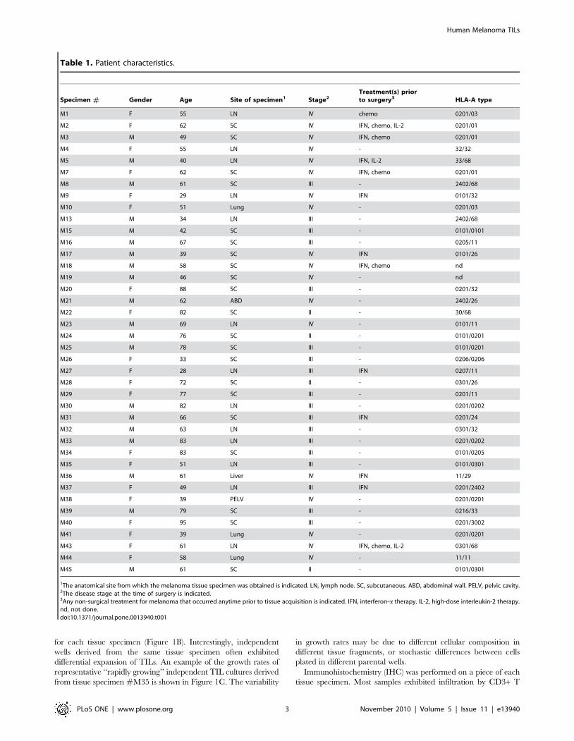
Tissue specimensA total of 40 melanoma tissue specimens were obtained from
patients undergoing surgical procedures under standard-of-care.
Patient characteristics are listed in Table 1. Three patients each
underwent surgical procedures on two occasions from which we
obtained tissue: Specimen #M2 and M7 were from the same
patient; specimen #M30 and M33 were from the same patient;
specimen #M38 and M41 were from the same patient. Most
specimens were obtained from subcutaneous lesions (n = 21), many
from nodal metastases (n = 13), and a few from other sites (lung,
liver, abdominal wall, pelvic cavity) (n = 6). The predominant
melanoma subtype was superficial spreading melanoma (n = 30)
(data not shown), which reflects the predominance of this
melanoma type in the general North American population [43].
The remaining histological types were nodular, acral lentiginous
or lentigo maligna melanoma (n = 7) or undetermined (n = 3) (data
not shown). Twenty specimens were from male patients, and
twenty from female patients. The average age at the time of tissue
acquisition was 60 years (+/2 17 years; median 61 years). Patients
were heavily skewed towards late stage disease, and thus matched
the target demographic for potential future adoptive cell therapy
clinical trials. There were 18 patients with stage IV melanoma at
the time of tissue acquisition, 18 with stage III, 4 with stage II and
none with stage I disease. Typing was performed at the HLA-A
locus in order to determine the appropriate panel of target cells for
assays of TIL function. Sixteen specimens were HLA-A*0201,
which is the most common HLA-A type amongst Caucasians [44].
TIL culturingTissues were processed by a variety of methods in preparation
for culturing TILs, depending in part on the amount of tissue
available. The specimen sizes obtained for this study varied widely,
from core biopsies to 3 cm3-sized samples. Single cell suspensions
were obtained by 1) enzymatic dissociation, 2) fine needle
aspirates, 3) mechanical dissociation using a Medimachine. Cells
obtained by these methods were plated and TILs were expanded
as described in the Materials and Methods. In addition, small
tissue fragments were also plated for TIL growth. In general, we
aimed to initiate TIL cultures from at least 8 tissue fragments and
106106 cells from enzymatically dissociated tissue; however this
varied depending on the size of the specimen.
Generally, TILs emerged from tissue fragments or began to
proliferate from single cell suspensions within 1–2 weeks of culture
initiation, and other cells such as tumor cells disappeared from the
cultures during that same time period. We observed that
expansion of TILs was more efficient following enzymatic
dissociation or plating tissue fragments (data not shown) and
therefore these methods will be used for processing tissues for our
planned clinical trials. Multiple independent bulk cultures of TILs
were maintained for each tissue specimen, with each independent
culture originating from approximately 1–2 parental wells. Each
culture was generally expanded for 4 weeks or until the minimum
number of TILs that would be needed for therapeutic protocols
were obtained (minimum 36107 TILs).
Our initial cultures were maintained in complete medium
containing commercially available human serum. We found that
TILs could be expanded from most melanoma tissues, but growth
rates were overall quite slow, with only 7 of 22 specimens (32%)
yielding at least one culture with $36107 cells within 4 weeks. We
therefore began to use plasma from healthy donors that was
obtained and processed at our own institution, instead of
commercially available serum. Growth rates of TIL cultures greatly
improved, with 13 of 18 tissue specimens (72%) yielding at least one
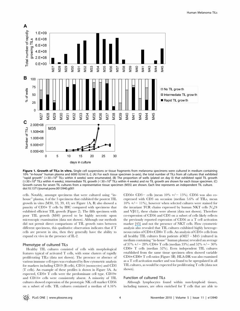
TIL culture reaching $36107 TILs within 4 weeks. In fact, most
specimens yielded multiple cultures exhibiting rapid growth rates
and therefore a total number of cells far exceeding the 36107
minimum (Figure 1A). We did not observe any differences in TIL
expansion between specimens obtained from different anatomical
sites (i.e. subcutaneous, visceral or nodal lesions) (data not shown).
For each of the 18 tissue specimens, the percentage of parental wells
that yielded TIL cultures with rapid growth rates (defined as
reaching $36107 TILs within 4 weeks), intermediate growth rates
(,36107 TILs within 4 weeks), or no TIL growth, were enumerated
Human Melanoma TILs
PLoS ONE | www.plosone.org 2 November 2010 | Volume 5 | Issue 11 | e13940
for each tissue specimen (Figure 1B). Interestingly, independent
wells derived from the same tissue specimen often exhibited
differential expansion of TILs. An example of the growth rates of
representative ‘‘rapidly growing’’ independent TIL cultures derived
from tissue specimen #M35 is shown in Figure 1C. The variability
in growth rates may be due to different cellular composition in
different tissue fragments, or stochastic differences between cells
plated in different parental wells.
Immunohistochemistry (IHC) was performed on a piece of each
tissue specimen. Most samples exhibited infiltration by CD3+ T
Table 1. Patient characteristics.
Specimen # Gender Age Site of specimen1 Stage2Treatment(s) priorto surgery3 HLA-A type
M1 F 55 LN IV chemo 0201/03
M2 F 62 SC IV IFN, chemo, IL-2 0201/01
M3 M 49 SC IV IFN, chemo 0201/01
M4 F 55 LN IV - 32/32
M5 M 40 LN IV IFN, IL-2 33/68
M7 F 62 SC IV IFN, chemo 0201/01
M8 M 61 SC III - 2402/68
M9 F 29 LN IV IFN 0101/32
M10 F 51 Lung IV - 0201/03
M13 M 34 LN III - 2402/68
M15 M 42 SC III - 0101/0101
M16 M 67 SC III - 0205/11
M17 M 39 SC IV IFN 0101/26
M18 M 58 SC IV IFN, chemo nd
M19 M 46 SC IV - nd
M20 F 88 SC III - 0201/32
M21 M 62 ABD IV - 2402/26
M22 F 82 SC II - 30/68
M23 M 69 LN IV - 0101/11
M24 M 76 SC II - 0101/0201
M25 M 78 SC III - 0101/0201
M26 F 33 SC III - 0206/0206
M27 F 28 LN III IFN 0207/11
M28 F 72 SC II - 0301/26
M29 F 77 SC III - 0201/11
M30 M 82 LN III - 0201/0202
M31 M 66 SC III IFN 0201/24
M32 M 63 LN III - 0301/32
M33 M 83 LN III - 0201/0202
M34 F 83 SC III - 0101/0205
M35 F 51 LN III - 0101/0301
M36 M 61 Liver IV IFN 11/29
M37 F 49 LN III IFN 0201/2402
M38 F 39 PELV IV - 0201/0201
M39 M 79 SC III - 0216/33
M40 F 95 SC III - 0201/3002
M41 F 39 Lung IV - 0201/0201
M43 F 61 LN IV IFN, chemo, IL-2 0301/68
M44 F 58 Lung IV - 11/11
M45 M 61 SC II - 0101/0301
1The anatomical site from which the melanoma tissue specimen was obtained is indicated. LN, lymph node. SC, subcutaneous. ABD, abdominal wall. PELV, pelvic cavity.2The disease stage at the time of surgery is indicated.3Any non-surgical treatment for melanoma that occurred anytime prior to tissue acquisition is indicated. IFN, interferon-a therapy. IL-2, high-dose interleukin-2 therapy.nd, not done.doi:10.1371/journal.pone.0013940.t001
Human Melanoma TILs
PLoS ONE | www.plosone.org 3 November 2010 | Volume 5 | Issue 11 | e13940
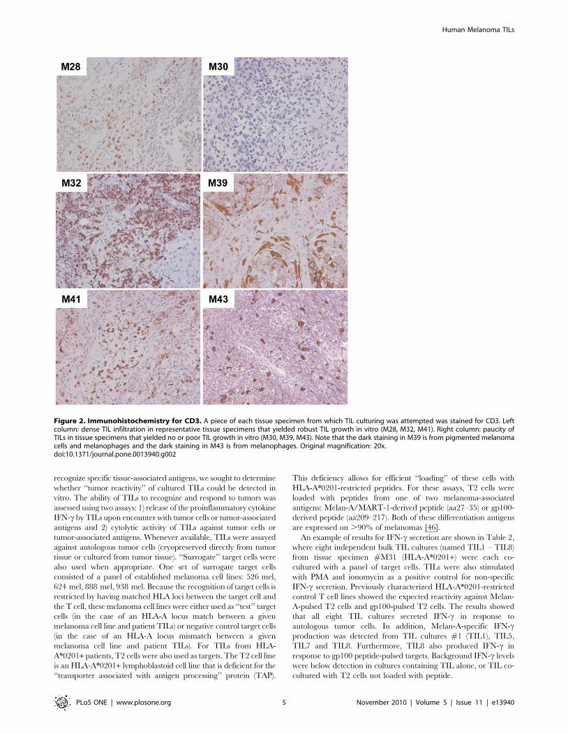
cells. Notably, amongst specimens that were cultured using ‘‘in-
house’’ plasma, 4 of the 5 specimens that exhibited the poorest TIL
growth in vitro (M30, 33, 39, 43; see Figure 1A, B) also showed a
paucity of CD3+ T cells by IHC compared with specimens that
exhibited efficient TIL growth (Figure 2). The fifth specimen with
poor TIL growth (M40) proved to be highly necrotic upon
microscopic examination (data not shown). Although our methods
did not permit direct comparisons of TIL growth rates between
different specimens, this qualitative observation indicates that if T
cells are present in situ, then they generally have the ability to
expand ex vivo in the presence of IL-2.
Phenotype of cultured TILsHealthy TIL cultures consisted of cells with morphological
features typical of activated T cells, with some clusters of rapidly
proliferating TILs (data not shown). The presence or absence of
various immune cell types was evaluated by flow cytometric analysis
for markers including CD19 (B cells), CD14 (monocytes) and CD3
(T cells). An example of these profiles is shown in Figure 3A. As
expected, CD3+ T cells were the predominant cell type. CD19+and CD14+ cells were consistently absent. A minority of TIL
cultures showed expression of the prototypic NK cell marker CD56
on a subset of cells. TIL cultures contained a median of 4.16%
CD56+ CD32 cells (mean 10% +/2 13%). CD56 was also co-
expressed with CD3 on occasion (median 5.6% of TILs, mean
10% +/2 11%), however when selected cultures were stained for
the invariant TCR chains expressed by human NKT cells (Va24
and Vb11), these chains were absent (data not shown). Therefore
co-expression of CD56 and CD3 on a subset of cells likely reflects
the previously reported expression of CD56 as a T cell activation
marker [45] and not the presence of NKT cells. Flow cytometric
analysis also revealed that TIL cultures exhibited highly heteroge-
neous ratios of CD4+:CD8+ T cells. An analysis of CD3+ cells from
all healthy TIL cultures from patients #M27 - M45 (cultured in
medium containing ‘‘in-house’’ human plasma) revealed an average
of 37% +/2 28% CD4+ T cells (median 33%) and 52% +/2 30%
CD8+ T cells (median 52%). Even independent TIL cultures
established from the same tissue specimen often showed variable
CD4+:CD8+ T cell ratios (Figure 3B). HLA-DR was also examined
as a T cell activation marker and was found to be upregulated in all
TIL cultures, as would be expected for proliferating T cells (data not
shown).
Function of cultured TILsAlthough lymphocytes found within non-lymphoid tissues,
including tumors, are often enriched for T cells that are able to
Figure 1. Growth of TILs in vitro. Single cell suspensions or tissue fragments from melanoma specimens were cultured in medium containing10% ‘‘in-house’’ human plasma and 6000 IU/ml IL-2. (A) For each tissue specimen (x-axis), the total number of TILs from all cultures that exhibited‘‘rapid growth’’ ($306106 TILs within 4 weeks) were enumerated. (B) The proportion of wells (plated on day 0) that exhibited rapid TIL growth($306106 TILs within 4 weeks), intermediate TIL growth (,306106 TILs within 4 weeks) and no TIL growth are shown for each tissue specimen. (C)Growth curves for seven TIL cultures from a representative tissue specimen (M35) are shown. Each line represents an independent TIL culture.doi:10.1371/journal.pone.0013940.g001
Human Melanoma TILs
PLoS ONE | www.plosone.org 4 November 2010 | Volume 5 | Issue 11 | e13940
recognize specific tissue-associated antigens, we sought to determine
whether ‘‘tumor reactivity’’ of cultured TILs could be detected in
vitro. The ability of TILs to recognize and respond to tumors was
assessed using two assays: 1) release of the proinflammatory cytokine
IFN-c by TILs upon encounter with tumor cells or tumor-associated
antigens and 2) cytolytic activity of TILs against tumor cells or
tumor-associated antigens. Whenever available, TILs were assayed
against autologous tumor cells (cryopreserved directly from tumor
tissue or cultured from tumor tissue). ‘‘Surrogate’’ target cells were
also used when appropriate. One set of surrogate target cells
consisted of a panel of established melanoma cell lines: 526 mel,
624 mel, 888 mel, 938 mel. Because the recognition of target cells is
restricted by having matched HLA loci between the target cell and
the T cell, these melanoma cell lines were either used as ‘‘test’’ target
cells (in the case of an HLA-A locus match between a given
melanoma cell line and patient TILs) or negative control target cells
(in the case of an HLA-A locus mismatch between a given
melanoma cell line and patient TILs). For TILs from HLA-
A*0201+ patients, T2 cells were also used as targets. The T2 cell line
is an HLA-A*0201+ lymphoblastoid cell line that is deficient for the
‘‘transporter associated with antigen processing’’ protein (TAP).
This deficiency allows for efficient ‘‘loading’’ of these cells with
HLA-A*0201-restricted peptides. For these assays, T2 cells were
loaded with peptides from one of two melanoma-associated
antigens: Melan-A/MART-1-derived peptide (aa27–35) or gp100-
derived peptide (aa209–217). Both of these differentiation antigens
are expressed on .90% of melanomas [46].
An example of results for IFN-c secretion are shown in Table 2,
where eight independent bulk TIL cultures (named TIL1 – TIL8)
from tissue specimen #M31 (HLA-A*0201+) were each co-
cultured with a panel of target cells. TILs were also stimulated
with PMA and ionomycin as a positive control for non-specific
IFN-c secretion. Previously characterized HLA-A*0201-restricted
control T cell lines showed the expected reactivity against Melan-
A-pulsed T2 cells and gp100-pulsed T2 cells. The results showed
that all eight TIL cultures secreted IFN-c in response to
autologous tumor cells. In addition, Melan-A-specific IFN-cproduction was detected from TIL cultures #1 (TIL1), TIL5,
TIL7 and TIL8. Furthermore, TIL8 also produced IFN-c in
response to gp100 peptide-pulsed targets. Background IFN-c levels
were below detection in cultures containing TIL alone, or TIL co-
cultured with T2 cells not loaded with peptide.
Figure 2. Immunohistochemistry for CD3. A piece of each tissue specimen from which TIL culturing was attempted was stained for CD3. Leftcolumn: dense TIL infiltration in representative tissue specimens that yielded robust TIL growth in vitro (M28, M32, M41). Right column: paucity ofTILs in tissue specimens that yielded no or poor TIL growth in vitro (M30, M39, M43). Note that the dark staining in M39 is from pigmented melanomacells and melanophages and the dark staining in M43 is from melanophages. Original magnification: 20x.doi:10.1371/journal.pone.0013940.g002
Human Melanoma TILs
PLoS ONE | www.plosone.org 5 November 2010 | Volume 5 | Issue 11 | e13940
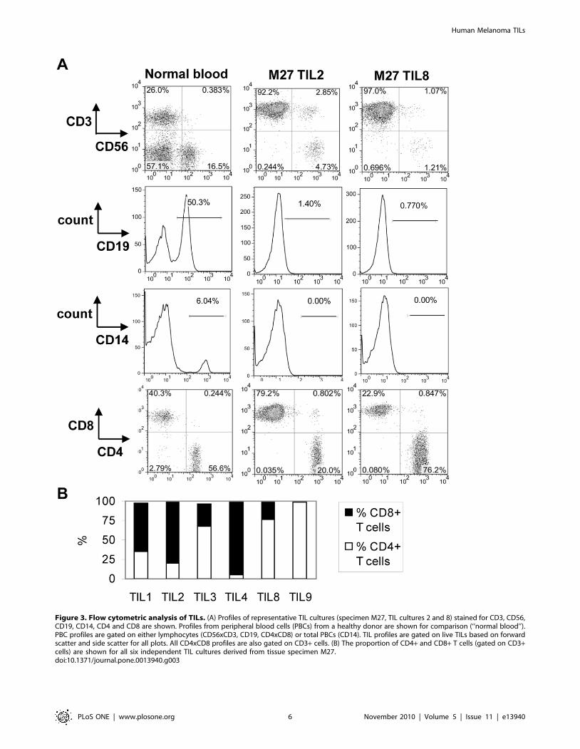
Figure 3. Flow cytometric analysis of TILs. (A) Profiles of representative TIL cultures (specimen M27, TIL cultures 2 and 8) stained for CD3, CD56,CD19, CD14, CD4 and CD8 are shown. Profiles from peripheral blood cells (PBCs) from a healthy donor are shown for comparison (‘‘normal blood’’).PBC profiles are gated on either lymphocytes (CD56xCD3, CD19, CD4xCD8) or total PBCs (CD14). TIL profiles are gated on live TILs based on forwardscatter and side scatter for all plots. All CD4xCD8 profiles are also gated on CD3+ cells. (B) The proportion of CD4+ and CD8+ T cells (gated on CD3+cells) are shown for all six independent TIL cultures derived from tissue specimen M27.doi:10.1371/journal.pone.0013940.g003
Human Melanoma TILs
PLoS ONE | www.plosone.org 6 November 2010 | Volume 5 | Issue 11 | e13940
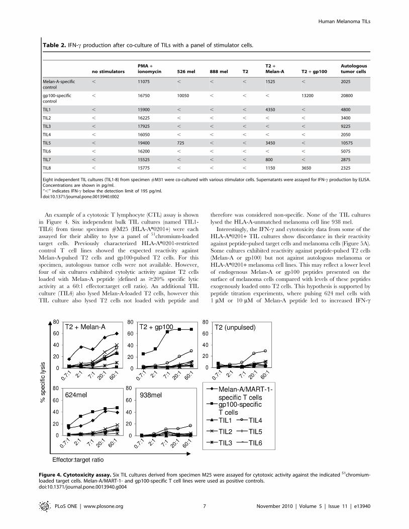
An example of a cytotoxic T lymphocyte (CTL) assay is shown
in Figure 4. Six independent bulk TIL cultures (named TIL1-
TIL6) from tissue specimen #M25 (HLA-A*0201+) were each
assayed for their ability to lyse a panel of 51chromium-loaded
target cells. Previously characterized HLA-A*0201-restricted
control T cell lines showed the expected reactivity against
Melan-A-pulsed T2 cells and gp100-pulsed T2 cells. For this
specimen, autologous tumor cells were not available. However,
four of six cultures exhibited cytolytic activity against T2 cells
loaded with Melan-A peptide (defined as $20% specific lytic
activity at a 60:1 effector:target cell ratio). An additional TIL
culture (TIL4) also lysed Melan-A-loaded T2 cells, however this
TIL culture also lysed T2 cells not loaded with peptide and
therefore was considered non-specific. None of the TIL cultures
lysed the HLA-A-unmatched melanoma cell line 938 mel.
Interestingly, the IFN-c and cytotoxicity data from some of the
HLA-A*0201+ TIL cultures show discordance in their reactivity
against peptide-pulsed target cells and melanoma cells (Figure 5A).
Some cultures exhibited reactivity against peptide-pulsed T2 cells
(Melan-A or gp100) but not against autologous melanoma or
HLA-A*0201+ melanoma cell lines. This may reflect a lower level
of endogenous Melan-A or gp100 peptides presented on the
surface of melanoma cells compared with levels of these peptides
exogenously loaded onto T2 cells. This hypothesis is supported by
peptide titration experiments, where pulsing 624 mel cells with
1 mM or 10 mM of Melan-A peptide led to increased IFN-c
Table 2. IFN-c production after co-culture of TILs with a panel of stimulator cells.
no stimulatorsPMA +ionomycin 526 mel 888 mel T2
T2 +Melan-A T2 + gp100
Autologoustumor cells
Melan-A-specificcontrol
, 11075 , , , 1525 , 2025
gp100-specificcontrol
, 16750 10050 , , , 13200 20800
TIL1 , 15900 , , , 4350 , 4800
TIL2 , 16225 , , , , , 3400
TIL3 , 17925 , , , , , 9225
TIL4 , 16050 , , , , , 2050
TIL5 , 19400 725 , , 3450 , 10575
TIL6 , 16200 , , , , , 5075
TIL7 , 15525 , , , 800 , 2875
TIL8 , 15775 , , , 1150 3650 2325
Eight independent TIL cultures (TIL1-8) from specimen #M31 were co-cultured with various stimulator cells. Supernatants were assayed for IFN-c production by ELISA.Concentrations are shown in pg/ml.‘‘,’’ indicates IFN-c below the detection limit of 195 pg/ml.doi:10.1371/journal.pone.0013940.t002
Figure 4. Cytotoxicity assay. Six TIL cultures derived from specimen M25 were assayed for cytotoxic activity against the indicated 51chromium-loaded target cells. Melan-A/MART-1- and gp100-specific T cell lines were used as positive controls.doi:10.1371/journal.pone.0013940.g004
Human Melanoma TILs
PLoS ONE | www.plosone.org 7 November 2010 | Volume 5 | Issue 11 | e13940
production by TILs, and conversely, decreasing the concentration
of Melan-A peptide on T2 cells led to decreased IFN-c production
(data not shown). Other cultures showed reactivity against
melanoma cells but not peptide-pulsed T2 cells. These TILs
may recognize other peptide epitopes of the Melan-A or gp100
proteins; it is also likely that the antigen specificity of these TILs is
much broader and may encompass other antigens.
All TIL cultures that yielded sufficient numbers of cells and for
which appropriate target cells were available were assayed for
tumor reactivity. Of the 40 tissue specimens that were obtained, 33
specimens yielded sufficient TILs to perform both the IFN-c and
CTL assays described above. Of these, appropriate target cells
were available for assaying TILs from 31 specimens. A positive
signal from either IFN-c production or CTL activity was taken as
an indication of ‘‘tumor reactivity’’. From these assays, 15
specimens yielded at least one TIL culture that exhibited tumor
reactivity in vitro. This result is comparable with the experience of
two other groups using similar methods [32,41].
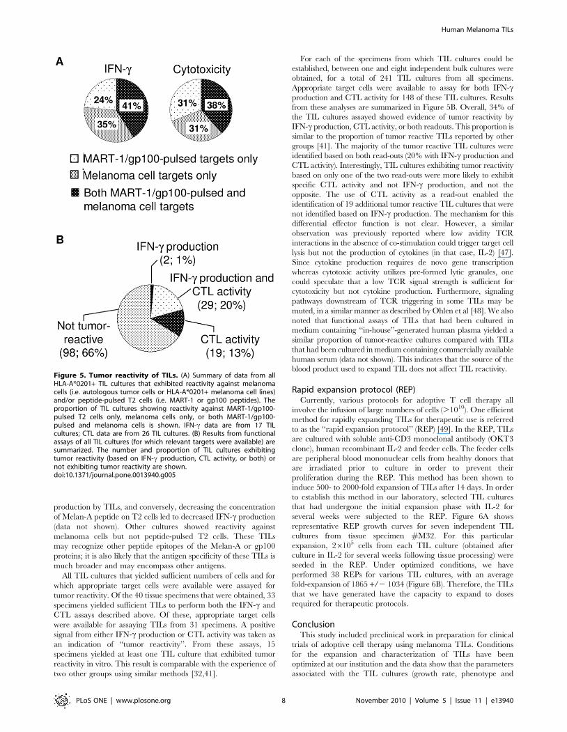
For each of the specimens from which TIL cultures could be
established, between one and eight independent bulk cultures were
obtained, for a total of 241 TIL cultures from all specimens.
Appropriate target cells were available to assay for both IFN-cproduction and CTL activity for 148 of these TIL cultures. Results
from these analyses are summarized in Figure 5B. Overall, 34% of
the TIL cultures assayed showed evidence of tumor reactivity by
IFN-c production, CTL activity, or both readouts. This proportion is
similar to the proportion of tumor reactive TILs reported by other
groups [41]. The majority of the tumor reactive TIL cultures were
identified based on both read-outs (20% with IFN-c production and
CTL activity). Interestingly, TIL cultures exhibiting tumor reactivity
based on only one of the two read-outs were more likely to exhibit
specific CTL activity and not IFN-c production, and not the
opposite. The use of CTL activity as a read-out enabled the
identification of 19 additional tumor reactive TIL cultures that were
not identified based on IFN-c production. The mechanism for this
differential effector function is not clear. However, a similar
observation was previously reported where low avidity TCR
interactions in the absence of co-stimulation could trigger target cell
lysis but not the production of cytokines (in that case, IL-2) [47].
Since cytokine production requires de novo gene transcription
whereas cytotoxic activity utilizes pre-formed lytic granules, one
could speculate that a low TCR signal strength is sufficient for
cytotoxicity but not cytokine production. Furthermore, signaling
pathways downstream of TCR triggering in some TILs may be
muted, in a similar manner as described by Ohlen et al [48]. We also
noted that functional assays of TILs that had been cultured in
medium containing ‘‘in-house’’-generated human plasma yielded a
similar proportion of tumor-reactive cultures compared with TILs
that had been cultured in medium containing commercially available
human serum (data not shown). This indicates that the source of the
blood product used to expand TIL does not affect TIL reactivity.
Rapid expansion protocol (REP)Currently, various protocols for adoptive T cell therapy all
involve the infusion of large numbers of cells (.1010). One efficient
method for rapidly expanding TILs for therapeutic use is referred
to as the ‘‘rapid expansion protocol’’ (REP) [49]. In the REP, TILs
are cultured with soluble anti-CD3 monoclonal antibody (OKT3
clone), human recombinant IL-2 and feeder cells. The feeder cells
are peripheral blood mononuclear cells from healthy donors that
are irradiated prior to culture in order to prevent their
proliferation during the REP. This method has been shown to
induce 500- to 2000-fold expansion of TILs after 14 days. In order
to establish this method in our laboratory, selected TIL cultures
that had undergone the initial expansion phase with IL-2 for
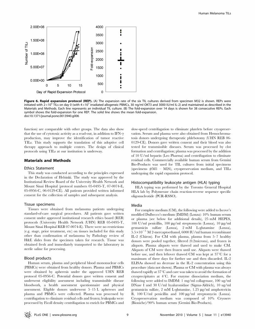
several weeks were subjected to the REP. Figure 6A shows
representative REP growth curves for seven independent TIL
cultures from tissue specimen #M32. For this particular
expansion, 26105 cells from each TIL culture (obtained after
culture in IL-2 for several weeks following tissue processing) were
seeded in the REP. Under optimized conditions, we have
performed 38 REPs for various TIL cultures, with an average
fold-expansion of 1865 +/2 1034 (Figure 6B). Therefore, the TILs
that we have generated have the capacity to expand to doses
required for therapeutic protocols.
ConclusionThis study included preclinical work in preparation for clinical
trials of adoptive cell therapy using melanoma TILs. Conditions
for the expansion and characterization of TILs have been
optimized at our institution and the data show that the parameters
associated with the TIL cultures (growth rate, phenotype and
Figure 5. Tumor reactivity of TILs. (A) Summary of data from allHLA-A*0201+ TIL cultures that exhibited reactivity against melanomacells (i.e. autologous tumor cells or HLA-A*0201+ melanoma cell lines)and/or peptide-pulsed T2 cells (i.e. MART-1 or gp100 peptides). Theproportion of TIL cultures showing reactivity against MART-1/gp100-pulsed T2 cells only, melanoma cells only, or both MART-1/gp100-pulsed and melanoma cells is shown. IFN-c data are from 17 TILcultures; CTL data are from 26 TIL cultures. (B) Results from functionalassays of all TIL cultures (for which relevant targets were available) aresummarized. The number and proportion of TIL cultures exhibitingtumor reactivity (based on IFN-c production, CTL activity, or both) ornot exhibiting tumor reactivity are shown.doi:10.1371/journal.pone.0013940.g005
Human Melanoma TILs
PLoS ONE | www.plosone.org 8 November 2010 | Volume 5 | Issue 11 | e13940
function) are comparable with other groups. The data also show
that the use of cytotoxic activity as a read-out, in addition to IFN-cproduction, may improve the identification of tumor reactive
TILs. This study supports the translation of this adoptive cell
therapy approach to multiple centers. The design of clinical
protocols using TILs at our institution is underway.
Materials and Methods
Ethics StatementThis study was conducted according to the principles expressed
in the Declaration of Helsinki. The study was approved by the
Institutional Review Board of the University Health Network and
Mount Sinai Hospital (protocol numbers 05-0495-T, 07-0074-E,
05-0956-C, 06-0129-CE). All patients provided written informed
consent for the collection of samples and subsequent analysis.
Tissue specimensTissues were obtained from melanoma patients undergoing
standard-of-care surgical procedures. All patients gave written
consent under approved institutional research ethics board (REB)
protocols (University Health Network (UHN) REB 05-0495-T,
Mount Sinai Hospital REB 07-0074-E). There were no restrictions
(e.g. stage, prior treatment, etc) on tissues included for this study
other than confirmation of melanoma by Pathology review of
H&E slides from the specimen taken for research. Tissue was
obtained fresh and immediately transported to the laboratory in
sterile saline for processing.
Blood productsHuman serum, plasma and peripheral blood mononuclear cells
(PBMCs) were obtained from healthy donors. Plasma and PBMCs
were obtained by apheresis under the approved UHN REB
protocol 05-0956-C. Potential donors gave written consent and
underwent eligibility assessment including transmissible disease
bloodwork, a health assessment questionnaire and physical
assessment. Eligible donors underwent 5–15 L aphereses and
plasma and PBMCs were collected. Plasma was processed by
centrifugation to eliminate residual cells and frozen; leukopaks were
processed by Ficoll density centrifugation to enrich for PBMCs and
slow-speed centrifugation to eliminate platelets before cryopreser-
vation. Serum and plasma were also obtained from Hemochroma-
tosis donors undergoing therapeutic phlebotomy (UHN REB 06-
0129-CE). Donors gave written consent and their blood was also
tested for transmissible diseases. Serum was processed by clot
formation and centrifugation; plasma was processed by the addition
of 10 U/ml heparin (Leo Pharma) and centrifugation to eliminate
residual cells. Commercially available human serum from Gemini
Bio-Products was used for TIL cultures from initial specimens
(specimens #M1 – M26), cryopreservation medium, and TILs
undergoing the rapid expansion protocol.
Histocompatibility leukocyte antigen (HLA) typingHLA typing was performed by the Toronto General Hospital
HLA lab by Polymerase chain reaction-reverse sequence specific
oligonucleotide (PCR-RSSO).
MediaFor complete medium (CM), the following were added to Iscove’s
modified Dulbecco’s medium (IMDM) (Lonza): 10% human serum
or plasma (see below for additional details), 25 mM HEPES,
100 U/ml penicillin, 100 mg/ml streptomycin (Lonza), 10 mg/ml
gentamicin sulfate (Lonza), 2 mM L-glutamine (Lonza),
5.561025 M 2-mercaptoethanol, 6000 IU/ml human recombinant
IL-2 (Chiron). For CM with plasma, plasma from 5–7 healthy
donors were pooled together, filtered (0.2microns), and frozen in
aliquots. Plasma aliquots were thawed and used to make CM.
Aliquots of CM were then frozen until use. Aliquots were thawed
before use, and then leftover thawed CM was kept at 37uC for a
maximum of three days for further use and then discarded. IL-2
ELISAs showed no decrease in the IL-2 concentration using this
procedure (data not shown). Plasma or CM with plasma was always
thawed rapidly at 37uC and care was taken to avoid the formation of
cryoprecipitates at 4uC. For enzyme dissociation medium, the
following were added to IMDM: 1 mg/ml collagenase, 100 ug/ml
DNase I and 30 U/ml hyaluronidase (Sigma-Aldrich), 10 ug/ml
gentamicin sulfate, 2 mM L-glutamine, 1.25 mg/ml amphotericin
B, 100 U/ml penicillin and 100 mg/ml streptomycin (Lonza).
Cryopreservation medium was composed of 10% Cryoserv
(Bioniche)/90% human serum (Gemini Bio-Products).
Figure 6. Rapid expansion protocol (REP). (A) The expansion rate of the six TIL cultures derived from specimen M32 is shown. REPs wereinitiated with 26105 TILs on day 0 (with 46107 irradiated allogeneic PBMCs, 30 ng/ml OKT3 and 3000 IU/ml IL-2) and maintained as described in theMaterials and Methods. Each line represents an individual TIL culture. (B) The fold-expansion over 14 days is shown for 38 consecutive REPs. Eachsymbol shows the fold-expansion for one REP. The solid line shows the mean fold-expansion.doi:10.1371/journal.pone.0013940.g006
Human Melanoma TILs
PLoS ONE | www.plosone.org 9 November 2010 | Volume 5 | Issue 11 | e13940
TIL culturingMethods for TIL culturing were the same as those used by the
Rosenberg group. Several methods were used to process tissue: 1)
enzymatic dissociation, 2) fine needle aspirates, 3) mechanical
dissociation using a Medimachine (Becton Dickinson) and/or 4)
directly plating small tissue fragments. For enzymatic dissociation,
tissue was first minced into ,1 mm3 pieces and then incubated on a
stir plate at room temperature until tissue was dissociated (1–18
hours). After passing through a 100micron nylon mesh, cells were
washed extensively before plating. For fine needle aspirates, cells were
collected through a 23 gauge needle and then washed. For
mechanical dissociation, small tissue pieces were loaded into
Medicons and then dissociated according to the manufacturer’s
instructions. Cells were then subjected to Ficoll gradient centrifugation
and the leukocyte-enriched layer collected for TIL growth. Single cell
suspensions were plated at 16106 total cells per well, in 24-well tissue
culture plates. For TIL growth from tissue fragments, one 1 mm3
fragment was placed into each well of 24-well plates. Cells were
cultured in 2 ml per well of CM (containing 6000 IU/ml of human
recombinant IL-2) in a 37uC, 5% CO2, humidified incubator. After
the first week in culture, 1 ml medium from each well was replaced
with fresh CM three times a week. Wells were maintained at a cell
concentration of 0.5–26106 cells/ml. Each independent TIL culture
was generally derived from 1–2 parental wells and upon subsequent
expansions, all daughter wells were combined, mixed and re-plated.
Flow cytometryCells were stained at 4uC for 30 minutes in buffer (2% fetal calf
serum/0.05% sodium azide/PBS) containing antibodies, washed,
and resuspended in 1% paraformaldehyde/PBS. Antibodies used
included: CD3-phycoerythrin (PE), CD4-fluorescein isothiocya-
nate (FITC), CD8-peridinin chlorophyll protein (PerCP), CD56-
allophycocyanin (APC), CD19-FITC, CD14-PerCP-Cyanine5.5.
Data was acquired on a FACSCalibur flow cytometer (BD) and
analyzed using FlowJo software.
Immunohistochemistry (IHC)4micron formalin-fixed paraffin-embedded sections were de-
waxed in 5 changes of xylene and brought down to water through
graded alcohols. Slides were pretreated with pepsin (1% pepsin in
0.01 N HCl (pH 2.0), 15 mins at 37uC). Endogenous peroxidase
and biotin activities were blocked respectively using 3% hydrogen
peroxide and avidin/biotin blocking kit (Vector labs.). After
blocking for 15 mins with 10% normal goat serum, sections were
incubated accordingly at room temperature with anti-human CD3
antibody (Dako) at 1:100 for 1 hour. This was followed by 30 mins
each with a biotinylated linking reagent (ID labs.) and horseradish
peroxidase-conjugated ultrastreptavidin labeling reagent (ID labs.).
After washing well in PBS, colour development was done with
freshly prepared NovaRed solution (Vector labs.). Finally, sections
were counterstained lightly with Mayer’s hematoxylin, dehydrated
in alcohols, cleared in xylene and mounted in Permount.
Functional assaysTIL cultures were assayed for reactivity against the following
panel of target cells, depending on availability and HLA-type: 1)
autologous tumor cells (cryopreserved single cell suspensions from
original tissue specimen and/or melanoma tumor cells propagated
in culture (in CM without IL-2) from the original tissue specimen),
2) melanoma cell lines (kind gifts from M. Dudley and S.
Rosenberg): 526 mel (HLA-A*0301/0201), 624 mel (HLA-
A*0301/0201), 888 mel (HLA-A*0101/2402), 938 mel (HLA-
A*0101/2402) (all expressing Melan-A/MART-1 and gp100), 3)
T2 lymphoblastoid cells (HLA-A*0201+, TAP-deficient) pulsed for
2 hours with 1 mM Melan-A/MART-1-derived peptide (aa27–35),
1 mM gp100-derived peptide (aa209–217), or unpulsed. Melan-A/
MART-1-specific and gp100-specific T cell lines were used as
positive control T cells. Stimulation of TILs with 10 ng/ml
Phorbol myristate acid (PMA) and 500 ng/ml ionomycin (Sigma)
was used as a positive control for non-specific cytokine production.
For IFN-c production, 56104 TILs were co-cultured with 56104
target cells overnight at 37uC and supernatants were assayed for
IFN-c concentration by ELISA according to the manufacturer’s
instructions (Endogen). For cytotoxic T lymphocyte (CTL) assays,
26103 51chromium-loaded target cells were plated in each well,
together with TILs at various effector:target ratios. After 4–5 hours
at 37uC, supernatants were transferred to LumaPlates (Perkin
Elmer) and counted using a TopCount counter (Perkin Elmer).
Maximal release was obtained using 1% Triton-X 100. Percent
specific lysis was calculated as ((experimental release – spontaneous
release)/(maximal release – spontaneous release))*100. Specific
tumor reactivity was defined for IFN-c as $200 pg/ml IFN-c in
response to autologous or HLA-A-matched target cells and .2-
fold over background response of unstimulated TILs or against
HLA-A-unmatched target cells. Specific tumor reactivity was
defined for cytotoxicity as $20% specific lysis at a 60:1
effector:target ratio against autologous or HLA-A-matched target
cells and #10% specific lysis at a 60:1 effector:target ratio against
HLA-A-unmatched target cells.
Rapid expansion protocol (REP)TILs were thawed and rested in CM (containing 6000 IU/ml
IL-2) for 1-3 days prior to initiating the REP. For initiation of
REPs, the following were combined in T175 tissue culture flasks:
16106 TILs, 26108 allogeneic feeder cells (peripheral blood
mononuclear cells pooled from 2–3 healthy donors and irradiated
(50 Gy) before use), 30 ng/ml OKT3 (Janssen-Ortho), 3000 IU/
ml IL-2 (Chiron) (no difference was seen in the expansion rate in
the REP using 3000 IU/ml versus 6000 IU/ml of IL-2 (data not
shown)), 75 ml CM and 75 ml AIM V serum-free medium
(Gibco). REPs were maintained in a 37uC, 5% CO2, humidified
incubator. On day 5, 80–90% of the media was replaced with
fresh medium (50%CM/50%AIM V) and 3000 IU/ml IL-2. Cells
were maintained at approximately 16106 cells/ml using AIM V
with 5% human serum and 3000 IU/ml IL-2 on day 7 and plain
AIM V medium and 3000 IU/ml IL-2 from day 8 onwards. Once
cultures exceeded the T715 flask capacity, cells were transferred
into 3 L Lifecell tissue culture bags (Baxter). This method was
scaled down as necessary (e.g. 26105 TIL per T25 flask).
Acknowledgments
We thank the melanoma patients for consenting to tissue being given to this
study and the blood donors for their time. We also thank Robert Buckman
for recruitment of blood donors, Karen Witiuk for conducting aphereses,
and Karen Chang and Nazir Jamal for advice on cell manufacturing
procedures. Valuable protocols, reagents and advice were kindly provided
by Mark Dudley and Steven Rosenberg.
Author Contributions
Conceived and designed the experiments: LTN DG DH AMJ IQ HM
PSO. Performed the experiments: LTN PHY JN NL AE WL JL DM MR.
Analyzed the data: LTN PHY JN NL DG AAH ES. Contributed reagents/
materials/analysis tools: AE WL JL DM MR DH AMJ IQ HM PAS MC
ES. Wrote the paper: LTN PSO.
Human Melanoma TILs
PLoS ONE | www.plosone.org 10 November 2010 | Volume 5 | Issue 11 | e13940
References
1. Shankaran V, Ikeda H, Bruce AT, White JM, Swanson PE, et al. (2001)IFNgamma and lymphocytes prevent primary tumour development and shape
tumour immunogenicity. Nature 410: 1107–1111.
2. Dunn GP, Old LJ, Schreiber RD (2004) The immunobiology of cancerimmunosurveillance and immunoediting. Immunity 21: 137–148.
3. Romero P, Dunbar PR, Valmori D, Pittet MJ, Ogg GS, et al. (1998) Ex vivostaining of metastatic lymph nodes by class I major histocompatibility complex
tetramers reveals high numbers of antigen-experienced tumor-specific cytotoxic
T lymphocytes. J Exp Med 188: 1641–1650.
4. Pittet MJ, Valmori D, Dunbar PR, Speiser DE, Lienard D, et al. (1999) High
frequencies of naive Melan-A/MART-1-specific CD8(+) T cells in a largeproportion of human histocompatibility leukocyte antigen (HLA)-A2 individuals.
J Exp Med 190: 705–715.
5. Kawakami Y, Dang N, Wang X, Tupesis J, Robbins PF, et al. (2000)Recognition of shared melanoma antigens in association with major HLA-A
alleles by tumor infiltrating T lymphocytes from 123 patients with melanoma.
J Immunother 23: 17–27.
6. Boon T, Coulie PG, Van den Eynde BJ, van der Bruggen P (2006) Human T cell
responses against melanoma. Annu Rev Immunol 24: 175–208.
7. Speiser DE, Lienard D, Rufer N, Rubio-Godoy V, Rimoldi D, et al. (2005)Rapid and strong human CD8+ T cell responses to vaccination with peptide,
IFA, and CpG oligodeoxynucleotide 7909. J Clin Invest 115: 739–746.
8. Schwartzentruber DJ, Lawson D, Richards J, Conry RM, Miller D, et al. (2009)
A phase III multi-institutional randomized study of immunization with the
gp100:209-217(210 M) peptide followed by high-dose IL-2 compared with high-dose IL-2 alone in patients with metastatic melanoma. J Clin Oncol 27: 18s.
9. Weber J (2008) Overcoming immunologic tolerance to melanoma: targeting
CTLA-4 with ipilimumab (MDX-010). Oncologist 13(Suppl 4): 16–25.: 16-25.
10. Ribas A (2008) Overcoming immunologic tolerance to melanoma: targeting
CTLA-4 with tremelimumab (CP-675,206). Oncologist 13(Suppl 4): 10–5.: 10-15.
11. Hodi FS, O’Day SJ, McDermott DF, Weber RW, Sosman JA, et al. (2010)
Improved Survival with Ipilimumab in Patients with Metastatic Melanoma.N Engl J Med 363: 711–723.
12. Blank C, Gajewski TF, Mackensen A (2005) Interaction of PD-L1 on tumor cells
with PD-1 on tumor-specific T cells as a mechanism of immune evasion:implications for tumor immunotherapy. Cancer Immunol Immunother 54:
307–314.
13. Berger R, Rotem-Yehudar R, Slama G, Landes S, Kneller A, et al. (2008) PhaseI safety and pharmacokinetic study of CT-011, a humanized antibody
interacting with PD-1, in patients with advanced hematologic malignancies.Clin Cancer Res 14: 3044–3051.
14. Brahmer JR, Drake CG, Wollner I, Powderly JD, Picus J, et al. (2010) Phase I
Study of Single-Agent Anti-Programmed Death-1 (MDX-1106) in RefractorySolid Tumors: Safety, Clinical Activity, Pharmacodynamics, and Immunologic
Correlates. J Clin Oncol 28: 3167–3175.
15. Dudley ME, Wunderlich J, Nishimura MI, Yu D, Yang JC, et al. (2001)Adoptive transfer of cloned melanoma-reactive T lymphocytes for the treatment
of patients with metastatic melanoma. J Immunother 24: 363–373.
16. Yee C, Thompson JA, Byrd D, Riddell SR, Roche P, et al. (2002) Adoptive T
cell therapy using antigen-specific CD8+ T cell clones for the treatment of
patients with metastatic melanoma: in vivo persistence, migration, andantitumor effect of transferred T cells. Proc Natl Acad Sci U S A 99:
16168–16173.
17. Vignard V, Lemercier B, Lim A, Pandolfino MC, Guilloux Y, et al. (2005)
Adoptive transfer of tumor-reactive Melan-A-specific CTL clones in melanoma
patients is followed by increased frequencies of additional Melan-A-specific Tcells. J Immunol 175: 4797–4805.
18. Hunder NN, Wallen H, Cao J, Hendricks DW, Reilly JZ, et al. (2008)
Treatment of metastatic melanoma with autologous CD4+ T cells against NY-ESO-1. N Engl J Med 358: 2698–2703.
19. Rosenberg SA, Packard BS, Aebersold PM, Solomon D, Topalian SL, et al.(1988) Use of tumor-infiltrating lymphocytes and interleukin-2 in the
immunotherapy of patients with metastatic melanoma. A preliminary report.
N Engl J Med 319: 1676–1680.
20. Kradin RL, Kurnick JT, Lazarus DS, Preffer FI, Dubinett SM, et al. (1989)
Tumour-infiltrating lymphocytes and interleukin-2 in treatment of advancedcancer. Lancet 1: 577–580.
21. Baars JW, Fonk JC, Scheper RJ, von Blomberg-van der Flier BM, Bril H, et al.
(1992) Treatment with tumour infiltrating lymphocytes and interleukin-2 inpatients with metastatic melanoma: a pilot study. Biotherapy 4: 289–297.
22. Dillman RO, Church C, Oldham RK, West WH, Schwartzberg L, et al. (1993)
Inpatient continuous-infusion interleukin-2 in 788 patients with cancer. TheNational Biotherapy Study Group experience. Cancer 71: 2358–2370.
23. Rosenberg SA, Yannelli JR, Yang JC, Topalian SL, Schwartzentruber DJ, et al.
(1994) Treatment of patients with metastatic melanoma with autologous tumor-infiltrating lymphocytes and interleukin 2. J Natl Cancer Inst 86: 1159–1166.
24. Ravaud A, Legrand E, Delaunay MM, Bussieres E, Coulon V, et al. (1995) Aphase I trial of repeated tumour-infiltrating lymphocyte (TIL) infusion in
metastatic melanoma. Br J Cancer 71: 331–336.
25. Goedegebuure PS, Douville LM, Li H, Richmond GC, Schoof DD, et al. (1995)Adoptive immunotherapy with tumor-infiltrating lymphocytes and interleukin-2
in patients with metastatic malignant melanoma and renal cell carcinoma: a pilot
study. J Clin Oncol 13: 1939–1949.
26. Reali UM, Martini L, Borgognoni L, Semino C, Pietra G, et al. (1998) Infusion
of in vitro expanded tumour-infiltrating lymphocytes and recombinant
interleukin-2 in patients with surgically resected lymph node metastases of
malignant melanoma: a pilot study. Melanoma Res 8: 77–82.
27. Queirolo P, Ponte M, Gipponi M, Cafiero F, Peressini A, et al. (1999) Adoptive
immunotherapy with tumor-infiltrating lymphocytes and subcutaneous recom-
binant interleukin-2 plus interferon alfa-2a for melanoma patients with
nonresectable distant disease: a phase I/II pilot trial. Melanoma Istituto
Scientifico Tumori Group. Ann Surg Oncol 6: 272–278.
28. Dreno B, Nguyen JM, Khammari A, Pandolfino MC, Tessier MH, et al. (2002)
Randomized trial of adoptive transfer of melanoma tumor-infiltrating lympho-
cytes as adjuvant therapy for stage III melanoma. Cancer Immunol Immunother
51: 539–546.
29. Dudley ME, Wunderlich JR, Robbins PF, Yang JC, Hwu P, et al. (2002) Cancer
regression and autoimmunity in patients after clonal repopulation with
antitumor lymphocytes. Science 298: 850–854.
30. Dudley ME, Wunderlich JR, Yang JC, Sherry RM, Topalian SL, et al. (2005)
Adoptive cell transfer therapy following non-myeloablative but lymphodepleting
chemotherapy for the treatment of patients with refractory metastatic
melanoma. J Clin Oncol 23: 2346–2357.
31. Dudley ME, Yang JC, Sherry R, Hughes MS, Royal R, et al. (2008) Adoptive
Cell Therapy for Patients With Metastatic Melanoma: Evaluation of Intensive
Myeloablative Chemoradiation Preparative Regimens. J Clin Oncol 26:
5233–5239.
32. Besser MJ, Shapira-Frommer R, Treves AJ, Zippel D, Itzhaki O, et al. (2009)
Minimally cultured or selected autologous tumor-infiltrating lymphocytes after a
lympho-depleting chemotherapy regimen in metastatic melanoma patients.
J Immunother 32: 415–423.
33. Morgan RA, Dudley ME, Wunderlich JR, Hughes MS, Yang JC, et al. (2006)
Cancer Regression in Patients After Transfer of Genetically Engineered
Lymphocytes. Science 314: 68–69.
34. Aoki Y, Takakuwa K, Kodama S, Tanaka K, Takahashi M, et al. (1991) Use of
adoptive transfer of tumor-infiltrating lymphocytes alone or in combination with
cisplatin-containing chemotherapy in patients with epithelial ovarian cancer.
Cancer Res 51: 1934–1939.
35. Belldegrun A, Pierce W, Kaboo R, Tso CL, Shau H, et al. (1993) Interferon-
alpha primed tumor-infiltrating lymphocytes combined with interleukin-2 and
interferon-alpha as therapy for metastatic renal cell carcinoma. J Urol 150:
1384–1390.
36. Ratto GB, Melioli G, Zino P, Mereu C, Mirabelli S, et al. (1995)
Immunotherapy with the use of tumor-infiltrating lymphocytes and interleu-
kin-2 as adjuvant treatment in stage III non-small-cell lung cancer. A pilot study.
J Thorac Cardiovasc Surg 109: 1212–1217.
37. Gardini A, Ercolani G, Riccobon A, Ravaioli M, Ridolfi L, et al. (2004)
Adjuvant, adoptive immunotherapy with tumor infiltrating lymphocytes plus
interleukin-2 after radical hepatic resection for colorectal liver metastases: 5-year
analysis. J Surg Oncol 87: 46–52.
38. Atkins MB, Kunkel L, Sznol M, Rosenberg SA (2000) High-dose recombinant
interleukin-2 therapy in patients with metastatic melanoma: long-term survival
update. Cancer J Sci Am 6(Suppl 1): S11–4.: S11-S14.
39. Bedikian AY, Millward M, Pehamberger H, Conry R, Gore M, et al. (2006) Bcl-
2 antisense (oblimersen sodium) plus dacarbazine in patients with advanced
melanoma: the Oblimersen Melanoma Study Group. J Clin Oncol 24:
4738–4745.
40. Wallen H, Thompson JA, Reilly JZ, Rodmyre RM, Cao J, et al. (2009)
Fludarabine modulates immune response and extends in vivo survival of
adoptively transferred CD8 T cells in patients with metastatic melanoma. PLoS
One 4: e4749.
41. Tran KQ, Zhou J, Durflinger KH, Langhan MM, Shelton TE, et al. (2008)
Minimally cultured tumor-infiltrating lymphocytes display optimal characteris-
tics for adoptive cell therapy. J Immunother 31: 742–751.
42. Besser MJ, Shapira-Frommer R, Treves AJ, Zippel D, Itzhaki O, et al. (2010)
Clinical responses in a phase II study using adoptive transfer of short-term
cultured tumor infiltration lymphocytes in metastatic melanoma patients. Clin
Cancer Res 16: 2646–2655.
43. Clark WH, Jr., Elder DE, Van Horn M (1986) The biologic forms of malignant
melanoma. Hum Pathol 17: 443–450.
44. Cao K, Hollenbach J, Shi X, Shi W, Chopek M, et al. (2001) Analysis of the
frequencies of HLA-A, B, and C alleles and haplotypes in the five major ethnic
groups of the United States reveals high levels of diversity in these loci and
contrasting distribution patterns in these populations. Hum Immunol 62:
1009–1030.
45. Pittet MJ, Speiser DE, Valmori D, Cerottini JC, Romero P (2000) Cutting edge:
cytolytic effector function in human circulating CD8+ T cells closely correlates
with CD56 surface expression. J Immunol 164: 1148–1152.
46. Barrow C, Browning J, MacGregor D, Davis ID, Sturrock S, et al. (2006) Tumor
antigen expression in melanoma varies according to antigen and stage. Clin
Cancer Res 12: 764–771.
Human Melanoma TILs
PLoS ONE | www.plosone.org 11 November 2010 | Volume 5 | Issue 11 | e13940
47. Bachmann MF, Sebzda E, Kundig TM, Shahinian A, Speiser DE, et al. (1996)
T cell responses are governed by avidity and costimulatory thresholds.Eur J Immunol 26: 2017–2022.
48. Ohlen C, Kalos M, Cheng LE, Shur AC, Hong DJ, et al. (2002) CD8(+) T cell
tolerance to a tumor-associated antigen is maintained at the level of expansionrather than effector function. J Exp Med 195: 1407–1418.
49. Dudley ME, Wunderlich JR, Shelton TE, Even J, Rosenberg SA (2003)
Generation of tumor-infiltrating lymphocyte cultures for use in adoptive transfer
therapy for melanoma patients. J Immunother 26: 332–342.
Human Melanoma TILs
PLoS ONE | www.plosone.org 12 November 2010 | Volume 5 | Issue 11 | e13940
Related Documents


![Tumour-infiltrating cytotoxic T lymphocytes in …...such as CD8+ and natural killer lymphocytes [17], indu-cing a cytotoxic cascade resulting in tumour cell death, while other TILs](https://static.cupdf.com/doc/110x72/5f4838f3212d137c1c54d55d/tumour-infiltrating-cytotoxic-t-lymphocytes-in-such-as-cd8-and-natural-killer.jpg)