Exosomes/microvesicles as a mechanism of cell-to-cell communication Giovanni Camussi 1 , Maria C. Deregibus 1 , Stefania Bruno 2 , Vincenzo Cantaluppi 1 and Luigi Biancone 1 1 Department of Internal Medicine, Centre for Molecular Biotechnology and Centre for Research in Experimental Medicine (CeRMS), Torino, Italy and 2 Sis-Ter, Palazzo Pignano, Italy Microvesicles (MVs) are circular fragments of membrane released from the endosomal compartment as exosomes or shed from the surface membranes of most cell types. An increasing body of evidence indicates that they play a pivotal role in cell-to-cell communication. Indeed, they may directly stimulate target cells by receptor-mediated interactions or may transfer from the cell of origin to various bioactive molecules including membrane receptors, proteins, mRNAs, microRNAs, and organelles. In this review we discuss the pleiotropic biologic effects of MVs that are relevant for communication among cells in physiological and pathological conditions. In particular, we discuss their potential involvement in inflammation, renal disease, and tumor progression, and the evidence supporting a bidirectional exchange of genetic information between stem and injured cells. The transfer of gene products from injured cells may explain stem cell functional and phenotypic changes without the need of transdifferentiation into tissue cells. On the other hand, transfer of gene products from stem cells may reprogram injured cells to repair damaged tissues. Kidney International (2010) 78, 838–848; doi:10.1038/ki.2010.278; published online 11 August 2010 KEYWORDS: exosomes; microvesicles; renal injury; stem cells Cell-to-cell communication is required to guarantee proper coordination among different cell types within tissues. Cells may communicate by soluble factors, 1 adhesion molecule- mediated cell-to-cell interactions including cytonemes that connect neighboring cells enabling ligand–receptor-mediated transfer of surface-associated molecules, or by tunneling nanotubules that establish conduits between cells, allowing the transfer of not only surface molecules but also cyto- plasmic components. 2,3 Recent studies have suggested that cells may also communicate by circular membrane fragments named microvesicles (MVs). 4 For a long time, MVs were considered to be inert cellular debris, and the frequently observed vesicles by electron microscopy in the interstitial space of tissues or in blood were considered the consequence of cell damage or the result of dynamic plasmamembrane turnover. 5 De Broe et al. 6 first suggested that circular plasmamembrane fragments released from human cells may result from a specific process and showed that they may carry functional membrane enzymes in the same ratio as the membrane of the cells of origin. However, only recent studies have assigned a defined function to the vesicles/exosomes released in the microenvironment by various cell types. Two distinct processes of vesicle release from the cells have been described. MVs may derive from the endosomal membrane compartment that after fusion with the plasma membrane are extruded from the cell surface of activated cells as exosomes. 7,8 Otherwise, MVs may take origin by direct budding from the cell plasma membrane as shedding vesicles. 9 As the vesicle population detectable both in vitro and in vivo is a mixed population of exosomes and shedding vesicles, we will refer to them collectively as MVs. Released MVs may remain in the extracellular space in proximity of the place of origin or may enter into the biological fluids reaching distant sites. This may explain the presence of MVs in the plasma, urine, milk, and cerebrospinal fluid. The bulk of MVs present in the circulation is derived from platelets, 10 and in less extent from other blood cells and endothelial cells. 11 The MVs derived from platelets are also designed as microparticles, 10 whereas those derived from polymorphonuclear leukocytes are also named ectosomes. 12 Finally, MVs released during morphogenesis of multicellular organisms are indicated as argosomes. 13 Besides normal cells, tumor cells may also release MVs, and in patients suffering from neoplastic diseases, tumor-derived MVs may be review http://www.kidney-international.org & 2010 International Society of Nephrology Received 9 April 2010; accepted 9 June 2010; published online 11 August 2010 Correspondence: Giovanni Camussi, Dipartimento di Medicina Interna, Ospedale Maggiore S. Giovanni Battista, Corso Dogliotti 14, Torino 10126, Italy. E-mail: [email protected] 838 Kidney International (2010) 78, 838–848

Welcome message from author
This document is posted to help you gain knowledge. Please leave a comment to let me know what you think about it! Share it to your friends and learn new things together.
Transcript
Exosomes/microvesicles as a mechanism ofcell-to-cell communicationGiovanni Camussi1, Maria C. Deregibus1, Stefania Bruno2, Vincenzo Cantaluppi1 and Luigi Biancone1
1Department of Internal Medicine, Centre for Molecular Biotechnology and Centre for Research in Experimental Medicine (CeRMS),Torino, Italy and 2Sis-Ter, Palazzo Pignano, Italy
Microvesicles (MVs) are circular fragments of membrane
released from the endosomal compartment as exosomes
or shed from the surface membranes of most cell types.
An increasing body of evidence indicates that they play
a pivotal role in cell-to-cell communication. Indeed, they
may directly stimulate target cells by receptor-mediated
interactions or may transfer from the cell of origin to various
bioactive molecules including membrane receptors, proteins,
mRNAs, microRNAs, and organelles. In this review we discuss
the pleiotropic biologic effects of MVs that are relevant for
communication among cells in physiological and
pathological conditions. In particular, we discuss their
potential involvement in inflammation, renal disease, and
tumor progression, and the evidence supporting a
bidirectional exchange of genetic information between stem
and injured cells. The transfer of gene products from injured
cells may explain stem cell functional and phenotypic
changes without the need of transdifferentiation into tissue
cells. On the other hand, transfer of gene products from stem
cells may reprogram injured cells to repair damaged tissues.
Kidney International (2010) 78, 838–848; doi:10.1038/ki.2010.278;
published online 11 August 2010
KEYWORDS: exosomes; microvesicles; renal injury; stem cells
Cell-to-cell communication is required to guarantee propercoordination among different cell types within tissues. Cellsmay communicate by soluble factors,1 adhesion molecule-mediated cell-to-cell interactions including cytonemes thatconnect neighboring cells enabling ligand–receptor-mediatedtransfer of surface-associated molecules, or by tunnelingnanotubules that establish conduits between cells, allowingthe transfer of not only surface molecules but also cyto-plasmic components.2,3 Recent studies have suggested that cellsmay also communicate by circular membrane fragmentsnamed microvesicles (MVs).4 For a long time, MVs wereconsidered to be inert cellular debris, and the frequentlyobserved vesicles by electron microscopy in the interstitialspace of tissues or in blood were considered the consequence ofcell damage or the result of dynamic plasmamembraneturnover.5 De Broe et al.6 first suggested that circularplasmamembrane fragments released from human cells mayresult from a specific process and showed that they maycarry functional membrane enzymes in the same ratio as themembrane of the cells of origin. However, only recent studieshave assigned a defined function to the vesicles/exosomesreleased in the microenvironment by various cell types.Two distinct processes of vesicle release from the cells havebeen described. MVs may derive from the endosomalmembrane compartment that after fusion with the plasmamembrane are extruded from the cell surface of activatedcells as exosomes.7,8 Otherwise, MVs may take origin by directbudding from the cell plasma membrane as shedding vesicles.9
As the vesicle population detectable both in vitro and in vivo isa mixed population of exosomes and shedding vesicles, we willrefer to them collectively as MVs. Released MVs may remain inthe extracellular space in proximity of the place of origin ormay enter into the biological fluids reaching distant sites. Thismay explain the presence of MVs in the plasma, urine, milk,and cerebrospinal fluid. The bulk of MVs present in thecirculation is derived from platelets,10 and in less extent fromother blood cells and endothelial cells.11 The MVs derived fromplatelets are also designed as microparticles,10 whereas thosederived from polymorphonuclear leukocytes are also namedectosomes.12 Finally, MVs released during morphogenesis ofmulticellular organisms are indicated as argosomes.13 Besidesnormal cells, tumor cells may also release MVs, and in patientssuffering from neoplastic diseases, tumor-derived MVs may be
r e v i e w http://www.kidney-international.org
& 2010 International Society of Nephrology
Received 9 April 2010; accepted 9 June 2010; published online 11
August 2010
Correspondence: Giovanni Camussi, Dipartimento di Medicina Interna,
Ospedale Maggiore S. Giovanni Battista, Corso Dogliotti 14, Torino 10126,
Italy. E-mail: [email protected]
838 Kidney International (2010) 78, 838–848

detected within the biological fluids.14,15 Therefore, MVs are anassorted population, differing in cellular origin, number, size,and antigenic composition,16 that are shed by various cell typesin physiological and pathological conditions.
FORMATION OF MVs
The release of MVs may be constitutive or consequent to cellactivation by soluble agonists, by physical or chemical stresssuch as the oxidative stress and hypoxia, and by shear stress.4
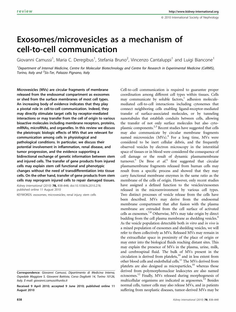
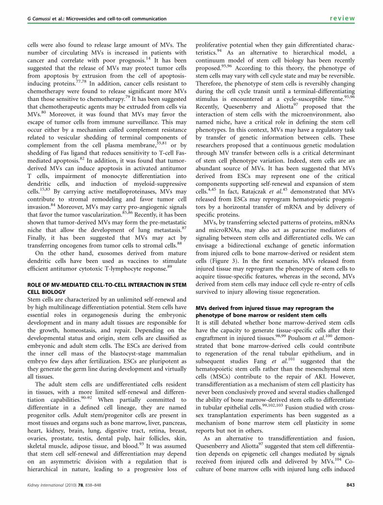
(i) Exosomes have an endosome origin and are a ratherhomogenous population with a size ranging from30 to 120 nm.7 They are stored as intraluminal vesicleswithin multivesicular bodies of the late endosome andare released when these multivesicular bodies fuse withthe cell membrane (Figure 1a). Our knowledge on themechanism of assembly and sorting of the exosomes isonly partial, because of the fact that a common sortingsignal for all cell types has not so far been identified.17
They are released by exocytosis through a mechanismdependent on cytoskeleton activation and under theregulation of p53 protein.18
(ii) Shedding vesicles are usually larger than exosomeswith size ranging from 100 nm to 1mm. Formation ofshedding vesicles takes place from the budding of smallcytoplasmic protrusions followed by their detachmentfrom the cell surface (Figure 1b). This process isdependent on calcium influx, calpain, and cytoskeletonreorganization.9 Schara et al.19 describe two physicalmechanisms involved in the formation of MVs andnanotubes: the curvature-mediated lateral redistributionof membrane components with the formation ofmembrane nanodomains and the plasma-mediatedattractive forces between membranes. The intracellularlevels of calcium ions modify the asymmetric phospho-lipid distribution of plasmamembranes by specificenzymes named flippase, floppase, and scramblase.20
The increase in calcium ions inhibits translocase andinduces activation of scramblase that translocatesphosphatydilserine from the inner leaflet of the cellmembrane bilayer to the outer. Therefore, MVs exposeon their surface large amounts of phosphatydilserine andare enriched in proteins associated with membrane lipidrafts.21 Moreover, the intracellular pathways that activatereorganization of cytoskeleton induce the detachment ofplasmamembrane protrusions from the cortical actin.Calcium ions by activation of calpain that cleaves tallinand activin and of gelsolin that cleaves actin-cappingproteins also favor the reorganization of cytoskeleton.22
Therefore, depending on the cell of origin and on themechanism of formation, MVs vary on size and mole-cular composition.
MV BIOLOGICAL ACTIVITIES
It is now recognized that MVs are an integral part of theintercellular microenvironment and may act as regulators of
cell-to-cell communication. This concept is based on the obser-vation that MVs released from a given cell type may interactthrough specific receptor ligands with other cells, leading to
Sheddingvesicles
Plasmamembrane
Membrane-sortingprocess
Exosomes
Degradativemultivesicular bodies
Exocyticmultivesicular bodies
Figure 1 | Schematic representation of exosome and sheddingvesicle formation. (a) Release of exosomes. Exosomes areaccumulated within the multivesicular bodies as a result ofendosome compartmentalization. The vesicles present inmultivesicular bodies may undergo degradation or exocytosis.The exocytic multivesicular bodies fuse with membrane after cellstimulation and release exosomes. (Upper inset) Representativetransmission electron microscopy showing exocytosis ofexosomes from the surface of a mesenchymal stem cell (originalmagnification � 15,000). (Lower inset) Representativetransmission electron microscopy showing a multivesicular bodywithin a mesenchymal stem cell (original magnification � 10,000).(b) Production of shedding vesicles from the cell surface.Shedding vesicles are sorted out from cytoplasm by buddingof cell plasmamembrane in response to cell stimulation. (Leftmicrograph) Transmission electron microscopy panel showingvesicles shed from the surface of an endothelial progenitor cell(original magnification � 10,000); the inset shows the highmagnification ultrastructure of a vesicle shed from an endothelialprogenitor (original magnification � 25,000). (Right micrograph)Transmission electron microscopy panel showing an aspect of cellmembrane budding in an endothelial progenitor cell duringmicrovesicle (MV) formation (original magnification � 15,000).The mechanisms involved in MV cargo as well as those involved inmembrane-sorting processes remain at present largely unknown.
Kidney International (2010) 78, 838–848 839
G Camussi et al.: Microvesicles and cell-to-cell communication r e v i e w
target cell stimulation directly or by transferring surfacereceptors.23,24 This implicates that MVs interact only with targetcells that specifically recognize rather than just with any cellpresent in the microenvironment.25 This interaction may eitherbe limited to a receptor-mediated binding to the surface of targetcells forming a platform for assembly of multimolecularcomplexes or leading to cell signaling, either to be followed byinternalization as a result of direct fusion or endocytic uptake bytarget cells.9 Once internalized, MVs can fuse their membraneswith those of endosomes, thus leading to a horizontal transfer oftheir content in the cytosol of target cells. Alternatively, they mayremain segregated within endosomes and be transferred tolysosomes or dismissed by the cells following the fusion with theplasmamembrane, thus leading to a process of transcytosis.9
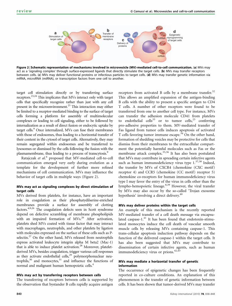
Ratajczak et al.4 proposed that MV-mediated cell-to-cellcommunication emerged very early during evolution as atemplate for the development of further more refinedmechanisms of cell communication. MVs may influence thebehavior of target cells in multiple ways (Figure 2).
MVs may act as signaling complexes by direct stimulation oftarget cells
MVs derived from platelets, for instance, have an importantrole in coagulation as their phosphatydilserine-enrichedmembranes provide a surface for assembly of clottingfactors.4,9,26 The coagulation defects seen in Scott syndromedepend on defective scrambling of membrane phospholipidswith an impaired formation of MVs.26 After activation,platelets shed MVs coated with tissue factor that may interactwith macrophages, neutrophils, and other platelets by ligationwith molecules expressed on the surface of these cells such as P-selectin.27 On the other hand, MVs released from neutrophilsexpress activated leukocyte integrin alpha M beta2 (Mac-1)that is able to induce platelet activation.28 Moreover, platelet-derived MVs, besides coagulation, trigger various cell responsesas they activate endothelial cells,29 polymorphonuclear neu-trophils,30 and monocytes,31 and influence the functions ofnormal and malignant human hemopoietic cells.4
MVs may act by transferring receptors between cells
The transferring of receptors between cells is supported bythe observation that bystander B cells rapidly acquire antigen
receptors from activated B cells by a membrane transfer.32
This allows an amplified expansion of the antigen-bindingB cells with the ability to present a specific antigen to CD4T cells. A number of other receptors were found to betransferred from one to another cell type. For instance, MVscan transfer the adhesion molecule CD41 from plateletsto endothelial cells33 or to tumor cells,23 conferringpro-adhesive properties to them. MV-mediated transfer ofFas ligand from tumor cells induces apoptosis of activatedT cells favoring tumor immune escape.34 On the other hand,formation of shedding vesicles may be protective for cells thatdismiss from their membranes to the extracellular compart-ment the potentially harmful molecules such as Fas or themembrane attack complex.35,36 It has also been postulatedthat MVs may contribute in spreading certain infective agentssuch as human immunodeficiency virus type 1.37,38 Indeed,the transfer by MVs of CXCR4 (chemokine (CXC motif)receptor 4) and CCR5 (chemokine (CC motif) receptor 5)chemokine co-receptors for human immunodeficiency virustype I may favor the entry of the virus in cells other than thelympho-hemopoietic lineage.8,39 However, the viral transferby MVs may also occur by the so-called ‘Trojan exosomehypothesis’ involving a direct delivery.40
MVs may deliver proteins within the target cells
An example of this mechanism is the recently reportedMV-mediated transfer of a cell death message via encapsu-lated caspase-1.41 It has been found that endotoxin-stimu-lated monocytes induce the cell death of vascular smoothmuscle cells by releasing MVs containing caspase-1. Thistrans-cellular apoptosis induction pathway depends on thefunction of the delivered caspase-1 within the target cells. Ithas also been suggested that MVs may contribute todissemination of certain infective agents, such as humanimmunodeficiency virus or prions.42,43
MVs may mediate a horizontal transfer of geneticinformation
The occurrence of epigenetic changes has been frequentlyreported in co-culture conditions. An explanation of thisphenomenon is the transfer of genetic information betweencells. It has been shown that tumor-derived MVs may transfer
Stimulation oftarget cells
Transfer ofmembranereceptors
Epigeneticreprogrammingof target cells
Delivery ofproteins
miRNA
mRNA
Figure 2 | Schematic representation of mechanisms involved in microvesicle (MV)-mediated cell-to-cell communication. (a) MVs mayact as a ‘signaling complex’ through surface-expressed ligands that directly stimulate the target cells. (b) MVs may transfer receptorsbetween cells. (c) MVs may deliver functional proteins or infectious particles to target cells. (d) MVs may transfer genetic information viamRNA, microRNA (miRNA), or transcription factors from one cell to another.
840 Kidney International (2010) 78, 838–848
r e v i e w G Camussi et al.: Microvesicles and cell-to-cell communication
not only surface determinants but also mRNA of tumorcells to monocytes.44 Ratajczak et al.45 demonstrated thatMVs derived from murine embryonic stem cells (ESCs)may induce an epigenetic reprogramming of target cells.ES-derived MVs were shown to improve survival ofhematopoietic stem/progenitor cells, to induce upregulationof early pluripotent and early hematopoietic markers, and toinduce phosphorylation of mitogen-activated protein kinasep42/44 and Akt. In addition, ES-derived MVs were shown toexpress mRNAs for several pluripotent transcription factorsthat can be delivered to target cells and translated to thecorresponding proteins. As RNase inhibited MV-mediatedbiological effect, the involvement of mRNA in the observedbiological effects was suggested.45 We demonstrated that MVsderived from human endothelial progenitor cells can alsoact as a vehicle for mRNA transport among cells.46 MVsgenerated from endothelial progenitor cells were incorpo-rated in normal endothelial cells by interaction with a4 andb1 integrins expressed on their surface and activated anangiogenic program.46 This effect was also proved in vivoin severe combined immunodeficient mice, whereMV-stimulated human endothelial cells subcutaneouslyimplanted within Matrigel organized in a patent vesselnetwork connected with the murine vasculature. RNasepretreatment of MVs abrogated their angiogenic activity eventhough they were internalized by endothelial cells, suggestinga critical role for RNA transfer following MV incorporation.The molecular analysis of mRNA indicated that MVs derivedfrom endothelial progenitor cells were shuttling a specificsubset of cellular mRNA, including mRNA associated withpathways relevant for angiogenesis such as the PI3K/AKTand endothelial nitric oxide synthase signaling pathways.Protein expression and functional studies demonstrated thatphosphatidylinositol 3-kinase and endothelial nitric oxidesynthase were upregulated in target cells after MV incorpora-tion. As a proof of transduction in target cells of mRNAdelivered from MVs, we used the green fluorescent protein(GFP) mRNA as reporter. Endothelial cells targeted withMVs carrying GFP mRNA produced the GFP proteins.46
More recently, we demonstrated that MVs derived fromhuman stem cells may also deliver in vivo human mRNA tomouse cells, resulting in protein translation.47,48 Yuan et al.49
have recently shown that besides mRNA, MVs may transferin target cells microRNA. They demonstrated that MVsderived from ESCs contain abundant microRNA and thatthey can transfer a subset of microRNAs to mouse embryonicfibroblasts in vitro. As microRNAs are naturally occurringregulators of protein translation, this observation opens thepossibility that stem cells can alter the expression of genesin neighboring cells by transferring microRNAs containedin MVs.
ROLE OF MVs IN INFLAMMATION AND IN CARDIOVASCULARAND RENAL DISEASES
Inflammation is sustained by multiple interactions amongcells. In this context, MVs may act at different stages of the
process by carrying either anti-inflammatory or pro-inflammatory factors.50 MVs derived from platelets andmacrophages were found to be accumulated in the lipid coreof the atherosclerotic plaques with the potential of triggeringpro-inflammatory, angiogenic, and thrombotic signals.51
These observations rise the possibility that targetingMVs may be a therapeutic strategy in atherosclerosis.9,50
Indeed, increased levels of MVs of mainly endothelialorigin were observed in cardiovascular pathology.52 Endo-thelial dysfunction is an initial event in the develop-ment of atherosclerosis and correlate with an unfavorablecardiovascular prognosis.53 Injured endothelial cells mayrelease MVs, which are considered as markers of endo-thelial dysfunction.54 Moreover, MVs have been implicatedin the modulation of inflammation, as at early stagesneutrophil-derived MVs may stimulate the production ofanti-inflammatory cytokines55,56 and at later stagesMVs released from fibroblasts may induce the productionof pro-inflammatory cytokines such as interleukin-6 andthe monocyte chemotactic protein 1 and metallo-proteinases.57
In experimental membranous glomerulonephritis, wefound that the vesicular shedding of terminal componentsof complement from the cell plasma membrane protectpodocytes from lyses.35 This, by reducing their surface andactivating the cytoskeleton, may favor retraction of footprocesses and disruption of the slit pore thus favoringproteinuria.35
Although in healthy subjects, circulating MVs are mainlyderived from platelets, in pathological conditions MVs mayderive from other cell types such as endothelial andinflammatory cells and erythrocytes. Augmented blood levelsof MVs have been found in various diseases such as pre-eclampsia,58 diabetes,59 acute coronary syndrome,60 severehypertension,61 multiple sclerosis,62 vasculitis,63 as well as inpatients with chronic renal failure.64,65
Circulating levels of MVs derived from endothelialcells correlate with arterial stiffness in hemodialysedpatients.64,66,67
The recent discovery of exosomes/MVs in normal urineopens the possibility of obtaining information on the cellof origin in physiological and pathological conditions.It is conceivable that the analysis of urinary MVs mayprovide protein biomarkers for the involvement of differentcellular components of the nephron.68 Indeed, it has beenrecently shown that fetuin-A present in urine exosomes is anovel biomarker of structural renal injury in experimentalmodels of cisplatin-induced nephrotoxicity and in intensivecare unit patients developing acute kidney injury (AKI).69
Moreover, a reduction in urinary exosomal levels ofaquaporin-1 has been associated with renal ischemia–reper-fusion injury in rats.70 Zhou et al.71 described the presenceof transcription factors in urinary exosomes in differentexperimental models of AKI (cisplatin and ischemia–reperfusion) and of podocyte injury (puromycin-treatedrats or podocin-V transgenic mice). In particular, the
Kidney International (2010) 78, 838–848 841
G Camussi et al.: Microvesicles and cell-to-cell communication r e v i e w
transcription factor activating transcription factor 3 wasassociated with AKI and the Wilms tumor 1 with an earlypodocyte injury.71
In the setting of transplantation, it has been shown thatthe exchange of exosomes between dendritic cells inlymphoid organs may constitute a potential mechanism bywhich passenger leukocytes transfer alloantigens to recipientantigen-presenting cells, leading to an increased generationof donor-reactive T cells.72 However, other studies showedthat dendritic cell-derived exosomes may induce tolerancerather than immune stimulation. In particular, exosomesisolated from bone marrow-derived dendritic cells adminis-tered before transplantation can modulate heart allo-graft rejection, prolonging survival.73 Moreover, dendritic
cell-derived exosomes administered after heart transplanta-tion in combination with short-term immunosuppressioncan induce regulatory responses that are able to modulateallograft rejection and to induce donor-specific allografttolerance.74
On the other hand, MVs derived from cytomegalovirus-infected endothelial cells can stimulate allogenicCD4þ memory T cells, providing a new potential mecha-nism by which cytomegalovirus can exacerbate allograftrejection.75
ROLE OF MVs IN TUMOR BIOLOGY
MVs derived from activated platelets were found to be able toinduce metastasis and angiogenesis in lung cancer.76 Tumor
miRNAmRNA
BM-derived ortissue-resident
stem cells
Tissue-residentstem cells
Tissue injurya
b
Tissuerepair
MV
EGF HGF MSP IGF-1 VEGF
Differentiation
miRNAmRNA
BM-derivedstem cells
MV
Re-differentiationDe-differentiationProliferation
Figure 3 | Schematic representation of bidirectional exchange of genetic information between stem cells and tissue-injuredcells mediated by microvesicles (MVs). (a) MVs released from tissue-injured cells may reprogram the phenotype of stem cells to acquiretissue-specific features by delivering to stem cells the mRNAs and/or microRNAs (miRNAs) of tissue cells. (b) MVs produced by stemcells recruited from the circulation or from resident stem cells may reprogram tissue-injured cells by delivering mRNA and/or miRNA that inducethe de-differentiation, the production of soluble paracrine mediators, and the cell cycle re-entry of these cells favoring tissue regeneration. BM,bone marrow; EGF, epiotermal growth factor; HGF, hepatocyte growth factor; IGF-1, insulin-like growth factor-1; MSP, macrophage stimulatingprotein; VEGF, vascular endothelial growth factor.
842 Kidney International (2010) 78, 838–848
r e v i e w G Camussi et al.: Microvesicles and cell-to-cell communication
cells were also found to release large amount of MVs. Thenumber of circulating MVs is increased in patients withcancer and correlate with poor prognosis.14 It has beensuggested that the release of MVs may protect tumor cellsfrom apoptosis by extrusion from the cell of apoptosis-inducing proteins.77,78 In addition, cancer cells resistant tochemotherapy were found to release significant more MVsthan those sensitive to chemotherapy.79 It has been suggestedthat chemotherapeutic agents may be extruded from cells viaMVs.80 Moreover, it was found that MVs may favor theescape of tumor cells from immune surveillance. This mayoccur either by a mechanism called complement resistancerelated to vesicular shedding of terminal components ofcomplement from the cell plasma membrane,35,81 or byshedding of Fas ligand that reduces sensitivity to T-cell Fas-mediated apoptosis.82 In addition, it was found that tumor-derived MVs can induce apoptosis in activated antitumorT cells, impairment of monocyte differentiation intodendritic cells, and induction of myeloid-suppressivecells.15,83 By carrying active metalloproteinases, MVs maycontribute to stromal remodeling and favor tumor cellinvasion.84 Moreover, MVs may carry pro-angiogenic signalsthat favor the tumor vascularization.85,86 Recently, it has beenshown that tumor-derived MVs may form the pre-metastaticniche that allow the development of lung metastasis.87
Finally, it has been suggested that MVs may act bytransferring oncogenes from tumor cells to stromal cells.88
On the other hand, exosomes derived from maturedendritic cells have been used as vaccines to stimulateefficient antitumor cytotoxic T-lymphocyte response.89
ROLE OF MV-MEDIATED CELL-TO-CELL INTERACTION IN STEMCELL BIOLOGY
Stem cells are characterized by an unlimited self-renewal andby high multilineage differentiation potential. Stem cells haveessential roles in organogenesis during the embryonicdevelopment and in many adult tissues are responsible forthe growth, homeostasis, and repair. Depending on thedevelopmental status and origin, stem cells are classified asembryonic and adult stem cells. The ESCs are derived fromthe inner cell mass of the blastocyst-stage mammalianembryo few days after fertilization. ESCs are pluripotent asthey generate the germ line during development and virtuallyall tissues.
The adult stem cells are undifferentiated cells residentin tissues, with a more limited self-renewal and differen-tiation capabilities.90–92 When partially committed todifferentiate in a defined cell lineage, they are namedprogenitor cells. Adult stem/progenitor cells are present inmost tissues and organs such as bone marrow, liver, pancreas,heart, kidney, brain, lung, digestive tract, retina, breast,ovaries, prostate, testis, dental pulp, hair follicles, skin,skeletal muscle, adipose tissue, and blood.93 It was assumedthat stem cell self-renewal and differentiation may dependon an asymmetric division with a regulation that ishierarchical in nature, leading to a progressive loss of
proliferative potential when they gain differentiated charac-teristics.94 As an alternative to hierarchical model, acontinuum model of stem cell biology has been recentlyproposed.95,96 According to this theory, the phenotype ofstem cells may vary with cell cycle state and may be reversible.Therefore, the phenotype of stem cells is reversibly changingduring the cell cycle transit until a terminal-differentiatingstimulus is encountered at a cycle-susceptible time.95,96
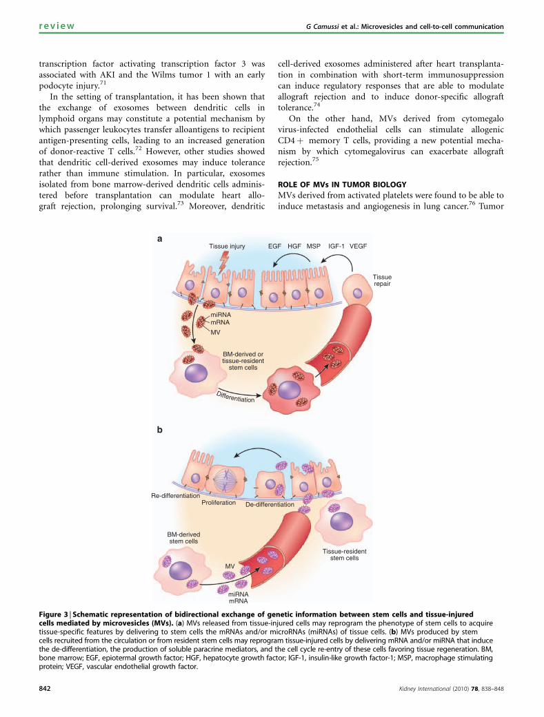
Recently, Quesenberry and Aliotta97 proposed that theinteraction of stem cells with the microenvironment, alsonamed niche, have a critical role in defining the stem cellphenotypes. In this context, MVs may have a regulatory taskby transfer of genetic information between cells. Theseresearchers proposed that a continuous genetic modulationthrough MV transfer between cells is a critical determinantof stem cell phenotype variation. Indeed, stem cells are anabundant source of MVs. It has been suggested that MVsderived from ESCs may represent one of the criticalcomponents supporting self-renewal and expansion of stemcells.4,45 In fact, Ratajczak et al.45 demonstrated that MVsreleased from ESCs may reprogram hematopoietic progeni-tors by a horizontal transfer of mRNA and by delivery ofspecific proteins.
MVs, by transferring selected patterns of proteins, mRNAsand microRNAs, may also act as paracrine mediators ofsignaling between stem cells and differentiated cells. We canenvisage a bidirectional exchange of genetic informationfrom injured cells to bone marrow-derived or resident stemcells (Figure 3). In the first scenario, MVs released frominjured tissue may reprogram the phenotype of stem cells toacquire tissue-specific features, whereas in the second, MVsderived from stem cells may induce cell cycle re-entry of cellssurvived to injury allowing tissue regeneration.
MVs derived from injured tissue may reprogram thephenotype of bone marrow or resident stem cells
It is still debated whether bone marrow-derived stem cellshave the capacity to generate tissue-specific cells after theirengraftment in injured tissues.98,99 Poulsom et al.100 demon-strated that bone marrow-derived cells could contributeto regeneration of the renal tubular epithelium, and insubsequent studies Fang et al.101 suggested that thehematopoietic stem cells rather than the mesenchymal stemcells (MSCs) contribute to the repair of AKI. However,transdifferentiation as a mechanism of stem cell plasticity hasnever been conclusively proved and several studies challengedthe ability of bone marrow-derived stem cells to differentiatein tubular epithelial cells.99,102,103 Fusion studied with cross-sex transplantation experiments has been suggested as amechanism of bone marrow stem cell plasticity in somereports but not in others.
As an alternative to transdifferentiation and fusion,Quesenberry and Aliotta97 suggested that stem cell differentia-tion depends on epigenetic cell changes mediated by signalsreceived from injured cells and delivered by MVs.104 Co-culture of bone marrow cells with injured lung cells induced
Kidney International (2010) 78, 838–848 843
G Camussi et al.: Microvesicles and cell-to-cell communication r e v i e w
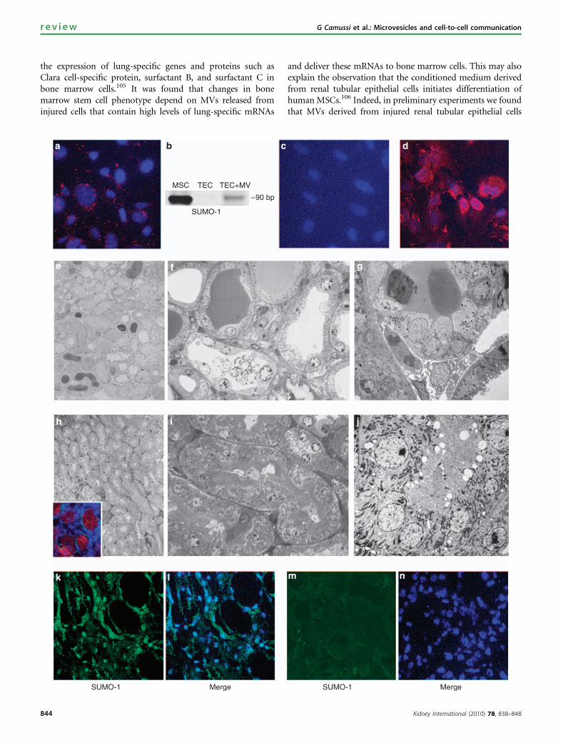
the expression of lung-specific genes and proteins such asClara cell-specific protein, surfactant B, and surfactant C inbone marrow cells.105 It was found that changes in bonemarrow stem cell phenotype depend on MVs released frominjured cells that contain high levels of lung-specific mRNAs
and deliver these mRNAs to bone marrow cells. This may alsoexplain the observation that the conditioned medium derivedfrom renal tubular epithelial cells initiates differentiation ofhuman MSCs.106 Indeed, in preliminary experiments we foundthat MVs derived from injured renal tubular epithelial cells
MSC TEC TEC+MV
SUMO-1
–90 bp
SUMO-1 Merge SUMO-1 Merge
844 Kidney International (2010) 78, 838–848
r e v i e w G Camussi et al.: Microvesicles and cell-to-cell communication
may induce expression of tubular cell markers in humanMSCs. One can speculate that MVs released from injuredtissue may reprogram not only bone marrow-derived stemcells, but also resident stem cells. Several studies indicate thepresence of resident stem cell populations within the kidneythat may contribute to renal repair.47,107,108
Taken together, these results suggest that MVs derivedfrom injured tissues mediated transfer of genetic informationthat could explain not only the plasticity and phenotypicchanges of stem cells, but also the functional effects withoutthe need of their transdifferentiation into tissue cells.
MVs derived from stem cells may reprogram cells survived toinjury and favor tissue regeneration
Experiments based on exogenous MSC administration inAKI demonstrate a functional and morphological recoveryfrom acute tubular injury induced by toxic and ischemia–reperfusion injury99,109,110 and a functional improvement inchronic renal failure.111 As these beneficial effects areassociated only with a transient recruitment of MSCwithin the renal vasculature with a minimal incorporationwithin the regenerating tubules,102,103 it has been suggestedthat MSC may provide a paracrine support to the repairof injured tissue.112 On the other hand, many studies ontubular repopulation after acute injury indicate a prominentcontribution of renal tubular cells.112,113 Strong support of aparacrine/endocrine mechanism for tissue repair comes fromexperiments of Bi et al.,114 showing that the administrationof conditioned medium from MSC is able to mimic thebeneficial effects of the stem cell therapy. They demonstratedthat MSC may favor renal regeneration independently fromengraftment within tubules by producing factors thatlimit apoptosis and enhance proliferation of tubular cells.A growing body of evidence supports the hypothesis ofa paracrine mechanism in bone marrow-derived stem celltherapy in other organs also, such as infarcted hearts.115
Indeed, the frequency of stem cell engraftment and
transdifferentiation or fusion to generate new cardiomyocytesand vascular cells appear too low to explain the beneficialeffects observed. Conversely, several studies indicate thatstem cell-released soluble factors may contribute to cardiacrepair and regeneration.116
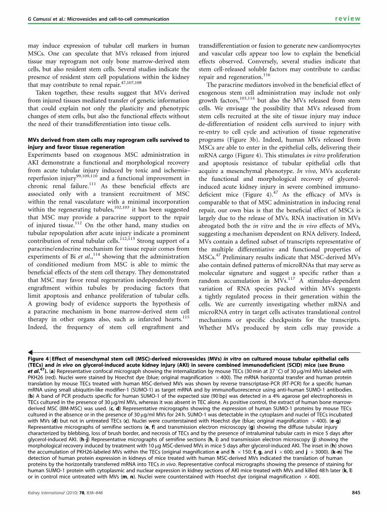
The paracrine mediators involved in the beneficial effect ofexogenous stem cell administration may include not onlygrowth factors,103,114 but also the MVs released from stemcells. We envisage the possibility that MVs released fromstem cells recruited at the site of tissue injury may inducede-differentiation of resident cells survived to injury withre-entry to cell cycle and activation of tissue regenerativeprograms (Figure 3b). Indeed, human MVs released fromMSCs are able to enter in the epithelial cells, delivering theirmRNA cargo (Figure 4). This stimulates in vitro proliferationand apoptosis resistance of tubular epithelial cells thatacquire a mesenchymal phenotype. In vivo, MVs acceleratethe functional and morphological recovery of glycerol-induced acute kidney injury in severe combined immuno-deficient mice (Figure 4).47 As the efficacy of MVs iscomparable to that of MSC administration in inducing renalrepair, our own bias is that the beneficial effect of MSCs islargely due to the release of MVs. RNA inactivation in MVsabrogated both the in vitro and the in vivo effects of MVs,suggesting a mechanism dependent on RNA delivery. Indeed,MVs contain a defined subset of transcripts representative ofthe multiple differentiative and functional properties ofMSCs.47 Preliminary results indicate that MSC-derived MVsalso contain defined patterns of microRNAs that may serve asmolecular signature and suggest a specific rather than arandom accumulation in MVs.117 A stimulus-dependentvariation of RNA species packed within MVs suggestsa tightly regulated process in their generation within thecells. We are currently investigating whether mRNA andmicroRNA entry in target cells activates translational controlmechanisms or specific checkpoints for the transcripts.Whether MVs produced by stem cells may provide a
Figure 4 | Effect of mesenchymal stem cell (MSC)-derived microvesicles (MVs) in vitro on cultured mouse tubular epithelial cells(TECs) and in vivo on glycerol-induced acute kidney injury (AKI) in severe combined immunodeficient (SCID) mice (see Brunoet al.47). (a) Representative confocal micrograph showing the internalization by mouse TECs (30 min at 37 1C) of 30 mg/ml MVs labeled withPKH26 (red). Nuclei were stained by Hoechst dye (blue; original magnification � 400). The mRNA horizontal transfer and human proteintranslation by mouse TECs treated with human MSC-derived MVs was shown by reverse transcriptase-PCR (RT-PCR) for a specific humanmRNA using small ubiquitin-like modifier-1 (SUMO-1) as target mRNA and by immunofluorescence using anti-human SUMO-1 antibodies.(b) A band of PCR products specific for human SUMO-1 of the expected size (90 bp) was detected in a 4% agarose gel electrophoresis inTECs cultured in the presence of 30 mg/ml MVs, whereas it was absent in TEC alone. As positive control, the extract of human bone marrow-derived MSC (BM-MSC) was used. (c, d) Representative micrographs showing the expression of human SUMO-1 proteins by mouse TECscultured in the absence or in the presence of 30mg/ml MVs for 24 h. SUMO-1 was detectable in the cytoplasm and nuclei of TECs incubatedwith MVs (d) but not in untreated TECs (c). Nuclei were counterstained with Hoechst dye (blue; original magnification � 400). (e–g)Representative micrographs of semifine sections (e, f) and transmission electron microscopy (g) showing the diffuse tubular injurycharacterized by blebbing, loss of brush border, and necrosis of TECs and by the presence of intraluminal tubular casts in mice 5 days afterglycerol-induced AKI. (h–j) Representative micrographs of semifine sections (h, i) and transmission electron microscopy (j) showing themorphological recovery induced by treatment with 10 mg MSC-derived MVs in mice 5 days after glycerol-induced AKI. The inset in (h) showsthe accumulation of PKH26-labeled MVs within the TECs (original magnification e and h � 150; f, g, and i � 600; and j � 3000). (k–n) Thedetection of human protein expression in kidneys of mice treated with human MSC-derived MVs indicated the translation of humanproteins by the horizontally transferred mRNA into TECs in vivo. Representative confocal micrographs showing the presence of staining forhuman SUMO-1 protein with cytoplasmic and nuclear expression in kidney sections of AKI mice treated with MVs and killed 48 h later (k, l)or in control mice untreated with MVs (m, n). Nuclei were counterstained with Hoechst dye (original magnification � 400).
Kidney International (2010) 78, 838–848 845
G Camussi et al.: Microvesicles and cell-to-cell communication r e v i e w
potential therapeutic strategy to avoid the possible mal-differentiation of stem cells once engrafted in the kidney inthe long term118 requires further investigations. We recentlyshowed that MV-mediated transfer of RNA-based informa-tion from human liver stem cells stimulates liver regenerationin a model of 75% hepatectomy.48
CONCLUSION
The main function of MVs is signaling through specificinteractions with target cells and transferring gene products.Therefore, they may participate in physiological andpathological processes. Gaining further insights into themolecular specificity of MVs may allow the identification ofthe cellular source and may provide new diagnostic tools.Indeed, an increasing body of evidence indicates thatMVs may offer prognostic information in various diseasessuch as chronic inflammation, cardiovascular and renaldiseases, pathological pregnancy, and tumors. The presenceof MVs in body fluid makes them readily accessible, and theirnumber, cellular origin, composition, and function can bedisease state dependent. Cancer cells, for example, shedMVs that might not only help tumor and metastasisdevelopment but also represent an important non-invadingdiagnostic tool especially with regard to the fact that theycontain genetic material under the form of RNA, whichcould be easily screened for cancer genetic markers.In addition, the recognition of the signals delivered byMVs may open new therapeutic strategies. The removalfrom plasma of harmful MVs may be beneficial inpathological conditions where MVs deliver thrombogenicand inflammatory signals or in tumors. On the otherhand, MVs derived from stem cells may reprogram alteredfunctions in target cells, suggesting that they could beexploited in regenerative medicine to repair damaged tissues.Moreover, MV-mediated transfer of genetic informationcould explain the observed plasticity and the functionaleffects of stem cells without the need of their transdiffer-entiation into tissue cells. Many points require furtherinvestigation: (1) the stimuli and the molecular pathwaysthat regulate the assembly within MVs of the biologicallyactive molecules that they shuttle; (2) the stimuli that triggertheir release; (3) the surface receptors that may conferselective specificity; (4) the full diagnostic potential of MVsin different pathological conditions; (5) the strategy toinhibit formation or to remove from circulation potentiallyharmful MVs; and (6) the therapeutic exploitation inregenerative medicine of the ability of MVs to modify thephenotype and function of target cells. The recognition ofthe importance of MVs may open new perspectives ofinvestigation.
DISCLOSUREAll the authors declared no competing interests.
ACKNOWLEDGMENTSOur research was supported by grants from Regione Piemonte,Piattaforme Biotecnologiche, progetto PiSTEM, and from Oncoprot.
REFERENCES1. Majka M, Janowska-Wieczorek A, Ratajczak J et al. Numerous growth
factors, cytokines, and chemokines are secreted by human CD34(+) cells,myeloblasts, erythroblasts, and megakaryoblasts and regulate normalhematopoiesis in an autocrine/paracrine manner. Blood 2001; 97:3075–3085.
2. Rustom A, Saffrich R, Markovic I et al. Nanotubular highways forintercellular organelle transport. Science 2004; 303: 1007–1010.
3. Sherer NM, Mothes W. Cytonemes and tunnelling nanotubules incell-cell communication and viral pathogenesis. Trends Cell Biol 2008; 18:414–420.
4. Ratajczak J, Wysoczynski M, Hayek F et al. Membrane-derivedmicrovesicles: important and underappreciated mediators of cell-to-cellcommunication. Leukemia 2006; 20: 1487–1495.
5. Siekevitz P. Biological membranes: the dynamics of their organization.Annu Rev Physiol 1972; 34: 117–140.
6. De Broe ME, Wieme RJ, Logghe GN et al. Spontaneous shedding ofplasma membrane fragments by human cells in vivo and in vitro.Clin Chim Acta 1977; 81: 237–245.
7. Heijnen HF, Schiel AE, Fijnheer R et al. Activated platelets release twotypes of membrane vesicles: microvesicles by surface shedding andexosomes derived from exocytosis of multivesicular bodies and alpha-granules. Blood 1999; 94: 3791–3799.
8. Rozmyslowicz T, Majka M, Kijowski J et al. Platelet- and megakaryocyte-derived microparticles transfer CXCR4 receptor to CXCR4-null cells andmake them susceptible to infection by X4-HIV. AIDS 2003; 17: 33–42.
9. Cocucci E, Racchetti G, Meldolesi J. Shedding microvesicles: artefacts nomore. Trends Cell Biol 2008; 19: 43–51.
10. George JN, Thoi LL, McManus LM et al. Isolation of human plateletmembrane microparticles from plasma and serum. Blood 1982; 60:834–840.
11. Martinez MC, Tesse A, Zobairi F et al. Shed membrane microparticlesfrom circulating and vascular cells in regulating vascular function.Am J Physiol Hearth Circ Physiol 2005; 288: H1004–H1009.
12. Hess C, Sadallah S, Hefti A et al. Ectosomes released by humanneutrophils are specialized functional units. J Immunol 1999; 163:4564–4573.
13. Greco V, Hannus M, Eaton S. Argosomes: a potential vehicle for thespread of morphogens through epithelia. Cell 2001; 106: 633–645.
14. Kim HK, Song KS, Park YS et al. Elevated levels of circulating plateletmicroparticles, VEGF, IL-6 and RANTES in patients with gastric cancer:possible role of a metastasis predictor. Eur J Cancer 2003; 39: 184–191.
15. Iero M, Valenti R, Huber V et al. Tumour-released exosomes and theirimplications in cancer immunity. Cell Death Differ 2008; 15: 80–88.
16. Diamant M, Tushuizen ME, Sturk A et al. Cellular microparticles: newplayers in the field of vascular disease? Eur J Clin Invest 2004; 34:392–401.
17. Johnstone RM. Exosomes biological significance: a concise review.Blood Cells Mol Dis 2006; 36: 315–321.
18. Yu X, Harris SL, Levine AJ. The regulation of exosome secretion: a novelfunction of the p53 protein. Cancer Res 2006; 66: 4795–4801.
19. Schara K, Jansa V, Sustar V et al. Mechanisms for the formation ofmembranous nanostructures in cell-to-cell communication. Cell Mol BiolLett 2009; 14: 636–656.
20. Hugel B, Martinez MC, Kunzelmann C et al. Membrane microparticles:two sides of the coin. Physiology (Bethesda) 2005; 20: 22–27.
21. Del Conde I, Shrimpton CN, Thiagarajan P et al. Tissue-factor-bearingmicrovesicles arise from lipids rafts and fuse with activated platelets toinitiate coagulation. Blood 2005; 106: 1604–1611.
22. Pap E, Pallinger E, Pasztoi M et al. Highlights of a new type ofintercellular communication: microvesicle-based information transfer.Inflamm Res 2009; 58: 1–8.
23. Janowska-Wieczorek A, Majka M, Kijowski J et al. Platelet-derivedmicroparticles bind to hematopoietic progenitor cells and enhance theirengraftment. Blood 2001; 98: 3143–3149.
24. Morel O, Toti F, Hugel B et al. Cellular microparticles: a disseminatedstorage pool of bioactive vascular effectors. Curr Opin Hematol 2004; 11:156–164.
25. Losche W, Scholz T, Temmler U et al. Platelet-derived microvesiclestransfer tissue factor to monocytes but not to neutrophils. Platelets2004; 15: 109–115.
26. Zwaal RF, Comfurius P, Bevers EM et al. Scott syndrome, a bleedingdisorder caused by defective scrambling of membrane phospholipids.Biochim Biophys Acta 2004; 1636: 119–128.
27. Polgar J, Matuskova J, Wagner DD. The P-selectin, tissue factor,coagulation triad. J Thromb Haemost 2005; 3: 1590–1596.
846 Kidney International (2010) 78, 838–848
r e v i e w G Camussi et al.: Microvesicles and cell-to-cell communication
28. Andrews RK, Berndt MC. Platelet physiology and thrombosis. ThrombRes 2004; 114: 447–453.
29. Barry OP, Pratico D, Lawson JA et al. Transcellular activation of plateletsand endothelial cells by bioactive lipids in platelet microparticles.J Clin Invest 1997; 99: 2118–2127.
30. Miyamoto S, Kowalska MA, Marcinkiewicz C et al. Interaction ofleukocytes with platelet microparticles derived from outdated plateletconcentrates. Thromb Haemost 1998; 80: 982–988.
31. Barry OP, Kazanietz MG, Pratico D et al. Arachidonic acid in plateletmicroparticles up-regulates cyclooxygenase-2-dependent prostaglandinformation via a protein kinase C/mitogen-activated protein kinase-dependent pathway. J Biol Chem 1999; 274: 7545–7556.
32. Quah BJ, Barlow VP, McPhun V et al. Bystander B cells rapidlyacquire antigen receptors from activated B cells by membranetransfer. Proc Natl Acad Sci USA 2008; 105: 4259–4264.
33. Barry OP, Pratico D, Savani RC et al. Modulation of monocyte-endothelialcell interactions by platelet microparticles. J Clin Invest 1998; 102:136–144.
34. Kim JW, Wieckowski E, Taylor DD et al. Fas ligand-positive membranousvesicles isolated from sera of patients with oral cancer induce apoptosisof activated T lymphocytes. Clin Cancer Res 2005; 11: 1010–1020.
35. Camussi G, Salvidio G, Biesecker G et al. Heymann antibodies inducecomplement-dependent injury of rat glomerular visceral epithelial cells.J Immunol 1987; 139: 2906–2914.
36. Pilzer D, Fishelson Z. Mortalin/GRP75 promotes release of membranevesicles from immune attacked cells and protection from complement-mediated lysis. Int Immunol 2005; 17: 1239–1248.
37. Fackler OT, Peterlin BM. Endocytic entry of HIV-1. Curr Biol 2000; 10:1005–1008.
38. Fevrier B, Raposo G. Exosomes: endosomal-derived vesicles shippingextracellular messages. Curr Opin Cell Biol 2004; 16: 415–421.
39. Mack M, Kleinschmidt A, Bruhl H et al. Transfer of the chemokinereceptor CCR5 between cells by membrane-derived microparticles:a mechanism for cellular human immunodeficiency virus 1 infection.Nat Med 2000; 6: 769–775.
40. Gould SJ, Booth AM, Hildreth JE. The Trojan exosome hypothesis.Proc Natl Acad Sci USA 2003; 100: 10592–10597.
41. Sarkar A, Mitra S, Mehta S et al. Monocyte derived microvesicles deliver acell death message via encapsulated caspase-1. PLoS One 2009; 4:e7140.
42. Facler OT, Peterlin BM. Endocytic entry of HIV-1. Curr Biol 2000; 10:1005–1008.
43. Fevrier B, Vilette D, Archer F et al. Cells release prions in association withexosomes. Proc Natl Acad Sci USA 2004; 101: 9683–9688.
44. Baj-Krzyworzeka M, Szatanek R, Weglarczyk K et al. Tumour-derivedmicrovesicles carry several surface determinants and mRNA of tumourcells and transfer some of these determinants to monocytes. CancerImmunol Immunother 2006; 55: 808–818.
45. Ratajczak J, Miekus K, Kucia M et al. Embryonic stem cell-derivedmicrovesicles reprogram hematopoietic progenitors: evidence forhorizontal transfer of mRNA and protein delivery. Leukemia 2006; 20:847–856.
46. Deregibus MC, Cantaluppi V, Calogero R et al. Endothelial progenitorcell derived microvesicles activate an angiogenic program inendothelial cells by a horizontal transfer of mRNA. Blood 2007; 110:2440–2448.
47. Bruno S, Grange C, Deregibus MC et al. Mesenchymal stem cell-derivedmicrovesicles protect against acute tubular injury. J Am Soc Nephrol2009; 20: 1053–1067.
48. Herrera MB, Fonsato V, Gatti S et al. Human liver stem cell-derivedmicrovesicles accelerate hepatic regeneration in hepatectomized rats.J Cell Mol Med 2010; 14: 1605–1618.
49. Yuan A, Farber EL, Rapoport AL et al. Transfer of microRNAs byembryonic stem cell microvesicles. PLoS One 2009; 4: e4722.
50. Ardoin SP, Shanahan JC, Pisetsky DS. The role of microparticles ininflammation and thrombosis. Scand J Immunol 2007; 66: 159–165.
51. Leroyer AS, Tedgui A, Boulanger CM. Role of microparticles inatherothrombosis. J Intern Med 2008; 263: 528–537.
52. Simak J, Gelderman MP. Cell membrane microparticles in blood andblood products: potentially pathogenic agents and diagnostic markers.Transfus Med Rev 2006; 20: 1–26.
53. Bonetti PO, Lerman LO, Lerman A. Endothelial dysfunction: a marker ofatherosclerotic risk. Arterioscler Thromb Vasc Biol 2003; 23: 168–175.
54. Horstman LL, Jy W, Jimenez JJ et al. Endothelial microparticles asmarkers of endothelial dysfunction. Front Biosci 2004; 9: 1118–1135.
55. Koppler B, Cohen C, Schlondorff D et al. Differential mechanisms of
microparticle transfer to B cells and monocytes: anti-inflammatoryproperties of microparticles. Eur J Immunol 2006; 36: 648–660.
56. Gasser O, Schifferli JA. Activated polymorphonuclear neutrophils
disseminate anti-inflammatory microparticles by ectocytosis.Blood 2004; 104: 2543–2548.
57. Distler JH, Pisetsky DS, Huber LC et al. Microparticles as regulators
of inflammation: novel players of cellular crosstalk in the rheumaticdiseases. Arthritis Rheum 2005; 52: 3337–3348.
58. Gonzalez-Quintero VH, Jimenez JJ, Jy W et al. Elevated plasmaendothelial microparticles in preeclampsia. Am J Obstet Gynecol 2003;
189: 589–593.59. Sabatier F, Darmon P, Hugel B et al. Type 1 and type 2 diabetic patients
display different patterns of cellular microparticles. Diabetes 2002; 51:
2840–2845.60. Bernal-Mizrachi L, Jy W, Jimenez JJ et al. High levels of circulating
endothelial microparticles in patients with acute coronary syndromes.
Am Heart J 2003; 145: 962–970.61. Preston RA, Jy W, Jimenez JJ et al. Effects of severe hypertension on
endothelial and platelet microparticles. Hypertension 2003; 41: 211–217.62. Minagar A, Jy W, Jimenez JJ et al. Elevated plasma endothelial
microparticles in multiple sclerosis. Neurology 2001; 56: 1319–1324.63. Combes V, Simon AC, Grau GE et al. In vitro generation of endothelial
microparticles and possible prothrombotic activity in patients withlupus anticoagulant. J Clin Invest 1999; 104: 93–102.
64. Amabile N, Guerin AP, Leroyer A et al. Circulating endothelialmicroparticles are associated with vascular dysfunction in patients
with end-stage renal failure. J Am Soc Nephrol 2005; 16: 3381–3388.65. Faure V, Dou L, Sabatier F et al. Elevation of circulating endothelial
microparticles in patients with chronic renal failure. J Thromb Haemost
2006; 4: 566–573.66. Boulanger CM, Amabile N, Guerin AP et al. In vivo shear stress
determines circulating levels of endothelial microparticles in end-stage
renal disease. Hypertension 2007; 49: 902–908.67. Dursun I, Poyrazoglu HM, Gunduz Z et al. The relationship between
circulating endothelial microparticles and arterial stiffness and
atherosclerosis in children with chronic kidney disease. Nephrol DialTransplant 2009; 24: 2511–2518.
68. Pisitkun T, Shen RF, Knepper MA. Identification and proteomic profilingof exosomes in human urine. Proc Natl Acad Sci USA 2004; 101:
13368–13373.69. Zhou H, Pisitkun T, Aponte A et al. Exosomal fetuin-A identified by
proteomics: a novel urinary biomarker for detecting acute kidney injury.
Kidney Int 2006; 70: 1847–1857.70. Sonoda H, Yokota-Ikeda N, Oshikawa S et al. Decreased abundance
of urinary exosomal aquaporin-1 in renal ischemia-reperfusion injury.
AM J Physiol Renal Physiol 2009; 297: F1006–F1016.71. Zhou H, Cheruvanky A, Hu X et al. Urinary exosomal transcription
factors, a new class of biomarkers for renal disease. Kidney Int 2008; 74:
613–621.72. Montecalvo A, Shufesky WJ, Stolz DB et al. Exosomes as a short-range
mechanism to spread alloantigen between dendritic cells duringT cell allorecognition. J Immunol 2008; 180: 3081–3090.
73. Peche H, Heslan M, Usal C et al. Presentation of donor major
histocompatibility complex antigens by bone marrow dendriticcell-derived exosomes modulates allograft rejection. Transplantation
2003; 76: 1503–1510.74. Peche H, Renaudin K, Beriou G et al. Induction of tolerance by exosomes
and short-term immunosuppression in a fully MHC-mismatched rat
cardiac allograft model. Am J Transplant 2006; 6: 1541–1550.75. Walker JD, Maier CL, Pober JS. Cytomegalovirus-infected human
endothelial cells can stimulate allogenic CD4+ memory T cells byreleasing antigenic exosomes. J Immunol 2009; 182: 1548–1559.
76. Janowska-Wieczorek A, Wysoczynski M, Kijowski J et al. Microvesicles
derived from activated platelets induce metastasis and angiogenesisin lung cancer. Int J Cancer 2005; 113: 752–760.
77. Abid Hussein MN, Boing AN, Sturk A et al. Inhibition of microparticle
release triggers endothelial cell apoptosis and detachment. ThrombHaemost 2007; 98: 1096–1107.
78. van Doormaal FF, Kleinjan A, Di Nisio M et al. Cell-derived microvesicles
and cancer. Neth J Med 2009; 67: 266–273.79. Safaei R, Larson BJ, Cheng TC et al. Abnormal lysosomal trafficking
and enhanced exosomal export of cisplatin in drug-resistant humanovarian carcinoma cells. Mol Cancer Ther 2005; 4: 1595–1604.
Kidney International (2010) 78, 838–848 847
G Camussi et al.: Microvesicles and cell-to-cell communication r e v i e w
80. Shedden K, Xie XT, Chandaroy P et al. Expulsion of small molecules in
vesicles shed by cancer cells: association with gene expression andchemosensitivity profiles. Cancer Res 2003; 63: 4331–4337.
81. Sims PJ, Faioni EM, Wiedmer T et al. Complement proteins C5b-9
cause release of membrane vesicles from the platelet surface thatare enriched in the membrane receptor for coagulation factor Va
and express prothrombinase activity. J Biol Chem 1988; 263:18205–18212.
82. Huber V, Fais S, Iero M et al. Human colorectal cancer cells induce T-cell
death through release of proapoptotic microvesicles: role in immuneescape. Gastroenterology 2005; 128: 1796–1804.
83. Valenti R, Huber V, Iero M et al. Tumor-released microvesicles as vehicles
of immunosuppression. Cancer Res 2007; 67: 2912–2915.84. Graves LE, Ariztia EV, Navari JR et al. Proinvasive properties of ovarian
cancer ascites-derived membrane vesicles. Cancer Res 2004; 64:7045–7049.
85. Al-Nedawi K, Meehan B, Rak J. Microvesicles: messengers and mediators
of tumor progression. Cell Cycle 2009; 8: 2014–2018.86. Bussolati B, Deregibus MC, Camussi G. Characterization of molecular
and functional alterations of tumor endothelial cells to design
anti-angiogenic strategies. Curr Vasc Pharmacol 2010; 8: 220–232.87. Jung T, Castellana D, Klingbeil P et al. CD44v6 dependence of
premetastatic niche preparation by exosomes. Neoplasia 2009; 11:1093–1105.
88. Skog J, Wurdinger T, van Rijn S et al. Glioblastoma microvesicles
transport RNA and proteins that promote tumour growth andprovide diagnostic biomarkers. Nat Cell Biol 2008; 10: 1470–1476.
89. Hao S, Bai O, Li F et al. Mature dendritic cells pulsed with exosomes
stimulate efficient cytotoxic T-lymphocyte responses and antitumorimmunita. Immunology 2007; 120: 90–102.
90. Mimeault M, Batra SK. Recent advances on the significance of stemcells in tissue regeneration and cancer therapies. Stem Cells 2006; 24:
2319–2345.91. Bryder D, Rossi DJ, Weissman IL. Hematopoietic stem cells: the
paradigmatic tissue-specific stem cell. Am J Pathol 2006; 169: 338–346.92. Mimeault M, Hauke R, Batra SK. Stem cells: a revolution in therapeutics-
recent advances in stem cell biology and their therapeutic applicationsin regenerative medicine and cancer therapies. Clin Pharmacol Ther
2007; 82: 252–264.93. Mimeault M, Batra SK. Recent progress on tissue-resident adult stem cell
biology and their therapeutic implications. Stem Cell Rev 2008; 4: 27–49.94. Till JE, McCulloch EA, Siminovitch L. A stochastic model of stem cell
proliferation, based on the growth of spleen colony-forming cells.
Proc Natl Acad Sci USA 1964; 51: 29–36.95. Colvin GA, Lambert JF, Moore BE et al. Intrinsic hematopoietic stem
cell/progenitor plasticity: inversions. J Cell Physiol 2004; 199: 20–31.96. Quesenberry P, Abedi M, Dooner M et al. The marrow cell continuum:
stochastic determinism. Folia Histochem Cytobiol 2005; 43: 187–190.97. Quesenberry PJ, Aliotta JM. The paradoxical dynamism of marrow stem
cells: considerations of stem cells, niches, and microvesicles. Stem CellRev 2008; 4: 137–147.
98. Bussolati B, Tetta C, Camussi G. Contribution of stem cells to kidney
repair. Am J Nephrol 2008; 28: 813–822.99. Humphreys BD, Bonventre JD. Mesenchymal stem cells in acute kidney
injury. Annu Rev Med 2008; 59: 311–325.
100. Poulsom R, Forbes SJ, Hodivala-Dilke K et al. Bone marrow contributesto renal parenchymal turnover and regeneration. J Pathol 2001; 195:229–235.
101. Fang TC, Otto WR, Rao J et al. Haematopoietic lineage-committed bonemarrow cells, but not cloned cultured mesenchymal stem cells,contribute to regeneration of renal tubular epithelium afterHgCl2-induced acute tubular injury. Cell Prolif 2008; 41: 575–591.
102. Duffield JS, Park KM, Hsiao LL et al. Restoration of tubular epithelial cellsduring repair of the postischemic kidney occurs independently of bonemarrow-derived stem cells. J Clin Invest 2005; 115: 743–755.
103. Togel F, Hu Z, Weiss K et al. Administered mesenchymal stem cellsprotect against ischemic acute renal failure through differentiation-independent mechanisms. Am J Physiol Renal Physiol 2005; 289: 31–42.
104. Aliotta JM, Sanchez-Guijo FM, Dooner GJ et al. Alteration of marrow cellgene expression, protein production, and engraftment into lung bylung-derived microvesicles: a novel mechanism for phenotypemodulation. Stem Cells 2007; 25: 2245–2256.
105. Dooner MS, Aliotta JM, Pimentel J et al. Conversion potential ofmarrow cells into lung cells fluctuates with cytokine-induced cell cycle.Stem Cells Dev 2008; 17: 207–219.
106. Baer PC, Bereiter-Hahn J, Missler C et al. Conditioned medium from renaltubular epithelial cells initiates differentiation of human mesenchymalstem cells. Cell Prolif 2009; 42: 29–37.
107. Fujigaki Y, Goto T, Sakakima M et al. Kinetics and characterization ofinitially regenerating proximal tubules in s3 segment in response tovarious degrees of acute tubular injury. Nephrol Dial Transplant 2006;21: 41–50.
108. Chen J, Park HC, Addabbo F et al. Kidney-derived mesenchymalstem cells contribute to vasculogenesis, angiogenesis and endothelialrepair. Kidney Int 2008; 74: 879–889.
109. Morigi M, Introna M, Imberti I et al. Human bone marrow mesenchymalstem cells accelerate recovery of acute renal injury and prolong survivalin mice. Stem Cells 2008; 26: 2075–2082.
110. Togel F, Cohen A, Zhang P et al. Autologous and allogeneic marrowstromal cells are safe and effective for the treatment of acute kidneyinjury. Stem Cells Dev 2009; 18: 475–485.
111. Choi S, Park M, Kim J et al. The role of mesenchymal stem cells in thefunctional improvement of chronic renal failure. Stem Cells Dev 2009; 18:521–529.
112. Humphreys BD, Valerius MT, Kobayashi A et al. Intrinsic epithelial cellsrepair the kidney after injury. Cell Stem Cell 2008; 2: 284–291.
113. Vogetseder A, Picard N, Gaspert A et al. The proliferation capacity of therenal proximal tubule involves the bulk of differentiated epithelial cells.Am J Physiol Cell Physiol 2008; 294: C22–C28.
114. Bi B, Schmitt R, Israilova M et al. Stromal cells protect against acutetubular injury via an endocrine effect. J Am Soc Nephrol 2007; 18:2486–2496.
115. Gnecchi M, Zhang Z, Ni A et al. Paracrine mechanisms in adult stemcell signaling and therapy. Circ Res 2008; 103: 1204–1219.
116. Caplan AI, Dennis JE. Mesenchymal stem cells as trophic mediators.J Cell Biochem 2006; 98: 1076–1084.
117. Collino F, Dezegibus MC, Bruno S et al. Microvesicles derived from adultbone marrow and tissue specific mesenchymal stem cells shuttleselected pattern of miRNAs. PLoS One 2010; 5: 1–15.
118. Kunter U, Rong S, Boor P et al. Mesenchymal stem cells preventprogressive experimental renal failure but maldifferentiate intoglomerular adipocytes. J Am Soc Nephrol 2007; 18: 1754–1764.
848 Kidney International (2010) 78, 838–848
r e v i e w G Camussi et al.: Microvesicles and cell-to-cell communication
Related Documents