Ji et al. Cell Death and Disease (2020)11:327 https://doi.org/10.1038/s41419-020-2510-4 Cell Death & Disease ARTICLE Open Access Exosomes derived from hucMSC attenuate renal fi brosis through CK1 δ / β -TRCP-mediated YAP degradation Cheng Ji 1 , Jiahui Zhang 1 , Yuan Zhu 2 , Hui Shi 1 , Siqi Yin 1 , Fengtian Sun 1 , Qiongni Wang 1 , Leilei Zhang 1 , Yongmin Yan 1 , Xu Zhang 1 , Wenrong Xu 1 and Hui Qian 1 Abstract Exosomes from human umbilical cord mesenchymal stem cells (hucMSC-Ex) have been suggested as novel nanomaterials for regenerative medicine. Here we explored the roles of hucMSC-Ex through regulating Yes-associated protein (YAP) in renal injury repair by using rat unilateral ureteral obstruction (UUO) models. Our study identified mechanical stress induced YAP nucleus expression and stimulated collagen deposition and interstitial fibrosis in the kidney. Then, infusion with hucMSC-Ex promoted YAP nuclear cytoplasmic shuttling and ameliorated renal fibrosis in UUO model. Interestingly, hucMSC-Ex delivered casein kinase 1δ (CK1δ) and E3 ubiquitin ligase β-TRCP to boost YAP ubiquitination and degradation. Knockdown of CK1δ and β-TRCP in hucMSC decreased the repairing effects of hucMSC-Ex on renal fibrosis. Our results suggest that hucMSC-Ex attenuates renal fibrosis through CK1δ/β-TRCP inhibited YAP activity, unveiling a new mechanism for the therapeutic effects of hucMSC-Ex on tissue injury and offering a potential approach for renal fibrosis treatment. Introduction The development of renal interstitial fibrosis (RIF) is linked to progressive renal injury and chronic kidney disease (CKD) 1 . And tubulointerstitial fibrosis is a very important and common pathological change in the pro- gress of CKD, which seriously affects the prognosis of kidney disease 2 . Initiated by diabetes, obstruction, and hypertension, it is characterized by increased production of growth factors and inflammatory factors 3,4 . Its main pathological features are inflammatory cell infiltration, tubular atrophy, capillary loss and accelerated proliferation of myofibroblasts, and excessive deposition of extracellular matrix (ECM) 5,6 . In recent years, more and more evidences show that EMT changes of renal tubules are the main pathway of myofibroblasts produc- tion in renal diseases 7 . Long-term tubulointerstitial fibrosis reduced the regenerative potential of the kidneys and led to a substantial decline in renal function 8 . At present, there is lack of effective treatment that can pre- vent the renal fibrosis progression 9,10 . Thus, it is imperative to enhance the understanding towards the pathogenesis of tubulointerstitial fibrosis and conse- quently find new therapeutic approaches 11,12 . Mesenchymal stem cells (MSCs) are multipotent adult stem cells that have been widely used in tissue regenera- tion 13,14 . MSCs have been reported to improve renal function, reduce renal damage, and inhibit chronic renal fibrosis 15–17 , where tissue damage was mainly repaired through paracrine mechanism 18 . Exosomes are cell- secreted membranous nano vesicles that mediate cell communication by delivering a variety of molecules, © The Author(s) 2020 Open Access This article is licensed under a Creative Commons Attribution 4.0 International License, which permits use, sharing, adaptation, distribution and reproduction in any medium or format, as long as you give appropriate credit to the original author(s) and the source, provide a link to the Creative Commons license, and indicate if changes were made. The images or other third party material in this article are included in the article’ s Creative Commons license, unless indicated otherwise in a credit line to the material. If material is not included in the article’s Creative Commons license and your intended use is not permitted by statutory regulation or exceeds the permitted use, you will need to obtain permission directly from the copyright holder. To view a copy of this license, visit http://creativecommons.org/licenses/by/4.0/. Correspondence: Wenrong Xu ([email protected]) or Hui Qian (lstmmmlst@163. com) 1 Zhenjiang Key Laboratory of High Technology Research on Exosomes Foundation and Transformation Application, Jiangsu Key Laboratory of Medical Science and Laboratory Medicine, School of Medicine, Jiangsu University, Zhenjiang, Jiangsu 212013, China 2 Department of Clinical Laboratory Medicine, the Affiliated People’s Hospital of Jiangsu University, 212002 Zhenjiang, China These authors contributed equally: Cheng Ji, Jiahui Zhang, Yuan Zhu Edited by Y. Wang Official journal of the Cell Death Differentiation Association 1234567890():,; 1234567890():,; 1234567890():,; 1234567890():,;

Welcome message from author
This document is posted to help you gain knowledge. Please leave a comment to let me know what you think about it! Share it to your friends and learn new things together.
Transcript
Ji et al. Cell Death and Disease (2020) 11:327
https://doi.org/10.1038/s41419-020-2510-4 Cell Death & Disease
ART ICLE Open Ac ce s s
Exosomes derived from hucMSC attenuate renalfibrosis through CK1δ/β-TRCP-mediated YAPdegradationCheng Ji1, Jiahui Zhang1, Yuan Zhu2, Hui Shi1, Siqi Yin1, Fengtian Sun1, Qiongni Wang1, Leilei Zhang1, Yongmin Yan1,Xu Zhang1, Wenrong Xu1 and Hui Qian1
AbstractExosomes from human umbilical cord mesenchymal stem cells (hucMSC-Ex) have been suggested as novelnanomaterials for regenerative medicine. Here we explored the roles of hucMSC-Ex through regulating Yes-associatedprotein (YAP) in renal injury repair by using rat unilateral ureteral obstruction (UUO) models. Our study identifiedmechanical stress induced YAP nucleus expression and stimulated collagen deposition and interstitial fibrosis in thekidney. Then, infusion with hucMSC-Ex promoted YAP nuclear cytoplasmic shuttling and ameliorated renal fibrosis inUUO model. Interestingly, hucMSC-Ex delivered casein kinase 1δ (CK1δ) and E3 ubiquitin ligase β-TRCP to boost YAPubiquitination and degradation. Knockdown of CK1δ and β-TRCP in hucMSC decreased the repairing effects ofhucMSC-Ex on renal fibrosis. Our results suggest that hucMSC-Ex attenuates renal fibrosis through CK1δ/β-TRCPinhibited YAP activity, unveiling a new mechanism for the therapeutic effects of hucMSC-Ex on tissue injury andoffering a potential approach for renal fibrosis treatment.
IntroductionThe development of renal interstitial fibrosis (RIF) is
linked to progressive renal injury and chronic kidneydisease (CKD)1. And tubulointerstitial fibrosis is a veryimportant and common pathological change in the pro-gress of CKD, which seriously affects the prognosis ofkidney disease2. Initiated by diabetes, obstruction, andhypertension, it is characterized by increased productionof growth factors and inflammatory factors3,4. Its mainpathological features are inflammatory cell infiltration,tubular atrophy, capillary loss and accelerated
proliferation of myofibroblasts, and excessive depositionof extracellular matrix (ECM)5,6. In recent years, moreand more evidences show that EMT changes of renaltubules are the main pathway of myofibroblasts produc-tion in renal diseases7. Long-term tubulointerstitialfibrosis reduced the regenerative potential of the kidneysand led to a substantial decline in renal function8. Atpresent, there is lack of effective treatment that can pre-vent the renal fibrosis progression9,10. Thus, it isimperative to enhance the understanding towards thepathogenesis of tubulointerstitial fibrosis and conse-quently find new therapeutic approaches11,12.Mesenchymal stem cells (MSCs) are multipotent adult
stem cells that have been widely used in tissue regenera-tion13,14. MSCs have been reported to improve renalfunction, reduce renal damage, and inhibit chronic renalfibrosis15–17, where tissue damage was mainly repairedthrough paracrine mechanism18. Exosomes are cell-secreted membranous nano vesicles that mediate cellcommunication by delivering a variety of molecules,
© The Author(s) 2020OpenAccessThis article is licensedunder aCreativeCommonsAttribution 4.0 International License,whichpermits use, sharing, adaptation, distribution and reproductionin any medium or format, as long as you give appropriate credit to the original author(s) and the source, provide a link to the Creative Commons license, and indicate if
changesweremade. The images or other third partymaterial in this article are included in the article’s Creative Commons license, unless indicated otherwise in a credit line to thematerial. Ifmaterial is not included in the article’s Creative Commons license and your intended use is not permitted by statutory regulation or exceeds the permitted use, you will need to obtainpermission directly from the copyright holder. To view a copy of this license, visit http://creativecommons.org/licenses/by/4.0/.
Correspondence: Wenrong Xu ([email protected]) or Hui Qian ([email protected])1Zhenjiang Key Laboratory of High Technology Research on ExosomesFoundation and Transformation Application, Jiangsu Key Laboratory of MedicalScience and Laboratory Medicine, School of Medicine, Jiangsu University,Zhenjiang, Jiangsu 212013, China2Department of Clinical Laboratory Medicine, the Affiliated People’s Hospital ofJiangsu University, 212002 Zhenjiang, ChinaThese authors contributed equally: Cheng Ji, Jiahui Zhang, Yuan ZhuEdited by Y. Wang
Official journal of the Cell Death Differentiation Association
1234
5678
90():,;
1234
5678
90():,;
1234567890():,;
1234
5678
90():,;
including nucleic acids, proteins and lipids, from donorcells to target cells19,20. MSC-derived exosomes have beenshown to exert therapeutic effects on spinal cord injury21,multiple sclerosis22, infarcted hearts23. Our researchfocused on human umbilical cord MSC-derived exosomes(hucMSC-Ex), it was beneficial to liver fibrosis24, diabeticmelitus25, and skin burn26. HucMSC-Ex could repaircisplatin-induced AKI by ameliorating oxidative stressand cell apoptosis, promoting cell proliferation in vivoand in vitro. hucMSC-Ex pretreatment promoted autop-hagy of renal tubular epithelial cells and reducedcisplatin-induced renal toxicity by transporting 14-3-3ζprotein27,28. However, whether hucMSC-Ex couldattenuate renal fibrosis remains to be studied.Being a key pathway involved in CKD progression,
Hippo signaling controls organ size and regulates tissueregeneration29,30. Along the Hippo pathway yes-associated protein (YAP) is an important effector pro-tein, playing an indispensable role in fibrosis31. As a co-factor YAP regulates TGF-β1 signaling by retaining acti-vated Smad2/3 in the nucleus32,33. YAP expression couldbe upregulated by a variety of factors such as mechanicalforces, leading to activation of myofibroblasts and exces-sive deposition of ECM34. YAP was a tissue mechan-osensor that the interaction of YAP and ECM formed afeed-forward loop resulting in kidney fibrosis35, indicatingthat YAP may be a new target for anti-fibrosis therapy.CK1δ and β-TRCP were the kinase ubiquitin system ofYAP protein degradation. Firstly, the protein substrateYAP was phosphorylated by CK1δ kinase at the serinesite, and then recognized by E3 ubiquitin ligase transprtedubiquitin molecules which binded to the substrate, thenhydrolyzed by proteasomes and promoted YAP degrada-tion. These led us to research the intrinsic relationshipbetween hucMSC-Ex, kinase ubiquitin system and YAP.In this study we explored whether hucMSC-Ex could
alleviate renal fibrosis induced by mechanical stress(unilateral ureteral obstruction (UUO) model). HucMSC-Ex infusion in rats showed that it could migrate todamaged kidney tissue and alleviated the damage of kid-ney tissue. Our results showed that hucMSC-Ex couldtransport CK1δ and β-TRCP system to promote YAPubiquitination and degradation, therefore inhibiting YAPactivation, lessening collagen deposition, and alleviatingrenal fibrosis. These findings provide a new strategy fortherapeutic treatment of renal fibrosis.
Material and methodsUUO rat modelThe SD rats (male) age were 8 weeks and the weight
were 250 g. The temperature of the housing condition waskept at 25 °C, the relative humidity was 50%, which pro-vided 12 h of light and 12 h of darkness. After 14 days, SDrats were anesthetized with 10% chloral hydrate. Briefly,
under general anesthesia, the left ureter was ligated at theureter-pelvic junction with 4-0 silk through a left flankincision. The right kidney was sham operated and theureter was not ligated as a control. After intervention, allanimals were euthanized and their kidneys were harvestedon the 14th day after operation. Normal rats were fed withregular diets. Animal Ethics Committee of the Universityof Jiangsu approved all animal protocols (2014280).
Isolation and characterization of exosomesExosomes were extracted and purified as previously
described23. The protein concentration, as the quantifi-cation of exosomes, was determined by using a BCAprotein assay kit (CWBIO, Beijing, China). The finalamount of exosomes used for in vitro cell study was160 μg/ml. For in vivo animal study, 200 μg exosomeswere used for each animal. The morphology of theextracted exosomes was observed by using transmissionelectron microscopy (FEI Tecnai 12, Philips, Nether-lands). The size of exosomes was analyzed by measuringthe rate of Brownian motion using the NanoSightLM10 system (nanosight tracking analysis, UK).
Cryo-TEMA cryo-TEM observation of exosomes solutions was
carried out in a controlled-environment vitrification sys-tem. The climate chamber temperature was 25–28 °C, andthe relative humidity was kept close to saturation toprevent evaporation from the sample during preparation.Two milliliters exosomes sample solution at room tem-perature was placed on a carbon-coated holey film sup-ported by a copper grid and gently blotted with filterpaper about 3 s to obtain a thin liquid film (20–200 nm)on the grid. The grid was quenched rapidly in liquidethane at −180 °C and then transferred to liquid nitrogen(−196 °C) for storage. The acceleration voltage was200 kV, and the working temperature was kept below−170 °C. The images were recorded digitally with acharge-coupled device camera (Gatan) under low-doseconditions with an under focus of approximately 3 μm.
Cell cultureUmbilical cords were obtained from the affiliated hospital
of Jiangsu University with the permission of mothers andwere freshly processed within 2 h. HucMSCs were isolatedas previously described24, and human lung fibroblasts Cells(HFL1) maintained in a low-glucose Dulbecco’s modifiedEagle medium (DMEM) containing 10% fetal bovine serum(Excell Bio, Australia). Rat renal proximal tubular (NRK-52E) cells were purchased from Stem Cell Bank of ChineseAcademy of Sciences and maintained in H-DMEM (Gibco,USA) containing 10% fetal bovine serum and 1% penicillinand streptomycin at 37 °C with 5% CO2. The cells at pas-sage 3 were used for the following studies.
Ji et al. Cell Death and Disease (2020) 11:327 Page 2 of 10
Official journal of the Cell Death Differentiation Association
Preparation of polyacrylamide hydrogelsHydrogels were prepared as previously described on
25mm coverslips were utilized36. Briefly, firstly glasscoverslips were etched using 0.1 NaOH, rinsed withddH2O, incubated in 0.5% gluteraldehyde in phosphate-buffered saline (PBS), and then acrylamide/bis-acrylamidemixtures polymerized between the functionalized cover-slip and a glass slide coated with dichlorodimethylsiloxane(Sigma-Aldrich).
Histology and immunohistochemistryFor histologic analysis, the kidneys were prepared by
perfusion of the rat through the left ventricle and slides ofthe kidney were prepared, fixed in 4% paraformaldehyde,embedded in paraffin and then cut into sections. Thesections were stained with hematoxylin and eosin (HE)and Sirius Red and Masson staining, and the histologicalchanges of renal tissues were observed under a micro-scope (DP73; Olympus, Tokyo, Japan). The expressionand localization of YAP (0.6 μg/ml, CST, USA), α-SMA(0.5 μg/ml, CST, USA), CK1δ (1.0 μg/ml, CST, USA), andβ-TRCP (1.0 μg/ml, CST, USA) in paraffin-embeddedsections were measured as previously described25.
Immunofluorescence stainingRenal tissue section placed in 4% paraformaldehyde at
4 °C for 12 h. Then permeabilized with PBS solutioncontaining 0.15% Triton X-100 for 30 min and incubatedwith 5% bovine serum albumin (BSA) for 1 h to blocknon-specific antibody binding. Kidney slices were thenincubated with the following primary antibodies: YAP(0.6 μg/ml, CST), α-SMA (0.5 μg/ml, CST), Collagen I(0.5 μg/ml, CST) at 4 °C for 24 h. The slices were washedwith PBS and then incubated with two secondary anti-body, Alexa Fluor 555-conjugated donkey anti-mouse IgGand FITC-conjugated goat anti-Rabbit IgG (0.1 μg/ml,Invitrogen, USA) overnight at 4 °C. The nucleus wasstained with Hoechst33342 (1:200, Sigma, USA). Theslides were visualized with a confocal microscope (Del-taVision Elite, GE, USA).
Western blot analysisTotal protein was extracted from tissues and cells by
using RIPA lysis buffer with proteinase inhibitors. Theprotein concentration was determined by using a BCAprotein assay kit. Equal amounts of protein were separatedon 12% SDS-PAGE gel and then transferred onto poly-vinylidene fluoride (PVDF) membranes. After blockadewith 5% skim milk for 1 h, the membranes were incubatedwith primary and the HRP-conjugated secondary antibodiesand detected by using ECL detection system (AmershamPharmacia Biotech, Little Chalfont, UK). The primaryantibodies were as follows: CD9, CD63, Alix (50 ng/ml,CST, USA), α-SMA (100 ng/ml, BioWorld, USA), TGF-β1,
Collagen I, FAP (100 ng/ml, SAB, USA), YAP, CK1δ(120 ng/ml, CSTc), β-TRCP (200 ng/ml, CST, USA), β-actin(50 ng/ml, CWBIO, China). The secondary antibodies wereHRP-conjugated goat anti-rabbit and goat anti-mouseantibodies (10 ng/ml, CWBIO, China).
Immunoprecipitation assaysCells were pretreatment 5 h with 20 µM MG132 before
immunoprecipitation. Cells were washed using PBS andsubsequently lysed in Western/IP lysis buffer (Bioword,China), followed by addition of carrier beads. Immuno-complexes were washed five times with NETN bufferbefore being resolved by SDS-PAGE and immunoblottedwith indicated antibodies.
Adenovirus mediated knockdown of CK1δ and β-TRCP inhucMSCsThe adenovira CK1δ shRNA and β-TRCP shRNA vec-
tor were generated by the vector ADV1(U6/CMV-GFP)with CK1δ and β-TRCP shRNA oligonucleotides.CK1δ shRNA oligonucleotide sequences are: 5′-GGG
CAAGCTCTATAAGATTCTTCCTCGAGGAAGAATCTTATAGAGCTTGCTTTTT-3′. β-TRCP shRNA oligo-nucleotide sequences are: 5′-CCGGGCGTTGTATTCGATTTGATAACTCGAGTTATCAAATCGAATACAACGCTTT-3′. The sequences of control shRNA are: 5′-CCGGGCAAGCTGACCCTGAAGTTCATCTCGAGATGAACTTCAGGGTCACGTTGCTTTTTG-3′. Recombi-nant adenovirus was produced by co-transfectingHEK293T cells with ADV1-CK1δ-shRNA or ADV1-β-TRCP-shRNA and PU1563 plasmid using Lipofecta-mine2000 (Invitrogen, Shanghai, China). The efficiency ofCK1δ an-d β-TRCP knockdown was evaluated by usingreal-time quantitative RT-PCR and western blot.HucMSCs were transduced with the prepared adenovirus(Adeno-CK1δ-shRNA, Adeno-β-TRCP-shRNA). Thestable cell lines were cultured in serum-free medium for48 h, the supernatants were collected and exosomes wereisolated for further study.
Statistical analysisThe data were analyzed using GraphPad Prism 5
(GraphPad, USA). All data are reported as the means ±standard deviation. Significant differences were evaluatedusing an unpaired Student’s t-test for the comparison oftwo groups and one-way ANOVA Tukey test formultiple-group comparisons. *P < 0.05 was consideredsignificant.
ResultsHucMSC-Ex alleviated renal fibrosis in UUO rats modelHucMSC-Ex were first extracted and isolated as pre-
viously described23. As nanoscale membrane vesicles,hucMSC-Ex size was determined to be 106.3 ± 37.3 nm
Ji et al. Cell Death and Disease (2020) 11:327 Page 3 of 10
Official journal of the Cell Death Differentiation Association
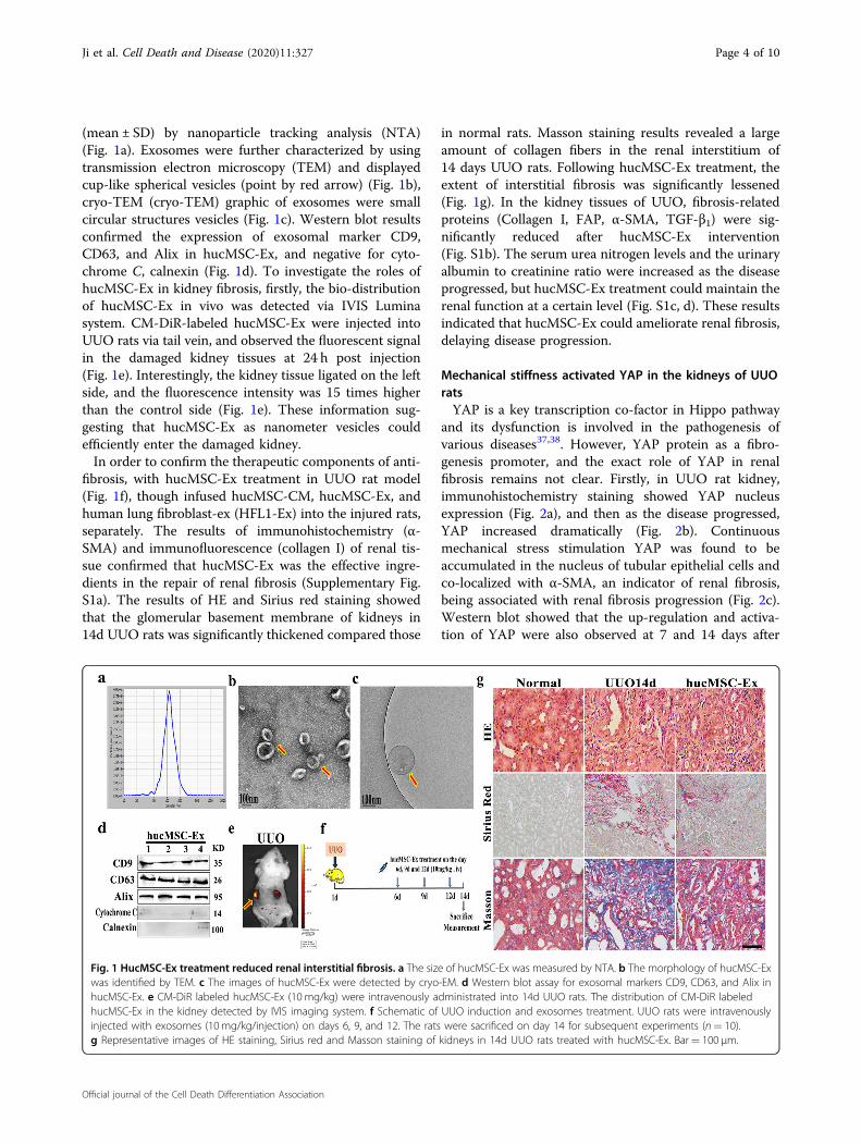
(mean ± SD) by nanoparticle tracking analysis (NTA)(Fig. 1a). Exosomes were further characterized by usingtransmission electron microscopy (TEM) and displayedcup-like spherical vesicles (point by red arrow) (Fig. 1b),cryo-TEM (cryo-TEM) graphic of exosomes were smallcircular structures vesicles (Fig. 1c). Western blot resultsconfirmed the expression of exosomal marker CD9,CD63, and Alix in hucMSC-Ex, and negative for cyto-chrome C, calnexin (Fig. 1d). To investigate the roles ofhucMSC-Ex in kidney fibrosis, firstly, the bio-distributionof hucMSC-Ex in vivo was detected via IVIS Luminasystem. CM-DiR-labeled hucMSC-Ex were injected intoUUO rats via tail vein, and observed the fluorescent signalin the damaged kidney tissues at 24 h post injection(Fig. 1e). Interestingly, the kidney tissue ligated on the leftside, and the fluorescence intensity was 15 times higherthan the control side (Fig. 1e). These information sug-gesting that hucMSC-Ex as nanometer vesicles couldefficiently enter the damaged kidney.In order to confirm the therapeutic components of anti-
fibrosis, with hucMSC-Ex treatment in UUO rat model(Fig. 1f), though infused hucMSC-CM, hucMSC-Ex, andhuman lung fibroblast-ex (HFL1-Ex) into the injured rats,separately. The results of immunohistochemistry (α-SMA) and immunofluorescence (collagen I) of renal tis-sue confirmed that hucMSC-Ex was the effective ingre-dients in the repair of renal fibrosis (Supplementary Fig.S1a). The results of HE and Sirius red staining showedthat the glomerular basement membrane of kidneys in14d UUO rats was significantly thickened compared those
in normal rats. Masson staining results revealed a largeamount of collagen fibers in the renal interstitium of14 days UUO rats. Following hucMSC-Ex treatment, theextent of interstitial fibrosis was significantly lessened(Fig. 1g). In the kidney tissues of UUO, fibrosis-relatedproteins (Collagen I, FAP, α-SMA, TGF-β1) were sig-nificantly reduced after hucMSC-Ex intervention(Fig. S1b). The serum urea nitrogen levels and the urinaryalbumin to creatinine ratio were increased as the diseaseprogressed, but hucMSC-Ex treatment could maintain therenal function at a certain level (Fig. S1c, d). These resultsindicated that hucMSC-Ex could ameliorate renal fibrosis,delaying disease progression.
Mechanical stiffness activated YAP in the kidneys of UUOratsYAP is a key transcription co-factor in Hippo pathway
and its dysfunction is involved in the pathogenesis ofvarious diseases37,38. However, YAP protein as a fibro-genesis promoter, and the exact role of YAP in renalfibrosis remains not clear. Firstly, in UUO rat kidney,immunohistochemistry staining showed YAP nucleusexpression (Fig. 2a), and then as the disease progressed,YAP increased dramatically (Fig. 2b). Continuousmechanical stress stimulation YAP was found to beaccumulated in the nucleus of tubular epithelial cells andco-localized with α-SMA, an indicator of renal fibrosis,being associated with renal fibrosis progression (Fig. 2c).Western blot showed that the up-regulation and activa-tion of YAP were also observed at 7 and 14 days after
Fig. 1 HucMSC-Ex treatment reduced renal interstitial fibrosis. a The size of hucMSC-Ex was measured by NTA. b The morphology of hucMSC-Exwas identified by TEM. c The images of hucMSC-Ex were detected by cryo-EM. d Western blot assay for exosomal markers CD9, CD63, and Alix inhucMSC-Ex. e CM-DiR labeled hucMSC-Ex (10 mg/kg) were intravenously administrated into 14d UUO rats. The distribution of CM-DiR labeledhucMSC-Ex in the kidney detected by IVIS imaging system. f Schematic of UUO induction and exosomes treatment. UUO rats were intravenouslyinjected with exosomes (10 mg/kg/injection) on days 6, 9, and 12. The rats were sacrificed on day 14 for subsequent experiments (n= 10).g Representative images of HE staining, Sirius red and Masson staining of kidneys in 14d UUO rats treated with hucMSC-Ex. Bar= 100 μm.
Ji et al. Cell Death and Disease (2020) 11:327 Page 4 of 10
Official journal of the Cell Death Differentiation Association
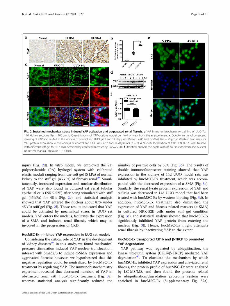
injury (Fig. 2d). In vitro model, we employed the 2Dpolyacrylamide (PA) hydrogel system with calibratedelastic moduli ranging from the soft gel (5 kPa) of normalkidney to the stiff gel (45 kPa) of fibrosis renal39. Simul-taneously, increased expression and nuclear distributionof YAP were also found in cultured rat renal tubularepithelial cells (NRK-52E) after being stimulated with stiffgel (45 kPa) for 48 h (Fig. 2e), and statistical analysisshowed that YAP entered the nucleus about 87% under45 kPa stiff gel (Fig. 2f). Those results indicated that YAPcould be activated by mechanical stress in UUO ratmodels. YAP enters the nucleus, facilitates the expressionof α-SMA and induced renal fibrosis, which may beinvolved in the progression of CKD.
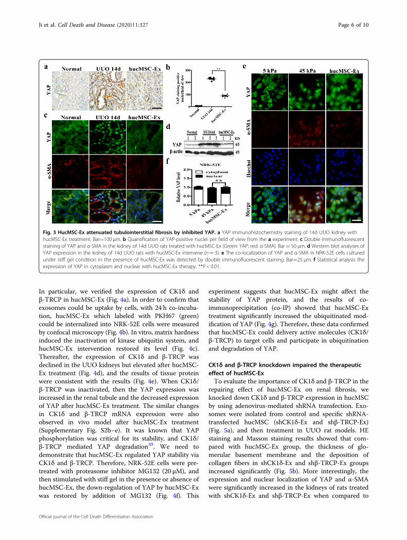
HucMSC-Ex inhibited YAP expression in UUO rat modelsConsidering the critical role of YAP in the development
of kidney diseases29, in this study, we found mechanicalpressure stimulation induced YAP nuclear translocation,interact with Smad2/3 to induce α-SMA expression andaggravated fibrosis; however, we hypothesized that thisnegative regulation could be neutralized by hucMSC-Extreatment by regulating YAP. The immunohistochemistryexperiment revealed that decreased numbers of YAP inobstructed renal with hucMSC-Ex treatment (Fig. 3a),whereas statistical analysis significantly reduced the
number of positive cells by 55% (Fig. 3b). The results ofdouble immunofluorescent staining showed that YAPexpression in the kidneys of 14d UUO model rats wasinhibited by hucMSC-Ex treatment, which was accom-panied with the decreased expression of α-SMA (Fig. 3c).Similarly, the renal lysate protein expression of YAP andα-SMA was decreased in 14d UUO model that had beentreated with hucMSC-Ex by western blotting (Fig. 3d). Inaddition, hucMSC-Ex treatment also diminished theexpression of YAP and fibrosis-related markers (α-SMA)in cultured NRK-52E cells under stiff gel condition(Fig. 3e), and statistical analysis showed that hucMSC-Exsignificantly inhibited YAP protein from entering thenucleus (Fig. 3f). Hence, hucMSC-Ex might attenuaterenal fibrosis by inactivating YAP to the extent.
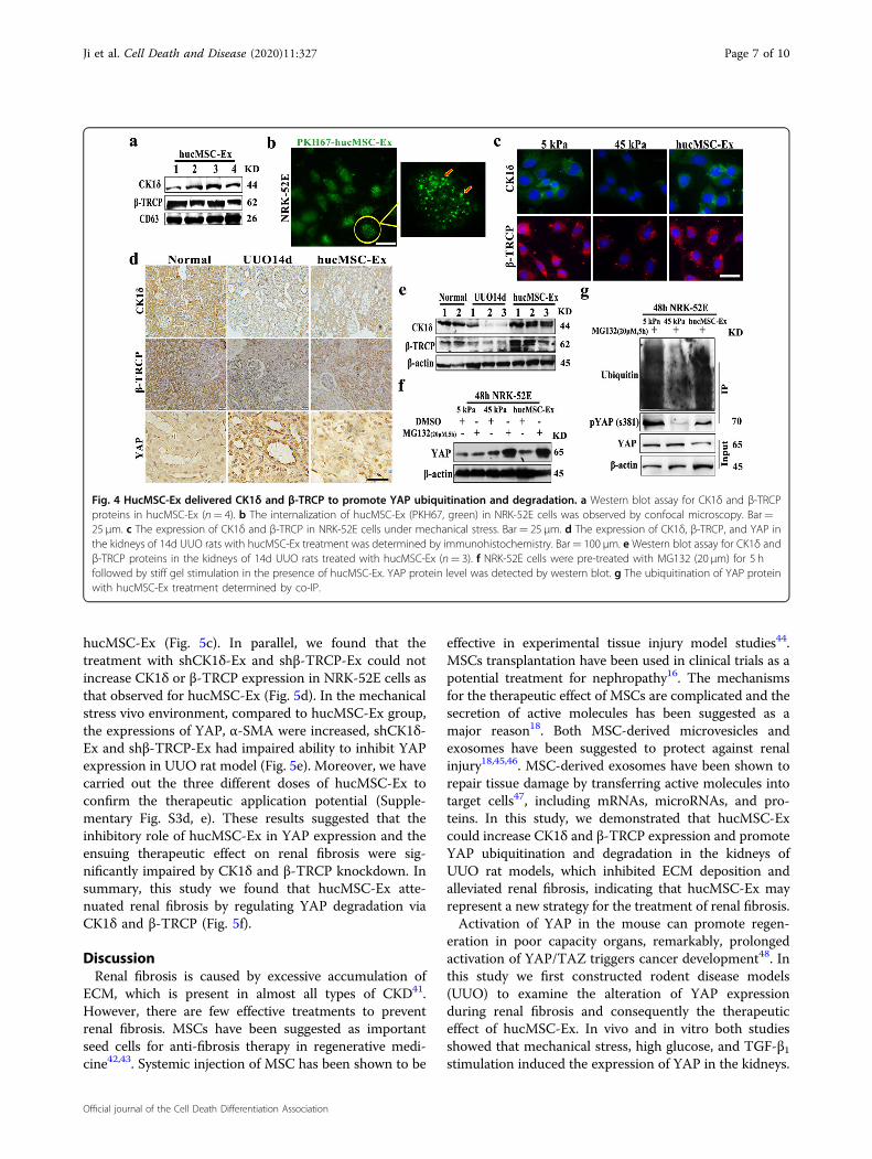
HucMSC-Ex transported CK1δ and β-TRCP to promotedYAP degradationYAP pathway was regulated by ubiquitination, the
kinase ubiquitin system (CK1δ/β-TRCP) mediated YAPdegradation40. To elucidate the mechanism by whichhucMSC-Ex inhibited YAP expression and alleviated renalfibrosis, the protein profile of hucMSC-Ex were analyzedby LC-MS/MS, and then found the proteins relatedto ubiquitination/degradation proteome system wereenriched in hucMSC-Ex (Supplementary Fig. S2a).
Fig. 2 Sustained mechanical stress induced YAP activation and aggravated renal fibrosis. a YAP immunohistochemistry staining of UUO 7d,14d kidney sections. Bar= 100 μm. b Quantification of YAP-positive nuclei per field of view from the a experiment. c Double immunofluorescentstaining of YAP and α-SMA in the kidneys of control and UUO (at 7 and 14 days) rats (Green: YAP; Red: α-SMA). Bar= 50 μm. d Western blot assay forYAP protein expression in the kidneys of control and UUO rats (at 7 and 14 days) rats (n= 3). e Nuclear localization of YAP in NRK-52E cells treatedwith different stiff gel for 48 h was detected by confocal microscopy. Bar=25 μm. f Statistical analysis the expression of YAP in cytoplasm and nuclearunder mechanical pressure. **P < 0.01.
Ji et al. Cell Death and Disease (2020) 11:327 Page 5 of 10
Official journal of the Cell Death Differentiation Association
In particular, we verified the expression of CK1δ andβ-TRCP in hucMSC-Ex (Fig. 4a). In order to confirm thatexosomes could be uptake by cells, with 24 h co-incuba-tion, hucMSC-Ex which labeled with PKH67 (green)could be internalized into NRK-52E cells were measuredby confocal microscopy (Fig. 4b). In vitro, matrix hardnessinduced the inactivation of kinase ubiquitin system, andhucMSC-Ex intervention restored its level (Fig. 4c).Thereafter, the expression of CK1δ and β-TRCP wasdeclined in the UUO kidneys but elevated after hucMSC-Ex treatment (Fig. 4d), and the results of tissue proteinwere consistent with the results (Fig. 4e). When CK1δ/β-TRCP was inactivated, then the YAP expression wasincreased in the renal tubule and the decreased expressionof YAP after hucMSC-Ex treatment. The similar changesin CK1δ and β-TRCP mRNA expression were alsoobserved in vivo model after hucMSC-Ex treatment(Supplementary Fig. S2b–e). It was known that YAPphosphorylation was critical for its stability, and CK1δ/β-TRCP mediated YAP degradation39. We need todemonstrate that hucMSC-Ex regulated YAP stability viaCK1δ and β-TRCP. Therefore, NRK-52E cells were pre-treated with proteasome inhibitor MG132 (20 μM), andthen stimulated with stiff gel in the presence or absence ofhucMSC-Ex, the down-regulation of YAP by hucMSC-Exwas restored by addition of MG132 (Fig. 4f). This
experiment suggests that hucMSC-Ex might affect thestability of YAP protein, and the results of co-immunoprecipitation (co-IP) showed that hucMSC-Extreatment significantly increased the ubiquitinated mod-ification of YAP (Fig. 4g). Therefore, these data confirmedthat hucMSC-Ex could delivery active molecules (CK1δ/β-TRCP) to target cells and participate in ubiquitinationand degradation of YAP.
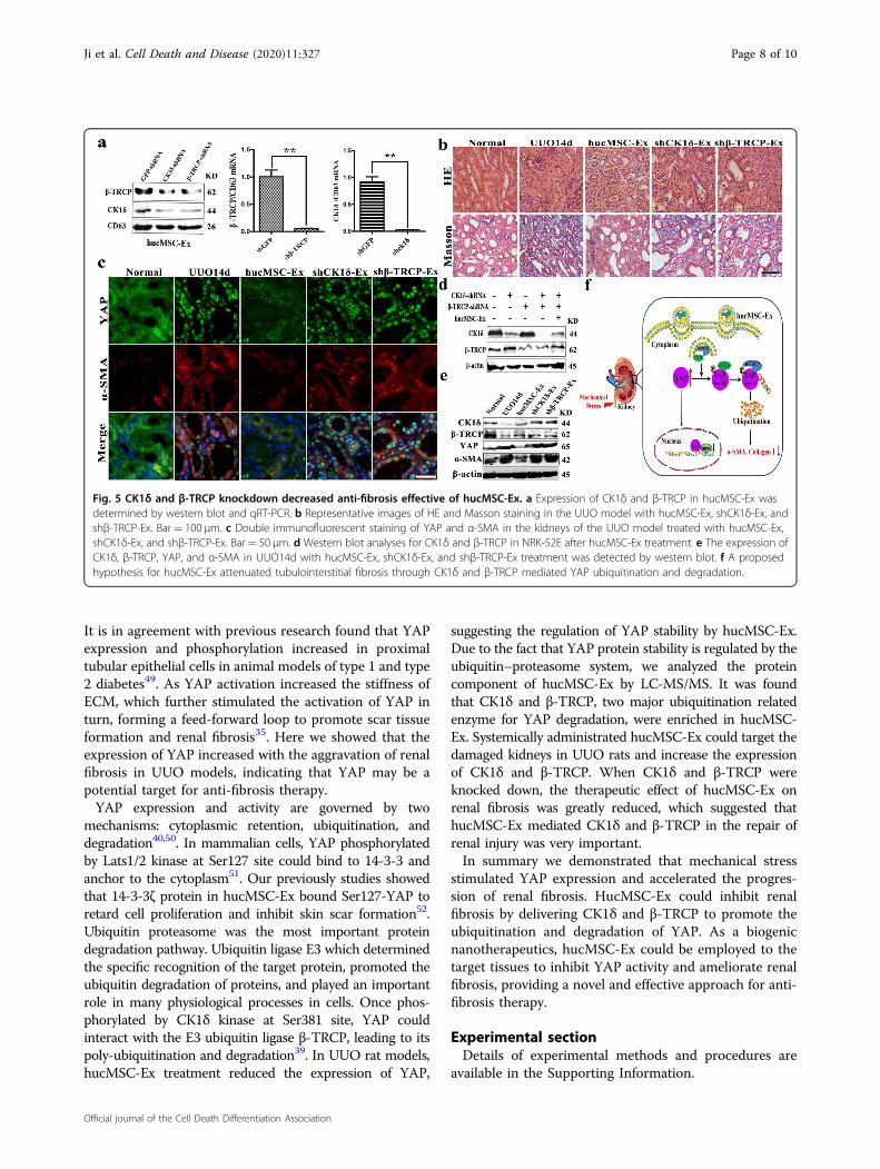
CK1δ and β-TRCP knockdown impaired the therapeuticeffect of hucMSC-ExTo evaluate the importance of CK1δ and β-TRCP in the
repairing effect of hucMSC-Ex on renal fibrosis, weknocked down CK1δ and β-TRCP expression in hucMSCby using adenovirus-mediated shRNA transfection. Exo-somes were isolated from control and specific shRNA-transfected hucMSC (shCK1δ-Ex and shβ-TRCP-Ex)(Fig. 5a), and then treatment in UUO rat models. HEstaining and Masson staining results showed that com-pared with hucMSC-Ex group, the thickness of glo-merular basement membrane and the deposition ofcollagen fibers in shCK1δ-Ex and shβ-TRCP-Ex groupsincreased significantly (Fig. 5b). More interestingly, theexpression and nuclear localization of YAP and α-SMAwere significantly increased in the kidneys of rats treatedwith shCK1δ-Ex and shβ-TRCP-Ex when compared to
Fig. 3 HucMSC-Ex attenuated tubulointerstitial fibrosis by inhibited YAP. a YAP immunohistochemistry staining of 14d UUO kidney withhucMSC-Ex treatment. Bar=100 μm. b Quantification of YAP-positive nuclei per field of view from the a experiment. c Double immunofluorescentstaining of YAP and α-SMA in the kidney of 14d UUO rats treated with hucMSC-Ex (Green: YAP; red: α-SMA). Bar= 50 μm. d Western blot analyses ofYAP expression in the kidney of 14d UUO rats with hucMSC-Ex intervene (n= 3). e The co-localization of YAP and α-SMA in NRK-52E cells culturedunder stiff gel condition in the presence of hucMSC-Ex was detected by double immunofluorescent staining. Bar=25 μm. f Statistical analysis theexpression of YAP in cytoplasm and nuclear with hucMSC-Ex therapy. **P < 0.01.
Ji et al. Cell Death and Disease (2020) 11:327 Page 6 of 10
Official journal of the Cell Death Differentiation Association
hucMSC-Ex (Fig. 5c). In parallel, we found that thetreatment with shCK1δ-Ex and shβ-TRCP-Ex could notincrease CK1δ or β-TRCP expression in NRK-52E cells asthat observed for hucMSC-Ex (Fig. 5d). In the mechanicalstress vivo environment, compared to hucMSC-Ex group,the expressions of YAP, α-SMA were increased, shCK1δ-Ex and shβ-TRCP-Ex had impaired ability to inhibit YAPexpression in UUO rat model (Fig. 5e). Moreover, we havecarried out the three different doses of hucMSC-Ex toconfirm the therapeutic application potential (Supple-mentary Fig. S3d, e). These results suggested that theinhibitory role of hucMSC-Ex in YAP expression and theensuing therapeutic effect on renal fibrosis were sig-nificantly impaired by CK1δ and β-TRCP knockdown. Insummary, this study we found that hucMSC-Ex atte-nuated renal fibrosis by regulating YAP degradation viaCK1δ and β-TRCP (Fig. 5f).
DiscussionRenal fibrosis is caused by excessive accumulation of
ECM, which is present in almost all types of CKD41.However, there are few effective treatments to preventrenal fibrosis. MSCs have been suggested as importantseed cells for anti-fibrosis therapy in regenerative medi-cine42,43. Systemic injection of MSC has been shown to be
effective in experimental tissue injury model studies44.MSCs transplantation have been used in clinical trials as apotential treatment for nephropathy16. The mechanismsfor the therapeutic effect of MSCs are complicated and thesecretion of active molecules has been suggested as amajor reason18. Both MSC-derived microvesicles andexosomes have been suggested to protect against renalinjury18,45,46. MSC-derived exosomes have been shown torepair tissue damage by transferring active molecules intotarget cells47, including mRNAs, microRNAs, and pro-teins. In this study, we demonstrated that hucMSC-Excould increase CK1δ and β-TRCP expression and promoteYAP ubiquitination and degradation in the kidneys ofUUO rat models, which inhibited ECM deposition andalleviated renal fibrosis, indicating that hucMSC-Ex mayrepresent a new strategy for the treatment of renal fibrosis.Activation of YAP in the mouse can promote regen-
eration in poor capacity organs, remarkably, prolongedactivation of YAP/TAZ triggers cancer development48. Inthis study we first constructed rodent disease models(UUO) to examine the alteration of YAP expressionduring renal fibrosis and consequently the therapeuticeffect of hucMSC-Ex. In vivo and in vitro both studiesshowed that mechanical stress, high glucose, and TGF-β1stimulation induced the expression of YAP in the kidneys.
Fig. 4 HucMSC-Ex delivered CK1δ and β-TRCP to promote YAP ubiquitination and degradation. a Western blot assay for CK1δ and β-TRCPproteins in hucMSC-Ex (n= 4). b The internalization of hucMSC-Ex (PKH67, green) in NRK-52E cells was observed by confocal microscopy. Bar=25 μm. c The expression of CK1δ and β-TRCP in NRK-52E cells under mechanical stress. Bar= 25 μm. d The expression of CK1δ, β-TRCP, and YAP inthe kidneys of 14d UUO rats with hucMSC-Ex treatment was determined by immunohistochemistry. Bar= 100 μm. e Western blot assay for CK1δ andβ-TRCP proteins in the kidneys of 14d UUO rats treated with hucMSC-Ex (n= 3). f NRK-52E cells were pre-treated with MG132 (20 μm) for 5 hfollowed by stiff gel stimulation in the presence of hucMSC-Ex. YAP protein level was detected by western blot. g The ubiquitination of YAP proteinwith hucMSC-Ex treatment determined by co-IP.
Ji et al. Cell Death and Disease (2020) 11:327 Page 7 of 10
Official journal of the Cell Death Differentiation Association
It is in agreement with previous research found that YAPexpression and phosphorylation increased in proximaltubular epithelial cells in animal models of type 1 and type2 diabetes49. As YAP activation increased the stiffness ofECM, which further stimulated the activation of YAP inturn, forming a feed-forward loop to promote scar tissueformation and renal fibrosis35. Here we showed that theexpression of YAP increased with the aggravation of renalfibrosis in UUO models, indicating that YAP may be apotential target for anti-fibrosis therapy.YAP expression and activity are governed by two
mechanisms: cytoplasmic retention, ubiquitination, anddegradation40,50. In mammalian cells, YAP phosphorylatedby Lats1/2 kinase at Ser127 site could bind to 14-3-3 andanchor to the cytoplasm51. Our previously studies showedthat 14-3-3ζ protein in hucMSC-Ex bound Ser127-YAP toretard cell proliferation and inhibit skin scar formation52.Ubiquitin proteasome was the most important proteindegradation pathway. Ubiquitin ligase E3 which determinedthe specific recognition of the target protein, promoted theubiquitin degradation of proteins, and played an importantrole in many physiological processes in cells. Once phos-phorylated by CK1δ kinase at Ser381 site, YAP couldinteract with the E3 ubiquitin ligase β-TRCP, leading to itspoly-ubiquitination and degradation39. In UUO rat models,hucMSC-Ex treatment reduced the expression of YAP,
suggesting the regulation of YAP stability by hucMSC-Ex.Due to the fact that YAP protein stability is regulated by theubiquitin–proteasome system, we analyzed the proteincomponent of hucMSC-Ex by LC-MS/MS. It was foundthat CK1δ and β-TRCP, two major ubiquitination relatedenzyme for YAP degradation, were enriched in hucMSC-Ex. Systemically administrated hucMSC-Ex could target thedamaged kidneys in UUO rats and increase the expressionof CK1δ and β-TRCP. When CK1δ and β-TRCP wereknocked down, the therapeutic effect of hucMSC-Ex onrenal fibrosis was greatly reduced, which suggested thathucMSC-Ex mediated CK1δ and β-TRCP in the repair ofrenal injury was very important.In summary we demonstrated that mechanical stress
stimulated YAP expression and accelerated the progres-sion of renal fibrosis. HucMSC-Ex could inhibit renalfibrosis by delivering CK1δ and β-TRCP to promote theubiquitination and degradation of YAP. As a biogenicnanotherapeutics, hucMSC-Ex could be employed to thetarget tissues to inhibit YAP activity and ameliorate renalfibrosis, providing a novel and effective approach for anti-fibrosis therapy.
Experimental sectionDetails of experimental methods and procedures are
available in the Supporting Information.
Fig. 5 CK1δ and β-TRCP knockdown decreased anti-fibrosis effective of hucMSC-Ex. a Expression of CK1δ and β-TRCP in hucMSC-Ex wasdetermined by western blot and qRT-PCR. b Representative images of HE and Masson staining in the UUO model with hucMSC-Ex, shCK1δ-Ex, andshβ-TRCP-Ex. Bar= 100 μm. c Double immunofluorescent staining of YAP and α-SMA in the kidneys of the UUO model treated with hucMSC-Ex,shCK1δ-Ex, and shβ-TRCP-Ex. Bar= 50 μm. d Western blot analyses for CK1δ and β-TRCP in NRK-52E after hucMSC-Ex treatment. e The expression ofCK1δ, β-TRCP, YAP, and α-SMA in UUO14d with hucMSC-Ex, shCK1δ-Ex, and shβ-TRCP-Ex treatment was detected by western blot. f A proposedhypothesis for hucMSC-Ex attenuated tubulointerstitial fibrosis through CK1δ and β-TRCP mediated YAP ubiquitination and degradation.
Ji et al. Cell Death and Disease (2020) 11:327 Page 8 of 10
Official journal of the Cell Death Differentiation Association
AcknowledgementsThis work was supported by the National Natural Science Foundation of China(Grant no. 81871496, 81971757), Zhenjiang Key Laboratory of High TechnologyResearch on Exosomes Foundation and Transformation Application (Grantss2018003), and project funded by the Priority Academic ProgramDevelopment of Jiangsu Higher Education Institutions (Phase III), the SixthTalent Peaks Project of Jiangsu Province(2018-YY-014).
Conflict of interestThe authors declare that they have no conflict of interest.
Publisher’s noteSpringer Nature remains neutral with regard to jurisdictional claims inpublished maps and institutional affiliations.
Supplementary Information accompanies this paper at (https://doi.org/10.1038/s41419-020-2510-4).
Received: 25 November 2019 Revised: 11 April 2020 Accepted: 14 April2020
References1. Falke, L. L., Gholizadeh, S., Goldschmeding, R., Kok, R. J. & Nguyen, T. Q. Diverse
origins of the myofibroblast—implications for kidney fibrosis. Nat. Rev.Nephrol. 11, 233–244 (2015).
2. Ferenbach, D. A. & Bonventre, J. V. Mechanisms of maladaptive repair after AKIleading to accelerated kidney ageing and CKD. Nat. Rev. Nephrol. 11, 264–276(2015).
3. Lu, Q. et al. Circulating miR-103a-3p contributes to angiotensin II-inducedrenal inflammation and fibrosis via a SNRK/NF-κB/p65 regulatory axis. Nat.Commun. 10, 21–45 (2019).
4. Komada, T. & Muruve, D. A. The role of inflammasomes in kidney disease. Nat.Rev. Nephrol. 15, 501–520 (2019).
5. Kramann, R., DiRocco, D. P. & Humphreys, B. D. Understanding the origin,activation and regulation of matrixproducing myofibroblasts for treatment offibrotic disease. J. Pathol. 231, 273–289 (2013).
6. Ying, W. Z. et al. Immunoglobulin light chains generate proinflammatory andprofibrotic kidney injury. J. Clin. Invest. 17, 2792–2806 (2019).
7. Liu, Y. Cellular and molecular mechanisms of renal fibrosis. Nat. Rev. Nephrol. 7,684–696 (2011).
8. Djudjaj, S. & Boor, P. Cellular and molecular mechanisms of kidney fibrosis.Mol.Aspects Med. 65, 16–36 (2019).
9. Rockey, D. C., Bell, P. D. & Hill, J. A. Fibrosis—a common pathway to organinjury and failure. N. Engl. J. Med. 372, 1138–1149 (2015).
10. Tang, T. T., Lv, L. L., Lan, H. Y. & Liu, B. C. Extracellular vesicles: opportunities andchallenges for the treatment of renal diseases. Front. Physiol. 19, 226 (2019).
11. Friedman, S. L., Sheppard, D. & Duffield, J. S. Therapy for fibrotic diseases:nearing the starting line. Sci. Transl. Med. 5, 167 (2013).
12. Wang, P. et al. Long noncoding RNA lnc-TSI inhibits renal fibrogenesis bynegatively regulating the TGF-β/Smad3 pathway. Sci. Transl. Med. 10,eaat2039 (2018).
13. Broekman, W. et al. Mesenchymal stromal cells: a novel therapy for thetreatment of chronic obstructive pulmonary disease? Thorax 73, 565–574(2018).
14. Lee, C. W., Chen, Y. F., Wu, H. H. & Lee, O. K. Historical perspectives andadvances in mesenchymal stem cell research for the treatment of liver dis-eases. Gastroenterology 154, 46–56 (2018).
15. Erpicum, P. et al. Infusion of third-party mesenchymal stromal cells after kidneytransplantation: a phase I-II, open-label, clinical study. Kidney Int. 95, 693–707(2019).
16. Sivanathan, K. N. & Coates, P. T. IL-17A-induced mesenchymal stem cells havepromising therapeutic value for clinical translation. Kidney Int. 93, 771–773(2018).
17. Kuppe, C. & Kramann, R. Role of mesenchymal stem cells in kidney injury andfibrosis. Curr. Opin. Nephrol. Hypertens. 25, 372–7 (2016).
18. Camussi, G., Deregibus, M. C., Bruno, S., Cantaluppi, V. & Biancone, L. Exo-somes/microvesicles as a mechanism of cell-to-cell communication. Kidney Int.78, 838–848 (2010).
19. Denzer, K., Kleijmeer, M. J., Heijnen, H. F., Stoorvogel, W. & Geuze, H. J. Exosome:from internal vesicle of the multivesicular body to intercellular signalingdevice. J. Cell Sci. 19, 3365–3374 (2000).
20. Chen, F. et al. Extracellular vesicle-packaged HIF-1α-stabilizing LncRNA fromtumour associated macrophages regulates aerobic glycolysis of breast cancercells. Nat. Cell Biol. 21, 498–510 (2019).
21. Huang, J. H. et al. Systemic administration of exosomes released frommesenchymal stromal cells attenuates apoptosis, inflammation, and promotesangiogenesis after spinal cord injury in rats. J. Neurotrauma 34, 3388–3396(2017).
22. Riazifar, M. et al. Stem cell-derived exosomes as nanotherapeutics for auto-immune and neurodegenerative disorders. ACS Nano. 13, 6670–6688 (2019).
23. Xiao, C. et al. Transplanted mesenchymal stem cells reduce autophagic flux ininfarcted hearts via the exosomal transfer of miR-125b. Circ. Res. 123, 564–578(2018).
24. Li, T. et al. Exosomes derived from human umbilical cord mesenchymal stemcells alleviate liver fibrosis. Stem Cells Dev. 22, 845–854 (2013).
25. Sun, Y. et al. Human mesenchymal stem cell derived exosomes alleviate type2 diabetes mellitus by reversing peripheral insulin resistance and relievingβ-cell destruction. ACS Nano 12, 7613–7628 (2018).
26. Zhang, B. et al. HucMSC exosome mediated Wnt4 signaling is required forcutaneous wound healing. Stem Cells 33, 2158–2168 (2015).
27. Wang, B. et al. Pre-Incubation with HucMSC-exosomes prevents cisplatin-induced nephrotoxicity by activating autophagy. Stem Cell Res. Ther. 8, 75(2017).
28. Jia, H. et al. HucMSC exosomes-delivered 14-3-3ζ enhanced autophagy viamodulation of ATG16L in preventing cisplatin-induced acute kidney injury.Am. J. Transl. Res. 10, 101–113 (2018).
29. Yu, F. X., Zhao, B. & Guan, K. L. Hippo pathway in organ size control, tissuehomeostasis, and cancer. Cell 163, 811–828 (2015).
30. Dupont, S. et al. Role of YAP/TAZ in Mechanotransduction. Nature 474,179–183 (2011).
31. Xu, J. et al. Involvement of the Hippo pathway in regeneration and fibro-genesis after ischaemic acute kidney injury: YAP is the key effector. Clin. Sci.(Lond.). 130, 349–363 (2016).
32. Huynh, P. & Chai, Z. Transforming growth factor β (TGFβ) and related mole-cules in chronic kidney disease (CKD). Clin. Sci. (Lond.). 133, 287–313 (2019).
33. Varelas, X. et al. The Crumbs complex couples cell density sensing to Hippo-dependent control of the TGF-β-SMAD pathway. Dev. Cell. 19, 831–844 (2010).
34. Liu, F. et al. Mechanosignaling through YAP and TAZ drives fibroblast acti-vation and fibrosis. Am. J. Physiol. Lung Cell Mol. Physiol. 308, 344–357 (2015).
35. Liang, M. et al. Yap/Taz deletion in Gli+ cell-derived myofibroblasts attenuatesfibrosis. J. Am. Soc. Nephrol. 28, 3278–3290 (2017).
36. Tse, J. R. & Engler, A. J. Preparation of hydrogel substrates with tunablemechanical properties. Curr. Protoc. Cell Biol. 10, 16 (2010).
37. Moya, I. M. & Halder, G. Hippo-YAP/TAZ signalling in organ regeneration andregenerative medicine. Nat. Rev. Mol. Cell Biol. 20, 211–226 (2019).
38. Wu, J. et al. Intercellular interaction dictates cancer cell ferroptosis via NF2-YAPsignalling. Nature 572, 402–406 (2019).
39. Zhao, B., Li, L., Tumaneng, K., Wang, C. Y. & Guan, K. L. A Coordinated phos-phorylation by Lats and CK1δ regulates YAP stability through SCF (beta-TRCP).Genes Dev. 24, 72–85 (2010).
40. Meng, Z., Moroishi, T. & Guan, K. L. Mechanisms of Hippo pathway regulation.Genes Dev. 30, 1–17 (2016).
41. Leaf, I. A. & Duffield, J. S. What can target kidney fibrosis? Nephrol. Dial.Transplant. 1, 89–97 (2017).
42. Zhuang, Q. et al. Mesenchymal stem cells in renal fibrosis: the flame ofcytotherapy. Stem Cells Int. 13, 8387350 (2019).
43. Agha E, E. I. et al. Mesenchymal stem cells in fibrotic disease. Cell Stem Cell 3,166–177 (2017).
44. Perico, N., Casiraghi, F. & Remuzzi, G. Clinical translation of mesenchymalstromal cell therapies in nephrology. J. Am. Soc. Nephrol. 29, 362–375 (2018).
45. Gatti, S. et al. Microvesicles derived from human adult mesenchymal stemcells protect against ischaemia-reperfusion-induced acute and chronic kidneyinjury. Nephrol. Dial. Transplant. 26, 1474–1483 (2011).
46. Bruno, S. et al. Mesenchymal stem cell-derived microvesicles protect againstacute tubular injury. J. Am. Soc. Nephrol. 20, 1053–1067 (2009).
Ji et al. Cell Death and Disease (2020) 11:327 Page 9 of 10
Official journal of the Cell Death Differentiation Association
47. Yeo, R. W. et al. Mesenchymal stem cell: an efficient mass producer of exo-somes for drug delivery. Adv. Drug Deliv. Rev. 65, 336–41 (2013).
48. Moroishi, T., Hansen, C. G. & Guan, K. L. The emerging roles of YAP and TAZ incancer. Nat. Rev. Cancer 15, 73–79 (2015).
49. Chen, J. & Harris, R. C. Interaction of the EGF receptor and the Hippo pathwayin the diabetic kidney. J. Am. Soc. Nephrol. 27, 1689–1700 (2016).
50. Lin, K. C. et al. Regulation of Hippo pathway transcription factor TEAD by p38MAPK-induced cytoplasmic translocation. Nat. Cell Biol. 28, 996–1002 (2017).
51. Zhao, B., Tumaneng, K. & Guan, K. L. The Hippo pathway in organ size control,tissue regeneration and stem cell self-renewal. Nat. Cell Biol. 13, 877–883(2011).
52. Zhang, B. et al. HucMSC exosome-delivered 14-3-3ζ orchestrates self-control ofthe Wnt response via modulation of YAP during cutaneous regeneration.Stem Cells 34, 2485–2500 (2016).
Ji et al. Cell Death and Disease (2020) 11:327 Page 10 of 10
Official journal of the Cell Death Differentiation Association
Related Documents