Original Investigation Exertional Fatigue in Patients With CKD Jamie H. Macdonald, PhD, 1 Lee Fearn, MSc, 1 Mahdi Jibani, FRCP, 2 and Samuele M. Marcora, PhD 3 Background: Fatigue is one of the most prevalent symptoms in chronic kidney disease (CKD). However, fatigue mechanisms are poorly understood due in part to nonspecific definitions. This study investigates exertional fatigue during simulated activities of daily living, focusing on oxygen delivery and utilization. Study Design: “Explanatory” matched-cohort study. Participants & Setting: 13 patients with CKD (stages 3b-4; mean age, 62 13 [SD] years) and 13 healthy controls, mean matched for age, height, body mass and composition, and physical activity level. Participants completed an incremental cycle ergometer test to simulate energy expenditure of typical activities of daily living. Factor: 4 exercise intensities: 1, 1.8, 2.4, and 3.1 metabolic equivalent tasks (METs). Outcomes: The primary outcome was exertional fatigue by rating of perceived exertion (RPE) on a 6-20 scale. Measurements: Other multidimensional measures of fatigue: UK Short Form Health Survey 36 (UK SF-36) Vitality and Functional Assessment of Chronic Illness Therapy–Fatigue (FACIT-Fatigue) subscales. Physi- ologic measures of the oxygen transport and utilization chain (expired gas analysis, cardiac output, and arterial oxygen content) and blood lactate. Results: RPE was increased in patients compared with controls at 2.4 (10.5 [ie, light] 2.7 vs 8.7 [very light] 1.7 units) and 3.1 (12.5 [somewhat hard] 2.6 vs 10.2 [light] 1.7 units) METs (interaction P 0.03), which was consistent with higher chronic fatigue in patients by both the UK SF-36 Vitality (P 0.01) and FACIT-Fatigue (P 0.004) subscales. Arterial oxygen content was decreased in patients (P 0.001), but cardiac output and oxygen extraction ratio were unchanged, decreasing oxygen delivery (P 0.04). Respiratory exchange ratio (P 0.004) and blood lactate production (P 0.002) were increased. Limitations: Those inherent to a matched-cohort study. Conclusions: Using a novel application of the outcome measure RPE, patients with non– dialysis- dependent CKD reported considerable exertional fatigue during simulated activities of daily living. Poor compensation for mild anemia contributed to this symptom. In addition to anemia, the entire oxygen transport chain needs to be targeted to treat fatigue in patients with CKD. Am J Kidney Dis. 60(6):930-939. © 2012 by the National Kidney Foundation, Inc. INDEX WORDS: Activities of daily living; anemia; exercise capacity; hemoglobin; oxygen delivery; perceived exertion; quality of life. I n chronic kidney disease (CKD), fatigue is re- ported as the most prevalent and severe symptom experienced by patients. 1-3 Such fatigue has been identified as the principal factor influencing quality of life in this population, 2,4,5 and fatigue also impacts on mortality. 6 However, fatigue is poorly investigated, diagnosed, and treated in patients with CKD. Scientific investigations in this area lack a precise definition of fatigue. Fatigue is recognized to be multidimensional in nature and consists of various subtypes, each of which may have distinct mecha- nisms. 7 It has been advocated that explanatory studies of specific fatigue subtypes are needed to clarify pathophysiology in relation to phenomenology. 7 One subtype of fatigue is exertional fatigue, defined as an increased sense of effort in relation to a task. 8 This subtype can be measured easily during physical tasks using the extensively validated rating of perceived exertion (RPE) scale. 9 By simulating normal activi- ties of daily living and simultaneously obtaining mea- surements of RPE, it becomes possible to define and measure a specific construct of fatigue of particular relevance to quality of life. 10 In addition to problems defining fatigue, the exact cause of fatigue in patients with CKD is unknown. One potential mechanism is an altered oxygen trans- port chain 11,12 ; oxygen delivery and/or oxygen utiliza- tion may be impaired in patients with CKD. 13 In this From the 1 College of Health and Behavioural Sciences, Bangor University; 2 Ysbyty Gwynedd Renal Unit, Betsi Cadwaladr Univer- sity Health Board, Bangor; and 3 Sports Therapy, Physical Activity and Health Research Group, Centre for Sports Studies, University of Kent at Medway, Kent, United Kingdom. Received February 16, 2012. Accepted in revised form June 19, 2012. Originally published online August 9, 2012. Address correspondence to Jamie H. Macdonald, PhD, College of Health and Behavioural Sciences, Bangor University, George Bldg, Bangor, Gwynedd, LL57 2PZ, United Kingdom. E-mail: [email protected] © 2012 by the National Kidney Foundation, Inc. 0272-6386/$36.00 http://dx.doi.org/10.1053/j.ajkd.2012.06.021 Am J Kidney Dis. 2012;60(6):930-939 930

Welcome message from author
This document is posted to help you gain knowledge. Please leave a comment to let me know what you think about it! Share it to your friends and learn new things together.
Transcript
Original Investigation
Exertional Fatigue in Patients With CKD
Jamie H. Macdonald, PhD,1 Lee Fearn, MSc,1 Mahdi Jibani, FRCP,2 andSamuele M. Marcora, PhD3
Background: Fatigue is one of the most prevalent symptoms in chronic kidney disease (CKD). However,fatigue mechanisms are poorly understood due in part to nonspecific definitions. This study investigatesexertional fatigue during simulated activities of daily living, focusing on oxygen delivery and utilization.
Study Design: “Explanatory” matched-cohort study.Participants & Setting: 13 patients with CKD (stages 3b-4; mean age, 62 � 13 [SD] years) and 13 healthy
controls, mean matched for age, height, body mass and composition, and physical activity level. Participantscompleted an incremental cycle ergometer test to simulate energy expenditure of typical activities of dailyliving.
Factor: 4 exercise intensities: 1, 1.8, 2.4, and 3.1 metabolic equivalent tasks (METs).Outcomes: The primary outcome was exertional fatigue by rating of perceived exertion (RPE) on a 6-20
scale.Measurements: Other multidimensional measures of fatigue: UK Short Form Health Survey 36 (UK SF-36)
Vitality and Functional Assessment of Chronic Illness Therapy–Fatigue (FACIT-Fatigue) subscales. Physi-ologic measures of the oxygen transport and utilization chain (expired gas analysis, cardiac output, and arterialoxygen content) and blood lactate.
Results: RPE was increased in patients compared with controls at 2.4 (10.5 [ie, light] � 2.7 vs 8.7 [very light] �1.7 units) and 3.1 (12.5 [somewhat hard] � 2.6 vs 10.2 [light] � 1.7 units) METs (interaction P � 0.03), which wasconsistent with higher chronic fatigue in patients by both the UK SF-36 Vitality (P � 0.01) and FACIT-Fatigue(P � 0.004) subscales. Arterial oxygen content was decreased in patients (P � 0.001), but cardiac output andoxygen extraction ratio were unchanged, decreasing oxygen delivery (P � 0.04). Respiratory exchange ratio(P � 0.004) and blood lactate production (P � 0.002) were increased.
Limitations: Those inherent to a matched-cohort study.Conclusions: Using a novel application of the outcome measure RPE, patients with non–dialysis-
dependent CKD reported considerable exertional fatigue during simulated activities of daily living. Poorcompensation for mild anemia contributed to this symptom. In addition to anemia, the entire oxygen transportchain needs to be targeted to treat fatigue in patients with CKD.Am J Kidney Dis. 60(6):930-939. © 2012 by the National Kidney Foundation, Inc.
INDEX WORDS: Activities of daily living; anemia; exercise capacity; hemoglobin; oxygen delivery; perceivedexertion; quality of life.
In chronic kidney disease (CKD), fatigue is re-ported as the most prevalent and severe symptom
experienced by patients.1-3 Such fatigue has beenidentified as the principal factor influencing quality oflife in this population,2,4,5 and fatigue also impacts onmortality.6 However, fatigue is poorly investigated,diagnosed, and treated in patients with CKD.
From the 1College of Health and Behavioural Sciences, BangorUniversity; 2Ysbyty Gwynedd Renal Unit, Betsi Cadwaladr Univer-sity Health Board, Bangor; and 3Sports Therapy, Physical Activityand Health Research Group, Centre for Sports Studies, Universityof Kent at Medway, Kent, United Kingdom.
Received February 16, 2012. Accepted in revised form June 19,2012. Originally published online August 9, 2012.
Address correspondence to Jamie H. Macdonald, PhD, Collegeof Health and Behavioural Sciences, Bangor University, GeorgeBldg, Bangor, Gwynedd, LL57 2PZ, United Kingdom. E-mail:[email protected]
© 2012 by the National Kidney Foundation, Inc.0272-6386/$36.00
http://dx.doi.org/10.1053/j.ajkd.2012.06.021930
Scientific investigations in this area lack a precisedefinition of fatigue. Fatigue is recognized to bemultidimensional in nature and consists of varioussubtypes, each of which may have distinct mecha-nisms.7 It has been advocated that explanatory studiesof specific fatigue subtypes are needed to clarifypathophysiology in relation to phenomenology.7 Onesubtype of fatigue is exertional fatigue, defined as anincreased sense of effort in relation to a task.8 Thissubtype can be measured easily during physical tasksusing the extensively validated rating of perceivedexertion (RPE) scale.9 By simulating normal activi-ties of daily living and simultaneously obtaining mea-surements of RPE, it becomes possible to define andmeasure a specific construct of fatigue of particularrelevance to quality of life.10
In addition to problems defining fatigue, the exactcause of fatigue in patients with CKD is unknown.One potential mechanism is an altered oxygen trans-port chain11,12; oxygen delivery and/or oxygen utiliza-
tion may be impaired in patients with CKD.13 In thisAm J Kidney Dis. 2012;60(6):930-939
Fatigue in CKD
regard, it increasingly is being recognized that oxygenutilization has a relatively minor role in CKD myop-athy and fatigue, evidenced by elegant studies show-ing normal mitochondrial function.14-17
In contrast, oxygen convection (from the lung tothe muscle capillary) and specifically anemia arecommonly cited causes of general fatigue in patientswith CKD.18 More recently, it has been advocated thatanemia treatment be targeted by patient-reported fa-tigue, although the optimal hemoglobin target is un-clear.19 Furthermore, the influence of mild anemia onexertional fatigue is ambiguous because when healthypersons are made anemic experimentally, they compen-sate to maintain oxygen delivery (eg, by elevatingcardiac output), especially at lower exercise intensi-ties, as typically experienced during activities of dailyliving.20-22 Experimental evidence contradicting a pri-mary role of mild anemia in fatigue also is provided bythe surprisingly minor impact of erythropoietin therapyon this symptom.23,24 Thus, some authors have con-cluded that oxygen conductance (muscle capillary tomitochondria) is of greater importance than oxygenconvection/anemia.25
Still, previous studies must be interpreted cau-tiously. These studies often investigated maximal26
and single-leg27 exercise paradigms, but submaximalexercise and whole-body exercise better simulate ac-tivities of daily living. When submaximal compari-sons have been made, exercise intensities relative toparticipants’ maximal exercise capacity often havebeen used,25 making interpretation of oxygen deliveryand utilization variables difficult due to different abso-lute exercise intensities. Furthermore, most studiesfocus on hemodialysis patients, although this sub-group makes up only a small fraction of patients withCKD.28 Whether fatigue also occurs in patients withmoderate CKD is underinvestigated, and whethersuch patients with CKD adapt to reduced oxygendelivery is unknown, making guidelines for how totreat conditions such as exertional fatigue and anemiadifficult to offer.29
Therefore, the aim of this study was to determinewhether exertional fatigue, assessed by RPE, is el-evated in patients with CKD during simulated activi-ties of daily living. A secondary aim was to determinethe role of oxygen delivery and utilization in exer-tional fatigue, investigating whole-body, submaximal,and absolute exercise intensities to better representactivities of daily living. We hypothesized that exer-tional fatigue, assessed by RPE, would be increased inpatients with CKD during submaximal exercise. Fur-thermore, we hypothesized that oxygen convection/anemia would be involved minimally in such elevatedexertional fatigue due to cardiac compensation main-
taining oxygen delivery, thus suggesting that otherAm J Kidney Dis. 2012;60(6):930-939
factors (such as poor oxygen conductance from musclecapillary to mitochondria) are of greater importanceto exertional fatigue in patients with CKD.
METHODS
Patients andStudyDesign
After ethical approval from the North West Wales ResearchEthics Committee, a matched-cohort study was completed. Usinga convenience sampling method, patients were approached duringroutine nephrology outpatient clinics at Gwynedd Hospital be-tween October 2007 and January 2008. Control participants wereapproached by e-mails and letters to friends and relatives ofpatients, hospital staff, and the local community between October2007 and June 2010. Inclusion criteria for patients were CKD stage3b (estimated glomerular filtration rate [eGFR] of 30-44 mL/min/1.73 m2) or 4 (eGFR of 15-29 mL/min/17.3 m2), as defined byeGFR (calculated using the isotope-dilution mass spectrometry–traceable 4-variable MDRD [Modification of Diet in Renal Dis-ease] Study equation)30 from 2 serum samples at least 3 monthsapart. Exclusion criteria for controls were current CKD stages 1-5and medications that elicit abnormal physiologic responses toexercise (eg, �-blockers). Exclusion criteria for both groups wereage younger than 18 years, history of renal replacement therapy oracute kidney injury, any uncontrolled metabolic condition (eg,gross obesity or uncontrolled diabetes), pulmonary or neuromuscu-lar disease, hemoglobin concentration �11 g/dL, and not beingable to self-ambulate for 50 m.
After written informed consent, all participants presented at theSchool of Sport, Health and Exercise Sciences between January2008 and July 2010 for testing procedures. All participants at-tended for testing on 2 occasions: visit 1 was to familiarizeparticipants with the protocol and use of the RPE scale, and visit 2was identical to visit 1 and was completed 7 days later. Data fromvisit 2 were used for analysis. Participants abstained from alcohol,caffeine, and strenuous exercise for 24 hours prior to appoint-ments, then consumed a standard light meal and water 2 hoursprior to arrival.
Assessment of Participant Characteristics andPsychologicalOutcomes
Height, body mass, and body composition were measured usinga wall-mounted stadiometer and bioelectrical impedance analyzer(TBF300MA; Tanita, www.tanita.co.uk). Physical activity levelwas assessed by 7-day pedometer counts (Digi-Walker SW-200;Yamax, www.yamax.co.uk). Cause of CKD was classified aspreviously described.31 To provide a general overview of qualityof life including fatigue, version 2 of the UK (English) Short FormHealth Survey 36 (SF-36) was selected.32 This survey is a UKEnglish translation of version 2 of the US (English) SF-36. TheVitality subscale was of particular interest and is presented herein.To provide a measure of chronic illness–associated fatigue, theFunctional Assessment of Chronic Illness Therapy–Fatigue (FACIT-Fatigue) subscale was selected due to good face validity to assessanemia-related fatigue.33 To determine whether affect may haveinfluenced these measures, the 20-item Positive and NegativeAffect Schedule (PANAS) was used to assess positive (the degreeto which the individual feels enthusiastic and alert) and negativeaffect (the degree to which the individual feels aversive moodstates and general distress) during the preceding week.34,35 Partici-pants then were familiarized with the 15-point 6-20 RPE scale9
using the provided standard written instructions and appropriateanchoring. Points 6, 13, and 20 equated to verbal descriptors of “noexertion at all,” “somewhat hard,” and “maximal exertion,” respec-
tively.931
Macdonald et al
Assessment of PhysiologicOutcomes
Exercise testing consisted of submaximal cycle ergometer exer-cise (Corival; Lode, www.lode.nl) with progressively increasingintensity. After measurements at rest, participants cycled at 60 rpmand 0 W, then intensity was increased by 25 W every 3 minutes to amaximum of 150 W or heart rate of 75% of predicted maximum(calculated as 220 � age). Only the 3 lowest exercise intensitieswere completed by all participants and hence their inclusion here.These intensities were selected to represent the energy expenditureof typical activities of daily living.36 In this sample, intensitiesrepresented: at rest, 1.00 � 0.33 metabolic equivalent tasks (METs;eg, sitting quietly); 0 W, 1.84 � 0.55 METs (eg, ironing); 25 W,2.38 � 0.52 METs (eg, food shopping); and 50 W, 3.10 � 0.68METs (eg, putting away household items), in which 1 METrepresents the ratio of work metabolic rate to the metabolic rate atrest or oxygen uptake of 3.5 mL/kg/min.36
In the last 30 seconds at each intensity, RPE was recorded andoutcome measurements were averaged and obtained. Minute venti-lation, oxygen uptake (consumption), carbon dioxide production,and respiratory exchange ratio were measured/calculated by anautomated gas analyzer (Metalyser 3B; Cortex, www.cortex-medical.de). Heart rate, stroke volume, cardiac output (Q̇), bloodpressure, and total peripheral resistance were obtained by integrat-ing a thoracic bioimpedance analyzer (Physioflow; Manatec, www.physioflow.com) and an automated blood pressure monitor (Tango;SunTech Biomedical, www.suntechmed.com). The validity of Q̇estimations using this thoracic bioimpedance analyzer has beenshown against the direct Fick method during submaximal cyclingexercise.37 Arterial oxygen saturation (SpO2) was determined byfingertip pulse oximetry (Onix 9500; Nonin, www.nonin.com).Blood outcome measurements were obtained from arterializedblood collected from the earlobe after topical application of avasodilating agent (Finalgon cream; Boehringer Pharma, www.boehringer-ingelheim.co.uk). Blood lactate and hemoglobin con-centrations were determined by handheld lactate analyzer (LactatePro LT1710; Akray, www.arkray.co.jp) and desktop photometer(HemoCue Ltd, www.hemocue.com). Using the measures de-scribed, mean arterial blood pressure, total systemic peripheralresistance, blood oxygen-carrying capacity, whole-body oxygendelivery, and whole-body oxygen extraction ratio were calculatedusing the equations shown in Box 1.38
Leg muscle deoxygenation was determined continuously byspatially resolved spectroscopy of the nondominant leg in thevastus lateralis. The deoxygenated hemoglobin signal is dependenton changes in oxygen extraction and generally is independent ofblood perfusion.39 After determination of subcutaneous fat depth,an adhesive probe of a continuous wave near-infrared spectros-copy instrument (Nimo; Nirox, www.nirox.it) was placed on the
Box 1. Equations Used in Assessment of Physiologic Outcomes
MAP � (0.66 � DBP) � (0.33 � SBP)TsPr � MAP/Q̇CaO2 � Hb � 1.39 � (SpO2/100)DO2 � Q̇ � Hb � 1.39 � (SpO2/100)O2ER � ([VO2]/DO2) � 100
Note: For MAP in mm Hg; TsPr, mm Hg/L/min; Q̇, L/min; CaO2,mL/L; Hb, g/L; SpO2, %; DO2, L O2/min; O2ER, %; and VO2, L/min.
Abbreviations and definitions: CaO2, blood oxygen-carryingcapacity; DBP, diastolic blood pressure; DO2, whole-body oxy-gen delivery; Hb, hemoglobin; MAP, mean arterial pressure;O2ER, whole-body oxygen extraction ratio; Q̇, cardiac output;SBP, systolic blood pressure; SpO2, arterial oxygen saturation;TsPr, total systemic peripheral resistance; VO2, oxygen uptake.
skin 15 cm above the proximal border of the patella and 5 cm
932
lateral to the midline of the thigh. The validity of this technique hasbeen shown previously for patient populations,40 whereas thereliability of this technique in our laboratory ranged from good (eg,coefficient of variation of deoxygenated hemoglobin is 4.8% at100 W) to adequate (coefficient of variation of deoxygenatedhemoglobin at rest is 10.2%), tested on 10 healthy people 1 weekapart.
Statistical Analyses
All data are presented as mean � standard deviation. Allstatistical analyses were completed using SPSS, version 18 (IBMCorp, www.ibm.com) and statistical significance was accepted atP � 0.05. After checking and correcting for relevant assump-tions,41 2-tailed independent-sample t tests were used to comparedemographic and psychological assessment data and also psycho-physiologic data obtained at rest. Analyses of variance were usedto compare data obtained during the incremental test with abetween factor of group (patient and control) and a repeated-measures factor of intensity (1.8, 2.4, and 3.1 METs). Significantmain effects and interactions were followed up with Tukey tests orstepdown Holm-Bonferroni tests, as appropriate.41,42 For the mainoutcome measure of RPE, effect size (eta squared, �2) also wasused to describe the proportion of the total variability in RPEattributable to group allocation, where 0.01 is small, 0.06 ismedium, and 0.14 is large effects.43
This study was powered to test the hypothesis that exertionalfatigue, assessed by RPE, would be increased in patients withCKD during submaximal exercise. During the incremental exer-cise test, a significant main effect of group (patient vs control) byanalysis of variance would provide support for the hypothesis.Using the tables of Bausell and Li44 (2002), 12 participants pergroup would result in an 80% chance of obtaining statisticalsignificance through the main effect of group. Effect size wasestimated to be 1.2, calculated from a study using a protocolsimilar to ours in patients with chronic fatigue syndrome, wherebyreported RPE values were 0.16 per watt in patients and 0.10 perwatt in controls (pooled SD � 0.05).10
RESULTS
Participant Characteristics
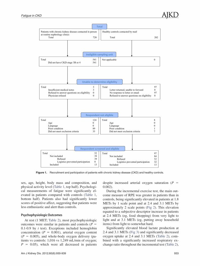
Recruitment and participation are shown in Fig 1.Mean eGFR in the patient group was 29 � 10 mL/min/1.73 m2; 5 patients were in stage 3b and 8 patientswere in stage 4 CKD. Causes of CKD were arterio-pathic (n � 1), diabetic (n � 3), infective/obstructive(n � 1), systemic (n � 1), and uncertain (n � 7).Mean values for all other clinical data and bloodvariables were within the ranges recommended by theRenal Association Standard (data not shown). Sevenpatients were prescribed an angiotensin-convertingenzyme inhibitor; 1 patient, an angiotensin II receptorantagonist; 5 patients, �-blockers; 2 patients, an�-blocker; 4 patients, a calcium channel blocker; 5patients, a lipid-lowering agent; 6 patients, a diuretic;and 5 patients, erythropoietin. Of the controls, 2 werehospital staff, 2 were friends of patients, and 9 werefrom local clubs and societies (eg, Rotary Club andWomen’s Institute). No participants were currentsmokers.
Matching of patients to controls was successful,
with no significant differences between groups forAm J Kidney Dis. 2012;60(6):930-939
with
Fatigue in CKD
sex, age, height, body mass and composition, andphysical activity level (Table 1, top half). Psychologi-cal measurements of fatigue were significantly el-evated in patients compared with controls (Table 1,bottom half). Patients also had significantly lowerscores of positive affect, suggesting that patients wereless enthusiastic and alert than controls.
PsychophysiologicOutcomes
At rest (1 MET; Table 2), most psychophysiologicoutcomes were similar in patients and controls (P �0.1-0.9 by t test). Exceptions included hemoglobinconcentration (P � 0.001), arterial oxygen content(P � 0.005), and whole-body oxygen delivery (pa-tients vs controls: 1,016 vs 1,249 mL/min of oxygen;
Total 124Age 0Language 0Prior condition 89Did not meet exclusion criteria 35
Total 52Not included 39
Refused 39Logistics prevented participation 0
Included 13
Respondent sc
Responde
Total 3Insufficient medical notes 3Refused to answer questions on eligibility 0Physician refused 0
Total 541Did not have CKD stage 3B or 4 541
Patients with chronic kidney disease contacted in personat routine nephrology clinics
Total 720
T
Unable to de
Ineligible
Figure 1. Recruitment and participation of patients
P � 0.05), which were all decreased in patients
Am J Kidney Dis. 2012;60(6):930-939
despite increased arterial oxygen saturation (P �0.002).
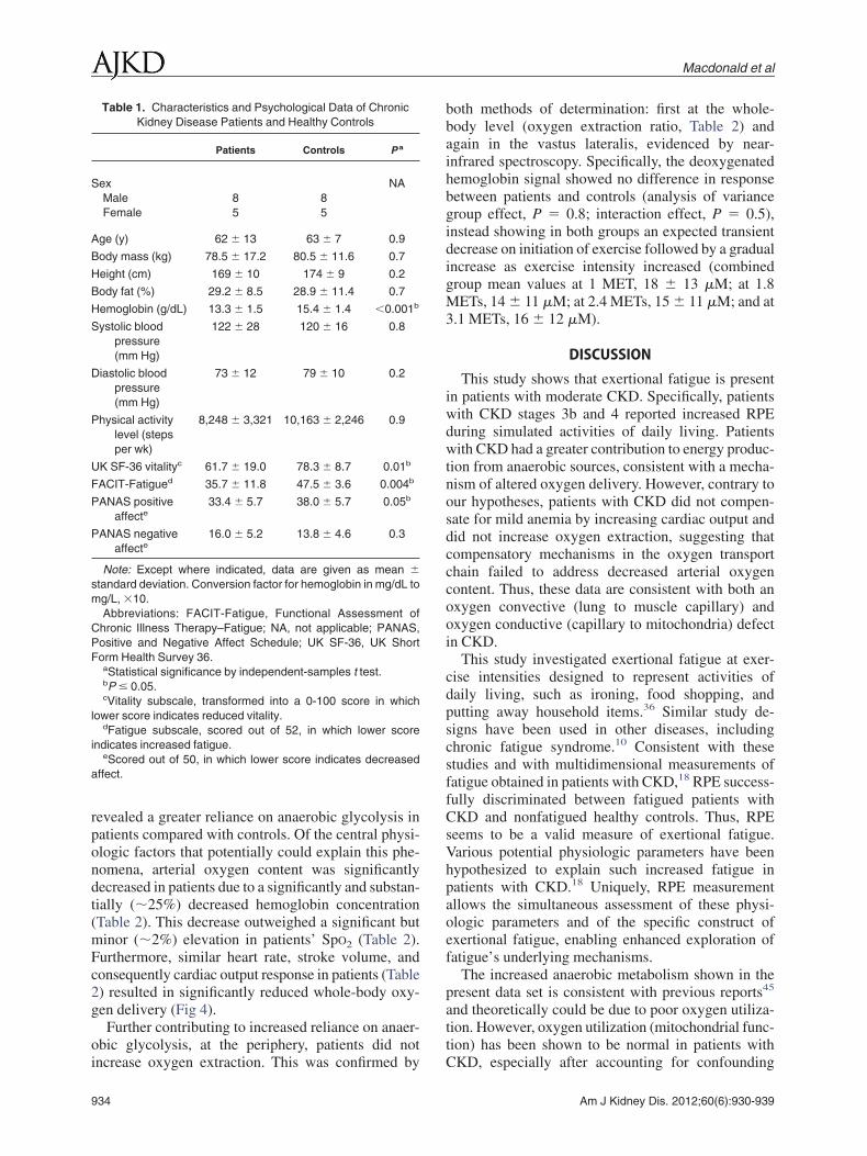
During the incremental exercise test, the main out-come measure of RPE was greater in patients than incontrols, being significantly elevated in patients at 1.8METs by 1 scale point and at 2.4 and 3.1 METs byapproximately 2 scale points (Fig 2). This elevationequated to a subjective descriptor increase in patientsat 2.4 METs (eg, food shopping) from very light tolight and at 3.1 METs (eg, putting away householditems) from light to somewhat hard.
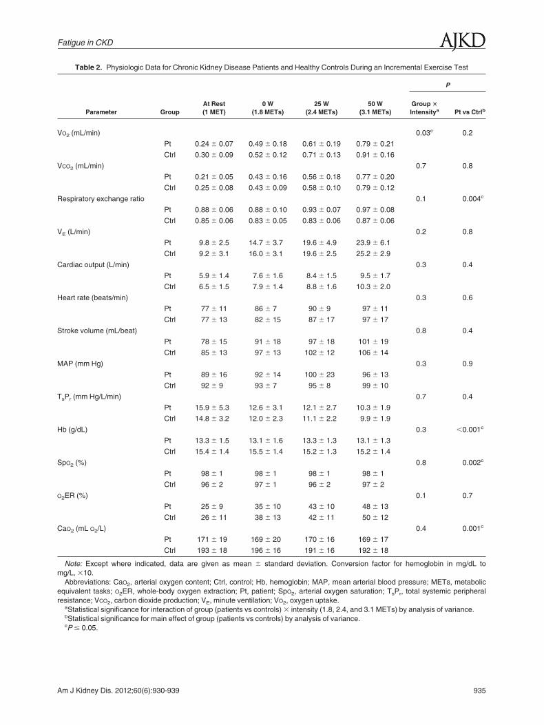
Significantly elevated blood lactate production at2.4 and 3.1 METs (Fig 3) and significantly decreasedoxygen uptake at 2.4 and 3.1 METs (Table 2), com-bined with a significantly increased respiratory ex-
otal 8Age 0Language 0Prior condition 1Did not meet exclusion criteria 7
otal 97Not included 84
Refused 52Logistics prevented participation 32
Included 13
d and eligible
t eligible
otal 97Letter returned, unable to forward 0No response to letter or email 97Refused to answer questions on eligibility 0
ealthy controls contacted by mail
Total 202
ot applicable 0
ne eligibility
ling unit
chronic kidney disease (CKD) and healthy controls.
T
T
reene
nt no
T
H
N
otal
termi
samp
change ratio throughout the incremental test (Table 2),
933
Macdonald et al
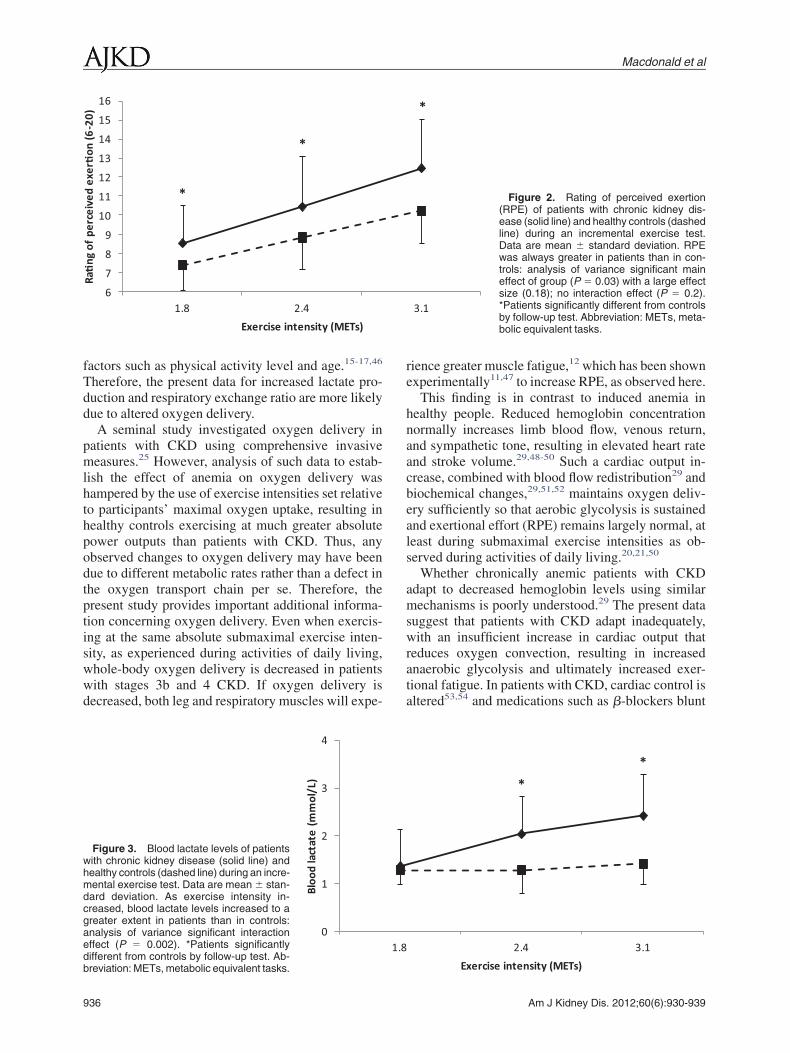
revealed a greater reliance on anaerobic glycolysis inpatients compared with controls. Of the central physi-ologic factors that potentially could explain this phe-nomena, arterial oxygen content was significantlydecreased in patients due to a significantly and substan-tially (�25%) decreased hemoglobin concentration(Table 2). This decrease outweighed a significant butminor (�2%) elevation in patients’ SpO2 (Table 2).Furthermore, similar heart rate, stroke volume, andconsequently cardiac output response in patients (Table2) resulted in significantly reduced whole-body oxy-gen delivery (Fig 4).
Further contributing to increased reliance on anaer-obic glycolysis, at the periphery, patients did not
Table 1. Characteristics and Psychological Data of ChronicKidney Disease Patients and Healthy Controls
Patients Controls P a
Sex NAMale 8 8Female 5 5
Age (y) 62 � 13 63 � 7 0.9
Body mass (kg) 78.5 � 17.2 80.5 � 11.6 0.7
Height (cm) 169 � 10 174 � 9 0.2
Body fat (%) 29.2 � 8.5 28.9 � 11.4 0.7
Hemoglobin (g/dL) 13.3 � 1.5 15.4 � 1.4 �0.001b
Systolic bloodpressure(mm Hg)
122 � 28 120 � 16 0.8
Diastolic bloodpressure(mm Hg)
73 � 12 79 � 10 0.2
Physical activitylevel (stepsper wk)
8,248 � 3,321 10,163 � 2,246 0.9
UK SF-36 vitalityc 61.7 � 19.0 78.3 � 8.7 0.01b
FACIT-Fatigued 35.7 � 11.8 47.5 � 3.6 0.004b
PANAS positiveaffecte
33.4 � 5.7 38.0 � 5.7 0.05b
PANAS negativeaffecte
16.0 � 5.2 13.8 � 4.6 0.3
Note: Except where indicated, data are given as mean �standard deviation. Conversion factor for hemoglobin in mg/dL tomg/L, �10.
Abbreviations: FACIT-Fatigue, Functional Assessment ofChronic Illness Therapy–Fatigue; NA, not applicable; PANAS,Positive and Negative Affect Schedule; UK SF-36, UK ShortForm Health Survey 36.
aStatistical significance by independent-samples t test.bP � 0.05.cVitality subscale, transformed into a 0-100 score in which
lower score indicates reduced vitality.dFatigue subscale, scored out of 52, in which lower score
indicates increased fatigue.eScored out of 50, in which lower score indicates decreased
affect.
increase oxygen extraction. This was confirmed by
934
both methods of determination: first at the whole-body level (oxygen extraction ratio, Table 2) andagain in the vastus lateralis, evidenced by near-infrared spectroscopy. Specifically, the deoxygenatedhemoglobin signal showed no difference in responsebetween patients and controls (analysis of variancegroup effect, P � 0.8; interaction effect, P � 0.5),instead showing in both groups an expected transientdecrease on initiation of exercise followed by a gradualincrease as exercise intensity increased (combinedgroup mean values at 1 MET, 18 � 13 �M; at 1.8METs, 14 � 11 �M; at 2.4 METs, 15 � 11 �M; and at3.1 METs, 16 � 12 �M).
DISCUSSION
This study shows that exertional fatigue is presentin patients with moderate CKD. Specifically, patientswith CKD stages 3b and 4 reported increased RPEduring simulated activities of daily living. Patientswith CKD had a greater contribution to energy produc-tion from anaerobic sources, consistent with a mecha-nism of altered oxygen delivery. However, contrary toour hypotheses, patients with CKD did not compen-sate for mild anemia by increasing cardiac output anddid not increase oxygen extraction, suggesting thatcompensatory mechanisms in the oxygen transportchain failed to address decreased arterial oxygencontent. Thus, these data are consistent with both anoxygen convective (lung to muscle capillary) andoxygen conductive (capillary to mitochondria) defectin CKD.
This study investigated exertional fatigue at exer-cise intensities designed to represent activities ofdaily living, such as ironing, food shopping, andputting away household items.36 Similar study de-signs have been used in other diseases, includingchronic fatigue syndrome.10 Consistent with thesestudies and with multidimensional measurements offatigue obtained in patients with CKD,18 RPE success-fully discriminated between fatigued patients withCKD and nonfatigued healthy controls. Thus, RPEseems to be a valid measure of exertional fatigue.Various potential physiologic parameters have beenhypothesized to explain such increased fatigue inpatients with CKD.18 Uniquely, RPE measurementallows the simultaneous assessment of these physi-ologic parameters and of the specific construct ofexertional fatigue, enabling enhanced exploration offatigue’s underlying mechanisms.
The increased anaerobic metabolism shown in thepresent data set is consistent with previous reports45
and theoretically could be due to poor oxygen utiliza-tion. However, oxygen utilization (mitochondrial func-tion) has been shown to be normal in patients with
CKD, especially after accounting for confoundingAm J Kidney Dis. 2012;60(6):930-939
Fatigue in CKD
Table 2. Physiologic Data for Chronic Kidney Disease Patients and Healthy Controls During an Incremental Exercise Test
Parameter GroupAt Rest(1 MET)
0 W(1.8 METs)
25 W(2.4 METs)
50 W(3.1 METs)
P
Group �
Intensitya Pt vs Ctrlb
VO2 (mL/min) 0.03c 0.2
Pt 0.24 � 0.07 0.49 � 0.18 0.61 � 0.19 0.79 � 0.21
Ctrl 0.30 � 0.09 0.52 � 0.12 0.71 � 0.13 0.91 � 0.16
VCO2 (mL/min) 0.7 0.8
Pt 0.21 � 0.05 0.43 � 0.16 0.56 � 0.18 0.77 � 0.20
Ctrl 0.25 � 0.08 0.43 � 0.09 0.58 � 0.10 0.79 � 0.12
Respiratory exchange ratio 0.1 0.004c
Pt 0.88 � 0.06 0.88 � 0.10 0.93 � 0.07 0.97 � 0.08
Ctrl 0.85 � 0.06 0.83 � 0.05 0.83 � 0.06 0.87 � 0.06
VE (L/min) 0.2 0.8
Pt 9.8 � 2.5 14.7 � 3.7 19.6 � 4.9 23.9 � 6.1
Ctrl 9.2 � 3.1 16.0 � 3.1 19.6 � 2.5 25.2 � 2.9
Cardiac output (L/min) 0.3 0.4
Pt 5.9 � 1.4 7.6 � 1.6 8.4 � 1.5 9.5 � 1.7
Ctrl 6.5 � 1.5 7.9 � 1.4 8.8 � 1.6 10.3 � 2.0
Heart rate (beats/min) 0.3 0.6
Pt 77 � 11 86 � 7 90 � 9 97 � 11
Ctrl 77 � 13 82 � 15 87 � 17 97 � 17
Stroke volume (mL/beat) 0.8 0.4
Pt 78 � 15 91 � 18 97 � 18 101 � 19
Ctrl 85 � 13 97 � 13 102 � 12 106 � 14
MAP (mm Hg) 0.3 0.9
Pt 89 � 16 92 � 14 100 � 23 96 � 13
Ctrl 92 � 9 93 � 7 95 � 8 99 � 10
TsPr (mm Hg/L/min) 0.7 0.4
Pt 15.9 � 5.3 12.6 � 3.1 12.1 � 2.7 10.3 � 1.9
Ctrl 14.8 � 3.2 12.0 � 2.3 11.1 � 2.2 9.9 � 1.9
Hb (g/dL) 0.3 �0.001c
Pt 13.3 � 1.5 13.1 � 1.6 13.3 � 1.3 13.1 � 1.3
Ctrl 15.4 � 1.4 15.5 � 1.4 15.2 � 1.3 15.2 � 1.4
SpO2 (%) 0.8 0.002c
Pt 98 � 1 98 � 1 98 � 1 98 � 1
Ctrl 96 � 2 97 � 1 96 � 2 97 � 2
O2ER (%) 0.1 0.7
Pt 25 � 9 35 � 10 43 � 10 48 � 13
Ctrl 26 � 11 38 � 13 42 � 11 50 � 12
CaO2 (mL O2/L) 0.4 0.001c
Pt 171 � 19 169 � 20 170 � 16 169 � 17
Ctrl 193 � 18 196 � 16 191 � 16 192 � 18
Note: Except where indicated, data are given as mean � standard deviation. Conversion factor for hemoglobin in mg/dL tomg/L, �10.
Abbreviations: CaO2, arterial oxygen content; Ctrl, control; Hb, hemoglobin; MAP, mean arterial blood pressure; METs, metabolicequivalent tasks; O2ER, whole-body oxygen extraction; Pt, patient; SpO2, arterial oxygen saturation; TsPr, total systemic peripheralresistance; VCO2, carbon dioxide production; VE, minute ventilation; VO2, oxygen uptake.
aStatistical significance for interaction of group (patients vs controls) � intensity (1.8, 2.4, and 3.1 METs) by analysis of variance.bStatistical significance for main effect of group (patients vs controls) by analysis of variance.
cP � 0.05.Am J Kidney Dis. 2012;60(6):930-939 935
Macdonald et al
factors such as physical activity level and age.15-17,46
Therefore, the present data for increased lactate pro-duction and respiratory exchange ratio are more likelydue to altered oxygen delivery.
A seminal study investigated oxygen delivery inpatients with CKD using comprehensive invasivemeasures.25 However, analysis of such data to estab-lish the effect of anemia on oxygen delivery washampered by the use of exercise intensities set relativeto participants’ maximal oxygen uptake, resulting inhealthy controls exercising at much greater absolutepower outputs than patients with CKD. Thus, anyobserved changes to oxygen delivery may have beendue to different metabolic rates rather than a defect inthe oxygen transport chain per se. Therefore, thepresent study provides important additional informa-tion concerning oxygen delivery. Even when exercis-ing at the same absolute submaximal exercise inten-sity, as experienced during activities of daily living,whole-body oxygen delivery is decreased in patientswith stages 3b and 4 CKD. If oxygen delivery isdecreased, both leg and respiratory muscles will expe-
6
7
8
9
10
11
12
13
14
15
16
1.8 2.4
Ra�
ng o
f pe
rcei
ved
exer
�on
(6-2
0)
Exercise intensity (METs)
*
*
0
1
2
3
4
Bloo
d la
ctat
e (m
mol
/L)
Figure 3. Blood lactate levels of patientswith chronic kidney disease (solid line) andhealthy controls (dashed line) during an incre-mental exercise test. Data are mean � stan-dard deviation. As exercise intensity in-creased, blood lactate levels increased to agreater extent in patients than in controls:analysis of variance significant interactioneffect (P � 0.002). *Patients significantlydifferent from controls by follow-up test. Ab-
breviation: METs, metabolic equivalent tasks.936
rience greater muscle fatigue,12 which has been shownexperimentally11,47 to increase RPE, as observed here.
This finding is in contrast to induced anemia inhealthy people. Reduced hemoglobin concentrationnormally increases limb blood flow, venous return,and sympathetic tone, resulting in elevated heart rateand stroke volume.29,48-50 Such a cardiac output in-crease, combined with blood flow redistribution29 andbiochemical changes,29,51,52 maintains oxygen deliv-ery sufficiently so that aerobic glycolysis is sustainedand exertional effort (RPE) remains largely normal, atleast during submaximal exercise intensities as ob-served during activities of daily living.20,21,50
Whether chronically anemic patients with CKDadapt to decreased hemoglobin levels using similarmechanisms is poorly understood.29 The present datasuggest that patients with CKD adapt inadequately,with an insufficient increase in cardiac output thatreduces oxygen convection, resulting in increasedanaerobic glycolysis and ultimately increased exer-tional fatigue. In patients with CKD, cardiac control isaltered53,54 and medications such as �-blockers blunt
3.1
*
Figure 2. Rating of perceived exertion(RPE) of patients with chronic kidney dis-ease (solid line) and healthy controls (dashedline) during an incremental exercise test.Data are mean � standard deviation. RPEwas always greater in patients than in con-trols: analysis of variance significant maineffect of group (P � 0.03) with a large effectsize (0.18); no interaction effect (P � 0.2).*Patients significantly different from controlsby follow-up test. Abbreviation: METs, meta-bolic equivalent tasks.
2.4 3.1
*
*
1.8
Exercise intensity (METs)
Am J Kidney Dis. 2012;60(6):930-939
Fatigue in CKD
sympathetic stimulation,29 potentially explaining theobserved lack of central compensation. Blood flowredistribution and biochemical changes at muscle levelalso may be inadequate and are worthy of furtherinvestigation.29,51,52
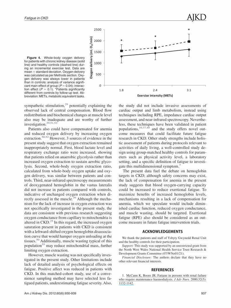
Patients also could have compensated for anemiaand reduced oxygen delivery by increasing oxygenextraction.50,52 However, 3 sources of evidence in thepresent study suggest that oxygen extraction remainedinappropriately normal. First, blood lactate level andrespiratory exchange ratio were increased, showingthat patients relied on anaerobic glycolysis rather thanincreased oxygen extraction to sustain aerobic glyco-lysis. Second, whole-body oxygen extraction ratio,calculated from whole-body oxygen uptake and oxy-gen delivery, was similar between patients and con-trols. Third, near-infrared spectroscopy measurementsof deoxygenated hemoglobin in the vastus lateralisdid not increase in patients compared with controls,indicative of unchanged oxygen extraction when di-rectly assessed in the muscle.39 Although the mecha-nism for the lack of increase in oxygen extraction wasnot specifically investigated in the present study, thedata are consistent with previous research suggestingoxygen conductance from capillary to mitochondria isaltered in CKD.25 In this regard, the increased oxygensaturation present in patients with CKD is consistentwith a leftward-shifted oxygen hemoglobin disassocia-tion curve that would hamper oxygen unloading at thetissues.50 Additionally, muscle wasting typical of thispopulation55 may reduce mitochondrial mass, furtherlimiting oxygen extraction.
However, muscle wasting was not specifically inves-tigated in the present study. Other limitations includelack of detailed analysis of psychological effects onfatigue. Positive affect was reduced in patients withCKD. In this matched-cohort study, use of a conve-nience sampling method may have selected less fa-
0
500
1000
1500
2000
2500
Oxy
gen
deliv
ery
(mL
O2/
min
)
Figure 4. Whole-body oxygen deliveryfor patients with chronic kidney disease (solidline) and healthy controls (dashed line) dur-ing an incremental exercise test. Data aremean � standard deviation. Oxygen deliverywas calculated as per Methods section. Oxy-gen delivery was always lower in patientsthan in controls: analysis of variance signifi-cant main effect of group (P � 0.04); interac-tion effect (P � 0.1). *Patients significantlydifferent from controls by follow-up test. Ab-breviation: METs, metabolic equivalent tasks.
tigued patients, underestimating fatigue severity. Also,
Am J Kidney Dis. 2012;60(6):930-939
the study did not include invasive assessments ofcardiac output and limb metabolism, instead usingtechniques including RPE, impedance cardiac outputassessment, and near-infrared spectroscopy. Neverthe-less, these techniques have been validated in patientpopulations,10,37,40 and the study offers novel out-come measures that could facilitate future fatigueresearch in CKD. Other study strengths include holis-tic assessment of patients during protocols relevant toactivities of daily living, a well-controlled study de-sign using group-matched healthy controls for param-eters such as physical activity level, a laboratorysetting, and a specific definition of fatigue to investi-gate this multidimensional symptom.7
The present data fuel the debate on hemoglobintargets in CKD; although safety concerns may exist,the lack of compensation for anemia in the presentstudy suggests that blood oxygen-carrying capacitycould be increased to reduce exertional fatigue. Tomaximize benefits of increased hemoglobin levels,mechanisms resulting in a lack of compensation foranemia, which we speculate would include dimin-ished cardiac function, reduced oxygen conductance,and muscle wasting, should be targeted. Exertionalfatigue (RPE) also should be considered as an out-come measure in future fatigue intervention trials.
ACKNOWLEDGEMENTSWe thank the patients and staff of Ysbyty Gwynedd Renal Unit
and the healthy controls for their participation.Support: This study was supported by an unrestricted grant from
the North West Wales National Health Service Trust Research &Development Grants Committee (07/WNo01/21).
Financial Disclosure: The authors declare that they have noother relevant financial interests.
REFERENCES1. McCann K, Boore JR. Fatigue in persons with renal failure
who require maintenance haemodialysis. J Adv Nurs. 2000;32(5):
1.8 2.4 3.1
Exercise intensity (METs)
* *
*
1132-1142.
937
Macdonald et al
2. Weisbord SD, Fried LF, Arnold RM, et al. Prevalence,severity, and importance of physical and emotional symptoms inchronic hemodialysis patients. J Am Soc Nephrol. 2005;16(8):2487-2494.
3. Murtagh FE, Addington-Hall J, Higginson IJ. The preva-lence of symptoms in end-stage renal disease: a systematic review.Adv Chronic Kidney Dis. 2007;14(1):82-99.
4. Laupacis A, Muirhead N, Keown P, Wong C. A disease-specific questionnaire for assessing quality of life in patients onhemodialysis. Nephron. 1992;60(3):302-306.
5. Curtin RB, Bultman DC, Thomas-Hawkins C, Walters BA,Schatell D. Hemodialysis patients’ symptom experiences: effectson physical and mental functioning. Nephrol Nurs J. 2002;29(6):562, 567-574.
6. Koyama H, Fukuda S, Shoji T, et al. Fatigue is a predictor forcardiovascular outcomes in patients undergoing hemodialysis.Clin J Am Soc Nephrol. 2010;5(4):659-666.
7. Portenoy RK. Fatigue. In: Max MB, Lynn J, eds. InteractiveTextbook of Clinical Symptom Research. Bethesda, MD: NationalInstitutes of Health; 2003:1-45.
8. Tiesinga LJ, Dassen TWN, Halfens RJG. Fatigue: a sum-mary of the definitions, dimensions and indicators. Nurs Diagn.1996;7(2):51-62.
9. Borg G. Borg’s Perceived Exertion and Pain Scales. Cham-paign, IL: Human Kinetics; 1998.
10. Wallman KE, Morton AR, Goodman C, Grove R. Physiolog-ical responses during a submaximal cycle test in chronic fatiguesyndrome. Med Sci Sports Exerc. 2004;36(10):1682-1688.
11. Marcora SM, Bosio A, de Morree HM. Locomotor musclefatigue increases cardiorespiratory responses and reduces perfor-mance during intense cycling exercise independently from meta-bolic stress. Am J Physiol Regul Integr Comp Physiol. 2008;294(3):R874-R883.
12. Amann M, Calbet JA. Convective oxygen transport andfatigue. J Appl Physiol. 2008;104(3):861-870.
13. Campistol JM. Uremic myopathy. Kidney Int. 2002;62(5):1901-1913.
14. Moore GE, Parsons DB, Stray-Gundersen J, et al. Uremicmyopathy limits aerobic capacity in hemodialysis patients. Am JKidney Dis. 1993;22(2):277-287.
15. Thompson CH, Kemp GJ, Barnes PR, et al. Uraemicmuscle metabolism at rest and during exercise. Nephrol DialTransplant. 1994;9(11):1600-1605.
16. Barany P, Wibom R, Hultman E, Bergstrom J. ATP produc-tion in isolated muscle mitochondria from haemodialysis patients:effects of correction of anaemia with erythropoietin. Clin Sci(Lond). 1991;81(5):645-653.
17. Miro O, Marrades RM, Roca J, et al. Skeletal musclemitochondrial function is preserved in young patients with chronicrenal failure. Am J Kidney Dis. 2002;39(5):1025-1031.
18. Jhamb M, Weisbord SD, Steel JL, Unruh M. Fatigue inpatients receiving maintenance dialysis: a review of definitions,measures, and contributing factors. Am J Kidney Dis. 2008;52(2):353-365.
19. Kliger AS, Fishbane S, Finkelstein FO. Erythropoieticstimulating agents and quality of a patient’s life: individualizinganemia treatment. Clin J Am Soc Nephrol. 2012;7(2):354-357.
20. Woodson RD, Wills RE, Lenfant C. Effect of acute andestablished anemia on O2 transport at rest, submaximal and maxi-mal work. J Appl Physiol. 1978;44(1):36-43.
21. Ekblom B, Goldbarg AN, Gullbring B. Response to exer-cise after blood loss and reinfusion. J Appl Physiol. 1972;33(2):175-180.
22. Ekblom B, Wilson G, Astrand PO. Central circulationduring exercise after venesection and reinfusion of red blood cells.
J Appl Physiol. 1976;40(3):379-383.938
23. Ross SD, Fahrbach K, Frame D, et al. The effect of anemiatreatment on selected health-related quality-of-life domains: asystematic review. Clin Ther. 2003;25(6):1786-1805.
24. Johansen KL, Finkelstein FO, Revicki DA, et al. Systematicreview of the impact of erythropoiesis-stimulating agents on fa-tigue in dialysis patients. Nephrol Dial Transplant. 2012;27(6):2418-2425.
25. Marrades RM, Roca J, Campistol JM, et al. Effects oferythropoietin on muscle O2 transport during exercise in patientswith chronic renal failure. J Clin Invest. 1996;97(9):2092-2100.
26. Painter P, Moore GE. The impact of recombinant humanerythropoietin on exercise capacity in hemodialysis patients. AdvRen Replace Ther. 1994;1(1):55-65.
27. Sala E, Noyszewski EA, Campistol JM, et al. Impairedmuscle oxygen transfer in patients with chronic renal failure. Am JPhysiol Regul Integr Comp Physiol. 2001;280(4):R1240-R1248.
28. Perlman RL, Finkelstein FO, Liu L, et al. Quality of life inchronic kidney disease (CKD): a cross-sectional analysis in theRenal Research Institute-CKD Study. Am J Kidney Dis. 2005;45(4):658-666.
29. Hebert PC, Van der LP, Biro G, Hu LQ. Physiologic aspectsof anemia. Crit Care Clin. 2004;20(2):187-212.
30. Levey AS, Coresh J, Balk E, et al. National Kidney Founda-tion practice guidelines for chronic kidney disease: evaluation,classification, and stratification. Ann Intern Med. 2003;139(2):137-147.
31. Maisonneuve P, Agodoa L, Gellert R, et al. Distribution ofprimary renal diseases leading to end-stage renal failure in theUnited States, Europe, and Australia/New Zealand: results from aninternational comparative study. Am J Kidney Dis. 2000;35(1):157-165.
32. Ware JE, Kosinski M, Bjorner JB, Turner-Bowker DM,Gandek B, Maruish ME. User’s Manual for the SF-36v2 HealthSurvey. 2nd ed. Lincoln, RI: QualityMetric; 2007.
33. Yellen SB, Cella DF, Webster K, Blendowski C, Kaplan E.Measuring fatigue and other anemia-related symptoms with theFunctional Assessment of Cancer Therapy (FACT) measurementsystem. J Pain Symptom Manage. 1997;13(2):63-74.
34. Crawford JR, Henry JD. The Positive and Negative AffectSchedule (PANAS): construct validity, measurement propertiesand normative data in a large non-clinical sample. Br J ClinPsychol. 2004;43(pt 3):245-265.
35. Watson D, Clark LA, Tellegen A. Development and valida-tion of brief measures of positive and negative affect: the PANASscales. J Pers Soc Psychol. 1988;54(6):1063-1070.
36. Ainsworth BE, Haskell WL, Herrmann SD, et al. 2011Compendium of Physical Activities: a second update of codes andMET values. Med Sci Sports Exerc. 2011;43(8):1575-1581.
37. Richard R, Lonsdorfer-Wolf E, Charloux A, et al. Non-invasive cardiac output evaluation during a maximal progressiveexercise test, using a new impedance cardiograph device. EurJ Appl Physiol. 2001;85(3-4):202-207.
38. Robergs RA, Keteyian SJ. Fundamentals of Exercise Physi-ology. 2nd ed. New York, NY: McGraw-Hill Higher Education;2003.
39. De Blasi RA, Ferrari M, Natali A, et al. Noninvasivemeasurement of forearm blood flow and oxygen consumption bynear-infrared spectroscopy. J Appl Physiol. 1994;76(3):1388-1393.
40. Van Beekvelt MC, Colier WN, Wevers RA, van EngelenBG. Quantitative measurement of oxygen consumption and fore-arm blood flow in patients with mitochondrial myopathies. AdvExp Med Biol. 1999;471:313-319.
41. Stevens JP. Applied Multivariate Statistics for the SocialSciences. 4th ed. London, England: Lawrence Erlbaum Associates;
2002.Am J Kidney Dis. 2012;60(6):930-939
Fatigue in CKD
42. Ludbrook J. Multiple comparison procedures updated. ClinExp Pharmacol Physiol. 1998;25(12):1032-1037.
43. Levine TR, Hullett CH. Eta squared, partial eta squared,and misreporting of effect size in communication research. HumCommun Res. 2002; 28(4):612-625.
44. Bausell R, Li Y. Power Analysis for Experimental Research.Cambridge, England: Cambridge University Press; 2002.
45. Parrish AE. The effect of minimal exercise on the bloodlactate in azotemic subjects. Clin Nephrol. 1981;16(1):35-39.
46. Moore GE, Bertocci LA, Painter PL. 31P-Magnetic reso-nance spectroscopy assessment of subnormal oxidative metabo-lism in skeletal muscle of renal failure patients. J Clin Invest.1993;91(2):420-424.
47. Romer LM, Polkey MI. Exercise-induced respiratory musclefatigue: implications for performance. J Appl Physiol. 2008;104(3):879-888.
48. Saltin B, Radegran G, Koskolou MD, Roach RC. Skeletalmuscle blood flow in humans and its regulation during exercise.Acta Physiol Scand. 1998;162(3):421-436.
49. Roach RC, Koskolou MD, Calbet JA, Saltin B. Arterial O
2content and tension in regulation of cardiac output and leg blood
Am J Kidney Dis. 2012;60(6):930-939
flow during exercise in humans. Am J Physiol. 1999;276(2, pt2):H438-H445.
50. Koskolou MD, Roach RC, Calbet JA, Radegran G, Saltin B.Cardiovascular responses to dynamic exercise with acute anemiain humans. Am J Physiol. 1997;273(4, pt 2):H1787-H1793.
51. Meierhenrich R, Jedicke H, Voigt A, Lange H. The effect oferythropoietin on lactate, pyruvate and excess lactate under physi-cal exercise in dialysis patients. Clin Nephrol. 1996;45(2):90-97.
52. Sproule BJ, Mitchell JH, Miller WF. Cardiopulmonaryphysiological responses to heavy exercise in patients with anemia.J Clin Invest. 1960;39(2):378-388.
53. Frei U, Nonnast-Daniel B, Schaffer J, Koch KM. Adversecardiovascular effects of partial correction of renal anemia byrecombinant human erythropoietin. Contrib Nephrol. 1990;82:72-78.
54. Painter P, Messer-Rehak D, Hanson P, Zimmerman SW,Glass NR. Exercise capacity in hemodialysis, CAPD, and renaltransplant patients. Nephron. 1986;42(1):47-51.
55. Macdonald JH, Phanish MK, Marcora SM, et al. Muscleinsulin-like growth factor status, body composition, and functionalcapacity in hemodialysis patients. J Ren Nutr. 2004;14(4):248-
252.939
Related Documents