Am J Transl Res 2014;6(5):422-438 www.ajtr.org /ISSN:1943-8141/AJTR0000794 Review Article Exercise training and immune crosstalk in breast cancer microenvironment: exploring the paradigms of exercise-induced immune modulation and exercise-induced myokines Jorming Goh 1 , Negin Niksirat 2 , Kristin L Campbell 2,3 1 Screening & Diagnostics Technologies Laboratory, Defence Medical and Environmental Research Institute, DSO National Laboratories, Republic of Singapore; 2 Rehabilitation Sciences, University of British Columbia, Vancouver B.C., Canada; 3 Cancer Prevention Center, University of British Columbia, Vancouver B.C., Canada Received May 14, 2014; Accepted June 16, 2014; Epub October 11, 2014; Published October 15, 2014 Abstract: Observational research suggests that exercise may reduce the risk of breast cancer and improve survival. One proposed mechanism for the protective effect of aerobic exercise related to cancer risk and outcomes, but has not been examined definitively, is the immune response to aerobic exercise. Two prevailing paradigms are proposed. The first considers the host immune response as modifiable by aerobic exercise training. This exercise-modulated immune-tumor crosstalk in the mammary microenvironment may alter the balance between tumor initiation and progression versus tumor suppression. The second paradigm considers the beneficial role of exercise-induced, skel- etal muscle-derived cytokines, termed “myokines”. These myokines exert endocrine-like effects on multiple organs, including the mammary glands. In this systematic review, we i) define the role of macrophages and T-cells in breast cancer initiation and progression; ii) address the two paradigms that support exercise-induced immunomodulation; iii) systematically assessed the literature for exercise intervention that assessed biomarkers relevant to both para- digms in human intervention trials of aerobic exercise training, in healthy women and women with breast cancer; iv) incorporated pre-clinical animal studies and non-RCTs for background discussion of putative mechanisms, through which aerobic exercise training modulates the immunological crosstalk, or the myokine-tumor interaction in the tumor microenvironment; and v) speculated on the potential biomarkers and mechanisms that define an exercise- induced, anti-tumor “signature”, with a view toward developing relevant biomarkers for future aerobic exercise in- tervention trials. Keywords: Exercise, breast cancer, immune, myokines, translational, immunotherapy Introduction Exercise training in women with early stage breast cancer, both during or following cancer treatment, enhances aerobic capacity, muscu- lar strength and physical function, as well as improves quality of life, anxiety and depression [1]. There is now substantial observational evi- dence that regular participation in moderate- to-vigorous intensity physical activity reduces risk of developing postmenopausal breast can- cer [2] and improves survival following a breast cancer diagnosis [3-5]. Several mechanisms for this protective effect have been proposed including alterations in sex hormones, meta- bolic hormones, DNA repair capacity and sys- temic low-grade inflammation [6, 7]. However, one proposed mechanism that has received less attention is host immune function. The potential impact of exercise on the immune system has been examined related to both ath- letic performance [8] and risk of illness, such as increased risk of upper respiratory track infec- tions in athletes with a heavy volume of training [9, 10]. More recently, there has been interest in the potential role of exercise in mitigating chronic disease outcomes that have an immune or inflammatory component, such as to prevent and reduce the risk of a breast cancer recur- rence [11]. Although research to date support a modulato- ry role of exercise and physical activity on

Welcome message from author
This document is posted to help you gain knowledge. Please leave a comment to let me know what you think about it! Share it to your friends and learn new things together.
Transcript
Am J Transl Res 2014;6(5):422-438www.ajtr.org /ISSN:1943-8141/AJTR0000794
Review Article Exercise training and immune crosstalk in breast cancer microenvironment: exploring the paradigms of exercise-induced immune modulation and exercise-induced myokines
Jorming Goh1, Negin Niksirat2, Kristin L Campbell2,3
1Screening & Diagnostics Technologies Laboratory, Defence Medical and Environmental Research Institute, DSO National Laboratories, Republic of Singapore; 2Rehabilitation Sciences, University of British Columbia, Vancouver B.C., Canada; 3Cancer Prevention Center, University of British Columbia, Vancouver B.C., Canada
Received May 14, 2014; Accepted June 16, 2014; Epub October 11, 2014; Published October 15, 2014
Abstract: Observational research suggests that exercise may reduce the risk of breast cancer and improve survival. One proposed mechanism for the protective effect of aerobic exercise related to cancer risk and outcomes, but has not been examined definitively, is the immune response to aerobic exercise. Two prevailing paradigms are proposed. The first considers the host immune response as modifiable by aerobic exercise training. This exercise-modulated immune-tumor crosstalk in the mammary microenvironment may alter the balance between tumor initiation and progression versus tumor suppression. The second paradigm considers the beneficial role of exercise-induced, skel-etal muscle-derived cytokines, termed “myokines”. These myokines exert endocrine-like effects on multiple organs, including the mammary glands. In this systematic review, we i) define the role of macrophages and T-cells in breast cancer initiation and progression; ii) address the two paradigms that support exercise-induced immunomodulation; iii) systematically assessed the literature for exercise intervention that assessed biomarkers relevant to both para-digms in human intervention trials of aerobic exercise training, in healthy women and women with breast cancer; iv) incorporated pre-clinical animal studies and non-RCTs for background discussion of putative mechanisms, through which aerobic exercise training modulates the immunological crosstalk, or the myokine-tumor interaction in the tumor microenvironment; and v) speculated on the potential biomarkers and mechanisms that define an exercise-induced, anti-tumor “signature”, with a view toward developing relevant biomarkers for future aerobic exercise in-tervention trials.
Keywords: Exercise, breast cancer, immune, myokines, translational, immunotherapy
Introduction
Exercise training in women with early stage breast cancer, both during or following cancer treatment, enhances aerobic capacity, muscu-lar strength and physical function, as well as improves quality of life, anxiety and depression [1]. There is now substantial observational evi-dence that regular participation in moderate-to-vigorous intensity physical activity reduces risk of developing postmenopausal breast can-cer [2] and improves survival following a breast cancer diagnosis [3-5]. Several mechanisms for this protective effect have been proposed including alterations in sex hormones, meta-bolic hormones, DNA repair capacity and sys-temic low-grade inflammation [6, 7]. However,
one proposed mechanism that has received less attention is host immune function.
The potential impact of exercise on the immune system has been examined related to both ath-letic performance [8] and risk of illness, such as increased risk of upper respiratory track infec-tions in athletes with a heavy volume of training [9, 10]. More recently, there has been interest in the potential role of exercise in mitigating chronic disease outcomes that have an immune or inflammatory component, such as to prevent and reduce the risk of a breast cancer recur-rence [11].
Although research to date support a modulato-ry role of exercise and physical activity on
Exercise and immune crosstalk in breast cancer microenvironment
423 Am J Transl Res 2014;6(5):422-438
immune function, the specific mechanisms relating exercise-induced immune responses to cancer outcomes in humans remains to be elu-cidated. Furthermore, the novel concept of skeletal muscle as an endocrine organ [12] has received substantial attention in muscle physi-ology research in the past decade. Bente Klarlund Pedersen and colleagues have con-ceptualized and demonstrated how contracting skeletal muscle is capable of secreting muscle-derived cytokines, which they termed “myo-kines”. These myokines exerted endocrine-like functions in other tissues and organs, including the adipose tissue, colon liver, and brain. The extensive body of work by Pedersen’s group as well as others [13-18], suggest that exercise may modulate the host immune response via skeletal muscle-organ crosstalk.
In this review, we will: i) define the prognostic role of macrophages and T-cells in breast can-cer; ii) outline two proposed paradigms that support exercise-induced immunomodulation; iii) systematically review the literature for human exercise intervention that assessed bio-markers for breast cancer risk and survival; iv) incorporate pre-clinical animal studies and non-RCTs to discuss putative mechanisms through which exercise training modulates the immunological crosstalk, or the myokine-tumor interaction in the tumor microenvironment; and v) speculate on the potential biomarkers and mechanisms that define an exercise-induced, anti-tumor “signature”, with a view toward developing relevant biomarkers for future exer-cise intervention trials.
The role of the immune system in breast can-cer and potential immune biomarkers
The immune system has a Janus-like duality in breast cancer; on the one hand, various facets of the immune system become co-opted to sup-port tumor growth. Conversely, the immune system remains capable of mounting an anti-tumor response against breast cancer cells. The balance between immunosuppression which allows tumor growth, versus immune rejection of neoplastic cells which limits tumor growth depends on the type of, and activation state of the leukocytes, as well as their cross-talk with other stromal cells and tumor cells in the tumor microenvironment [19]. This review will not cover all the immune cells involved in the cancer immunology literature, but rather
focus on the role of macrophages and T cells in mediating tumor response and identified inflammatory biomarkers of breast cancer risk.
Macrophages
Tissue macrophages originate from circulating monocytes, and are found in large abundance in the breast tumor microenvironment; a trait associated with poor prognosis in cancer patients [20]. Specifically, a 77% increased risk of mortality was associated with increased double immunostaining of cluster of differentia-tion (CD)68+ and proliferating cell nuclear anti-gen (PCNA)+ macrophages in human breast adenocarcinomas [20]. These tumor-associat-ed macrophages (TAMs) represent the largest proportion of leukocytes in the breast tumor microenvironment and are attractive targets for immunotherapy.
TAMs have been broadly classified into two dif-ferent subsets, the M1-polarized macrophages, and the M2-polarized macrophages [21]. M1 macrophages have been termed classically activated macrophages, in that they are acti-vated by T-helper (Th)1-type cytokines, such as interferon (IFN)-γ as well as molecules associ-ated with pathogen associated molecular pat-terns (PAMPs) and damage associated molecu-lar patterns (DAMPs) [22]. DAMPs, such as high mobility group box protein (HMGB)-1 which is secreted extracellularly, stimulates the produc-tion of IFN-γ from natural killer (NK) cells, with potential implications for tumor death [23].
Some notable characteristics of M1 macro-phages include low expression of Th2-related cytokines, such as interleukin (IL)-4 and IL-10, and high expression of Th1-related cytokines such as IL-12, tumor necrosis factor (TNF)-α and IL-6 [19]. In addition, M1 macrophages are able to induce tumor destruction in vitro. C57BL/6 female mice bearing the Lewis Lung Carcinoma (3LLC) treated with microspheres containing IL-12 had significantly reduced pri-mary tumor mass compared with placebo treat-ed controls [24]. IL-12 can activate tumoricidal NK cells [25], as well as activate Th1 cells to mediate tumor killing [26]. IL-12 also induces the extracellular secretion of IL-15 from TAMs. Importantly, the IL-12-mediated cytotoxic activ-ity of M1 macrophages appeared to be depen-dent on the presence of IL-15, where prior blockade with IL-15 antibody resulted in an

Exercise and immune crosstalk in breast cancer microenvironment
424 Am J Transl Res 2014;6(5):422-438
attenuated effect on shrinking primary tumor mass.
The utility of IL-12 and IL-15 as anti-tumor ther-apeutics has been documented in other pre-clinical models. For instance, intraperitoneal injection of IL-12 into AB6F1 mice bearing Sa1 ascites was successful in clearing tumors in 40% of the mice [27]. As well, the IL-12 treat-ment resulted in massive macrophage infiltra-tion in the Sa1 ascites, comprising up to 75% of the total cell population, compared with 9%, in untreated conditions. Comes et al. [28] report-ed a synergistic anti-tumor effect of combining IL-12 and IL-15. In this study, N592 small cell lung cancer cells were transfected with plasmid vectors carrying IL-12, IL-15 or both, and subse-quently implanted in female athymic CD1 mice. The investigators found that N592 cells that produced only either IL-12 or IL-15 had either modest or significant effects on attenuating tumor growth in vivo, whereas mice implanted with N592 cells expressing both cytokines had complete abrogation of tumor growth. As well, immunohistochemical analyses revealed that mice implanted with the combined treatment had higher expression of inducible nitric oxide (iNOS) synthase, TNF-α and IFN-γ in resected tumor tissue. These molecular characteristics suggest that the macrophage subsets in these IL-12/IL-15-treated mice belong to the M1 phenotype.
M2 macrophages are myeloid cells belonging to the opposite extreme of polarized TAMs. They are activated by typical Th2-type cyto-kines such as IL-4, IL-10 and IL-13 and partici-pate in tumor angiogenesis [29], extra-cellular matrix (ECM) degradation and invasion into stromal parenchyma, metastasis, and recruit-ment of immunosuppressive T cells [30]. Furthermore, an increased M1 compared to M2 population in the tumor microenvironment may reflect better prognosis, at least in non-small cell lung cancer [31, 32]. Tumors resected from patients showed an association between increased median survival times (92.7 months versus 7.7 months) and high percentages of M1 macrophages in the tumor islets (>75% versus <5%) [32].
T-cells
T-cells are generally divided into CD4+ T helper (Th), CD8+ cytotoxic T-cells and regulatory T
cells (Tregs). The increased presence of infil-trating T lymphocytes, especially CD8+ T-cells, is associated with a favorable prognosis. For instance, BALB/c mice were injected with lung alveolar carcinoma cell line-1 and then treated with IL-12 and a NO inhibitor (N-nitro-L-arginine methyl ester; L-NAME) [33]. Treatment of these mice resulted in enhanced viability and prolif-eration of CD8+ T-cells, compared with treating with IL-12 alone. In another study [34], adoptive transfer of CD8+ T-cells was effective in mediat-ing tumor regression in mice implanted with B16 melanoma cells.
Clinical studies in humans have also shown positive correlations between intratumoral T-cell infiltration and favorable prognosis in breast cancer treatment [35-37]. A pathologic complete response to neoadjuvant treatment in breast cancer patients was associated with greater immunohistochemical staining of CD3+, CD8+ and forkhead box (FOX) P3 T-cells in resected tumor samples obtained at diagnosis [35]. In another study [36], increased concen-tration of CD3+CD4+ or CD8+CD28+ cells in peripheral blood was associated with improved survivorship for patients with metastatic breast cancer, whereas patients with increased blood concentration of CD8+CD28- cells was associ-ated with shorter survival time. Interestingly, patients with greater number of CD8+CD28- cells in peripheral blood also presented with decreased IFN-γ. This suggests that IFN-γ-mediated tumor destruction depends on the co-stimulatory signals of CD28 for CD8+ T-cell activation.
Immune biomarkers of breast cancer progno-sis in peripheral blood and tumor microenvi-ronment
The oncology community routinely samples peripheral blood to obtain a snapshot of gen-eral immune function of the cancer patient in order to monitor if the patient can maintain the planned chemotherapy schedule. However, the circulating leukocytes at the time of treatment may also provide an idea of activation state of the immune cells that are recruited to eliminate the tumor and in turn serve as blood-borne bio-marker of tumor progression. Breast cancer patients have been shown to demonstrate a higher concentration of peripheral blood CD14+ CD16+ monocytes compared with healthy con-trols, with the number of the pro-inflammatory

Exercise and immune crosstalk in breast cancer microenvironment
425 Am J Transl Res 2014;6(5):422-438
CD16+ monocytes correlating negatively with tumor size and early stage tumor growth [38]. The intriguing findings suggest that this specific subset of monocytes represent an early recruit-ment of pro-inflammatory monocytes into the tumor microenvironment. Furthermore, this monocyte subset becomes less frequent as the tumor progresses, implying its potential utility in early diagnosis. CD14+CD16+ monocytes are also found in the tumor tissue, which raises the question if these are the same population of monocytes that will become polarized into M1 and M2 macrophages? While the answer is presently unknown, this represents a relevant paradigm to explore in future pre-clinical and clinical studies.
In addition to monocytes subsets as potential biomarkers in breast cancer, T-cell subsets such as CD3+CD8+CD28+ cells may be useful prognosticators of outcomes, such as respon-siveness to therapy and likelihood of residual disease and recurrence. In addition, the intra-cellular and extracellular gene and protein expression of cytokines associated with T-cell subsets as well as monocytes/macrophages can be further quantitated to give an indication of the molecular signatures associated with disease progress. These cytokines and chemo-kines include the Th-1/Th-2 or M1/M2 related cytokines such as IFN-γ, TNF-α, transforming growth factor (TGF)-β, IL-4, IL-6, IL-10, IL-12, IL-13, and IL-15.
Analyzing peripheral blood leukocytes for sub-set differences, as well as gene and protein expression may account for their functional sta-tus, but may miss a complete picture of how these leukocytes interact with the tumors with-in the microenvironment. Combining both the blood-borne and intratumoral panel from tis-sue collected through needle biopsy, to exam-ine plausible biomarkers will be more useful than either alone, in determining immune response during cancer treatment.
Exercise modulates immune function and inflammation
Currently, there are two intriguing concepts in the areas of exercise and the immune response that have potential implications in cancer biol-ogy. One concept deals with the host immune response to exercise, specifically, changes in functional status of cells of the immune sys-
tem, such as monocytes and T-cells. The sec-ond concept is that cytokine production is not restricted to immune cells alone; that other organs, such as skeletal muscle, can be induced to secrete “myokines” during exercise. The two concepts remain disparate areas of inquiry, despite potential overlap. Thus, there is a current knowledge gap which needs to be addressed: do the “myokines” crosstalk with the immune system, and do they contribute to the protective effects of exercise that is consis-tently reported in epidemiological studies?
Cancer has been described as an inflammatory disease [39], in that the immune cells found within the tumor microenvironment promote a feedback loop of pro-inflammatory signaling via the up-regulation and production of specific cytokines and chemokines. These inflammato-ry signals further recruit other leukocytes that can be immunosuppressive. The protective effect of exercise may be attributed to its ability to modulate the host immune response, with direct effects on the immune-tumor cross-talk, or via the mobilization of myokines from con-tracting skeletal muscle, which indirectly influ-ences cancer via anti-inflammatory and meta-bolic pathways.
Acute aerobic exercise influences monocyte characteristics in humans
The effects of exercise on the biological func-tions of human monocytes have been investi-gated. In general, the type, intensity and dura-tion of exercise appear to play a role in modulating the behavior of human monocytes. Acute aerobic exercise resulted in increased: i) monocyte mobilization [40-43], ii) insulin bind-ing by monocytes [8], and iii) density of beta-adrenergic receptors on monocyte surfaces [44]. However, acute prolonged cycling (4 h at 70% of anaerobic threshold) did not result in changes in either oxidative burst or phagocyto-sis of monocytes [45]. Monocyte subsets also appeared to be altered differentially by the duration of acute aerobic exercise, with a bout of exhaustive exercise of short duration (approx. 87 min) increasing CD14+ bright and CD14+ low cell counts, whereas a bout of exhaustive exer-cise of long duration (100 km, 4 h) resulted in an increase in CD14 bright+ and a decrease in CD14+ low cells [46]. A seminal study by Steppich et al. [47] showed that CD14+CD16+ monocytes were mobilized after a short bout of

Exercise and immune crosstalk in breast cancer microenvironment
426 Am J Transl Res 2014;6(5):422-438
Figure 1. Preferred Reporting Items for Sys-tematic Reviews and Meta-Analyses (PRIS-MA) study flow diagram. Abbreviations: RCT, randomized controlled trial.
cycling (2 min at 400 W) above anaerobic threshold, increasing by almost 2-fold com-pared with pre-exercise values.
The production of different cytokines and che-mokines by monocytes is instrumental in recruiting other immune cells, as well as con-tributing to antigen presentation and stimula-tion of cellular cytotoxicity against malignant cells. In addition, the cytokine profile may reflect the polarization state of macrophage and T cells. In this regard, a brief 30-minute bout of cycling was able to induce the up-regu-lation of TNF-α, IL-4 and IL-6 in Th cells, IFN-γ, and IL-4 in monocytes, as determined by flow cytometry [48]. The results of this study sug-gested that a short acute bout of aerobic exer-
cise (30 min at 80% VO2max) does not necessar-ily skew the immune profile towards either a Th1/M1 or Th2/M2 state, given that both polar-ized states are extremes in the continuum. Instead, it seems that acute aerobic exercise may up-regulate both pro-and anti-inflammato-ry immune mediators, which can alert and acti-vate the leukocytes to cellular stress and danger.
Exercise training status is related to mono-cytes subset and cytokine profile in humans
Training status may influence the functionality of monocytes. CD64 expression is relevant to IgG binding and phagocytosis, and its surface expression on monocytes was reported to be

Exercise and immune crosstalk in breast cancer microenvironment
427 Am J Transl Res 2014;6(5):422-438
Table 1. Results of systematic review of literature of aerobic exercise on outcomes of interest by group
Author, Year Sample size
Participant charac-teristics Age: (yrs) BMI (kg/m2) Mean (SD)
Intervention Outcome measure Pre-intervention Mean (SD) or (95% confidence interval)
Post-intervention Mean (SD) or (95% confidence interval)
Pre-from post-intervention (Mean change)
Source of blood, Assay
Breast Cancer Survivors
Fairey, 2005
n=53 Age:Ex: 59 (5) Con: 58 (6)BMI:Ex: 29.4 (7.4)Con: 29. (6.1)
15 wks, 3 x/wk (supervised), 70-75% VO2max, 15 min for wk 1-3, increased by 5 min every 3 wk and 35 min for wk 13-15
NK cell cytotoxic activity
Ex: 55.5 (12.1)Con: 58.0 (12.9)
Ex: 61.4 (9.8) Con: 56.4 (10.5)
Ex: 5.9**Con: -1.6
Peripheral blood mono-nuclear cells, ELISA
Spontaneous ([3H]-thymidine dpm × 106 cells)
Ex: 863 (425)Con: 776 (417)
Ex: 1.042 (290) Con: 811 (247)
Ex: 179**Con: 35
CRP (mg/L) Ex: 5.19 (3.56)Con: 4.28 (3.05)
Ex: 3.79 (2.30)Con: 4.39 (3.87)
Ex: -1.4Con: 0.11
Payne, 2008
n=20 Age:All: 64.7 (6.3)BMI: NR
14 wks, 4 x/wk, moder-ate walking activity 20 min/session
IL-6 Range: 2-29 (ng/ml) Serum IL-6, NR
Nieman, 1994
n=12 Age:Ex: 60.8 (4.0) Con: 51.2 (4.7)Weight: (Kg)Ex: 67.6 (3.7)Con: 75.5 (9.8)
8 wks, 3 x /wk (super-vised) 75% HRmax, 60 min/session
NK cells- (109/1) Ex: 0.3 (0.1) Con: 0.2 (0.1)
Ex: 0.3 (0.1)Con: 0.2 (0.1)
Ex: 0Con: 0
Whole blood, chromium release assay for all measuresLymphocyte (109/1) Ex: 1.4 (0.2)
Con: 1.4 (0.2)Ex: 1.1 (0.2)Con: 1.6 (0.3)
Ex: -0.3Con: 0.2
Neutrophil (109/1) Ex: 3.7 (0.3)Con: 3.8 (0.7)
Ex: 3.0 (0.4)Con: 3.9 (0.8)
Ex: -0.7Con: 0.1
Total leukocytes (109/1)
Ex: 5.7 (0.3)Con: 5.9 (0.9)
Ex: 4.9 (0.4)Con: 6.1 (0.9)
Ex: -0.8Con: 0.2
Sprod, 2012
n=21 Age:TCC: 54.3 (3.6)SST: 52.7 (2.1)BMI:Ex: 24.9 (1.9)Con: 25.0 (1.4)
12 wks, 3 times/wk (supervised), Intensity>NA, 60 min/session
IL-6 (pg/mL) Ex: 2.63 (1.32) Con: 2.44 (0.56)
Ex: 4.63 (2.32)Con: 2.42 (0.55)
Ex: 2Con: -0.01
Serum, ELISA
IL-8 (pg/mL) Ex: 9.37 (1.80)Con: 11.06 (2.57)
Ex: 9.69 (2.05)Con: 7.24 (1.93)
Ex: 0.32Con: -3.82
Jones, 2012
n=115 Age: Ex: 60.5 (7.0) Con: 60.9 (6.8) BMI:Ex: 30.2 (4.0)Con: 30.4 (3.8)
24 wks, 5 days/wk, 3 days/wk (supervised), 2 days/wk (home-based), 60-75% HR max, at least 45 min by eight wk of trial.
IL-6 (pg/mL)CRP (mg/L)
Ex: 3.55 (6.29)Con: 1.91 (1.01)
Ex: 3.59 (6.03)Con: 1.91 (1.19)
Ex: 0.04Con: 0
Serum, ELISA
Ex: 2.47 (2.35)Con: 2.43 (2.55)
Ex: 2.39 (2.26)Con: 2.23 (2.60)
Ex: -0.08Con: -0.2
TNF-α (pg/mL) Ex: 1.15 (0.52)Con: 1.28 (0.60)
Ex: 1.17 (0.40)Con: 1.35 (0.63)
Ex: 0.02Con: 0.08
Ergun, 2013
n=40 Age:Ex: 49.7 (8.3) Con: 50.3 (10.4)BMI:Ex: 26.6 (4.4)Con: 28.6 (5.1)
12 wks, 3 days/wk, 45 min/day and brisk walking for 30 min/day
IL-6 Ex: 3.30 (2.18)Con: 2.23 (1.84)
Ex: 2.89 (1.89)Con: 2.18 (1.92)
Ex: 0.7Con: -0.05
Serum and whole blood, ELISA
IL-8 Ex: 10.37 (3.60) Ex: 7.76 (3.10) Ex: -2.61*
Con: 9.99 (3.70) Con: 8.68 (3.33) Con: -1.31
TNF-α Ex: 11.12 (3.25) Ex: 11.85 (4.13) Ex: 0.73
Con: 11.99 (5.74) Con: 12.98 (5.11) Con: 0.99
RANTES Ex: 169.30 (27.14) Ex: 161.47 (30.77) Ex: -7.89
Con: 167.36 (25.89) Con: 169.05 (25.64) Con: 1.69
MCP-1 Ex: 20.05 (6.05)Con: 19.60 (6.79)
Ex: 20.64 (8.75)Con: 22.98 (10.15)
Ex: 0.59Con: 3.38*

Exercise and immune crosstalk in breast cancer microenvironment
428 Am J Transl Res 2014;6(5):422-438
Premenopausal women
Arikawa, 2011
n=319 Age:Ex: 25.2 (3.4)Con: 25.2 (3.5) BMI:<25: 101 (66%)25-30: 30 (19.6%)>30: 22 (14.4%) Age:All: 22.3 (3.3)Weight: 73.4 (11.6) Kg
16 wks, 5 days/wk (supervised), 65%-70>85% HRmax, 45 min/session
CRP (mg.L-1) Ex (162): 5.02 (4.17-6.03)Con (149): 3.94 (3.25-4.78)
Ex: 4.32 (3.60-5.19)Con: 3.90 (3.22-4.73)
Ex: -0.7**Con: -0.04
Plasma, Multiplex Bead based Assay
SAA (mg.L-1) Ex: 4.60 (3.83-5.53)Con: 3.56 (2.94-4.32)
Ex: 4.04 (3.42-4.78)Con: 3.72 (3.12-4.44)
Ex: -0.56Con: 0.16
Horne, 1997
n=9 12 wks, 3 times/wk (supervised), 90% of VO2max, 30-42 min/session
TNFα (pg/ml) Ex: 5.6 (3.7)Con: NR
Ex: 17.6 (6.4)Con: 11.2 (15.9)
Ex: 12**Con: NA
Whole blood, ELISA
Jimenez, 2007
n=8 Age:All: 22.3 (3.3)Weight: 73.4 (11.6) Kg
One session of acute exercise, 65% VO2max, 2 hours
Leukocyte (103.mL-1) Ex: 0Con: 4.4 (0.4) SEM
Ex: -1.6 (1.1)Con: 5.1 (0.5)
Ex: -1.6**Con: 0.7
Whole blood, Assay (leukocytes) and culture supernatant (cytokines)
Lymphocyte (103.mL-1) Ex: 1.8 (0.2) SEMCon: 1.5 (0.2)
Ex: 1.7 (0.1) Con: 1.7 (0.1)
Ex: 0.1**Con: 0.2
LPS-stimulated ΔTNF-α (%)
Ex: 0Con: 0
Ex: -38 (6)% SEMCon: 10
Ex: -38**Con: 10
Monocyte (103.mL-1) Ex: 0.39 (0.04) SEMCon: 0.36 (0.03) SEM
Ex: 0.58 (0.05)Con: 0.35 (0.03)
Ex: 0.19**Con: 0.01
LPS-stimulated ΔIL-10 (%)
Ex: 0Con: 0
Ex: 79 (19) SEMCon: 0
Ex: 79Con: 0
Nehlsen-Cannarel-la, 1990
n=36 Age: Ex: 36 (1.6)Con: 32.8 (1.4)BMI:Ex: 28.3 (0.7)Con: 27.8 (0.8)
15 wk, 5 days/wk (supervised), 60% HR reserve, 45 min/session
Total Leukocyte (109.I-1)
Ex: 6.03 (0.23) (SE)Con: 5.96 (0.39)
Ex: 6.38 (0.31)Con: 6.79 (0.33)
Ex: 0.35Con: 0.83
Whole blood, lympho-cyte subsets
Lymphocyte (109.I-1) Ex: 2.40 (0.17) (SE)Con: 2.17 (0.12)
Ex: 2.24 (0.16)Con: 2.35 (0.13)
Ex: -0.16**Con: 0.18
T cells (CD5) (109.I-1) Ex: 1.87 (0.14) (SE)Con: 1.73 (0.1)
Ex: 1.71 (0.12)Con: 1.84 (0.1)
Ex: -0.16**Con: 0.11
B cells (CD20) (109.I-1) Ex: 0.274 (0.03) (SE)Con: 0.157 (0.02)
Ex: 0.286 (0.041)Con: 0.306 (0.025)
Ex: 0.012**Con: 0.149
T helper cells (CD4) (109.I-1)
Ex: 1.22 (0.1) (SE)Con: 1.09 (0.08)
Ex: 1.10 (0.07) Con: 1.13 (0.06)
Ex: -0.12Con: 0.4
T cytotoxic (CD8) (109.I-1)
Ex: 0.684 (0.053) (SE)Con: 0.698 (0.050)
Ex: 0.614 (0.047) Con: 0.712 (0.067)
Ex: 0.561Con: 0.067
Postmenopausal women
Camp-bell, K.L. 2008
n=100 Age: Ex: 54.4 (7.1)Con: 53.7 (5.6)
12 months, 6 d/wk, at least 3 days/wk (super-vised) and home-based sessions 60-85% of HRmax, 60 min/day
CRP (mg/L) Ex: 2.08 (1.48-2.91)Con: 2.16 (1.57-2.97)
Ex: 1.87 (1.35-2.59)Con: 2.16 (1.57-2.98)
Ex: -0.21Con: 0
Plasma, Latex-enhanced nephelom-etry by high-sensitivity assays BMI:
Ex: 28.9 (5.5)Con: 28.5 (4.8)
Exercise and immune crosstalk in breast cancer microenvironment
429 Am J Transl Res 2014;6(5):422-438
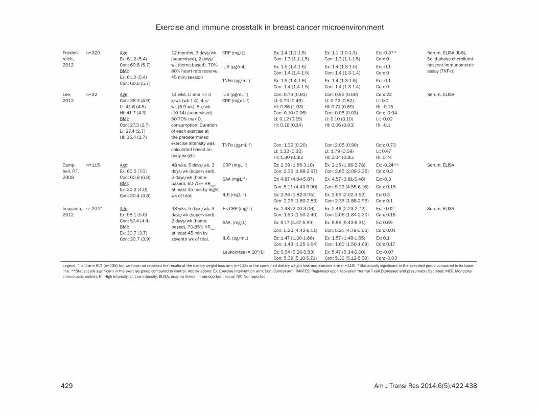
Frieden-reich, 2012
n=320 Age: Ex: 61.2 (5.4) Con: 60.6 (5.7)BMI: Ex: 61.2 (5.4)Con: 60.6 (5.7)
12 months, 3 days/wk (supervised), 2 days/wk (home-based), 70%-80% heart rate reserve, 45 min/session
CRP (mg/L) Ex: 1.4 (1.2-1.6)Con: 1.3 (1.1-1.5)
Ex: 1.1 (1.0-1.3) Con: 1.3 (1.1-1.5)
Ex: -0.3**Con: 0
Serum, ELISA (IL-6), Solid-phase chemilumi-nescent immunometric assay (TNF-α)
IL-6 (pg/mL) Ex: 1.5 (1.4-1.6) Con: 1.4 (1.4-1.5)
Ex: 1.4 (1.3-1.5) Con: 1.4 (1.3-1.4)
Ex: -0.1Con: 0
TNFα (pg/mL) Ex: 1.5 (1.4-1.6)Con: 1.4 (1.4-1.5)
Ex: 1.4 (1.3-1.5) Con: 1.4 (1.3-1.4)
Ex: -0.1Con: 0
Lee, 2012
n=22 Age:Con: 38.3 (4.9)LI: 41.6 (4.5)HI: 41.7 (4.3)BMI:Con: 27.3 (2.7)LI: 27.4 (2.7)HI: 25.4 (2.7)
14 wks, LI and HI: 3 x/wk (wk 1-4), 4 x/wk (5-9 wk), 5 x/wk (10-14) (supervised) 50-70% max O2 consumption, Duration of each exercise at the predetermined exercise intensity was calculated based on body weight
IL-6 (pg·mL–1)CRP (mg·dL–1)
Con: 0.73 (0.81)LI: 0.70 (0.49)HI: 0.86 (1.03)Con: 0.10 (0.06)LI: 0.12 (0.15)HI: 0.16 (0.14)
Con: 0.95 (0.60)LI: 0.72 (0.62)HI: 0.71 (0.69)Con: 0.06 (0.03) LI: 0.10 (0.10)HI: 0.06 (0.03)
Con: 22LI: 0.2HI: -0.15Con: -0.04LI: -0.02HI: -0.1
Serum, ELISA
TNFα (pg·mL–1) Con: 1.32 (0.20) LI: 1.32 (0.32)HI: 1.30 (0.35)
Con: 2.05 (0.90)LI: 1.79 (0.58) HI: 2.04 (0.85)
Con: 0.73LI: 0.47HI: 0.74
Camp-bell, P.T, 2008
n=115 Age:Ex: 60.5 (7.0) Con: 60.9 (6.8)BMI:Ex: 30.2 (4.0)Con: 30.4 (3.8)
48 wks, 5 days/wk, 3 days/wk (supervised), 2 days/wk (home-based), 60-75% HRmax, at least 45 min by eight wk of trial.
CRP (mg·L–1) Ex: 2.39 (1.85-3.10)Con: 2.36 (1.88-2.97)
Ex: 2.15 (1.66-2.78)Con: 2.65 (2.09-3.36)
Ex: -0.24**Con: 0.2
Serum, ELISA
SAA (mg·L–1) Ex: 4.87 (4.04-5.87) Ex: 4.57 (3.81-5.48) Ex: -0.3
Con: 5.11 (4.43-5.90) Con: 5.29 (4.55-6.16) Con: 0.18
IL-6 (mg·L–1) Ex: 2.36 (1.82-3.05)Con: 2.26 (1.80-2.83)
Ex: 2.66 (2.02-3.52)Con: 2.36 (1.88-2.96)
Ex: 0.3 Con: 0.1
Imayama, 2012
n=204^ Age:Ex: 58.1 (5.0) Con: 57.4 (4.4)BMI:Ex: 30.7 (3.7)Con: 30.7 (3.9)
48 wks, 5 days/wk, 3 days/wk (supervised), 2 days/wk (home-based), 70-85% HRmax, at least 45 min by seventh wk of trial.
Hs-CRP (mg/L) Ex: 2.48 (2.00-3.06)Con: 1.90 (1.50-2.40)
Ex: 2.46 (2.23-2.72)Con: 2.06 (1.84-2.30)
Ex: -0.02Con: 0.16
Serum, ELISA
SAA, (mg/L) Ex: 5.17 (4.47-5.99) Ex: 5.86 (5.43-6.31) Ex: 0.69
Con: 5.20 (4.42-6.11) Con: 5.21 (4.79-5.66) Con: 0.01
IL-6, (pg/mL) Ex: 1.47 (1.30-1.66)Con: 1.43 (1.25-1.64)
Ex: 1.57 (1.48-1.65)Con: 1.60 (1.50-1.69)
Ex: 0.1 Con: 0.17
Leukocytes (× 109/L) Ex: 5.54 (5.26-5.83)Con: 5.39 (5.10-5.71)
Ex: 5.47 (5.34-5.60)Con: 5.36 (5.12-5.50)
Ex: -0.07Con: -0.03
Legend: ^, a 4-arm RCT (n=438) but we have not reported the results of the dietary weight loss arm (n=118) or the combined dietary weight loss and exercise arm (n=116). *Statistically significant in the specified group compared to its base-line. **Statistically significant in the exercise group compared to control. Abbreviations: Ex, Exercise intervention arm; Con, Control arm; RANTES, Regulated upon Activation Normal T-cell Expressed and presumably Secreted; MCP, Monocyte chemotactic protein; HI, High intensity; LI, Low intensity; ELISA, enzyme-linked immunosorbent assay; NR, Not reported.
Exercise and immune crosstalk in breast cancer microenvironment
430 Am J Transl Res 2014;6(5):422-438
higher in endurance athletes than sedentary controls [49]. In a combined endurance and resistance training intervention in elderly men and women, a significant decrease in the inflammatory CD14+CD16+ monocyte subsets was observed, as well as lower endotoxin-stim-ulated TNF-α production in vitro [50]. This find-ing supports the anti-inflammatory effects mediated by regular exercise, given that the CD14+CD16+ monocyte subset is related to a pro-inflammatory profile as well as an early can-cer stage [38].
In a more clinically relevant study [51], breast cancer survivors responded to 5 weeks of cycle ergometry training (5 days per week, for 30-40 min per session at 60% of heart rate reserve) by exhibiting a decrease in circulating concen-trations of monocytes at rest. Functionally, exercise training showed a divergent effect on phagocytosis, depending on whether the stimu-lus used was enzyme treated sheep red blood cells (RBCs) or Anti-D loaded human RBCs [51]. In a different study, breast cancer survivors were randomized into a control or a combined aerobic and strength training program (3 days per week, 90 min per session) for 2 months [52]. Peripheral blood concentrations of the immune-related cytokines such as TNF-α, IFN-γ, IL-1α, IL4, IL-6, IL-10, IL-12, IL-13, IL15, as well as chemokines such as monocyte chemotactic protein (MCP)-1, and stromal derived factor (SDF)-1α were unchanged after the exercise training program.
Exercise affects tissue macrophage behavior in pre-clinical mouse studies
To date, no exercise studies in cancer survivors have reported on both human tissues and blood markers collected before and after an intervention. Therefore, pre-clinical studies, predominantly in animal models, that are rele-vant to exercise-induced immunomodulation in cancer provide some insight. Similar to studies performed on human monocytes, acute exer-cise, such as a single bout of exhaustive tread-mill running, inhibited sarcoma cell growth in vitro by peritoneal macrophages of NMRI mice [53]. As well, short-term (several days) exercise training studies in mice of various genetic back-grounds demonstrated an increased in vitro cytotoxic effect of peritoneal macrophages against adenocarcinoma cells [54, 55].
Exercise training for cancer prevention may involve a shift in macrophage polarization in the tumor microenvironment [56]. The cyto-kines induced by exercise are frequently asso-ciated with M1 or Th1 cells. A previous study [57] showed that MHC IIhigh tumor-associated macrophages appeared in the tumor microenvi-ronment during the early stages of the disease. These cells also expressed genes that are syn-onymous with the M1 macrophage phenotype (IL-6, IL-12, iNOS). In contrast, MHC IIlow tumor-associated macrophages were found during the later stages of tumor progression, and expressed genes that are associated with the M2 macrophage phenotype (Ym1, fizz1, Arg1, IL-10, TGF-beta).
T-cells
A bout of acute treadmill running (45min at 80% VO2max) increased relative changes in CD8+ cells in young, healthy and endurance-trained human volunteers, resulting in a decline in the CD4+/CD8+ cell ratio in peripheral blood [58]. Comparatively, 3 months of aerobic training with cycle ergometry did not result in differenc-es in phytohemagglutin (PHA)- or pokeweed mitogen (PMA)-stimulated T-cell proliferation in healthy young men [59].
Well-trained athletes provide a useful bench-mark with which to compare exercise-induced changes in immune function from the normal, sedentary population. It was reported recently that compared with sedentary individuals, pe-ripheral blood obtained under resting condi-tions from endurance-trained athletes present-ed with higher concentrations of IL-10, as well as a greater population of CD4+CD25+CD127low Tregs [60]. It is possible that prolonged periods of intensive endurance training up-regulates an anti-inflammatory profile in order to suppress pro-inflammatory signalling due to muscle dam-age.
Exercise-induced, skeletal muscle-derived cytokines possess endocrine-like properties
Role of exercise-induced, skeletal muscle-derived cytokines or “myokines”
An exciting breakthrough in exercise immunol-ogy research is the novel paradigm conceptual-ized by Bente Pedersen and research group in Copenhagen. Pedersen et al. have proposed

Exercise and immune crosstalk in breast cancer microenvironment
431 Am J Transl Res 2014;6(5):422-438
that the contracting skeletal muscle is an endo-crine organ; secreting specific cytokines, known as myokines [12, 62]. To date, at least 12 myo-kines have been reported.
Myokines with reported paracrine functions on the original tissue or organ of secretion have been reported and include leukemia inhibitory factor (LIF) [15] and IL-15 [17]. Other myokines purported to exhibit endocrine functions include the well-studied IL-6 [63], Irisin [14], calprotectin [16], myonectin [18], Oncostatin M (OSM) [64] and secreted protein acidic and rich in cysteine (SPARC) [13]. The mechanistic con-cept is that skeletal muscle contraction results in the transient rise of myokine concentration, such as IL-6 from contracting muscles, and subsequent release of other anti-inflammatory cytokines, such as IL-1 receptor agonist (IL-1ra) and IL-10 [65]. This in turn reduces the number of pro-inflammatory cytokines, with a subse-quent inhibition of migratory mononuclear cells such as macrophages and T-cells towards the inflamed microenvironment, such as adipose tissue [66].
Therefore, this line of research suggests that exercise training, and the accumulation of repeated bouts of skeletal muscle contraction may mediate an anti-inflammatory milieu, which may be beneficial for the primary preven-tion of cancer. In other words, muscle contrac-tion during exercise induces the secretion of cytokines such as IL-6, which further recruits other anti-inflammatory cytokines such as IL-1ra and IL-10. Changes in the circulating con-centrations of the pro- and anti-inflammatory cytokines may also shift the circulating mono-cyte or T-cell population to that of a M2 or Th2 profile. A M2 or Th2 profile may be beneficial given that during carcinogenesis, there is increased pro-inflammatory signaling due to increased oxidative stress and cellular/tissue damage. The possibility is that regular aerobic exercise training may prevent cancer initiation by reducing pro-inflammatory signaling. Thus, from a primary prevention perspective, aerobic exercise training induces an anti-inflammatory profile, at least in whole blood. However, aero-bic exercise training may induce a subtle shift in the subsets of T-cells and macrophages to be may play a role in reducing risk of cancer (e.g. Th1 and M1 polarized), consistent with animal models of breast cancer.
Some myokines are putative candidates for exercise-induced chemoprotection, including SPARC, OSM and IL-15. SPARC is a glycoprotein involved in multiple cellular functions, such as proliferation, apoptosis, as well as mitigating the interactions between cells and the extracel-lular matrix [67]. Further, a C allele polymor-phism was found to be more frequent in patients with hepatocellular carcinoma (HCC) than in healthy controls [68]. Aoi et al. [13] found an increase in circulating SPARC protein concentrations after an acute aerobic exercise (30 min of treadmill run at 30 m/min) in wild type mice and healthy human volunteers. Wild-type mice given 4 weeks of progressive aerobic exercise training (20 min at 18 m/min to 60 min at 30 m/min) and subsequent injections of azoxymethane (AOM; which induces colon tumorigenesis) presented with decreased aber-rant crypts compared with sedentary mice. Conversely, SPARC-null mice had greater aber-rant crypts in their colons after they were given the same treatments. Increased SPARC protein expression was also detected in both C2C12 myocytes that were stretched in vitro and in the culture media, compared with non-stretched myocytes. The results from this innovative study suggest that SPARC may serve as a myo-kine, and can potentially alter the interaction of the tumor cells with the extra-cellular matrix.
In a similar study by Hojman et al. [64], an increase in serum concentrations of OSM was observed after an acute, 1-hour bout of swim session in mice. This increase was most evi-dent immediately post-exercise, and returned to baseline values after 2 hours. Importantly, serum taken from exercising animals increased caspase activity in MCF-7 tumor cells in vitro. As well, incubating the tumor cells with human recombinant OSM resulted in in vitro growth inhibition after 5 days. To ascertain that skele-tal muscle was a source of OSM secretion, elec-trical stimulation of murine C2C12 myocytes was performed and the media from stimulated and non-stimulated cells were then added to MCF-7 cells. MCF-7 cells treated with serum from electrically stimulated myocytes showed a 100% increase in caspase activity, which was attenuated in half when anti-OSM antibodies were added to the culture.
Other putative cytokines/myokines such as IL-15 may also be relevant in exercise-induced

Exercise and immune crosstalk in breast cancer microenvironment
432 Am J Transl Res 2014;6(5):422-438
cancer protection. However, unlike IL-6 which has been demonstrated to be increased by up to 100-fold after an acute bout of prolonged aerobic exercise, the plasma concentrations of IL-15 were reported to be unchanged [69] or increased by up to 1-fold in young healthy men [70]. These conflicting differences may be attributed to training status in subjects from the two studies, with untrained subjects appar-ently demonstrating an increase in circulating IL-15 after exercise, compared with trained sub-jects. A more plausible explanation is that in the Ostrowski et al. study, data for IL-15 was only available for 2 subjects, limiting the ability to interpret the data.
In summary, the majority of the studies that investigated the biological functions of myo-kines have not considered their interactions with the immune system, and specifically, how these myokines can cross-talk with immune cells to potentially impact the process of tumor-igenesis. With the exception of SPARC protein [13] and OSM [64], the majority of myokines has not been studied in the context of immune-tumor crosstalk after exercise training, and are attractive targets for future work.
Systematic review of the effect of aerobic exercise on immune function: human inter-vention trials in women without breast cancer and following a breast cancer diagnosis
In an effort to examine the currently available literature on the role of aerobic exercise on immune function, a systematic literature review was performed related to cancer prevention (i.e., in women who had not had a breast cancer diagnosis) and related to cancer outcomes in “patients” (i.e., women currently undergoing adjuvant cancer treatment; namely chemother-apy and/or radiation) and “survivors” (i.e., women who have completed adjuvant cancer treatment, but hormone therapy may be ongo-ing). The search was limited to: full text English-language; randomized controlled trials (RCT) of aerobic exercise in women; and published in peer-reviewed journals. Search terms related to breast cancer (e.g., breast neoplasm, breast cancer, breast tumor) and exercise (e.g., aero-bic exercise, running) were used and modified for the individual databases (MEDLINE and EMBASE) (See Supplemental Table 1). Studies were excluded if multiple cancer sites were included and separate information for women
with breast cancer was not available. In order to address the two key paradigms, the search included immunological markers, specifically monocytes, T cells, lymphocytes and leuko-cytes, and inflammation markers proposed to be affected by exercise and relevant to cancer prevention and survival, namely, interleukins (IL-1, 6, 18, 10, 12, 15), chemokine (CCLs) and (CXCLs), tumor necrosis factor (TNF-α), Serum Amyloid A (SAA), and C-reactive protein (CRP).
The search yielded 1684 potentially relevant articles, which were then screened for inclusion by title and abstract (Figure 1). All eligible papers underwent a full text review and for the eligible papers, the reference list was hand-searched to identify any additional relevant studies, which yielded one additional study [71]. Duplicate papers from a single study were handled by including individual studies rather than individual papers. Two papers [72, 73] from a single study were identified and there-fore counted as one single study. The search strategy identified 15 studies that met the inclusion criteria and relevant data was extract-ed on study design and study outcomes (Table 1).
Four out of 15 studies were in premenopausal women [74-77], five in postmenopausal women [71, 78-81] and six in breast cancer survivors [11, 72, 82-85]. Sample sizes ranged from 8 to 320, and the mean number of participants was 100. The aerobic exercise interventions were a mean of 23 weeks in length (range, 8-48 weeks). In 10 out of 15 studies, the inflamma-tory and immunological markers were specified as the primary outcome [11, 71-78, 82, 83] and as a secondary outcome in the remaining five studies [79-81, 84, 85].
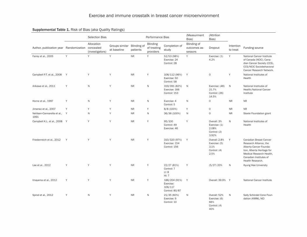
The quality and risk of bias for all studies were assessed using criteria from the Cochrane Collaboration risk of bias assessment protocol, risk of bias was low in all included studies (data not shown) [86].
Aerobic exercise training interventions
The length of the exercise interventions ranged from 8 to 48 weeks, while one study reported the result of a single acute exercise bout [76]. The frequency of exercise interventions ranged from three times per week to six times per week. The intensity of the prescribed exercise moderate to vigorous (i.e., 60-85% of maximum
Exercise and immune crosstalk in breast cancer microenvironment
433 Am J Transl Res 2014;6(5):422-438
heart rate), however, exercise training intensity was not reported in three studies [82, 84, 85]. The duration of each exercise session ranged from 15 to 60 minutes or was calculated with consideration of subjects’ body weight in one study [81]. The duration of the acute exercise study was a two-hour exercise bout [76]. For supervised interventions, 11 RCTs reported attendance as the mean adherence rate of 84%; ranging from 49% to 98% [11, 71-74, 76, 78-80, 82, 83, 85]. Three studies did not report the adherence rate [75, 77, 81].
Impact of aerobic exercise training on bio-markers of interest in healthy female popula-tions
Five studies reported significant changes in immunological and inflammation markers due to aerobic exercise among pre- and post-meno-pausal healthy women with no prior diagnosis of cancer [71, 74-76, 79]. In premenopausal women, significant reduction in the pro-inflam-matory cytokine TNF-α was reported in a 12-week aerobic exercise intervention [75], and in one study of an acute 2-hour exercise bout [76]. In addition to changes in concentra-tions of TNF-α, a significant reduction of exer-cise-induced production of IL-10 after 120 min of exercise, and a significant mobilization of blood leukocytes and monocytes were also noted in the recovery period of the acute exer-cise study [76]. A 16-week aerobic exercise intervention in premenopausal women signifi-cantly reduced the plasma CRP concentrations in the exercise group compared to control; how-ever, no significant reduction of SAA was observed [74]. In a 15-week aerobic exercise intervention among obese premenopausal women, the percentage and number of lympho-cytes, numbers of T cells (CD5+), and B cells (CD20+) significantly decreased compared to controls [77]. However, no significant changes in total leukocytes, T helper cell (CD4+), and T cytotoxic cell (CD8+) numbers were found.
In postmenopausal women, similar reductions in CRP were noted with aerobic exercise inter-ventions of varying lengths in some [71, 79], but not all studies [78, 80, 81]. No changes were observed in serum SAA [71, 80], IL-6 [79-81], TNF-α [71, 79, 81] and total leukocytes [80] among postmenopausal women in differ-ent aerobic exercise interventions. Seven stud-ies investigated the factors that may mediate the effect of aerobic exercise and adjusted the
model for baseline BMI, adherence rate, body fat and physical fitness [71, 74, 78-80, 81, 83]. Three studies found that exercise-induced sig-nificant reduction of CRP concentrations were independent of weight loss [74, 78, 81].
Impact of aerobic exercise training on bio-markers of interest patients or breast cancer survivors
No studies examined the effect of aerobic exer-cise on inflammation and immunological mark-ers in breast cancer patients (i.e., during che-motherapy or radiation treatment). Six studies examined the influence of aerobic exercise intervention on inflammation and immunologi-cal markers among breast cancer survivors [11, 72, 73, 82-85]. Two studies examined CRP [73, 83] and while a slight reduction was report-ed in one study [73], it was not statistically sig-nificant and no change was reported in another study [83]. Two studies examined IL-8 [82, 85], and a reduction was noted in one study [82], but not in the other [85]. No significant changes were observed in serum IL-6 [82-85], and TNF-α concentrations [82, 83]. One study examined the impact of an aerobic exercise intervention on Regulated upon Activation Normal T-cell Expressed and secreted (RANTES), and while a greater reduction was noted in the exercise group [82], no statistically significant changes were reported in RANTES or monocyte chemotactic protein-1 (MCP-1) concentrations compared to the education group. In addition, comparing the post-treat-ment to baseline concentrations, interleukin-8 was significantly decreased in the home exer-cise group while concentrations of MCP-1 were significantly increased in the education group. Two studies examined natural killer cell cyto-toxic activity [11, 72]. The first study observed an increased natural killer cell cytotoxic activi-ty, total lytic unit and spontaneous lymphocyte proliferation in postmenopausal breast cancer survivors with a 15-week exercise intervention [72]. However, in the second study, natural kill-er cell activity (NKCA), lymphocyte and total leu-kocytes concentrations were not altered signifi-cantly by the aerobic exercise intervention, although NKCA tended to be higher in the exer-cise group [11].
Summary of results
Our systematic review of the literature revealed that although biomarkers of immune function
Exercise and immune crosstalk in breast cancer microenvironment
434 Am J Transl Res 2014;6(5):422-438
and inflammation have been examined in healthy women or breast cancer survivors, the results have been mixed and very few RCTs included biomarkers relevant to immunomodu-lation, as opposed to a more generic inflamma-tory profile. Furthermore, only some investiga-tors performed flow cytometry [11, 72] to determine changes in immune subsets or changes in intracellular concentrations of cyto-kines after acute exercise or exercise training.
Protective molecular phenotypes related to exercise-induced immunomodulation
Determination of an exercise-induced molecu-lar “signature” could provide a rationale to examine the effect of exercise combined with chemotherapy or immunotherapy in mediating disease prognosis. The putative mechanisms are likely to involve direct modulation of mono-cyte/macrophage and T-cell functions, as well as indirect modulation of myokine (OSM, SPARC) crosstalk with stromal and tumor cells from the tumor microenvironment. In fact the integration of these two paradigms begets the compelling questions: do myokines communi-cate with innate and adaptive immune cells, and what is the direction of the cross-talk? Gaps in the exercise immunology literature rep-resent new opportunities for cross-disciplinary collaborations. In addition, long-term profiling of chronic aerobic exercise training on leuko-cyte and myokine expression in individuals without a diagnosis of cancer could help to elu-cidation a more complete understanding of the immune response to long-term aerobic exer-cise training, specifically how aerobic exercise modulates gene and protein expression of molecular mediators involved in the immune surveillance and recognition of potential malignancies.
Conclusion
We envision a future where aerobic exercise training may play a larger role in an integrated approach to enhance breast cancer prevention and treatment outcomes. The immune system plays an instrumental role in surveying the host microenvironment for potential malignancies, and its efficacy can be modulated with exercise training. In order for the research and clinical community to improve breast cancer preven-tion and treatment outcomes, it is necessary to elucidate the potential mechanisms involved.
Proposed mechanisms include immune-rele-vant pathways, as well as myokine signaling. We suggest that these two areas represent a significant opportunity to further understand how exercise can positively influence immune cross-talk relevant to breast cancer prevention and treatment outcomes.
Disclosure of conflict of interest
None.
Address correspondence to: Jorming Goh, Screening and Diagnostics Technologies Laboratory, Defence Medical and Environmental Research Institute, DSO National Laboratories, 27 Medical Drive, Republic of Singapore (117510). Tel: 65-6485-7208; Fax: 65-6485-7226; E-mail: [email protected]
References
[1] Speck RM, Courneya KS, Mâsse LC, Duval S, Schmitz KH. An update of controlled physical activity trials in cancer survivors: a systematic review and meta-analysis. J Cancer Surviv 2010; 4: 87-100.
[2] Friedenreich CM, Neilson HK. State of the epi-demiological evidence on physical activity and cancer prevention. Eur J Cancer 2010; 46: 2593-2604.
[3] Loprinzi PD, Cardinal BJ, Winters-Stone K, Smit E, Loprinzi CL. Physical activity and the risk of breast cancer recurrence: a literature review. Oncol Nurs For 2012; 39: 269-274.
[4] Schmidt ME, Chang-Claude J, Vrieling A, Sei-bold P, Heinz J, Obi N, Flesch-Janys D, Stein-dorf K. Association of pre-diagnosis physical activity with recurrence and mortality among women with breast cancer. Int J Cancer 2013; 133: 1431-1440.
[5] Sternfeld B, Weltzien E, Quesenberry CP Jr, Castillo AL, Kwan M, Slattery ML, Caan BJ. Physical activity and risk of recurrence and mortality in breast cancer survivors: findings from the LACE study. Cancer Epidemiol Bio-markers Prev 2009; 18: 87-95.
[6] Ballard-Barbash R, Friedenreich CM, Courneya KS, Siddiqi SM, McTiernan A, Alfano CM. Physi-cal activity, biomarkers, and disease outcomes in cancer survivors: a systematic review. J Natl Cancer Inst 2012; 104: 815-840.
[7] Campbell KL, McTiernan A. Exercise and bio-markers for cancer prevention studies. J Nutr 2007; 137: 161S-169S.
[8] Koivisto VA, Soman V, Conrad P, Hendler R, Nadel E, Felig P. Insulin binding to monocytes in trained athletes: changes in the resting

Exercise and immune crosstalk in breast cancer microenvironment
435 Am J Transl Res 2014;6(5):422-438
state and after exercise. J Clin Invest 1979; 64: 1011-1015.
[9] McCarthy DA, Perry JD, Melsom RD, Dale MM. Leukocytosis induced by exercise. Br Med J 1987; 295: 636.
[10] Nieman DC, Johanssen LM, Lee JW. Infectious episodes in runners before and after a roadra-ce. J Sports Med Phys Fitness 1989; 29: 289-296.
[11] Nieman DC, Cook VD, Henson DA, Suttles J, Rejeski WJ, Ribisl PM, Fagoaga OR, Nehlsen-Cannarella SL. Moderate exercise training and natural killer cell cytotoxic activity in breast cancer patients. Int J Sports Med 1995; 16: 334-337.
[12] Pedersen BK. Exercise-induced myokines and their role in chronic disease. Brain Behav Im-mun 2011; 25: 811-816.
[13] Aoi W, Naito Y, Takagi T, Tanimura Y, Takanami Y, Kawai Y, Sakuma K, Hang LP, Mizushima K, Hirai Y, Koyama R, Wada S, Higashi A, Kokura S, Ichikawa H, Yoshikawa T. A novel myokine, secreted protein acidic and rich in cysteine (SPARC), suppresses colon tumorigenesis via regular exercise. Gut 2013; 62: 882-889.
[14] Boström P, Wu J, Jedrychowski MP, Korde A, Ye L, Lo JC, Rasbach KA, Boström EA, Choi JH, Long JZ, Kajimura S, Zingaretti MC, Vind BF, Tu H, Cinti S, Højlund K, Gygi SP, Spiegelman BM. A PGC-1α-dependent myokine that drives brown-fat-like development of white fat and thermogenesis. Nature 2012; 481: 463-468.
[15] Broholm C, Mortensen OH, Nielsen S, Aker-strom T, Zankari A, Dahl B, Pedersen BK. Exer-cise induces expression of leukaemia inhibito-ry factor in human skeletal muscle. J Physiol 2008; 586: 2195-2201.
[16] Mortensen OH, Andersen K, Fischer C, Nielsen AR, Nielsen S, Akerström T, Aastrøm MB, Borup R, Pedersen BK. Calprotectin is released from human skeletal muscle tissue during exercise. J Physiol 2008; 586; 3551-3562.
[17] Quinn LS, Anderson BG, Strait-Bodey L, Stroud AM, Argilés JM. Oversecretion of interleukin-15 from skeletal muscle reduces adiposity. Am J Physiol Endocrinol Metab 2009; 296: E191-E202.
[18] Seldin MM, Peterson JM, Byerly MS, Wei Z, Wong GW. Myonectin (CTRP15), a novel myo-kine that links skeletal muscle to systemic lipid homeostasis. J Biol Chem 2012; 287: 11968-11980.
[19] Mukhtar RA, Nseyo O, Campbell MJ, Esserman LJ. Tumor-associated macrophages in breast cancer as potential biomarkers for new treat-ments and diagnostics. Expert Rev Mol Diagn 2011; 11: 91-100.
[20] Campbell MJ, Tonlaar NY, Garwood ER, Huo D, Moore DH, Khramtsov AI, Au A, Baehner F,
Chen Y, Malaka DO, Lin A, Adeyanju OO, Li S, Gong C, McGrath M, Olopade OI, Esserman LJ. Proliferating macrophages associated with high grade, hormone receptor negative breast cancer and poor clinical outcome. Breast Can-cer Res Treat 2011; 128: 703-11.
[21] Heusinkveld M, Van Der Burg S. Identification and manipulation of tumor associated macro-phages in human cancers. J Transl Med 2011; 9: 216.
[22] Laoui D, Movahedi K, Van Overmeire E, Van den Bossche J, Schouppe E, Mommer C, Niko-laou A, Morias Y, De Baetselier P, Van Ginder-achter JA. Tumor-associated macrophages in breast cancer: distinct subsets, distinct func-tions. Int J Dev Biol 2011; 55: 861-867.
[23] DeMarco RA, Fink MP, Lotze MT. Monocytes promote natural killer cell interferon gamma production in response to the endogenous danger signal HMGB1. Mol Immunol 2005; 42: 433-444.
[24] Watkins SK, Li B, Richardson KS, Head K, Egil-mez NK, Zeng Q, Suttles J, Stout RD. Rapid re-lease of cytoplasmic IL-15 from tumor associ-ated macrophages is an initial and critical event in IL-12 initiated tumor regression. Eur J Immunol 2009; 39: 2126-2135.
[25] Hagemann T, Lawrence T, McNeish I, Charles KA, Kulbe H, Thompson RG, Robinson SC, Balkwill FR. “Re-educating” tumor-associated macrophages by targeting NF-kappaB. J Exp Med 2008; 205: 1261-1268.
[26] Emtage PC, Clarke D, Gonzalo-Daganzo R, Junghans RP. Generating potent Th/Tc1 T cell adoptive immunotherapy doses using human IL-12: harnessing the immunomodulatory po-tential of IL-12 without the in vivo associated toxicity. J Immunother 2003; 26: 97-106.
[27] Tsung K, Dolan JP, Tsung YL, Norton JA. Macro-phages as effector cells in interleukin 12-in-duced T cell-dependent tumor rejection. Can-cer Res 2002; 62: 5069-5075.
[28] Comes A, Di Carlo E, Musiani P, Rosso O, Meaz-za R, Chiodoni C, Colombo MP, Ferrini S. IFN-gamma-independent synergistic effects of IL-12 and IL-15 induce anti-tumor immune responses in syngeneic mice. Eur J Immunol 2002; 32: 1914-1923.
[29] Leek RD, Lewis CE, Whitehouse R, Greenall M, Clarke J, Harris AL. Association of macrophage infiltration with angiogenesis and prognosis in invasive breast carcinoma. Cancer Res 1996; 56: 4625-4629.
[30] Menetrier-Caux C, Faget J, Biota C, Gobert M, Blay JY, Caux Cl. Innate immune recognition of breast tumor cells mediates CCL22 secretion favoring Treg recruitment within tumor environ-ment. OncoImmunol 2012; 1: 759-761.

Exercise and immune crosstalk in breast cancer microenvironment
436 Am J Transl Res 2014;6(5):422-438
[31] Ma J, Liu L, Che G, Yu N, Dai F, You Z. The M1 form of tumor-associated macrophages in non-small cell lung cancer is positively associated with survival time. BMC Cancer 2010; 10: 112.
[32] Ohri CM, Shikotra A, Green RH, Waller DA, Bradding P. Macrophages within NSCLC tu-mour islets are predominantly of a cytotoxic M1 phenotype associated with extended sur-vival. Eur Respir J 2009; 33: 118-126.
[33] Kilinc MO, Aulakh KS, Nair RE, Jones SA, Alard P, Kosiewicz MM, Egilmez NK. Reversing tumor immune suppression with intratumoral IL-12: activation of tumor-associated T effector/memory cells, induction of T suppressor apop-tosis, and infiltration of CD8+ T effectors. J Im-munol 2006; 177: 6962-73.
[34] Hinrichs CS, Kaiser A, Paulos CM, Cassard L, Sanchez-Perez L, Heemskerk B, Wrzesinski C, Borman ZA, Muranski P, Restifo NP. Type 17 CD8+ T cells display enhanced antitumor im-munity. Blood 2009; 114: 596-599.
[35] Lee HJ, Seo JY, Ahn JH, Ahn SH, Gong G. Tumor-associated lymphocytes predict response to neoadjuvant chemotherapy in breast cancer patients. J Breast Cancer 2013; 16: 32-39.
[36] Song G, Wang X. Elevated level of peripheral CD8+CD28- T lymphocytes are an independent predictor of progression-free survival in pa-tients with metastatic breast cancer during the course of chemotherapy. Cancer Immunol Im-munother 2013; 62: 1123-1130.
[37] West NR, Milne K, Truong PT, Macpherson N, Nelson BH, Watson PH. Tumor-infiltrating lym-phocytes predict response to anthracycline-based chemotherapy in estrogen receptor-neg-ative breast cancer. Breast Cancer Res 2011; 13: R126.
[38] Feng AL, Zhu JK, Sun JT, Yang MX, Neckenig MR, Wang XW, Shao QQ, Song BF, Yang QF, Kong BH, Qu X. CD16+ monocytes in breast cancer patients: expanded by monocyte che-moattractant protein-1 and may be useful for early diagnosis. Clin Exp Immunol 2011; 164: 57-65.
[39] Coussens LM, Werb Z. Inflammation and can-cer. Nature 2002; 420: 860-867.
[40] Field CJ, Gougeon R, Marliss EB. Circulating mononuclear cell numbers and function dur-ing intense exercise and recovery. J Appl Physi-ol 1991; 71: 1089-1097.
[41] Gabriel H, Schwarz L, Born P, Kindermann W. Differential mobilization of leucocyte and lym-phocyte subpopulations into the circulation during endurance exercise. Eur J Appl Physiol Occup Physiol 1992; 65: 529-534.
[42] Michel G, Vocke T, Fiehn W, Weicker H, Schwarz W, Bieger WP. Bidirectional alteration of insulin receptor affinity by different forms of physical exercise. Am J Physiol 1984; 246: E153-E159.
[43] Tvede N, Pedersen BK, Hansen FR, Bendix T, Christensen LD, Galbo H, Halkjaer-Kristensen J. Effect of physical exercise on blood mono-nuclear cell subpopulations and in vitro prolif-erative responses. Scand J Immunol 1989; 29: 383-389.
[44] Ratge D, Wiedemann A, Kohse KP, Wisser H. Alterations of beta-adrenoceptors on human leukocyte subsets induced by dynamic exer-cise: effect of prednisone. Clin Exp Pharmacol Physiol 1988; 15: 43-53.
[45] Scharhag J, Meyer T, Gabriel HH, Schlick B, Faude O, Kindermann W. Does prolonged cy-cling of moderate intensity affect immune cell function? Br J Sports Med 2005; 39: 171-177.
[46] Gabriel H, Urhausen A, Brechtel L, Müller HJ, Kindermann W. Alterations of regular and ma-ture monocytes are distinct, and dependent on intensity and duration of exercise. Eur J Appl Physiol Occup Physiol 1994; 69: 179-181.
[47] Steppich B, Dayyani F, Gruber R, Lorenz R, Mack M, Ziegler-Heitbrock HW. Selective mobi-lization of CD14+CD16+ monocytes by exercise. Am J Physiol 2000; 279: C578-C586.
[48] Zaldivar F, Wang-Rodriguez J, Nemet D, Schwindt C, Galassetti P, Mills PJ, Wilson LD, Cooper DM. Constitutive pro- and anti-inflam-matory cytokine and growth factor response to exercise in leukocytes. J Appl Physiol 2006; 100: 1124-113.
[49] Gabriel H, Rothe G, Korpys M, Schmitz G, Kin-dermann W. Enhanced expression of HLA-DR, Fc gamma receptor 1 (CD64) and leukocyte common antigen (CD45) indicate activation of monocytes in regenerative training periods of endurance athletes. Int J Sports Med 1997; 18: 136-141.
[50] Timmerman KL, Flynn MG. Exercise training-induced lowering of inflammatory (CD14+
CD16+) monocytes: a role in the anti-inflamma-tory influence of exercise? J Leukol Biol 2008; 84: 1271-1278.
[51] Peters C, Lötzerich H, Niemeir B, Schüle K, Uhlenbruck G. Exercise, cancer and the im-mune response of monocytes. Anticancer Res 1995; 15: 175-179.
[52] Gomez AM, Martinez C, Fiuza-Luces C, Herrero F, Pérez M, Madero L, Ruiz JR, Lucia A, Ramírez M. Exercise training and cytokines in breast cancer survivors. Int J Sports Med 2011; 32: 461-467.
[53] Lötzerich H, Fehr HG, Appell HJ. Potentiation of cytostatic but not cytolytic activity of murine macrophages after running stress. Int J Sports Med 1990; 11: 61-65.
[54] Woods JA, Davis JM, Mayer EP, Ghaffar A, Pate RR. Exercise increases inflammatory macro-phage antitumor cytotoxicity. J Appl Physiol 1993; 75: 879-886.
Exercise and immune crosstalk in breast cancer microenvironment
437 Am J Transl Res 2014;6(5):422-438
[55] Woods JA, Davis JM, Mayer EP, Ghaffar A, Pate RR. Effects of exercise on macrophage activa-tion for antitumor cytotoxicity. J Appl Physiol 1994; 76: 2177-2185.
[56] Goh J, Kirk EA, Lee SX, Ladiges WC. Exercise, physical activity and breast cancer: the role of tumor-associated macrophages. Exerc Immu-nol Rev 2012; 18: 158-176.
[57] Wang B, Li Q, Qin L, Zhao S, Wang J, Chen X. Transition of tumor-associated macrophages from MHC class II (hi) to MHC class II (low) me-diates tumor progression in mice. BMC Immu-nol 2011; 12: 43.
[58] Nieman DC, Miller AR, Henson DA, Warren BJ, Gusewitch G, Johnson RL, Davis JM, Butter-worth DE, Herring JL, Nehlsen-Cannarella SL. Effect of high-versus moderate-intensity exer-cise on lymphocyte subpopulations and prolif-erative response. Int J Sports Med 1994; 15: 199-206.
[59] Mitchell JB, Paquet AJ, Pizza FX, Starling RD, Holtz RW, Grandjean PW. The effect of moder-ate aerobic training on lymphocyte prolifera-tion. Int J Sports Med 1996; 17: 384-389.
[60] Handzlik MK, Shaw AJ, Dungey M, Bishop NC, Gleeson M. The influence of exercise training status on antigen-stimulated IL-10 production in whole blood culture and numbers of circulat-ing regulatory T cells. Eur J Appl Physiol 2013; 113: 1839-1848.
[61] Goh J, Tsai J, Bammler TK, Farin FM, Endicott EE, Ladiges WC. Exercise training in transgenic mice is associated with attenuation of early breast cancer growth in a dose-dependent manner. PLoS One 2013; 8: e80123.
[62] Mathur N, Pedersen BK. Exercise as a mean to control low-grade systemic inflammation. Me-diators Inflamm 2008; 2008: 109502.
[63] Pedersen BK. A muscular twist on the fate of fat. N Engl J Med 2012; 366: 1544-1545.
[64] Hojman P, Dethlefsen C, Brandt C, Hansen J, Pedersen L, Pedersen BK. Exercise-induced muscle-derived cytokines inhibit mammary cancer cell growth. Am J Physiol Endocrinol Metab 2011; 301: E504-E510.
[65] Steensberg A, Fischer CP, Keller C, Møller K, Pedersen BK. IL-6 enhances plasma IL-1ra, IL-10 and cortisol in humans. Am J Physiol Endo-crinol Metab 2003; 285: E433-E437.
[66] Kawanishi N, Yano H, Yokogawa Y, Suzuki K. Exercise training inhibits inflammation in adi-pose tissue via both suppression of macro-phage infiltration and acceleration of pheno-typic switching from M1 to M2 macrophages in high-fat-diet-induced obese mice. Exerc Immu-nol Rev 2010; 16: 105-118.
[67] Inoue M, Senju S, Hirata S, Ikuta Y, Hayashida Y, Irie A, Harao M, Imai K, Tomita Y, Tsunada T, Furukawa Y, Ito T, Nakamura Y, Baba H, Nishimura Y. Identification of SPARC as a can-
didate target antigen for immunotherapy of various cancers. Int J Cancer 2010; 127: 1393-1403.
[68] Segat L, Milanese M, Pirulli D, Trevisiol C, Lupo F, Salizzoni M, Amoroso A, Crovella S. Secreted protein acidic and rich in cysteine (SPARC) gene polymorphism association with hepato-cellular carcinoma in Italian patients. J Gastro-enterol Hepatol 2009; 24: 1840-1846.
[69] Ostrowski K, Hermann C, Bangash A, Schjer-ling P, Nielsen JN, Pedersen BK. A trauma-like elevation of plasma cytokines in humans in response to treadmill running. J Physiol 1998; 513: 889-894.
[70] Tamura Y, Watanabe K, Kantani T, Hayashi J, Ishida N, Kaneki M. Upregulation of circulating IL-15 by treadmill running healthy individuals: is IL-15 an endocrine mediator of the benefi-cial effects of endurance exercise? Endocr J 2011; 58: 211-215.
[71] Campbell PT, Campbell KL, Wener MH, Wood BL, Potter JD, McTiernan A, Ulrich CM. A year-long exercise intervention decreases CRP among obese postmenopausal women. Med Sci Sports Exerc 2009; 41: 1533-1539.
[72] Fairey AS, Courneya KS, Field CJ, Bell GJ, Jones LW, Mackey JR. Randomized controlled trial of exercise and blood immune function in post-menopausal breast cancer survivors. J Appl Physiol 2005; 98: 1534-1540.
[73] Fairey AS, Courneya KS, Field CJ, Bell GJ, Jones LW, Martin BS, Mackey JR. Effect of exercise training on C-reactive protein in postmeno-pausal breast cancer survivors: A randomized controlled trial. Brain Behav Immun 2005; 19: 381-388.
[74] Arikawa AY, Thomas W, Schmitz KH, Kurzer MS. Sixteen weeks of exercise reduces C-reac-tive protein levels in young women. Med Sci Sports Exerc 2011; 43: 1002-1009.
[75] Horne L, Bell G, Fisher B, Warren S, Janowska-Wieczorek A. Interaction between cortisol and tumour necrosis factor with concurrent resis-tance and endurance training. Clin J Sport Med 1997; 7: 247-251.
[76] Jimenez C, Melin B, Savourey G, Launay JC, Alonso A, Mathieu J. Effects of passive hyper-thermia versus exercise-induced hyperthermia on immune responses: Hormonal implications. Eur Cytokine Netw 2007; 18: 154-161.
[77] Nehlsen-Cannarella SL, Nieman DC, Balk-Lam-berton AJ, Markoff PA, Chritton DB, Gusewitch G, Lee JW. The effects of moderate exercise training on immune response. Med Sci Sports Exerc 1991; 23: 64-70.
[78] Campbell KL, Campbell PT, Ulrich CM, Wener M, Alfano CM, Foster-Schubert K, Rudolph RE, Potter JD, McTiernan A. No reduction in C-reac-tive protein following a 12-month randomized controlled trial of exercise in men and women.
Exercise and immune crosstalk in breast cancer microenvironment
438 Am J Transl Res 2014;6(5):422-438
Cancer Epidemiol Biomarkers Prev 2008; 17: 1714-1718.
[79] Friedenreich CM, Neilson HK, Woolcott CG, Wang Q, Stanczyk FZ, McTiernan A, Jones CA, Irwin ML, Yasui Y, Courneya KS. Inflammatory marker changes in a yearlong randomized ex-ercise intervention trial among postmenopaus-al women. Cancer Prev Res 2012; 5: 98-108.
[80] Imayama I, Ulric CM, Alfano CM, Wang C, Xiao L, Wener MH, Campbell KL, Duggan C, Foster-Schubert KE, Kong A, Mason CE, Wang CY, Blackburn GL, Bain CE, Thompson HJ, McTier-nan A. Effects of a caloric restriction weight loss diet and exercise on inflammatory bio-markers in overweight/obese postmenopaus-al women: A randomized controlled trial. Can-cer Res 2012; 72: 2314-2326.
[81] Lee MG, Park KS, Kim DU, Choi SM, Kim HJ. Effects of high-intensity exercise training on body composition, abdominal fat loss, and car-diorespiratory fitness in middle-aged korean females. Appl Physiol Nutr Metab 2012; 37: 1019-1027.
[82] Ergun M, Eyigor S, Karaca B, Kisim A, Uslu R. Effects of exercise on angiogenesis and apop-tosis-related molecules, quality of life, fatigue and depression in breast cancer patients. Eur J Cancer Care 2013; 22: 626-37.
[83] Jones SB, Thomas GA, Hesselsweet SD, Alva-rez-Reeves M, Yu H, Irwin ML. Effect of exercise on markers of inflammation in breast cancer survivors: The yale exercise and survivorship study. Cancer Prev Res 2013; 6: 109-118.
[84] Payne JK, Held J, Thorpe J, Shaw H. Effect of exercise on biomarkers, fatigue, sleep distur-bances, and depressive symptoms in older women with breast cancer receiving hormonal therapy. Oncol Nurs Forum 2008; 35: 635-642.
[85] Sprod LK, Janelsins MC, Palesh OG, Carroll JK, Heckler CE, Peppone LJ, Mohile SG, Morrow GR, Mustian KM. Health-related quality of life and biomarkers in breast cancer survivors par-ticipating in tai chi chuan. J Cancer Surviv 2012; 6: 146-154.
[86] Higgins JPT, Green S. Cochrane Handbook for Systematic Reviews of Interventions Version 5.0.2. The Cochrane Collaboration 2009.
Exercise and immune crosstalk in breast cancer microenvironment
Supplemental Table 1. Risk of Bias (aka Quality Ratings)
Selection Bias Performance Bias (Measurment Bias)
(Attrition Bias)
Author, publication year RandomizationAllocation concealed (investigators)
Groups similar at baseline
Blinding of patients
Blinding of treating providers
Completion of study
Blinding of outcomes as-sessors
Dropout Intention to treat Funding source
Fairey et al., 2005 Y Y Y NR Y 52/53 (98%)Exercise: 24Control: 28
Y Exercise: (1) 4.2%
Y National Cancer Institute of Canada (NCIC), Cana-dian Cancer Society (CCS), CCS/NCIC Sociobehavioral Cancer Research Network.
Campbell P.T. et al., 2008 Y Y Y NR Y 108/112 (96%)Exercise: 50Control: 58
Y 0 Y National Institutes of Health
Arikawa et al., 2011 Y N Y NR N 319/391 (82%)Exercise: 166Control: 153
N Exercise: (46) 21.7%Control: (26) 14.5%
N National Institutes of Health/National Cancer Institute
Horne et al., 1997 Y N Y NR N Exercise: 4Control: 5
N 0 NR NR
Jimenez et al., 2007 Y Y Y NR Y 8/8 (100%) Y 0 NR NR
Nehlsen-Cannarella et al., 1991
Y N Y NR N 36/36 (100%) N 0 NR Steele Foundation grant
Campbell K.L. et al., 2008 Y Y Y NR Y 95/100Control: 49Exercise: 46
Y Overall: 3%Exercise: (1) 2.08% Control: (2) 3.92%
N National Institutes of Health
Friedenreich et al., 2012 Y Y Y NR Y 310/320 (97%)Exercise: 154Control: 156
Y Overall: 2.8%Exercise: (5) 3.1%Control: (4) 2.5%
Y Canadian Breast Cancer Research Alliance, the Alberta Cancer Founda-tion, Alberta Heritage for Medical Research Health, Canadian Institutes of Health Research.
Lee et al., 2012 Y Y Y NR Y 22/27 (81%)Control: 7LI: 8HI: 7
Y (5/27) 20% N Kyung Hee University
Imayama et al., 2012 Y Y Y NR Y 186/204 (91%)Exercise: 106/117Control: 80/87
Y Overall: 39.9% Y National Cancer Institute
Sprod et al., 2012 Y N Y NR N 21/35 (60%)Exercise: 9 Control: 10
N Overall: 52% Exercise: (6) 66%Control: (4) 40%
N Sally Schindel Cone Foun-dation (KMM), NCI

Exercise and immune crosstalk in breast cancer microenvironment
Nieman et al., 1995 Y N Y NR N 16/20 (80%)Exercise: 8Control: 8
N Overall: 40%Exercise: (2) (10%)Control: (2) (10%)
N National Institute of Aging
Payne et al., 2008 Y N Y NR N 18/20 (90%)Exercise: 9Control: 9
N Overall: 10%Exercise: (1) (10%)Control: (1) (10%)
N National Institute of Nurs-ing Research
Ergun et al., 2013 Y Y Y NR Y 38/40 (95%)Exercise: 18Control: 20
Y 2/40 (5%)Exercise: 2
NR Ege University Medical Faculty BAP project
Jones et al., 2013 Y Y Y NR Y 67/68 (99%)Exercise: 36Control: 31
Y 1/68 (1.5%)Control: 1
Y American Cancer Society
Abbreviations: Y, yes; N, no; NR, not reported.
Related Documents