Excision of an Unstable Pathogenicity Island in Salmonella enterica Serovar Enteritidis Is Induced during Infection of Phagocytic Cells Tania S. Quiroz 1 , Pamela A. Nieto 1 , Hugo E. Tobar 1 , Francisco J. Salazar-Echegarai 1 , Rodrigo J. Lizana 1 , Carolina P. Quezada 2 , Carlos A. Santiviago 2 , Daniela V. Araya 1 , Claudia A. Riedel 3 , Alexis M. Kalergis 1,4 , Susan M. Bueno 1 * 1 Departamento de Gene ´ tica Molecular y Microbiologı ´a, Facultad de Ciencias Biolo ´ gicas, Millennium Institute on Immunology and Immunotherapy, Pontificia Universidad Cato ´ lica de Chile, Santiago, Chile, 2 Departamento de Bioquı ´mica y Biologı ´a Molecular, Facultad de Ciencias Quı ´micas y Farmace ´ uticas, Universidad de Chile, Santiago, Chile, 3 Facultad de Ciencias Biolo ´ gicas y Facultad de Medicina, Millennium Institute on Immunology and Immunotherapy, Universidad Andre ´ s Bello, Santiago, Chile, 4 Departamento de Reumatologı ´a, Facultad de Medicina, Pontificia Universidad Cato ´ lica de Chile, Santiago, Chile Abstract The availability of the complete genome sequence of several Salmonella enterica serovars has revealed the presence of unstable genetic elements in these bacteria, such as pathogenicity islands and prophages. This is the case of Salmonella enterica serovar Enteritidis (S. Enteritidis), a bacterium that causes gastroenteritis in humans and systemic infection in mice. The whole genome sequence analysis for S. Enteritidis unveiled the presence of several genetic regions that are absent in other Salmonella serovars. These regions have been denominated ‘‘regions of difference’’ (ROD). In this study we show that ROD21, one of such regions, behaves as an unstable pathogenicity island. We observed that ROD21 undergoes spontaneous excision by two independent recombination events, either under laboratory growth conditions or during infection of murine cells. Importantly, we also found that one type of excision occurred at higher rates when S. Enteritidis was residing inside murine phagocytic cells. These data suggest that ROD21 is an unstable pathogenicity island, whose frequency of excision depends on the environmental conditions found inside phagocytic cells. Citation: Quiroz TS, Nieto PA, Tobar HE, Salazar-Echegarai FJ, Lizana RJ, et al. (2011) Excision of an Unstable Pathogenicity Island in Salmonella enterica Serovar Enteritidis Is Induced during Infection of Phagocytic Cells. PLoS ONE 6(10): e26031. doi:10.1371/journal.pone.0026031 Editor: Roy Martin Roop II, East Carolina University School of Medicine, United States of America Received January 13, 2011; Accepted September 15, 2011; Published October 19, 2011 Copyright: ß 2011 Quiroz et al. This is an open-access article distributed under the terms of the Creative Commons Attribution License, which permits unrestricted use, distribution, and reproduction in any medium, provided the original author and source are credited. Funding: Support for this study was provided by: FONDEYT nu 11075060, FONDECYT nu 1070352, FONDECYT nu 1050979, FONDECYT nu 1040349, FONDECYT 1100971, FONDECYT 1100092, SavinMuco-Path-INCO-CT-2006-032296; IFS#B/3764-1, Anillo ADI-08/2006, VRAID-INICIO 20/2007, Red 15 PBCT, and Millennium Nucleus on Immunology and Immunotherapy (P-07-088-F). HET and PAN are CONICYT Fellows. The funders had no role in study design, data collection and analysis, decision to publish, or preparation of the manuscript. Competing Interests: The authors have declared that no competing interests exist. * E-mail: [email protected] Introduction S. Enteritidis is a Gram negative bacterium and the etiological agent of several foodborne diseases in humans [1]. This bacterium belongs to the subspecies I of the species Salmonella enterica, whose members cause systemic diseases in warm-blooded animals [2,3]. The ability of S. Enteritidis to cause a systemic disease in the host is due to its capacity to survive and replicate inside eukaryotic cells, especially within epithelial and phagocytic cells [4]. This feature of S. Enteritidis promotes the establishment of systemic disease in mammals and birds after ingestion of contaminated food or water [5]. As for many other Enterobacteria, the complete genome of S. Enteritidis has been sequenced and analyzed [2]. That informa- tion has allowed the identification of several genetic regions absent in the genome of other Salmonella serovars, such as Typhimurium. These distinctive gene clusters, denominated ‘‘Regions of difference’’ (ROD), could have been acquired by means of lateral gene transfer [2]. One of such regions is ROD21, a pathogenicity island found only in the chromosome of S. Enteritidis, S. Gallinarum and S. Dublin, but absent in other Salmonella serovars whose whole genome has been sequenced [2,6]. Similar to other pathogenicity islands described in Enterobacteria, ROD21 is located next to a gene coding for a tRNA. Previous reports have shown that genomic islands of Enterobacteria located near tRNA genes are unstable, because they excise from the bacterial chromosome [7–9]. For instance, it has been described that pathogenicity islands SHI-1 and SRL of Shigella flexneri excise from the bacterial chromosome in laboratory growth conditions [10], as well as the high pathogenicity island of Yersinia pseudotuberculosis [11,12]. In Salmonella, previous reports have shown that SPI-7 of serovar Typhi, a 133 kb genomic island adjacent to a pheU tRNA gene, excises from the chromosome and gets lost at low rate in laboratory growth conditions [13]. In addition, it has been recently described that the prophage-like element wSE14 of S. Enteritidis (another S. Enteritidis ROD) also excises spontaneously from the chromosome under standard culture conditions [14]. The few reports that have identified specific conditions that promote the excision and transfer of genomic islands have focused on bacteria infecting plants. For instance, the Pseudomonas syringae pv. Phaseolicola genomic island 1 (PPHGI-1) excises from the chromosome and is transferred to recipient strains at high rates PLoS ONE | www.plosone.org 1 October 2011 | Volume 6 | Issue 10 | e26031

Welcome message from author
This document is posted to help you gain knowledge. Please leave a comment to let me know what you think about it! Share it to your friends and learn new things together.
Transcript
Excision of an Unstable Pathogenicity Island inSalmonella enterica Serovar Enteritidis Is Induced duringInfection of Phagocytic CellsTania S. Quiroz1, Pamela A. Nieto1, Hugo E. Tobar1, Francisco J. Salazar-Echegarai1, Rodrigo J. Lizana1,
Carolina P. Quezada2, Carlos A. Santiviago2, Daniela V. Araya1, Claudia A. Riedel3, Alexis M. Kalergis1,4,
Susan M. Bueno1*
1 Departamento de Genetica Molecular y Microbiologıa, Facultad de Ciencias Biologicas, Millennium Institute on Immunology and Immunotherapy, Pontificia Universidad
Catolica de Chile, Santiago, Chile, 2 Departamento de Bioquımica y Biologıa Molecular, Facultad de Ciencias Quımicas y Farmaceuticas, Universidad de Chile, Santiago,
Chile, 3 Facultad de Ciencias Biologicas y Facultad de Medicina, Millennium Institute on Immunology and Immunotherapy, Universidad Andres Bello, Santiago, Chile,
4 Departamento de Reumatologıa, Facultad de Medicina, Pontificia Universidad Catolica de Chile, Santiago, Chile
Abstract
The availability of the complete genome sequence of several Salmonella enterica serovars has revealed the presence ofunstable genetic elements in these bacteria, such as pathogenicity islands and prophages. This is the case of Salmonellaenterica serovar Enteritidis (S. Enteritidis), a bacterium that causes gastroenteritis in humans and systemic infection in mice.The whole genome sequence analysis for S. Enteritidis unveiled the presence of several genetic regions that are absent inother Salmonella serovars. These regions have been denominated ‘‘regions of difference’’ (ROD). In this study we show thatROD21, one of such regions, behaves as an unstable pathogenicity island. We observed that ROD21 undergoes spontaneousexcision by two independent recombination events, either under laboratory growth conditions or during infection ofmurine cells. Importantly, we also found that one type of excision occurred at higher rates when S. Enteritidis was residinginside murine phagocytic cells. These data suggest that ROD21 is an unstable pathogenicity island, whose frequency ofexcision depends on the environmental conditions found inside phagocytic cells.
Citation: Quiroz TS, Nieto PA, Tobar HE, Salazar-Echegarai FJ, Lizana RJ, et al. (2011) Excision of an Unstable Pathogenicity Island in Salmonella enterica SerovarEnteritidis Is Induced during Infection of Phagocytic Cells. PLoS ONE 6(10): e26031. doi:10.1371/journal.pone.0026031
Editor: Roy Martin Roop II, East Carolina University School of Medicine, United States of America
Received January 13, 2011; Accepted September 15, 2011; Published October 19, 2011
Copyright: � 2011 Quiroz et al. This is an open-access article distributed under the terms of the Creative Commons Attribution License, which permitsunrestricted use, distribution, and reproduction in any medium, provided the original author and source are credited.
Funding: Support for this study was provided by: FONDEYT nu 11075060, FONDECYT nu 1070352, FONDECYT nu 1050979, FONDECYT nu 1040349, FONDECYT1100971, FONDECYT 1100092, SavinMuco-Path-INCO-CT-2006-032296; IFS#B/3764-1, Anillo ADI-08/2006, VRAID-INICIO 20/2007, Red 15 PBCT, and MillenniumNucleus on Immunology and Immunotherapy (P-07-088-F). HET and PAN are CONICYT Fellows. The funders had no role in study design, data collection andanalysis, decision to publish, or preparation of the manuscript.
Competing Interests: The authors have declared that no competing interests exist.
* E-mail: [email protected]
Introduction
S. Enteritidis is a Gram negative bacterium and the etiological
agent of several foodborne diseases in humans [1]. This bacterium
belongs to the subspecies I of the species Salmonella enterica, whose
members cause systemic diseases in warm-blooded animals [2,3].
The ability of S. Enteritidis to cause a systemic disease in the host is
due to its capacity to survive and replicate inside eukaryotic cells,
especially within epithelial and phagocytic cells [4]. This feature of
S. Enteritidis promotes the establishment of systemic disease in
mammals and birds after ingestion of contaminated food or water
[5].
As for many other Enterobacteria, the complete genome of S.
Enteritidis has been sequenced and analyzed [2]. That informa-
tion has allowed the identification of several genetic regions absent
in the genome of other Salmonella serovars, such as Typhimurium.
These distinctive gene clusters, denominated ‘‘Regions of
difference’’ (ROD), could have been acquired by means of lateral
gene transfer [2]. One of such regions is ROD21, a pathogenicity
island found only in the chromosome of S. Enteritidis, S.
Gallinarum and S. Dublin, but absent in other Salmonella serovars
whose whole genome has been sequenced [2,6]. Similar to other
pathogenicity islands described in Enterobacteria, ROD21 is
located next to a gene coding for a tRNA. Previous reports have
shown that genomic islands of Enterobacteria located near tRNA
genes are unstable, because they excise from the bacterial
chromosome [7–9]. For instance, it has been described that
pathogenicity islands SHI-1 and SRL of Shigella flexneri excise from
the bacterial chromosome in laboratory growth conditions [10], as
well as the high pathogenicity island of Yersinia pseudotuberculosis
[11,12]. In Salmonella, previous reports have shown that SPI-7 of
serovar Typhi, a 133 kb genomic island adjacent to a pheU tRNA
gene, excises from the chromosome and gets lost at low rate in
laboratory growth conditions [13]. In addition, it has been recently
described that the prophage-like element wSE14 of S. Enteritidis
(another S. Enteritidis ROD) also excises spontaneously from the
chromosome under standard culture conditions [14].
The few reports that have identified specific conditions that
promote the excision and transfer of genomic islands have focused
on bacteria infecting plants. For instance, the Pseudomonas syringae
pv. Phaseolicola genomic island 1 (PPHGI-1) excises from the
chromosome and is transferred to recipient strains at high rates
PLoS ONE | www.plosone.org 1 October 2011 | Volume 6 | Issue 10 | e26031

during the infection of host plants [15]. In addition, the frequency
of transfer to different strains of the same species is enhanced in
vitro when plant apoplastic fluids are added to bacteria undergoing
transformation, suggesting that excision and transfer of this
genomic island can be influenced by components derived from
the host [15]. Also, it has been described that the density of the
bacterial population is another factor that influences the excision
rate of genomic islands, as it was observed for the symbiotic
bacterium Mesorhizobium loti [16]. Furthermore, there is one single
study in bacteria causing disease in animals describing that the
excision of the Vibrio Pathogenicity Island 2 from Vibrio cholerae can
be induced at low temperatures and after UV irradiation [17].
However, the conditions or signals responsible of promoting or
preventing the excision of unstable genomic islands in Salmonella
remain largely unknown. Furthermore, whether the excision of
these genetic elements contributes to pathogenicity is an important
question that requires to be addressed.
Here we show that ROD21 is an unstable Salmonella
pathogenicity island that can excise from the bacterial chromo-
some due to at least two different and independent recombination
events. However, only site-specific recombination could lead to
ROD21 loss, suggesting that this pathogenicity island may be kept
as an episomal element inside the bacterium. Of major importance
was the observation that the excision rate of ROD21 increases
when S. Enteritidis resides inside phagocytic cells, such as dendritic
cells and macrophages. These results suggest that the excision
frequency of ROD21 can be enhanced by specific environmental
conditions taking place inside phagocytic cells during the oxidative
stress response against intracellular bacteria.
Results
Characterization of ROD21 in the genome of S. EnteritidisROD21 is a 26,687 bp DNA fragment located between
coordinates 2,061,170 and 2,087,657 in the S. Enteritidis PT4
NCTC13349 genome. ROD21 is found in a region of the S.
Enteritidis chromosome that is common to the genome of S.
Typhimurium strains LT2 and 14028 (Fig. 1A). ROD21 is found
next to an asparagine tRNA gene (asnT-2 or attL) and at its right
end is delimited by 24 base pairs (bp), 22 of which are identical to
the 39 end of asnT (Fig. 1B). This element was denominated direct
repeated sequence (DRS or attR). Furthermore, near ROD21
there are other two asparagine tRNA genes located in the same
orientation as asnT-2 (asnT-1 and asnT-3 in this study). asnT-1 is
located 971 bp upstream of asnT-2, while asnT-3 is located
11,487 bp downstream of the DRS. A fourth asparagine tRNA
gene (asnW) is located 9,943 pb downstream of the DRS, but in
the opposite direction when compared to the other asnT genes.
ROD21 harbors 29 coding sequences that are similar to genes
previously described in other bacteria, or that have functions
already assigned in Salmonella (Table 1). Interestingly, some regions
of this pathogenicity island have a genetic organization similar to a
genomic island found in Uropathogenic Escherichia coli strain
CFT073 [18] (Table 1). One of the genes in ROD21 that has an
assigned function is SEN1975 (or tlpA), which encodes a virulence
protein that has a Toll/Interleukin-1 receptor (TIR) domain [19].
A previous report has described that this protein interferes with the
signaling derived from Toll-like receptor engagement and NF-kB
activity in epithelial cells. Furthermore, mutant strains of S.
Enteritidis lacking this gene cause a less severe disease in mice
[19]. ROD21 also contains the gene SEN1993, encoding a protein
homologous to HnsT from uropathogenic E. coli, which releases
the suppression of virulence genes in these pathogenic bacteria
[20]. Another gene found in this pathogenicity island is SEN1978
that encodes a putative type IV pilin protein. In addition, ROD21
harbors genes coding for proteins belonging to conjugation
systems: TraD (SEN1979) and MobA/MobL (SEN1980) (Fig. 1
and Table 1). Another CDS with assigned function is SEN1970,
which encodes a putative integrase sharing 74% similarity to the
integrase in prophage P4. Based on the presence of att sites and the
integrase gene found in ROD21, we hypothesized that this
pathogenicity island would excise from the S. Enteritidis
chromosome.
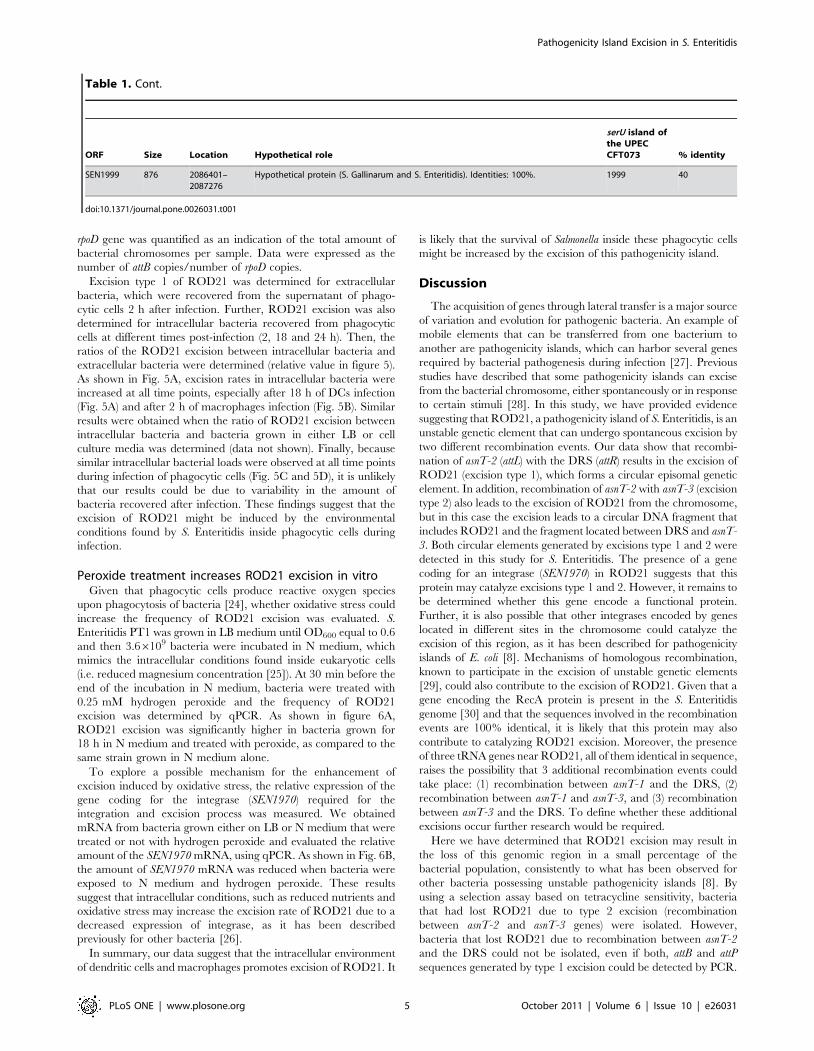
At least two different recombination events promoteROD21 excision from the bacterial chromosome
Next, we tested whether ROD21 is able to undergo spontane-
ous excision from the bacterial chromosome. Because ROD21 is
flanked by three copies of asnT and delimited by the DRS, at least
two types of recombination events may take place: recombination
between asnT-2 and the DRS (excision type 1) and recombination
between asnT-2 and asnT-3 (excision type 2). Either one of these
recombination events might result in the complete excision of this
new pathogenicity island (Fig. 2).
To evaluate whether these potential recombinations can occur,
we performed a PCR reaction using primers that hybridize
upstream and downstream of asnT genes and the DRS. As shown
in Fig. 2B and 2C, these hypothetical excision events yield two
different attB and attP sequences, which could be detected by PCR
using several different primer combinations. To detect these
excisions, the genomic DNA of four S. Enteritidis strains was
obtained as described in materials and methods and tested by
PCR. Conventional PCR amplifications failed to produce
measurable amounts of the expected PCR products for each of
the attB and attP sequences in each of the S. Enteritidis strains
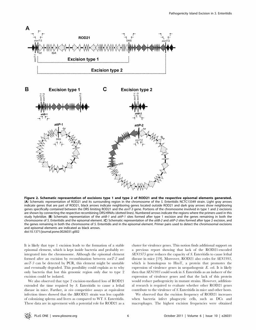
evaluated (data not shown). To increase the sensitivity of detection,
nested PCRs were performed as described in material and
methods and the expected sized amplicons were obtained: 591
and 657 bp for the chromosomal attB-1 and attB-2 respectively
(Fig. 3A) and 958 and 1058 for the episomal attP-1 and attP-2
respectively (Fig. 3B). To corroborate the specificity of these PCR
products, the DNA fragments obtained from S. Enteritidis PT1
were sequenced. As shown in Fig. 3, each of the obtained PCR
products matched the expected attB (Fig. 3A) and attP (Fig. 3B)
sequences. These data suggest that both recombination events
occurred at low frequency when bacteria grew to stationary phase
in LB medium.
Excision type 2 results in the loss of ROD21 from thechromosome of S. Enteritidis
Then, we evaluated whether the excision of ROD21 results in
the loss of this pathogenicity island from the bacterial genome. To
evaluate this possibility, we inserted the genes tetA and tetR
downstream the gene SEN1975 in the strain of S. Enteritidis
Phagotype 1 (PT1), to generate the ROD21::tetRA strain (Fig. 4A).
The genes tetA and tetR confer resistance to tetracycline, but also
prevent the growth of tetracycline-resistant bacteria in a medium
containing zinc chloride and fusaric acid (BM medium), as
described previously [21]. Therefore, only those Salmonella strains
sensitive to tetracycline will grow in BM medium [21]. Nested
PCR reactions showed that excision type 1 and type 2 (Fig. 2A)
occurred in the modified ROD21::tetRA strain, as efficiently as
observed for the wild type (WT) strain (data not shown). To
evaluate if these excisions caused ROD21 loss, the ROD21::tetRA
strain was grown in LB medium and then seeded on solid BM
medium, as described in materials and methods. Bacteria were
incubated 36 h at 37uC to select for tetracycline sensitive bacteria.
Pathogenicity Island Excision in S. Enteritidis
PLoS ONE | www.plosone.org 2 October 2011 | Volume 6 | Issue 10 | e26031
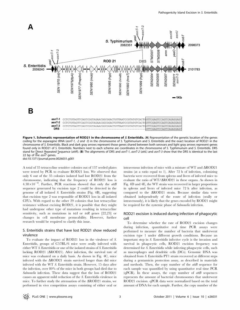
A total of 35 tetracycline sensitive colonies out of 137 seeded plates
were tested by PCR to evaluate ROD21 loss. We observed that
only 6 out of the 35 colonies isolated had lost ROD21 from the
chromosome, indicating that the frequency of ROD21 loss is
4.3861028. Further, PCR reactions showed that only the attB
sequence generated by excision type 2 could be detected in the
genome of all isolated S. Enteritidis strains (Fig. 4B), suggesting
that excision type 2 was responsible of ROD21 loss in all isolated
CFUs. With regard to the other 29 colonies that lost tetracycline
resistance without excising ROD21, it is possible that they might
had undergone other type of mutations resulting in tetracycline
sensitivity, such as mutations in tetA or tetR genes [22,23] or
changes in cell membrane permeability. However, further
research would be required to clarify this issue.
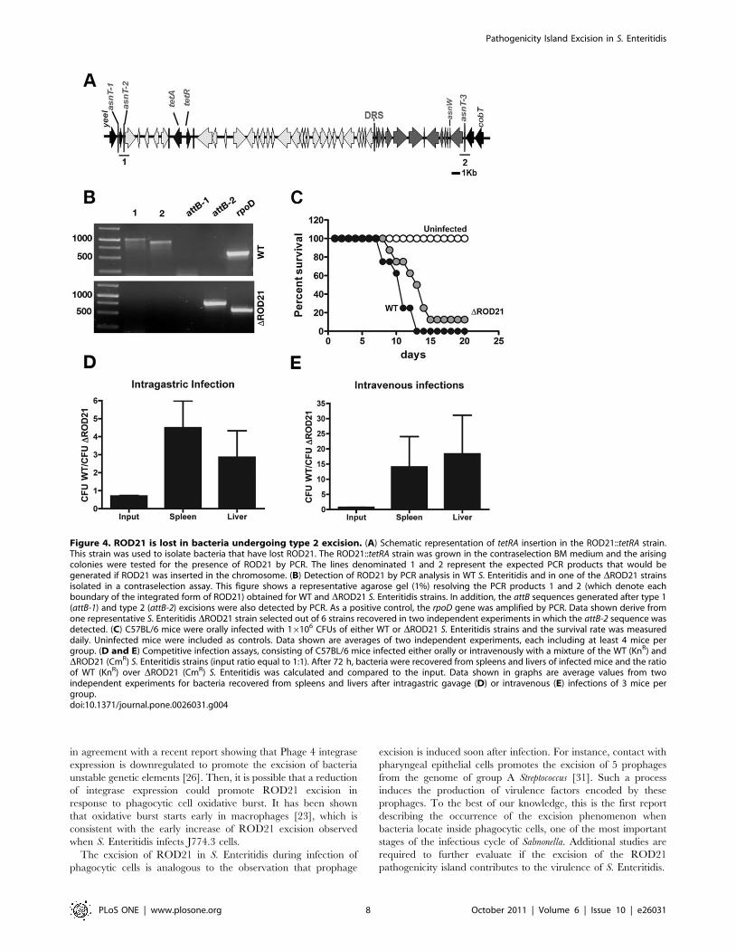
S. Enteritidis strains that have lost ROD21 show reducedvirulence
To evaluate the impact of ROD21 loss in the virulence of S.
Enteritidis, groups of C57BL/6 mice were orally infected with
either WT S. Enteritidis or one of the isolated strains of S. Enteritidis
lacking ROD21 (DROD21). After infection, the survival rate of
mice was evaluated on a daily basis. As shown in Fig. 4C, mice
infected with the DROD21 strain survived longer than did mice
infected with the WT S. Enteritidis strain. However, 15 days after
the infection, over 80% of the mice in both groups had died due to
Salmonella infection. These data suggest that the loss of ROD21
causes an apparent mild reduction of the S. Enteritidis virulence in
mice. To further study the attenuation of the DROD21 strains, we
performed in vivo competition assays consisting of either oral or
intravenous infection of mice with a mixture of WT and DROD21
strains (at a ratio equal to 1). After 72 h of infection, colonizing
bacteria were recovered from spleens and livers of infected mice to
evaluate the ratio of WT/DROD21 in these organs. As shown in
Fig. 4D and 4E, the WT strain was recovered in larger proportions
in spleens and livers of infected mice 72 h after infection, as
compared to the DROD21 strain. Because similar data were
obtained independently of the route of infection (orally or
intravenously), it is likely that the genes encoded by ROD21 might
be required for the systemic phase of Salmonella infection.
ROD21 excision is induced during infection of phagocyticcell
To determine whether the rate of ROD21 excision changes
during infection, quantitative real time PCR assays were
performed to measure the number of bacteria that underwent
excision type 1 under different growth conditions. Because an
important step in S. Enteritidis infective cycle is the invasion and
survival in phagocytic cells, ROD21 excision frequency was
determined for S. Enteritidis while infecting phagocytic cells, such
as macrophages and dendritic cells (DCs). Genomic DNA was
obtained from S. Enteritidis PT1 strain recovered at different steps
during a gentamicin protection assay, as described in materials
and methods. Then, the copy number of the attB sequence for
each sample was quantified by using quantitative real time PCR
(qPCR). In these assays, the copy number of attB sequences
represents the amount of bacterial chromosomes that underwent
ROD21 excision. qPCR data were normalized based on the total
amount of DNA for each sample. Further, the copy number of the
Figure 1. Schematic representation of ROD21 in the chromosome of S. Enteritidis. (A) Representation of the genetic location of the genescoding for the asparagine tRNA (asnT-1, -2 and -3) in the chromosome of S. Typhimurium and S. Enteritidis and the exact location of ROD21 in thechromosome of S. Enteritidis. Black and dark gray arrows represent those genes shared between both serovars and light gray arrows represent genesfound only in ROD21 of S. Enteritidis. Numbers next to each scheme are coordinates in the chromosome of S. Typhimurium and S. Enteritidis. DRSstand for Direct Repeated Sequence (attR). (B) The alignments of DRS and asnT-1, asnT-2 (attL) and asnT-3 show that the DRS is identical to the last22 bp of the asnT genes.doi:10.1371/journal.pone.0026031.g001
Pathogenicity Island Excision in S. Enteritidis
PLoS ONE | www.plosone.org 3 October 2011 | Volume 6 | Issue 10 | e26031
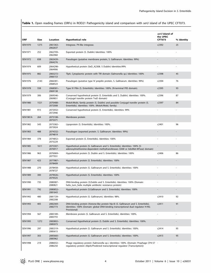
Table 1. Open reading frames (ORFs) in ROD21 Pathogenicity island and comparison with serU island of the UPEC CFT073.
ORF Size Location Hypothetical role
serU island ofthe UPECCFT073 % identity
SEN1970 1275 2061363–2062637
Integrase. P4-like integrase. c2392 25
SEN1971 252 2062709–2062960
Exported protein (S. Dublin) Identities: 100% - -
SEN1972 838 2063439–2064276
Pseudogen (putative membrane protein, S. Gallinarum. Identities: 99%) - -
SEN1974 609 2064298–2064906
Hypothetical protein (SeD_A2308. S Dublin) Identities:99% - -
SEN1975 882 2065272–2066153
TlpA. Cytoplasmic protein with TIR domain (Salmonella sp.) Identities: 100% c2398 45
SEN1976 2143 2066381–2068523
Pseudogen (putative type IV prepilin protein, S. Gallinarum. Identities: 99%) c2394 76
SEN1978 558 2068581–2069138
Type IV Pilin (S. Enteritidis). Identities: 100%. (N-terminal PilS domain). c2395 93
SEN1979 306 2069546–2069851
Conserved hypothetical protein (S. Enteritidis and S. Dublin). Identities: 100%.(Conjugal transfer protein TraD domain)
c2396 87
SEN1980 1521 2070488–2072008
MobA/MobL family protein (S. Dublin) and possible Conjugal transfer protein (S.Enteritidis). Identities: 100%. (MobA/MobL family).
c2397 84
SEN1981 915 2072032–2072946
Conserved hypothetical protein (S. Enteritidis). Identities: 99% - -
SEN1981A 264 2073108–2073371
Membrane protein
SEN1982 543 2073382–2073924
Lipoprotein (S. Enteritidis). Identities: 100%. c2401 96
SEN1983 488 2074332–2074819
Pseudogen (exported protein. S. Gallinarum. Identities: 99%) - -
SEN1984 378 2074852–2075229
Exported protein (S. Enteritidis). Identities: 100% - -
SEN1985 1611 2075307–2076917
Hypothetical protein (S. Gallinarum and S. Enteritidis). Identities: 100% (S-adenosylmethionine-dependent methyltransferases (SAM or AdoMet-MTase) domain)
- -
SEN1986 963 2076969–2077931
Hypothetical protein (S. Dublin and S. Enteritidis). Identities: 100% c2406 86
SEN1987 423 2077987–2078409
Hypothetical protein (S. Enteritidis). Identities: 100% - -
SEN1988 270 2078458–2078727
Hypothetical protein (S. Gallinarum and S. Enteritidis). Identities: 100% - -
SEN1989 300 2079026–2079325
Hypothetical protein (S. Enteritidis). Identities: 100% - -
SEN1990 735 2080087–2080821
DNA-binding protein (S.Dublin and S. Enteritidis). Identities: 100% (Domain:helix_turn_helix multiple antibiotic resistance protein)
- -
SEN1991 792 2080852–2081643
Hypothetical protein (S.Gallinarum and S. Enteritidis). Identities: 100% - -
SEN1992 480 2081729–2082208
Hypothetical protein (S. Gallinarum). Identities: 98%. c2410 92
SEN1993 405 2082369–2082773
DNA-binding protein (histone-like protein hlp-II) (S. Gallinarum and S. Enteritidis).Identities: 100% (Domain: global DNA-binding transcriptional dual regulator H-NS;Provisional)
c2411 91
SEN1994 567 2083189–2083755
Membrane protein (S. Gallinarum and S. Enteritidis). Identities: 100%. - -
SEN1995 1272 2083802–2085073
Conserved Hypothetical protein (S. Dublin and S. Enteritidis). Identities: 100%. - -
SEN1996 297 2085319–2085615
Hypothetical protein (S. Gallinarum and S. Enteritidis). Identities: 100%. c2414 95
SEN1997 303 2085660–2085962
Hypothetical protein (S. Gallinarum and S. Enteritidis). Identities: 100%. c2415 95
SEN1998 219 2086032–2086250
Phage regulatory protein (Salmonella sp.). Identities: 100%. (Domain: Prophage CP4-57regulatory protein (AlpA)/Predicted transcriptional regulator [Transcription])
- -
Pathogenicity Island Excision in S. Enteritidis
PLoS ONE | www.plosone.org 4 October 2011 | Volume 6 | Issue 10 | e26031
rpoD gene was quantified as an indication of the total amount of
bacterial chromosomes per sample. Data were expressed as the
number of attB copies/number of rpoD copies.
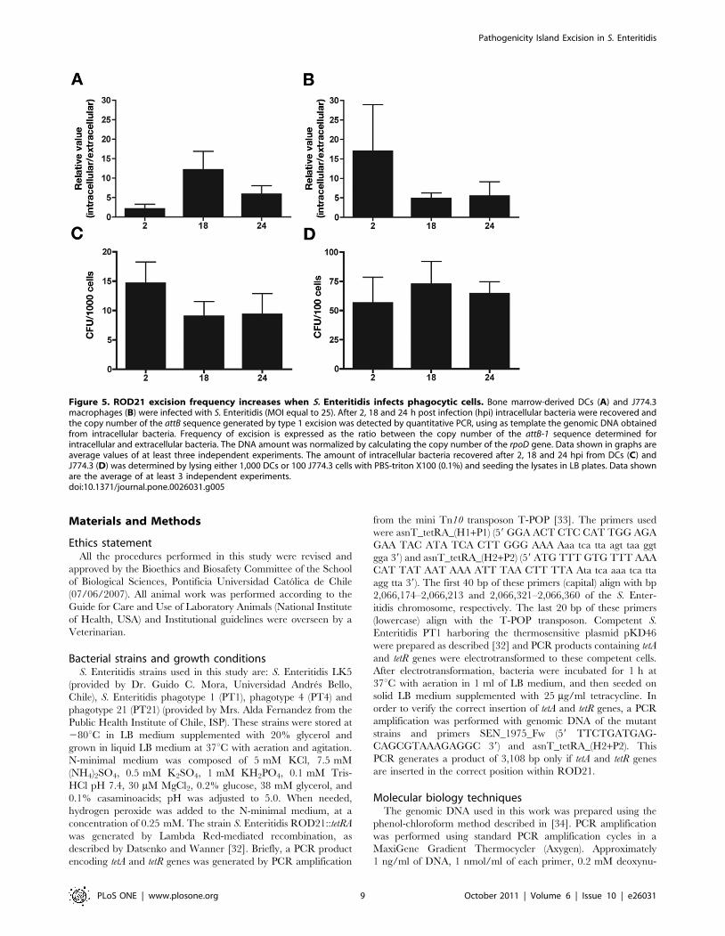
Excision type 1 of ROD21 was determined for extracellular
bacteria, which were recovered from the supernatant of phago-
cytic cells 2 h after infection. Further, ROD21 excision was also
determined for intracellular bacteria recovered from phagocytic
cells at different times post-infection (2, 18 and 24 h). Then, the
ratios of the ROD21 excision between intracellular bacteria and
extracellular bacteria were determined (relative value in figure 5).
As shown in Fig. 5A, excision rates in intracellular bacteria were
increased at all time points, especially after 18 h of DCs infection
(Fig. 5A) and after 2 h of macrophages infection (Fig. 5B). Similar
results were obtained when the ratio of ROD21 excision between
intracellular bacteria and bacteria grown in either LB or cell
culture media was determined (data not shown). Finally, because
similar intracellular bacterial loads were observed at all time points
during infection of phagocytic cells (Fig. 5C and 5D), it is unlikely
that our results could be due to variability in the amount of
bacteria recovered after infection. These findings suggest that the
excision of ROD21 might be induced by the environmental
conditions found by S. Enteritidis inside phagocytic cells during
infection.
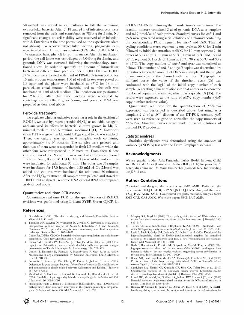
Peroxide treatment increases ROD21 excision in vitroGiven that phagocytic cells produce reactive oxygen species
upon phagocytosis of bacteria [24], whether oxidative stress could
increase the frequency of ROD21 excision was evaluated. S.
Enteritidis PT1 was grown in LB medium until OD600 equal to 0.6
and then 3.66109 bacteria were incubated in N medium, which
mimics the intracellular conditions found inside eukaryotic cells
(i.e. reduced magnesium concentration [25]). At 30 min before the
end of the incubation in N medium, bacteria were treated with
0.25 mM hydrogen peroxide and the frequency of ROD21
excision was determined by qPCR. As shown in figure 6A,
ROD21 excision was significantly higher in bacteria grown for
18 h in N medium and treated with peroxide, as compared to the
same strain grown in N medium alone.
To explore a possible mechanism for the enhancement of
excision induced by oxidative stress, the relative expression of the
gene coding for the integrase (SEN1970) required for the
integration and excision process was measured. We obtained
mRNA from bacteria grown either on LB or N medium that were
treated or not with hydrogen peroxide and evaluated the relative
amount of the SEN1970 mRNA, using qPCR. As shown in Fig. 6B,
the amount of SEN1970 mRNA was reduced when bacteria were
exposed to N medium and hydrogen peroxide. These results
suggest that intracellular conditions, such as reduced nutrients and
oxidative stress may increase the excision rate of ROD21 due to a
decreased expression of integrase, as it has been described
previously for other bacteria [26].
In summary, our data suggest that the intracellular environment
of dendritic cells and macrophages promotes excision of ROD21. It
is likely that the survival of Salmonella inside these phagocytic cells
might be increased by the excision of this pathogenicity island.
Discussion
The acquisition of genes through lateral transfer is a major source
of variation and evolution for pathogenic bacteria. An example of
mobile elements that can be transferred from one bacterium to
another are pathogenicity islands, which can harbor several genes
required by bacterial pathogenesis during infection [27]. Previous
studies have described that some pathogenicity islands can excise
from the bacterial chromosome, either spontaneously or in response
to certain stimuli [28]. In this study, we have provided evidence
suggesting that ROD21, a pathogenicity island of S. Enteritidis, is an
unstable genetic element that can undergo spontaneous excision by
two different recombination events. Our data show that recombi-
nation of asnT-2 (attL) with the DRS (attR) results in the excision of
ROD21 (excision type 1), which forms a circular episomal genetic
element. In addition, recombination of asnT-2 with asnT-3 (excision
type 2) also leads to the excision of ROD21 from the chromosome,
but in this case the excision leads to a circular DNA fragment that
includes ROD21 and the fragment located between DRS and asnT-
3. Both circular elements generated by excisions type 1 and 2 were
detected in this study for S. Enteritidis. The presence of a gene
coding for an integrase (SEN1970) in ROD21 suggests that this
protein may catalyze excisions type 1 and 2. However, it remains to
be determined whether this gene encode a functional protein.
Further, it is also possible that other integrases encoded by genes
located in different sites in the chromosome could catalyze the
excision of this region, as it has been described for pathogenicity
islands of E. coli [8]. Mechanisms of homologous recombination,
known to participate in the excision of unstable genetic elements
[29], could also contribute to the excision of ROD21. Given that a
gene encoding the RecA protein is present in the S. Enteritidis
genome [30] and that the sequences involved in the recombination
events are 100% identical, it is likely that this protein may also
contribute to catalyzing ROD21 excision. Moreover, the presence
of three tRNA genes near ROD21, all of them identical in sequence,
raises the possibility that 3 additional recombination events could
take place: (1) recombination between asnT-1 and the DRS, (2)
recombination between asnT-1 and asnT-3, and (3) recombination
between asnT-3 and the DRS. To define whether these additional
excisions occur further research would be required.
Here we have determined that ROD21 excision may result in
the loss of this genomic region in a small percentage of the
bacterial population, consistently to what has been observed for
other bacteria possessing unstable pathogenicity islands [8]. By
using a selection assay based on tetracycline sensitivity, bacteria
that had lost ROD21 due to type 2 excision (recombination
between asnT-2 and asnT-3 genes) were isolated. However,
bacteria that lost ROD21 due to recombination between asnT-2
and the DRS could not be isolated, even if both, attB and attP
sequences generated by type 1 excision could be detected by PCR.
ORF Size Location Hypothetical role
serU island ofthe UPECCFT073 % identity
SEN1999 876 2086401–2087276
Hypothetical protein (S. Gallinarum and S. Enteritidis). Identities: 100%. 1999 40
doi:10.1371/journal.pone.0026031.t001
Table 1. Cont.
Pathogenicity Island Excision in S. Enteritidis
PLoS ONE | www.plosone.org 5 October 2011 | Volume 6 | Issue 10 | e26031
It is likely that type 1 excision leads to the formation of a stable
episomal element, which is kept inside bacteria and probably re-
integrated into the chromosome. Although the episomal element
formed after an excision by recombination between asnT-2 and
ansT-3 can be detected by PCR, this element might be unstable
and eventually degraded. This possibility could explain as to why
only bacteria that lost this genomic region only due to type 2
excision could be isolated.
We also observed that type 2 excision-mediated loss of ROD21
extended the time required by S. Enteritidis to cause a lethal
disease in mice. Further, in vivo competitive assays at equivalent
infection times showed that the DROD21 strain was less capable
of colonizing spleens and livers as compared to WT S. Enteritidis.
These data are in agreement with a potential role for ROD21 as a
cluster for virulence genes. This notion finds additional support on
a previous report showing that lack of the ROD21-encoded
SEN1975 gene reduces the capacity of S. Enteritidis to cause lethal
disease in mice [19]. Moreover, ROD21 also codes for SEN1993,
which is homologous to HnsT, a protein that promotes the
expression of virulence genes in uropathogenic E. coli. It is likely
then that SEN1993 could work in S. Enteritidis as an inducer of the
expression of virulence genes and that the lack of this protein
would reduce pathogenicity in mutant strains. However, addition-
al research is required to evaluate whether other ROD21 genes
contribute to the virulence of S. Enteritidis in mice and other hosts.
We observed that the excision frequency of ROD21 increases
when bacteria infect phagocytic cells, such as DCs and
macrophages. The highest excision frequencies were obtained
Figure 2. Schematic representation of excisions type 1 and type 2 of ROD21 and the respective episomal elements generated.(A) Schematic representation of ROD21 and its surrounding region in the chromosome of the S. Enteritidis NCTC13349 strain. Light gray arrowsindicate genes that are part of ROD21, black arrows indicate neighboring genes located outside ROD21 and dark gray arrows show neighboringgenes specifically contained between the DRS limiting ROD21 and the asnT-3 gene. Portions of the chromosome involved in type 1 and 2 excisionsare shown by connecting the respective recombining DRS/tRNAs (dotted lines). Numbered arrows indicate the regions where the primers used in thisstudy hybridize. (B) Schematic representation of the attB-1 and attP-1 sites formed after type 1 excision and the genes remaining in both thechromosome of S. Enteritidis and the episomal element. (C) Schematic representation of the attB-2 and attP-2 sites formed after type 2 excision, andthe genes remaining in both the chromosome of S. Enteritidis and in the episomal element. Primer pairs used to detect the chromosomal excisionsand episomal elements are indicated as black arrows.doi:10.1371/journal.pone.0026031.g002
Pathogenicity Island Excision in S. Enteritidis
PLoS ONE | www.plosone.org 6 October 2011 | Volume 6 | Issue 10 | e26031
for bacteria recovered from the intracellular space of infected cells.
It is possible that recognition of signals present inside host cells
would lead to the activation of proteins involved in ROD21
excision. As a consequence of excision, transcription of genes
contained in these unstable genetic elements could be enhanced,
as described for unstable elements found in Corynebacterium
glutamicum [22]. Increased expression of potential virulence genes
contained in ROD21 when Salmonella locates inside host cells could
be relevant for its ability to survive intracellularly.
The conditions present in the Salmonella-containing vacuole,
such as low pH, high amounts of reactive oxygen species, nitric
oxide, reduced Mg+2 concentration, among others, could act as
signals that promote production or activation of proteins involved
in the excision process. In agreement with this notion, our results
show that the presence of hydrogen peroxide increases the
frequency of ROD21 excision. Furthermore, the transcription of
the integrase gene (SEN1970) is reduced when bacteria are grown
in N medium and exposed to hydrogen peroxide. These data are
Figure 3. ROD21 excision can be generated by means of two different recombination events. Amplification of attB and attP sequencesgenerated after type 1 and type 2 excisions were detected by nested PCR in LK5, PT4, PT1 and PT21 strains of S. Enteritidis, using primer pairsdescribed in Table 2 and in Figure 2. PCR products for attB (A) and attP (B) sequences for each type of excision were resolved in 1% agarose gels. Thesequence of each PCR product was obtained (chromatograms in each Figure) and compared with the attB and attP sequences deduced for type 1and type 2 excisions (labeled as theoretical). attB and attP sequences are highlighted in red in both alignments and chromatograms. Expected size foreach PCR product: 591 bp for type 1 excision attB, 657 bp for type 2 excision attB, 995 bp for type 1 excision attP and 1050 bp for type 2 excisionattP.doi:10.1371/journal.pone.0026031.g003
Pathogenicity Island Excision in S. Enteritidis
PLoS ONE | www.plosone.org 7 October 2011 | Volume 6 | Issue 10 | e26031
in agreement with a recent report showing that Phage 4 integrase
expression is downregulated to promote the excision of bacteria
unstable genetic elements [26]. Then, it is possible that a reduction
of integrase expression could promote ROD21 excision in
response to phagocytic cell oxidative burst. It has been shown
that oxidative burst starts early in macrophages [23], which is
consistent with the early increase of ROD21 excision observed
when S. Enteritidis infects J774.3 cells.
The excision of ROD21 in S. Enteritidis during infection of
phagocytic cells is analogous to the observation that prophage
excision is induced soon after infection. For instance, contact with
pharyngeal epithelial cells promotes the excision of 5 prophages
from the genome of group A Streptococcus [31]. Such a process
induces the production of virulence factors encoded by these
prophages. To the best of our knowledge, this is the first report
describing the occurrence of the excision phenomenon when
bacteria locate inside phagocytic cells, one of the most important
stages of the infectious cycle of Salmonella. Additional studies are
required to further evaluate if the excision of the ROD21
pathogenicity island contributes to the virulence of S. Enteritidis.
Figure 4. ROD21 is lost in bacteria undergoing type 2 excision. (A) Schematic representation of tetRA insertion in the ROD21::tetRA strain.This strain was used to isolate bacteria that have lost ROD21. The ROD21::tetRA strain was grown in the contraselection BM medium and the arisingcolonies were tested for the presence of ROD21 by PCR. The lines denominated 1 and 2 represent the expected PCR products that would begenerated if ROD21 was inserted in the chromosome. (B) Detection of ROD21 by PCR analysis in WT S. Enteritidis and in one of the DROD21 strainsisolated in a contraselection assay. This figure shows a representative agarose gel (1%) resolving the PCR products 1 and 2 (which denote eachboundary of the integrated form of ROD21) obtained for WT and DROD21 S. Enteritidis strains. In addition, the attB sequences generated after type 1(attB-1) and type 2 (attB-2) excisions were also detected by PCR. As a positive control, the rpoD gene was amplified by PCR. Data shown derive fromone representative S. Enteritidis DROD21 strain selected out of 6 strains recovered in two independent experiments in which the attB-2 sequence wasdetected. (C) C57BL/6 mice were orally infected with 16106 CFUs of either WT or DROD21 S. Enteritidis strains and the survival rate was measureddaily. Uninfected mice were included as controls. Data shown are averages of two independent experiments, each including at least 4 mice pergroup. (D and E) Competitive infection assays, consisting of C57BL/6 mice infected either orally or intravenously with a mixture of the WT (KnR) andDROD21 (CmR) S. Enteritidis strains (input ratio equal to 1:1). After 72 h, bacteria were recovered from spleens and livers of infected mice and the ratioof WT (KnR) over DROD21 (CmR) S. Enteritidis was calculated and compared to the input. Data shown in graphs are average values from twoindependent experiments for bacteria recovered from spleens and livers after intragastric gavage (D) or intravenous (E) infections of 3 mice pergroup.doi:10.1371/journal.pone.0026031.g004
Pathogenicity Island Excision in S. Enteritidis
PLoS ONE | www.plosone.org 8 October 2011 | Volume 6 | Issue 10 | e26031
Materials and Methods
Ethics statementAll the procedures performed in this study were revised and
approved by the Bioethics and Biosafety Committee of the School
of Biological Sciences, Pontificia Universidad Catolica de Chile
(07/06/2007). All animal work was performed according to the
Guide for Care and Use of Laboratory Animals (National Institute
of Health, USA) and Institutional guidelines were overseen by a
Veterinarian.
Bacterial strains and growth conditionsS. Enteritidis strains used in this study are: S. Enteritidis LK5
(provided by Dr. Guido C. Mora, Universidad Andres Bello,
Chile), S. Enteritidis phagotype 1 (PT1), phagotype 4 (PT4) and
phagotype 21 (PT21) (provided by Mrs. Alda Fernandez from the
Public Health Institute of Chile, ISP). These strains were stored at
280uC in LB medium supplemented with 20% glycerol and
grown in liquid LB medium at 37uC with aeration and agitation.
N-minimal medium was composed of 5 mM KCl, 7.5 mM
(NH4)2SO4, 0.5 mM K2SO4, 1 mM KH2PO4, 0.1 mM Tris-
HCl pH 7.4, 30 mM MgCl2, 0.2% glucose, 38 mM glycerol, and
0.1% casaminoacids; pH was adjusted to 5.0. When needed,
hydrogen peroxide was added to the N-minimal medium, at a
concentration of 0.25 mM. The strain S. Enteritidis ROD21::tetRA
was generated by Lambda Red-mediated recombination, as
described by Datsenko and Wanner [32]. Briefly, a PCR product
encoding tetA and tetR genes was generated by PCR amplification
from the mini Tn10 transposon T-POP [33]. The primers used
were asnT_tetRA_(H1+P1) (59 GGA ACT CTC CAT TGG AGA
GAA TAC ATA TCA CTT GGG AAA Aaa tca tta agt taa ggt
gga 39) and asnT_tetRA_(H2+P2) (59 ATG TTT GTG TTT AAA
CAT TAT AAT AAA ATT TAA CTT TTA Ata tca aaa tca tta
agg tta 39). The first 40 bp of these primers (capital) align with bp
2,066,174–2,066,213 and 2,066,321–2,066,360 of the S. Enter-
itidis chromosome, respectively. The last 20 bp of these primers
(lowercase) align with the T-POP transposon. Competent S.
Enteritidis PT1 harboring the thermosensitive plasmid pKD46
were prepared as described [32] and PCR products containing tetA
and tetR genes were electrotransformed to these competent cells.
After electrotransformation, bacteria were incubated for 1 h at
37uC with aeration in 1 ml of LB medium, and then seeded on
solid LB medium supplemented with 25 mg/ml tetracycline. In
order to verify the correct insertion of tetA and tetR genes, a PCR
amplification was performed with genomic DNA of the mutant
strains and primers SEN_1975_Fw (59 TTCTGATGAG-
CAGCGTAAAGAGGC 39) and asnT_tetRA_(H2+P2). This
PCR generates a product of 3,108 bp only if tetA and tetR genes
are inserted in the correct position within ROD21.
Molecular biology techniquesThe genomic DNA used in this work was prepared using the
phenol-chloroform method described in [34]. PCR amplification
was performed using standard PCR amplification cycles in a
MaxiGene Gradient Thermocycler (Axygen). Approximately
1 ng/ml of DNA, 1 nmol/ml of each primer, 0.2 mM deoxynu-
Figure 5. ROD21 excision frequency increases when S. Enteritidis infects phagocytic cells. Bone marrow-derived DCs (A) and J774.3macrophages (B) were infected with S. Enteritidis (MOI equal to 25). After 2, 18 and 24 h post infection (hpi) intracellular bacteria were recovered andthe copy number of the attB sequence generated by type 1 excision was detected by quantitative PCR, using as template the genomic DNA obtainedfrom intracellular bacteria. Frequency of excision is expressed as the ratio between the copy number of the attB-1 sequence determined forintracellular and extracellular bacteria. The DNA amount was normalized by calculating the copy number of the rpoD gene. Data shown in graphs areaverage values of at least three independent experiments. The amount of intracellular bacteria recovered after 2, 18 and 24 hpi from DCs (C) andJ774.3 (D) was determined by lysing either 1,000 DCs or 100 J774.3 cells with PBS-triton X100 (0.1%) and seeding the lysates in LB plates. Data shownare the average of at least 3 independent experiments.doi:10.1371/journal.pone.0026031.g005
Pathogenicity Island Excision in S. Enteritidis
PLoS ONE | www.plosone.org 9 October 2011 | Volume 6 | Issue 10 | e26031

cleoside triphosphates, 1.5 mM MgCl2 and 50 U/ml of Taq DNA
polymerase (Invitrogen) were used in these amplifications. Nested
PCR was performed as described above using 1 ml of a first PCR
product as a template. PCR products were resolved by
electrophoresis in 1% agarose gels containing 0.5 mg/ml ethidium
bromide and visualized under UV light. To determine the
specificity of the amplification reaction, some PCR products were
gel-purified and cloned into the pCRH-2.1H-TOPO cloning
plasmid, according to the manufacturer’s instructions (Invitrogen)
and sequenced by the Sequencing Facility at the Pontificia
Universidad Catolica de Chile.
Total RNA was obtained from each bacterial sample using
TRIzol reagent (Invitrogen) and purified with RNeasy Mini kit
(QIAGEN) according to the manufacturer instructions. After
purification, RNA was treated with the DNA Free kit (Ambion) to
remove contaminating genomic DNA. DNAse-treated RNA was
tested for DNA contamination amplifying rpoD gene by PCR.
Samples with no positive amplification up to cycle 30 were
considered to be DNA free. cDNA synthesis was performed using
the ImpromII Reverse Transcription System (Promega) following
the manufacturer instructions. One mg of RNA was used as a
template and cDNA synthesis was performed using random
hexamers. Reactions with no RNA or no reverse transcriptase
were included to rule out gDNA contamination.
Isolation of tetracycline-sensitive S. EnteritidisS. Enteritidis PT1 ROD21::tetRA was grown in LB medium
supplemented with 25 mg/ml tetracycline at 37uC, with aeration
provided by shaking, until an optical density at 600 nm (OD600)
equal to 2.0 was reached. Serial dilutions were performed in sterile
phosphate-buffered saline (PBS) to obtain 100 CFU in a volume of
100 ml. This volume was plated on LB agar supplemented with
tetracycline and incubated at 37uC for 18 h. One colony was
selected and grown in 2 ml of LB medium without antibiotics at
37uC for 18 h. Serial dilutions of this culture medium were
prepared and aliquots of 100 ml containing 106 CFU were plated
on solid Bochner-Maloy medium (5 g/l tryptone, 5 g/l yeast
extract, 10 g/l NaCl, 10 g/l NaH2PO4 N H2O, 50 mg/l
chlortetracycline hydrochloride, 12 mg/l fusaric acid, 0.1 mM
ZnCl2, 15 g/l agar) [21]. For each experiment, 100 plates were
seeded with 16106 CFU each and the amount of tetracycline
sensitive colonies was quantified. In addition, aliquots of 100 ml
containing approximately 100 CFU were plated on LB agar to
determine the exact CFU seeded in each BM plate. After 36 h of
incubation at 37uC, tetracycline-sensitive colonies were selected
and grown in liquid BM medium, at 37uC for 4 hours, then
patched in LB agar and replica plated in LB agar containing
tetracycline and incubated overnight at 37uC. Spontaneous loss of
ROD21 in tetracycline-sensitive CFU was confirmed by PCR,
using primers pairs indicated in Table 2 and in Fig. 2. The
frequency of ROD21 loss was calculated dividing the number of
tetracycline-sensitive colonies obtained by the total number of
CFU seeded in each experiment.
Mouse infection assaysGroups of 4 male C57BL/6 mice (5–6 week age) were used to
evaluate the virulence of strain S. Enteritidis PT1 DROD21.
Infections with WT or DROD21 S. Enteritidis PT1 strains were
performed by growing these bacteria in LB medium at 37uC until
an OD600 equal to 0.6 was reached. The volume of bacterial
culture containing 16106 CFUs was centrifuged in a refrigerated
microcentrifuge (CT15RE Hitachi) at 10,0006 g for 5 min.
Bacterial pellets were thoroughly resuspended in 20 ml of PBS and
used to orally infect mice. Infective doses were corroborated by
seeding serial dilutions of the bacterial inoculum onto LB plates.
After infection, survival rate was recorded on a daily basis. To
perform competitive assays, a kanamycin or chloramphenicol
resistance gene was introduced between genes putA (SEN0986A)
and putP (SEN0987) in WT and DROD21 S. Enteritidis,
respectively, using the methodology described by Datsenko and
Wanner [32]. These bacteria were grown until OD600 equal to 0.6
was reached and 16106 CFU of each strain was mixed,
resuspended in 200 ml PBS and used to infect groups of C57BL/
6 mice, by intragastric gavage. To perform intravenous infection,
1,000 CFU of each strain were mixed, resuspended in 20 ml PBS
and injected in the lateral tail vein using an insulin syringe (G29
needle). The initial proportion of kanamycin and chloramphenicol
resistant bacteria was determined by seeding bacteria in plates of
Figure 6. Exposure to peroxide induces the excision of ROD21from the S. Enteritidis chromosome. S. Enteritidis strain PT1 wasgrown in LB medium and then approximately 36109 CFUs weretransferred to fresh LB medium (LB) or to N-minimal medium (N), andincubated for 2 or 18 additional hours. Hydrogen peroxide was addedat a final concentration equal to 0.25 mM during the last 30 min ofincubation (N+H) and either genomic DNA or RNA was isolated. (A) Thefrequency of attB-1 excision was quantified by qPCR using genomicDNA and was expressed as a relative value equal to the ratio betweenthe copy number of attB-1 over the copy number of rpoD gene. (B)SEN1970 expression was determined by qPCR using cDNA andexpressed as a relative value (the ratio between the copy number ofSEN1970 and the copy number of rpoD). The results are the average ofthree independent experiments. **; ,0.01, one-way ANOVA and Tukeypost test.doi:10.1371/journal.pone.0026031.g006
Pathogenicity Island Excision in S. Enteritidis
PLoS ONE | www.plosone.org 10 October 2011 | Volume 6 | Issue 10 | e26031
solid LB, LB/kanamycin (50 mg/ml) and LB/chloramphenicol
(10 mg/ml) that were incubated for 12–16 h at 37uC. After 72 h of
infection, mice were euthanized and livers and spleens were
recovered and homogenized with two slides in a Petri dish, with
2 ml PBS. To determine the total amount of both kanamycin and
chloramphenicol resistant bacteria, serial dilutions of the homog-
enized tissue were seeded in solid LB, LB/Kanamycin (50 mg/ml)
and LB/Chloramphenicol (10 mg/ml) medium and incubated for
12–16 h at 37uC. Then, colonies were counted and the
competitive index was calculated as follows: (CFUWT/CFU-
DROD21) Input/(CFUWT/CFUDROD21) Output. The word ‘‘input’’
refers to the proportion of bacteria used to infect mice and
‘‘output’’ to the bacteria recovered from organs.
Phagocytic cells assaysThe monocyte/macrophage J774.3 cell line used in this study
was kindly provided by Dr. Marıa Ines Becker (Biosonda S.A.,
Chile). J774.3 cells were routinely grown in high-glucose DMEM
medium (GIBCO, Invitrogen), supplemented with 10% Fetal
Bovine Serum (HyClone) and 1 mM HEPES (GIBCO, Invitro-
gen) in T75 bottles. Cells were incubated at 37uC and 5% CO2
until 95% of confluence. Before infection assays, cells were treated
with 0.1 mg/ml trypsine (HyClone) for 5 min, recovered in 50 ml
polypropylene tubes, and centrifuged at 1,8006 g for 5 min at
room temperature. After three washes with supplemented DMEM
medium, cell number and viability was determined in a
haemocytometer, using the trypan blue staining (1 mg/ml,
Invitrogen). 56105 cells/ml were seeded in 24 well-plates and
incubated overnight at 37uC and 5% CO2. DCs were prepared
from bone marrow precursors of C57BL/6 mice. Cells were
incubated in complete RPMI 1640 medium supplemented with
5% FCS (Hyclone), 2 mM glutamine, 1 mM non-essential amino
acids, 1 mM pyruvate, 1 mM HEPES, and 10 ng/ml of
recombinant murine GM-CSF (Peprotech). All cell culture media
were acquired from GIBCO (Invitrogen). Culture media was
replaced every 2 days. After 6 days, the phenotype of DCs was
analyzed by flow cytometry for the expression of the surface
markers CD11c, CD86 and CD40, which revealed over 70%
CD11c+ with an immature phenotype. Before the infection assays,
DCs were washed three times with PBS and then culture media
was replaced with complete RPMI medium without antibiotics.
DCs and macrophages were infected with S. Enteritidis PT1 at a
multiplicity of infection (MOI) equal to 25. The MOI was
confirmed by plating serial dilutions of bacterial cultures on LB
agar. After 1 h of incubation at 37uC and 5% CO2, the
supernatant of infected cells was recovered and stored for genomic
DNA preparation. Cells were washed two times with PBS and
1 ml of appropriate medium supplemented with gentamicin
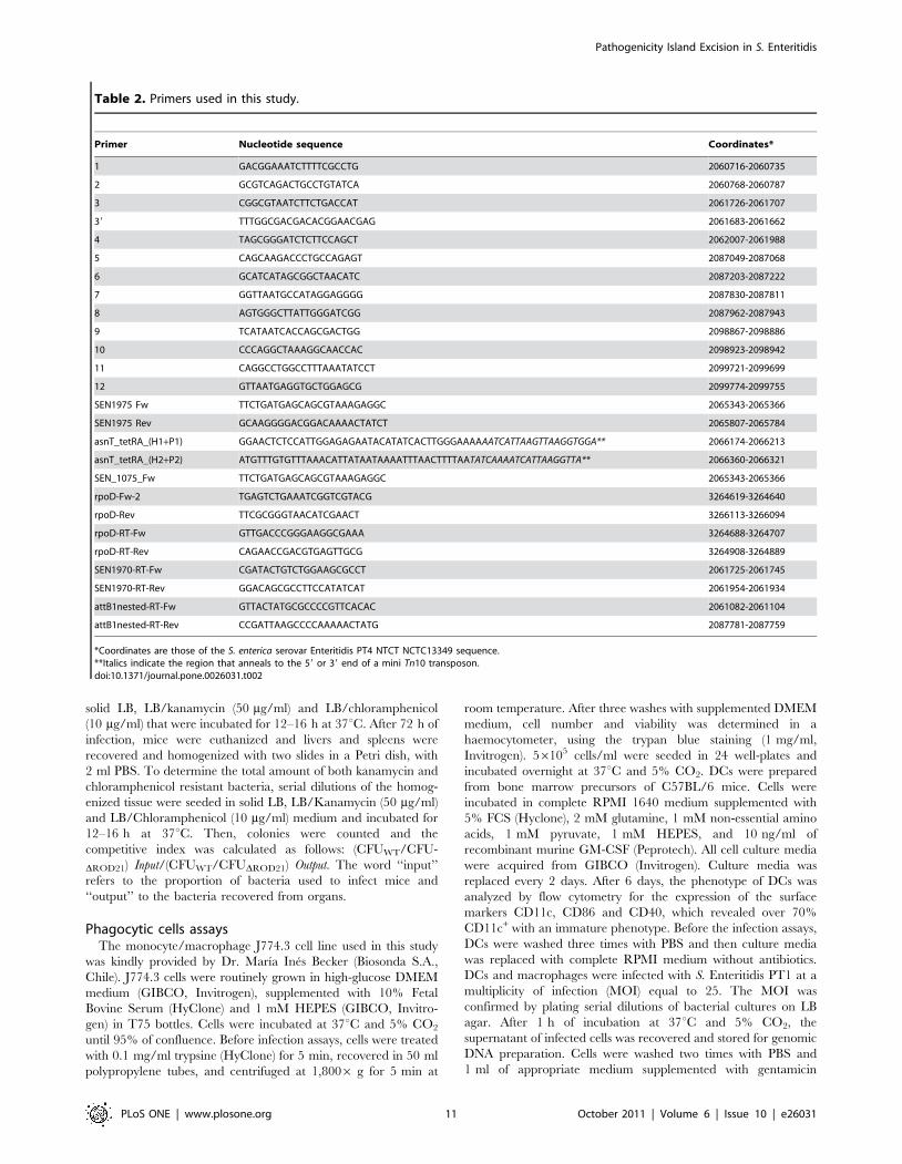
Table 2. Primers used in this study.
Primer Nucleotide sequence Coordinates*
1 GACGGAAATCTTTTCGCCTG 2060716-2060735
2 GCGTCAGACTGCCTGTATCA 2060768-2060787
3 CGGCGTAATCTTCTGACCAT 2061726-2061707
39 TTTGGCGACGACACGGAACGAG 2061683-2061662
4 TAGCGGGATCTCTTCCAGCT 2062007-2061988
5 CAGCAAGACCCTGCCAGAGT 2087049-2087068
6 GCATCATAGCGGCTAACATC 2087203-2087222
7 GGTTAATGCCATAGGAGGGG 2087830-2087811
8 AGTGGGCTTATTGGGATCGG 2087962-2087943
9 TCATAATCACCAGCGACTGG 2098867-2098886
10 CCCAGGCTAAAGGCAACCAC 2098923-2098942
11 CAGGCCTGGCCTTTAAATATCCT 2099721-2099699
12 GTTAATGAGGTGCTGGAGCG 2099774-2099755
SEN1975 Fw TTCTGATGAGCAGCGTAAAGAGGC 2065343-2065366
SEN1975 Rev GCAAGGGGACGGACAAAACTATCT 2065807-2065784
asnT_tetRA_(H1+P1) GGAACTCTCCATTGGAGAGAATACATATCACTTGGGAAAAAATCATTAAGTTAAGGTGGA** 2066174-2066213
asnT_tetRA_(H2+P2) ATGTTTGTGTTTAAACATTATAATAAAATTTAACTTTTAATATCAAAATCATTAAGGTTA** 2066360-2066321
SEN_1075_Fw TTCTGATGAGCAGCGTAAAGAGGC 2065343-2065366
rpoD-Fw-2 TGAGTCTGAAATCGGTCGTACG 3264619-3264640
rpoD-Rev TTCGCGGGTAACATCGAACT 3266113-3266094
rpoD-RT-Fw GTTGACCCGGGAAGGCGAAA 3264688-3264707
rpoD-RT-Rev CAGAACCGACGTGAGTTGCG 3264908-3264889
SEN1970-RT-Fw CGATACTGTCTGGAAGCGCCT 2061725-2061745
SEN1970-RT-Rev GGACAGCGCCTTCCATATCAT 2061954-2061934
attB1nested-RT-Fw GTTACTATGCGCCCCGTTCACAC 2061082-2061104
attB1nested-RT-Rev CCGATTAAGCCCCAAAAACTATG 2087781-2087759
*Coordinates are those of the S. enterica serovar Enteritidis PT4 NTCT NCTC13349 sequence.**Italics indicate the region that anneals to the 59 or 39 end of a mini Tn10 transposon.doi:10.1371/journal.pone.0026031.t002
Pathogenicity Island Excision in S. Enteritidis
PLoS ONE | www.plosone.org 11 October 2011 | Volume 6 | Issue 10 | e26031
50 mg/ml was added to cell cultures to kill the remaining
extracellular bacteria. After 2, 18 and 24 h of infection, cells were
removed from the wells and centrifuged at 7836 g for 5 min. No
significant changes on cell viability were observed after infection
with S. Enteritidis at the time points used on the experiments (data
not shown). To recover intracellular bacteria, phagocytic cells
were treated with 1 ml of lysis solution (19% ethanol, 0.1% SDS,
1% saturated basic phenol) for 30 min on ice. After the incubation
period, the cell lysate was centrifuged at 7,0436 g for 5 min, and
genomic DNA was extracted following the methodology men-
tioned above. In order to quantify the amount of intracellular
bacteria at different time points, either 100,000 DCs or 10,000
J774.3 cells were treated with 1 ml of PBS-0.1% triton X-100 for
15 min at room temperature. 100 ml of cell lysates were plated on
LB agar and the plates were incubated at 37uC for 18 h. In
parallel, an equal amount of bacteria used to infect cells was
incubated in 1 ml of cell medium. The incubation was performed
for 2 h and after that time bacteria were recovered by
centrifugation at 7.0436 g for 5 min, and genomic DNA was
prepared as described above.
Peroxide treatmentTo evaluate whether oxidative stress has a role in the excision of
ROD21, we used hydrogen peroxide (H2O2) as an oxidative agent
and analyzed its effects in bacterial cultures grown in LB, N-
minimal medium, and N-minimal medium+H2O2. S. Enteritidis
strain PT1 was grown in LB until OD600 equal to 0.6 was reached.
Then, the culture was split in 6 samples, each containing
approximately 36109 bacteria. The samples were pelleted and
then two of them were resuspended in fresh LB medium while the
other four were resuspended in N medium. From these 6 new
cultures, two of the N cultures were incubated for an additional
1.5 hour. Next, 0.25 mM H2O2 (Merck) was added and cultures
were incubated for additional 30 min. The other two N samples
were incubated for 17.5 hours, then 0.25 mM H2O2 (Merck) was
added and cultures were incubated for additional 30 minutes.
After the H2O2 treatment, all samples were pelleted and stored at
280uC until analyzed. Genomic DNA or total RNA was prepared
as described above.
Quantitative real time PCR assaysQuantitative real time PCR for the quantification of ROD21
excisions was performed using Brilliant SYBR Green QPCR kit
(STRATAGENE), following the manufacturer’s instructions. The
reaction mixture contained 2 ml of genomic DNA as a template
and 0.12 pmol/ml of each primer. Standard curves for attB-1 and
rpoD were generated using serial dilutions of a plasmid containing
the corresponding PCR fragment for attB-1 and rpoD. Thermal
cycling conditions were: segment 1; one cycle at 50uC for 2 min
followed by initial denaturation at 95uC for 10 min; segment 2, 40
cycles of 30 s at 95uC, 1 min at 58uC, 1 min at 72uC and 15 s at
80uC; segment 3, 1 cycle of 1 min at 95uC, 30 s at 55uC and 30 s
at 95uC. The copy number of attB-1 and rpoD was calculated as
follows: The number of attB-1 and rpoD copies was determined by
the ratio between the amount of DNA in a sample and the weight
of one molecule of the plasmid with the insert. To graph the
standard curve, the value of the threshold cycle (Ct) was
confronted with the log10 of the initial copy number of each
sample, generating a linear relationship that allows us to know the
number of copies of the sample, which has a specific Ct [35]. The
results were expressed as the ratio of attB-1 copy number/rpoD
copy number (relative value).
Quantitative real time for the quantification of SEN1970
expression was performed as described above, but using as a
template 2 ml of a 1021 dilution of the RT-PCR reaction. rpoD
were used as reference gene to normalize the copy numbers of
SEN1970. Standard curves were made of serial dilutions of
purified PCR products.
Statistic analysesStatistics significance was determined using the analyses of
variance (ANOVA) test with the Prism Graphpad software.
Acknowledgments
We are grateful to Mrs. Alda Fernandez (Public Health Institute, Chile)
and Dr. Guido Mora (Universidad Andres Bello, Chile) for providing S.
Enteritidis strains and Dr. Marıa Ines Becker (Biosonda S.A.) for providing
the J774.3 cells.
Author Contributions
Conceived and designed the experiments: SMB AMK. Performed the
experiments: TSQ HET RJL PAN FJS CPQ DVA. Analyzed the data:
TSQ PAN AMK SMB. Contributed reagents/materials/analysis tools:
SMB CAR CAS AMK. Wrote the paper: SMB PAN AMK.
References
1. Guard-Petter J (2001) The chicken, the egg and Salmonella Enteritidis. Environ
Microbiol 3: 421–430.
2. Thomson NR, Clayton DJ, Windhorst D, Vernikos G, Davidson S, et al. (2008)
Comparative genome analysis of Salmonella Enteritidis PT4 and Salmonella
Gallinarum 287/91 provides insights into evolutionary and host adaptation
pathways. Genome Res 18: 1624–1637.
3. Cotter PA, DiRita VJ (2000) Bacterial virulence gene regulation: an evolutionary
perspective. Annu Rev Microbiol 54: 519–565.
4. Bueno SM, Gonzalez PA, Carreno LJ, Tobar JA, Mora GC, et al. (2008) The
capacity of Salmonella to survive inside dendritic cells and prevent antigenpresentation to T cells is host specific. Immunology 124: 522–533.
5. Gantois I, Ducatelle R, Pasmans F, Haesebrouck F, Gast R, et al. (2009)
Mechanisms of egg contamination by Salmonella Enteritidis. FEMS Microbiol
Rev 33: 718–738.
6. Porwollik S, Santiviago CA, Cheng P, Florea L, Jackson S, et al. (2005)
Differences in gene content between Salmonella enterica serovar Enteritidis isolatesand comparison to closely related serovars Gallinarum and Dublin. J Bacteriol
187: 6545–6555.
7. Middendorf B, Hochnut B, Leipold K, Dobrindt U, Blum-Oehler G, et al.
(2004) Instability of pathogenicity islands in uropathogenic Escherichia coli 536.J Bacteriol 186: 3086–3096.
8. Hochhut B, Wilde C, Balling G, Middendorf B, Dobrindt U, et al. (2006) Role of
pathogenicity island-associated integrases in the genome plasticity of uropatho-
genic Escherichia coli strain 536. Mol Microbiol 61: 584–595.
9. Murphy RA, Boyd EF (2008) Three pathogenicity islands of Vibrio cholerae can
excise from the chromosome and form circular intermediates. J Bacteriol 190:636–647.
10. Turner SA, Luck SN, Sakellaris H, Rajakumar K, Adler B (2001) Nested deletionsof the SRL pathogenicity island of Shigella flexneri 2a. J Bacteriol 183: 5535–5543.
11. Lesic B, Bach S, Ghigo JM, Dobrindt U, Hacker J, et al. (2004) Excision of thehigh-pathogenicity island of Yersinia pseudotuberculosis requires the combined
actions of its cognate integrase and Hef, a new recombination directionalityfactor. Mol Microbiol 52: 1337–1348.
12. Bach S, Buchrieser C, Prentice M, Guiyoule A, Msadek T, et al. (1999) Thehigh-pathogenicity island of Yersinia enterocolitica Ye8081 undergoes low-
frequency deletion but not precise excision, suggesting recent stabilization inthe genome. Infect Immun 67: 5091–5099.
13. Bueno SM, Santiviago CA, Murillo AA, Fuentes JA, Trombert AN, et al. (2004)Precise excision of the large pathogenicity island, SPI7, in Salmonella enterica
serovar Typhi. J Bacteriol 186: 3202–3213.
14. Santiviago CA, Blondel CJ, Quezada CP, Silva CA, Tobar PM, et al. (2010)
Spontaneous excision of the Salmonella enterica serovar Enteritidis-specificdefective prophage-like element phiSE14. J Bacteriol 192: 2246–2254.
15. Lovell HC, Mansfield JW, Godfrey SA, Jackson RW, Hancock JT, et al. (2009)Bacterial evolution by genomic island transfer occurs via DNA transformation in
planta. Curr Biol 19: 1586–1590.
16. Ramsay JP, Sullivan JT, Jambari N, Ortori CA, Heeb S, et al. (2009) A LuxRI-
family regulatory system controls excision and transfer of the Mesorhizobium loti
Pathogenicity Island Excision in S. Enteritidis
PLoS ONE | www.plosone.org 12 October 2011 | Volume 6 | Issue 10 | e26031
strain R7A symbiosis island by activating expression of two conserved
hypothetical genes. Mol Microbiol 73: 1141–1155.17. Almagro-Moreno S, Napolitano MG, Boyd EF (2010) Excision dynamics of
Vibrio pathogenicity island-2 from Vibrio cholerae: role of a recombination
directionality factor VefA. BMC Microbiol 10: 306.18. Williamson HS, Free A (2005) A truncated H-NS-like protein from
enteropathogenic Escherichia coli acts as an H-NS antagonist. Mol Microbiol55: 808–827.
19. Newman RM, Salunkhe P, Godzik A, Reed JC (2006) Identification and
characterization of a novel bacterial virulence factor that shares homology withmammalian Toll/interleukin-1 receptor family proteins. Infect Immun 74:
594–601.20. Thomson N, Baker S, Pickard D, Fookes M, Anjum M, et al. (2004) The role of
prophage-like elements in the diversity of Salmonella enterica serovars. J Mol Biol339: 279–300.
21. Maloy SR, WD Nunn (1981) Selection for loss of tetracycline resistance by
Escherichia coli. J Bacteriol 145: 1110–1111.22. Moyed HS, Bertrand KP (1983) Mutations in multicopy Tn10 tet plasmids that
confer resistance to inhibitory effects of inducers of tet gene expression.J Bacteriol 155: 557–564.
23. McMurry LM, Stephan M, Levy SB (1992) Decreased function of the class B
tetracycline efflux protein Tet with mutations at aspartate 15, a putativeintramembrane residue. J Bacteriol 174: 6294–6297.
24. Araya DV, Quiroz TS, Tobar HE, Lizana RJ, Quezada CP, et al. (2010)Deletion of a prophage-like element causes attenuation of Salmonella enterica
serovar Enteritidis and promotes protective immunity. Vaccine 28: 5458–5466.25. Deiwick J, Nikolaus T, Erdogan S, Hensel M (1999) Environmental regulation of
Salmonella pathogenicity island 2 gene expression. Mol Microbiol 3: 1759–1773.
26. Moyed HS, Nguyen TT, Bertrand KP (2009) Multicopy Tn10 tet plasmids
confer sensitivity to induction of tet gene expression. J Bacteriol 155: 549–556.
27. Juhas M, van der Meer JR, Gaillard M, Harding RM, Hood DW (2009)
Genomic islands: tools of bacterial horizontal gene transfer and evolution. FEMS
Microbiol Rev 33: 376–393.
28. Manson JM, Gilmore MS (2006) Pathogenicity island integrase cross-talk: a
potential new tool for virulence modulation. Mol Microbiol 61: 555–559.
29. Lesic B, Carniel E (2005) Horizontal transfer of the high-pathogenicity island of
Yersinia pseudotuberculosis. J Bacteriol 187: 3352–3358.
30. Kobayashi H, Miyamoto T, Hashimoto Y, Kiriki M, Motomatsu A, et al. (2005)
Identification of factors involved in recovery of heat-injured Salmonella
Enteritidis. J Food Prot 68: 932–941.
31. Banks DJ, Lei B, Musser JM (2003) Prophage induction and expression of
prophage-encoded virulence factors in group A Streptococcus serotype M3 strain
MGAS315. Infect Immun 71: 7079–7086.
32. Datsenko KA, Wanner BL (2000) One-step inactivation of chromosomal genes
in Escherichia coli K-12 using PCR products. Proc Natl Acad Sci U S A 97:
6640–6645.
33. Hidalgo AA, Trombert AN, Castro-Alonso JC, Santiviago CA, Tesser BR, et al.
(2004) Insertions of mini-Tn10 transposon T-POP in Salmonella enterica sv. typhi.
Genetics 167: 1069–1077.
34. Sambrook J, Fritsch EF, Maniatis T (1982) Molecular cloning: a laboratory
manual. Cold Spring HarborNY: Cold Spring Harbor Laboratory. pp 545.
35. Higuchi R, Fockler C, Dollinger G, Watson R (1993) Kinetic PCR analysis: real-
time monitoring of DNA amplification reactions. Biotechnology (NY) 11:
1026–1030.
Pathogenicity Island Excision in S. Enteritidis
PLoS ONE | www.plosone.org 13 October 2011 | Volume 6 | Issue 10 | e26031
Related Documents