UMEÅ UNIVERSITY MEDICAL DISSERTATIONS New Series No. 11 – 58 ISSN 0346-6612 ISBN 978-91-7264-512-7 ______________________________________________________ Department of Medical Biosciences, Pathology, University of Umeå, Sweden 2008 EXAMINING THE PROSTATE STROMA AND VASCULATURE - Importance and potential as targets for therapy Anna Johansson

Welcome message from author
This document is posted to help you gain knowledge. Please leave a comment to let me know what you think about it! Share it to your friends and learn new things together.
Transcript
UMEÅ UNIVERSITY MEDICAL DISSERTATIONS New Series No. 11 – 58 ISSN 0346-6612 ISBN 978-91-7264-512-7 ______________________________________________________
Department of Medical Biosciences, Pathology,
University of Umeå, Sweden 2008
EXAMINING THE PROSTATE STROMA AND VASCULATURE
- Importance and potential as targets for therapy
Anna Johansson

Copyright © Anna Johansson ISSN 0346-6612, New Series No. 11 – 58 ISBN 978-91-7264-512-7 Printed by Print och Media Umeå, Sweden, 2008

To my family

ABSTRACT Background. Recent studies in cancer research have focused on the reciprocal interaction between cancer cells and their microenvironment. Tumour growth is angiogenesis dependent and the rate of angiogenesis correlates with a poor prognosis in many different cancers. We have shown that the rate of angiogenesis correlates with prognosis in Prostate Cancer (PC). We have also observed that the vasculature is involved during the involution of the prostate in rodents subsequent to hormonal ablation. Patients with metastatic PC are subjected to hormonal ablation therapy – a therapy unfortunately not curative. Our ambition is therefore to find means to enhance the effects of castration therapy of prostate tumours, possibly by a simultaneous inhibition of angiogenesis and of growth factors populating the tumour stroma. The angiopoietins are a family of growth factors that regulate angiogenesis by direct effects on endothelial cells in a context dependent manner. The purpose of this thesis was therefore to examine the role of the angiopoietins and the stroma in general in PC and to explore their potential as novel targets. Materials and Methods. We have had at our disposal access to clinical materials in the form of paraffin embedded samples from untreated PC patients with a long follow up. We have also used animal tumour models and in vitro cell culture systems followed by immunohistochemistry, in situ hybridization, western blotting, laser micro dissection, and quantitative real-time PCR for evaluation of the experiments. Results. In paper I, we found a significant correlation between high levels of angiopoietin 2 (Ang 2) and high vascular density, histological grade, metastases and poor prognosis in PC patients. In the second paper we found that the receptor for the angiopoietins, Tie 2, and the ligand Ang 1 mediated the decrease in vascular stability observed after castration treatment. This was not observed in prostate tumours subsequent to hormonal ablation (paper III), nor was there a decrease of other growth factor receptors. In summary (paper III), we found that a combined inhibition of the tumour stroma in terms of an inhibition of the PDGF-Rs by the use of Imatinib, and the vasculature in terms of a perturbed Tie 2 signalling, inhibited tumour growth. Finally, in paper IV, we found that Imatinib inhibited the castration induced influx of mast cells after castration therapy. The mast cells expressed high levels of FGF 2 and epiregulin, and inhibition of mast cell function inhibited tumour growth, by inhibiting angiogenesis. Conclusions. We have observed that the tumour stroma is of particular importance for tumour growth in PC. Targeting the tumour microenvironment, and in particular by a simultaneous inhibition of the vasculature and stroma, could prove beneficial for patients with advanced PC.

ORIGINAL PAPERS This thesis is based on the following papers, which are referred to in the text by their
Roman numerals.
I. Anna J:son Lind, Pernilla Wikström, Torvald Granfors, Lars Egevad, Pär Stattin,
Anders Bergh. Angiopoietin 2 expression is related to histological grade, vascular
density, metastases, and outcome in prostate cancer. The Prostate 62:394-399 (2005)
II. Anna Johansson, Stina Häggström Rudolfsson, Pernilla Wikström, Anders Bergh.
Altered levels of angiopoietin 1 and Tie 2 are associated with androgen-regulated
vascular regression and growth in the ventral prostate in adult mice and rats.
Endocrinology 146(8):3463-3470 (2005).
III. Anna Johansson, Jonathan Jones, Kristian Pietras, Sigrid Kilter, Åsa Skytt, Stina
Häggström Rudolfsson, Anders Bergh. A stroma targeted therapy enhances castration
effects in a transplantable rat prostate tumour model. The Prostate Nov 1;67(15):1664-
76 (2007).
IV. Anna Johansson, Stina Häggström Rudolfsson, Sofia Halin, Kristian Pietras, Anders
Bergh. Mast Cell Targeted Therapy – a Novel Approach to Inhibit Angiogenesis and
Prostate Tumour Growth. Manuscript.
POPULÄRVETENSKAPLIG SAMMANFATTNING Bakgrund. Prostatacancer är den vanligaste cancerformen bland svenska män. Varje år diagnostiseras 10 000 fall och 25 % av dem kommer att dö av sjukdomen. Avancerad prostata cancer, med dottertumörer i framför allt skelettet, behandlas med kastrationsterapi, det vill säga att man tar bort det manliga könshormonet testosteron. Anledningen till detta är att prostatatumörer till del är beroende av testosteron för sin tillväxt. Tyvärr är denna behandling inte botande, tumörerna växer efter en tid till igen och patienten avlider några få år efter behandling. Att kunna förstärka effekten och förhindra återfall är därför av yttersta vikt. Tidigare studier i vår grupp har visat att den normala och den elakartade prostatans tillväxt också delvis bestäms av hur mycket blodkärl som finns både i och utanför tumörområdet. Vid genomgång av ett stort patientmaterial har vi också sett att mängden kärl ger en indikation om hur prognosen ser ut; ju fler kärl desto sämre prognos. Vi försöker därför kombinera kastrationsbehandling med andra behandlingar som slår ut blodkärl och stödjeceller för att uppnå kraftigare och mer långvarig behandlingseffekt. Material och metoder. Vi har framförallt använt oss av djurmodeller med prostatacancer och vävnadsprover, från patienter med prostatacancer. Djuren har kastrerats och sedan fått tilläggsbehandling som slagit mot blodkärl med omkringliggande stödjevävnader. Vävnaderna har sedan analyserats med mikroskopering där vi bestämt mängden kärl, hur fort kärlen växt, mängden tumör samt konstaterat om tumörens celler delat på sig eller dött. Resultat. I den första studien fann vi genom att undersöka biopsier från patienter med prostatacancer att hög produktion av ett protein, som heter Angiopoietin 2 (Ang 2), och som stimulerar blodkärlstillväxt, korrelerar med aggressiv cancer och medför kortare överlevnad. I nästa studie undersökte vi om Ang 2 tillsammans med ytterligare medlemmar i samma ”proteinfamilj”, nämligen Angiopoietin 1 (Ang 1) och deras ”antenn” på blodkärl Tie 2, hade någon betydelse för den normala prostatans blodkärlsminskning och blodkärlsökning efter hormonell behandling. Det hade de. I studie 3 gick vi vidare och undersökte två olika sorters tumörer i råttor för att få reda på om det kunde bero på angiopoietinerna att det inte uppstår någon blodkärlsminskning i tumörer efter kastrering. Vi fann att så kunde vara fallet, den normala regleringen av angiopoietinerna var borta i tumörerna. Vi fann även att en annan familj av kärlreglerande proteiner, de s.k. PDGFs, inte heller svarade normalt på kastrering. Då gick vi vidare och designade en terapi som skulle slå ut angiopoietinerna och PDGFs efter kastrering av djuren. Vi fann att denna kombinerade behandling dödade blodkärlen och minskade tumörtillväxten efter 4 veckors behandling medan de djur som bara kastrerats relapsade efter 4 veckor. Till vår förvåning upptäckte vi också att det blev mycket färre så kallade mastceller i de behandlade djurens tumörer. Mastceller är inflammatoriska celler som i andra tumörmodeller visat sig spela en stor roll för tumörtillväxt. I studie 4 gick vi därför vidare med den upptäckten och undersökte vilken betydelse mastcellerna hade för tillväxt av prostatacancer. Vi fann att mastcellerna ansamlades efter kastrationsterapi i patienter och djur, att de producerade

flera proteiner som aktivt stimulerar blodkärl och att vi kunde hämma tumörtillväxt genom att slå ut mastcellerna. Slutsats. I dag är inte kastrationsbehandling botande. Vi har sett att en kombinerad behandling inriktad mot blodkärl och inflammatoriska celler (mastceller) tillsammans eller utan kastrationsterapi ger en starkt hämmande effekt på prostatacancertillväxt i flera olika råttumörmodeller. På grund av detta borde fler kliniska studier uppmärksamma betydelsen av att förstärka kastrationsbehandlingen genom att samtidigt slå ut blodkärl med omkringliggande stödjevävnad och mastceller.
CONTENTS INTRODUCTION................................................................................................................ - 1 -
The Prostate ...................................................................................................................... - 1 - Prostate Anatomy and function..................................................................................... - 1 - Prostate Development ................................................................................................... - 2 - Prostate Growth Control ............................................................................................... - 2 -
Prostate Cancer................................................................................................................. - 3 - General Background, Epidemiology & Etiology.......................................................... - 3 - Classification of Prostate Tumours; PSA Test and Gleason Score ............................... - 4 - Treatment of local and metastatic disease..................................................................... - 5 - Hormone Refractory Prostate Cancer (HRPC) ............................................................. - 5 -
Angiogenesis...................................................................................................................... - 6 - Process and Function .................................................................................................... - 6 - The Angiopoietins ........................................................................................................ - 7 - Other regulators of vascular maturation and integrity ................................................ - 12 -
Tumour Vascular Biology................................................................................................ - 13 - Tumour angiogenesis.................................................................................................. - 13 -
The Concept “Tumour Stroma” ...................................................................................... - 14 - Composition and reciprocal interaction with tumour cells ......................................... - 14 - Recruitment of inflammatory cell types and endothelial progenitors ......................... - 16 -
Significance ..................................................................................................................... - 17 - Specific aims............................................................................................................... - 18 -
MATERIALS AND METHODS....................................................................................... - 19 - Animal models of PC ....................................................................................................... - 19 - Animal Studies in Vivo..................................................................................................... - 19 -
The Dunning Tumours (paper III and IV) .................................................................. - 19 - Wild Type Mouse and Rat Prostate Studies (paper II) ............................................... - 20 -
Patient Materials ............................................................................................................. - 21 - Västerås Project (paper I) ........................................................................................... - 21 - Human freeze biopsies from poorly differentiated tumours (unpublished observations) ...- 22 -
Protein Analysis............................................................................................................... - 22 - Isolation of Protein Extracts (paper II - III) ................................................................ - 22 - Western Blot (paper II - IV) ....................................................................................... - 22 -
RNA Analysis ................................................................................................................... - 23 - RNA Preparation tissues and cell lines (paper II-IV) ................................................. - 23 - Laser Micro dissection techniques (paper II and IV).................................................. - 23 - cDNA synthesis (paper II – IV) .................................................................................. - 24 -
Quantitative Real-Time RT-PCR and array analyses (paper II – IV) ......................... - 24 - Immunohistochemistry (IHC) .......................................................................................... - 24 -
Tissue Staining (paper I- IV) ...................................................................................... - 24 - Ultrasound....................................................................................................................... - 25 -
Gray Scale Ultrasound (paper III)............................................................................... - 25 - Statistics........................................................................................................................... - 25 -
RESULTS AND DISCUSSION......................................................................................... - 26 - Paper I............................................................................................................................. - 26 - Paper II............................................................................................................................ - 28 - Paper III .......................................................................................................................... - 29 - Paper IV .......................................................................................................................... - 31 -
GENERAL DISCUSSION AND FUTURE PERSPECTIVES....................................... - 35 - Inhibition of the angiopoietin system in human patients ................................................. - 35 - Pros and Cons with anti angiogenic therapies................................................................ - 36 -
CONCLUSIONS................................................................................................................. - 38 -
REFERENCES................................................................................................................... - 39 -
ACKNOWLEDGMENTS.................................................................................................. - 56 -
ABBREVATIONS Ang Angiopoietin AR Androgen Receptor BrdU Bromodeoxyuridine CAF Cancer Associated Fibroblast cDNA Complementary Deoxyribonucleic Acid CML Chronic Myelogenous Leukaemia DLP Dorsolateral Prostate ECM Extracellular Matrix EGF Epidermal Growth Factor EPC Endothelial Progenitor Cell Erk Extracellular Signal-Regulated Kinase FGF Fibroblast Growth Factor GIST Gastrointestinal Stromal Tumour GS Gleason Score HE Hematoxylin Eosin IFP Interstitial Fluid Pressure IHC Immunohistochemistry MAPK Mitogen-Activated Protein Kinase MMP Matrix Metalloproteinase mRNA Messenger Ribonucleic Acid MVD Microvessel Density PBS Phosphate Buffer Saline PBST Phosphate Buffer Saline Tween PDGF Platelet Derived Growth Factor PI3-K Phosphatidylinositol 3-Kinase PSA Prostate Specific Antigen RTK Receptor Tyrosine Kinase RT-PCR Polymerase Chain Reaction SD Standard Deviation SDS Sodeum Dodecyl Sulfate SMC Smooth Muscle Cell TEM Tie 2 Expressing Monocyte TRAMP Transgenic Adenocarcinoma of the Mouse Prostate TSP Trombospondin TUNEL Terminal Deoxynucleotidyl Transferase biotin-dUTP Nick End Labelling TURP Transurethral Resection of the Prostate VEGF Vascular Endothelial Growth Factor VP Ventral Prostate

INTRODUCTION
- 1 -
INTRODUCTION
The Prostate
Prostate Anatomy and function ANATOMY. The human prostate is an alobular structure divided into three different zones (transitional, central, peripheral) enclosed by a pseudocapsule that surrounds the urethra just below the urinary bladder in males (McNeal et al 1988). This is in contrast to the prostate in rodents which is divided into several different lobes (anterior, ventral, lateral, and dorsolateral) with lobe specific morphological and functional differences (Sugimura et al 1986, Kinbara et al 1996). Regardless, the models used to study PC biology are mainly animal models, and more specifically, models in rodents. In humans, the gland is composed of a dense fibromuscular stroma and ducts that are made up of a basal epithelial cell layer and a columnar epithelium. In rodents however the stroma is composed of blood vessels and lymph spaces, and a thin periglandular smooth muscle layer. FUNCTION. The main function of the prostate is to provide proteins and ions that together with the seminal vesicles produce most of the ejaculate. The prostate secretion is composed of enzymes that relates to semen gelation, coagulation, and liquefaction. The most well known secreted enzyme, is prostate specific antigen (PSA), a serine protease also known as kallikrein III, which is produced by the columnar epithelium in the gland (reviewed by Balk et al 2003). Although the secreted proteins from the prostate aid in the fertilization process, the prostate is not required for reproduction. PSA. Under normal conditions, PSA is secreted apically into the ductal lumina and removed by ejaculation. However, when the basal epithelial cell layer and the basal membrane are disrupted as occurs in chronic inflammation and cancer, PSA can leak into the surrounding stroma and vasculature and thus be detected in blood (reviewed by Brawer et al 1989). Due to its function as a protease, it rapidly alters the availability of growth factors in the stroma, such as for example cleaving insulin-like growth factor binding protein 3 (IGF BP3) thus liberating and increasing IGF 1 signalling (Cohen et al 1992, 1994).
INTRODUCTION
- 2 -
Prostate Development The prostate gland starts as a protrusion of the urogenital sinus mesenchyme (UGM) which is derived from embryonic mesoderm, at around 10 weeks of fetal development in humans. This is initiated upon androgen receptor (AR) signalling to induce epithelial budding, proliferation and differentiation to form ductal structures. This occurs concurrently with the differentiation of the prostate stroma that shifts from a vimentin positive cell population towards a smooth muscle and fibroblastic cell stroma. The ARs are not expressed in the developing prostatic buds and studies have shown that prostate development is initiated and maintained by paracrine signalling from the AR regulated mesenchyme (Cooke et al 1991). The epithelial ARs which appear later during development, are on the other hand requisite for maintenance of the differentiation status of the epithelium (Cunha et al 1991, Donjacour et al 1993).
Prostate Growth Control ACTION OF TESTOSTERONE. Testosterone is produced by the Leydig cells in the testis and is subsequently converted into the active form, dihydrotestosterone (DHT) by 5α- reductase, located in the prostate stroma primarily, but also in the epithelium. The secretion of testosterone from the testis is stimulated by the hypothalamus through luteinising hormone releasing hormone (LHRH) that activates the pituitary gland to produce luteinising hormone (LH), which activates the leydig cells. Testosterone in turn, negatively regulates the release of LHRH from the hypothalamus, creating a negative feed-back loop. HORMONAL ABLATION. In 1966, Charles Higgins was awarded the Nobel Prize for the discovery that castration led to shrinkage of the prostate gland (Huggins et al 1941, 1943). This regression is most extensive in distal regions of prostatic ducts which can be visualised as a withdrawal of the gland towards the urethra. Surprisingly however, the exact mechanisms behind this effect were not investigated, and are still not completely understood. It was believed, and assumed, that the castration induced epithelial cell death and decreased proliferation was mediated through inactivated AR signalling in the epithelium. However recently, two studies showed that it is in fact the AR in the stroma that mediates the effects observed in the epithelium (Kurita et al 2001, Cooke et al 1997). In these two beautifully executed studies, chimeric mice expressing stroma AR and or epithelium AR in the uterus and the prostate respectively were treated with hormonal therapy and the effects on epithelial cell apoptosis and proliferation measured. What they found was that the AR on the epithelium did not contribute significantly to the reduced proliferation and increased apoptosis seen in the epithelium. Mice expressing AR receptors in the stroma but not in the epithelium, responded similar to hormonal ablation therapy, as
INTRODUCTION
- 3 -
mice expressing AR in the stroma and epithelium simultaneously. And mice expressing AR in the epithelium, but not in the surrounding stroma, did not respond to castration at all. Other researchers have found that the ARs on the epithelium mainly function as a break to maintain the functional differentiation status and restraining proliferation of epithelial cells by using knock outs that specifically lacked ARs in the epithelium (Simanainen et al. 2007, Wu et al 2007). In addition, studies from Umeå and subsequently confirmed by others, have showed that castration may induce prostate shrinkage by a decrease in blood flow (Lekås et al 1997, Shabsigh et al 1998, Shibata et al 2004). In line with this, an increased endothelial cell (EC) apoptosis precedes the inhibitory effects on the epithelium after castration, in both normal (Lissbrant et al 2001) and prostate tumour models (Jain et al 1998). Similarly, tumour cells that lack the expression of ARs respond to castration treatment when growing in an androgen sensitive environment (Halin et al 2007). In addition, normal prostate tissue regression and growth – which occurs during hormonal therapy – is angiogenesis dependent (Frank-Lissbrant et al 1998, Shibata et al 2004). The studies have also been extended and individual growth factors disseminated and shown to play a pivotal role in the controlled shrinkage and growth of the gland, mediated by effects in the stroma. Such growth factors include Vascular Endothelial Growth Factor (VEGF) (Lissbrant et al 2004, Shibata et al 2004), a very potent growth and permeability factor for ECs (Senger et al 1983).
Prostate Cancer
General Background, Epidemiology & Etiology GENERAL BACKGROUND. Cancer is a multistage, heterogeneous disease that affects people world wide. It is characterised as an imbalance between cell growth and cell death resulting in tumour expansion. Prostate cancer (PC) is the most common cancer related death in Sweden, and second most common in the western world among men. Roughly 10 000 men will be diagnosed with PC in Sweden every year, and around 25 % of these will die from their disease (Swedish Cancer Registry 2004). It is estimated that 50 % of the elderly male population in Sweden have PC but the vast majority of them will never become aware of it. PC has also been shown to be multi focal and the median amount of individual tumours found in one single patient at diagnosis is three, although the contribution of the different tumours is contradictive (reviewed by Meiers et al 2007). It is also believed that all men will get prostate cancer if they only live long enough, and don’t die from other causes. Herein lies the problem with prostate cancer which will be described in more detail below.
INTRODUCTION
- 4 -
EPIDEMIOLOGY AND ETIOLOGY. Studies of the incidence of PC in the different parts of the world have lead to the idea that diet plays a significant role. For instance, death from PC in Asia is extremely rare although small symptom free foci of cancer are as common as in the West. This has lead to the hypothesis that for example soy which contains phytoestrogens would have a protective role in the progression of PC, since soy is consumed in large quantities in Asia, but to a lesser extent in the West. Also tomatoes, broccoli and pomegranates have been implicated. In contrast, meat and high fat intake have been shown to increase the risk of developing PC perhaps explaining the high incidence in the western world. The impact of these studies has been underscored by the observation that Asian men that have moved to the US and there adopted an American diet, rapidly develops the same risk for PC (Haenszel et al 1968). However, genetics have also been shown to play a significant role, although genetic studies on PC are difficult to perform due to the high risk of developing PC even without the genetic contribution. Nevertheless, genetic studies have implicated genes regulating inflammation, such as for example IL-1 receptor and macrophage inhibitory cytokine gene-1 (Lindmark et al 2004, 2005, reviewed by Sun et al 2007).
Classification of Prostate Tumours; PSA Test and Gleason Score PSA SCREENING. In contrast to the scenario in Sweden, PSA screening is actively performed in the US. A normal level of PSA in the blood ranges from 0 to 3 ng /ml. Unfortunately the PSA test diagnoses many patients with PC that does not have the disease (low specificity), and vice verse, it fails to detect patients that have PC (low sensitivity). This could be due to the fact that the majority of patients that are being tested for PSA lie within the range of 3-10 ng / ml, which unfortunately does not give any indications for the individual patient. These moderately elevated levels could also be caused by other conditions besides cancer, such as inflammation or benign hyperplasia (BPH). Considering that the side effects of PC treatment include incontinence, impotence, loss of libido, and possibly the outgrowth of dormant undetected micro metastases, a high specificity screening test for the entire population for PC is crucial. Several clinical studies are now undertaken to enhance the specificity and the sensitivity of the PSA test. Measuring free versus bound PSA and PSA doubling time (PSA-DT) has been suggested to improve the test (ASCO, PC Symposium, San Francisco, CA, 2006). Nevertheless, mortality in PC is fairly similar in countries with active screening compared to those without (Quinn et al 2002).
INTRODUCTION
- 5 -
GLEASON SCORE. Biopsies from the prostate are taken when the PSA levels in the blood are elevated. Usually, 6-12 small biopsies are taken with a needle through the rectal wall, with or without ultrasound as guidance. However, the location of the tumours is not always evident at ultrasound which makes the sampling unrepresentative. The differentiation status of the biopsies are determined and scored according to Gleason Score (GS) that correlates with prognosis in large statistical cohorts (Gleason et al 1977). This method was established in the 1960s where the two numbers represents the most common and the second most common area, graded on a differentiation scale ranging from 1 to 5, where 5 represents a low differentiated tumour. Unfortunately like the PSA test, the majority of patients diagnosed have a GS of 6-7 (3+3, 3+4 or 4+3) which does not give any indications for the individual patient. In addition, the problems with representative sampling results in inaccurate Gleason scoring if only highly differentiated tumours are found. On the contrary, if a poorly differentiated tumour is found then the high Gleason score will be more accurate and indicate a poor prognosis. OTHER METHODS TO DETERMINE DISEASE STAGE. Treatment of PC is different for local, locally advanced and metastatic disease and therefore it is vital to determine whether or not the cancer has spread. Methods currently used for these purposes include Magnetic Resonance imaging (MRI), digital rectal exam (DRE) and Bone scintography, although the latter is unable to detect micro metastasis with low activity in bone. In summary one might say that PC is not hard to find, what is hard is to determine its aggressiveness.
Treatment of local and metastatic disease Local PC is treated with radical prostatectomy, irradiation or watchful waiting. Again, the preferential choice for the treatment of local PC is different in different countries. A patient in the US is more likely to be recommended a radical prostatectomy than a patient in Sweden. Once the PC has spread to the preferential sites, i.e. bone and lymph nodes, the treatment is purely palliative in the form of chemical or surgical castration (Huggins 1943). This method has shown to be especially useful to relieve patients from the pains associated with bone metastases. The rationale for this therapy is that also prostate tumours are dependent on androgens for their survival, although this effect is short lived and followed by a subsequent relapse.
Hormone Refractory Prostate Cancer (HRPC) A few years after hormonal ablation therapy, the PC relapses and thus becomes androgen independent, or supersensitive due to its ability to grow in the presence of the small amounts of testosterone in peripheral blood, that are still available. However, recent data
INTRODUCTION
- 6 -
have shown that the local levels of testosterone within the prostate are not markedly decreased after castration therapy, possibly by an up regulation of genes converting adrenal androgens to testosterone (Stanbrough et al, 2006, Mostaghel et al 2007). Thus the prostate can convert adrenal steroids to androgens by enzymes present in the tumour cells. The median overall survival after initiation of androgen ablation therapy is roughly 2 to 3 years. Although several clinical trials using chemotherapies with or without anti angiogenic treatment (see below) aim at treating HRPC, the overall survival benefit from these trials is no more than a few months or even weeks. For example a phase III clinical trial in the US for the treatment of HRPC is currently ongoing using Docetaxel and Prednisone with or without a monoclonal antibody towards VEGF (Avastin). Another study somewhat highlights the desperateness in the field of HRPC when anti angiogenic treatment in the form of Avastin and thalidomide, chemotherapy in the form of docetaxel, and the steroid prednisone all together, are being tested in a phase II clinical trial for HRPC. Preliminary results showed that 86 % of the patients had a PSA decline of > 50 % and a disappearance of bone lesions on multiple bone scans (ASCO, PC Symposium, San Francisco, CA, 2006). Interestingly, the preference for radical prostatectomy in the US does not prevent the prevalence of HRPC. In approximately 1/4 to 1/3 of the cases in both Sweden and the US, HRPC is diagnosed even though the primary tumour has been removed. This suggests that the removal of the prostate did not alter the outcome and that earlier markers to detect PC before it has spread are necessary. Most alarmingly, it also suggests that the formation of micro metastasis could be an early event in PC progression.
Angiogenesis
Process and Function THE DISCOVERY. The articles written by Dr Judah Folkman and colleges in the early 70’s are rapidly becoming some of the most cited articles in the medical history (Folkman et al 1971 (a), Folkman et al 1971 (b)). There they presented world changing evidence that tumours are dependent on a vascular bed, and that each tumour must be able to attract blood vessels and induce and sustain their own vascular supply, or they will not grow beyond a size of 1 mm3. This led to the hypothesis that tumour growth should be able to be inhibited, or at least perturbed, if one could design an anti angiogenic therapy. More than 30 years later, the process of angiogenesis, or the manner in which new blood vessels are formed from existing endothelial cells (ECs) has been extensively investigated and today more than 60 anti angiogenic agents are currently in clinical trials for various types of cancer (ASCO Annual Meeting 2007). In addition, a handful of drugs were recently approved by the food and drug administration (FDA) in the US for treatment of among other things metastatic colon cancer (reviewed by Folkman 2006).
INTRODUCTION
- 7 -
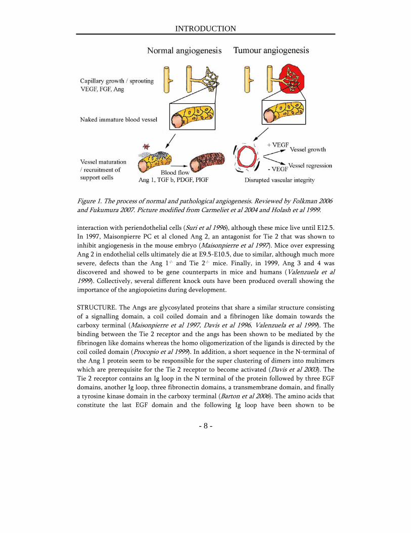
REGULATION. Angiogenesis is regulated via proteins that act on endothelial cells (directly or indirectly) and either stimulate or inhibit their survival, differentiation, migration or proliferation (reviewed by Folkman 2006). In quiescent tissues there is a balance of pro- and anti angiogenic molecules that results in a steady state of vessel growth (Hanahan et al 1996). When pro-angiogenic molecules are up regulated or anti angiogenic molecules are repressed there is an angiogenic switch that results in the formation of new blood vessels. The process of sprouting angiogenesis is divided into four steps, the first being the proteolytic breakdown of the basal membrane and the extra cellular matrix. The next two steps involve the proliferation and migration of the endothelial cells into a new tubules structure. Finally, the newly formed vessel is allowed to mature and become fully functional by the stabilization of recruited pericytes and smooth muscle cells (fig 1) (reviewed by Folkman 2006). However, there is also an alternative mechanism that can explain neovascular growth termed intersuceptive angiogenesis. This type of angiogenesis involves the splitting of one existing capillary into two and is especially important during development when there is a need for several new capillaries and moderate resources to increase the number of ECs. One of the earliest, and most studied regulators, is Vascular Endothelial Growth Factor (VEGF), or Vascular Permeability Factor (VPF), since it was discovered based on its ability to induce leakage of the vasculature (Senger et al 1983). Nowadays, several tens of inhibitors and stimulators have been found and characterized, such as for example Endostatin, Angiostatin, Trombospondin, Platelet Epidermal Growth Factor (PEDF), Epidermal Growth Factor (EGF), Platelet Derived Growth Factor (PDGF), Fibroblast Growth Factor (FGF) and recently, a family of growth factors called the Angiopoietins (Ang).
The Angiopoietins DISCOVERY AND FUNCTION. The endothelial receptor tyrosine kinase (RTK), tek, or Tie 2, was discovered in 1994 when it was found to play a critical role in the vasculogenesis of the embryo (Dumont et al 1994). Vasculogenesis is separated from the process of angiogenesis in the sense that vasculogenesis is the formation of new blood vessels through the differentiation of endothelial progenitor cells into mature ECs and thereby the establishment of a new blood vessels network. Tie 2 knock out mice die at embryonic day 9.5 (E9.5) due to a poorly established vasculature. The endothelial cells are fewer; they show an abnormal morphology and the blood vessels lack pericytic coverage. Shortly after, Sato et al showed that there was another receptor, named Tie 1, and that Tie 1 and Tie 2 had distinct roles during embryogenesis (Sato et al 1995). One year later, the first ligand for the receptor was found, Angiopoietin 1 (Ang 1), due to its ability to bind the extra cellular domain of the Tie 2 receptor (Davis et al 1996). The vascular phenotype of Ang 1-/- mice resembles that of the Tie 2-/- mouse, i.e. dilated ruptured blood vessels and a poor
INTRODUCTION
- 8 -
Figure 1. The process of normal and pathological angiogenesis. Reviewed by Folkman 2006 and Fukumura 2007. Picture modified from Carmeliet et al 2004 and Holash et al 1999. interaction with periendothelial cells (Suri et al 1996), although these mice live until E12.5. In 1997, Maisonpierre PC et al cloned Ang 2, an antagonist for Tie 2 that was shown to inhibit angiogenesis in the mouse embryo (Maisonpierre et al 1997). Mice over expressing Ang 2 in endothelial cells ultimately die at E9.5-E10.5, due to similar, although much more severe, defects than the Ang 1-/- and Tie 2-/- mice. Finally, in 1999, Ang 3 and 4 was discovered and showed to be gene counterparts in mice and humans (Valenzuela et al 1999). Collectively, several different knock outs have been produced overall showing the importance of the angiopoietins during development. STRUCTURE. The Angs are glycosylated proteins that share a similar structure consisting of a signalling domain, a coil coiled domain and a fibrinogen like domain towards the carboxy terminal (Maisonpierre et al 1997, Davis et al 1996, Valenzuela et al 1999). The binding between the Tie 2 receptor and the angs has been shown to be mediated by the fibrinogen like domains whereas the homo oligomerization of the ligands is directed by the coil coiled domain (Procopio et al 1999). In addition, a short sequence in the N-terminal of the Ang 1 protein seem to be responsible for the super clustering of dimers into multimers which are prerequisite for the Tie 2 receptor to become activated (Davis et al 2003). The Tie 2 receptor contains an Ig loop in the N terminal of the protein followed by three EGF domains, another Ig loop, three fibronectin domains, a transmembrane domain, and finally a tyrosine kinase domain in the carboxy terminal (Barton et al 2006). The amino acids that constitute the last EGF domain and the following Ig loop have been shown to be
INTRODUCTION
- 9 -
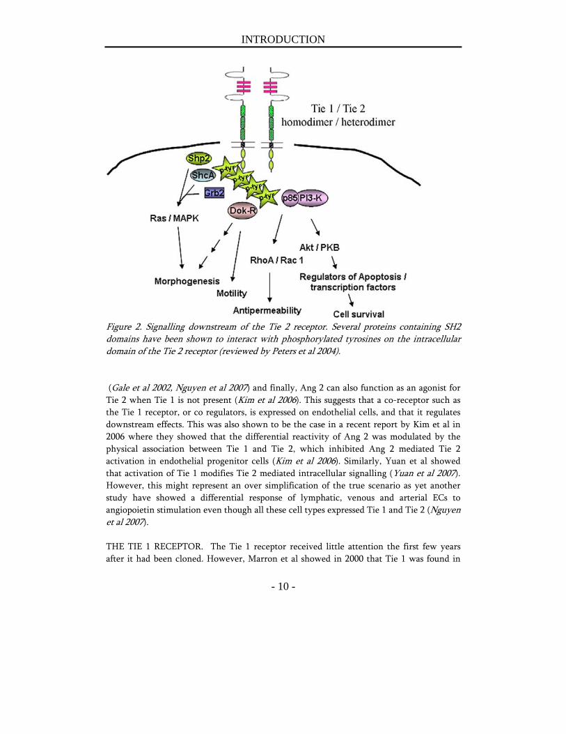
responsible for the binding to Ang 1 (Fiedler et al 2003). In addition, a number of tyrosine residues (Huang et al 1995, Audero et al 2004, Kontos CD 1998, Jones et al 2003) have been identified as major phosphorylation sites and required for downstream signalling via the Ras /MAPK and PI3-K pathways respectively (fig 2). The Tie 1 receptor shares a high degree of homology with Tie 2, especially the cytoplasmic part of the receptor. SIGNALLING DOWNSTREAM OF THE TIE 2 RECEPTOR. As suggested by the developmental experiments using knock out mice, the angiopoietin family consists of agonists (Ang 1 and Ang 4) and antagonists (Ang 2 and Ang 3) that have opposite effects. Ang 1 exert its effect primarily on already established vasculature, in contrast to for example VEGF that regulate the formation of new blood vessels (Senger et al 1983) that in turn are dependent on VEGF for their survival up until a critical moment where withdrawal of VEGF stimulation no longer lead to vessel regression (Dor et al 2002). Ang 1 binds and activates the receptor tyrosine kinase Tie 2 on endothelial cells as a tetramer (Davis et al 2003, Kim et al 2005) which induces receptor dimerization, autophosphorylation, and a downstream signalling cascade ultimately activating the PI3-K / MAPK pathway (fig 2). After binding of Ang 1, and to a lesser extent of Ang 2, the Tie 2 receptor is internalized and degraded via the ubiquitous pathway (Bogdanovic et al 2006). The net results after Ang 1 stimulation are enhancement of endothelial cell survival (Papapetropoulos et al 2000, Harfouche et al 2002, Harfouche et al 2003), vascular stability due to recruitment of pericytes and smooth muscle cells (Thurston et al 1999, Thurston et al 2000), endothelial cell migration and sprouting (Witzenbichler et al 1998, Koblizek et al 1998, Kim et al 2000), and anti inflammatory effects (Gamble et al 2000, Kim et al 2001, Kim et al 2002, Pizurki et al 2003, Joussen et al 2002). The mechanism behind Ang 1 recruitment of SMCs has been shown to involve endothelial derived heparin binding EGF-like growth factor (HB-EGF) and the Erb1 and Erb2 receptors on SMCs (Iivanainen et al 2003), as well as PDGF-B and TGFβ (Nishishita et al 2004). Recently, it was also shown that COMP-Ang 1, a soluble, stable and potent form of Ang 1 in vivo, stimulated wound healing through an increase in angiogenesis, lymph angiogenesis and increased blood flow (Cho et al 2006). The prevailing role for Ang 2 for many years has been as an antagonist of Ang 1 and Tie 2. However, novel data have started to illustrate the roles of Ang 2 as much more complex. For example it was recently shown that Ang 2 can activate the receptor if the receptor is exposed to Ang 2 in a high dose or during a long time of exposure (Teichert-Kuliszewska et al 2001). In addition, Daly C et al found that Ang 2 can activate the Tie 2 receptor and the downstream target Akt on stressed ECs after inhibition of the PI3-K / Akt pathway, and inhibit vascular leak (Daly et al 2006). Furthermore, Ang 2 was also shown to play a pivotal role in the autocrine sensitization of ECs to TNF α induced up regulation of cell adhesion molecules (Fiedler et al 2006). Ang 2 has also been shown to induce lymph angiogenesis
INTRODUCTION
- 10 -
Figure 2. Signalling downstream of the Tie 2 receptor. Several proteins containing SH2 domains have been shown to interact with phosphorylated tyrosines on the intracellular domain of the Tie 2 receptor (reviewed by Peters et al 2004). (Gale et al 2002, Nguyen et al 2007) and finally, Ang 2 can also function as an agonist for Tie 2 when Tie 1 is not present (Kim et al 2006). This suggests that a co-receptor such as the Tie 1 receptor, or co regulators, is expressed on endothelial cells, and that it regulates downstream effects. This was also shown to be the case in a recent report by Kim et al in 2006 where they showed that the differential reactivity of Ang 2 was modulated by the physical association between Tie 1 and Tie 2, which inhibited Ang 2 mediated Tie 2 activation in endothelial progenitor cells (Kim et al 2006). Similarly, Yuan et al showed that activation of Tie 1 modifies Tie 2 mediated intracellular signalling (Yuan et al 2007). However, this might represent an over simplification of the true scenario as yet another study have showed a differential response of lymphatic, venous and arterial ECs to angiopoietin stimulation even though all these cell types expressed Tie 1 and Tie 2 (Nguyen et al 2007). THE TIE 1 RECEPTOR. The Tie 1 receptor received little attention the first few years after it had been cloned. However, Marron et al showed in 2000 that Tie 1 was found in
INTRODUCTION
- 11 -
human ECs in complex with Tie 2, although stimulation with the Angs did not result in a tyrosine phosphorylation cascade downstream of the receptor (Marron et al 2000). In contrast, Kontos et al showed that Tie 1 ectopically expressed in NIH 3T3 cells activated the PI3-K and Akt pathway and inhibited UV irradiation induced apoptosis (Kontos et al 2002). However, at that time, the ligand for the Tie 1 receptor was not found. Recently it was shown that COMP-Ang 1 and Ang 4 does indeed stimulate Tie 1 phosphorylation at tyrosine 1113 in endothelial cells and that this activation is enhanced by Tie 2 (Saharinen et al 2005). Why previous studies failed to show this ability of Tie 1 to become phosphorylated on key residues, is not known. SIGNIFICANCE IN HUMAN TUMOURS. The importance of the angiopoietins in not only normal development and angiogenesis, but also in tumour angiogenesis, has been shown in several different tumours in vitro and in vivo. High expression of Ang 2 correlates with poor prognosis in gastric carcinoma (Etoh et al 2001), non small cell lung cancer (Tanaka et al 2002), and breast cancer (Sfiligoi et al 2003, Dales et al 2004). In a more recent study, high Ang 2 together with high VEGF levels in breast cancer patients lead to a worse disease free survival (Tsutsui et al 2006). High Ang 2 expression has also been observed in metastatic colon cancer (Ogawa et al 2004 and Ellis et al 2002), hepatocellular cancer (Tanaka et al 1999), astrocytomas (Zagzag et al 1999), and ovarian cancer (Zhang et al 2003). In addition, the Tie 2 receptor itself was also recently reported to be involved in hepatocellular carcinoma (Tanaka et al 2002). At a glance, the literature presents some conflicting data as high levels of Ang 1 also has showed to be related with tumour malignancy in plasma cell tumours (Nakayama et al 2004) and ovarian cancer (Martoglio et al 2000). Animal studies over expressing the angiopoietins show similar conflicting results. Ang 1 over expression in MCF-7 breast cancer cells (Hayes et al 2000), colon cancer cells (Stoeltzing et al 2003) and in squamous cell carcinoma (Hawighorst et al 2002) impairs tumour growth. On the contrary, over expression of Ang 1 in a rat glioma model showed a significant stimulation of tumour angiogenesis and growth (Machein et al 2004). Ang 2 has also been shown to both stimulate and inhibit tumour growth in a variety of tumour models (Yu et al 2001, Tanaka et al 1999, Holash et al 1999, Etoh et al 2001, Cao et al 2007). However, accumulating data and a more thorough investigation of the literature, suggest the existence of an Angiopoietin switch, that changes the ratio of Ang 1 > Ang 2 that supports a stabilized quiescent vasculature, to an Ang 2 > Ang 1 ratio, that causes pericyte drop out and susceptibility for the ECs to respond to other angiogenic stimuli, such as VEGF. The latter is believed to take place in tumours and was recently reviewed by Tait (Tait et al 2004).
INTRODUCTION
- 12 -
THE INTEGRINS. To further complicate matters, the angiopoietins have been shown to bind the α5β1 and αvβ5 integrins (Carlson et al 2001). Furthermore, Ang 2 induces glioma cell invasion and stimulates breast cancer metastasis through the α5β1 integrin mediated pathway (Hu et al 2006, Imanishi et al 2007). In addition, Tie 2 has been shown to interact with the α5β1 integrin and thereby regulate ECs response to Ang 1 (Cascone et al 2005). Such non-endothelial functions of the angiopoietins most certainly play a pivotal role in tumour formation and growth and may also contribute to the conflicting results regarding the angiopoietins. Nevertheless, the molecular mechanisms by which the angiopoietins exert their effects in different contexts are most important to elucidate in order to evaluate the angiopoietins as therapeutic targets. REGULATION BY HYPOXIA AND OTHER GROWTH FACTORS. Hypoxia has been shown to regulate the expression of the angs and Tie 2 (Mandriota et al 1998, Oh et al 1999). This was shown in more detail to involve hypoxia inducible factor 1 alpha (HIF-1α) that regulates the expression of VEGF, Ang 2 and Ang 4 (Yamakawa et al 2003) in primary human ECs. In addition, the authors found that inhibition of the Tie 2 receptor or VEGF receptor 2 partially inhibited HIF-1α induced tube formation. VEGF has also been shown to directly alter Tie 1 and Tie 2 receptor complex (Tsiamis et al 2002) or the Tie 2 receptor in itself (Findley et al 2007). Stimulation of HUVECs with VEGF resulted in the cleavage of the extra cellular domain of Tie 1 and the endo domain bound to Tie 2. A similar study showed that both Tie 1 and Tie 2 increased in response to hypoxia in HUVECs and this was correlated with the increase in the transcription factor for Tie 2, namely NERF2 (Christensen et al 2002). They also observed that Ang 1 positively regulated the expression of NERF2 and Tie 2 in quiescent cells. Finally, the levels of Ang 2 protein in vascular smooth muscle cells have been found to be decreased by PDGF but up regulated by IGF through posttranscriptional and transcriptional modifications respectively (Phelps et al 2006).
Other regulators of vascular maturation and integrity In addition to the angiopoietins, other growth factors have also been shown to regulate vascular stability. Among the most characterized, are the PDGFs. This family of peptide growth factors includes four ligands (PDGF A-D) and two receptors, PDGF-R α and β. The ligands homo- or hetero-dimerizes and bind the receptor which then forms homo- or heterodimers depending on which receptor the target cell expresses and which ligand binds (reviewed by Östman et al 2007). Activation of the receptors leads to the recruitment and complex formation with proteins containing SH2 domains, thus activating pathways such as the PI3K- Grb2/Sos1- SHP2- and GTPase activating protein for Ras pathways (Heldin et al 2002, Soskic et al 1999). All activated receptors stimulate proliferation of among others, connective tissue cells (Heldin et al 1999). However, the stimulatory effects
INTRODUCTION
- 13 -
on migration of smooth muscle cells and fibroblasts are primarily mediated via the β homo- and heterodimers. In addition, the vascular phenotype in PDGF-R β knock-out mice (Soriano et al 1994) phenocopies the effects in PDGF-B knock outs (Levéen et al 1994), with a defect in the smooth muscle cells of the blood vessel wall resulting in severe bleeding, highlighting the importance of PDGF-R β signalling for maintaining vascular maturation and integrity. Interestingly, in vivo experiments have shown that the α receptor can not replace the β receptors effect in vascular development (Klinghoffer et al 2001). In the majority of tumour models, the PDGF-receptors α and β are expressed by pericytes and fibroblasts in the tumour stroma, and not in the tumour cells or ECs themselves (Pietras et al 2001, 2002, Lindahl et al 1997, Westermark et al 1993). The ligands however, are expressed during development by epithelial cells (reviewed by Betsholtz 2003) but in many tumours, PDGF isoforms have been shown to be co-expressed with the receptors in the tumour stroma (reviewed by Heldin et al 1999, Östman et al 2001). In prostate tumours however, a study by Chott et al. have observed mRNA expression of the PDGF-R α in bone marrow metastasis in HRPC (Chott et al 1999). Similarly, other studies have shown that the α and β receptors are expressed in prostatic intraepithelial neoplasia (PIN) and adenocarcinoma but not in BPH or normal prostatic epithelium (Fudge et al 1994, 1996). Furthermore, immunocytochemical analyses of three commonly used prostatic cell lines showed expression of both receptors (Kübler et al. 2005). In addition, the laboratory run by Fidler IJ has shown in several studies the expression of PDGF-Rs on tumour associated ECs in metastasis of prostate-, breast-, ovarian- and pancreatic origin (Apte et al 2004, Uehara et al 2003, Lev et al 2005, Hwang et al 2003). Collectively, these studies indicate the importance of the PDGF system in PC and merits further investigations.
Tumour Vascular Biology
Tumour angiogenesis The morphology of tumour blood vessels somewhat support a scenario where the expression of Ang 2 is higher than Ang 1. Tumour blood vessels are leaky with many openings and a discontinuous or absent basement membrane often lacking perivascular cells and therefore grow excessively in an uncontrolled manner. These are sometimes referred to as spaghetti vessels. Furthermore, the ECs are disorganized, tortuous and dilated with uneven diameters, and excessive branching and shunts, resulting in a chaotic and variable tumour blood flow which leaves hypoxic and acidic areas within the tumour (reviewed by Fukumura et al 2007). In contrast to normal physiological angiogenesis, when the newly formed blood vessels are matured and inhibited to grow, tumour blood vessels lack the ability to terminate vessel growth and sustain as leaky immature blood vessels,
INTRODUCTION
- 14 -
prone to respond to any additional angiogenic stimuli (fig 1). In addition, tumour ECs (TEMs) have themselves properties distinct from normal ECs such as a higher rate of proliferation (reviewed by Ruoslahti 2002) and an altered production of genes and surface markers (St Croix et al 2000, Joyce et al 2003). IMATINIB. This defective vascular phenotype, in combination with a poorly functioning of the lymphatics (reviewed by Fukumura 2007) causes the interstitial fluid pressure (IFP) in tumours to be abnormally high. The latter inhibits drug delivery to tumours and illustrates a setback in cancer therapy. The use of Imatinib, a tyrosine kinase inhibitor that targets the PDGF-Rs, c-Kit, and the bcr-abl translocation, has lead to a lowering of IFP in various tumour models and subsequently an enhanced efficacy of radio immunotherapy and chemotherapy (Pietras et al 2002, 2003, Baranowska-Kortylewicz et al 2005). The mechanisms behind a lowering of the IFP by Imatinib treatment are thought to be mediated at least in part by fibroblasts that lose their contractility (Heuchel et al 1999). Due to its ability to inhibit the activation of the above mentioned receptors, Imatinib was recently approved for the treatment of gastrointestinal tumours (GIST) and different forms of leukemia. However, Imatinib was also recently shown to inhibit the macrophage colony-stimulating factor (M-CSF) receptor c-fms (Dewar et al 2005), indicating that it affects not only malignant cells, but also non-malignant hematopoietic cells. The importance of this finding needs to be assessed but implies novel therapeutic targets for Imatinib.
The Concept “Tumour Stroma”
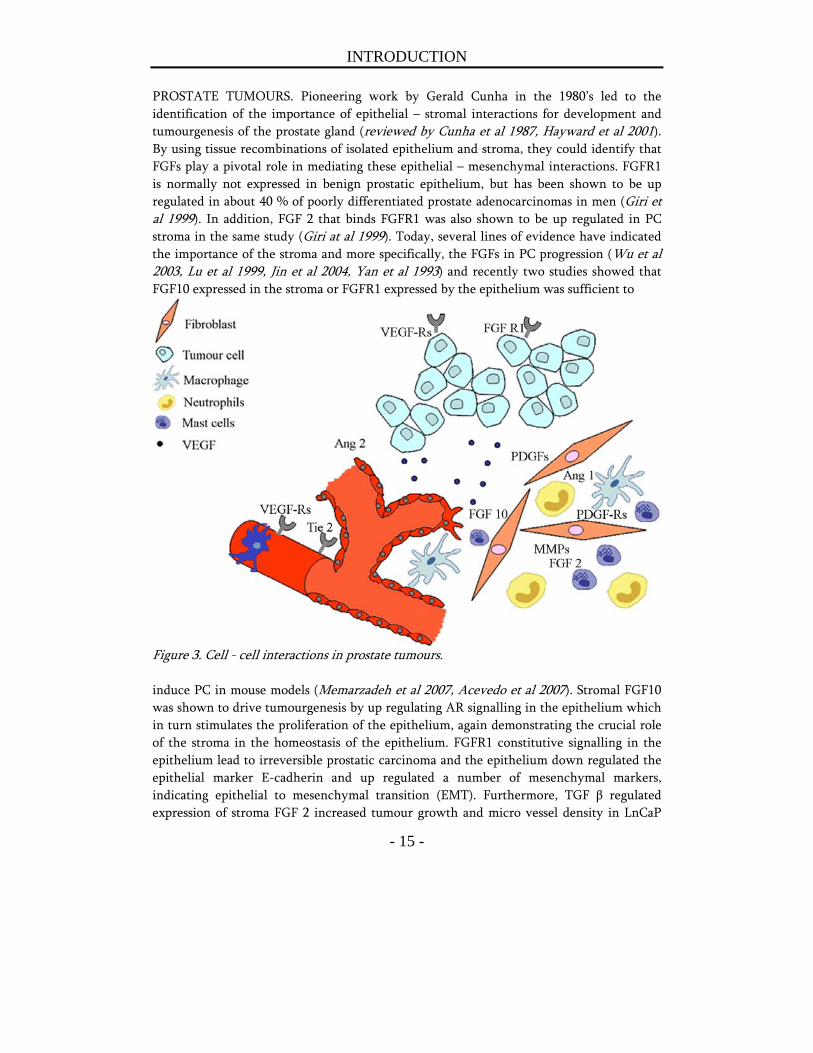
Composition and reciprocal interaction with tumour cells The tumour stroma has received more and more attention during the last decade and it is now well established that the tumour stroma is different from normal stroma in terms of composition and the expression of growth factors, cytokines, angiogenic factors and proteolytic enzymes. It consists of fibroblasts, myofibroblasts, endothelial cells, pericytes, and a variety of inflammatory cells. Interestingly, most of the enzymatic activity needed for the tumour to invade the surrounding ECM and to form metastasis, is actually supplied by host cells in the tumour microenvironment (Coussens et al 2000). For example Ang 1 and Ang 2 stimulate the production of MMP-2 and inhibit the expression of certain TIMPs (Hu et al 2003, Kim et al 2000). In addition, the angiopoietins themselves have been shown to stimulate MMP9 expression in ECs (Das et al 2003). There is also evidence for an up regulation of host derived Ang 2 by VEGF produced by tumour cells in a paracrine manner to support angiogenesis during tumour growth (Zhang et al 2003) (fig 3).
INTRODUCTION
- 15 -
PROSTATE TUMOURS. Pioneering work by Gerald Cunha in the 1980’s led to the identification of the importance of epithelial – stromal interactions for development and tumourgenesis of the prostate gland (reviewed by Cunha et al 1987, Hayward et al 2001). By using tissue recombinations of isolated epithelium and stroma, they could identify that FGFs play a pivotal role in mediating these epithelial – mesenchymal interactions. FGFR1 is normally not expressed in benign prostatic epithelium, but has been shown to be up regulated in about 40 % of poorly differentiated prostate adenocarcinomas in men (Giri et al 1999). In addition, FGF 2 that binds FGFR1 was also shown to be up regulated in PC stroma in the same study (Giri at al 1999). Today, several lines of evidence have indicated the importance of the stroma and more specifically, the FGFs in PC progression (Wu et al 2003, Lu et al 1999, Jin et al 2004, Yan et al 1993) and recently two studies showed that FGF10 expressed in the stroma or FGFR1 expressed by the epithelium was sufficient to
Figure 3. Cell - cell interactions in prostate tumours. induce PC in mouse models (Memarzadeh et al 2007, Acevedo et al 2007). Stromal FGF10 was shown to drive tumourgenesis by up regulating AR signalling in the epithelium which in turn stimulates the proliferation of the epithelium, again demonstrating the crucial role of the stroma in the homeostasis of the epithelium. FGFR1 constitutive signalling in the epithelium lead to irreversible prostatic carcinoma and the epithelium down regulated the epithelial marker E-cadherin and up regulated a number of mesenchymal markers, indicating epithelial to mesenchymal transition (EMT). Furthermore, TGF β regulated expression of stroma FGF 2 increased tumour growth and micro vessel density in LnCaP
INTRODUCTION
- 16 -
tumours (Yang et al 2008). In addition, loss of TGF β R signalling in the prostate stroma is common in PC patients (Turley et al 2007, Tu et al 2003) and a fibroblast specific knock out of the TGF β II R leads to prostate cancer and squamouse cell carcinoma in the forestomach (Bhowmick et al 2004). Finally, cancer associated fibroblasts (CAFs) have been shown to support angiogenesis and promote prostate tumour growth when co-injected with initiated non-tumourigenic prostate epithelial cells (Olumi et al 1999). Our group have found other indications for stromal – epithelium interactions. For example, in PC patients an induction of apoptosis after castration therapy in the tumour epithelium has been related to a requirement in the down regulation of IGF-1 in the tumour stroma (Ohlson et al 2007). In addition, we have observed that an androgen insensitive prostate tumour respond to castration treatment when growing in an androgen dependent environment (Halin et al 2007). Also the vasculature specifically has been implicated in the reciprocal communication with the epithelium during prostate cancer progression. For instance, endoglin and von Willebrand factor positive micro vessels, correlates with prognosis in PC patients (Lissbrant et al 1997, Wikström et al 2002). In animal models, several angiogenic growth factors including VEGF relates to metastatic potential (Häggström et al 2000). In models of androgen ablation therapy to highly differentiated androgen sensitive prostate tumours studies have revealed that the vasculature may be unresponsive to castration treatment in some but not in other tumours (Lekås et al 1997, Lissbrant et al 2001). These findings suggest most alarmingly that the tumour microenvironment itself could support the relapse of the tumour by a continuous support to the epithelium.
Recruitment of inflammatory cell types and endothelial progenitors Not only is there a reciprocal interaction between the tumour cells and the host in the local tumour microenvironment, but also in the manner in which cells are attracted to the tumour from the periphery. The tumour shapes its own stroma necessary for its survival and recruits progenitors from the local environment and possibly from the bone marrow (reviewed by Ganss et al 2006). For example immune cells such as macrophages are actively recruited to the tumour stroma where they promote tumour progression and metastasis and are related to poor clinical outcome (Pollard et al 2004, Lissbrant et al 2000, Loberg et al 2007). In addition, the accumulation of mast cells in PC has been correlated with a worse prognosis (Nonomura et al 2007) and there is also data that shows an increase in the amount of mast cells and macrophages in the rodent prostate after castration therapy (Franck Lissbrant et al 1998). The importance of the latter has not been thoroughly examined but is most likely of great importance. Mast cells are loaded with cytokines, chemokines, growth factors, and angiogenic factors which all have the possibility to
INTRODUCTION
- 17 -
directly or indirectly induce angiogenesis and thus support tumour growth. In fact, inhibition of mast cell degranulation by the use of sodium cromoglycate (Lomudal) completely inhibits the growth of Myc induced pancreatic tumours due to an underprovided ability to induce angiogenesis (Soucek et al 2007). ENDOTHELIAL PROGENITOR CELLS (EPCs). CAFs have also been shown to produce elevated levels of stromal cell derived factor 1 (SDF-1) that recruit EPCs in some animal tumour models and boosts angiogenesis (Ceradini et al 2004, De Falco et al 2004). However, in other animal models, the recruitment of endothelial precursors is regulated by high levels of VEGF produced by the tumour cells (Lyden et al 2001). In the RipTag5 mouse model of pancreatic cancer, chemokines were shown to regulate the homing of EPCs into neovessels in later stages, but not during early stages of tumour progression (Spring et al 2005). The observation that different tumours recruit EPCs via different pathways suggests that the organ specific tumour microenvironment play a pivotal role. Palma and colleagues (De Palma et al 2005) reported Tie 2 expressing monocyte (TEMs) cells of hematopoietic origin that supported neovascularization in vivo. Interestingly, removal of those cells completely inhibited human glioma neovascularization in the mouse brain. In contrast, in the TRAMP mouse, the contribution of EPCs to the primary tumour seems low as it only increases from 0 % to 14 % in poorly differentiated tumours (Li et al 2004). The latter has been supported by findings in human patient material where the contribution of EPCs to the neovascularization in tumours has been low to non existent (Peters et al 2005). The increased levels of VEGF in the tumour in turn, has been shown to at least partly be due to an increased number of infiltrating neutrophils expressing matrix metalloprotease type 9 (MMP9) that releases extra cellular bound VEGF in a mouse model of multistage carcinogenesis (Nozawa et al 2006). The reasons why different animal models and also human studies show different contributions of EPCs to the tumour vasculature are not known but suggest tumour stage and or species specific regulations. Collectively, these data suggest that the tumour stroma and infiltrating inflammatory cell types are crucial for tumour growth and progression and that the tumour cells are synchronised with the stroma cells in a highly coordinated manner to achieve tumour progression. This also opens up the possibility to use the tumour stroma as a target in cancer therapy since the stroma at least in theory, should be genetically more stable, and thus less able to acquire resistance to therapies, than the tumour cells themselves.
Significance Anti angiogenic therapy shows great promise in a variety of cancer models. In the prostate, the vasculature has been shown to be crucial for normal and malignant tumour growth and a prognostic factor in tumour progression which imposes a role as a therapeutic target. The
INTRODUCTION
- 18 -
function of the Angs has not been studied in prostate biology or PC and therefore this thesis aimed at elucidating the origin and effect of the angiopoietins in normal and malignant prostate. Secondly, the tumour stroma has received more and more attention during the last decade. Inflammatory cells types recruited to the tumour stroma, and the tumour stroma itself, are of significance in several tumour models and therefore, the second aim was to evaluate the effects of inhibiting the tumour stroma with the broad spectrum inhibitor Imatinib mesylate, which targets several cell types populating the tumour stroma.
Specific aims
To study the expression and potential importance of the Angs in normal and
malignant prostate tissue in humans and in rodents
To examine the role of the angiopoietins and to some extent other stroma derived
factors during hormonal ablation therapy and testosterone stimulated growth of normal
and malignant prostate tissue in order to elucidate their potential role as targets for
enhancing castration effects in prostate tumours
To study the significance of the enhanced influx of inflammatory cells in PC after
castration therapy and evaluate their potential as a novel targets
MATERIALS AND METHODS
- 19 -
MATERIALS AND METHODS
Animal models of PC Animal models of PC include poorly differentiated androgen independent tumour cell lines, highly differentiated androgen dependent transplantable prostate tumours, and the transgenic adenocarcinoma of the mouse prostate (TRAMP). Considering the pivotal role of the prostate stroma in both normal and malignant transformation, we have employed normal immunocompetent mice and rats for studies regarding castration effects where the vasculature is hormonally regulated in a coordinated fashion with the epithelium. In paper III and IV, we used the Dunning H and PAP tumours which are transplantable androgen dependent tumours composed of tumour cells and a prostatic tumour stroma. The downside is of course that pieces of the tumour is grown subcutaneously (s.c) on the back of Copenhagen rats somewhat altering the natural local milieu. However, ARs are expressed throughout the growing tumour strongly suggesting that the stroma is of prostatic origin. In addition, the tumours response to castration closely resembles that of human patients in regard to initial response and subsequent relapse. In paper IV, we also used the less differentiated androgen insensitive AT-1 tumour cell lines that were injected into the VP of normal immunocompetent rats. The TRAMP mouse was not used in these studies since there are large differences in disease progression at a certain time point. Without access to proper imaging techniques to more precisely establish tumour stage, pharmacological studies in the TRAMP mouse are hard to perform.
Animal Studies in Vivo
The Dunning Tumours (paper III and IV) In the early 1960s Dr. W.F Dunning found a spontaneously growing papillary adenocarcinoma in the DLP in a 22 months old male rat. This tumour, named R3327, was transplanted and grew slowly, maintaining its properties of the rat DLP in terms of androgen sensitivity and expression of 5-alpha reductase activity (Dunning 1963). A relapse of the tumour was also observed after hormonal withdrawal, similar to the scenario in human PC patients and the model was evaluated and shown to be appropriate for the studying of prostate cancer (Smolev et al 1977). Subsequent passages of that tumour gave rise to the well differentiated and hormonally sensitive R3327 H and PAP tumours (only transplantable as tumour pieces), as well as the less differentiated and less hormone sensitive AT-1 tumour (transplantable as cell line). Serial passages of the AT1 tumour then
MATERIALS AND METHODS
- 20 -
subsequently gave rise to the low differentiated and highly metastatic cell line MatLyLu (Metastatic AT-Lymph Node and Lung) (Tennant et al 2000). Now, several more cell lines with differences in differentiation status and metastatic potential have been selected and are in use. CELL CULTURE AND ORTOTHOPIC IMPLANTATION OF TUMOUR CELLS INTO THE RAT PROSTATE. AT1 cells were grown according to the manufacturers instructions (RPMI with 10 % Fetal Calf Serum (FCS), 50 μg/ml gentamycin, 2.5 μM dextametasone, and 0.2 % Na-Bic) in 37°C, 5 % CO2. Before implantation, the cells were detached from the cell culture vessel with Trypsin (1X), counted using a Burkel chamber and diluted in RPMI to the appropriate volume. During anaesthesia, approximately 2000 AT1 cells were injected into the left VP in a volume of 10 μl. TRANSPLANTABLE R3327 H AND PAP TUMOURS. Ten week old male Copenhagen/Fisher rats, supplied from Taconic, Copenhagen Denmark, were inoculated with two small pieces of the highly differentiated androgen-sensitive Dunning R3327 PAP prostate adenocarcinoma (originally obtained from Dr Normal N Altman, Papanicolai Cancer Research Institute, Miami, FL, USA) or Dunning H tumours (kindly provided by Dr JT Isaacs, John Hopkins University, Baltimore, USA) subcutaneously (s.c) on each flank of the rat. After approximately 6 months, the tumours measured 0.6 cm3 and the experiments were initiated. ADMINISTRATION OF ORAL AND INTRA TUMOURAL DRUGS. The broad spectrum tyrosine kinase receptor inhibitor Imatinib mesylate was dissolved in 1 ml of sterile PBS prior to each administration and feed orally by the use of a thin soft plastic probe to avoid harm to the oesophagus. Imatinib was administered twice a day (75 mg / kg morning and 50 mg / kg evening) for one or four weeks in total. The control group received 1 ml of PBS morning and night during the same time period. The soluble Tie 2 receptor (s Tie 2 Fc) was diluted in sterile PBS and injected intra tumoural at a dose of 0.4 ug / cm3 based on unpublished observations that showed this dose to be highly efficient in stimulating the accumulation of intravascular leukocytes. The control group received an equal volume of sterile PBS.
Wild Type Mouse and Rat Prostate Studies (paper II) CASTRATION AND TESTOSTERONE REPLACEMENT MODEL. Adult male Sprague-Dawley rats and C57 black mice (B&K, Stockholm, Sweden) were sedated with pentobarbital (60 mg / kg), castrated, and examined after 1, 3, and 7 - 10 days after castration. In addition, 7 days castrated rats were treated with daily subcutaneous injections of long-acting testosterone esters (Sustanon, Organon, Oss, The Netherlands, 10 mg / kg)
MATERIALS AND METHODS
- 21 -
and studied 1, 2 or 3 days thereafter. Intact animals were used as controls. In a separate experiment 7 days after castration, one group was given an intra - prostatic injection of 10 μg of s Tie 2 - Fc (Tie 2 - Fc mouse chimera, R&D Systems, UK) delivered in 5 μl basement membrane extract (Matrigel, BD Biosciences, Sweden). The second group was given Matrigel loaded with phosphate buffered saline (PBS) using a 10 μl Hamilton syringe. The third group was not operated on. These three groups were given s.c injections of Sustanon (10 mg / kg) at day 7, 8, and 9 after castration. SACRIFICE OF ANIMALS AND TISSUE HANDLING. One hour before sacrifice all animals were injected with bromodeoxyuridine (BrdU, Sigma MO, USA)) intra peritoneally (i.p) to mark proliferating cells. After sedation some of the animals were fixed by vascular perfusion with Bouins fluid and the tissue of interest was removed, weighed and post fixed with Bouins solution for 24 h and then embedded in paraffin, as previously described (Lissbrant et al 2004). In paper III and IV, the tissues were fixed in 4 % formalin over night, followed by 24 hours in 70 % ethanol. For Western blot, quantitative reverse transcription polymerase chain reaction (qRT-PCR), and some tissue staining procedures, the tissues were removed and frozen in liquid nitrogen. All animal experiments were approved by the local ethic committee in Umeå, Sweden. The animals were housed in a well-ventilated room under controlled light (12 h light / 12 h dark), temperature (25 ◦C), and humidity (40-60 %). Pelleted food and water were available ad libitum. The animals tolerated the treatments well.
Patient Materials
Västerås Project (paper I) Between 1970s’ and 1980s’, specimens were collected from patients who underwent transurethral resection (TUR) at the Central Hospital in Västerås, Sweden, due to obstructive voiding problems, and later where histological examination showed cancer. Samples were formalin-fixed, paraffin-embedded and graded according to Gleason classification. Other parameters such as local tumour stage and the presence of metastases were investigated. The patients had not received any treatment prior to diagnosis and were left untreated until symptoms occurred. At that time point, they were subjected to androgen ablation or radiotherapy for palliative purposes. The material has been described in more detail previously (Stattin et al 1997).
MATERIALS AND METHODS
- 22 -
Human freeze biopsies from poorly differentiated tumours (unpublished observations) Specimens from patients who had undergone a radical prostatectomy were selected and sectioned. The material was used for laser micro dissection and pressure catapulting (LMPC) and RT-PCR, as described below.
Protein Analysis
Isolation of Protein Extracts (paper II - III) CELL LINES AND TISSUES. Protein from cells were extracted using lysis buffer containing 0.5 % NP-40, 0.5 % Na DOC, 0.1 % SDS, 50 mM Tris-HCL (pH = 7.5), 150 mM NaCl, 1 mM EDTA (pH 8.0), 1 mM NaF, and Complete Protease Inhibitor Cocktail (Boehringer Mannheim, Germany) followed by centrifugation at 20000 x g in 4 °C for 30 min. The supernatant was isolated and the concentration determined using the BCA Protein assay reagent Kit (Pierce Chemical Co., IL, USA). Frozen tissues were homogenized using a Micro Dismembrator (B. Braun Biotech International GmbH, Melsungen, Germany) at 2000 rpm for 45 seconds. They were then added to an adequate amount of the lysis buffer described above, or alternatively, 7 M urea (rat tissues, paper II), 2 M thio-urea, 4 % CHAPS, and 30 mM Tris pH 8.5). Samples were mixed and incubated on ice for 30 minutes. For urea extracted proteins, an appropriate amount of benzonase (Merck KGaA, Darmstadt, Germany) was added to the mixture to reduce viscosity caused by nucleic acids. The supernatants were isolated following centrifugation (20000 x g, 4°C, 30 min) and the protein concentration was determined using the BCA Protein assay reagent Kit as described above. Concentrations of urea extracted protein samples were determined using the 2-D Quant Kit (Amersham Biosciences, Uppsala, Sweden) according to protocol.
Western Blot (paper II - IV) All samples were separated by 7.5-12.5 % SDS-PAGE under reducing conditions and subsequently transferred to a Hybond-P PVDF Membrane (Amersham). The membranes were blocked in 5 % milk followed by primary (Ang 1 and Ang 2, US Biological, USA; Tie 2 and VEGF, Santa Cruz Biotechnology, Inc., Santa Cruz, CA) and secondary (anti rabbit IgG, Amersham Biosciences, and anti-goat, Jackson Laboratory, Pierce Biotechnology, Rockford IL, US) antibody incubations. Enhanced chemiluminescence plus (ECL-Plus) (Amersham Biosciences) was used to visualize protein expression. In paper II, blocking experiments
MATERIALS AND METHODS
- 23 -
were performed in order to verify that the bands shown corresponded to the proteins of interest. A western blot was run where half of the filter was incubated with an antibody: peptide solution (1:40 w/w) and the other half incubated with antibody as previously described.
RNA Analysis
RNA Preparation tissues and cell lines (paper II-IV) Generally, RNA was prepared using Trizol (Invitrogen, Stockholm, Sweden). Briefly, frozen samples were homogenized using a Micro Dismembrator (B. Braun Biotech International GmbH, Melsungen, Germany) at 2000 rpm for 45 seconds. After that, the powder was added to 0.8 ml Trizol. Chloroform was added, the samples vortexed, and incubated at room temperature for 3 minutes. Following centrifugation, the supernatant was isolated and isopropanol used to precipitate the RNA. The RNA was washed with 70 % ethanol and dissolved in sterile DEPC-treated water. RNA from cell lines were handled as described above, except that trizol was added to the wells following a wash with PBSA. The concentration was measured using a nanodrop and the integrity of the RNA was determined on a 1 % agarose gel.
Laser Micro dissection techniques (paper II and IV) LASER CAPTURE MICRODISSECTION (LCM). The VP from two intact rats was removed and frozen in liquid nitrogen. Five μm thin sections were stained with Mayer’s Hematoxylin (Sigma-Aldrich, MO, USA) and Eosin B (Sigma Diagnostics, MO, USA) and dehydrated by increasing the percentage of ethanol according to protocol from Arcturus (Mountain View, CA). A laser capture microscope (PixCell II, Arcturus) was subsequently used to isolate cells from selected areas with a laser diameter of 15 μm and a power of 47 mW. The regions of interest were captured on special caps (HS CapSure, Arcturus) and RNA was prepared according to instructions in the PicoPure RNA isolation Kit (Arcturus). Briefly, the tissue was added to extraction buffer, incubated at 42 °C for 30 min, dissolved in ethanol and run through a specially designed column. After 2 washing steps, the RNA was eluted with elution buffer supplied in the kit. LASER MICRODISSECTION AND PRESSURE CATAPULTING (LMPC). Six μm frozen sections of tumour tissue from animals or patient biopsies were mounted on UV treated PALM Membrane Slides (P.A.L.M Microlaser Technologies AG, Bernried, Germany). The sections were stained with toluidine blue to visualise mast cells or Mayer’s hematoxylin (Sigma-Aldrich) and eosin B (Sigma Diagnostics) to visualise nuclei’s. Approximately
MATERIALS AND METHODS
- 24 -
100 000 μm2 of mast cells from rat 4 week castrated rat tumours or an approximate area of 100 000 μm2 of epithelium versus stroma from normal and malignant human prostate tissues, were micro dissected and isolated and used for RNA extraction with trizol or with PicoPure RNA isolation Kit (Arcturus) as described above.
cDNA synthesis (paper II – IV) RNA from tissues, cell lines and from isolated mast cells was used in a first-strand cDNA synthesis using Superscript II (Invitrogen, Sweden) in a 10 μl reaction. Briefly, total RNA isolated was mixed with 2.5 μM random hexamers (Applied Biosystems, Sundbyberg, Sweden) and 5 mM dNTP’s. After incubation at 65 °C for 5 min, the samples were chilled on ice. Finally, first-Strand buffer, 0.1M DTT, 20 U RNAsin (Promega, Madison, WI, USA), and 100 U of superscript II, was added and the cDNA synthesis initiated with 10 min at 25 °C followed by 50 min incubation at 42 °C and a finalising inactivation step at 70 °C for 15 min.
Quantitative Real-Time RT-PCR and array analyses (paper II – IV) Quantification of the mRNA of interest was performed using Real-time quantitative PCR with the Light Cycler SYBR Green 1 technology (Roche Diagnostics, Bromma, Sweden). The PCR was run in a 20 μl reaction including 0.5 μM primers, 2 μl enzyme mix supplied in the kit, 3-4 mM MgCl2 and 2 μl cDNA (ranging from very low to high concentrations). The PCR product was subjected to melting curve analysis and run on 2 % agarose gels to verify the specificity of the product. Each experiment included positive and negative controls. Data was analysed using the LightCycler Software 3.5.3 (Roche Molecular Biochemical, Bromma, Sweden). Array analyses were described in detail in paper IV.
Immunohistochemistry (IHC)
Tissue Staining (paper I- IV) STEREOLOGY AND IHC. To quantify the volume density of blood vessels, the fraction of factor VIII (DAKO, Stockholm, Sweden) stained blood vessels (lumina and vascular walls) was related to prostate volume using a light microscope at high magnification. The fraction of stabilised blood vessels was calculated by quantifying the number of factor VIII positive vessels in relation to smooth muscle cell actin (SMA) positive blood vessels. The fraction of
MATERIALS AND METHODS
- 25 -
tumour cells, tumour volume, and mast cells were measured with similar methods. The quantification was performed by one or two researchers on blinded samples and the results showed as mean values. For detailed protocols on the immunohistochemical analysis see paper I-IV.
Ultrasound
Gray Scale Ultrasound (paper III) Grey scale ultrasound was performed using a linear transducer (Acuson Sequoia 512; Seimens) with a 15 MHz frequency. Tumour size was measured in a transaxial, coronal and sagital plane and tumour volume was calculated using the formula for an elliptical object.
Statistics In general, all statistical analyses were performed with non parametric tests not requiring normal distributed data (paper I – IV). A p-value ≤ 0.05 was considered statistically significant. Statistical analysis was performed using the Statistical Package for the Social Sciences, SPSS 12.0.1 software (SPSS Inc., Chicago, IL).
RESULTS AND DISCUSSION
- 26 -
RESULTS AND DISCUSSION
Paper I Since the stroma in normal and malignant prostate has been shown to be of importance for the homeostasis of epithelium, and as the role of the angiopoietins in the prostate was largely unknown, we started to explore the potential importance and expression of the angiopoietins in the malignant prostate. We found that Ang 2, but not Ang 1, was increased in tumour epithelium and that the expression was correlated with histological grade, vascular density, metastasis and outcome in PC patients. The material used was formalin embedded samples from patients that had undergone TURP due to voiding problems and where histological analyses showed cancer and where no subsequent treatment had been performed. This material is unique in several ways. Firstly, it allows one to investigate the natural causes for disease retrospectively, with no interfering of disease progression (and thus potential selection), such as chemotherapy, radiotherapy, anti angiogenic therapy, or surgery. Second, the material is large with several hundreds of patients involved with all clinical observations reported in the clinical records which increase the accuracy of the screening. Finally, this material has been extensively studied through the years and parameters such as vascular density, angiogenic growth factors, tumour cell apoptosis and proliferation and much more, is already known. The finding that Ang 2 was significantly up regulated in PC patients and that this was correlated with a worse prognosis, was not surprising, although it had never been shown in prostate cancer. Several similar studies in other tumours have shown similar results (Ogawa et al 2004, Ellis et al 2002, Tanaka et al 1999, Zagzag et al 1999, Zhang et al 2003). However, the reasons for this up regulation are more speculative. Prostate tumours have shown to be hypoxic with a variable blood flow, and Ang 2 has been shown to be up regulated by hypoxia (Yamakawa et al 2003). Ang 2 has also been shown to recruit macrophages from the periphery and macrophages in turn have been shown to stimulate PC growth (Loberg et al 2007, Lissbrant et al 2000). Whether this is a cause or a consequence is not known. Furthermore, Ang 2 is said to destabilize the vasculature by inhibiting Ang 1 ability to exert its function. The result is a leaky vasculature prone to respond to any additional angiogenic factor around, such as, for example VEGF. In fact, VEGF was recently shown to directly modulate Tie 2 signalling by cleaving the Tie 2 receptor forming a soluble fragment (Findley et al 2007). In our patient material, VEGF has also been shown to be up regulated in high grade tumours, and thus the two growth factors could work in concert to switch on angiogenesis. This would hypothetically result in a higher vascular density, which indeed is observed in this patient material, in terms of an increased fraction of
RESULTS AND DISCUSSION
- 27 -
CD31+ staining. Ang 2 was previously shown to be up regulated by VEGF producing tumour cells in ovarian cancer (Zhang et al 2003), but whether Ang 2 was up regulated after or before VEGF in this model is not known. Ang 1 was not increased in the same manner which resulted in a change of the angiopoietin switch, with a higher expression of Ang 2 than Ang 1, the contrary of what is seen in most normal tissues where the vasculature is quiescent. This study was conducted to evaluate the importance of the angiopoietins in PC by the use of a well studied unique material. However, the results from this study should be validated with the use of other techniques where the expression of the angiopoietins in normal and malignant PC tissue could be quantified with more accuracy. For example, the use of LMPC in combination with qRT-PCR or western blot to isolate normal and tumour epithelium from patients with different GS is an attractive idea and currently under investigation. Although most reports on the location of Ang 2 has indicated that Ang 2 is expressed in the tumour stroma, particularly at sites of vascular remodelling, we found the expression in humans to be restricted primarily to the tumour cells. Interestingly, a recent report investigating the role of constitutive FGFR1 signalling in PC progression revealed an increase in vascular volume which was not dependent on a continuous FGFR1 activation (Winter et al 2007). Instead, this angiogenic phenotype was shown to be upheld and mediated by among others VEGF and the angiopoietins. In more detail, Ang 1 expression was lost and Ang 2 expression was up regulated and located to the tumour epithelium, altering the angiopoietin switch. The importance of our findings is highlighted by unpublished observations that the Tie 2 receptor is not EC specific in PC. We have found the expression of Tie 2 (but not Tie 1) mRNA and protein on PC epithelium in patients with high grade PC with the use of LMPC combined with q RT-PCR and in addition immunohistochemistry. We have also found the expression of Tie 2 on a few human metastatic PC cell lines, including the well used LnCaP. Current studies are now focusing on establishing the biological effect of the Tie 2 receptor on prostate tumour cells by in vitro studies and Tie 2-Lac Z transgenic mice crossed with the TRAMP mouse. Other researchers have found that the ectopic expression of Tie 2 on for example fibroblasts, where Tie 1 is not expressed, results in the activation of Tie 2 upon binding of Ang 2, but not Ang 1 (Kim et al 2006). This switch in agonist antagonist function has been suggested to be regulated by Tie 1. Therefore, in these model systems, Ang 2 activation of Tie 2 has lead to an activation of the PI3-K / MAPK pathways ultimately resulting in an increase in migration and survival signals. If our unpublished findings prove to be correct, the increase in Ang 2 on prostate tumour epithelium could initiate autocrine signalling to the epithelium where Tie 2 is expressed, leading to enhanced survival and migration capacity of the tumour cells.
RESULTS AND DISCUSSION
- 28 -
Paper II Since the angiopoietins seemed to be of relevance in PC, we then wanted to examine the role of the angiopoietins in prostate biology in more detail. We therefore sought to investigate the importance of angiopoietins during hormonal treatment of normal prostate tissue. Previous results have shown that regression of the prostate following castration therapy in principal is mediated by effects in the stroma, and more specifically by a vascular regression preceding the decrease in the epithelium (Lekås et al 1997, Lissbrant et al 1998, Lissbrant et al 2001). The rationale to study the normal response to castration in the prostate is that in tumours, the response is only temporary and the initial decrease in tumour regression is followed by a subsequent relapse. HRPC is not curable and the median survival for these patients is less than 3 years. Therefore, our hypothesis is that castration therapy should be used in combination with other anti-stromal therapies. Since the angiopoietins are known to stimulate vascular growth and maturation in other model systems and our first study indicated an importance also in prostate cancer, the role of the angiopoietins during vascular remodelling was examined. We found that both Ang 1 and Tie 2 proteins were down regulated by androgen withdrawal and up regulated by androgen stimulation. We also found a significant decrease in the fraction of SMA+ blood vessels after androgen withdrawal and a subsequent increase after androgen stimulation. Our hypothesis was that this regulation of SMA+ vessels was mediated by the angiopoietins, and that interference with Tie 2 signalling should impair vascular stability and possibly also the regrowth of the prostate gland after androgen stimulation. To test this, we used a soluble Tie 2 receptor that we injected into castrated mice together with matrigel. The mice were then followed over 3 days and after that, the VP dissected out, weighed and morphological parameters determined. We did find a perturbed increase in the stability of the vessels (at least in terms of SMA+ vessels) but we did not find a difference in weight of the gland. This could however be due to the relative short time of treatment. Recently, a study by Wang et al showed in a similar experiment that after an additional 3 days of inhibition of Tie 2 signalling, the re growth of the gland is significantly inhibited by s Tie 2 (Wang et al 2007). Due to technical difficulties we did not however pursue such an experiment. Based on these results it appears that inhibition of Tie 2 signalling impairs prostate growth. Other vascular growth factors, such as for example VEGF, have been shown to play a role in the re growth of the gland following testosterone treatment and androgen stimulated prostate growth can be inhibited by anti-VEGF treatment (Lissbrant et al 2004, Wang et al 2007). VEGF however, plays an earlier role in angiogenesis compared to the angiopoietins, by stimulating neovascularization, and not primarily vessel maturation. Therefore, it was interesting to find that also factors stimulating vessel maturation play a role in the regulation of normal prostate gland growth. It should be mentioned however, that we did
RESULTS AND DISCUSSION
- 29 -
not investigate other pericyte markers such as desmin, NG2, or PDGF-Rβ expression in this experiment. In addition, in vivo assays to test the function of these vessels (for example with MRI, or i.v. injection of lectin) were not performed but should be investigated in the future to more precisely pin point the effects of inhibited Tie 2 signalling during growth of the prostate gland. In addition, we also investigated the location of synthesis of the angiopoietins with the use of LCMP and qRT-PCR in the VP of normal Copenhagen rats. We found that the Tie 2 receptor was expressed in the stroma as expected but the angiopoietins showed a different expression pattern compared to the human prostate tissue studied in paper I. The angiopoietins were, in addition to the expression in the epithelium, also found in the stroma, although Ang 1 was preferentially expressed in the stroma. Similar results were obtained with in situ hybridization (unpublished observations). This could be due to the differences in methodology, that is measuring mRNA versus protein, or differences among species. Few other studies have investigated the location of the angiopoietins in the prostate, and those that have, show similar conflicting results (Wurmbach et al 2000, Wang et al 2007). Later studies showed that also PDGF-Rβ was down regulated after hormonal ablation in the mice VP. Thus, it would have been interesting to explore the effects of Imatinib, possibly in combination with inhibitors of ECs such as s Tie 2, on the re growth of the gland. The finding that both SMA+ cells and PDGF-R β expression decreases is intriguing in terms of the potential of a general mechanism by which the normal tissue down regulates the number of pericytes and fibroblasts in response to androgen withdrawal. This is in line with previous findings that not only tumours are coordinated and regulated by events in the stroma - and in this case pericytes in particular, but also normal tissues (Lissbrant et al 2004).
Paper III Based on our second work where we found that the angiopoietins supported the growth of the vasculature after testosterone treatment in the normal prostate, we then wanted to investigate if a lack of angiopoietin regulation in prostate tumours could explain the lack of stroma response following castration therapy. In our third work, we demonstrated by using the androgen sensitive Dunning H tumour model that a combined therapy, that targeted both ECs and perivascular cells in combination with castration therapy, increased the efficacy of castration therapy. Castration alone, led to a second wave of angiogenesis and a subsequent tumour growth, whereas the combined treatment partly inhibited the growth
RESULTS AND DISCUSSION
- 30 -
of the vasculature, up regulated tumour cell apoptosis and completely inhibited tumour growth after four weeks of treatment. The Dunning H tumour model is a transplantable tumour which is grown s.c. on the back of normal Copenhagen rats, composed of both prostate stroma and epithelial cells. Although it is grown in an ectopic environment, its relevance as a model to study tumour responses to androgen deprivation therapy has been proven by its similarity to the responses in PC patients (Wu et al 2003, Yan et al 1993). Another aspect is that the tumours grow slowly, thus more similar to the situation in patients, although making research more time consuming. As mentioned above, patients with advanced prostate cancer respond initially to castration therapy with a temporarily reduction in tumour volume followed by a subsequent relapse. This is also seen in the Dunning H tumour model and surprisingly, we found that it was the tumour stroma itself, and not the tumour epithelial cells, that lacked the ability to inactivate the AR. This suggested that the tumour micro environment was androgen insensitive and interestingly, clinical studies in PC patients have showed similar results with an androgen independent tumour stroma (Ohlson et al 2007). Interestingly, activity of the AR in the tumour stroma re-occurs during a suspected local relapse in PC patients (Wikström et al 2007). Interestingly, we found that Imatinib strongly up regulated the expression of the endogenous angiogenesis inhibitor trombospondin 2 (TSP 2) after one and four weeks of treatment. Several studies have highlighted the importance of TSP 1 in PC (Colombel et al 2005, Vallbo et al 2004, Doll et al 2001) but few have investigated the role of TSP2 (Vallbo 2004). Both TSPs are matri cellular proteins expressed in the extra cellular space with very potent anti angiogenic properties (reviewed by Ren et al 2006, Grant et al 2005, Zhang et al 2007). Both TSP 1 and 2 are potent inhibitors of VEGF and FGF 2 induced angiogenesis (reviewed by Lawler and Detmar 2004). Interestingly, androgens in prostate tumours repress the transcription of TSP 1 and androgen withdrawal increases the expression of TSP 1 and leads to a temporary decrease in the amount of blood vessels (Colombel et al 2005). However, with continued exposure PC angiogenesis and growth is relapsed probably due to the redundancy of other angiogenic growth factors in the local milieu. TSP 1, FGF 2 and VEGF have been shown to represent key regulators of angiogenesis in the prostate (Doll et al 2001), and thus the up regulation of TSP 2 in combination with our observed decrease in FGF 2 mRNA (paper IV), most likely contributed to the decrease in angiogenesis in our Dunning H tumour model. The importance of endogenous angiogenesis inhibitors was also observed in a study were the researchers found that restoration of the normal physiological levels of three different endogenous angiogenesis inhibitors, including TSP 1, retarded tumour growth by specifically inhibiting EC proliferation and migration (Sund et al 2005). On the contrary, removal of the inhibitors resulted in an enhanced angiogenesis and a 2- to 3- fold increase in tumour growth (Sund et al 2005).
RESULTS AND DISCUSSION
- 31 -
We found that a combinatorial therapy targeting pericytes and endothelial cells was most efficient and blocked the second wave of angiogenesis observed in this model four weeks after castration. Interestingly, other studies now indicate that there are reciprocal interactions between different growth factors produced by endothelial cells and pericytes. For instance, Nissen et al recently showed that FGF 2 and PDGF-BB up regulated the expression of each others receptors on pericytes and endothelial cells respectively, thereby enhancing neovascularization and pulmonary metastasis formation in vivo (Nissen et al 2007). It is most likely that similar regulatory mechanisms exist between other growth factors, making combined targeted therapies attractive options. In that sense, Imatinib show great promise in terms of its inhibition of several different RTKs. In contrast, the individual contribution from each receptor inhibited, is difficult to precisely determine, requiring isolated inhibitors. In study IV, we tried to determine the contribution of the depletion of mast cells in the tumour stroma. Considering our unpublished observations that Tie 2 is expressed by prostatic tumour cells in both patients and animal models, we cannot exclude the possibility that the inhibition of Tie 2 in our study was not exclusive to the ECs. Blocking Tie 2 signalling with the soluble receptor increased the amount of intravascular leukocytes as expected and had a slight effect on the proliferation in the stroma. However, the effects on vascular volume and stability were minor and we also observed minor effects on the epithelium, which could be direct effects. Unfortunately, immunohistochemistry for Tie 2 expression has proven difficult, but the problem could be solved by the use of LMPC in combination with qRT-PCR to locate Tie 2 in our tumour model. It would be interesting to test this stroma targeted therapy in more advanced prostate cancer models such as for example the TRAMP mouse. Difficulties that have to be overcome are the ability to systemically inhibit the Tie 2 receptor and to be able to monitor disease progression with more accuracy. Secondly, TRAMP mice display a large heterogeneity in terms of disease progression and therefore a large number of animals are needed. However, future studies should aim at elucidating the power of this type of therapy in poorly differentiated prostate cancer models.
Paper IV In the final study we set out to determine the importance of diminishing mast cells in prostate tumours. In the third paper, the anti tumour effects were assumed to primarily be mediated by inhibiting pericyte - EC interactions (blocking effects of angiopoietins and PDGF). However, we also made two observations that made us want to examine the role of mast cells in prostate tumours. First, mast cells increased in tumours after androgen deprivation therapy and second, Imatinib treatment resulted in a dramatic decrease in
RESULTS AND DISCUSSION
- 32 -
toluidine blue positively stained mast cells one week after therapy. The eradication of mast cells by Imatinib treatment has not been shown previously, although Imatinib is a potent inhibitor of c-Kit signalling which is pivotal for mast cell development and survival (reviewed by Kitamura et al 1995). We found that mast cells home to prostate tumours, and that they secrete angiogenic factors including FGF 2 which in several other studies have shown to be important for not only prostate carcinogenesis, but also other tumours (Pietras et al 2008, Yang et al 2008, Nissen et al 2007). Depletion of mast cells with Imatinib or interference with mast cell function with sodium cromoglycate, prior to tumour cell injection inhibited tumour take and growth by inhibiting angiogenesis. Mast cells derive from a myeloid progenitor cell in the periphery and are differentiated out into mature mast cells via a progenitor. Mast cells line the side of blood vessel walls as undifferentiated agranular cells, but upon stimuli from the micro environment, they extravasate into the tissue. Interestingly, the final differentiation, which is accompanied by granule formation, occurs after they have arrived in the tissues. Since we used toluidine blue to detect mast cells and toluidine blue stains the granule of the mast cells, it is therefore possible that Imatinib inhibits the formation of granules and thus the final differentiation of the mast cells. This is most likely mediated via its inhibitory effect of c-Kit, which is known to be pivotal for mast cell function. Therefore, the mast cells could possibly still be present in our animals treated with Imatinib, and could probably be detected by other means. The importance of this is however minor, since the mast cells in that case are most likely inactivated and not releasing any of their granules. For our human samples, we stained the mast cells with an antibody towards tryptase, the most abundant protein produced in human mast cells. We did not find a large number of mast cells by staining our human sections with toluidine blue and this is line with previous findings that have showed that tryptase staining is a much more sensitive method to detect activated mast cells in human tissues, than toluidine blue (Porzionato et al 2004). Tryptase, however, has not been found in sufficient amounts in rat mast cells (Gallwitz et al 2007), and therefore we did not analyse our rat tumours for tryptase expression. Imatinib has been shown to lower the IFP in several different tumour models and this in turn has been shown to increase the efficacy of chemotherapy, drug delivery and radiation therapy. Our finding also suggest another way for Imatinib to lower the IFP by eradicating mast cells and thus the major source of histamine – a very potent inducer of vascular leakage and lowering of blood flow. However, it remains to be determined in our tumour model if Imatinib inhibited the influx of mast cells, or the final differentiation of the mast cells in the tumour stroma. One way to investigate this would be to use markers that also detect undifferentiated inactivated mast cells.
RESULTS AND DISCUSSION
- 33 -
Several chemokines were up regulated by castration therapy. For instance Ccr4 and Ccr3, Cxcl5, IL-13, IL-18, IL-1 α, IL-4, IL-8R α. Many of these chemokines have been shown to attract mast cells or be produced by mast cells (Juremalm et al 2002, 2005, Marshall & Jawdat 2004). These findings also gave some insight into the function of mast cells in prostate tumours. Several inflammatory mediators such as complement 3 and 5 (C3 and C5) together with TNF α were up regulated and has been described in the literature to be produced by mast cells (Brzezińska-Blaszczyk et al 2007) suggesting that mast cells home to prostate tumours and aid in the formation and maintenance of an inflammatory response. Hypoxia itself has also been shown to recruit mast cells in immature rats (Jin et al 2007) and was up regulated by castration treatment. Since inflammation is very common in the prostate and has been shown to be a trigger for the formation of tumours in several other organs including the prostate (de Marzo et al 2007), and promote growth of tumours already established, our finding that castration therapy stimulate an inflammatory response might not be beneficial for individual patients. An inflammatory response per se might not be solely detrimental; inflammatory cells may for example assist in the clearance of cell debris occurring after castration therapy. However, the persistence and the continuous increase in inflammatory cell types several weeks after castration therapy, and their contribution to angiogenesis by producing highly angiogenic factors such as FGF 2, most likely stimulate prostate tumour growth. Inflammatory cell types have been shown to be a rich source of MMP 9, which in turn has shown to cleave matrix bound VEGF from the extra cellular space and mediate the angiogenic switch (Bergers et al 2000). We did not find MMP 3 or 9 expression in our mast cells, but that does not exclude the possibility that MMP 9 is expressed in mast cells that home to prostate tumours, since there are limitations in terms of sensitivity of LMPC on single cells combined with RT-PCR. Interestingly, we did find a strong expression of FGF 2 in our isolated mast cells and FGF 2 has shown to stimulate angiogenesis in several different ways, including by up regulating VEGF in the stroma (Tsunoda et al 2007). In addition, Pietras et al showed recently that the inhibitory angiogenic effect by Imatinib treatment in a cervix cancer tumour model was mediated by an inhibition of FGF 2 produced by CAFs (Pietras et al 2008). They did not find the expression of FGF 2 to be co-localized with endothelial cells, leukocytes, mast cells, macrophages or certain other inflammatory cell types using FACS analyses followed by RT-PCR and immunohistochemistry. Differences in the source of FGF 2 could be tumour specific or species specific, however, the importance of FGF 2 in mediating the anti angiogenic phenotype of Imatinib treatment is conclusive. We did not specifically block the expression of FGF 2 in our tumour model to compare the effects with the effects of Imatinib or sodium cromoglycate, although that would further strengthen our hypothesis that mast cells and their production of FGF 2 play a major role in prostate tumourgenesis and is a major target for Imatinib treatment. However, FGF 2 has been reported to exert mitogenic signalling
RESULTS AND DISCUSSION
- 34 -
through FGFR1 expressed on endothelial cells whose over expression in the epithelium recently was shown to be sufficient to drive prostatic carcinoma (Acevedo et al 2007). It would be interesting to extend these studies using other prostate cancer models with a higher degree of metastases formation. As mentioned in the introduction, castration therapy is principally used on patients with advanced PC with metastases in bone. Unfortunately however, rodents with prostate cancer do not develop spontaneous bone lesions which is rather interesting in itself but as a consequence there is no relevant animal model to study PC bone metastases. Models currently in use are mainly immuno deficient mice where human tumour cells are injected directly into the tibia. Since we have observed the importance of inflammatory cells, and in particular mast cells, in the castration response of tumours, we have not employed these models. Instead we are currently developing models where tumour cells are growing in the bone in immune competent animals. By using such models we could start to explore the important interactions between tumour cells and the tumour microenvironment in metastases. However, in order to study the process of metastases formation other models are necessary.
GENERAL DISCUSSION
- 35 -
GENERAL DISCUSSION AND FUTURE PERSPECTIVES The focus of cancer therapy has during the last few decades switched from a tumour cell specific concept to targets also in the tumour micro environment. In the case of prostate cancer, castration effects of prostate tumours are primarily mediated by events, or the lack of events, in the stroma. The results from this thesis indicate that a target against the tumour stroma both in combination with castration therapy, but also on its own, reduces prostate tumour angiogenesis and growth. This merits further studies to elucidate the specific role of the angiopoietins, the PDGFs and inflammatory cell types and in particular mast cells, in prostate tumours and metastases. In these studies, we have observed that the angiopoietins are expressed in human prostate cancer and that the expression of Ang 2 – the ligand most frequently observed to be up regulated in many different tumours – is also up regulated in prostate cancer. Secondly, we have observed that the angiopoietins in part regulate the stability of the blood vessel during hormonal deprivation stimulation therapy of the normal prostate, but that this system fails during castration treatment of prostate tumours. Thirdly, a therapy that simultaneously targets the receptor for the angiopoietins and the receptor for the PDGFs, inhibits angiogenesis and tumour growth of an androgen sensitive prostate tumour compared to castration alone. These effects are mediated via inhibitory effects on perivascular cells and ECs as well as an inhibition of mast cells. Finally, mast cells that home to prostate tumours express FGF2 a very potent pro angiogenic growth factor and mast cell depletion with Imatinib or inhibition of mast cell function with sodium cromoglycate inhibits the growth of an ortothopic low differentiated prostate tumour model.
Inhibition of the angiopoietin system in human patients These studies indicate that the angiopoietins are of relevance in prostate tumourgenesis and that they could represent novel targets for the treatment of prostate cancer. However, these studies combined with other researchers studies, have also highlighted the complexity of the angiopoietin system with ligands acting in a context dependent manner, switching in agonist – antagonist function and the potential for the receptor to be up regulated on other cells than ECs. This merits further studies to identify single specific inhibitors for the angiopoietin system in specific tumour models that could resemble the heterogeneity observed in prostate cancer patients. Interestingly, an inhibitor for Ang 1 / 2 interaction with the Tie 2 receptor has been tested in the US for the treatment of a range of advanced solid tumours (ASCO Annual Meeting 2007). Unpublished data revealed interesting effects
GENERAL DISCUSSION
- 36 -
on tumour inhibition with low toxicity when combined with different forms of chemotherapy. In addition, preclinical experiments involving a selective Ang 2 inhibitor leads to a suppression of angiogenesis and tumour growth in several different tumour models (Oliner et al 2004) clearly indicating the potential of Ang 2 inhibition in the right context.
Pros and Cons with anti angiogenic therapies Considering that angiogenesis is implicated in several diseases like atherosclerosis, obesity, diabetes, hair loss, arthritis and not only in tumours, the clinical significance of both angiogenic and anti angiogenic therapies could be very large indeed. Overall, the idea to target non malignant cells in a tumour should hypothetically reduce the risk of relapsing tumours, since endothelial cells and or stroma cells are genetically more stable than tumour cells that can accumulate adaptive mutations during the course of therapy to acquire drug resistance. In addition, since the tumour stroma is different from its normal counterpart, a therapy against the tumour stroma cells or a deregulated growth factor is somewhat specific to the tumour microenvironment. However, it is important to also realize its limitations, at least when it comes to tumour biology. Since it is crucial for tumours to establish their own vasculature, there are several growth factors that are ready to step in and take over, if one growth factor is blocked. That sort of evasion has been observed in late pancreatic islet tumours, where anti angiogenic targeting of VEGF led to a second wave of angiogenesis supported by among others, FGF and hypothetically also the angiopoietins (Casanovas et al 2005). Another sort of evasion has also been observed in clinical studies where Imatinib has showed strong initial effects for patients with chronic myelogenous leukemia (CML) but in patients with more advanced stages of disease, CML relapses with Imatinib-resistant tumour cell variants (Weisberg et al 2007). Although many studies have focused on the importance of individual angiogenic growth factors, it is only a few studies that have addressed the reciprocal interplay between growth factors in tumour models in vivo (Nissen et al 2007). In the latter, FGF 2 and PDGF-BB was shown to synergistically promote tumour neovascularization and metastases through a regulation of the receptors for FGF 2 and PDGF-BB on vascular smooth muscle cells and ECs respectively. At least in theory, a therapeutic option that would inhibit such angiogenic loops should prove more efficient than inhibitors only targeting for example VEGF. In line with this, we found that the combined treatment in paper III, targeting both pericytes and ECs, was more efficient that any of the separate treatments on their own. Another aspect is that vasculatory regulators exert their effects at different tumour stages when the vasculature is differently matured. As mentioned, VEGF primarily induces the formation of new blood vessels whereas the angiopoietins and the PDGFs stabilize already
GENERAL DISCUSSION
- 37 -
formed vessels. Therefore, anti angiogenic therapy needs to be adjusted for tumour stage. In fact, Isayeva et al showed that anti-VEGF therapy of the TRAMP mouse model for PC only delays tumour progression when administered early on before a significant rise in VEGF levels is observed (Isayeva et al 2007). Therefore, it is speculated that the efficacy of angiogenesis inhibitors would be more effective if given earlier in the course of disease progression or in combination with other therapeutic interventions such as for example castration therapy. Today, most clinical studies that aim to inhibit angiogenesis in prostate tumours are performed on patients with HRPC. These patients already have an aggressive late stage disease where the efficacy of any treatment would be hard to find indeed. This thesis suggests that different strategies to inhibit the tumour stroma combined with castration therapy, at an earlier time point during disease progression, could be an attractive option.
CONCLUSIONS
- 38 -
CONCLUSIONS
The Angiopoietins are expressed in the human prostate and the expression of Ang
2 correlates with histological grade, vascular density, metastases and clinical outcome
Inhibition of Tie 2 signalling partly inhibits the increase in stability observed after
testosterone treatment showing that Tie 2 is involved in the regulation of blood vessel
stability in the prostate after hormonal therapy
A combined blockage of the Tie 2 receptor with the broad spectrum inhibitor
Imatinib together with castration treatment inhibits tumour growth and decreases
angiogenesis compared to castration alone
Imatinib on its own inhibits the influx of inflammatory cell types, in particular
mast cells, after castration therapy in an ortothopic rat prostate cancer model. Depletion of
mast cells with Imatinib or inhibition of mast cell function with mast cell specific drugs
perturbs tumour growth
REFERENCES
- 39 -
REFERENCES
1. Acevedo VD, Gangula RD, Freeman KW, Li R, Zhang Y, Wang F, Ayala GE, Peterson LE, Ittmann M, Spencer DM Inducible FGFR-1 activation leads to irreversible prostate adenocarcinoma and an epithelial-to-mesenchymal transition Cancer Cell. 2007 Dec;12(6):559-71
2. Apte SM, Fan D, Killion JJ, Fidler IJ Targeting the platelet-derived growth factor receptor in antivascular therapy for human ovarian carcinoma Clin Cancer Res. 2004 Feb 1;10(3):897-908
3. Audero E, Cascone I, Maniero F, Napione L, Arese M, Lanfrancone L, Bussolino F Adaptor ShcA protein binds tyrosine kinase Tie2 receptor and regulates migration and sprouting but not survival of endothelial cells J Biol Chem. 2004 Mar 26;279(13):13224-33
4. Balk SP, Ko Y-J, Bubley GJ. Biology of Prostate Specific Antigen. 2003 J Clin Oncol 21; 2(15): 383-391
5. Baranowska-Kortylewicz J, Abe M, Pietras K, Kortylewicz ZP, Kurizaki T, Nearman J, Paulsson J, Mosley RL, Enke CA, Ostman A. Effect of platelet-derived growth factor receptor-beta inhibition with STI571 on radioimmunotherapy Cancer Res. 2005 Sep 1;65(17):7824-31
6. Barton WA, Tzvetkova-Robev D, Miranda EP, Kolev MV, Rajashankar KR, Himanen JP, Nikolov DB Crystal structures of the Tie2 receptor ectodomain and the angiopoietin-2-Tie2 complex Nat Struct Mol Biol. 2006 Jun;13(6):524-32
7. Bergers G, Brekken R, McMahon G, Vu TH, Itoh T, Tamaki K, Tanzawa K, Thorpe P, Itohara S, Werb Z, Hanahan D Matrix metalloproteinase-9 triggers the angiogenic switch during carcinogenesis Nat Cell Biol. 2000 Oct;2(10):737-44
8. Betsholtz C Biology of platelet-derived growth factors in development Birth Defects Res C Embryo Today. 2003 Nov;69(4):272-85
9. Bhowmick NA, Chytil A, Plieth D, Gorska AE, Dumont N, Shappell S, Washington MK, Neilson EG, Moses HL TGF-beta signaling in fibroblasts modulates the oncogenic potential of adjacent epithelia Science. 2004 Feb 6;303(5659):848-51
10. Bogdanovic E, Nguyen VP, Dumont DJ Activation of Tie2 by angiopoietin-1 and angiopoietin-2 results in their release and receptor internalization J Cell Sci. 2006 Sep 1;119(Pt 17):3551-60
11. Brawer MK, Lange PH Prostate-specific antigen in management of prostatic carcinoma Urology. 1989 May;33(5 Suppl):11-6
12. Brzezińska-Błaszczyk E, Pietrzak A, Misiak-Tłoczek AH Tumour necrosis factor (TNF) is a potent rat mast cell chemoattractant J Interferon Cytokine Res. 2007 Nov;27(11):911-9
REFERENCES
- 40 -
13. Cao Y, Sonveaux P, Liu S, Zhao Y, Mi J, Clary BM, Li CY, Kontos CD, Dewhirst MW Systemic overexpression of angiopoietin-2 promotes tumour microvessel regression and inhibits angiogenesis and tumour growth. Cancer Res. 2007 Apr 15;67(8):3835-44
14. Carlson TR, Feng Y, Maisonpierre PC, Mrksich M, Morla AO Direct cell adhesion to the angiopoietins mediated by integrins J Biol Chem. 2001 Jul 13;276(28):26516-25
15. Carmeliet P Manipulating angiogenesis in medicine J Intern Med. 2004 May;255(5):538-61
16. Casanovas O, Hicklin DJ, Bergers G, Hanahan D Drug resistance by evasion of antiangiogenic targeting of VEGF signaling in late-stage pancreatic islet tumours Cancer Cell. 2005 Oct;8(4):299-309
17. Cascone I, Napione L, Maniero F, Serini G, Bussolino F Stable interaction between alpha5beta1 integrin and Tie2 tyrosine kinase receptor regulates endothelial cell response to Ang-1 J Cell Biol. 2005 Sep 12;170(6):993-1004
18. Ceradini DJ, Kulkarni AR, Callaghan MJ, Tepper OM, Bastidas N, Kleinman ME, Capla JM, Galiano RD, Levine JP, Gurtner GC Progenitor cell trafficking is regulated by hypoxic gradients through HIF-1 induction of SDF-1 Nat Med. 2004 Aug;10(8):858-64
19. Cho CH, Sung HK, Kim KT, Cheon HG, Oh GT, Hong HJ, Yoo OJ, Koh GY COMP-angiopoietin-1 promotes wound healing through enhanced angiogenesis, lymphangiogenesis, and blood flow in a diabetic mouse model Proc Natl Acad Sci U S A. 2006 Mar 28;103(13):4946-51
20. Chott A, Sun Z, Morganstern D, Pan J, Li T, Susani M, Mosberger I, Upton MP, Bubley GJ, Balk SP Tyrosine kinases expressed in vivo by human prostate cancer bone marrow metastases and loss of the type 1 insulin-like growth factor receptor Am J Pathol. 1999 Oct;155(4):1271-9
21. Christensen RA, Fujikawa K, Madore R, Oettgen P, Varticovski L NERF2, a member of the Ets family of transcription factors, is increased in response to hypoxia and angiopoietin-1: a potential mechanism for Tie2 regulation during hypoxia J Cell Biochem. 2002;85(3):505-15
22. Cohen P, Graves HC, Peehl DM Kamarei M, Giudice LC, Rosenfeld RG. Prostate-specific antigen (PSA) is an insulin-like growth factor binding protein-3 protease found in seminal plasma J Clin Endocrinol Metab. 1992 Oct;75(4):1046-53
23. Cohen P, Peehl DM, Graves HC, Rosenfeld RG Biological effects of prostate specific antigen as an insulin-like growth factor binding protein-3 protease. J Endocrinol. 1994 Sep;142(3):407-15
24. Colombel M, Filleur S, Fournier P, Merle C, Guglielmi J, Courtin A, Degeorges A, Serre CM, Bouvier R, Clézardin P, Cabon F Androgens repress the expression of the angiogenesis inhibitor thrombospondin-1 in normal and neoplastic prostate Cancer Res. 2005 Jan 1;65(1):300-8
REFERENCES
- 41 -
25. Cooke PS, Buchanan DL, Young P, Setiawan T, Brody J, Korach KS, Taylor J, Lubahn DB, Cunha GR Stromal estrogen receptors mediate mitogenic effects of estradiol on uterine epithelium Proc Natl Acad Sci U S A. 1997 Jun 10;94(12):6535-40
26. Cooke PS, Young P, Cunha GR. Androgen receptor expression in developing male reproductive organs Endocrinology. 1991 Jun;128(6):2867-73
27. Coussens LM, Tinkle CL, Hanahan D, Werb Z MMP-9 supplied by bone marrow-derived cells contributes to skin carcinogenesis Cell. 2000 Oct 27;103(3):481-90
28. Cunha GR, Donjacour A Stromal-epithelial interactions in normal and abnormal prostatic development Prog Clin Biol Res. 1987;239:251-72
29. Cunha GR, Young P. Inability of Tfm (testicular feminization) epithelial cells to express androgen-dependent seminal vesicle secretory proteins in chimeric tissue recombinants. Endocrinology. 1991 Jun;128(6):3293-8
30. Dales JP, Garcia S, Carpentier S, Andrac L, Ramuz O, Lavaut MN, Allasia C, Bonnier P, Charpin C Long-term prognostic significance of neoangiogenesis in breast carcinomas: comparison of Tie-2/Tek, CD105, and CD31 immunocytochemical expression Hum Pathol. 2004 Feb;35(2):176-83
31. Daly C, Pasnikowski E, Burova E, Wong V, Aldrich TH, Griffiths J, Ioffe E, Daly TJ, Fandl JP, Papadopoulos N, McDonald DM, Thurston G, Yancopoulos GD, Rudge JS Angiopoietin-2 functions as an autocrine protective factor in stressed endothelial cells. Proc Natl Acad Sci U S A. 2006 Oct 17;103(42):15491-6
32. Das A, Fanslow W, Cerretti D, Warren E, Talarico N, McGuire P Angiopoietin/Tek interactions regulate mmp-9 expression and retinal neovascularization Lab Invest. 2003 Nov;83(11):1637-45
33. Davis S, Aldrich TH, Jones PF, Acheson A, Compton DL, Jain V, Ryan TE, Bruno J, Radziejewski C, Maisonpierre PC, Yancopoulos GD. Isolation of angiopoietin-1, a ligand for the TIE2 receptor, by secretion-trap expression cloning. Cell. 1996 Dec 27;87(7):1161-9
34. Davis S, Papadopoulos N, Aldrich TH, Maisonpierre PC, Huang T, Kovac L, Xu A, Leidich R, Radziejewska E, Rafique A, Goldberg J, Jain V, Bailey K, Karow M, Fandl J, Samuelsson SJ, Ioffe E, Rudge JS, Daly TJ, Radziejewski C, Yancopoulos GD Angiopoietins have distinct modular domains essential for receptor binding, dimerization and superclustering Nat Struct Biol. 2003 Jan;10(1):38-44
35. De Falco E, Porcelli D, Torella AR, Straino S, Iachininoto MG, Orlandi A, Truffa S, Biglioli P, Napolitano M, Capogrossi MC, Pesce M SDF-1 involvement in endothelial phenotype and ischemia-induced recruitment of bone marrow progenitor cells Blood. 2004 Dec 1;104(12):3472-82
36. De Marzo AM, Platz EA, Sutcliffe S, Xu J, Grönberg H, Drake CG, Nakai Y, Isaacs WB, Nelson WG Inflammation in prostate carcinogenesis Nat Rev Cancer. 2007 Apr;7(4):256-69
REFERENCES
- 42 -
37. De Palma M, Venneri MA, Galli R, Sergi Sergi L, Politi LS, Sampaolesi M, Naldini L Tie2 identifies a hematopoietic lineage of proangiogenic monocytes required for tumour vessel formation and a mesenchymal population of pericyte progenitors Cancer Cell. 2005 Sep;8(3):211-26
38. Dewar AL, Cambareri AC, Zannettino AC, Miller BL, Doherty KV, Hughes TP, Lyons AB Macrophage colony-stimulating factor receptor c-fms is a novel target of imatinib. Blood. 2005 Apr 15;105(8):3127-32
39. Doll JA, Reiher FK, Crawford SE, Pins MR, Campbell SC, Bouck NP Thrombospondin-1, vascular endothelial growth factor and fibroblast growth factor-2 are key functional regulators of angiogenesis in the prostate Prostate. 2001 Dec 1;49(4):293-305
40. Donjacour AA, Cunha GR. Assessment of prostatic protein secretion in tissue recombinants made of urogenital sinus mesenchyme and urothelium from normal or androgen-insensitive mice. Endocrinology. 1993 Jun;132(6):2342-50
41. Dor Y, Djonov V, Abramovitch R, Itin A, Fishman GI, Carmeliet P, Goelman G, Keshet E. Conditional switching of VEGF provides new insights into adult neovascularization and pro-angiogenic therapy EMBO J. 2002 Apr 15;21(8):1939-47
42. Dumont DJ, Gradwohl G, Fong GH, Puri MC, Gertsenstein M, Auerbach A, Breitman ML Dominant-negative and targeted null mutations in the endothelial receptor tyrosine kinase, tek, reveal a critical role in vasculogenesis of the embryo. Genes Dev. 1994 Aug 15;8(16):1897-909
43. Dunning WF. PC in the rat. Natl Cancer Inst Monogr. 1963 Oct;12:351-69. 44. Ellis LM, Ahmad S, Fan F, Liu W, Jung YD, Stoeltzing O, Reinmuth N, Parikh AA
Angiopoietins and their role in colon cancer angiogenesis Oncology (Williston Park). 2002 Apr;16(4 Suppl 3):31-5
45. Etoh T, Inoue H, Tanaka S, Barnard GF, Kitano S, Mori M Angiopoietin-2 is related to tumour angiogenesis in gastric carcinoma: possible in vivo regulation via induction of proteases Cancer Res. 2001 Mar 1;61(5):2145-53
46. Fiedler U, Krissl T, Koidl S, Weiss C, Koblizek T, Deutsch U, Martiny-Baron G, Marmé D, Augustin HG Angiopoietin-1 and angiopoietin-2 share the same binding domains in the Tie-2 receptor involving the first Ig-like loop and the epidermal growth factor-like repeats J Biol Chem. 2003 Jan 17;278(3):1721-7
47. Fiedler U, Reiss Y, Scharpfenecker M, Grunow V, Koidl S, Thurston G, Gale NW, Witzenrath M, Rosseau S, Suttorp N, Sobke A, Herrmann M, Preissner KT, Vajkoczy P, Augustin HG Angiopoietin-2 sensitizes endothelial cells to TNF-alpha and has a crucial role in the induction of inflammation. Nat Med. 2006 Feb;12(2):235-9
48. Findley CM, Cudmore MJ, Ahmed A, Kontos CD VEGF induces Tie2 shedding via a phosphoinositide 3-kinase/Akt dependent pathway to modulate Tie2 signaling. Arterioscler Thromb Vasc Biol. 2007 Dec;27(12):2619-26
REFERENCES
- 43 -
49. Folkman J Angiogenesis Annu Rev Med. 2006;57:1-18 50. Folkman J Tumour angiogenesis: therapeutic implications N Engl J Med. 1971 (a)
Nov 18;285(21):1182-6. 51. Folkman J, Merler E, Abernathy C, Williams G Isolation of a tumour factor
responsible for angiogenesis J Exp Med. 1971 (b) Feb 1;133(2):275-88. 52. Franck-Lissbrant I, Häggström S, Damber JE, Bergh A. Testosterone stimulates
angiogenesis and vascular regrowth in the ventral prostate in castrated adult rats Endocrinology. 1998 Feb;139(2):451-6
53. Fudge K, Bostwick DG, Stearns ME Platelet-derived growth factor A and B chains and the alpha and beta receptors in prostatic intraepithelial neoplasia Prostate. 1996 Nov;29(5):282-6
54. Fudge K, Wang CY, Stearns ME Immunohistochemistry analysis of platelet-derived growth factor A and B chains and platelet-derived growth factor alpha and beta receptor expression in benign prostatic hyperplasias and Gleason-graded human prostate adenocarcinomas Mod Pathol. 1994 Jun;7(5):549-54
55. Fukumura D, Jain RK Tumour microvasculature and microenvironment: targets for anti-angiogenesis and normalization Microvasc Res. 2007 Sep-Nov;74(2-3):72-84
56. Gale NW, Thurston G, Hackett SF, Renard R, Wang Q, McClain J, Martin C, Witte C, Witte MH, Jackson D, Suri C, Campochiaro PA, Wiegand SJ, Yancopoulos GD Angiopoietin-2 is required for postnatal angiogenesis and lymphatic patterning, and only the latter role is rescued by Angiopoietin-1 Dev Cell. 2002 Sep;3(3):411-23
57. Gallwitz M, Enoksson M, Hellman L Expression profile of novel members of the rat mast cell protease (rMCP)-2 and (rMCP)-8 families, and functional analyses of mouse mast cell protease (mMCP)-8 Immunogenetics. 2007 May;59(5):391-405
58. Gamble JR, Drew J, Trezise L, Underwood A, Parsons M, Kasminkas L, Rudge J, Yancopoulos G, Vadas MA Angiopoietin-1 is an antipermeability and anti-inflammatory agent in vitro and targets cell junctions Circ Res. 2000 Sep 29;87(7):603-7
59. Ganss R. Tumour stroma fosters neovascularization by recruitment of progenitor cells into the tumour bed J Cell Mol Med. 2006 Oct-Dec;10(4):857-65
60. Giri D, Ropiquet F, Ittmann M Alterations in expression of basic fibroblast growth factor (FGF) 2 and its receptor FGFR-1 in human prostate cancer. Clin Cancer Res. 1999 May;5(5):1063-71
61. Gleason DF. The Veteran's Administration Cooperative Urologic Research Group: histologic grading and clinical staging of prostatic carcinoma. In Tannenbaum M (ed.) Urologic Pathology: The Prostate. Lea and Febiger, Philadelphia, 1977; 171-198
62. Grant MA, Kalluri R. Structural basis for the functions of endogenous angiogenesis inhibitors. Cold Spring Harb Symp Quant Biol. 2005;70:399-410
REFERENCES
- 44 -
63. Haenszel W, Kurihara M Studies of Japanese migrants. I. Mortality from cancer and other diseases among Japanese in the United States. J Natl Cancer Inst. 1968 Jan;40(1):43-68
64. Häggström S, Bergh A, Damber JE Vascular endothelial growth factor content in metastasizing and nonmetastasizing Dunning prostatic adenocarcinoma Prostate. 2000 Sep 15;45(1):42-50
65. Halin S, Hammarsten P, Wikström P, Bergh A. Androgen-insensitive prostate cancer cells transiently respond to castration treatment when growing in an androgen-dependent prostate environment Prostate. 2007 Mar 1;67(4):370-7
66. Hammarsten P, Halin S, Wikstöm P, Henriksson R, Rudolfsson SH, Bergh A. Inhibitory effects of castration in an orthotopic model of androgen-independent prostate cancer can be mimicked and enhanced by angiogenesis inhibition Clin Cancer Res. 2006 Dec 15;12(24):7431-6
67. Hanahan D, Folkman J. Patterns and emerging mechanisms of the angiogenic switch during tumourgenesis. Cell. 1996 Aug 9;86(3):353-64
68. Harfouche R, Gratton JP, Yancopoulos GD, Noseda M, Karsan A, Hussain SN Angiopoietin-1 activates both anti- and proapoptotic mitogen-activated protein kinases FASEB J. 2003 Aug;17(11):1523-5
69. Harfouche R, Hasséssian HM, Guo Y, Faivre V, Srikant CB, Yancopoulos GD, Hussain SN Mechanisms which mediate the antiapoptotic effects of angiopoietin-1 on ECs Microvasc Res. 2002 Jul;64(1):135-47.
70. Hawighorst T, Skobe M, Streit M, Hong YK, Velasco P, Brown LF, Riccardi L, Lange-Asschenfeldt B, Detmar M. Activation of the tie2 receptor by angiopoietin-1 enhances tumour vessel maturation and impairs squamous cell carcinoma growth Am J Pathol. 2002 Apr;160(4):1381-92
71. Hayes AJ, Huang WQ, Yu J, Maisonpierre PC, Liu A, Kern FG, Lippman ME, McLeskey SW, Li LY Expression and function of angiopoietin-1 in breast cancer Br J Cancer. 2000 Nov;83(9):1154-60
72. Hayward SW, Wang Y, Cao M, Hom YK, Zhang B, Grossfeld GD, Sudilovsky D, Cunha GR Malignant transformation in a nontumourigenic human prostatic epithelial cell line Cancer Res. 2001 Nov 15;61(22):8135-42
73. Heldin CH, Eriksson U, Ostman A New members of the platelet-derived growth factor family of mitogens Arch Biochem Biophys. 2002 Feb 15;398(2):284-90
74. Heldin CH, Westermark B Mechanism of action and in vivo role of platelet-derived growth factor Physiol Rev. 1999 Oct;79(4):1283-316
75. Heuchel R, Berg A, Tallquist M, Ahlén K, Reed RK, Rubin K, Claesson-Welsh L, Heldin CH, Soriano P Platelet-derived growth factor beta receptor regulates interstitial fluid homeostasis through phosphatidylinositol-3' kinase signalling Proc Natl Acad Sci U S A. 1999 Sep 28;96(20):11410-5
REFERENCES
- 45 -
76. Holash J, Maisonpierre PC, Compton D, Boland P, Alexander CR, Zagzag D, Yancopoulos GD, Wiegand SJ Vessel cooption, regression, and growth in tumours mediated by angiopoietins and VEGF Science. 1999 Jun 18;284(5422):1994-8
77. Holash J, Wiegand SJ, Yancopoulos GD New model of tumour angiogenesis: dynamic balance between vessel regression and growth mediated by angiopoietins and VEGF Oncogene. 1999 Sep 20;18(38):5356-62
78. Hu B, Guo P, Fang Q, Tao HQ, Wang D, Nagane M, Huang HJ, Gunji Y, Nishikawa R, Alitalo K, Cavenee WK, Cheng SY Angiopoietin-2 induces human glioma invasion through the activation of matrix metalloprotease-2 Proc Natl Acad Sci U S A. 2003 Jul 22;100(15):8904-9
79. Hu B, Jarzynka MJ, Guo P, Imanishi Y, Schlaepfer DD, Cheng SY Angiopoietin 2 induces glioma cell invasion by stimulating matrix metalloprotease 2 expression through the alphavbeta1 integrin and focal adhesion kinase signaling pathway Cancer Res. 2006 Jan 15;66(2):775-83
80. Huang L, Turck CW, Rao P, Peters KG GRB2 and SH-PTP2: potentially important endothelial signaling molecules downstream of the TEK/TIE2 receptor tyrosine kinase Oncogene. 1995 Nov 16;11(10):2097-103
81. Huggins C, Steven RE and Hodges CV, Studies on prostatic cancer. Arch. Sug. 43:209–223, 1941
82. Huggins C. Endocrine control of prostatic cancer. Science. 1943 Jun 18;97(2529):541-544
83. Hwang RF, Yokoi K, Bucana CD, Tsan R, Killion JJ, Evans DB, Fidler IJ Inhibition of platelet-derived growth factor receptor phosphorylation by STI571 (Gleevec) reduces growth and metastasis of human pancreatic carcinoma in an orthotopic nude mouse model. Clin Cancer Res. 2003 Dec 15;9(17):6534-44.
84. Iivanainen E, Nelimarkka L, Elenius V, Heikkinen SM, Junttila TT, Sihombing L, Sundvall M, Maatta JA, Laine VJ, Yla-Herttuala S, Higashiyama S, Alitalo K, Elenius K Angiopoietin-regulated recruitment of vascular smooth muscle cells by endothelial-derived heparin binding EGF-like growth factor FASEB J. 2003 Sep;17(12):1609-21
85. Imanishi Y, Hu B, Jarzynka MJ, Guo P, Elishaev E, Bar-Joseph I, Cheng SY Angiopoietin-2 stimulates breast cancer metastasis through the alpha(5)beta(1) integrin-mediated pathway Cancer Res. 2007 May 1;67(9):4254-63
86. Isayeva T, Chanda D, Kallman L, Eltoum IE, Ponnazhagan S Effects of sustained antiangiogenic therapy in multistage prostate cancer in TRAMP model Cancer Res. 2007 Jun 15;67(12):5789-97
87. Jain RK, Safabakhsh N, Sckell A, Chen Y, Jiang P, Benjamin L, Yuan F, Keshet E Endothelial cell death, angiogenesis, and microvascular function after castration in an androgen-dependent tumour: role of vascular endothelial growth factor. Proc Natl Acad Sci U S A. 1998 Sep 1;95(18):10820-5
REFERENCES
- 46 -
88. Jin C, Wang F, Wu X, Yu C, Luo Y, McKeehan WL Directionally specific paracrine communication mediated by epithelial FGF9 to stromal FGFR3 in two-compartment premalignant prostate tumours. Cancer Res. 2004 Jul 1;64(13):4555-62
89. Jin Y, Silverman AJ, Vannucci SJ Mast cell stabilization limits hypoxic-ischemic brain damage in the immature rat Dev Neurosci. 2007;29(4-5):373-84.
90. Jones N, Chen SH, Sturk C, Master Z, Tran J, Kerbel RS, Dumont DJ A unique autophosphorylation site on Tie2/Tek mediates Dok-R phosphotyrosine binding domain binding and function Mol Cell Biol. 2003 Apr;23(8):2658-68
91. Joussen AM, Poulaki V, Tsujikawa A, Qin W, Qaum T, Xu Q, Moromizato Y, Bursell SE, Wiegand SJ, Rudge J, Ioffe E, Yancopoulos GD, Adamis AP Suppression of diabetic retinopathy with angiopoietin-1 Am J Pathol. 2002 May;160(5):1683-93
92. Joyce JA, Laakkonen P, Bernasconi M, Bergers G, Ruoslahti E, Hanahan D Stage-specific vascular markers revealed by phage display in a mouse model of pancreatic islet tumourgenesis Cancer Cell. 2003 Nov;4(5):393-403
93. Juremalm M, Nilsson G Chemokine receptor expression by mast cells Chem Immunol Allergy. 2005;87:130-44
94. Juremalm M, Olsson N, Nilsson G Selective CCL5/RANTES-induced mast cell migration through interactions with chemokine receptors CCR1 and CCR4 Biochem Biophys Res Commun. 2002 Sep 27;297(3):480-5
95. Kim I, Kim HG, Moon SO, Chae SW, So JN, Koh KN, Ahn BC, Koh GY Angiopoietin-1 induces endothelial cell sprouting through the activation of focal adhesion kinase and plasmin secretion Circ Res. 2000 May 12;86(9):952-9
96. Kim I, Moon SO, Park SK, Chae SW, Koh GY Angiopoietin-1 reduces VEGF-stimulated leukocyte adhesion to endothelial cells by reducing ICAM-1, VCAM-1, and E-selectin expression Circ Res. 2001 Sep 14;89(6):477-9.
97. Kim I, Oh JL, Ryu YS, So JN, Sessa WC, Walsh K, Koh GY Angiopoietin-1 negatively regulates expression and activity of tissue factor in ECs FASEB J. 2002 Jan;16(1):126-8
98. Kim KL, Shin IS, Kim JM, Choi JH, Byun J, Jeon ES, Suh W, Kim DK Interaction between Tie receptors modulates angiogenic activity of angiopoietin 2 in endothelial progenitor cells. Cardiovasc Res. 2006 Dec 1;72(3):394-402
99. Kim KT, Choi HH, Steinmetz MO, Maco B, Kammerer RA, Ahn SY, Kim HZ, Lee GM, Koh GY Oligomerization and multimerization are critical for angiopoietin-1 to bind and phosphorylate Tie 2 J Biol Chem. 2005 May 20;280(20):20126-31
100. Kinbara H, Cunha GR. Ductal heterogeneity in rat dorsal-lateral prostate Prostate. 1996 Jan;28(1):58-64
101. Kitamura Y, Tsujimura T, Jippo T, Kasugai T, Kanakura Y Regulation of development, survival and neoplastic growth of mast cells through the c-kit receptor Int Arch Allergy Immunol. 1995 May-Jun;107(1-3):54-6
REFERENCES
- 47 -
102. Klinghoffer RA, Mueting-Nelsen PF, Faerman A, Shani M, Soriano P The two PDGF receptors maintain conserved signaling in vivo despite divergent embryological functions. Mol Cell. 2001 Feb;7(2):343-54
103. Koblizek TI, Weiss C, Yancopoulos GD, Deutsch U, Risau W Angiopoietin-1 induces sprouting angiogenesis in vitro Curr Biol. 1998 Apr 23;8(9):529-32
104. Kontos CD, Cha EH, York JD, Peters KG The endothelial receptor tyrosine kinase Tie1 activates phosphatidylinositol 3-kinase and Akt to inhibit apoptosis Mol Cell Biol. 2002 Mar;22(6):1704-13.
105. Kontos CD, Stauffer TP, Yang WP, York JD, Huang L, Blanar MA, Meyer T, Peters KG Tyrosine 1101 of Tie2 is the major site of association of p85 and is required for activation of phosphatidylinositol 3-kinase and Akt Mol Cell Biol. 1998 Jul;18(7):4131-40
106. Kübler HR, van Randenborgh H, Treiber U, Wutzler S, Battistel C, Lehmer A, Wagenpfeil S, Hartung R, Paul R In vitro cytotoxic effects of imatinib in combination with anticancer drugs in human prostate cancer cell lines Prostate. 2005 Jun 1;63(4):385-94
107. Kurita T, Wang YZ, Donjacour AA, Zhao C, Lydon JP, O'Malley BW, Isaacs JT, Dahiya R, Cunha GR. Paracrine regulation of apoptosis by steroid hormones in the male and female reproductive system. Cell Death Differ. 2001 Feb;8(2):192-200.
108. Lawler J, Detmar M Tumour progression: the effects of thrombospondin-1 and -2 Int J Biochem Cell Biol. 2004 Jun;36(6):1038-45
109. Lekås E, Johansson M, Widmark A, Bergh A, Damber JE Decrement of blood flow precedes the involution of the ventral prostate in the rat after castration. Urol Res. 1997;25(5):309-14.
110. Lev DC, Kim SJ, Onn A, Stone V, Nam DH, Yazici S, Fidler IJ, Price JE Inhibition of platelet-derived growth factor receptor signaling restricts the growth of human breast cancer in the bone of nude mice Clin Cancer Res. 2005 Jan 1;11(1):306-14.
111. Levéen P, Pekny M, Gebre-Medhin S, Swolin B, Larsson E, Betsholtz C Mice deficient for PDGF B show renal, cardiovascular, and hematological abnormalities Genes Dev. 1994 Aug 15;8(16):1875-87
112. Li H, Gerald WL, Benezra R Utilization of bone marrow-derived endothelial cell precursors in spontaneous prostate tumours varies with tumour grade Cancer Res. 2004 Sep 1;64(17):6137-43
113. Lindahl P, Johansson BR, Levéen P, Betsholtz C Pericyte loss and microaneurysm formation in PDGF-B-deficient mice. Science. 1997 Jul 11;277(5323):242-5
114. Lindmark F, Jonsson BA, Bergh A, Stattin P, Zheng SL, Meyers DA, Xu J, Grönberg H Analysis of the macrophage scavenger receptor 1 gene in Swedish hereditary and sporadic PC Prostate. 2004 May 1;59(2):132-40
115. Lindmark F, Zheng SL, Wiklund F, Bälter KA, Sun J, Chang B, Hedelin M, Clark J, Johansson JE, Meyers DA, Adami HO, Isaacs W, Grönberg H, Xu J. Interleukin-1
REFERENCES
- 48 -
receptor antagonist haplotype associated with prostate cancer risk Br J Cancer. 2005 Aug 22;93(4):493-7
116. Lissbrant IF, Hammarsten P, Lissbrant E, Ferrara N, Rudolfsson SH, Bergh A Neutralizing VEGF bioactivity with a soluble chimeric VEGF-receptor protein flt(1-3)IgG inhibits testosterone-stimulated prostate growth in castrated mice Prostate. 2004 Jan 1;58(1):57-65
117. Lissbrant IF, Lissbrant IF, Lissbrant E, Damber JE, Bergh A, Blood vessels are regulators of growth, diagnostic markers and therapeutic targets in PC. Scand J Urol Nephrol. 2001 Dec;35(6):437-52
118. Lissbrant IF, Stattin P, Damber JE, Bergh A. Vascular density is a predictor of cancer-specific survival in prostatic carcinoma Prostate. 1997 Sep 15;33(1):38-45
119. Lissbrant IF, Stattin P, Wikstrom P, Damber JE, Egevad L, Bergh A Tumour associated macrophages in human prostate cancer: relation to clinicopathological variables and survival Int J Oncol. 2000 Sep;17(3):445-51
120. Loberg RD, Ying C, Craig M, Yan L, Snyder LA, Pienta KJ. CCL2 as an important mediator of prostate cancer growth in vivo through the regulation of macrophage infiltration Neoplasia. 2007 Jul;9(7):556-6
121. Lu W, Luo Y, Kan M, McKeehan WL Fibroblast growth factor-10. A second candidate stromal to epithelial cell andromedin in prostate. J Biol Chem. 1999 Apr 30;274(18):12827-34
122. Lyden D, Hattori K, Dias S, Costa C, Blaikie P, Butros L, Chadburn A, Heissig B, Marks W, Witte L, Wu Y, Hicklin D, Zhu Z, Hackett NR, Crystal RG, Moore MA, Hajjar KA, Manova K, Benezra R, Rafii S Impaired recruitment of bone-marrow-derived endothelial and hematopoietic precursor cells blocks tumour angiogenesis and growth Nat Med. 2001 Nov;7(11):1194-201
123. Machein MR, Knedla A, Knoth R, Wagner S, Neuschl E, Plate KH Angiopoietin-1 promotes tumour angiogenesis in a rat glioma model. Am J Pathol. 2004 Nov;165(5):1557-70
124. Maisonpierre PC, Suri C, Jones PF, Bartunkova S, Wiegand SJ, Radziejewski C, Compton D, McClain J, Aldrich TH, Papadopoulos N, Daly TJ, Davis S, Sato TN, Yancopoulos GD Angiopoietin-2, a natural antagonist for Tie2 that disrupts in vivo angiogenesis Science. 1997 Jul 4;277(5322):55-60
125. Mandriota SJ, Pepper MS Regulation of angiopoietin-2 mRNA levels in bovine microvascular endothelial cells by cytokines and hypoxia Circ Res. 1998 Oct 19;83(8):852-9
126. Marron MB, Hughes DP, Edge MD, Forder CL, Brindle NP Evidence for heterotypic interaction between the receptor tyrosine kinases TIE-1 and TIE-2 J Biol Chem. 2000 Dec 15;275(50):39741-6
127. Marshall JS, Jawdat DM. Mast cells in innate immunity. J Allergy Clin Immunol. 2004 Jul;114(1):21-7
REFERENCES
- 49 -
128. Martoglio AM, Tom BD, Starkey M, Corps AN, Charnock-Jones DS, Smith SK Changes in tumourgenesis- and angiogenesis-related gene transcript abundance profiles in ovarian cancer detected by tailored high density cDNA arrays. Mol Med. 2000 Sep;6(9):750-65
129. McNeal JE, Redwine EA, Freiha FS, Stamey TA. Zonal distribution of prostatic adenocarcinoma. Correlation with histologic pattern and direction of spread. Am J Surg Pathol. 1988 Dec;12(12):897-906
130. Meiers I, Waters DJ, Bostwick DG. Preoperative prediction of multifocal PC and application of focal therapy: review 2007. Urology 2007 Dec;70(6 Suppl):3-8
131. Memarzadeh S, Xin L, Mulholland DJ, Mansukhani A, Wu H, Teitell MA, Witte ON Enhanced paracrine FGF10 expression promotes formation of multifocal prostate adenocarcinoma and an increase in epithelial androgen receptor Cancer Cell. 2007 Dec;12(6):572-85
132. Mostaghel EA, Page ST, Lin DW, Fazli L, Coleman IM, True LD, Knudsen B, Hess DL, Nelson CC, Matsumoto AM, Bremner WJ, Gleave ME, Nelson PS Intraprostatic androgens and androgen-regulated gene expression persist after testosterone suppression: therapeutic implications for castration-resistant PC Cancer Res. 2007 May 15;67(10):5033-41
133. Nakayama T, Yao L, Tosato G Mast cell-derived angiopoietin-1 plays a critical role in the growth of plasma cell tumours J Clin Invest. 2004 Nov;114(9):1317-25
134. Nguyen VP, Chen SH, Trinh J, Kim H, Coomber BL, Dumont DJ Differential response of lymphatic, venous and arterial endothelial cells to angiopoietin-1 and angiopoietin-2 BMC Cell Biol. 2007 Mar 6;8:10
135. Nishishita T, Lin PC Angiopoietin 1, PDGF-B, and TGF-beta gene regulation in endothelial cell and smooth muscle cell interaction J Cell Biochem. 2004 Feb 15;91(3):584-93
136. Nissen LJ, Cao R, Hedlund EM, Wang Z, Zhao X, Wetterskog D, Funa K, Bråkenhielm E, Cao Y Angiogenic factors FGF2 and PDGF-BB synergistically promote murine tumour neovascularization and metastasis J Clin Invest. 2007 Oct;117(10):2766-77
137. Nonomura N, Takayama H, Nishimura K, Oka D, Nakai Y, Shiba M, Tsujimura A, Nakayama M, Aozasa K, Okuyama A Decreased number of mast cells infiltrating into needle biopsy specimens leads to a better prognosis of PC Br J Cancer. 2007 Oct 8;97(7):952-6
138. Nozawa H, Chiu C, Hanahan D Infiltrating neutrophils mediate the initial angiogenic switch in a mouse model of multistage carcinogenesis Proc Natl Acad Sci U S A. 2006 Aug 15;103(33):12493-8
139. Ogawa M, Yamamoto H, Nagano H, Miyake Y, Sugita Y, Hata T, Kim BN, Ngan CY, Damdinsuren B, Ikenaga M, Ikeda M, Ohue M, Nakamori S, Sekimoto M, Sakon M, Matsuura N, Monden M Hepatic expression of ANG2 RNA in metastatic colorectal cancer. Hepatology. 2004 Feb;39(2):528-39
REFERENCES
- 50 -
140. Oh H, Takagi H, Suzuma K, Otani A, Matsumura M, Honda Y Hypoxia and vascular endothelial growth factor selectively up-regulate angiopoietin-2 in bovine microvascular ECs J Biol Chem. 1999 May 28;274(22):15732-9
141. Ohlson N, Bergh A, Stattin P, Wikström P Castration-induced epithelial cell death in human prostate tissue is related to locally reduced IGF-1 levels. Prostate. 2007 Jan 1;67(1):32-40
142. Oliner J, Min H, Leal J, Yu D, Rao S, You E, Tang X, Kim H, Meyer S, Han SJ, Hawkins N, Rosenfeld R, Davy E, Graham K, Jacobsen F, Stevenson S, Ho J, Chen Q, Hartmann T, Michaels M, Kelley M, Li L, Sitney K, Martin F, Sun JR, Zhang N, Lu J, Estrada J, Kumar R, Coxon A, Kaufman S, Pretorius J, Scully S, Cattley R, Payton M, Coats S, Nguyen L, Desilva B, Ndifor A, Hayward I, Radinsky R, Boone T, Kendall R Suppression of angiogenesis and tumour growth by selective inhibition of angiopoietin-2 Cancer Cell. 2004 Nov;6(5):507-16
143. Olumi AF, Grossfeld GD, Hayward SW, Carroll PR, Tlsty TD, Cunha GR Carcinoma-associated fibroblasts direct tumour progression of initiated human prostatic epithelium Cancer Res. 1999 Oct 1;59(19):5002-11
144. Östman A, Heldin CH Involvement of platelet-derived growth factor in disease: development of specific antagonists Adv Cancer Res. 2001;80:1-38
145. Östman A, Heldin CH PDGF Receptors as Targets in Tumour Treatment Adv Cancer Res. 2007;97:247-74
146. Papapetropoulos A, Fulton D, Mahboubi K, Kalb RG, O'Connor DS, Li F, Altieri DC, Sessa WC Angiopoietin-1 inhibits endothelial cell apoptosis via the Akt/survivin pathway J Biol Chem. 2000 Mar 31;275(13):9102-5
147. Peters BA, Diaz LA, Polyak K, Meszler L, Romans K, Guinan EC, Antin JH, Myerson D, Hamilton SR, Vogelstein B, Kinzler KW, Lengauer C Contribution of bone marrow-derived endothelial cells to human tumour vasculature Nat Med. 2005 Mar;11(3):261-2
148. Peters KG, Kontos CD, Lin PC, Wong AL, Rao P, Huang L, Dewhirst MW, Sankar S Functional significance of Tie2 signaling in the adult vasculature Recent Prog Horm Res. 2004;59:51-71
149. Phelps ED, Updike DL, Bullen EC, Grammas P, Howard EW Transcriptional and posttranscriptional regulation of angiopoietin-2 expression mediated by IGF and PDGF in vascular smooth muscle cells Am J Physiol Cell Physiol. 2006 Feb;290(2):C352-61
150. Pietras K, Ostman A, Sjöquist M, Buchdunger E, Reed RK, Heldin CH, Rubin K Inhibition of platelet-derived growth factor receptors reduces interstitial hypertension and increases transcapillary transport in tumours Cancer Res. 2001 Apr 1;61(7):2929-34
151. Pietras K, Pahler J, Bergers G, Hanahan D. Functions of paracrine PDGF signalling in the proangiogenic tumour stroma revealed by pharmacological targeting. PLoS 2008 Jan; 5(1):0123-0138
REFERENCES
- 51 -
152. Pietras K, Rubin K, Sjöblom T, Buchdunger E, Sjöquist M, Heldin CH, Ostman A Inhibition of PDGF receptor signaling in tumour stroma enhances antitumour effect of chemotherapy Cancer Res. 2002 Oct 1;62(19):5476-84
153. Pietras K, Stumm M, Hubert M, Buchdunger E, Rubin K, Heldin CH, McSheehy P, Wartmann M, Ostman A STI571 enhances the therapeutic index of epothilone B by a tumour-selective increase of drug uptake Clin Cancer Res. 2003 Sep 1;9(10 Pt 1):3779-87
154. Pizurki L, Zhou Z, Glynos K, Roussos C, Papapetropoulos A Angiopoietin-1 inhibits endothelial permeability, neutrophil adherence and IL-8 production Br J Pharmacol. 2003 May;139(2):329-36
155. Pollard JW Tumour-educated macrophages promote tumour progression and metastasis Nat Rev Cancer. 2004 Jan;4(1):71-8
156. Porzionato A, Macchi V, Parenti A, De Caro R The distribution of mast cells in the human area postrema. J Anat. 2004 Feb;204(2):141-7
157. Procopio WN, Pelavin PI, Lee WM, Yeilding NM Angiopoietin-1 and -2 coiled coil domains mediate distinct homo-oligomerization patterns, but fibrinogen-like domains mediate ligand activity J Biol Chem. 1999 Oct 15;274(42):30196-201
158. Quinn M, Babb P Patterns and trends in prostate cancer incidence, survival, prevalence and mortality. Part I: international comparisons BJU Int. 2002 Jul;90(2):162-73
159. Ren B, Yee KO, Lawler J, Khosravi-Far R Regulation of tumour angiogenesis by thrombospondin-1 Biochim Biophys Acta. 2006 Apr;1765(2):178-88
160. Ruoslahti E Specialization of tumour vasculature Nat Rev Cancer. 2002 Feb;2(2):83-90
161. Saharinen P, Kerkelä K, Ekman N, Marron M, Brindle N, Lee GM, Augustin H, Koh GY, Alitalo K Multiple angiopoietin recombinant proteins activate the Tie1 receptor tyrosine kinase and promote its interaction with Tie 2 J Cell Biol. 2005 Apr 25;169(2):239-43
162. Sato TN, Tozawa Y, Deutsch U, Wolburg-Buchholz K, Fujiwara Y, Gendron-Maguire M, Gridley T, Wolburg H, Risau W, Qin Y. Distinct roles of the receptor tyrosine kinases Tie-1 and Tie-2 in blood vessel formation. Nature. 1995 Jul 6;376(6535):70-4.
163. Senger DR, Galli SJ, Dvorak AM, Perruzzi CA, Harvey VS, Dvorak HF. Tumour cells secrete a vascular permeability factor that promotes accumulation of ascites fluid. Science. 1983 Feb 25;219(4587):983-5
164. Sfiligoi C, de Luca A, Cascone I, Sorbello V, Fuso L, Ponzone R, Biglia N, Audero E, Arisio R, Bussolino F, Sismondi P, De Bortoli M Angiopoietin-2 expression in breast cancer correlates with lymph node invasion and short survival. Int J Cancer. 2003 Feb 10;103(4):466-74
165. Shabsigh A, Chang DT, Heitjan DF, Kiss A, Olsson CA, Puchner PJ, Buttyan R Rapid reduction in blood flow to the rat ventral prostate gland after castration:
REFERENCES
- 52 -
preliminary evidence that androgens influence prostate size by regulating blood flow to the prostate gland and prostatic endothelial cell survival. Prostate. 1998 Aug 1;36(3):201-6
166. Shibata Y, Kashiwagi B, Arai S, Fukabori Y, Suzuki K, Honma S, Yamanaka H Direct regulation of prostate blood flow by vascular endothelial growth factor and its participation in the androgenic regulation of prostate blood flow in vivo. Endocrinology. 2004 Oct;145(10):4507-12.
167. Simanainen U, Allan CM, Lim P, McPherson S, Jimenez M, Zajac JD, Davey RA, Handelsman DJ Disruption of prostate epithelial androgen receptor impedes prostate lobe-specific growth and function Endocrinology. 2007 May;148(5):2264-72
168. Smolev JK, Heston WD, Scott WW, Coffey DS Characterization of the Dunning R3327H prostatic adenocarcinoma: an appropriate animal model for prostatic cancer Cancer Treat Rep. 1977 Mar-Apr;61(2):273-87
169. Soriano P Abnormal kidney development and hematological disorders in PDGF beta-receptor mutant mice Genes Dev. 1994 Aug 15;8(16):1888-96
170. Soskic V, Görlach M, Poznanovic S, Boehmer FD, Godovac-Zimmermann J Functional proteomics analysis of signal transduction pathways of the platelet-derived growth factor beta receptor Biochemistry. 1999 Feb 9;38(6):1757-64
171. Soucek L, Lawlor ER, Soto D, Shchors K, Swigart LB, Evan GI Mast cells are required for angiogenesis and macroscopic expansion of Myc-induced pancreatic islet tumours. Nat Med. 2007 Oct;13(10):1211-8
172. Spring H, Schüler T, Arnold B, Hämmerling GJ, Ganss R Chemokines direct endothelial progenitors into tumour neovessels Proc Natl Acad Sci U S A. 2005 Dec 13;102(50):18111-6
173. St Croix B, Rago C, Velculescu V, Traverso G, Romans KE, Montgomery E, Lal A, Riggins GJ, Lengauer C, Vogelstein B, Kinzler KW Genes expressed in human tumour endothelium Science. 2000 Aug 18;289(5482):1197-202
174. Stanbrough M, Bubley GJ, Ross K, Golub TR, Rubin MA, Penning TM, Febbo PG, Balk SP Increased expression of genes converting adrenal androgens to testosterone in androgen-independent PC Cancer Res. 2006 Mar 1;66(5):2815-25
175. Stattin P, Bergh A, Karlberg L, Tavelin B, Damber JE Long-term outcome of conservative therapy in men presenting with voiding symptoms and prostate cancer Eur Urol. 1997;32(4):404-9
176. Stoeltzing O, Ahmad SA, Liu W, McCarty MF, Wey JS, Parikh AA, Fan F, Reinmuth N, Kawaguchi M, Bucana CD, Ellis LM Angiopoietin-1 inhibits vascular permeability, angiogenesis, and growth of hepatic colon cancer tumours Cancer Res. 2003 Jun 15;63(12):3370-7
177. Sugimura Y, Cunha GR, Donjacour AA Morphogenesis of ductal networks in the mouse prostate Biol Reprod. 1986 Jun;34(5):961-71
REFERENCES
- 53 -
178. Sun J, Turner A, Xu J, Grönberg H, Isaacs W Genetic variability in inflammation pathways and prostate cancer risk Urol Oncol. 2007 May-Jun;25(3):250-9
179. Sund M, Hamano Y, Sugimoto H, Sudhakar A, Soubasakos M, Yerramalla U, Benjamin LE, Lawler J, Kieran M, Shah A, Kalluri R. Function of endogenous inhibitors of angiogenesis as endothelium-specific tumour suppressors. Proc Natl Acad Sci U S A. 2005 Feb 22;102(8):2934-9
180. Suri C, Jones PF, Patan S, Bartunkova S, Maisonpierre PC, Davis S, Sato TN, Yancopoulos GD Requisite role of angiopoietin-1, a ligand for the TIE2 receptor, during embryonic angiogenesis Cell. 1996 Dec 27;87(7):1171-80
181. Tait CR, Jones PF Angiopoietins in tumours: the angiogenic switch J Pathol. 2004 Sep;204(1):1-10
182. Tanaka F, Ishikawa S, Yanagihara K, Miyahara R, Kawano Y, Li M, Otake Y, Wada H Expression of angiopoietins and its clinical significance in non-small cell lung cancer Cancer Res. 2002 Dec 1;62(23):7124-9
183. Tanaka S, Mori M, Sakamoto Y, Makuuchi M, Sugimachi K, Wands JR Biologic significance of angiopoietin-2 expression in human hepatocellular carcinoma J Clin Invest. 1999 Feb;103(3):341-5
184. Tanaka S, Sugimachi K, Yamashita Yi Y, Ohga T, Shirabe K, Shimada M, Wands JR, Sugimachi K Tie2 vascular endothelial receptor expression and function in hepatocellular carcinoma. Hepatology. 2002 Apr;35(4):861-7
185. Teichert-Kuliszewska K, Maisonpierre PC, Jones N, Campbell AI, Master Z, Bendeck MP, Alitalo K, Dumont DJ, Yancopoulos GD, Stewart DJ Biological action of angiopoietin-2 in a fibrin matrix model of angiogenesis is associated with activation of Tie 2 Cardiovasc Res. 2001 Feb 16;49(3):659-70
186. Tennant TR, Kim H, Sokoloff M, Rinker-Schaeffer CW The Dunning model Prostate. 2000 Jun 1;43(4):295-3
187. Thurston G, Rudge JS, Ioffe E, Zhou H, Ross L, Croll SD, Glazer N, Holash J, McDonald DM, Yancopoulos GD. Angiopoietin-1 protects the adult vasculature against plasma leakage. Nat Med. 2000 Apr;6(4):460-3
188. Thurston G, Suri C, Smith K, McClain J, Sato TN, Yancopoulos GD, McDonald DM Leakage-resistant blood vessels in mice transgenically overexpressing angiopoietin-1 Science. 1999 Dec 24;286(5449):2511-4
189. Tsiamis AC, Morris PN, Marron MB, Brindle NP Vascular endothelial growth factor modulates the Tie-2:Tie-1 receptor complex Microvasc Res. 2002 Mar;63(2):149-58
190. Tsunoda S, Nakamura T, Sakurai H, Saiki I Fibroblast growth factor-2-induced host stroma reaction during initial tumour growth promotes progression of mouse melanoma via vascular endothelial growth factor A-dependent neovascularization Cancer Sci. 2007 Apr;98(4):541-8
191. Tsutsui S, Inoue H, Yasuda K, Suzuki K, Takeuchi H, Nishizaki T, Higashi H, Era S, Mori M Angiopoietin 2 expression in invasive ductal carcinoma of the breast: its
REFERENCES
- 54 -
relationship to the VEGF expression and microvessel density Breast Cancer Res Treat. 2006 Aug;98(3):261-6
192. Tu WH, Thomas TZ, Masumori N, Bhowmick NA, Gorska AE, Shyr Y, Kasper S, Case T, Roberts RL, Shappell SB, Moses HL, Matusik RJ The loss of TGF-beta signaling promotes prostate cancer metastasis Neoplasia. 2003 May-Jun;5(3):267-77
193. Turley RS, Finger EC, Hempel N, How T, Fields TA, Blobe GC The type III transforming growth factor-beta receptor as a novel tumour suppressor gene in PC Cancer Res. 2007 Feb 1;67(3):1090-8
194. Uehara H, Kim SJ, Karashima T, Shepherd DL, Fan D, Tsan R, Killion JJ, Logothetis C, Mathew P, Fidler IJ Effects of blocking platelet-derived growth factor-receptor signaling in a mouse model of experimental prostate cancer bone metastases J Natl Cancer Inst. 2003 Mar 19;95(6):458-70
195. Valenzuela DM, Griffiths JA, Rojas J, Aldrich TH, Jones PF, Zhou H, McClain J, Copeland NG, Gilbert DJ, Jenkins NA, Huang T, Papadopoulos N, Maisonpierre PC, Davis S, Yancopoulos GD. Angiopoietins 3 and 4: diverging gene counterparts in mice and humans Proc Natl Acad Sci U S A. 1999 Mar 2;96(5):1904-9
196. Vallbo C, Wang W, Damber JE The expression of thrombospondin-1 in benign prostatic hyperplasia and prostatic intraepithelial neoplasia is decreased in prostate cancer. BJU Int. 2004 Jun;93(9):1339-43.
197. Wang GM, Kovalenko B, Huang Y, Moscatelli D Vascular endothelial growth factor and angiopoietin are required for prostate regeneration Prostate. 2007 Apr 1;67(5):485-99
198. Weisberg E, Manley PW, Cowan-Jacob SW, Hochhaus A, Griffin JD Second generation inhibitors of BCR-ABL for the treatment of imatinib-resistant chronic myeloid leukaemia Nat Rev Cancer. 2007 May;7(5):345-56
199. Westermark B, Heldin CH Platelet-derived growth factor. Structure, function and implications in normal and malignant cell growth Acta Oncol. 1993;32(2):101-5
200. Wikström P, Lissbrant IF, Stattin P, Egevad L, Bergh A Endoglin (CD105) is expressed on immature blood vessels and is a marker for survival in PC Prostate. 2002 Jun 1;51(4):268-75
201. Wikström P, Ohlson N, Stattin P, Bergh A. Nuclear androgen receptors recur in the epithelial and stromal compartments of malignant and non-malignant human prostate tissue several months after castration therapy Prostate. 2007 Sep 1;67(12):1277-84
202. Winter SF, Acevedo VD, Gangula RD, Freeman KW, Spencer DM, Greenberg NM Conditional activation of FGFR1 in the prostate epithelium induces angiogenesis with concomitant differential regulation of Ang-1 and Ang-2 Oncogene. 2007 Jul 26;26(34):4897-907
203. Witzenbichler B, Maisonpierre PC, Jones P, Yancopoulos GD, Isner JM Chemotactic properties of angiopoietin-1 and -2, ligands for the endothelial-specific receptor tyrosine kinase Tie2. J Biol Chem. 1998 Jul 17;273(29):18514-21
REFERENCES
- 55 -
204. Wu CT, Altuwaijri S, Ricke WA, Huang SP, Yeh S, Zhang C, Niu Y, Tsai MY, Chang C. Increased prostate cell proliferation and loss of cell differentiation in mice lacking prostate epithelial androgen receptor. Proc Natl Acad Sci U S A. 2007 Jul 31;104(31):12679-84. Epub 2007 Jul 25
205. Wu X, Jin C, Wang F, Yu C, McKeehan WL Stromal cell heterogeneity in fibroblast growth factor-mediated stromal-epithelial cell cross-talk in premalignant prostate tumours Cancer Res. 2003 Aug 15;63(16):4936-44
206. Wurmbach JH, Hammerer P, Sevinc S, Huland H, Ergün S The expression of angiopoietins and their receptor Tie-2 in human prostate carcinoma Anticancer Res. 2000 Nov-Dec;20(6D):5217-20
207. Yamakawa M, Liu LX, Date T, Belanger AJ, Vincent KA, Akita GY, Kuriyama T, Cheng SH, Gregory RJ, Jiang C Hypoxia-inducible factor-1 mediates activation of cultured vascular endothelial cells by inducing multiple angiogenic factors Circ Res. 2003 Oct 3;93(7):664-73
208. Yan G, Fukabori Y, McBride G, Nikolaropolous S, McKeehan WL Exon switching and activation of stromal and embryonic fibroblast growth factor (FGF)-FGF receptor genes in prostate epithelial cells accompany stromal independence and malignanceMol Cell Biol. 1993 Aug;13(8):4513-22.
209. Yang F, Strand DW, Rowley DR Fibroblast growth factor-2 mediates transforming growth factor-beta action in prostate cancer reactive stroma Oncogene. 2008 Jan 17;27(4):450-9
210. Yu Q, Stamenkovic I Angiopoietin-2 is implicated in the regulation of tumour angiogenesis Am J Pathol. 2001 Feb;158(2):563-70
211. Yuan HT, Venkatesha S, Chan B, Deutsch U, Mammoto T, Sukhatme VP, Woolf AS, Karumanchi SA Activation of the orphan endothelial receptor Tie1 modifies Tie2-mediated intracellular signaling and cell survival FASEB J. 2007 Oct;21(12):3171-83
212. Zagzag D, Hooper A, Friedlander DR, Chan W, Holash J, Wiegand SJ, Yancopoulos GD, Grumet M In situ expression of angiopoietins in astrocytomas identifies angiopoietin-2 as an early marker of tumour angiogenesis Exp Neurol. 1999 Oct;159(2):391-400
213. Zhang L, Yang N, Park JW, Katsaros D, Fracchioli S, Cao G, O'Brien-Jenkins A, Randall TC, Rubin SC, Coukos G Tumour-derived vascular endothelial growth factor up-regulates angiopoietin-2 in host endothelium and destabilizes host vasculature, supporting angiogenesis in ovarian cancer Cancer Res. 2003 Jun 15;63(12):3403-12
214. Zhang X, Lawler J. Thrombospondin based antiangiogenic therapy. 2007 Microvasc Res, doi:10.1016/j.mvr.2007.04.007

- 56 -
ACKNOWLEDGMENTS … Å så sidan som alla läser, och därmed också kanske den viktigaste?! Väldigt många har bidragit till min avhandling på ett eller annat vis. Här följer ett speciellt tack till… Anders Bergh, my supervisor who always trusted me to try my own crazy ideas, or gave me support and guiding when I needed that. Thank you for inspiring me throughout the years and for providing me with an open and solid base. I will always be most grateful for this!! Stina Häggström Rudolfsson, my co-supervisor that helped me with everything from running the Light cycler to tackle my Ph D disease and for being such a good friend. You helped me more than you can imagine. Thank you!! Pernilla Wikström, my second co-supervisor. Thank you for taking the time to listen to my questions whenever I needed to. You are extraordinary ambitious and you have been a strong motivator for me. Sigrid “Siggan / rat surgeon” Kilter, without you none of these experiments would have been possible. Your dedication for your work is truly exceptional and I owe it all to you for helping me with all the weekend and night shifts. In addition, thank you for being such a great friend and giving me support throughout my years as a Ph D student. Co-Authors and collaborators; Torvald Granfors, Lars Egevad, Pär Stattin and Kristian Pietras. Thank you for your comments, ideas and valuable inputs! Kerstin Widing (Widings Acupunture), what would I have done without you? You can stick a needle in me anytime!!! ☺ Thank you also for being my mentor and “bollplank” whenever I needed it. You are truly an amazing woman and I wish I could have some of your girl power. Pernilla Andersson, Åsa Skytt, Elisabeth Dahlberg, Birgitta Ekblom, and former member Ulla-Stina Spetz, without you girls the Berghs lab would stop!!! Thank you so much for your great technical expertise and never ending kind support. “Lunchgänget” Maria, Nina, Kristina med flera. Tack för att ni sätter lite guldkant på tillvaron, men nu tar vi väl och lägger ner björken..? ☺

- 57 -
Everyone else in the Bergh lab Nina (former room mate – I miss you!), Lotta (nice to have you back, although when you read this, you’ll be gone again! , Sofia (when are we going to have that glass of wine, girl?!), Peter “ourowncomputerexpert/technician” Hammarsten – thank you for helping me connect the printer, Ulrica (good luck on your big day! I’ll be cheering), Andreas and new members Emma and Josip. Karin Bodén, övriga sekreterare och Åsa Lundsten. Tack för all hjälp inför avhandlingsarbetet och disputationen, mycket mycket uppskattat! Även stort Tack för all annan hjälp under åren, ni är verkligen toppen! Alla övriga på institutionen för Medicinsk Biovetenskap. Visst är det väl en bra arbetsplats vi har??? Erica Nygren, Johanna Olsson and Therese Norrsken former students that contributed substantially to my projects. Thank you!! I appreciated it tons, and thank you Erica for letting me use your pictures! Lilla Orm, one of my closest friends who have been there for me since childhood. Thank you! Now I hope I can see some more of you and your precious little Agnes!!! All my other friends, past and present. Thank you for sharing everything fun with me! My Engli… oups sorry, Jonathan just reminded me… WELSH family, thank you for taking me to your hearts and for being so wonderful to William. I really appreciate everything you do for us. Mamma & Pappa, tack för allt ni någonsin gjort för mig, för att ni stöttat mig och för att ni alltid trott på mig. Jag älskar er! Andreas, Lisa och Elisa, hoppas vi kan ses lite mer nu! Jag behöver uppdatera mig på Elisas framgångar! Jonathan, you are the most amazing person I know. Sometimes I just want to strangle you for being so clever, but most of the time, it feels awesome that you and I are going to spend the future together and that in a few months time we’ll have no more winters for years to come!!! Love you very much! William, my son. For being absolutely everything to me. I would do anything for you. Always.
Related Documents