University of North Dakota UND Scholarly Commons Physical erapy Scholarly Projects Department of Physical erapy 2007 Examination and Treatment for a Patient with a Distal Radius Fracture Chuck McCullough University of North Dakota Follow this and additional works at: hps://commons.und.edu/pt-grad Part of the Physical erapy Commons is Scholarly Project is brought to you for free and open access by the Department of Physical erapy at UND Scholarly Commons. It has been accepted for inclusion in Physical erapy Scholarly Projects by an authorized administrator of UND Scholarly Commons. For more information, please contact [email protected]. Recommended Citation McCullough, Chuck, "Examination and Treatment for a Patient with a Distal Radius Fracture" (2007). Physical erapy Scholarly Projects. 505. hps://commons.und.edu/pt-grad/505

Welcome message from author
This document is posted to help you gain knowledge. Please leave a comment to let me know what you think about it! Share it to your friends and learn new things together.
Transcript

University of North DakotaUND Scholarly Commons
Physical Therapy Scholarly Projects Department of Physical Therapy
2007
Examination and Treatment for a Patient with aDistal Radius FractureChuck McCulloughUniversity of North Dakota
Follow this and additional works at: https://commons.und.edu/pt-grad
Part of the Physical Therapy Commons
This Scholarly Project is brought to you for free and open access by the Department of Physical Therapy at UND Scholarly Commons. It has beenaccepted for inclusion in Physical Therapy Scholarly Projects by an authorized administrator of UND Scholarly Commons. For more information,please contact [email protected].
Recommended CitationMcCullough, Chuck, "Examination and Treatment for a Patient with a Distal Radius Fracture" (2007). Physical Therapy ScholarlyProjects. 505.https://commons.und.edu/pt-grad/505
Examination and Treatment for a Patient with a Distal Radius Fracture
By
Chuck McCullough PT, CEES, CHT Physical Therapist
Certified Ergonomic Evaluation Specialist Certified Haud Therapist
PT 995 Scholarly Project December 2007
A Scholarly Project Submitted to the Graduate Faculty of the Department of Physical Therapy
School of Medicine University of North Dakota
in partial fulfillment of the requirements for the degree of
Doctor of Physical Therapy
Grand Forks, North Dakota December 2007


2
This Scholarly Project, submitted by Charles McCullough in partial fulfillment of the requirements for the Degree of Doctor of Physical Therapy from the University of North Dakota, has been read by the Advisor and Chairperson of Physical Therapy under whom the work has been done and is hereby approved.
(Chairperson, Physical Therapy)

Title
Department
Degree
PERMISSION
Examination and Treatment for a Patient with a Distal Radius Fracture
Physical Therapy
Doctor of Physical Therapy
3
In presenting this Scholarly Project in partial fulfillment of the requirements for a graduate degree from the University of North Dakota, I agree that the Department of Physical Therapy shall make it freely available for inspection. I further agree that pennission for extensive copying for scholarly purposes may be granted by the professor who supervised my work or, in her absence, by the Chairperson of the department. It is understood that any copying or publication or other use of this Scholarly Project or part thereoffor fmancial gain shall not be allowed without my written pennission. It is also understood that due recognition shall be given to me and the University of North Dakota in any scholarly use which may be made of any material in this Scholarly Project.
Signature(s) rn~" LUJfj./
Date II-Of- 07
4
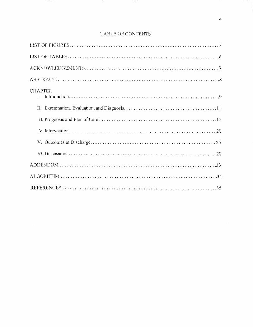
TABLE OF CONTENTS
LIST OF FIGURES ............................................................. 5
LIST OF TABLES .............................................................. 6
ACKNOWLEDGEMENTS ...................................................... 7
ABSTRACT ................................................................... 8
CHAPTER 1. Introduction ............................................................. 9
II. Examination, Evaluation, and Diagnosis ...................................... ll
III. Prognosis and Plan of Care ................................................ 18
IV. Intervention ............................................................ 20
V. Outcomes at Discharge ................................................... 25
VI. Discussion .............................................................. 28
ADDENDUM ................................................................ 33
ALGORITHM ................................................................ 34
REFERENCES ............................................................... 35

5
LIST OF FIGURES
1. Patient Rated Wrist Evaluation ........................................... 16
2. Addendum - home exercise program ...................................... 33
3. Algorithm ............................................................ 34

6
LIST OF TABLES
L Goals ............................................................... 13
2. Shoulder active range of motion .......................................... 14
3. Wrist range of motion measurements ...................................... 15
4. Problem list. ......................................................... 17
5. Wrist range of motion measurements at discharge ............................ 25
6. Goals summary ....................................................... 26
7. Costs of care for this patient ............................................. 32

7
ACKNOWLEDGEMENTS
! would like to aclmowledge the support and assistance from the entire staff of the University of North Dakota Physical Therapy Department. I would also like to give a special thank you to my
advisor, Bev Johnson, for all of the hard work with the preparation of this project and for the tireless dedication to my education.

8
ABSTRACT
Examination and Treatment for a Patient with a Distal Radius Fracture
BACKGROUND AND PURPOSE: This article describes the eight month outpatient physical
therapy management of 67 year old female who had an open fracture of the left distal radius that
was stabilized internally with K-wire and externally with the application of external fixator.
Initially, this patient presented with a need for patient education on wound care, edema control
techniques, splinting, and a maintenance program for finger and shoulder range of motion. The
physical therapy intervention progressed to range of motion, strengthening, joint mobilization,
and functional exercises for the wrist and hand. The purpose of this article is to describe the
interventions used for this patient, the results of these interventions, and the rationale for these
interventions. DESCRIPTION: This case was unique due to the addition of a splint that was
worn with the external fixator. Treatment of this patient included wound care, therapeutic
exercise, joint mobilization, edema control techniques, patient education, and splinting.
RESULTS: Following PT intervention, this patient achieved objective improvement of range of
motion and grip strength as well as improvements in functional activities of the hand.
DISCUSSION: Treatment guidelines outlined by the American Society of Hand Therapists
provided the framework and foundation for evaluation and treatment for this individual and were
deemed appropriate for this individual. While these guidelines were useful for the evaluation
and treatment ofthis patient, adaptations, such as the splint, were implemented.
KEY WORDS: Distal radius fracture, wrist pain, Patient-Rated Wrist Evaluation.

CHAPTER I
Introduction
This article describes the eight month outpatient physical therapy management of a 67
year old female, who had an open fracture of the left distal radius that was stabilized externally.
In this case study, the physician chose to use an external fixator rather than internal fixation
because it was quickly and easily applied, she had an open fracture, and he felt that he would
achieve sufficient bony aligmnent with the external fixator. This article will examine the
techniques used to rehabilitate this patient.
9
Distal radius fractures are one of the most common fractures in the United States. Studies
have shown that in the USA, a Caucasian female at 50 years old has a IS % lifetime risk of this
type of fracture.; Where as a Caucasian man of the same age has a lifetime risk of 2 %. Many of
these types of fractures occur from falls. One study noted that falls are the most prevalent
mechanism of injury.;; Often, a closed reduction and internal fixation is performed to restore the
wrist anatomy but there are times that an external fixator is used by the physician instead.
External fixation is a surgical treatment used to set bone fractures in which a cast with a closed
reduction would not allow proper alignment of the fracture.;;; In this kind of reduction, holes are
drilled into uninjured areas of bones around the fracture and special bolts or wires are screwed
into the holes. Outside the body, a rod or a curved piece of metal with special ball-and-socket
joints joins the bolts to make a rigid support. This technique is indicated when the individual is
mentally and physically healthy. The external fixator's goal is to restore proper wrist anatomy
and optimizing results for the best functional outcome by providing sufficient anatomical
alignment, maintain carpal alignment, and maintain proper length.
10
The rehabilitation of an individual who has undergone external fixation can be rigorous.
The main concern for the use of an external fixator is the potential for additional wrist and hand
stiffness.'" >V v Subsequently, the various rehabilitative techniques and treatment guidelines that
are applied for individuals receiving rehabilitative services following a distal radius fracture need
to be examined. Also, the usage of a custom orthoplastic splint in conjunction with the external
fixator is not a common practice but was used with this particular patient. There is very little
evidence supporting this procedure and only one case study in the literature that has examined
this course of action.vi This case study will not debate the pros and cons ofthis physician's
choice to support the wrist with an orthoplastic brace but only examine the physical therapy
techniques used and the possible benefits to the patient.
Studies have shown that functional and anatomical results indicate excellent to good
ratings in more then 80 % ofthe cases when an external fixator is usediii While this is a
promising study, additional studies have noted a gap in our current knowledge. A recent meta
analysis ofrehabilitative techniques used in the treatment of distal radius fractures in adults
noted that there was not enough evidence available to determine the best form of rehab for these
patients.vii Subsequently, the need for research, case studies, and assessments of these techniques
is warranted.
A theoretical frame work for treatment of individuals post application of an external
fixator is based on guidelines outlined by the American Society of Hand Therapists (ASHT).viii
This provided the frame work and foundation of evaluation and treatment of the patient I will
discuss in this article. While the suggestions and recommendations from the ASHT were
beneficial, the unique addition of a splint in conjunction with the external fixator necessitated a
distinctive approach and facilitated the development ofthis case study.
11
CHAPTER II
Examination, Evaluation, and Diagnosis
The patient was a 67 year old left handed female who injured herself when she fell
performing gardening activities on May 4th, 2005. She thought she may have stepped in a hole
and fell towards her left side onto her outstretched arm. She immediately noticed pain and
deformity of her wrist and an open wound over the ulnar side of her wrist. She sought treatment
immediately at the local emergency room and was seen by the orthopedic surgeon that day for
evaluation. The physician's evaluation noted that she had an open laceration ahove the distal
ulna with an intra-articular comminuted fracture of the distal radius. She was seen in surgery the
same day and underwent irrigation and debridement of her open wound on the distal ulna, as
well as internal stabilization with percutaneous pins through the distal radius and an external
fixator to provide additional stabilization. The fixator was applied with two percutaneous pins at
the mid shaft ofthe radius proximally and distally using two pins positioned at the mid shaft at
the second metacarpal. K-wire was used to stabilize the radius to address the intraarticular
fracture.
She then followed up with the orthopedic surgeon in his clinic five days later on May 9th,
2005. At that point in time, physical therapy treatment was initiated to hcgin education and to
fahricate a custom splint. This patient's orthopedic physician requested the splint in order to
provide additional support at the hand. With the splint extending into the palm, he felt that this
patient would feel more support and would be more apt to use her hand thus trying to minimize
loss of motion or stiffness at the fingers and thumb. The patient's chief complaints included the
following: pain, swelling, and decreased function of the left hand and wrist.
12
Prior to the injury this individual was fully independent and did not require any assistive
devices for activities of daily living (ADL) or ambulation. She presented a past medical history
of previous left and right total hip replacements due to osteoarthritis, history of low back pain,
non smoker, and post menopausal. She denied a history of falls. While she currently denied
any cardiac problems or current cardiac treatment, her family history was significant for heart
disease and arthritis. She described herself as an active retired teacher with no history of
smoking, drug, or alcohol usage. Specific questioning about possible osteoporosis was denied
and she denied previous fractures ofthe spine, upper extremity, and lower extremity. Shc
reported no specific exercise routine that she was currently engaged in on a regular basis. She
felt that her leisure activities and assisting with the care of her grandchildren kept her busy.
Leisure activities and hobbies included: gardening, crocheting, baking for her grandchildren, and
playing with and assisting with her grandchildren's care. She lived in a home with only her
husband. She did state that her grandchildren did come to visit often and she did assist with the
care of these children. She reported that she was left hand dominant and denied having any
problems when using the right upper extremity.
Since fracture and external fixator placement, she reported significant loss of function.
The pain, swelling, and stiffuess significantly affected her function. She reported all her leisure
activities were considerably limited as well as her personal care. She was now requiring her
husband to assist with daily cares, such as getting dressed and driving. She was unable to assist
with the care of her grandchildren and reported to me that this was a major complaint. She saw
her role as a grandmother significantly changed, which she felt was adversely affecting the
quality of her life.
13
Goals were mutually discussed and agreed upon by the patient and the physical therapist.
These initial goals (Table J) were established during the physical therapy evaluation at 12 weeks
post fracture and modified two additional times.
T hi 1 I "f I I a e : m la. goa s 1. The patient will present f,'11p strength of 60 pounds or better during hand
dynamometer testing within 2 months. 2. The patient will score a 10 or lower when completing the PRWE within 2 months. 3.The patient will report being able to perfonn baking activities in the kitchen without
assist within 2 months.
Her physician started her on oral antibiotics after the initial injury and on the same day of
surgery. She had completed the series at the time of second physical therapy visit. She denied
any allergic reaction to the antibiotics. She was prescribed Tylenol with co dine on the day of
injury and used it for the first two weeks status post surgery. She reported no side effects. This
medication's common side effects include the following: nausea, vomiting, constipation,
Iighthcadcdness, dizziness, drowsiness, flushing, vision changes, or mental/mood changes.
These side effects could limit or inhibit her ability to participate in rehabilitation or home
exercise activity. She later used over the counter Tylenol. Its side effects are rarc. The most
serious side effect is liver damage due to large doses, chronic use or concomitant use with
alcohol or other drugs that also damage the liver. She presented no adverse signs or symptoms
or undesirable effects of this medication.
Initial physical therapy visit occurred on May 9th, 2005. Assessment was based on
clinical assessment recommendations from the American Society of Hand Therapists (ASHT)'X
Assessment began with visual inspection of the extremity. At the wound sites there were no sign
of infection, redness or drainage. There were no asymmetries in temperature of the skin on the
left wrist radial side compared to the right side with no signs of infection around the pin areas. It
was noted that the physician instructed the patient to perfonn Hydrogen Peroxide wound
14
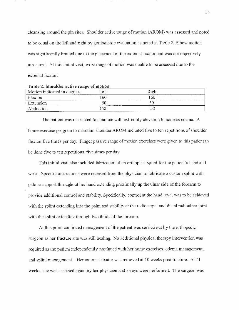
cleansing around the pin sites. Shoulder active range of motion (AROM) was assessed and noted
to be equal on the left and right by goniometric evaluation as noted in Table 2. Elbow motion
was significantly limited due to the placement of the external fixator and was not objectively
measured. At this initial visit, wrist range of motion was unable to be assessed due to the
external fixator.
Table 2- Shoulder active range of motion Motion indicated in degrees Left Right Flexion 160 160 Extension 50 50 Abduction 150 150
The patient was instructed to continue with extremity elevation to address edema. A
home exercise program to maintain shoulder AROM included five to ten repetitions of shoulder
flexion five times per day. Finger passive range of motion exercises were given to this patient to
be done five to ten repetitions, five times per day
This initial visit also included fabrication of an orthoplast splint for the patient's hand and
wrist. Specific instructions were received from the physician to fabricate a custom splint with
palmar support throughout her hand extending proximally up the ulnar side of the forearm to
provide additional control and stability. Specifically, control at the hand level was to be achieved
with the splint extending into the palm and stability at the radiocarpal and distal radioulnar joint
with the splint extending throngh two thirds ofthe forearm.
At this point continued management of the patient was carried out by the orthopedic
surgeon as her fracture site was still healing. No additional physical therapy intervention was
required as the patient independently continued with her home exercises, edema management,
and splint management. Her external fixator was removed at 10 weeks post fracture. At 11
weeks, she was assessed again by her physician and x-rays were performed. The surgeon was

15
satisfied with stability and ordered rehabilitation to begin on July 20th, 2005, 12 weeks post
fracture.
A second examination of this individual was perfonned. Wounds were closed with minimal
scar and no signs of infection. Prior to exam, the patient was given the Patient-Rated Wrist
Evaluation (PRWE). See figure 1 for a sample of the PRWK Her initial score for the PRWE
was 133. This tool was developed at the Hand and Upper Limb Center, St Joseph Health Center,
London, Ontario, Canada. The PRWE has been shown to provide a brief, reliable, and valid
measurement of patient rated pain and disability.x The tool was chosen because it was a one
page, easy to fill out, and could be used intennittently to gather data for evaluation of outcomes.
Table 3 describes the initial wrist range of motion. Grip strength was measured using the second
grip position with the right at 50 pounds and left at five pounds. A Jamar hand dynamometer
was used to assess grip strength; standard protocoL'x This form of grip strength assessment has
been found to be a valid and reliable method oftesting.x' It was also noted that the patient could
not make a full fist The amount of flexion lag was not measured.
T hi 3 I .. I f f t a e mha WrIst range 0 mo IOn measuremcn s. Motion indicated in degrees RightAROM Left AROM/PROM Flexion: 60 40/44 Extension: 60 10112 Radial deviation: 10 0/4 Ulnar deviation: 40 20/30 Pronation: 80 10120 Supination: 80 5110

16
Figure 1: Patieut Rated Wrist Evaluatiou
PATIENT RATED WRIST EVALUATION
, , , , 5 B , , 9
0 1 :l 4 5 B , " B 10
0 4 'J , , , 0 10
" , , 4 , S l , 1) 16
0 , , 4 5 " 1 5 '9 " 0 , 4 S , , 9 10
0 .2 4 '! , 0 10
0 1 1 4 4 , S , , 10 -~-~~ ..
0 1 2 , 4 , , 7 , 0 10
0 1 4 5 , 7 , 8 10
Q , 4 , , , 1l 10
A systems review was performed at this second visit. Cardiovascular and pulmonary
assessment notes that she has had general edema at the right wrist and hand, but not the left and
no lower extremity edema was noted. Blood pressure was at 132/84 with a heart rate of 72 beats
per minute. At the initial evaluation, integumentary system looked good and she had pins in
17
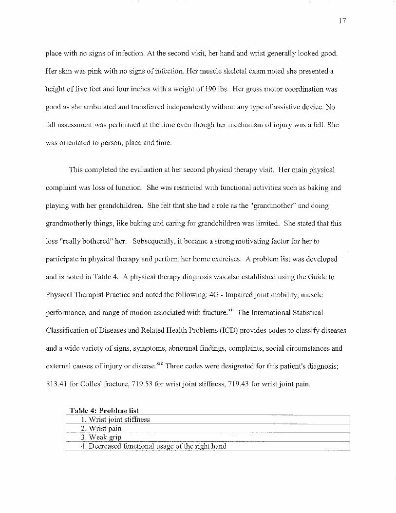
place with no signs of infection. At the second visit, her hand and wrist generally looked good.
Her skin was pink with no signs of infection. Her muscle skeletal exam noted she presented a
height of five feet and four inches with a weight of 190 Ibs. Her gross motor coordination was
good as she ambulated and transferred independently without any type of assistive device. No
fall assessment was performed at the time even though her mechanism of injury was a fall. She
was orientated to person, place and time.
This completed the evaluation at her second physical therapy visit. Her main physical
complaint was loss of function. She was restricted with fimctional activities such as baking and
playing with her grandchildren. She felt that she had a role as the "grandmother" and doing
grandmotherly things, like baking and caring for grandchildren was limited. She stated that this
loss "really bothered" her. Subsequently, it became a strong motivating factor for her to
participate in physical therapy and perform her home exercises. A problem list was developed
and is noted in Table 4. A physical therapy diagnosis was also established using the Guide to
Physical Therapist Practice and noted the following: 4G - Impaired joint mobility, muscle
performance, and range of motion associated with fracture. xii The International Statistical
Classification of Diseases and Related Health Problems (ICD) provides codes to classify diseases
and a widc variety of signs, symptoms, abnoTInal findings, complaints, social circumstances and
external causes of injury or disease.xiii Three codes were designated for this patient's diagnosis;
813.41 for Colles' fracture, 719.53 for wrist joint stiffiless, 719.43 for wrist joint pain.
Table 4: Problem list 1. Wrist joint stiffness 2. Wrist pain 3. Weak grip 4. Decreased functional usage of the right hand

18
CHAPTER III
Prognosis and Plan of Care
This patient's prognosis was good. This individual had a strong desire to achieve her
goals and return to her previous level of activity. Prognosis for this type of fracture is generally
good. One study noted functional and anatomic results indicated excellent to good ratings in
more than 80% ofthe cases after a five year follow Up:ii Another study used the PRWE to assess
outcomes in patients over 65 years of age with external fixation of a distal radius fracture six
months post surgery. xiv According to the PRWE results in this study two patients had no pain
and no functional disability; five had minimal; five had mild pain and functional disability; and
one had moderate degree and frequency of pain with moderate ±unctional disability.
Subsequently, I felt that there was good evidence that this patient would have a good outcome.
In addition, this individual was very motivated and I felt comfortable treating this type of
problem.
Occasionally, the treating orthopedic physician will just give the patient a pamphlet of
exercises to do on their own for self-directed rehabilitation. Watt et alxv investigated the use of
physical therapy intervention versus independent patient exercise. The results found that patients
who attended physical therapy achieved average wrist extension of 55 degrees versus 38 degrees
with the group who received no active therapy. xv Mean grip strength was also better with the
therapy group by seven kilograms over the group receiving no active therapy. This specific
patient was more complicated with an external fixator and a notable slow healing time prior to
PT starting than in comparison to the people in this particular study. In spite of this fact, I still
felt that her prognosis was good because our plan of care did include comprehensive
management by a Physical Therapist.

The plan of care and goals were discussed and mutually agreed upon. In clinic physical
therapy treatment was used as well as exercises to be done on a home basis. These initial goals
were set up at the 12 weeks post fracture physical therapy evaluation and listed in Table I.
19

20
CHAPTER IV
Intervention
This patient was seen for 16 visits over the course of eight months. The first visit was for
the splinting then she was not seen until 12 weeks post fracture. This patient was seen twice a
week for four weeks. Treatment was diminished to once a week for the next four weeks. The
frequency diminished to one visit per month over the course of the next three months. At eight
months status post fracture this patient was discharged from physical therapy as she has reached
maximum achievement towards her goals.
I coordinated efforts with her orthopedic surgeon. His office is within the same building as
my office and subsequent communication was easily accomplished. An example of this level of
communication would be the directives for the splint fabrication. Specific instructions were
received fi·om the physician to fabricate a palmar support throughout her hand to provide
additional control and stability. While I followed the basic principles and techniques of splinting,
a pure custom fabrication of a splint was made for this individual based on this physician'S
instructions.
Assessment was based on clinical assessment and recommendations from American
Society of Hand Therapists." This text is the standard for evaluation of upper extremity and
specifically, hand therapy assessment. It provides explanation and justification on various
evaluation tcchniques and procedures used for the assessment of this paticnt throughout her care.
Intervention techniques
My initial encounter with this patient was for fabrication of a protective orthoplast splint.
The splint was recommended by the surgeon to provide additional stabilization, beyond his
surgical technique. The use of splints is not very common in this situation so a custom splint
was needed. Even though this was a custom splint, the basic fundamentals of splinting were

followed: the greater the surface area the greater the support, folds in the material increases
strength, longer level anus provide more support. XV;
21
North Coast medical Spectrum II material was used for the splint as it provided stability
and draped easier on the ann when fabricating and customizing the splint. On the palmar side of
her hand, from the MCP joints to the hypothenar eminence, the material provided palmer side
support. The Spectrum II material was continued on this palmar side to the distal two-thirds of
the foreanu. A soft strap was secured on the dorsal side of the splint just distal from the second
metacarpal pins to secure the hand in the splint. Another strap was placed at the area of the wrist
and another proximally from the two stabilizer pins at the radius using soft cushioned strapping.
The patient was given instruction, demonstration, and practice about the application and removal
ofthe brace and she was independent with both. The cost of this splint was minimal and
fabrication was easy with very little time needed.
Wound care assessment was perfonned at the initial visit with a visual inspection of the
extremity. At the wound sites there was no sign of infection, redness or drainage. There were
no asymmetries in temperature of the skin on the left radial side compared to the right radius side
of the foreann with no signs of infection around the pin areas. It was noted that the physician
started a series of oral antibiotic medication and instructed the patient to perfonn Hydrogen
Peroxide wound cleansing around the pin sites, as he was concerned about infection. At her
second visit, after the pins were removed, the wounds continued to note no signs of infection and
were closed within two weeks. There was no significant scar fonnation.
At the time of the second physical therapy visit and continuing for the next six physical
therapy visits, heat was used prior to in clinic range of motion activities. Also, the patient was
instructed to use a heating pad (she chose to usc her electric heating pad) at home for 10 minutes
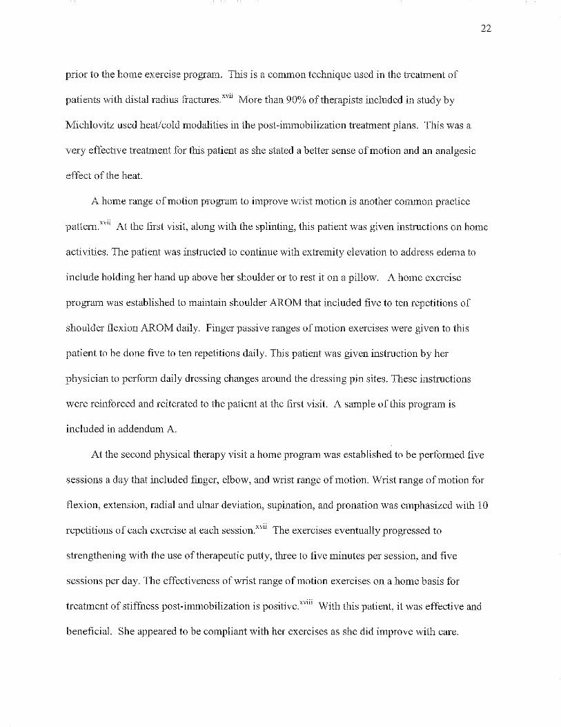
prior to the home exercise program. This is a common technique used in the treahnent of
patients with distal radius fractures. xvii More than 90% of therapists included in study by
Michlovitz used heat/cold modalities in the post-immobilization treahuent plans. This was a
very effective treatment for this patient as she stated a better sense of motion and an analgesic
effect of the heat.
22
A home range of motion program to improve wrist motion is another common practice
pattern. xvii At the first visit, along with the splinting, this patient was given instructions on home
activities. The patient was instructed to continue with extremity elevation to address edema to
include holding her hand up above her shoulder or to rest it on a pillow. A home exercise
program was established to maintain shoulder AROM that included five to ten repetitions of
shoulder flexion AROM daily. Finger passive ranges of motion exercises were given to this
patient to be done five to ten repetitions daily. This patient was given instruction by her
physician to perfonn daily dressing changes around the dressing pin sites. Thesc instructions
were reinforced and reiterated to the patient at the first visit. A sample ofthis program is
included in addendum A.
At the second physical therapy visit a home program was established to be perfonned five
sessions a day that included finger, elbow, and wrist range of motion. Wrist range of motion for
flexion, extension, radial and ulnar deviation, supination, and pronation was emphasized with 10
repetitions of each exercise at each session.xvu The exercises eventually progressed to
strengthening with the use of therapeutic putty, three to five minutes per session, and five
sessions per day. The effectiveness of wrist range of motion exercises on a home basis for
treatment of stiffness post-immobilization is positive. xviii With this patient, it was effective and
beneficial. She appeared to be compliant with her exercises as she did improve with care.

23
As this patient continued with her home exercises, in clinic treatment continued from visit
two through visit number ten in which gentle stretching, range of motion, and grade II and grade
III joint mobilizations at the wrist and fingers was perfonned. During these visits, after the usage
of a moist heat pad, I manually mobilized the wrist and fingers to assist with improving range of
motion and after this passive mobilization; active motion was perfonned with simple active wrist
flexion, extension, radial deviation, ulnar deviation, and wrist circumduction. MacDennid'"
found that these techniques are a commonly used approach. xix xx Hands-on stretches and joint
mobilization glides are useful in selected patients having difficulty maintaining their active range
of motion solely with the use of active exercise. Patients may require these hands-on techniques
and scar massage/management to restore full tissue mobility. As noted in the initial evaluation,
she was lacking range of motion. Subsequently, she needed assist with improving these values.
It appeared that as she improved with her range of motion her function improved.
Strengthening exercises to improve grip was also needed due to her poor grip strength and
function. Exercises to build functional strength are common techniques used by physical
therapists for many dysfunctions and that exercise is a common useful tool when treating distal
radius fractures. xxi The home exercise program continued with functional strengthening with
therapeutic putty and a stress ball to build strength. She had a strong desire and physical need to
improve her functional strength in order to achieve her goals. She perfonned various types of
grips and pinches with the therapeutic putty or the soft foam ball. I recommended that she does
three to five minutes of the motions for five sessions per day. There are many journals and texts
that recommend or justifY the need for these techniques and reinforce that the strengthening
activities are common practice patterns when treating patients with distal radius fractures. xxii

24
Regardless, this patient definitely benefited from this type of intervention as noted with objective
grip improvement as well as improved PRWE score.

25
CHAPTER V
Outcomes at Discharge
At eight months post fracture this patient was discharged from physical therapy as she
has reached maximum achievement of her goals. Final PRWE performed and her score was a 19
indicating mild pain and functional disability. Final range of motion measurements are provided
in Table 5. The final grip strength measurements were right at 50 pounds and left at 40 pounds.
Table 5 Wrist range of motion measurements at discharge . Motion indicated in degrees RightAROM Left AROM Flexion: 60 50 Extension: 60 55 Radial deviation: 10 10 Ulnar deviation: 40 35 Pronation: 80 80 S upinati on: 80 60
Her response to the intervention was noted to be positive as she was able to return to
previous activities in the kitchen and all ADLs without assistance. She did well with physical
therapy intervention with no adverse effects, no decrease in function with the interventions, and
she made objective improvement with trealment.
She did have some impainnents and functional limitations at discharge. While her grip did
not achieve 60 pounds, weakness was still noted even though she was subjectively reporting no
functional problems. This was evident from the PRWE score. As noted previously, the PRWE
clinometric tool was used to track and describe changes in functional status of the patient. Initial
testing noted that she really could not function, a 138 is very high number indicating a large
subjective amount of pain and physical limitations. Her final PRWE noted good functional
improvement, not at a zero but scored a 19. While she has some limitations she was going to
continue with her rehab on her own. I would anticipate continued improvement. Rikili, iii
reported improvements for up to five years post smgery. Her level of accomplishment of

26
anticipated goals and expected outcomes was very good. The goals that were set at the second
visit are noted below. The target date to achieve these goals was two months. While she made
progress to these goals she did not meet them. While I felt that they were worthy goals, I
overestimated her progress. Subsequently, I needed to reset them after two months and then
reset them again after another two month period of physical therapy intervention. The final
outcome took longer than initially expected and Table 6 summarizes the final outcomes. I did
anticipate that she would eventually meet the goals, but we needed to allow additional time for
rehabilitation.
T bl 6 G I a e : oa s summary Goal Summary I. The patient will present grip strength of 60 although not met, significant progress pounds or better with testing with the hand dynamometer within 2 months. 2. The patient will score a 10 or lower when although not met, significant progress completing the PRWE within 2 months 3. The patient will report being able to perform met baking activities in the kitchen without assist within 2 months.
This patient was fully compliant with attendance to physical therapy treatment sessions and
appeared to follow the exercise program instructions properly. She gave me no reason to doubt
her. She could verbally and physically reproduce the exercises so I believed that she was doing
them on her own. Risk reduction and prevention issues were minimal. She was provided one on
one care by me, no aides or physical therapy assistants provided care. She could safely perfom1
her exercises in clinic and at home, all exercise in clinic done under my supervision, and I made
sure she had pictures of the program to insure that she did exercises correctly. I did use moist
heat prior to exercises and made sure that the risk of bum was minimized by utilizing the heat
only 10 minutes and providing sufficient layering.
The patient satisfaction with physical therapy intervention was not formally measured.

27
She appeared to be satisfied with the results. I have seen her and visited with her in public since
discharge from rehabilitation and she reports no problems. My personal level of satisfaction was
met as she had a good outcome in the end.

28
CHAPTER VI
Discllssion
Generally, the outcome of this individual indicated that the physical therapy intervention
was effective. The best explanation that I can give for the results is that we followed evaluation
and treatment principles that were reliable, valid, and effective. Studies oflong term results of
external fixation of distal radius fractures have indicated excellent to good outcomesiii iv xiv
The eight month period of rehabilitation and eventual final evaluation was shorter than a
long term follow up study. Specifically, Rikliiii did follow up studies after five years. The eight
month period for my patient was sufficient for this situation and she did have a pleasing
outcome. Subsequently, I can conclude that my patient did very well with no complications.
Reflex sympathetic dystrophy can be a problem in patients with type of fracture and
stabilization, but in the end, this patient has no such complications in the eight months of rehab
under my careiv
At the time of discharge the patient was pleased with her progress. There was
improvement in function as measured by the PRWE as well as increase in strength and range of
motion. With the improvement in her function and with the lower PRWE score, the clinical
implications of this observation lead me to believing that the PRWE was an effective tool.
Recommendations for future studies of the usage of the PR WE, specifically with patients with
and external fixator would be beneficial.
Because this patient was compliant and was able to understand and perform the exercises,
I was able to decrease our frequency of treatment over time and look towards a more long term
approach for her rehabilitation. Quite often a home exercise program is used to address this type
of patient.xvii xxii xxiii Studies have shown that adherence to a hand therapy program helps
29
improve initial outcomes after distal radial fracture. xxiv The important clinical implication of this
observation in this study supports the use of a home exercise program. I believe that a home
program can improve long lenu outcomes as evident in this patient by her achievement and
progress to her goals. Future studies to look at specific exercises for these types of patients will
be helpful in order to find the most effective exercises and techniques available. Home exercise
adherence was noted to be one ofthe most important predictors of outcomes.xx" Clinically, it
appears that with this was an important factor in order to achieve her goals. I believe this patient
definitely benefited from adherence to her home exercise program. The physical therapy
sessions were important to help monitor her progress and to improve adherence to her home
exercise program. When treating a patient with a distal radius fracture, the treating physical
therapist should not only prescribe an appropriate home program but should make a point of
monitoring and improving patient compliance to that home program.
! believe the guidelines for trealment and rehabilitation established by the American
Society of Hand Therapists was beneficial with assisting in the clinical evaluation and
intervention of this patient. They did provide effective guidelines and should be used by
therapists treating this type of injury. This will also provide improved reliability between
therapists with testing. For example, to test the grip strength, a standard procedure was used
with the patient in the same position each time. This was good as I was accurately testing the
grip the same way each time and could be easily and reliably reproduced by another therapist if
needed.
The splint usage was a unique aspect of trealment for this patient. This has been the only
patient that I have used orthoplastic splinting in conjunction with an external fixator. Her
orthopedic physician was pleased with the healing of this patient's radius and felt that the splint

30
was a useful tool in this healing. I do not anticipate consistent usage of this type of splinting in
the future but this was a good example of using the fundamentals of splinting in unique or
difficult situations.
More evidence is needed to help therapists treat people with distal radius fractures. In
2006, Handoll vii published a systemic review of management of patients with distal radius
fracture. The author's conclusion was that with the available evidence from randomized
controlled trials, there was insufficient evidence to establish the relative effectiveness of the
various interventions used in the rehabilitation of adults with fractures of the distal radius. The
study was able to only look at 15 trials as many were excluded. This leads to the conclusion that
additional high quality, randomized control studies are needed to provide further and adequate
evidence.
Reflective Practice
Based on the outcomes ofthis study, I would proceed similarly with the examination,
evaluation, and intervention. The algorithm in figure 3 demonstrates that decision making
process of the examination, evaluation, and intervention. There are things that I have learned and
will improve upon based on this study. The mechanism of injury was a fall. In the future, fall
assessment could benefit the patient by detenllining risk for additional injury as well as possible
prevention of problems in the future. My goal setting will also change. I needed to extend the
target dates for the goals with this patient. The two month goal was too soon and needed to look
longer tenn. The one to two month period may be better suited for short tenu goals. I did not set
a specific pain scale goal for this patient. The first section of the PRWE includes a pain scale
that not only allows the patient to rate their pain levels at rest but with functional activities so I
felt that this clinometric tool addressed this issue sutIiciently. ! have included this tool in my

practice for the past 5 years and plan on continuing its usage not just for distal radius fractures
but other patients with wrist pain from surgery or injury.
31
Usage of a splint with the external fixator is extremely rare. Additional evidence and
reasons to use it is definitely an area that would need further exploration. The practice is
uncommon so I do not anticipate research on this in the future. The rational in this case was
based on clinical need and by the physician treating the fracture. The ability to adapt to different
and new situations or conditions by the therapist is crucial in these unusual circumstances.
No other disciplines were needed in the care of this individual. I did make it a point to
work closely with her orthopedic surgeon to follow the care recommended and to keep him
infonned on the patient's progress. Medically she was stable and no need for referral was
required.
Considering the movement towards pay for perfonnance for physical therapy services
and considering the constraint on health care dollars, I did note the cost of the equipment and of
the physical therapy sessions for this patient. This patient was seen for 16 visits over the course
of eight months. Dollar amounts are summarized in Table 7. She was seen initially for splinting
then physical therapy restarted at 12 weeks from fracture. The splint cost was minimal at $31.21
for the material and the cost to the patient was $40.00. An advanced beneficiary notice (ABN)
was used for splint costs as Medicare would not cover the splint. This assured my billing
department that the patient was fully aware of the costs and was expected to pay for the amount
not covered by Medicare. Our clinic "accepts assignment" from Medicare so we received full
payment ofthe evaluation and exercise costs plus full payment ofthe splint was received leading
to 100% reimbursement. There was no co-pay needed for each visit and she had already met her
32
deductible when she had the initial emergency room visit and surgery. This was a very
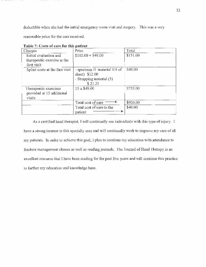
reasonable price for the care received.
T bl 7 C a e : f osts 0 care f h' or t IS patient Charges Price Total
Initial evaluation and $102.00 + $49.00 $151.00 therapeutic exercise at the first visit Splint costs at the first visit - spectrum II material 114 of $40.00
sheet) $12.00 - Strapping material (3)
$ 21.21 Therapeutic exercises 15 x $49.00 $735.00 provided at 15 additional visits
Total cost of care ~ $926.00 Total cost of care to the $40.00 patient ~
As a certified hand therapist, I will continually see individuals with this type of injury. I
have a strong interest in this specialty area and will continually work to improve my care of all
my patients. In order to achieve this goal, I plan to continue my education with attendance to
fracture management classes as well as reading journals. The Journal of Hand Therapy is an
excellent resource that I have been reading for the past five years and will continue this practice
to further my education and knowledge base.

Addendum A: home exercise program
"'''''''''''0',,,,,,,,,",,,,,,,,,,,,,,,,,,,,,,.,
,\\'"hl,",·,,'"'"'''''
11,,1.' .IL"" I
"'T"J""",hd,,,,<A"''''I~''';''J
""""."" ,·",.,1,,,11,,,,,,
U,i",o"~t=d."".,,., ""mt"f""<=,,"~
R"",~...1ll..-"m., D,---L-><-,u=,,,,,"'-,
"NG"" _" ""'.' Pll' /D[P Com_, fI"'''''' {">,,,'OS',,,,,,,,
Lr,."'h~lun< .. '", __ "0''' ",Uth=,."",
Rq<~--1!L,.m" r",_L.~=p«"'"
_",""I'L,_"~tr,,,,<,,,,,,,,",,,,,",,"_,"","jm""
H,,,,,,~"m" '''---2_=~=''''''''''
SII:JL LDcR C-'.RE TIPS
co _ ,wod tall and erec, so your shQulders mil na\urdlh
roll ba,kwilrd
""0'" ','"~"",'",",,',,',"'~,n,;.", "'"" ',,,'Cr. ~;, g.ml. '''"lngmg
_ .;e,oi50 slo"'l~ and carduil) if,ou hove. ilmi\ed r_1nge ofmo\lQn
DO:"'T _ c~ntinu' to e,erciso ifpain "presen! _ ,'0 b"ond comiortable =ge l'f motion _ ;Iuo,; ,ho~:j",_ '-~" can "C"!~ neck ?roblen:s - m ,~ lift or feed! heavy obJecLS above head
- Use stool or wait for =istanc~
fll\GER _ 29 DIP b.-Tension (Assi'Tne)
Use otherhand lO hold fingertip <TnIighT. Lei go and try lO maintain position.
Repe.T.-lL limes. Do ~ •• s:siQns per day.
FlNGER- 34 Flexor Tendon Gliding (Active Full Fist)
STraighlen all ftngcrs. Ihen make a fist. bendi"g altjoinl!;
Repeal JL times.. Do ~se"ion. per day_
"'''' ""'~"''''ffi1'' " W,~ ,,,",,,' "",","" ,oJ. " -d,d,",f,,~ld~n"_
cl=~,Appl"~,",d,,...· ~ ~"'p,,,,,~,,fi,,,c,,of
0,'",1=0
H~d...1ll..-=,d,
;;,,~~,::~""..,., ~)
d.<I!!1t>_""""",~,,,-, 'f"""'~"hd'"~'mfi'" ~if"""
'd*
~
"'" ~i" ""'''' ,,=,bL "~d ",o",b ~d ~,,,"' J~"" K«I'0",a""J"'""'"~"'''''''",'''''<''f",'m "*" --1!L "~,,, [1" --->- ,e""",> r= It.,
F ron' ,,,odln~' l'r <;:-1r'c PO;11100. placo arm,,, "d."p"l,mfomord Slow],' falSe arm, ol'e, hood unlii
''''lc"lSf,!,
Repeat _,"_,imes Do ~ ,">sion; per day
1'"ccl,,11
\
fNGER - 37 MP, PIP, DIP Comrosite Flexion (PassiYe Srr'tch)
U,e other hond 10 bend ___ finger
al all drree joinT'.
Hold_ ,"_ second,
Repe.t .-lL limos. Do_' _ ,,,,ion, per d.y.
)
FINGER - 33 Flexor T.ndo" Gliding (ACTive Book Fist)
With fingers and knuckle, slnIight, bend middle and lip joint'. Do not bend l:u-go knu<kle,.
Rep.at -1.Q... Time,. Do ~ ",,,;on, per da),.
I'"~' [ 01 [
33

33
Addendum A: home exercise program

34
Algorithm - Examination and Treatment for a Patient with a Distal Radius Fracture
I Primary Stage/Immobilization Stage: Week 1 - Week 11. Immobilization by external fixation. Splint
I fabrication, wound care education_ shoulder and fine:er ROM exercises. and complete PRWE clinometric.
~ j Yes I Assess wound: any signs of Refer back infection?
5::,~ to
Fabricate splint unable to fit kl Assess non-immobile and/or poor vascular status?
I No I ioints: Any loss of I
~ Yes: Restore/maintain range of motion of uninvolved joints and instruct client in independent home exercise program and precautions. I No I
~ ~ Secondary Stage/Mobilization Stage: Begins at Week 12. Orthopedic physician has deemed hacture stable. Complete new PRWE. Assess active, passive, and resisted wrist motion; Assess strength with grip and pinch tests
~l ~ No loss of motion or Yes, there is a loss of active, Yes, there is a loss of strength: Instruct in strength, equal left passive, and/or resisted motion: gJip and pinch strength training for home
and right, or minimal Instruct in exercise program/begin exercises and in clinic treatments.
difference left vs. treatment in clinic. right: instruct in
~J ~ ,.- home program.
Continued deficits Re assess objective tests at physical therapy visits I
Tertiary Stage/Late Strengthening Stage: Begins at Week 16-20. Final orthopedic physician evaluation completed. Complete new PRWE. Assess active, passive, and resisted wrist motion; Assess strength with grip and pinch tests; perform additional ADL tests for home or work related tasks.
/~ /' Deficits noted with testing: Continue treatment as stated in secondary phase as needed. No deficits: Full return to functional tasks, Exercises: More aggressive PRWE score less than 20. Discharge from skilled progressive strengthening, physical therapy services to continue with home resistive functional activities. based exercise program. ADLIWork: Begin work conditioning, ADL, or hardening program as indicated. Job modifications as needed. Focus on specific functional exercise program in clinic and with home program.
/ Re assess at subsequent physical therapy visits I

35
References:
ii
iii
iv
v
vi
vii
viii
ix
xi
XH
Xlii
xiv
Singer BR, Mclauchlan GJ, Robinson CN, Christie J. Epidemiology of fractures and 15 thousand adults: influence the age of gender. J Bone Joint Surg, 1998; 80: 243-248.
Cummings SR, Kelsey JL, Nevitt MC. Epidemiology of Osteoporosis and Osteoprotic fractures. Epidemiol Rev 1985; 7:178-208.
Rikli DA, Kupfer K, Bodoky A. Long tenn results of the external fixation of distal radius fractures. J Trauma 1998; 44: 970-976.
Hageman JH, et aL External fixation for unstable intra articular distal radius fractures in women older then 55 years. Injury 2005; 36: 339-344.
Margaliot Z, Haase SC, Kotsis S, Kim HM, Chung KC. A meta-analysis of outcomes of external fixation versus plate osteosynthesis for unstable distal radial ttactures. J Hand Surg [Am}. 2005; 30 (6): 1185-1199.
Pesco MS, Altner Pc. A protective orthoplast splint in the treatment of a patient with a Colles' fracture by external fixation. J Hand Ther, 1993; 6(1 ):39-4 L
Handoll HHG, Madhok R, Howe TE. Rehabilitation for distal radial ttactures in adults. Cochrane Database of Systemic Reviews 2006, Issue 3. Art. No.: CD003324. DOl: 10.1002114651858.CD003324.pub2
American Society of Hand Therapists. General Principles of Distal RadialFracture Management. Chicago, IL ASHT, 1998.
Amelican Society of Hand Therapists: Clinical Assessment Recommendations.2nd ed. Chicago, IL: ASHT; 1992.
MacDennid J, Turgeon T, Richards RS, Beadle M, and Roth JH. Patient rating of pain and disability: A reliable and valid measurement tooL J Orthop Trauma. 1998; 12 (8): 577-586
Mathiowetz V, Weber K, Volland G, Kashman N. Reliability and validity of grip and pinch strength evaluations. J Hand Surg. 1984; 9: 222-226.
American Physical Therapy Association. Guide to Physical Therapist Practice. Available at www.apta.org. Accessed on September 30, 2007.
Medilexicon ICD-9 and ICD9CM Codes Search. Available at http://www.mcdilcxicon.comlicd9codes.php. Accessed on September 30,2007
Kamiloski V, Kasapinova K. External fixation in patients with age over 65 years with distal radius ttacture. Prilozi~2006; 27(2):189-199.
xv
xvii
xviii
xix
xx
xxi
xxii
xxiii
xxiv
36
Watt CF, Taylor NF, and Baskus K. Do Colles' fracture patients benefit from routine referral to physiotherapy following cast removal? Arch Orthop Trauma Surg. 2000; 120: 413-415.
Brand P. Splinting: Principles and techniques. In: Hunter J, Mackin E, Callahan A, eds. Rehabilitation of the Hand: Surgery and Therapy, Volume 1 and 11. . Sl. Louis, MO: Mosby-Yearbook, Inc; 2001: 1581-1616
Michlovitz SL, LaStayo PC, Alzner S, Watson E. Distal radius fractures: therapy practice patterns. J Hand Ther. 2001; 14(4):249-57.
Maciel JS, Taylor NF, McIlveen C. A randomised clinical trial of activity-focused physiotherapy on patients with distal radius fractures. Arch Orthop Trauma Surg. 2005; J 25(8):515-20.
MacDennid JC. Hand therapy management of intra-articular fractures with open reduction and pi plate fixation: a therapist's perspective. Tech Hand Up Extrem Surg. 2004; 8(4): 2 J 9-223.
MacDennid JC. Treatment of distal radius fractures with external fixation: technical considerations for rehabilitation. Tech Hand Up Extrem Surg.2002; 6(4): 213-218.
Reiss B. Therapist management of distal radius fractures. Rehabilitation of the Hand: Surgery and Therapy, Volume J and lI. [n: Hunter J, Mackin E, Callahan A, eds. st. Louis, MO. Mosby-Yearbook, Inc; 2001: 337-352.
Collins DC. Management and rehabilitation of distal radius fractures. Orthop Chn North Am. 1993; 24(2):365-78.
Reiss B. Therapist management of distal radius fractures. Rehabilitation of the Hand: Surgery and Therapy, Volume I and JI. In: Hunter J, Mackin E, Callahan A, eds. st. Louis, MO. Mosby-Yearbook, Inc; 2001: 337-352.
Lyngcoln A, Taylor N, Pizzari T, Baskus K. The relationship between adherence to hand therapy and short term outcome after distal radius fracture. J Hand Therapy. 2005; 18 (1): 2-8.
Related Documents