Full Citation: Cheney, N., Clune, J., & Lipson, H. Evolved Electrophysiological Soft Robots. In ALIFE 14: The Fourteenth Conference on the Synthesis and Simulation of Living Systems (Vol. 14, pp. 222-229) Evolved Electrophysiological Soft Robots Nicholas Cheney 1 , Jeff Clune 2 , and Hod Lipson 1 1 Creative Machines Lab, Cornell University. Ithaca, NY 2 Evolving Artificial Intelligence Lab, University of Wyoming. Laramie, WY [email protected], [email protected], [email protected] Abstract The embodied cognition paradigm emphasizes that both bod- ies and brains combine to produce complex behaviors, in con- trast to the traditional view that the only seat of intelligence is the brain. Despite recent excitement about embodied cogni- tion, brains and bodies remain thought of, and implemented as, two separate entities that merely interface with one an- other to carry out their respective roles. Previous research co-evolving bodies and brains has simulated the physics of bodies that collect sensory information and pass that infor- mation on to disembodied neural networks, which then pro- cesses that information and return motor commands. Biologi- cal animals, in contrast, produce behavior through physically embedded control structures and a complex and continuous interplay between neural and mechanical forces. In addition to the electrical pulses flowing through the physical wiring of the nervous system, the heart elegantly combines control with actuation, as the physical properties of the tissue itself (or defects therein) determine the actuation of the organ. In- spired by these phenomena from cardiac electrophysiology (the study of the electrical properties of heart tissue), we in- troduce electrophysiological robots, whose behavior is dic- tated by electrical signals flowing though the tissue cells of soft robots. Here we describe these robots and how they are evolved. Videos and images of these robots reveal lifelike behaviors despite the added challenge of having physically embedded control structures. We also provide an initial ex- perimental investigation into the impact of different imple- mentation decisions, such as alternatives for sensing, actua- tion, and locations of central pattern generators. Overall, this paper provides a first step towards removing the chasm be- tween bodies and brains to encourage further research into physically realistic embodied cognition. Introduction and Background The fields of evolutionary robotics and artificial life have seen a great deal of emphasis on embodied cognition in recent years [Cheney et al. (2013); Bongard (2013); Ri- effel et al. (2013); Auerbach and Bongard (2012); Hiller and Lipson (2012a); Lehman and Stanley (2011); Auer- bach and Bongard (2010a,b); Pfeifer et al. (2007); Hornby et al. (2001); Lipson and Pollack (2000)]. There is even a paradigm called embodied cognition, which argues that the specifics of the embodiment (such as the morphology) are Figure 1: Current flowing through an evolved creature. The legend for voltage within each cell (colors) is given in Fig. 3. vital parts of the resulting behavior of the system: It argues that the co-evolutionary connection between body and brain is more deeply intertwined than the body simply acting as a minimal physical interface between the brain and the envi- ronment [Pfeifer and Bongard (2006)]. Recent work in evolutionary robotics has shown that complex behaviors can arise when co-evolving bodies and brains. At one end of the spectrum, Auerbach and Bon- gard (2010b) demonstrated the evolution of physical struc- tures that had no joints or actuators, and evolved to cover the largest distance in a controlled fall due to gravity. While that work exemplifies the evolution of behavior emerging from morphology alone, it does not co-evolve any actuation or control. Auerbach and Bongard (2010a) then evolved the placement of CPG controlled rotational joints between cel- lular spheres, thus co-evolving morphology and control. Cheney et al. (2013) evolved locomoting soft robots made of multiple different materials: two passive voxels of differing rigidity and two actuated voxel types that ex- panded cyclically via out-of-phase central pattern genera- tors (CPGs). While this work added a variety of soft ma- terials and a new type of actuation, the pairing of muscle types directly to a CPG again reflected a focus on evolving morphology rather than sophisticated neural control. Many examples in the literature include the co-evolution of a robot morphology with an artificial neural network con- troller [Sims (1994); Lipson and Pollack (2000); Hornby et al. (2001); Lehman and Stanley (2011)]. These studies (and many more like them) involve what might be called “ghost” networks: artificial neural networks that provide control to the body, yet do not have any physical embodi-

Welcome message from author
This document is posted to help you gain knowledge. Please leave a comment to let me know what you think about it! Share it to your friends and learn new things together.
Transcript
Full Citation: Cheney, N., Clune, J., & Lipson, H. Evolved Electrophysiological Soft Robots.In ALIFE 14: The Fourteenth Conference on the Synthesis and Simulation of Living Systems (Vol. 14, pp. 222-229)
Evolved Electrophysiological Soft Robots
Nicholas Cheney1, Jeff Clune2, and Hod Lipson1
1 Creative Machines Lab, Cornell University. Ithaca, NY2 Evolving Artificial Intelligence Lab, University of Wyoming. Laramie, WY
[email protected], [email protected], [email protected]
Abstract
The embodied cognition paradigm emphasizes that both bod-ies and brains combine to produce complex behaviors, in con-trast to the traditional view that the only seat of intelligence isthe brain. Despite recent excitement about embodied cogni-tion, brains and bodies remain thought of, and implementedas, two separate entities that merely interface with one an-other to carry out their respective roles. Previous researchco-evolving bodies and brains has simulated the physics ofbodies that collect sensory information and pass that infor-mation on to disembodied neural networks, which then pro-cesses that information and return motor commands. Biologi-cal animals, in contrast, produce behavior through physicallyembedded control structures and a complex and continuousinterplay between neural and mechanical forces. In additionto the electrical pulses flowing through the physical wiringof the nervous system, the heart elegantly combines controlwith actuation, as the physical properties of the tissue itself(or defects therein) determine the actuation of the organ. In-spired by these phenomena from cardiac electrophysiology(the study of the electrical properties of heart tissue), we in-troduce electrophysiological robots, whose behavior is dic-tated by electrical signals flowing though the tissue cells ofsoft robots. Here we describe these robots and how they areevolved. Videos and images of these robots reveal lifelikebehaviors despite the added challenge of having physicallyembedded control structures. We also provide an initial ex-perimental investigation into the impact of different imple-mentation decisions, such as alternatives for sensing, actua-tion, and locations of central pattern generators. Overall, thispaper provides a first step towards removing the chasm be-tween bodies and brains to encourage further research intophysically realistic embodied cognition.
Introduction and Background
The fields of evolutionary robotics and artificial life haveseen a great deal of emphasis on embodied cognition inrecent years [Cheney et al. (2013); Bongard (2013); Ri-effel et al. (2013); Auerbach and Bongard (2012); Hillerand Lipson (2012a); Lehman and Stanley (2011); Auer-bach and Bongard (2010a,b); Pfeifer et al. (2007); Hornbyet al. (2001); Lipson and Pollack (2000)]. There is even aparadigm called embodied cognition, which argues that thespecifics of the embodiment (such as the morphology) are
Figure 1: Current flowing through an evolved creature. Thelegend for voltage within each cell (colors) is given in Fig. 3.
vital parts of the resulting behavior of the system: It arguesthat the co-evolutionary connection between body and brainis more deeply intertwined than the body simply acting as aminimal physical interface between the brain and the envi-ronment [Pfeifer and Bongard (2006)].
Recent work in evolutionary robotics has shown thatcomplex behaviors can arise when co-evolving bodies andbrains. At one end of the spectrum, Auerbach and Bon-gard (2010b) demonstrated the evolution of physical struc-tures that had no joints or actuators, and evolved to coverthe largest distance in a controlled fall due to gravity. Whilethat work exemplifies the evolution of behavior emergingfrom morphology alone, it does not co-evolve any actuationor control. Auerbach and Bongard (2010a) then evolved theplacement of CPG controlled rotational joints between cel-lular spheres, thus co-evolving morphology and control.
Cheney et al. (2013) evolved locomoting soft robotsmade of multiple different materials: two passive voxelsof differing rigidity and two actuated voxel types that ex-panded cyclically via out-of-phase central pattern genera-tors (CPGs). While this work added a variety of soft ma-terials and a new type of actuation, the pairing of muscletypes directly to a CPG again reflected a focus on evolvingmorphology rather than sophisticated neural control.
Many examples in the literature include the co-evolutionof a robot morphology with an artificial neural network con-troller [Sims (1994); Lipson and Pollack (2000); Hornbyet al. (2001); Lehman and Stanley (2011)]. These studies(and many more like them) involve what might be called“ghost” networks: artificial neural networks that providecontrol to the body, yet do not have any physical embodi-
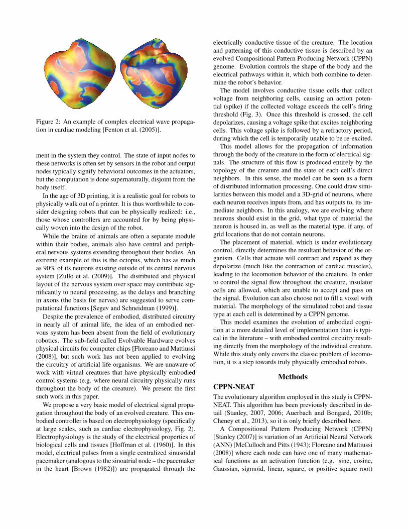
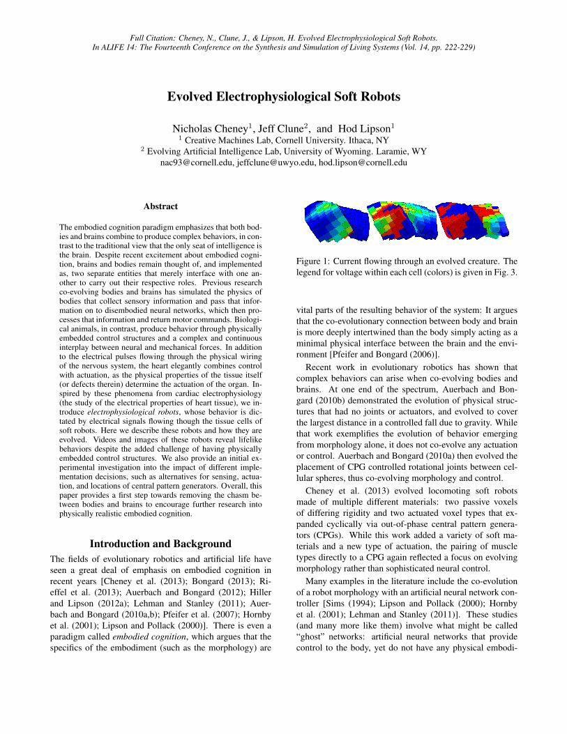
Figure 2: An example of complex electrical wave propaga-tion in cardiac modeling [Fenton et al. (2005)].
ment in the system they control. The state of input nodes tothese networks is often set by sensors in the robot and outputnodes typically signify behavioral outcomes in the actuators,but the computation is done supernaturally, disjoint from thebody itself.
In the age of 3D printing, it is a realistic goal for robots tophysically walk out of a printer. It is thus worthwhile to con-sider designing robots that can be physically realized: i.e.,those whose controllers are accounted for by being physi-cally woven into the design of the robot.
While the brains of animals are often a separate modulewithin their bodies, animals also have central and periph-eral nervous systems extending throughout their bodies. Anextreme example of this is the octopus, which has as muchas 90% of its neurons existing outside of its central nervoussystem [Zullo et al. (2009)]. The distributed and physicallayout of the nervous system over space may contribute sig-nificantly to neural processing, as the delays and branchingin axons (the basis for nerves) are suggested to serve com-putational functions [Segev and Schneidman (1999)].
Despite the prevalence of embodied, distributed circuitryin nearly all of animal life, the idea of an embodied ner-vous system has been absent from the field of evolutionaryrobotics. The sub-field called Evolvable Hardware evolvesphysical circuits for computer chips [Floreano and Mattiussi(2008)], but such work has not been applied to evolvingthe circuitry of artificial life organisms. We are unaware ofwork with virtual creatures that have physically embodiedcontrol systems (e.g. where neural circuitry physically runsthroughout the body of the creature). We present the firstsuch work in this paper.
We propose a very basic model of electrical signal propa-gation throughout the body of an evolved creature. This em-bodied controller is based on electrophysiology (specificallyat large scales, such as cardiac electrophysiology, Fig. 2).Electrophysiology is the study of the electrical properties ofbiological cells and tissues [Hoffman et al. (1960)]. In thismodel, electrical pulses from a single centralized sinusoidalpacemaker (analogous to the sinoatrial node – the pacemakerin the heart [Brown (1982)]) are propagated through the
electrically conductive tissue of the creature. The locationand patterning of this conductive tissue is described by anevolved Compositional Pattern Producing Network (CPPN)genome. Evolution controls the shape of the body and theelectrical pathways within it, which both combine to deter-mine the robot’s behavior.
The model involves conductive tissue cells that collectvoltage from neighboring cells, causing an action poten-tial (spike) if the collected voltage exceeds the cell’s firingthreshold (Fig. 3). Once this threshold is crossed, the celldepolarizes, causing a voltage spike that excites neighboringcells. This voltage spike is followed by a refractory period,during which the cell is temporarily unable to be re-excited.
This model allows for the propagation of informationthrough the body of the creature in the form of electrical sig-nals. The structure of this flow is produced entirely by thetopology of the creature and the state of each cell’s directneighbors. In this sense, the model can be seen as a formof distributed information processing. One could draw simi-larities between this model and a 3D-grid of neurons, whereeach neuron receives inputs from, and has outputs to, its im-mediate neighbors. In this analogy, we are evolving whereneurons should exist in the grid, what type of material theneuron is housed in, as well as the material type, if any, ofgrid locations that do not contain neurons.
The placement of material, which is under evolutionarycontrol, directly determines the resultant behavior of the or-ganism. Cells that actuate will contract and expand as theydepolarize (much like the contraction of cardiac muscles),leading to the locomotion behavior of the creature. In orderto control the signal flow throughout the creature, insulatorcells are allowed, which are unable to accept and pass onthe signal. Evolution can also choose not to fill a voxel withmaterial. The morphology of the simulated robot and tissuetype at each cell is determined by a CPPN genome.
This model examines the evolution of embodied cogni-tion at a more detailed level of implementation than is typi-cal in the literature – with embodied control circuitry result-ing directly from the morphology of the individual creature.While this study only covers the classic problem of locomo-tion, it is a step towards truly physically embodied robots.
Methods
CPPN-NEAT
The evolutionary algorithm employed in this study is CPPN-NEAT. This algorithm has been previously described in de-tail (Stanley, 2007, 2006; Auerbach and Bongard, 2010b;Cheney et al., 2013), so it is only briefly described here.
A Compositional Pattern Producing Network (CPPN)[Stanley (2007)] is variation of an Artificial Neural Network(ANN) [McCulloch and Pitts (1943); Floreano and Mattiussi(2008)] where each node can have one of many mathemat-ical functions as an activation function (e.g. sine, cosine,Gaussian, sigmoid, linear, square, or positive square root)
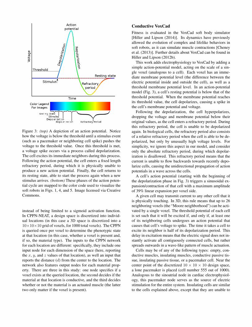
Figure 3: (top) A depiction of an action potential. Noticehow the voltage is below the threshold until a stimulus event(such as a pacemaker or neighboring cell spike) pushes thevoltage to the threshold value. Once this threshold is met,a voltage spike occurs via a process called depolarization.The cell excites its immediate neighbors during this process.Following the action potential, the cell enters a fixed lengthrefractory period, during which it is physically unable toproduce a new action potential. Finally, the cell returns toits resting state, able to start the process again when a newstimulus arrives. (bottom) These phases of the action poten-tial cycle are mapped to the color code used to visualize thesoft robots in Figs. 1, 4, and 5. Image licensed via CreativeCommons.
instead of being limited to a sigmoid activation function.In CPPN-NEAT, a design space is discretized into individ-ual locations (in this case a 3D space is discretized into a10⇥10⇥10 grid of voxels, for 1000 total voxels). The CPPNis queried once per voxel to determine the phenotypic stateat that location (in this case, whether a voxel is present and,if so, the material type). The inputs to the CPPN networkfor each location are different: specifically, they include oneinput node for each dimension of the space (here, reportingthe x, y, and z values of that location), as well an input thatreports the distance (d) from the center to the location. Thenetwork also features output nodes for each material prop-erty. There are three in this study: one node specifies if avoxel exists at the queried location, the second decides if thematerial at that location is conductive, and the third decideswhether or not the material is an actuated muscle (the lattertwo only matter if the voxel is present).
Conductive VoxCad
Fitness is evaluated in the VoxCad soft body simulator[Hiller and Lipson (2014)]. Its dynamics have previouslyallowed the evolution of complex and lifelike behaviors insoft robots, as it can simulate muscle contractions [Cheneyet al. (2013)]. Further details about VoxCad can be found inHiller and Lipson (2012b).
This work adds electrophysiology to VoxCad by adding asimple action-potential model, acting on the scale of a sin-gle voxel (analogous to a cell). Each voxel has an imme-diate membrane potential level (the difference between theelectric potential inside and outside the cell), as well as athreshold membrane potential level. In an action-potentialmodel (Fig. 3), a cell’s resting potential is below that of thethreshold potential. When the membrane potential reachesits threshold value, the cell depolarizes, causing a spike inthe cell’s membrane potential and voltage.
Following the depolarization, the cell hyperpolarizes,dropping the voltage and membrane potential below theiroriginal values, as the cell enters a refractory period. Duringthis refractory period, the cell is unable to be depolarizedagain. In biological cells, the refractory period also consistsof a relative refractory period when the cell is able to be de-polarized, but only by unusually high voltage levels. Forsimplicity, we ignore this aspect in our model, and consideronly the absolute refractory period, during which depolar-ization is disallowed. This refractory period means that thecurrent is unable to flow backwards towards recently depo-larize cells, causing the unidirectional propagation of actionpotentials in a wave across the cells.
A cell’s action potential (starting with the beginning ofthe depolarization phase in Fig. 3) triggers a sinusoidal ex-pansion/contraction of that cell with a maximum amplitudeof 39% linear expansion per voxel side.
A given cell may transmit current to any other cell that itis physically touching. In 3D, this rule means that up to 26neighboring voxels (the “Moore neighborhood”) can be acti-vated by a single voxel. The threshold potential of each cellis set such that it will be excited if, and only if, at least oneof its neighboring cells undergoes an action potential thatcauses that cell’s voltage to spike. The time it takes a cell toexcite its neighbor is half of its depolarization period. Thisdelay in excitation means that the electric signal does not in-stantly activate all contiguously connected cells, but ratherspreads outwards in a wave-like pattern of muscle actuation.
Cells may be of any of the following types: empty, con-ductive muscles, insulating muscles, conductive passive tis-sue, insulating passive tissue, or a pacemaker cell. Near thecenter point of the discretized 10 ⇥ 10 ⇥ 10 design space,a lone pacemaker is placed (cell number 555 out of 1000).Analogous to the sinoatrial node in cardiac electrophysiol-ogy, this pacemaker node serves as the source of electricstimulation for the entire system. Insulating cells are similarto the cells explained above, except that they are unable to
accept current from neighboring cells and thus never reachtheir threshold potential or produce an action potential.
In this model, the refractory period lasts 5 times as long asthe depolarization period. This means that at least 5 voxelsmust separate the leading edges of two serial action poten-tial waves. Since the pacemaker is placed in the center ofthe 10 ⇥ 10 ⇥ 10 space, approximately one wave of actionpotentials would exist at any given time in a setup with a uni-form cube of entirely conductive material – where a wave ofaction potentials would propagate uninterrupted, with a newone starting around the time the first reaches the outer edgeof the space. We chose this setup to encourage the evolutionof static gaits, which can be more robust and transferable toreality than dynamic gaits [Belter et al. (2008)].
The length of the expansion/contraction period of eachnode is set equal to the refractory period, such that each cellis guaranteed to be fully returned to its original size beforeits next actuation cycle begins.
Task and Fitness Evaluation
Following Cheney et al. (2013), we evolve these electro-physiological robots for locomotion over flat ground. Thissimple task and environment make fitness evaluation easy.Despite its simplicity, the task is a classic problem in thefield, and has been repeatedly shown to produce an arrayof complex morphologies and interesting behaviors [Cheneyet al. (2013); Clune et al. (2009, 2011); Auerbach and Bon-gard (2014); Lehman and Stanley (2011)].
Each creature is simulated for 20 times the length of anexpansion/contraction cycle. Its displacement between thestarting coordinates and the creature’s final center of mass(in the xy plane) is recorded. In an effort to discourage de-signs that might excite as many cells as possible, and to en-courage designs with sparse spindles of connectivity (simi-lar to the peripheral nervous system), the distance traveledis multiplied by 1 � (# of conductive cells)
1000 . Thus the fit-ness function incentivizes minimizing the amount of con-ductive tissue and maximizing the distance traveled. Whilea multi-objective technique may be ideal in finding the opti-mal tradeoff between these goals, we follow previous CPPN-NEAT research in using this single, multi-part fitness func-tion [Cheney et al. (2013); Auerbach and Bongard (2009)].
Experimental Parameters
Unless otherwise noted, each treatment described belowconsists of 48 independent runs (with identical initial con-ditions across treatments). Each run consists of a populationsize of 30 individuals evolved for 1000 generations. Unlessotherwise noted, all other parameters are consistent with Ch-eney et al. (2013).
Statistical Reporting
Because the data are not normally distributed, all plots showmedian fitness (thick, center lines) bracketed by two thin
lines that represent 95% bootstrapped confidence intervalsof the median [Sokal and Rohlf (1995)]. For the samereason, all p-values are generated with the non-parametricMann-Whitney-Wilcoxon Rank Sum test, which does notassume normality. Reported p-values compare the distancetraveled by the top organism for each of the 48 runs at thefinal (1000th) generation. Plots report distance traveled, notadjusted fitness (which penalized for the number of conduct-ing voxels as explained previously).
Results
Since this is the first study of evolved electrophysiologicalrobots, there are many unanswered questions regarding thedesign and implementation of such a system. Many arbitrarydesign choices were made during the initial implementation.Here, we examine the impact of some of these choices.
As with many explorations in evolved virtual organisms,one of the main goals is complex, natural-appearing behav-ior. However, there are no satisfactory metrics for the “natu-ralness” or complexity of evolved behaviors. For this reason,we must rely on our qualitative, subjective assessments. Avideo of the evolved behaviors can be seen on the “CornellCreative Machines Lab” Youtube channel, or found directlyat this link: http://goo.gl/CvJp4l. We believe thebehaviors are interesting, complex, and lifelike – at least asmuch as in Cheney et al. (2013) – despite the added chal-lenges of evolving physically embedded control.
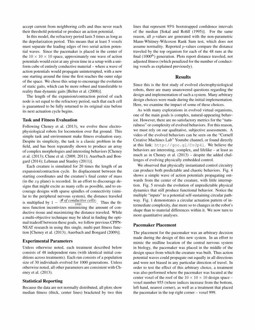
We observed that physically instantiated control circuitrycan produce both predictable and chaotic behaviors. Fig. 4shows a simple wave of action potentials propagating out-wards from the center of the creature, with little interrup-tion. Fig. 5 reveals the evolution of unpredictable physicaldynamics that still produce functional behavior. Notice themultiple “inputs” to a potential self-sustaining circular path-way. Fig. 1 demonstrates a circular actuation pattern of in-termediate complexity, due more so to changes in the robot’sshape than to material differences within it. We now turn tomore quantitative analyses.
Pacemaker Placement
The placement for the pacemaker was an arbitrary decisionmade during the design of this new system. In an effort tomimic the midline location of the central nervous systemin biology, the pacemaker was placed in the middle of thedesign space from which the creature was built. Thus actionpotential waves could propagate out equally in all directionsand were not biased in any particular direction of travel. Inorder to test the effect of this arbitrary choice, a treatmentwas also performed where the pacemaker was located at thecenter voxel of the roof of the 10 ⇥ 10 ⇥ 10 design space –voxel number 955 (where indices increase from the bottom,left hand, nearest corner), as well as a treatment that placedthe pacemaker in the top right corner – voxel 999.
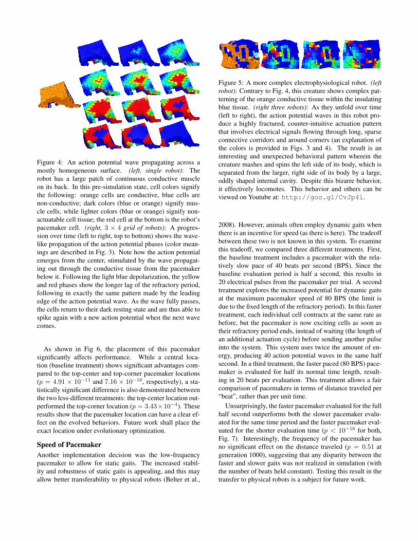
Figure 4: An action potential wave propagating across amostly homogeneous surface. (left, single robot): Therobot has a large patch of continuous conductive muscleon its back. In this pre-simulation state, cell colors signifythe following: orange cells are conductive, blue cells arenon-conductive; dark colors (blue or orange) signify mus-cle cells, while lighter colors (blue or orange) signify non-actuatable cell tissue; the red cell at the bottom is the robot’spacemaker cell. (right, 3 ⇥ 4 grid of robots): A progres-sion over time (left to right, top to bottom) shows the wave-like propagation of the action potential phases (color mean-ings are described in Fig. 3). Note how the action potentialemerges from the center, stimulated by the wave propagat-ing out through the conductive tissue from the pacemakerbelow it. Following the light blue depolarization, the yellowand red phases show the longer lag of the refractory period,following in exactly the same pattern made by the leadingedge of the action potential wave. As the wave fully passes,the cells return to their dark resting state and are thus able tospike again with a new action potential when the next wavecomes.
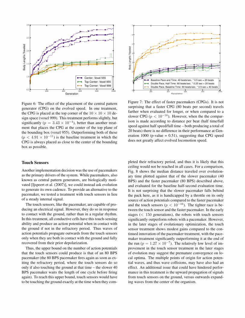
As shown in Fig 6, the placement of this pacemakersignificantly affects performance. While a central loca-tion (baseline treatment) shows significant advantages com-pared to the top-center and top-corner pacemaker locations(p = 4.91 ⇥ 10�11 and 7.16 ⇥ 10�16, respectively), a sta-tistically significant difference is also demonstrated betweenthe two less-different treatments: the top-center location out-performed the top-corner location (p = 3.43⇥10�4). Theseresults show that the pacemaker location can have a clear ef-fect on the evolved behaviors. Future work shall place theexact location under evolutionary optimization.
Speed of Pacemaker
Another implementation decision was the low-frequencypacemaker to allow for static gaits. The increased stabil-ity and robustness of static gaits is appealing, and this mayallow better transferability to physical robots (Belter et al.,
Figure 5: A more complex electrophysiological robot. (leftrobot): Contrary to Fig. 4, this creature shows complex pat-terning of the orange conductive tissue within the insulatingblue tissue. (right three robots): As they unfold over time(left to right), the action potential waves in this robot pro-duce a highly fractured, counter-intuitive actuation patternthat involves electrical signals flowing through long, sparseconnective corridors and around corners (an explanation ofthe colors is provided in Figs. 3 and 4). The result is aninteresting and unexpected behavioral pattern wherein thecreature mashes and spins the left side of its body, which isseparated from the larger, right side of its body by a large,oddly shaped internal cavity. Despite this bizarre behavior,it effectively locomotes. This behavior and others can beviewed on Youtube at: http://goo.gl/CvJp4l.
2008). However, animals often employ dynamic gaits whenthere is an incentive for speed (as there is here). The tradeoffbetween these two is not known in this system. To examinethis tradeoff, we compared three different treatments. First,the baseline treatment includes a pacemaker with the rela-tively slow pace of 40 beats per second (BPS). Since thebaseline evaluation period is half a second, this results in20 electrical pulses from the pacemaker per trial. A secondtreatment explores the increased potential for dynamic gaitsat the maximum pacemaker speed of 80 BPS (the limit isdue to the fixed length of the refractory period). In this fastertreatment, each individual cell contracts at the same rate asbefore, but the pacemaker is now exciting cells as soon astheir refractory period ends, instead of waiting (the length ofan additional actuation cycle) before sending another pulseinto the system. This system uses twice the amount of en-ergy, producing 40 action potential waves in the same halfsecond. In a third treatment, the faster paced (80 BPS) pace-maker is evaluated for half its normal time length, result-ing in 20 beats per evaluation. This treatment allows a faircomparison of pacemakers in terms of distance traveled per“beat”, rather than per unit time.
Unsurprisingly, the faster pacemaker evaluated for the fullhalf second outperforms both the slower pacemaker evalu-ated for the same time period and the faster pacemaker eval-uated for the shorter evaluation time (p < 10�16 for both,Fig. 7). Interestingly, the frequency of the pacemaker hasno significant effect on the distance traveled (p = 0.51 atgeneration 1000), suggesting that any disparity between thefaster and slower gaits was not realized in simulation (withthe number of beats held constant). Testing this result in thetransfer to physical robots is a subject for future work.
0 200 400 600 800 1000Generation
0.5
1.0
1.5
2.0
2.5
3.0B
ody
Leng
ths
Trav
eled
Center: Voxel 555Top Center: Voxel 955Top Corner: Voxel 999
Figure 6: The effect of the placement of the central patterngenerator (CPG) on the evolved speed. In one treatment,the CPG is placed at the top corner of the 10⇥ 10⇥ 10 de-sign space (voxel 999). This treatment performs slightly, butsignificantly (p = 3.43 ⇥ 10�4), better than another treat-ment that places the CPG at the center of the top plane ofthe bounding box (voxel 955). Outperforming both of these(p < 4.91 ⇥ 10�11) is the baseline treatment in which theCPG is always placed as close to the center of the boundingbox as possible.
Touch Sensors
Another implementation decision was the use of pacemakersas the primary drivers of the system. While pacemakers, alsoknown as central pattern generators, are biologically moti-vated [Ijspeert et al. (2007)], we could instead ask evolutionto generate its own cadence. To provide an alternative to thepacemaker, we tested a treatment with touch sensors in lieuof a steady internal signal.
The touch sensors, like the pacemaker, are capable of pro-ducing an electrical signal. However, they do so in responseto contact with the ground, rather than in a regular rhythm.In this treatment, all conductive cells have this touch-sensingability and produce an action potential when in contact withthe ground if not in the refractory period. Thus waves ofaction potentials propagate outwards from the touch sensorsonly when they are both in contact with the ground and fullyrecovered from their prior depolarization.
Thus, the upper bound on the number of action potentialsthat the touch sensors could produce is that of an 80 BPSpacemaker (the 80 BPS pacemaker fires again as soon as ex-iting the refractory period, where the touch sensors do soonly if also touching the ground at that time – the slower 40BPS pacemaker waits the length of one cycle before firingagain). To reach this upper bound, touch sensors would haveto be touching the ground exactly at the time when they com-
0 200 400 600 800 1000Generation
0
1
2
3
4
5
6
7
Bod
yLe
ngth
sTr
avel
ed
Baseline Pace and Time: 40 beats/sec. * 0.5 sec = 20 beatsDouble Pace, Half Time: 80 beats/sec. * 0.25 sec = 20 beatsDouble Pace, Baseline Time: 80 beats/sec. * 0.5 sec = 40 beats
Figure 7: The effect of faster pacemakers (CPGs). It is notsurprising that a faster CPG (80 beats per second) travelsfarther when evaluated for longer, or when compared to aslower CPG (p < 10�16). However, when the the compar-ison is made according to distance per beat (half time/fullspeed against half speed/full time – both producing a total of20 beats) there is no difference in their performance at Gen-eration 1000 (p-value = 0.51), suggesting that CPG speeddoes not greatly affect evolved locomotion speed.
pleted their refractory period, and thus it is likely that thisceiling would not be reached in all cases. For a comparison,Fig. 8 shows the median distance traveled over evolution-ary time plotted against that of the slower pacemaker (40BPS) and the faster pacemaker (80 BPS) described above,and evaluated for the baseline half-second evaluation time.It is not surprising that the slower pacemaker falls behindthe pack here, as it is handicapped by a throttle on its onlysource of action potentials compared to the faster pacemakerand the touch sensors (p < 10�16). The tighter race is be-tween the touch sensor and the faster pacemaker. In the earlystages (< 150 generations), the robots with touch sensorssignificantly outperform robots with a pacemaker. However,in the later stages of evolutionary optimization, the touchsensor treatment shows modest gains compared to the con-tinued innovation of the pacemaker treatment, with the pace-maker treatment significantly outperforming it at the end ofthe run (p = 1.27 ⇥ 10�7). The relatively low level of im-provement in the touch sensor treatment in the later stagesof evolution may suggest the premature convergence on lo-cal optima. The multiple points of origin for action poten-tial waves, and thus wave collisions, may have also had aneffect. An additional issue that could have hindered perfor-mance in this treatment is the upward propagation of signalsfrom touch sensors on the ground, versus outwards expand-ing waves from the center of the organism.

0 200 400 600 800 1000Generation
0
1
2
3
4
5
6
7B
ody
Leng
ths
Trav
eled
Baseline Pace and Time: 40 beats/secDouble Pace, Baseline Time: 80 beats/secTouch Sensors (No Pacemaker; up to 80 beats/sec)
Figure 8: The performance of touch sensors vs. central pat-tern generators. The touch sensor treatment produces anexpected number of beats with the upper bound set by thefaster (80 beat/sec) CPG. Despite early evolvability leadingto a statistically significant advantage in the first 150 gener-ations, in later generations the touch sensor setup is unableto produce creatures that travel as far as the faster CPG setup(p = 1.27 ⇥ 10�7 at Gen. 1000). Artificially throttled, theslow CPG is unable to compete with either (p < 10�16).
Expansion/Contraction Cycle
In the soft robot evolution system described by Cheney et al.(2013), regular, quickly repeating, and coupled out-of-phasesinusoidal action cycles defined the expansion and contrac-tion of cells. In this model, which does not feature the samecomplimentary muscle types, the question of actuation cycleis not entirely clear. In an attempt to explore this, here wetest the effectiveness of contraction-then-expansion phasecycles against expansion-then-contraction cycles (Fig. 9).These treatments take place on the baseline (slow) pace-maker setup, as to not allow continuous and quickly repeat-ing expansion/contraction cycles, but rather to have a breakbetween actuations. Despite the same number of beats (andthus the same amount of overall expansion and contraction)in both setups, the contraction-then-expansion setup per-forms significantly better (p = 1.94⇥ 10�3). While the rea-son for this difference is not entirely clear, it may be due, inpart, to a larger continuous expansion period from the troughof the sine wave to its peak (continuous expansion from min-imum to maximum size) in the contraction-then-expansiontreatment. In contrast, the expansion-then-contraction setupincludes a full-cycle length pause in the middle of its expan-sionary period. This explanation would suggest that morelocomotion tends to come from pushing than pulling, whichis in line with our observations from viewing videos of theevolved behaviors.
0 200 400 600 800 1000Generation
0.5
1.0
1.5
2.0
2.5
3.0
3.5
Bod
yLe
ngth
sTr
avel
ed
Expansion-then-ContractionContraction-then-Expansion
Figure 9: Unlike the regularly occurring actuation cycles ofCheney et al. (2013), the electrophysiological actuations inthis paper do not have a necessary order: either expansionor contraction can occur first. It turns out that performanceis significantly higher when muscles contract first and thenexpand, rather than vice versa (p = 1.94⇥ 10�3).
Discussion
This work reduces the separation between bodies and brainsin research into embodied cognition. We did so by em-bedding the control systems into the physical simulation ofthe robot’s morphology. Perhaps most interesting about thiswork is that the complex and interesting behaviors are thedirect result of the morphology of the creatures, as the con-trol is woven directly into the structure of the organisms. Inthis work the size of the creatures was limited for computa-tional reasons, but in future work we plan to explore largerdesign spaces. We also plan to test different ways of imple-menting electrophysiological robots and to challenge themto perform more difficult tasks.
Conclusion
We have introduced electrophysiological robots, which areinspired by the electrical properties of cardiac tissue. Thebehavior of these robots is governed by electrical signalsflowing though the evolved cells of soft robots. We de-scribed these robots and how they are evolved, including theevolution of interesting behaviors, despite the added chal-lenge of physically embedded control structures. We alsoprovided an initial experimental investigation into differentimplementation decisions, such as alternatives for sensing,actuation, and central pattern generator locations. We be-lieve that this paper provides a first step towards removingthe gulf between brains and bodies to encourage further re-search into physically realistic embodied cognition.
Acknowledgments
This work was supported by DARPA Open ManufacturingGrant W911NF-12-1-0449 and NASA Space TechnologyResearch Fellowship NNX13AL37H for Nicholas Cheney.
References
Auerbach, J. and Bongard, J. C. (2009). How robot morphologyand training order affect the learning of multiple behaviors.In IEEE Congress on Evol. Comp., 2009, pages 39–46. IEEE.
Auerbach, J. E. and Bongard, J. C. (2010a). Dynamic resolution inthe co-evolution of morphology and control. In ALife XII.
Auerbach, J. E. and Bongard, J. C. (2010b). Evolving CPPNs togrow three-dimensional physical structures. In Proc. of theGenetic & Evolutionary Comp. Conf., pages 627–634. ACM.
Auerbach, J. E. and Bongard, J. C. (2012). On the relation-ship between environmental and morphological complexityin evolved robots. In Proc. of Genetic & Evol. Comp. Conf.,pages 521–8. ACM.
Auerbach, J. E. and Bongard, J. C. (2014). Environmental influenceon the evolution of morphological complexity in machines.PLoS computational biology, 10(1):e1003399.
Belter, D., Kasinski, A., and Skrzypczynski, P. (2008). Evolvingfeasible gaits for a hexapod robot by reducing the space ofpossible solutions. In Intelligent Robots and Systems, 2008.IROS 2008. IEEE/RSJ International Conference on, pages2673–2678. IEEE.
Bongard, J. C. (2013). Evolutionary robotics. Comm. of the ACM,56(8):74–83.
Brown, H. F. (1982). Electrophysiology of the sinoatrial node.Physiological Reviews, 62(2):505–530.
Cheney, N., MacCurdy, R., Clune, J., and Lipson, H. (2013). Un-shackling evolution: evolving soft robots with multiple mate-rials and a powerful generative encoding. In Proc. of the 15thGenetic and Evol. Comp. Conf., pages 167–174. ACM.
Clune, J., Beckmann, B., Ofria, C., and Pennock, R. (2009). Evolv-ing coordinated quadruped gaits with the HyperNEAT gen-erative encoding. In Proc. of the IEEE Congress on Evol.Comp., pages 2764–71.
Clune, J., Stanley, K. O., Pennock, R. T., and Ofria, C. (2011). Onthe performance of indirect encoding across the continuum ofregularity. IEEE Trans. on Evol. Comp., 15(4):346–67.
Fenton, F. H., Cherry, E. M., Karma, A., and Rappel, W.-J. (2005).Modeling wave propagation in realistic heart geometries us-ing the phase-field method. Chaos: An InterdisciplinaryJournal of Nonlinear Science, 15(1):013502.
Floreano, D. and Mattiussi, C. (2008). Bio-inspired artificial intel-ligence: theories, methods, and technologies. MIT Press.
Hiller, J. and Lipson, H. (2014). Dynamic simulation of soft mul-timaterial 3d-printed objects. Soft Robotics, 1(1):88–101.
Hiller, J. D. and Lipson, H. (2012a). Automatic design and manu-facture of soft robots. IEEE Trans. on Rob., 28(2):457–466.
Hiller, J. D. and Lipson, H. (2012b). Dynamic simulation of softheterogeneous objects. ArXiv:1212.2845.
Hoffman, B. F., Cranefield, P. F., and Johnston, F. D. (1960). Elec-trophysiology of the heart.
Hornby, G. S., Pollack, J. B., et al. (2001). Body-brain co-evolutionusing l-systems as a generative encoding. In Proc. of the Ge-netic and Evolutionary Comp. Conf., pages 868–875.
Ijspeert, A. J., Crespi, A., Ryczko, D., and Cabelguen, J. M. (2007).From swimming to walking with a salamander robot drivenby a spinal cord model. Science, 315(5817):1416–1420.
Lehman, J. and Stanley, K. O. (2011). Evolving a diversity of vir-tual creatures through novelty search and local comp. In Proc.of 13th Genetic & Evol. Comp. Conf, pages 211–8. ACM.
Lipson, H. and Pollack, J. B. (2000). Automatic design and manu-facture of robotic lifeforms. Nature, 406(6799):974–978.
McCulloch, W. and Pitts, W. (1943). A logical calculus of theideas immanent in nervous activity. Bulletin of mathematicalbiology, 5(4):115–133.
Pfeifer, R. and Bongard, J. C. (2006). How the body shapes theway we think: a new view of intelligence. MIT press.
Pfeifer, R., Lungarella, M., and Iida, F. (2007). Self-organization,embodiment, and biologically inspired robotics. science,318(5853):1088–1093.
Rieffel, J., Knox, D., Smith, S., and Trimmer, B. (2013). Growingand evolving soft robots. Artificial Life, (Early Access):1–20.
Segev, I. and Schneidman, E. (1999). Axons as computing devices:basic insights gained from models. Journal of Physiology-Paris, 93(4):263–270.
Sims, K. (1994). Evolving 3d morphology and behavior by com-petition. Artificial life, 1(4):353–372.
Sokal, R. and Rohlf, F. (1995). Biometry: the principles and prac-tice of statistics in biological research. WH Freeman.
Stanley, K. O. (2006). Exploiting regularity without development.In Proceedings of the AAAI Fall Symposium on Developmen-tal Systems, page 37. AAAI Press Menlo Park, CA.
Stanley, K. O. (2007). Compositional pattern producing networks:A novel abstraction of development. Genetic Programmingand Evolvable Machines, 8(2):131–162.
Zullo, L., Sumbre, G., Agnisola, C., Flash, T., and Hochner, B.(2009). Nonsomatotopic organization of the higher motorcenters in octopus. Current Biology, 19(19):1632–1636.
Related Documents