Current Biology 23, 1–7, June 17, 2013 ª2013 Elsevier Ltd All rights reserved http://dx.doi.org/10.1016/j.cub.2013.05.003 Report Evolutionary Origin of the Turtle Shell Tyler R. Lyson, 1,2,3, * Gabe S. Bever, 4,5 Torsten M. Scheyer, 6 Allison Y. Hsiang, 1 and Jacques A. Gauthier 1,3 1 Department of Geology and Geophysics, Yale University, 210 Whitney Avenue, New Haven, CT 06511, USA 2 Department of Vertebrate Zoology, National Museum of Natural History, Smithsonian Institution, Washington, DC 20560, USA 3 Division of Vertebrate Paleontology, Yale Peabody Museum of Natural History, New Haven, CT 06511, USA 4 New York Institute of Technology, College of Osteopathic Medicine, Old Westbury, NY 11568, USA 5 Division of Paleontology, American Museum of Natural History, New York, NY 10024, USA 6 Pala ¨ ontologisches Institut und Museum, Universita ¨ t Zu ¨ rich, Karl-Schmid-Strasse 4, 8006 Zu ¨ rich, Switzerland Summary The origin of the turtle shell has perplexed biologists for more than two centuries [1]. It was not until Odontochelys semitestacea [2] was discovered, however, that the fossil and developmental data [3–8] could be synthesized into a model [9] of shell assembly that makes predictions for the as-yet unestablished history of the turtle stem group. We build on this model by integrating novel data for Eunotosaurus africanus—a Late Guadalupian (w260 mya) [10] Permian reptile inferred to be an early stem turtle [11]. Eunotosaurus expresses a number of relevant characters, including a reduced number of elongate trunk vertebrae (nine), nine pairs of T-shaped ribs, inferred loss of inter- costal muscles, reorganization of respiratory muscles to the ventral side of the ribs, (sub)dermal outgrowth of bone from the developing perichondral collar of the ribs, and paired gastralia that lack both lateral and median elements. These features conform to the predicted sequence of character acquisition and provide further sup- port that E. africanus, O. semitestacea, and Proganochelys quenstedti represent successive divergences from the turtle stem lineage. The initial transformations of the model thus occurred by the Middle Permian, which is congruent with molecular-based divergence estimates [12–15] for the lineage, and remain viable whether turtles originated inside or outside crown Diapsida. Results Unlike the bony covering of other ‘‘shelled’’ amniotes (e.g., anguid lizards, ankylosaur dinosaurs, armadillos, and placo- donts), the carapace of turtles is not simply a composite of dermal ossifications but rather integrates outgrowths of intra- membranous bone from the perichondrium of the developing ribs and thoracolumbar vertebrae [3–8]. Whether the origin of the turtle shell also involved melding of overlying osteoderms (composite model) or not (de novo model) was vigorously debated throughout the 20 th and early 21 st centuries (see [16]) with support falling largely along disciplinary lines [3–9, 17–25]. Paleontological explanations relied heavily on the composite model [19–25], but their efficacy was hampered by the large morphological gap separating the earliest, fully shelled turtles (e.g., Proganochelys quenstedti [26]) from all other known groups. In contrast, developmental biologists promoted the de novo model and viewed the lack of clear tran- sitional fossils as support for a rapid evolution of the shell, perhaps coincident with the appearance of a bone morphoge- netic protein (BMP) developmental pathway critical to shell construction in modern turtles [3–9, 27]. The lack of osteo- derms in the recently discovered stem turtle Odontochelys semitestacea [2] strongly supports the de novo model of shell origination and liberates the paleontological search for the even deeper history of the turtle stem from its previously self-imposed constraint of osteoderm-bearing forms. For example, the long-recognized similarities between turtles and the osteoderm-free, Late Guadalupian Permian reptile Eunotosaurus africanus (Figure 1 and Figure S1 available online) [11, 23, 24, 28, 29] can now be viewed as homologous, with Eunotosaurus lying just deep to Odontochelys on the tur- tle stem [11]. Our study tests the role of Eunotosaurus in under- standing the origin of turtles and their shell by determining whether its morphology conforms to the predictions of the Kuratani et al. [9] model of turtle shell evolution and develop- ment. To this end, we employ new specimens and novel data sources (including rib histology; Figures 1, 2, and 3 and the Supplemental Experimental Procedures) in a phylogenetic analysis of shell-related features (Figure 4 and the Supple- mental Experimental Procedures). We summarize our results by expanding the Kuratani et al. [9] model to include the turtle stem deep to Odontochelys. Morphology GM 86/341 is the only specimen of Eunotosaurus africanus with a complete cervical and trunk vertebral series (see section I of the Supplemental Experimental Procedures for a complete list of the material examined). Nine elongated trunk vertebrae and nine pairs of dorsal ribs are clearly present, which differs from the traditional reconstruction of ten trunk vertebrae and ten pairs of ribs [23, 30, 31]. A distinct change in vertebral length, neural spine shape, and rib morphology occurs between presacral vertebrae 6 and 7 (Figures 1A–1C and S1A–S1C). We argue this is the transition between the cervical and trunk regions (sensu [32]). The cervical vertebrae are short with a bulbous neural spine [24]. They have long, slender ribs that are round in cross-section and unexpanded distally. Cer- vical rib 6 (dorsal rib 1 of other authors) is long and mostly round in cross-section except for a small, middle portion that is distinctly broadened. Thus, there are six, not five, cervical vertebrae and nine, not ten, dorsal vertebrae with nine pairs of distinctly broadened dorsal ribs. The dorsal ribs are T-shaped in cross-section and contact each other for most of their length. The first eight pairs are oriented slightly poste- riorly, whereas the last pair projects slightly anteriorly. Unlike other specimens (USNM 23099, SAM 4328, and BMNH 4949), the last pair of ribs in GM 86/341 articulates with, but is not *Correspondence: [email protected] Please cite this article in press as: Lyson et al., Evolutionary Origin of the Turtle Shell, Current Biology (2013), http://dx.doi.org/ 10.1016/j.cub.2013.05.003

Welcome message from author
This document is posted to help you gain knowledge. Please leave a comment to let me know what you think about it! Share it to your friends and learn new things together.
Transcript
Current Biology 23, 1–7, June 17, 2013 ª2013 Elsevier Ltd All rights reserved http://dx.doi.org/10.1016/j.cub.2013.05.003
ReportEvolutionary Origin of the Turtle Shell
Tyler R. Lyson,1,2,3,* Gabe S. Bever,4,5 Torsten M. Scheyer,6
Allison Y. Hsiang,1 and Jacques A. Gauthier1,31Department of Geology and Geophysics, Yale University,210 Whitney Avenue, New Haven, CT 06511, USA2Department of Vertebrate Zoology, National Museum ofNatural History, Smithsonian Institution, Washington,DC 20560, USA3Division of Vertebrate Paleontology, Yale Peabody Museumof Natural History, New Haven, CT 06511, USA4New York Institute of Technology, College of OsteopathicMedicine, Old Westbury, NY 11568, USA5Division of Paleontology, American Museum of NaturalHistory, New York, NY 10024, USA6Palaontologisches Institut und Museum, Universitat Zurich,Karl-Schmid-Strasse 4, 8006 Zurich, Switzerland
Summary
The origin of the turtle shell has perplexed biologists formore than two centuries [1]. It was not until Odontochelyssemitestacea [2] was discovered, however, that the fossiland developmental data [3–8] could be synthesized into amodel [9] of shell assembly that makes predictions forthe as-yet unestablished history of the turtle stem group.We build on this model by integrating novel data forEunotosaurus africanus—a Late Guadalupian (w260 mya)[10] Permian reptile inferred to be an early stem turtle [11].Eunotosaurus expresses a number of relevant characters,including a reduced number of elongate trunk vertebrae(nine), nine pairs of T-shaped ribs, inferred loss of inter-costal muscles, reorganization of respiratory muscles tothe ventral side of the ribs, (sub)dermal outgrowth ofbone from the developing perichondral collar of the ribs,and paired gastralia that lack both lateral and medianelements. These features conform to the predictedsequence of character acquisition and provide further sup-port that E. africanus, O. semitestacea, and Proganochelysquenstedti represent successive divergences from the turtlestem lineage. The initial transformations of the model thusoccurred by the Middle Permian, which is congruent withmolecular-based divergence estimates [12–15] for thelineage, and remain viable whether turtles originated insideor outside crown Diapsida.
Results
Unlike the bony covering of other ‘‘shelled’’ amniotes (e.g.,anguid lizards, ankylosaur dinosaurs, armadillos, and placo-donts), the carapace of turtles is not simply a composite ofdermal ossifications but rather integrates outgrowths of intra-membranous bone from the perichondrium of the developingribs and thoracolumbar vertebrae [3–8]. Whether the origin ofthe turtle shell also involved melding of overlying osteoderms(composite model) or not (de novo model) was vigorously
debated throughout the 20th and early 21st centuries (see[16]) with support falling largely along disciplinary lines [3–9,17–25]. Paleontological explanations relied heavily on thecomposite model [19–25], but their efficacy was hamperedby the large morphological gap separating the earliest, fullyshelled turtles (e.g., Proganochelys quenstedti [26]) from allother known groups. In contrast, developmental biologistspromoted the de novomodel and viewed the lack of clear tran-sitional fossils as support for a rapid evolution of the shell,perhaps coincident with the appearance of a bone morphoge-netic protein (BMP) developmental pathway critical to shellconstruction in modern turtles [3–9, 27]. The lack of osteo-derms in the recently discovered stem turtle Odontochelyssemitestacea [2] strongly supports the de novo model of shellorigination and liberates the paleontological search for theeven deeper history of the turtle stem from its previouslyself-imposed constraint of osteoderm-bearing forms. Forexample, the long-recognized similarities between turtlesand the osteoderm-free, Late Guadalupian Permian reptileEunotosaurus africanus (Figure 1 and Figure S1 availableonline) [11, 23, 24, 28, 29] can now be viewed as homologous,with Eunotosaurus lying just deep to Odontochelys on the tur-tle stem [11]. Our study tests the role ofEunotosaurus in under-standing the origin of turtles and their shell by determiningwhether its morphology conforms to the predictions of theKuratani et al. [9] model of turtle shell evolution and develop-ment. To this end, we employ new specimens and novel datasources (including rib histology; Figures 1, 2, and 3 and theSupplemental Experimental Procedures) in a phylogeneticanalysis of shell-related features (Figure 4 and the Supple-mental Experimental Procedures). We summarize our resultsby expanding the Kuratani et al. [9] model to include the turtlestem deep to Odontochelys.
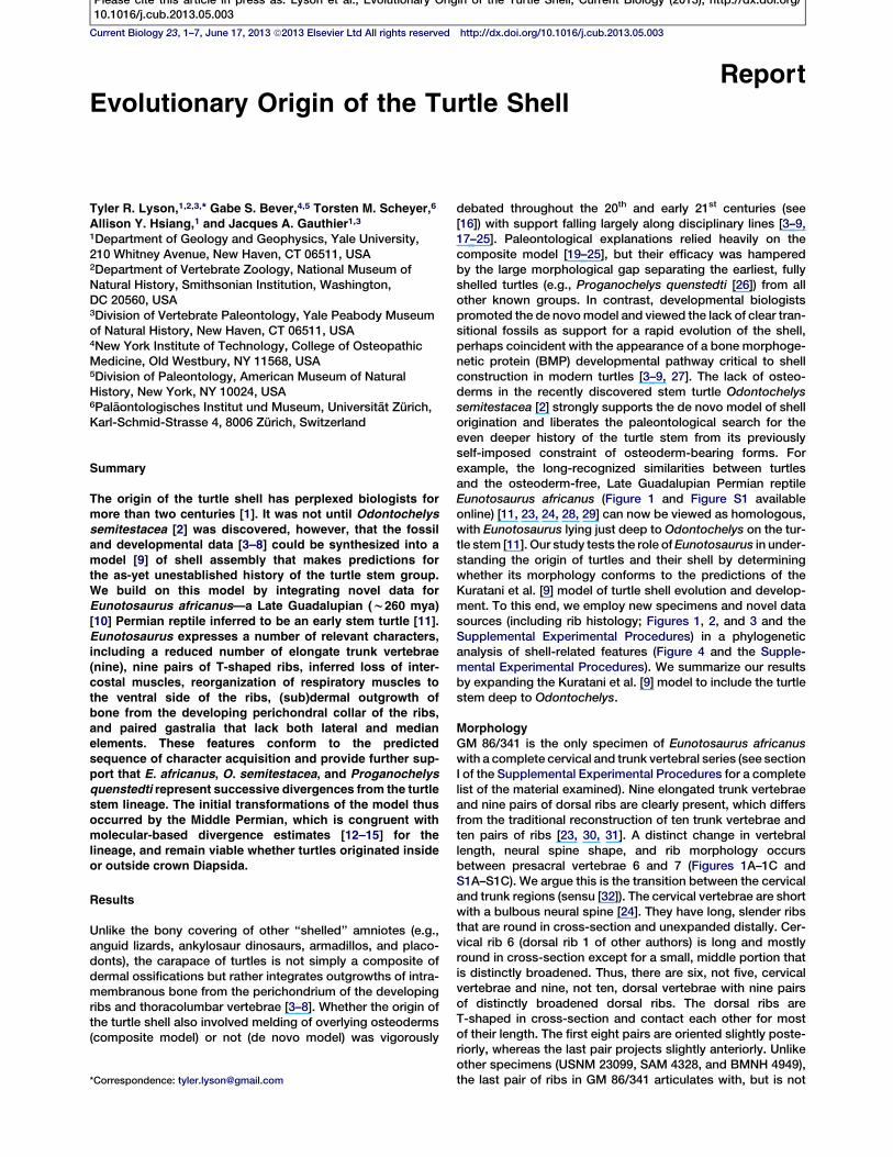
MorphologyGM 86/341 is the only specimen of Eunotosaurus africanuswith a complete cervical and trunk vertebral series (see sectionI of the Supplemental Experimental Procedures for a completelist of the material examined). Nine elongated trunk vertebraeand nine pairs of dorsal ribs are clearly present, which differsfrom the traditional reconstruction of ten trunk vertebrae andten pairs of ribs [23, 30, 31]. A distinct change in vertebrallength, neural spine shape, and rib morphology occursbetween presacral vertebrae 6 and 7 (Figures 1A–1C andS1A–S1C). We argue this is the transition between the cervicaland trunk regions (sensu [32]). The cervical vertebrae are shortwith a bulbous neural spine [24]. They have long, slender ribsthat are round in cross-section and unexpanded distally. Cer-vical rib 6 (dorsal rib 1 of other authors) is long and mostlyround in cross-section except for a small, middle portion thatis distinctly broadened. Thus, there are six, not five, cervicalvertebrae and nine, not ten, dorsal vertebrae with nine pairsof distinctly broadened dorsal ribs. The dorsal ribs areT-shaped in cross-section and contact each other for mostof their length. The first eight pairs are oriented slightly poste-riorly, whereas the last pair projects slightly anteriorly. Unlikeother specimens (USNM 23099, SAM 4328, and BMNH 4949),the last pair of ribs in GM 86/341 articulates with, but is not*Correspondence: [email protected]
Please cite this article in press as: Lyson et al., Evolutionary Origin of the Turtle Shell, Current Biology (2013), http://dx.doi.org/10.1016/j.cub.2013.05.003
fused to, the corresponding vertebra. That this conditionreflects size/age variation is supported by the smaller size ofGM 86/341.
Several specimens preserve a complete, articulated shoul-der girdle (AM 5999, NMQR 3299, GM 777, SAM K 1133, andSAM K 7909). The scapula is situated vertically and rostral tothe dorsal ribcage (Figures S1D and S1E). The clavicles areslender elements with a distinct dorsal process (Figure S1E).A slender cleithrum, which was hypothesized by Cox [23], isconfirmed. Its ventral end contacts the dorsal tip of the clavicle(Figure S1E). Paired gastralia lacking lateral and medial ele-ment(s) are preserved in NMQR 3299 (Figures 1F and S1C)and BP/1/7024.
Microanatomy and HistologyThe dorsal ribs of Eunotosaurus africanus (Figures 2B, 2F, andS2) and Proganochelys quenstedti (Figure 2G) are T-shaped incross-section (longitudinal or parasagittal section in referenceto the body axis) proximally, with a broadened surface and arounded visceral aspect that is arranged vertically (Figure 2B).In both taxa, the vertical portion of the ‘‘T’’ includes a gentlebulge distally (Figures 2F and 2G; see [33]).In GM 86/341, the anterior-most part of the broadened part
of the rib deviates ventrally and tapers to a sharp edge sothat the outer bone surface is straight to slightly concave.The posterior part does not deviate ventrally but maintains aconstant thickness, and it ends in an upturned blunt tip such
Figure 1. Newly Described Eunotosaurus africanus Material
(A) Photograph (left) and illustration (right) of GM 86/341 in dorsal view.(B) Photograph (left) and illustration (right) of GM 86/341 in ventral view.(C) Close-up photographs of the neck region of GM 86/341 in dorsal (top) and ventral (bottom) views showing differences between cervical (shortcentra with bulbous neural spines, and elongate ribs) and dorsal (greatly elongate centra, with long neural spines, and anteroposterior broadenedribs) vertebrae.(D) Photograph (left) and illustration (right) of QR 3299 in dorsal view.(E) Photograph (left) and illustration (right) of QR 3299 in ventral view.See also Figure S1 for red/blue stereophotographs of each specimen.
Current Biology Vol 23 No 122
Please cite this article in press as: Lyson et al., Evolutionary Origin of the Turtle Shell, Current Biology (2013), http://dx.doi.org/10.1016/j.cub.2013.05.003

that the internal (visceral) bone surface is slightly convex.A tapering anterior portion cannot be confirmed in NHM PV R4949, but its posterior portion is either horizontal or slightlydownturned due to its more posterior position in the trunk.Three phases of bone deposition, an initial phase and two suc-cessive phases, are recognizable (see section II of the Supple-mental Experimental Procedures). No interwoven dermalstructural fibers or remnants of cartilage are apparent any-where in the sectioned rib.
Although of similar overall size, both sectioned Eunotosau-rus ribs differ somewhat in cortical bone thickness, as wellas in internal trabecular thickness and arrangement, withNHM PV R 4949 appearing more robust overall. In this spec-imen, growth marks are visible in the cortical bone as lightand dark banding in normal transmitted light, but histologicaldetails (e.g., Sharpey’s fibers) are otherwise not well pre-served. The source of these variations is unclear.
Three discrete phases of bone deposition are visible, aninitial and two successive phases (see the SupplementalExperimental Procedures for a detailed histological descrip-tion). In the initial phase, a central large cavity (representingthe rib primordium) gets encased by parallel-fibered bone(PFB). In the next phase, thin sheets of anteriorly and posteri-orly extending bone are added dorsally and interior trabeculae
spread out from the bone around the central ovoid cavity, thuscreating a diploe structure. In the final phase, the drop-shapedbulge of the ventral shaft of the T-shaped rib is deposited later-ally and ventrally to the initial tube structure. During growth,inconspicuous Sharpey’s fibers are locally present only atthe posterior margin of the ventral shaft.
Phylogenetic AnalysisFifteen morphological characters and one taxon, the shelledSinosaurosphargis yunguiensis, were added to the mor-phological data set of Lyson et al. [11] (SupplementalExperimental Procedures, sections III–VI). In turn, this dataset is that of deBraga and Rieppel [34], with Li et al.’s [2]addition of Odontochelys semitestacea and one morpholog-ical character and Lyson et al.’s [11] addition of Eunotosaurusafricanus and Proganochelys quenstedti and seven mor-phological characters. Two most parsimonious trees (con-fidence interval [CI] = 0.3333, retention index = 0.6785,consistency index = 0.2262) with 723 steps were obtained(Figure S3). In addition, we performed a Bayesian phyloge-netic analysis, and the resulting topology is identical to thatof the parsimony analysis. There is strong support for aturtle + Eunotosaurus clade (bootstrap = 69% and posteriorprobability = 95%).
Figure 2. Histological Data from the Ribs of Eunotosaurus africanus and Proganochelys quenstedti
(A) Illustration of Eunotosaurus (GM 86/341) showing where the left third dorsal rib was sectioned histologically (red line).(B) Histological section in normal (left) and polarized (right) light showing the T shape of the rib in cross-section (see ‘‘1’’ in F, which shows the approximateplace the histological section was taken as compared to where the section was taken for Proganochelys). Black arrows indicate the presence and orien-tation of Sharpey’s fibers.(C–I) Images in (C), (D) and (E) are seen in normal transmitted (upper) and cross-polarized light using a lambda compensator (lower). Image in (H) is seen innormal transmitted and the one in (I) is seen in cross-polarized light.(C) Close-up view of the posterior diploe portion of the T-shaped rib. Thin external and internal compact layers frame interior cancellous bone, which iscomposed of thin trabeculae. Note the thin ring-like structure at internal (visceral) surface of the rib.(D) Close-up view of themidshaft region of the rib. Note central ovoid cavity surrounded by periosteal parallel-fibered bone (PFB). Sharpey’s fibers (ShF) arepresent in the posterior part of this region (blue colors). White arrows indicate the orientation of insertion of the ShF.(E) Close-up of the drop-shaped bulge, which consists internally of highly vascularized woven bone tissue (WFB) and externally of PFB. Sharpey’s fibers(white arrows) are absent from the anterior and ventral parts of the bulge. White arrows indicate presence and orientation of Sharpey’s fibers.(F and G) Eunotosaurus specimen (F, left; GM 71) showing the change in cross section of the rib (F, right) as you move distally compared to the change incross section of the rib/costal morphology (G, left) of Proganochelys (G, right).(H) Histological section of Proganochelys (MB.R. 3449.2) taken from the right costal 7? at approximately level three (see the corresponding number in G).(I) Close-up view of the external cortex, which is composed mainly of interwoven structural fibers (ISF). Numerous parallel Sharpey’s fibers insert into thebone tissue at high angles (ShF). A few scattered secondary osteons (SO) are visible.See also Figure S2.
Evolution of the Turtle Shell3
Please cite this article in press as: Lyson et al., Evolutionary Origin of the Turtle Shell, Current Biology (2013), http://dx.doi.org/10.1016/j.cub.2013.05.003

Discussion
Eunotosaurus, which previously was excluded from globalanalyses of amniote relationships, was recently recovered assister to undisputed turtles [11]. Likewise, the addition ofturtles to the latest ‘‘parareptile’’ data set (i.e., [35]) yieldsa Eunotosaurus + turtle clade [11]. The previously unde-scribed Eunotosaurus material provides compelling addi-tional morphological support for a privileged relationshipwith turtles.
Strengthening Support for a Turtle-Eunotosaurus CladeOur examination of previously undescribed specimens ofEunotosaurus, as well as the comparative histology ofEunotosaurus ribs, strengthens support for Eunotosaurus asboth a stem turtle and a critical transitional form in the evolu-tion of the turtle body plan. Gross morphological featuresrelated to the shell that are shared between Eunotosaurusand turtles now include trunk vertebrae reduced to nine(shared with Odontochelys), nine pairs of broadeneddorsal ribs (shared with Odontochelys semitestacea andProganochelys quenstedti), elongation of trunk vertebraewith length exceeding width by four times or more (sharedwith all undisputed stem and crown turtles), cross-section ofribs T-shaped proximally developing into a gentle ventralbulge distally (shared with several early turtles, includingO. semitestacea [2], P. quenstedti [26], and Palaeochersistalampayensis [36]), and paired gastralia that do not overlapmedially and lack both a central medial element and lateralelement(s) (shared with all turtles and parareptiles in whichgastralia are preserved, and lacking in all diapsids that havea central medial and/or lateral element(s); e.g., [37]). Each ofthese characters optimizes as unambiguous synapomorphiesof Eunotosaurus and undisputed turtles among reptiles. Inaddition, the cartilaginous rib primordium of Eunotosauruschanges position in relation to the developing T-shaped rib,which is also the case in the developing costals of hard-shelledturtles [38] (Figure S2). In both Eunotosaurus and hard-shelledturtles, the proximal end of the primordial rib cartilage is situ-ated more dorsally, whereas distally it becomes progressivelyventral, finally touching the internal cortical bone (NHM PV R4949; Figure S2). It should be noted that this change in positionof the primordial rib cartilage is not seen in the trionychidApalone ferox (Figure S2), in which the primordial rib cartilagemaintains a fairly constant position. This appears to be anautapomorphy for soft-shelled turtles.
The morphology of Eunotosaurus is consistent with theexplicit prediction of Kuratani et al. [9] that the early stagesof the turtle shell, prior to the emergence of Odontochelys,
was marked by a vertical scapula positioned rostral to the rib-cage. This condition is expressed in Eunotosaurus, in contrastto some other putative turtle sister groups, which have a scap-ula dorsal to the ribcage (see [39]). Furthermore, the grossmorphology of Eunotosaurus only differs from undisputedstem turtles, such as Odontochelys, in sharing fewer derivedcharacters with crown turtles, as would be expected for anearlier member of the turtle stem. For example, Eunotosauruslacks the derived conditions of neural plates, a hypoischium,and a co-ossified plastron (though all of the bones that formthe plastron are present in Eunotosaurus).The rib histology of Eunotosaurus provides further support
for a sister-group relationship with undisputed turtles (Figures2, 3, and S2). The nature of the bone tissue found in the hori-zontal flange and the shaft region of the rib is compatiblewith periosteal and appositional bone growth (‘‘Zuwachskno-chen’’ sensu [40]). The histological data indicate three phasesof bone morphogenesis in Eunotosaurus (Figure 3). Periostealbone is first deposited around a circular to ovoid cartilaginousrib primordium and is then followed by successive resorptionof the cartilage tissue. This phase is found in all amniotesexamined thus far. Second, outgrowth of bone trabeculaeand bony sheets forms the dorsally broadened, horizontalflange part of the rib. Such outgrowth of dermal bone fromthe developing perichondral/periosteal collar of the rib is aunique synapomorphy (CI = 1.00) of Eunotosaurus and turtles.Finally, the visceral portion of the rib is reinforced by succes-sive deposition of parallel fibered periosteal andwoven endos-teal bone, and remodeling processes of the dorsal diploestructure set in. This last phase of bone development differsprofoundly from all other amniotes examined and is consid-ered an autapomorphy of Eunotosaurus. There is no evidenceof metaplastic ossification at any stage of rib formation, unlikein undisputed turtles in which the costals incorporateinterwoven structural fibers (i.e., metaplastically ossifiedintegumentary layer [33, 41]). However, this stage of costaldevelopment normally occurs late in development, mostlyposthatchling [5]. Thus, the absence of this feature might beexpected deep in the turtle stem, especially if the featureevolved through terminal addition at some point between thedivergence of Eunotosaurus and Proganochelys.The local presence of Sharpey’s fibers at only the posterior
part of the ventral shaft in both the first and second successivephase of bone deposition is here interpreted as an insertion ofrespiratory or locomotory muscles (or associated ligaments)into the rib, which stayed active throughout the ontogenetictimeframe recorded in the thin sections. The Sharpey’s fibersare not considered indicative of intercostal muscles becausethey are only found on the posterior portion of the rib, and
Figure 3. Comparative Rib Development in Amniotes
The first stage of development is similar in the three groups (left, generalized amniote; middle, Eunotosaurus africanus; and right, generalized turtle. (Sub)dermal outgrowth of bone from the perichondral/periosteal collar of the developing rib is a developmental feature shared by Eunotosaurus and turtles.Eunotosaurus exhibits subsequent stages of rib development that we interpret as autoapomorphic. See also Figure S2.
Current Biology Vol 23 No 124
Please cite this article in press as: Lyson et al., Evolutionary Origin of the Turtle Shell, Current Biology (2013), http://dx.doi.org/10.1016/j.cub.2013.05.003
intercostal muscles normally insert on both the anterior andposterior portion of the ribs (Figure S2). Furthermore, even inanimals with anteroposterior broadened ribs and poorly devel-oped intercostal musculature (e.g., the mammal Cyclopesdidactylus; Figure S2), Sharpey’s fibers are still present onboth the anterior and posterior surfaces of the rib. Thus, therestriction of Sharpey’s fibers to the posterior side of the ribis an unambiguous synapomorphy (CI = 1.00) shared onlywith turtles. We interpret these fibers to reflect not an inter-costal muscle per se, but rather a muscle used in locomotionor more directly in respiration. Given the observation thatthe dermis in turtles completely ossifies, numerous mus-cles involved in locomotion (e.g., m. testocoracoideus) andrespiration (e.g., m. diaphragamaticus and m. transversus
abdominis) are obliged to insert on the ventral portion of theribs. This differs from other amniote ribs, in which intercostalmuscles extend between the ribs, with no muscles insertingon the ventral face of the rib [42, 43] (Figure S2).
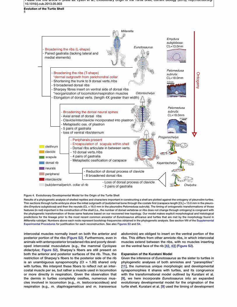
Expansion of the Kuratani ModelGiven the inference of Eunotosaurus as the sister to turtles inphylogenetic analyses of both amniotes and ‘‘parareptiles’’[11], the numerous unique morphologic and developmentalsynapomorphies it shares with turtles, and its congruencewith the transformational model outlined by Kuratani et al.[9], we here incorporate Eunotosaurus into an expandedevolutionary developmental model for the origination of theturtle shell. Kuratani et al. [9] used the timing of development
Figure 4. Evolutionary Developmental Model for the Origin of the Turtle Shell
Results of a phylogenetic analysis of shelled reptiles and characters important in constructing a shell are plotted against the ontogeny of pleurodire turtles.Thin sections through turtle embryos show the initial outgrowth of (sub)dermal bone through the costals first (carapace length [CL] = 13.0 mm in the pleuro-dire Emydura subglobosa) and then the neurals (CL = 18.0 mm in the pleurodire Pelomedusa subrufa). The timing of ontogenetic transformations of thosefeatures (in red) important in the construction of the shell (i.e., the number of dorsal vertebrae or ribs does not change through ontogeny) is congruent withthe phylogenetic transformation of those same features based on our recovered tree topology. Our model makes explicit morphological and histologicalpredictions for the lineage prior to the most recent common ancestor of Eunotosaurus africanus and turtles that are met by the morphology found inMilleretta rubidgei. Numbers above each node represent bootstrap frequencies obtained in the phylogenetic analysis. See section VIII of the SupplementalExperimental Procedures for justification for each reconstruction. See also Figures S3 and S4.
Evolution of the Turtle Shell5
Please cite this article in press as: Lyson et al., Evolutionary Origin of the Turtle Shell, Current Biology (2013), http://dx.doi.org/10.1016/j.cub.2013.05.003

of key shell features to predict ancestral morphologies andsecondarily place fossils within this developmental frame-work (Supplemental Experimental Procedures, section VII).We extend this model to include the developmental timingof a more comprehensive list of individual characters thatare essential to building a turtle shell (Figure 4). Outgrowthof membrane bone from the perichondral/periosteal collarof the developing rib occurs first, followed by outgrowth ofbone from the neural spines of the trunk vertebrae, and finallyby the acquisition of peripheral bones and the encapsulationof the scapula within the shell [7, 8] (Figure 4). The phyloge-netic sequence exhibited by Eunotosaurus (broadened ribsT-shaped in cross-section, dermal outgrowth of bone fromthe perichondral/periosteal collar of the rib, and reorganiza-tion of locomotion and respiratory muscles), Odontochelys(broadened neurals), and Proganochelys (acquisition ofperipherals and encapsulation of scapula within shell) isconsistent with the evolutionary developmental model. Thisexpanded model pulls the initial transformations of the turtleshell back to at least the late Middle Permian. But, this isstill younger than all recent molecular-based divergenceestimates for Pan-Testudines [12, 13] and thus is viable irre-spective of whether turtles originated inside or outside ofcrown Diapsida. To be clear, our phylogenetic analysisdoes recover the Eunotosaurus + turtle clade outsideDiapsida, which places it in conflict with molecular basedtopologies [12–15].
Our model for the origin of the turtle shell makes a number ofmorphological and histological predictions for stem turtlesthat existed or diverged prior to the most recent commonancestor of Eunotosaurus and extant turtles. For example,based on the early development of outgrowth of membranebone from the ribs of extant turtles (e.g., [38]), the model pre-dicts that earlier stem turtles had slightly broadened ribswith some intramembranous outgrowth of bone from the peri-chondral/periosteal collar of the rib. Based on the inferencethat the unique abdominal muscle ventilation system of turtles[44], in which the muscles attach to the ventral portion of thecarapace [45], arose from a basal amniote with costal ventila-tion [46], the model also predicts that early stem turtles likelyhad both intercostal muscles (unlike Eunotosaurus) and mus-cles beginning to insert on the ventral side of the trunk/dorsalribs. Histological data for Milleretta rubidgei has yet to be ob-tained, but this moderately broad-ribbed species, inferred inphylogenetic analyses to have diverged from the turtle stemearlier than Eunotosaurus [11, 30, 47], meets many of thesemorphological predictions (Figure 4). The dorsal ribs ofMiller-etta display an intermediate condition between the rod-likeribs found in stem amniotes (i.e., Limnoscelis paludis) andthe broad ribs found in Eunotosaurus and Odontochelys. Welook forward to testing the predictions of the expanded modelthrough further study of this taxon.
Supplemental Information
Supplemental Information includes Supplemental Experimental Proceduresand four figures and can be foundwith this article online at http://dx.doi.org/10.1016/j.cub.2013.05.003.
Acknowledgments
We thank the following institutions and their curators/staff for accessto material under their care: Albany Museum (Grahamstown), NaturalHistory Museum (London), Institute of Paleontology and Paleoanthro-pology (Beijing), Council for Geosciences (Pretoria), National Museum
(Bloemfontein), Bernard Price Institute (Johannesburg), South AfricanMuseum (Cape Town), Staatliches Museum fur Naturkunde (Stuttgart),United States National Museum (Washington, D.C.), and Yale PeabodyMuseum of Natural History (New Haven). M. Fox (Yale Peabody Museum[YPM]) is thanked for her careful preparation of the fossil material. B. Roach(YPM) made the beautiful illustrations used herein. W. Joyce is thanked forhis help with Figure 2. The vertebrate paleontology group at the PIMUZ isthanked for various assistances and discussions. K. deQueiroz, M.S.Y.Lee, M. Laurin, and an anonymous reviewer had useful comments thatimproved the manuscript. External funding for this project was providedby an NSF Graduate Research Fellowship and a Smithsonian InstitutionPeter Buck Fellowship to T.R.L. and a Swiss National Science Foundationgrant (SNSF no. 31003A_127053) to T.M.S.
Received: January 3, 2013Revised: April 3, 2013Accepted: May 1, 2013Published: May 30, 2013
References
1. Agassiz, L. (1857). Contributions to the Natural History of the UnitedStates of America (Boston: Little, Brown and Company).
2. Li, C., Wu, X.-C., Rieppel, O., Wang, L.-T., and Zhao, L.-J. (2008). Anancestral turtle from the Late Triassic of southwestern China. Nature456, 497–501.
3. Burke, A.C. (1989). Development of the turtle carapace: implications forthe evolution of a novel bauplan. J. Morphol. 199, 363–378.
4. Burke, A.C. (1991). The development and evolution of the turtle bodyplan. Inferring intrinsic aspects of the evolutionary process from exper-imental embryology. Am. Zool. 31, 616–627.
5. Gilbert, S.F., Loredo, G.A., Brukman, A., and Burke, A.C. (2001).Morphogenesis of the turtle shell: the development of a novel structurein tetrapod evolution. Evol. Dev. 3, 47–58.
6. Cebra-Thomas, J., Tan, F., Sistla, S., Estes, E., Bender, G., Kim, C.,Riccio, P., and Gilbert, S.F. (2005). How the turtle forms its shell: a para-crine hypothesis of carapace formation. J. Exp. Zoolog. B Mol. Dev.Evol. 304, 558–569.
7. Gilbert, S.F., Cebra-Thomas, J.A., and Burke, A.C. (2008). How the turtlegets its shell. In Biology of Turtles, J. Wyneken, M.H. Godfrey, and V.Bels, eds. (Boca Raton: CRC Press), pp. 1–16.
8. Sanchez-Villagra, M.R., Muller, H., Sheil, C.A., Scheyer, T.M.,Nagashima, H., and Kuratani, S. (2009). Skeletal development in theChinese soft-shelled turtle Pelodiscus sinensis (Testudines:Trionychidae). J. Morphol. 270, 1381–1399.
9. Kuratani, S., Kuraku, S., and Nagashima, H. (2011). Evolutionary devel-opmental perspective for the origin of turtles: the folding theory for theshell based on the developmental nature of the carapacial ridge. Evol.Dev. 13, 1–14.
10. Rubidge, B.S., Erwin, D.H., Ramezani, J., Bowring, S.A., and de Klerk,W.J. (2013). High-precision temporal calibration of Late Permian verte-brate biostratigraphy: U-Pb zircon constraints from the KarooSupergroup, South Africa. Geology 41, 363–366.
11. Lyson, T.R., Bever, G.S., Bhullar, B.A.S., Joyce, W.G., and Gauthier, J.A.(2010). Transitional fossils and the origin of turtles. Biol. Lett. 6, 830–833.
12. Hugall, A.F., Foster, R., and Lee, M.S.Y. (2007). Calibration choice, ratesmoothing, and the pattern of tetrapod diversification according to thelong nuclear gene RAG-1. Syst. Biol. 56, 543–563.
13. Shen, X.-X., Liang, D., Wen, J.Z., and Zhang, P. (2011). Multiple genomealignments facilitate development of NPCL markers: a case study oftetrapod phylogeny focusing on the position of turtles. Mol. Biol. Evol.28, 3237–3252.
14. Crawford, N.G., Faircloth, B.C., McCormack, J.E., Brumfield, R.T.,Winker, K., and Glenn, T.C. (2012). More than 1000 ultraconserved ele-ments provide evidence that turtles are the sister group of archosaurs.Biol. Lett. 8, 783–786.
15. Lyson, T.R., Sperling, E.A., Heimberg, A.M., Gauthier, J.A., King, B.L.,and Peterson, K.J. (2012). MicroRNAs support a turtle + lizard clade.Biol. Lett. 8, 104–107.
16. Lyson, T.R., and Gilbert, S.F. (2009). Turtles all the way down: logger-heads at the root of the chelonian tree. Evol. Dev. 11, 133–135.
17. Nagashima, H., Kuraku, S., Uchida, K., Ohya, Y.K., Narita, Y., andKuratani, S. (2007). On the carapacial ridge in turtle embryos: its
Current Biology Vol 23 No 126
Please cite this article in press as: Lyson et al., Evolutionary Origin of the Turtle Shell, Current Biology (2013), http://dx.doi.org/10.1016/j.cub.2013.05.003
developmental origin, function and the chelonian body plan.Development 134, 2219–2226.
18. Nagashima, H., Sugahara, F., Takechi, M., Ericsson, R., Kawashima-Ohya, Y., Narita, Y., and Kuratani, S. (2009). Evolution of the turtlebody plan by the folding and creation of new muscle connections.Science 325, 193–196.
19. Hay, O.P. (1922). On the phylogeny of the shell of the Testudinata andthe relationships of Dermochelys. J. Morphol. 36, 421–445.
20. Gregory, W.K. (1946). Pareiasaurs versus placodonts as near ancestorsto the turtles. Bulletin of the American Museum of Natural History 86,275–326.
21. Lee,M.S.Y. (1997). Reptile relationships turn turtle. Nature 389, 245–246.22. Joyce, W.G., Lucas, S.G., Scheyer, T.M., Heckert, A.B., and Hunt, A.P.
(2009). A thin-shelled reptile from the Late Triassic of North Americaand the origin of the turtle shell. Proc. Biol. Sci. 276, 507–513.
23. Cox, C.B. (1969). The problematic Permian reptile Eunotosaurus. Bull.Brit. Mus. Nat. Hist. 18, 165–196.
24. Gow, C.E., and de Klerk, B. (1997). First record of Eunotosaurus(Amniota: Parareptilia) from the Eastern Cape. Palaeontologia Africana34, 27–31.
25. Hill, R.V. (2005). Integration of morphological data sets for phylogeneticanalysis of Amniota: the importance of integumentary characters andincreased taxonomic sampling. Syst. Biol. 54, 530–547.
26. Gaffney, E.S. (1990). The comparative osteology of the Triassic TurtleProganochelys. Bulletin of the American Museum of Natural History194, 1–263.
27. Rieppel, O. (2001). Turtles as hopeful monsters. Bioessays 23, 987–991.28. Seeley, H. (1892). On a new reptile from Welte Vreden (Beaufort West)
Eunotosaurus africanus (Seeley). Quarterly Journal of the GeologicalSociety 48, 583–585.
29. Watson, D.M.S. (1914). Eunotosaurus africanus Seeley, and theancestry of the Chelonia. Proceedings of the Zoological Society ofLondon 1914, 1011–1020.
30. Gow, C.E. (1997a). A reassessment of Eunotosaurus africanus Seeley(Amniota: Parareptilia). Palaeontologia Africana 34, 33–42.
31. Muller, J., Scheyer, T.M., Head, J.J., Barrett, P.M., Werneburg, I.,Ericson, P.G.P., Pol, D., and Sanchez-Villagra, M.R. (2010). Homeoticeffects, somitogenesis and the evolution of vertebral numbers in recentand fossil amniotes. Proc. Natl. Acad. Sci. USA 107, 2118–2123.
32. Gauthier, J.A., Kearney, M., Maisano, J.A., Rieppel, O., and Behlke, A.(2012). Assembling the squamate Tree of Life: perspectives from thephenotype and the fossil record. Bulletin of the Peabody Museum ofNatural History 53, 3–308.
33. Scheyer, T.M., and Sander, P.M. (2007). Shell bone histology indicatesterrestrial palaeoecology of basal turtles. Proc. Biol. Sci. 274, 1885–1893.
34. DeBraga, M., and Rieppel, O. (1997). Reptile phylogeny and the affinitiesof turtles. Zool. J. Linn. Soc. 120, 281–354.
35. Muller, J., and Tsuji, L.A. (2007). Impedance-matching hearing inPaleozoic reptiles: evidence of advanced sensory perception at an earlystage of amniote evolution. PLoS ONE 2, e889.
36. Sterli, J., Rafael, S., de la Fuente, M.S., and Rougier, G.W. (2007).Anatomy and relationships of Palaeochersis talampayensis, a LateTriassic turtle fromArgentina. Palaeontographica Abteilung A 281, 1–61.
37. Dilkes, D.W. (1998). The Early Triassic rhynchosaurMesosuchus browniand the interrelationships of basal archosauromorph reptiles. Philos.Trans. R. Soc. Lond. B Biol. Sci. 353, 501–541.
38. Scheyer, T.M., Brullmann, B., and Sanchez-Villagra, M.R. (2008). Theontogeny of the shell in side-necked turtles, with emphasis on thehomologies of costal and neural bones. J. Morphol. 269, 1008–1021.
39. Lyson, T.R., and Joyce, W.G. (2012). Evolution of the turtle bauplan: thetopological relationship of the scapula relative to the ribcage. Biol. Lett.8, 1028–1031.
40. Starck, D. (1979). Vergleichende Anatomie der Wirbeltiere, Volume 2(Berlin: Springer).
41. Scheyer, T.M., Sander, P.M., Joyce, W.G., Bohme, W., and Witzel, U.(2007). A plywood structure in the shell of fossil and living soft-shelledturtles (Trionychidae) and its evolutionary implications. OrganismsDiversity & Evolution 7, 136–144.
42. Jenkins, F.A., Jr. (1970). Anatomy and function of expanded ribs incertain edentates and primates. J. Mammal. 51, 288–301.
43. Tsuihiji, T. (2007). Homologies of the longissimus, iliocostalis, andhypaxial muscles in the anterior presacral region of extant diapsida.J. Morphol. 268, 986–1020.
44. Landberg, T., Mailhot, J.D., and Brainerd, E.L. (2003). Lung ventilationduring treadmill locomotion in a terrestrial turtle, Terrapene carolina.J. Exp. Biol. 206, 3391–3404.
45. Shah, R.V. (1962). A comparative study of the respiratory muscles inChelonia. Brevoria 161, 1–16.
46. Brainerd, E.L., and Owerkowicz, T. (2006). Functional morphology andevolution of aspiration breathing in tetrapods. Respir. Physiol.Neurobiol. 154, 73–88.
47. Tsuji, L.A., and Muller, J. (2009). Assembling the history of theParareptilia: phylogeny, diversification, and a new definition of theclade. Fossil Record 12, 71–81.
Evolution of the Turtle Shell7
Please cite this article in press as: Lyson et al., Evolutionary Origin of the Turtle Shell, Current Biology (2013), http://dx.doi.org/10.1016/j.cub.2013.05.003
Related Documents