Evolutionary Genomics of the HAD Superfamily: Understanding the Structural Adaptations and Catalytic Diversity in a Superfamily of Phosphoesterases and Allied Enzymes A. Maxwell Burroughs 1,2 , Karen N. Allen 2,3 Debra Dunaway-Mariano 4 and L. Aravind 1 ⁎ 1 National Center for Biotechnology Information, National Library of Medicine, National Institutes of Health, Bethesda, MD 20894, USA 2 Bioinformatics Program, Boston University, Boston, MA 02215, USA 3 Department of Physiology and Biophysics, Boston University School of Medicine, 715 Albany Street, Boston, MA 02118-2394, USA 4 Department of Chemistry, University of New Mexico, Albuquerque, NM 87131, USA The HAD (haloacid dehalogenase) superfamily includes phosphoesterases, ATPases, phosphonatases, dehalogenases, and sugar phosphomutases acting on a remarkably diverse set of substrates. The availability of numerous crystal structures of representatives belonging to diverse branches of the HAD superfamily provides us with a unique opportunity to reconstruct their evolutionary history and uncover the principal determinants that led to their diversification of structure and function. To this end we present a comprehensive analysis of the HAD superfamily that identifies their unique structural features and provides a detailed classification of the entire superfamily. We show that at the highest level the HAD superfamily is unified with several other superfamilies, namely the DHH, receiver (CheY-like), von Willebrand A, TOPRIM, classical histone deacetylases and PIN/FLAP nuclease domains, all of which contain a specific form of the Rossmannoid fold. These Rossmannoid folds are distinguished from others by the presence of equivalently placed acidic catalytic residues, including one at the end of the first core β-strand of the central sheet. The HAD domain is distinguished from these related Rossmannoid folds by two key structural signatures, a “squiggle” (a single helical turn) and a “flap” (a beta hairpin motif) located immediately downstream of the first β-strand of their core Rossmanoid fold. The squiggle and the flap motifs are predicted to provide the necessary mobility to these enzymes for them to alternate between the “open” and “closed” conformations. In addition, most members of the HAD superfamily contains inserts, termed caps, occurring at either of two positions in the core Rossmannoid fold. We show that the cap modules have been independently inserted into these two stereotypic positions on multiple occasions in evolution and display extensive evolutionary diversification independent of the core catalytic domain. The first group of caps, the C1 caps, is directly inserted into the flap motif and regulates access of reactants to the active site. The second group, the C2 caps, forms a roof over the active site, and access to their internal cavities might be in part regulated by the movement of the flap. The diversification of the cap module was a major factor in the exploration of a vast substrate space in the course of the evolution of this superfamily. We show that the HAD superfamily contains 33 major families distributed across the three superkingdoms of life. Analysis of the phyletic patterns suggests that at least five distinct HAD proteins are traceable to the last universal common ancestor (LUCA) of all extant organisms. While these prototypes diverged prior to the emergence Abbreviations used: HAD, haloacid dehalogenase; PNKP, polynucleotide kinase phosphatase; KDO 8-P, deoxy-D- mannose-octulosonate 8-phosphate; CTD, carboxyl-terminal domain; PNKP, polynucleotide kinase phosphatase; LUCA, last universal common ancestor. E-mail address of the corresponding author: [email protected] doi:10.1016/j.jmb.2006.06.049 J. Mol. Biol. (2006) 361, 1003–1034 0022-2836/$ - see front matter. Published by Elsevier Ltd.

Welcome message from author
This document is posted to help you gain knowledge. Please leave a comment to let me know what you think about it! Share it to your friends and learn new things together.
Transcript

doi:10.1016/j.jmb.2006.06.049 J. Mol. Biol. (2006) 361, 1003–1034
Evolutionary Genomics of the HAD Superfamily:Understanding the Structural Adaptations and CatalyticDiversity in a Superfamily of Phosphoesterases andAllied Enzymes
A. Maxwell Burroughs1,2, Karen N. Allen2,3
Debra Dunaway-Mariano4 and L. Aravind1⁎
1National Center forBiotechnology Information,National Library of Medicine,National Institutes of Health,Bethesda, MD 20894, USA2Bioinformatics Program,Boston University, Boston,MA 02215, USA3Department of Physiology andBiophysics, Boston UniversitySchool of Medicine, 715Albany Street, Boston,MA 02118-2394, USA4Department of Chemistry,University of New Mexico,Albuquerque, NM 87131, USAAbbreviations used: HAD, haloacmannose-octulosonate 8-phosphate;last universal common ancestor.E-mail address of the correspondi
0022-2836/$ - see front matter. Publishe
The HAD (haloacid dehalogenase) superfamily includes phosphoesterases,ATPases, phosphonatases, dehalogenases, and sugar phosphomutasesacting on a remarkably diverse set of substrates. The availability ofnumerous crystal structures of representatives belonging to diversebranches of the HAD superfamily provides us with a unique opportunityto reconstruct their evolutionary history and uncover the principaldeterminants that led to their diversification of structure and function. Tothis end we present a comprehensive analysis of the HAD superfamily thatidentifies their unique structural features and provides a detailedclassification of the entire superfamily. We show that at the highest levelthe HAD superfamily is unified with several other superfamilies, namelythe DHH, receiver (CheY-like), von Willebrand A, TOPRIM, classicalhistone deacetylases and PIN/FLAP nuclease domains, all of which containa specific form of the Rossmannoid fold. These Rossmannoid folds aredistinguished from others by the presence of equivalently placed acidiccatalytic residues, including one at the end of the first core β-strand of thecentral sheet. The HAD domain is distinguished from these relatedRossmannoid folds by two key structural signatures, a “squiggle” (a singlehelical turn) and a “flap” (a beta hairpin motif) located immediatelydownstream of the first β-strand of their core Rossmanoid fold. Thesquiggle and the flap motifs are predicted to provide the necessary mobilityto these enzymes for them to alternate between the “open” and “closed”conformations. In addition, most members of the HAD superfamilycontains inserts, termed caps, occurring at either of two positions in thecore Rossmannoid fold. We show that the cap modules have beenindependently inserted into these two stereotypic positions on multipleoccasions in evolution and display extensive evolutionary diversificationindependent of the core catalytic domain. The first group of caps, the C1caps, is directly inserted into the flap motif and regulates access of reactantsto the active site. The second group, the C2 caps, forms a roof over the activesite, and access to their internal cavities might be in part regulated by themovement of the flap. The diversification of the cap module was a majorfactor in the exploration of a vast substrate space in the course of theevolution of this superfamily. We show that the HAD superfamily contains33 major families distributed across the three superkingdoms of life.Analysis of the phyletic patterns suggests that at least five distinct HADproteins are traceable to the last universal common ancestor (LUCA) of allextant organisms. While these prototypes diverged prior to the emergence
id dehalogenase; PNKP, polynucleotide kinase phosphatase; KDO 8-P, deoxy-D-CTD, carboxyl-terminal domain; PNKP, polynucleotide kinase phosphatase; LUCA,
ng author: [email protected]
d by Elsevier Ltd.

1004 Evolutionary Genomics of the HAD Superfamily
of the LUCA, the major diversification in terms of both substrate specificityand reaction types occurred after the radiation of the three superkingdomsof life, primarily in bacteria. Most major diversification events appear tocorrelate with the acquisition of new metabolic capabilities, especiallyrelated to the elaboration of carbohydrate metabolism in the bacteria. Thenewly identified relationships and functional predictions provided here arelikely to aid the future exploration of the numerous poorly understoodmembers of this large superfamily of enzymes.
Published by Elsevier Ltd.
Keywords: Rossmann fold; catalytic diversity; lateral gene transfer; substratespecificity; domain mobility
*Corresponding authorIntroduction
All cellular organisms depend extensively uponthe biochemical reactions related to organo-phos-phoesters and phosphoanhydrides. Hence, it is notsurprising that an enormous diversity of phospho-hydrolases have evolved on multiple occasions tocatalyze the dephosphorylation of various com-pounds.1,2 The majority of cellular phosphohydro-lases belong to a relatively small set of evolutionarilydistinct superfamilies, which are almost entirelydedicated to the catalysis of such reactions. Theselarge superfamilies include the P-loop NTPases,which is the largest monophyletic assemblage ofnucleotide triphosphatases encoded by cellulargenomes,3 the RNase H fold of ATPases, includingactin, Hsp70 and their relatives,4,5 the DHH,6 HD,7PHP,8 HAD,9,10 calcineurin-like,11 synaptojanin-like,12 and the Receiver domain (CheY) super-families.13,14 They span the entire range of structuralbasic classes with α-helical forms, such as the HDsuperfamily,15 the beta-barrels such as the CYTHsuperfamily of phosphohydrolases,16 three-layeredα/β sandwiches, such as P-loop NTPases,3 HAD17
and DHH,18,19 α/β barrels such as the PHP phos-phoesterases,20 and four- layered α/β-sandwichessuch as the calcineurin-like21 and synaptojanin-likephosphoesterases.22The HAD superfamily, named after the archetypal
enzyme haloacid dehalogenase,9 includes enzymescatalyzing carbon or phosphoryl group transferreactions on a diverse range of substrates, using anactive site aspartate in nucleophilic catalysis (Figure1(a)). The majority of the enzymes in this super-family are involved in phosphoryl transfer, i.e.phosphate monoester hydrolases (phosphatases) orphosphoanhydride hydrolase P-type ATPases.These include variations such as a phosphonoace-taldehyde hydrolase (phosphonatase) and phospho-transferases, such as β-phosphoglucomutase andα-mannophosphomutase. Each of the phospho-transferase enzymes requires a Mg2+ cofactor forcatalysis9,10 (Figure 1(b)). The carbon group transferreaction (Figure 1(a)) catalyzed by haloalkanoic aciddehalogenase (HAD)23 is unique in that it does notutilize a metal ion cofactor, and that a waternucleophile attacks the Asp C_O in the hydrolysispartial reaction.
The HAD superfamily is represented in theproteomes of organisms from all three superking-doms of life, and have colonized numerous verydisparate biological functions, which vary in theirdegree of essentiality to the cell. We are primarilyinterested in understanding how the catalytic plat-form of the HAD superfamily has been adaptedthrough evolution to act on a wide range ofsubstrates, a process which has been termed the“evolutionary exploration of substrate space”.24 Theaccumulation of over 40 X-ray crystal structures andthe enormous amount of sequence data availablethrough genome sequencing projects have made theHAD superfamily amenable to understanding thisprocess of evolution. Accordingly, here we present acomprehensive natural classification of the HADsuperfamily using the information derived fromrelevant sequence and structural elements, phyleticdistribution patterns, and phylogenetic tree analy-sis. This classification system offers a model forunderstanding the diversification of enzymes andallows us to predict important functional residues orregions in members of the superfamily havingunknown function.
Results and Discussion
Structural and functional aspects of the HADsuperfamily
Structural core of the HAD superfamily
To provide the basic context for a structure–function analysis of the HAD superfamily we firstdefine its essential structural core, and compare itto other structurally related folds. The core catalyticdomain of the HAD superfamily contains a three-layered α/β sandwich comprised of repeating β-αunits which adopt the topology typical of theRossmannoid class of α/β folds. The central sheetis parallel and is typically comprised of at least fivestrands in a 54123 strand order (Figures 2(a) and 3).These strands are hereinafter referred to as S1–S5.The HAD fold is distinguished from all otherrelated Rossmannoid folds by two key structuralmotifs (Figure 3). First, immediately downstreamof strand S1 is a unique, approximately six residue

Figure 1. HAD reaction mechanisms. A schematic representation of the reaction pathway in carbon transfer and in phosphoryl transfer is depicted. The left panel shows themajor types of reactions known to be catalyzed by the HAD superfamily can be distinguished by the identity of the leaving group of the substrate, the site of hydrolysis of theintermediate, and the identity of the phosphoryl acceptor group. Moieties originating from the substrate or solvent are colored blue and those originating from the enzyme arecolored red. The right panel shows a schematic of the active-site template for phosphoryl transferases showing interactions of the substrate with the catalytic motifs (contributed bythe core domain) and substrate specificity determinants (usually contributed by the cap domain). Residues contributed by each motif are coded: the substrate specificitycomponent is colored in blue, while residues from each of the motifs are given a separate color.
1005Evolutionary
Genom
icsof
theHAD
Superfam
ily

Figure 2. HAD catalytic domain. Cartoon representa-tions of the structure of the HAD fold with close-ups ofdifferent active site configurations. Beta strands arecolored blue while alpha-helices are colored red. (a) Topand side views are shown from deoxy-D-mannose-octulosonate 8-phosphate phosphatase (8KDO) fromHaemophilus influenzae (PDB: 1K1E). The top view (upperleft) reveals the typical spatial orientations of theconserved residues involved in catalysis, which aredepicted as stick and ball figures. Conserved residuesand two conserved structural motifs, the flap and thesquiggle, are labeled. The side view (upper right) showsthe Rossmann-like fold of the HAD superfamily, and thelocation of the cap domain relative to the core domain. Thesquiggle motif, central to the active site, is colored pink.Although HAD proteins typically employ a Mg2+ incatalysis, the tan sphere in this crystal structure representsa cobalt ion, a metal used for crystallization.103 (b) Close-up active site views of two HAD representatives withdistinct motif IV signatures. The left panel (MDP-1 fromthe MDP-1/FkbH family) has a motif IV DD signature(PDB: 1U7P) while the right panel (AphA from the AcidPhosphatase family) has a motif IV DxxxD signature(PDB: 1N9K).
1006 Evolutionary Genomics of the HAD Superfamily
structural motif which assumes a nearly completesingle helical turn, not unlike those found in thecatalytic domains of unrelated enzymes of thepolymerase–β-fold.25 We term this motif the“squiggle”. In some members of the HAD super-family the squiggle forms hydrogen bonds betweenthe ith and i+5th position resulting in the rare pi-helix conformation.26 Second, downstream of thesquiggle there is a β-hairpin turn formed by twostrands projecting from the core of the domain(Figure 2(a)). We term this structural motif the“flap”. The squiggle and flap structural motifs playessential roles in HAD superfamily catalysis (seebelow for details).Sequence comparisons have shown that practi-
cally all members of the HAD superfamily containfour highly conserved sequence motifs.10 Sequencemotif I corresponds to strand S1 and the DxDsignature is present at the end of this strand(Figures 1(b) and 4). The carboxylate group of thefirst Asp and the backbone C_O of the second Aspcoordinate the Mg2+ cofactor (Figure 1(b)). Addi-tionally, the first Asp in motif I acts as a nucleophile
that forms an aspartyl-intermediate during ca-talysis.27–31 In phosphatase and phosphomutasemembers of the superfamily the second acidicresidue acts as a general acid-base. It binds and,in many cases, protonates the substrate leavinggroup in the first step and deprotonates thenucleophile of the second step.32 In the ATPases,the occurrence of a threonine at this position allowsfor a reduced rate of aspartyl phosphate hydrolysis,which may allow for the time lag necessary for theconsequent conformational change. In the phos-phonatases, there is an alanine instead of the secondaspartate, which is consistent with the unique roleplayed by the enamine intermediate (formed withthe insert domain, see below) as a general acid-basecatalyst in aspartyl phosphate hydrolysis by theseproteins.Motif II corresponds to the S2 strand, which is
characterized by a highly conserved threonine or aserine at its end (Figures 1(b) and 4). Motif III iscentered on a conserved lysine that occurs aroundthe N terminus of the helix located upstream of S4(Figures 2, 3 and 4). Motif II and motif IIIcontribute to the stability of the reaction inter-mediates of the hydrolysis reaction. The lysine inmotif III is reminiscent of the basic residues termedarginine fingers that stabilize the negative chargeon reaction intermediates in many other phospho-hydrolases, particularly those of the P-loop NTPasefold.33 It is likely that they play a similar role evenin the HAD hydrolases. An analysis of the availablestructures shows that the lysine in motif III mayoccur in either of two structural contexts indifferent HAD hydrolases. In the P-type ATPases,acid phosphatases, phosphoserine phosphatasesand the Cof hydrolases the lysine is incorporatedinto the helix immediately preceding strand S4.However, in all other HAD hydrolases it emergesfrom the loop immediately prior to the helix. Onaccount of this difference in the secondary structurecontext of the lysine, motif III is poorly conservedrelative to the other motifs. The poor localconservation beyond the functionally critical basicresidue is also comparable to the regions bearingthe arginine finger in the AAA+ ATPases.3 Motif IVmaps to strand S4 and the conserved acidicresidues located at its end. These terminal acidicresidues of motif IV typically exhibit one of threebasic signatures: DD, GDxxxD, or GDxxxxD (wherex is any amino acid) ((Figures 1(b), 2 and 4)). Theseacidic residues along with those in motif I arerequired for coordinating the Mg ion in the activesite.27,32,34–39 Motifs I–IV are spatially arrangedaround a single “binding cleft” at the C-terminalend of the strands of the central sheet that formsthe active site of the HAD superfamily (Figure 1(b)). This binding cleft is partly covered by the β-hairpin flap occurring after S1 (Figures 2(a) and 3).Additional inserts occurring between the twostrands of the flap or in the region immediatelyafter S3 provide extensive shielding for the catalyticcavity. These inserts, termed caps, often contributeresidues required for specificity or auxiliary

Figure 3. Rossmannoid domains. Topology diagrams of domains representative of the major divisions of Rossmann-like folds with catalytic acidic residues. Two versions of the HADdomain (P-type ATPase HAD and BcbFHAD) that showsignificant modifications to the classic HAD domain are also shown. Strands are shown as arrows with the arrowhead onthe C-terminal end and are labeled from S1 to S6 in the classic HAD, with equivalent strands marked with eq in otherRossmannoid domains. The HAD domain of BcbF is an obligate dimer, and strands from the two dimers are differentiatedas A and B (e.g. S1A and S1B). The initial strand containing the catalytic D residue is rendered in yellow; other core strandsconserved across all members of the domain are in blue; non-conserved elements that may have been absent from theancestral state of a domain are in gray. The HADC1 cap insertion point is represented as a bright green line and the C2 capinsertion point is represented as an orange line. Broken lines indicate secondary structures elements not present in allmembers sharing the domain. The pink loop in the HAD domain represents the conserved squiggle. Residues conservedacross all members of a particular domain, including the initial catalytic D residue, are shown.
1007Evolutionary Genomics of the HAD Superfamily
catalytic functions, and play a central role in the re-actions catalyzed by most HAD hydrolases28,40,41(see below for further discussion).
Relationship of the HAD superfamily to otherRossmannoid folds
The topology of the central β-sheet of the HADfold makes it a typical representative of theRossmannoid class of three-layered α/β sandwichfolds (Figure 3). It shares with other Rossmanoidfolds the general location of the active site formed byresidues at the C-terminal end of the central sheet.More specifically, the HAD fold shares with otherRossmannoid fold enzymes a critical substrate-binding site in the loop between S1 and the down-stream α-helix, and a second active site residuepositioned immediately downstream of the strandoccurring after the crossover in the β-sheet, i.e.
strand S4 (Figure 3).42 Amongst the Rossmannoidfolds two major divisions can be recognized: (1) thenucleotide binding domains with a nucleotidebinding loop between strand 1 and the helix afterit. This group includes many large monophyleticassemblages of proteins, namely the classic Ross-mann NAD/FAD-dependent dehydrogenases,43Sir2-like deacetylases,44 the S-AdoMet-bindingmethyltransferases,45–47 the GTPase FtsZ,48 the ISO-COT fold49 and the HUP superclass (class I tRNAsynthetases, HIGH nucleotidyltransferases, USPA,photolyase and electron transport flavoprotein).42Most members of this division are characterized byspecific signatures, often glycine-rich, in theirnucleotide-binding loops. (2) The second divisioncomprises phosphohydrolases or divalent cation-chelating domains with a conserved acidic residue inthe loop between the first strand and the helix thatcomes after it. This division includes the HAD

1008 Evolutionary Genomics of the HAD Superfamily
superfamily, whose DxD motif is found in this loop,and several other enzymes superfamilies withsimilar active site configurations. These superfami-lies are the DHH domain phosphoesterases (e.g. theDNAse involved in repair and recombination,RecJ),6 the receiver or CheY domain of the two-component signaling system,13,14 the TOPRIM
Figure 4 (legend
domain, which is the shared catalytic domain ofthe topoisomerases and DnaG-type primases,50 thePIN/5′-3′ nuclease domain,51 the classical histonedeacetylases/arginases52 and the von Willebrandtfactor A (vWA) domain53 (second division onlydepicted in Figure 3). Most members of this divisionare also unified by a second acidic residue, which is
on page 1010)

1009Evolutionary Genomics of the HAD Superfamily
borne at the end of the strand adjacent to the firststrand, which occurs after the crossover of the sheetto the opposite side (left of strand S1 in Figure 3).Like the HAD domains, the receiver domain formsan aspartyl phosphate intermediate,54 whichreceives a phosphate from a histidinyl-phosphate
Figure 4 (legen
on the histidine kinase.54–56 Because of the mechan-istic similarity, the receiver domain has previouslybeen claimed to be a member of the HAD fold.57,58However, a careful examination of the active siteorganization and sheet topology of the receiverdomains (Figure 3) shows that it does not share any
d on next page)

1010 Evolutionary Genomics of the HAD Superfamily
of the other specific features conserved throughoutthe HAD superfamily beyond the phosphorylatedaspartate and other generic features of the acidicactive-site-containing division of Rossmannoidfolds (Figure 3).Of the other Rossmannoid folds of this division,
the DHH phosphoesterases contain a DxD signa-ture, and the histone deacetylases/arginases a DxHsignature at the end of strand 1, which chelate ametal ion, just as in the HAD superfamily. How-ever, these enzymes also contain their own char-acteristic motifs further downstream (Figure 3) andthere is no evidence for any aspartylphosphateintermediate being formed.52,59,60 In the PIN/5′-3′nuclease domains, a catalytic Mg2+ is chelated bythe acidic residues including those occurring at theend of the S1 equivalent and the strand immedi-ately to its left51 activates a water for nucleophilicattack. In the TOPRIM domains of primases andtopoisomerases the acidic residue at the end of thefirst strand is always a glutamate (Figure 3) thatacts as a general acid or base in the hydrolysis of thephosphoester bond or polynucleotide transfer.50,61The DXD motif is instead borne at the end of thestrand left of S1 (Figure 3) and coordinates a Mg2+.In the vWA domain the first aspartate is part of theso called MIDAS metal-binding motif (DxSxS53,62),which is critical for metal chelation by thesedomains. Thus, different superfamilies of thisdivision of the Rossmannoid folds, despite similarlypositioned acidic catalytic residues and metalcoordination sites, have acquired very distinctcatalytic mechanisms. Large to moderate insertswithin the core Rossmannoid domain are also seenin the TOPRIM, PIN/5′-3′ nuclease domains,histone deactylase/arginase and DHH superfami-lies, suggesting that they might also form caps
Figure 4. Multiple sequence alignment of HAD-domain cstructural regions. Structural regions not conserved, includinresidues in these excised regions. The top line of the alignmemotifs considered essential for HAD domain catalytic activitySecondary structure motifs are colored and labeled in the secrepresenting α-helices, and pink representing the squiggle mstructural elements; E for β-strand regions, H for α-helical regiresidues are shaded in yellow (A,C,F,I,L,M,V,Wand Y) while cin yellow (I,L and V). Conserved small residues appear in grepositive residues appear in blue (K and R), and negative residuprotein name, species name abbreviation, the GenBank GIdemarcated by underscores. PDB codes are shaded in orangfollows: Agam, Anopheles gambiae; Ana, Nostoc sp.; At, ArabiBacillus cereus; Bhal, Bacillus halodurans; Blic, Bacillus lichenifoClostridium acetobutylicum; Cfum, Choristoneura fumiferana; CgCydia pomonella granulovirus; Cvio, Chromobacterium violaceumEnterococcus faecalis; Exsp, Exiguobacterium sp.; Gzea, GibberellaHs,Homo sapiens; Hsp,Halobacterium sp.; Llac, Lactococcus lactisMjan, Methanocaldococcus jannaschii; Msp., Mesorhizobium sp.;Oryctolagus cuniculus; Osa, Oryza sativa; Pae, Pseudomonas aerputida; Psp., Pseudomonas sp.; Psyr, Pseudomonas syringae; Ptor, Pnorvegicus; Rpal, Rhodopseudomonas palustris; Rsol, Ralstonia solaaureus; Sc, Saccharomyces cerevisiae; Sent, Salmonella enterica; SepSchizosaccharomyces pombe; Ssui, Streptococcus suis; Syn, SThermosynechococcus elongatus; Tery, Trichodesmium erythraeumVfab, Vicia faba; Xaut, Xanthobacter autotrophicus; Ylip,pseudotuberculosis; Zmay, Zea mays. Alignment was produced
controlling access to the active site area, analogousto the HAD superfamily.
Structural variations in the core Rossmannoiddomain of the HAD superfamily
The core Rossmannoid fold of the HAD super-family is generally not prone to many modificationsbeyond the insertion of the cap modules. However,the central sheet often shows lateral modificationscorresponding to the two ends of the sheet. Theancestral condition of the HAD appears to havebeen the five-stranded central sheet (Figure 3), towhich a major division of the HAD superfamilyappears to have added a C-terminal β-α unit afterthe fifthf strand-helix unit (S6), extending thesandwich further (at the left side of the sheet inFigure 3). The additional strand S6 was lost on rareoccasions in members of this six-stranded division,especially in the context of C-terminal domainfusions. Likewise, on the opposite side (right endof the sheet in Figure 3) there are inserts ofadditional strands, which stack in the same planeas the core strands to extend the sheet. The simplestof these is a β-hairpin, which folds back and extendsthe central sheet, and is the defining feature of alarge clade within the HAD superfamily thatincludes the sucrose phosphate phosphatases, thephosphomannomutases, the trehalose phosphatephosphatases, mannosyl-3-phosphoglycerate phos-phatases and the cof-type phosphatases (Figure 3).A second independent insert in the “right side” ofthe sheet is seen in the P-type ATPases in the form ofan additional α-β unit immediately after S3 (Figure3, bottom left). This additional strand is accommo-dated in the sheet between the S2 and S3 and is aunique and defining feature of the P-type ATPases.
ontaining proteins. The alignment shows only conservedg cap regions, are replaced with numbers denoting thent indicates the approximate areas of the four conserved. Conserved residues of these motifs are shaded in gray.ond line of the alignment, blue representing β-sheets, redotif. The third line of the alignment designates secondaryons, and – for coil regions. Widely conserved hydrophobiconserved aliphatic residues appear in gray and are shadeden (G,A and S), hydroxy residues appear in teal (S and T),es appear in red (E and D). Sequences are identified by thenumber, and if applicable the PDB code; identifiers aree for added emphasis. Species names are abbreviated asdopsis thaliana; BPRB69, enterobacteria phage RB69; Bcer,rmis; Bmel, Brucella melitensis; Bs, Bacillus subtilis; Cace,la, Candida glabrata; Cneo, Cryptococcus neoformans; CpGV,; Dm, Drosophila melanogaster; Ec, Escherichia coli; Efae,zeae; Hinf, Haemophilus influenzae; Hp, Helicobacter pylori;
; Mbur,Methanococcoides burtonii; Mgri,Magnaporthe grisea;Ncr, Neurospora crassa; Npun, Nostoc punctiforme; Ocun,uginosa; Pmar, Prochlorococcus marinus; Pput, Pseudomonasicrophilus torridus; Rgel, Rubrivivax gelatinosus; Rnor,Rattusnacearum; Saga, Streptococcus agalactiae; Sau, Staphylococcusi, Staphylococcus epidermidis; Smut, Streptococcus mutans; Sp,ynechococcus sp.; Taci, Thermoplasma acidophilum; Telo,; Tmar, Thermotoga maritima; Tnig, Tetraodon nigroviridis;Yarrowia lipolytica; Yp, Yersinia pestis; Ypse, Yersiniawith the aid of the Chroma program.196

1011Evolutionary Genomics of the HAD Superfamily
The most dramatic modification, however, is seenin the proteobacterial BcbF family of phosphatases,which exist as obligate dimers in the catalytic form(Figure 3, bottom right). In these proteins the heliximmediately downstream of the conserved lysine inmotif III is replaced by a loop, which displaces thestrand S4 away from the core sheet and places it ananti-parallel configuration, where it stacks with theremaining three strands (S1–S3) of the secondmonomer in a parallel configuration (Figure 3).Thus, the S4 appears to be swapped between the twomonomers and two identical active sites are formedby a combination of two monomers, one monomersupplying motifs I, II and III and the other monomersupplying motif IV associated with the swappedstrand (Figure 3). Given that this configuration has avery limited phyletic spread, this dramatic modifica-tion appears to have evolved rather recently througha relatively simple process. We suspect that theancestral version was a five-stranded version, whichprobably functioned as a tightly associated dimerwith the active sites in each ancestral monomerfacing in opposite directions (head–tail dimer). Insuch a head–tail dimer, accidental swapping ofstrand 4 between the monomeric subunits couldhave re-constituted a functional active enzyme,thereby allowing the emergence of the configurationseen in the BcbF family.
Cap modules of the HAD superfamily
The most notable inserts seen in the HAD super-family are the caps,which, despite their diversity, canbe classified in three generic categories: (1) C0 caps,the structurally simplest representatives of the HADsuperfamily, have only small inserts in either of thetwo points of cap insertion. (2) The C1 are defined asinserts occurring in themiddle of the β-hairpin of theflapmotif, and fold into a structural unit distinct fromthe core domain. (3) The C2 caps are defined as in-serts occurring in the linker immediately after strandS3 (Figure 3). Most representatives of the HADsuperfamily have either a C1 cap or a C2, though infew cases proteins may simultaneously possess C1and C2 caps.The simplest C0 state with no elaboration of β-
hairpin or additional inserts in the C2 position arerather infrequent in the HAD superfamily and areseen in proteins such as deoxy-D-mannose-octulo-sonate 8-phosphate (KDO 8-P) phosphatase (Figure5). Slightly longer inserts are seen in the polynucleo-tide phosphatases, which have a long loop separat-ing the two β-strands of the flap. In the case of theCTD phosphatases and MDP-1 like phosphatases,this basic condition is elaborated further, with theaddition of a strand between the two sheets formingthe β-hairpin; resulting in a cap in the form of three-stranded sheet. Some of these phosphatases havealso acquired a rudimentary C2 cap in the form of along loop that extends out of the core domain.The classical C1 caps belong to two distinct
structural classes, the α-helical C1 caps and the capwith the unique α+β fold seen in the P-type ATPases
(Figure 5). The most basic α-helical cap in the formof bi-helical α-hairpin is observed in the acidphosphatase and the cN-I nucleotidase families(Figure 5). The next level of complexity is the tetra-helical bundle, which is the form of the C1 cap seenin the majority of HAD domains with a cap in thisposition. It includes three general subclasses thatmay be distinguished based on structural propertiesand conserved interactions. The first subclass,represented by β-phosphoglucomutases and deox-yribonucleotidases, has conserved contacts betweenthe descending arm of the cap domain and thesecond helix of the Rossmannoid core. The secondsubclass seen in haloacid dehalogenases and theirclose relatives (see below) has conserved contactsinvolving the loop between the second and thirdhelices of the cap and the linker between strand S3and the core helix downstream of it. The thirdsubclass, typified by the phosphoserine phosphatasefamily, shows contacts in the region between thethird and fourth α-helices of the C1 cap and asmaller C2 cap that is unique to this family. Despitesharing the same topology, these three categories oftetra-helical C1 caps share little primary sequenceconservation, and show notable differences in thepacking of the helices. The largest helical caps areseen in the form of the globular multi-helical bundlefound in the uncharacterized Zr25 family, with acore formed by eight prominent helices (Figure 5).Secondary structure prediction for the cN-II nucleo-tidase and Eyes absent (EYA) families reveals thepresence of large caps, which are predicted to formmulti-helical bundles similar to the Zr25 (the cap ofcN-II has developed an additional beta meander).The P-type ATPase C1 caps are unrelated to the
helical caps and searches of the PDB database withthe DALI program63,64 do not recover any knownfold. However, an analysis of the P-type ATPasecaps showed that they contain an internal duplica-tion of a simple α+β unit, with a core sheet formedby a three-stranded β-meander (Figure 5). Thissuggests they possibly arose from a single ancestralunit, which in turn could have itself emerged from aprecursor resembling the C0 caps of the CTDphosphatases and MDP1 via the addition of asmall α-helical hairpin to the three-stranded sheet.Subsequent duplication of this unit appears to havegenerated the C1 cap seen in extant P-type ATPases(Figure 5). However, the C1 cap of the extant P-typeATPases manifests considerable variability both interms of sequence as well as in the form of someadditional insertion and deletions. Thus, in the mostparsimonious scenario, the classical C1 caps appearto have been independently invented at least twice.All the known α-helical caps can be conservativelypictured as an evolutionary series of α-helicalbundles of increasing complexity emerging throughserial duplication from a basic bihelical precursor,along with rapid sequence divergence and reorga-nization of the helical packing (Figure 5).There are two major unrelated types of classical
C2 caps, respectively, seen in the Cof-type phos-phatases and the NagD-like phosphatases and its

Figure 5. Topology diagrams of selected C0 and C1 cap HAD domains. Representatives are identified with the PDBcode followed by one or more HAD family/clade name(s). With the exception of the P-type ATPase representative,strands are shown as blue arrows with the arrowhead on the C-terminal side and helices are represented by red coils.Central to the diagram is the ancestral strand–strand C0 cap. Remnants of the basal C0 cap can be seen in the smallstrands leading into and out of all other C1 caps. Arrows refer to the likely evolutionary progression leading to thediversification of C1 caps. Broken arrows pointing to the 1O08 tetra-helix cap domain reflect two possible progressionscenarios. The 1SU4 P-type ATPase cap is colored to accentuate a possible duplication event. The first unit of the cap iscolored in yellow and the second is colored in green. Other pieces of the cap that likely developed around theduplication event, including a strand–strand motif at the C terminus of the cap and a single helix linking the two units,are rendered in gray.
1012 Evolutionary Genomics of the HAD Superfamily
relatives (Figure 6). Both these types of C2 caps aredistinctly α+β with a core β-sheet containing atleast three strands. However, in structural similar-ity searches with the DALI program63,64 andthrough manual examination of topologies, wewere unable to detect any convincing similarity toother folds in the protein universe, or betweenthemselves. In addition to these major classes of C2caps there is a yet another small, unique C2 capfound in the histidinol phosphatase family. In theCof-type phosphatases we observed a remarkablediversification of the C2 cap through accretion ofsecondary structure elements to a basic unit with athree-stranded anti-parallel β-sheet (Figure 6). Themost basic version, seen in the protein Ta0175(PDB: 1L6R) from Thermoplasma acidophilum,65contains a three-stranded antiparallel sheet. Aslightly more complex form is seen in the treha-
lose-6-phosphatase ortholog (1U02) from the sameorganism, where a strand is added to the sheet atthe N terminus. In some other forms (e.g. YedPfrom Escherichia coli, PDB: 1XVI) there is entire β-αunit, instead of single strand, added to the Nterminus of the ancestral unit (Figure 6). In theuncharacterized phosphatase Tm0651 from Thermo-toga maritima (PDB: 1NF2),26 this trend is furtherexaggerated via the addition of three α-β units tothe ancestral unit. In the related YwpJ (1NRW) fromBacillus subtilis, in contrast, we observe elaborationvia duplication of a helix in one of the α-β units.Thus, as in the case of the helical C1 caps, it appearsthat the C2 caps of the Cof-type phosphatasesevolved through a process of serial addition ofsimple secondary structure units, most probablythrough duplications limited to the N-terminalregion of the cap.

Figure 6. Topology diagrams of selected HAD C2 cap domains. Representatives are identified with PDB code andfamily/clade name. Strands are shown as arrows rendered in blue with the arrowhead on the C-terminal side. The mainsection of the diagram contains arrows that show the likely evolutionary progression of C2 caps in the Cof Hydrolaseclade. The depiction of 1L6R represents the most likely ancestral state. The boxes to the right illustrate three additionalindependent innovations of the C2 caps in the NagD clade, the HisB clade, and the phosphoserine phosphatase (PSP)family. The depiction of the HisB C2 cap is predicted, as no structure is currently available. Orange Cs represent the likelylocations of putative metal-chelating cysteine residues.
1013Evolutionary Genomics of the HAD Superfamily
The C2 cap of the NagD-like phosphatases is an α/β domain with a core four-stranded parallel β-sheet,with an additional N-terminal antiparallel strand.The parallel configuration of the sheet, combinedwith the lack of specific similarities to any otherknown domain, suggests that itmight have arisen viaa duplication of the core domain which also has aparallelβ-sheet. However, at the sequence level thereis no significant similaritywith the core domain. Thisgroup of C2 caps also contains a unique beta hairpininserted after the third strand (Figure 6). An exami-nation of the sequence of the C2 caps of the histidinolphosphatase family reveals a conserved CxHx(6-13)CxC signature (where x is any amino acid). Thissuggests that this C2 cap is stabilized through thechelation of a divalent metal ion, and is likely toassume a simple flap-like structure (Figure 6).Several lineages of the HAD superfamily simulta-
neously possess both C1 and C2 caps, both of whichmay be similarly sized, or one of them may be thedominant cap. In the case of the enzymes with C0caps such as the CTD phosphatase family and the
related ROP9/38K family there is sometimes anadditional C2 cap in the form of a small β-hairpin.Similarly, small β-hairpin C2 caps are also seen inthe phosphoserine phosphatase and the pyrimidine5- nucleotidase families, which also contain helicalC1 caps (Figure 6). In an archaeal sub-family of thephosphoserine phosphatases, typified by the proteinAF1437 (PDB: 1Y8A), a small C2 cap assuming theform of a tri-helical bundle is seen, suggesting thatthere have been multiple independent innovationsof such smaller C2 caps. In all these families the C1cap is clearly the dominant cap with the C2 cappacking against it and probably providing anadditional solvent exclusion module (see below).
Role of the cap modules in the catalytic mechanismof the HAD superfamily
Several studies have revealed that HAD en-zymes with C1 caps are likely to follow a similarcatalytic cycle comprised of the steps outlinedbelow.17,27,32,37,66–68 The enzyme in the “open”

1014 Evolutionary Genomics of the HAD Superfamily
configuration allows the substrate (typically a phos-phoester) to enter the active site. Once the substrateis bound the enzyme assumes the “closed” configu-ration and the Mg2+ in the active site interacts withthe negatively charged phosphate, preparing it fornucleophilic attack by the first conserved aspartateat the end of strand one (Figure 1(a)). As a result anacyl phosphate intermediate is formed with thecarboxyl group of this aspartate.27–31 Subsequently,the enzyme enters the open configuration again andallows the leaving group to escape (Figure 1(a)). Inthe open state bulk solvent enters the active site anda water is deprotonated by the second aspartate ofstrand one; hydrolyzing the acyl phosphate inter-mediate and returning the enzyme to the nativestate.32 A variation on this theme is seen in thehaloacid dehalogenases which release a halide ionalong with the formation of a regular esterlinkage.69 In the phosphonatases and sugar phos-phate mutases there are differences in the initial andthe terminal stages of the reaction, respec-tively27,32,38,70–72 (Figure 1(a)) but the core phos-phoryl transfer mechanism remains the same.Key aspects of the HAD catalytic mechanism that
emerged from these studies are: (1) the alternationbetween open and close states and (2) a preliminaryreaction favored by solvent exclusion and a sub-sequent step favored by extensive solvent contact.The principal features of the core domain respon-sible for this process are the squiggle and the flap.The squiggle, being close to a helical conformation,appears to be a structure that can be alternativelytightly or loosely wound (Figures 2(a) and 3). Thisdifferential winding in turn induces a movement inthe flap immediately juxtaposed to the active site(Figures 2(a) and 3) and alternatively results in theclosed and open states. Given the strict conservationof the squiggle and the flap across the HADsuperfamily found herein, they are likely to be partof a universal essential functional feature of thissuperfamily. The conformational changes in thesquiggle and flap are likely to comprise the minimalapparatus for solvent exclusion and access at theactive site of these enzymes. Given this ground state,natural selection appears to have favored theemergence of cap modules as they made the processof solvent exclusion and acyl phosphate formationmore efficient. In addition to aiding the basiccatalytic mechanism the emergence of diverse capsalso provided a means of substrate recognition bysupplying new surfaces for interaction with sub-strates, which was not afforded by the ancestralactive site alone.28,40,41The simplest structures add the cap to the flap
motif itself, so as to completely seal the active site inthe closed state (Figure 7). Thus, the flap region wasa hotspot for the insertion of the various C1 caps,which appears to suggest intense natural selectionfor efficient solvent exclusion.27,32,38,69,70,73 In thecase of the HAD enzymes with C2 caps there is noevidence from either biochemical or structuralstudies, thus far, for extensive movement of thecap itself to result in open and closed states. How-
ever, an examination of the internal cavities of theavailable structures of the HAD enzymes with C2caps shows that the C2 cap forms a cavernousstructure over the active site with the flap sealing offthe aperture to this cavity (Figure 7). This impliesthat although the C2 caps likely lack mobilitycomparable to the C1 caps, even in these cases thesquiggle–flap elements likely exhibit drastic move-ments similar to that observed in C1 caps. As a resultthere would be an open state in which the substrate,solvent and leaving group can be exchanged withthe active site cavity and a closed state where theflap occludes the cavity formed by the C2 capcompletely and excludes the solvent. In most caseswhere both C1 and C2 caps are present such as thephosphoserine phosphatase family, the C1 cap is theprincipal functional moiety that closes the activesite. The subsidiary C2 cap packs against the C1 capand completes the occlusion by sealing off potentialchannels to the active site that exist in these C1 caps.In most of the C0 Caps the rudimentary caps forms acrater-like structure associated with the active site(e.g. MDP-1 and the CTD phosphatase families)(Figure 7). In the case of the polynucleotide kinasephosphatases (PNKP) this crater-like structure isalso walled by a unique insert occurring immedi-ately after strand S4 with motif IV. These crater-likeaccesses to the active site of the C0 cap enzymes areunlikely to completely occlude the solvent, but theirsubstrates are large molecules (proteins and poly-nucleotides), which may block the rest of the activesite from solvent while being bound to it. AnotherC0 cap enzyme, the 8KDO phosphatase, adopts anunusual strategy for solvent exclusion by using theparticularly elongated strands of its flap to form atetramer interface. As a result, each monomer in thetetrameric unit forms a cap over the active site in theadjacent monomer, effectively performing the samefunction of solvent exclusion (Figure 7). A similarstrategy of occlusion via cooperation between twosubunits is also seen in the aberrant BcbF family,which shows strand swapping between adjacentsubunits of the obligate dimer.
Natural classification of the HAD superfamily
Identification and clustering of the HAD superfamilyenzymes
We identified all available structures of the HADsuperfamily by using the DALI program63 tosearch the PDB database with the coordinates ofpreviously well-known HAD domains. HADstructures were typically recovered with Z-scores>9.0 regardless of the type of cap present in thestructure initiating the search, suggesting strong,detectable relationships between all members ofthe superfamily (see Materials and Methods andSupplementary Data). We then defined the con-served sequence features (along with their struc-tural cognates) of all HAD superfamily enzymes bymeans of a structure-based sequence alignmentof all available structures (Figure 4). Individual

Figure 7. Interaction of cap modules with the active site in the HAD superfamily. Molecular surface diagramsillustrating possible different roles played by the cap domain in substrate recognition and solvent exclusion in differentHAD families. The top left shows proximity of primitive C0 and C2 caps to the active site crater found in CTDphosphatases. The top right depicts the role of the C0 cap in tetramerization in 8KDO phosphatases. The middle diagramsshow the open and closed states associated with C1 caps, as well as the presence of the β-hairpin C2 cap in thephosphoserine phosphatase family, likely adding an extra layer of solvent exclusion. The bottom diagrams depict theputative role of the C0 cap as a gate to the active site in immobile C2 cap-dominantHAD lineages.With the exception of thetop right depiction, core domains are colored green; C0/C1 cap inserts are colored yellowwhile C2 cap regions are coloredblue. In the top right 8KDO phosphatase tetramer rendering, the cap domains are colored yellow while the core domainfrom each monomer is distinctly colored. Crystal structures are denoted by PDB identifiers followed by family names.
1015Evolutionary Genomics of the HAD Superfamily
sequences from this alignment were used to initiateiterative PSI-BLAST searches74 to identify allpossible members of the HAD superfamily in theNR database (Materials and Methods and Supple-mentary Data). Searches were carried out untilexhaustion, recovering sequence representativesfrom known families of HAD domain-containingproteins. For example, a search initiated with thesequence of the crystal structure of 8KDO phos-phatase from H. influenzae(gi: 20150626, PDB:
1K1E) returns other members of the 8KDOphosphatase family in the first PSI-BLAST itera-tion. In subsequent iterations, sequences from theCof hydrolase assemblage (gi: 28373517, iteration 2,E-value: 4e-07), P-type ATPase family (gi: 82407772,iteration 3, E-value: 4e-11), and phosphoserinephosphatase family (gi: 18160539, iteration 6, E-value: 0.002) were recovered. A search initiatedwith the sequence of a crystal structure from theNagD family (gi: 47169464, PDB: 1VJR) recovered

Figure 8. Reconstructed evolutionary scenario for theHAD superfamily. Inferred evolutionary history of HADdomainfamilies. The chart shows relative temporal eras that are demarcated by vertical black lines representingmajor evolutionarytransitions. Individual HAD lineages are listed in a column on the right side of the chart. Horizontal colored lines illustratethe maximum depth to which a HAD domain family can be traced relative to the temporal periods corresponding to themajor transitions. Broken horizontal lines indicate that the lineage cannot be traced to a definite starting point. Green Xs andOs lying over horizontal lines indicate position of the lysine finger in a particular HAD lineage or set of HAD lineages. An Xindicates the lysine finger is found in the coil region immediately preceding the α-helix N-terminal to S4. AnO indicates thelysine finger is incorporated within the aforementioned helix. Red letters to the right of the HAD domain family namesrepresent generalized substrate type(s) known to be processed by a family: N, nucleotide; P, protein; S, sugar. Color key:pink, universal; dark blue, bacteria and eukaryota; brown, virus; green, archaea; light blue, bacteria; orange, eukaryota.
1016 Evolutionary Genomics of the HAD Superfamily

Table 1. Natural classification of HAD superfamily
I. HADS WITH C0 CAPSA. Basal 5-stranded core assemblageThree-stranded C0 cap, 5-stranded core, rudimentary C2 cap in the form of simple extended hairpin if present, DD motif IV
MDP-1/FkbH family DxDxTxW motif I and DD motif IVFkbH/BryA subfamily (several Bacteria)MDP-1 tyrosine phosphatase subfamily (plants, animals, fungi, kinetoplastids)FFDDE motif IV and H near motif III
PDB: 1U7OSSO0580 subfamily (Several archaea) TWN motif II and DDR motif IV
RNA polymerase _carboxyl _terminal _domain (CTD) phosphatase familyPsr1p subfamily (animals, fungi, slime molds, plants, kinetoplastids, Giardia, apicomplexa, ciliates)
PDB: 1TA0Nem1p-dullard subfamily (animals, fungi)Tim50p subfamily (animals, fungi, plants)Fcp1p-CPL subfamily (animals, fungi, plants, slime molds, Cryptosporidium)355R subfamily (iridoviruses)Ublcp1 subfamily (animals, fungi, plants, slime molds)HSPC129 subfamily (animals, plants, slime molds)
38K/ROP9 phosphatase family (DDxxxN motif IV)38K subfamily (Baculoviruses). Conserved R in core helix 1, DW in core helix 5;ROP9 subfamily (Apicomplexa) HSGG motif in C0 cap
BcbF family (proteobacteria, bacteriophages). Unusual dimer-formed via strand swapping in core domainPDB: 1XPJ
Polynucleotide kinase phosphatase (PNKP) family D in core helix 1, Dx3K in core helix 3, SGR motif IIBacteriophage (PseT) subfamily (bacteriophages)
PDB: 1LTQEukaryotic (PNKP) subfamily (eukaryotes) variable inserts between D residues of motif IV
B. 8KDO (3-Deoxy-D-manno-octulosonate-8-phosphate) phosphatase family (bacteria, Methanobacterium, vertebrates) (GDxxxD motif IV,GGxGAxRE motif at phosphatase C-term), tetramerizes through flap strandsPDB: 1K1E
C. Yhr100c family (Firmicutes, Cyanobacteria,Deinococcus-Thermus, Thermotoga, plants, fungi, slimemolds) conservedWin core helix 2, R core helix 3
II. C1 CAP-CONTAINING HAD PROTEINSSimple Bi-helical Cap FamiliesA. Acid Phosphatase familyBi-helical cap
non-specific acid phosphatases (NSAPs)AphA subfamily (Streptomyces, enterobacteria)PDB: 1N9K
P4 subfamily (several bacteria) NPxYGxWE motif at phosphatase C-term;VSP subfamily (plants, Streptomyces, Coxiella, Legionella) GYR preceding motif IV
B. cN-I nucleotidase family bi-helical cap (vertebrates, proteobacteria, Thermosynechococcus, Arthrobacter)
Tetra-helical C1 cap assemblageC. Motif IV DD assemblage
Phosphonotase family DxG motif IClassic phosphonotase subfamily (proteobacteria) DFG motif in squiggle, small helical segment downstream of S5
PDB: 1RQN, 1FEZPA2803 subfamily (Pseudomonas) degenerate subfamily with loss of cap
Sdt1p-Epoxide Hydrolase C-terminal domain familysEHCT/Acad10 subfamily (animals, several α-proteobacteria)
PDB: 1S8O, 1EK1PHM8-SDT1 subfamily (fungi, plants, microsporidians, proteobacteria)YrfG subfamily (proteobacteria)YihX subfamily (most bacteria, some fungi, plants)
Deoxyribonucleotidase family (vertebrates, fungi (Cryptococcus only), plants, Giardia, several bacteria, bacteriophages (caudoviruses),mimivirus) W at the end of strand 6
PDB: 1MH9HerA-associated family (cyanobacteria, plants) WGY motif at end of strand 5, TxK motif IIβ-phosphoglucomutase (BPGM) family KPxP motif IIIβ-PGM proper subfamily (mainly firmicutes, some actinobacteria, E.coli, Thermotoga) conserved H, GxxR in cap domain
PDB: 1O08CbbY subfamily (plants, cyanobacteria, Chlamydia, Legionella, Yersinia, several α-proteobacteria) conserved H in cap domainDOG(2-deoxyglucose-6-phosphate phosphatase) subfamily (fungi, several bacteria,methanogenic euryarchaea) conservedHGin capdomainYniC subfamily (most bacteria, fungi, animals, plants, Giardia)
PDB: 1TE2
D. dehalogenase-Enolase-phosphatase assemblagedehr (dehalogenase-related) familydehr subfamily I (most bacteria, many archaea, fungi, animals, plants)Isr subfamily (plants) EWE motif I, SNxxxE motif IV
(continued on next page)
1017Evolutionary Genomics of the HAD Superfamily

II. C1 CAP-CONTAINING HAD PROTEINSD. dehalogenase-Enolase-phosphatase assemblage
dehr (dehalogenase-related) familydehr subfamily II (most bacteria, with sporadic transfers to various eukaryotes and archaea)PDB: 1ZRN, 1QQ5
Enolase-phosphatase family (animals, fungi, γ-proteobacteria, cyanobacteria, Aquifex, Streptomyces) conserved FVxxxLFPY andDxKxxxLKxLQGxxW regions in cap domain
Bcs3 family (some proteobacteria and Streptococcus)VNG2608C family (cyanobacteria and some euryarchaea)
E. PSP-P5N-1 assemblageP5N-1 (Pyrimidine 5-nucleotidase) family (animals) Additional small C2 cap presentPhosphoserine Phosphatase (PSP) familySerB subfamily (bacteria, some euryarchaea, fungi (recent bacterial transfers), animals, plants)
PDB: 1F5S, 1NNLThrH subfamily (few proteobacteria)
PDB: 1RKUphosphoserine:homoserine phosphotransferase
PHOSPHO1 subfamily (fungi, animals, plants, bacteria (mainly firmicutes), several Archaea; generates inorganic phosphate forskeletal matrix mineralization; small C2 cap contains 3 conserved cysteine residues that may be involved in metal chelation
CicA subfamily (proteobacteria, actinobacteria, Parachlamydia, Porphyromonas)NapD subfamily (several bacteria, some filamentous ascomycetes, Methanosarcina)AF1437 subfamily (few archaea) C2 cap has three helices stacking above C1 cap
PDB: 1Y8AMultihelical C1 cap assemblage
G. cN-II nucleotidase family β-hairpin insertion in core domain after motif IIIcN-II subfamily 1 (animals, slime molds)cN-II subfamily 2 (animals, plants, slime molds)cN-II subfamily 3 (animals, plants, slime molds, Legionella, Bdellovibrio) cystolic 5′-nucleotidases
H. EYA (Eyes Absent) family (animals)
I. Zr25 family (Staphylococcus) Insertion in core domain after motif IIIPDB: 1QYI
P-type ATPase familyStrand 3.1 present between strands S2 and S3, DKTGT motif I, GDGXND motif IV, unique α+β cap with conserved K
Type I subfamily (bacteria, archaea, eukaryotes) heavy metal, K+ transporting pumpsType II subfamily (bacteria, eukaryotes) Ca2+, Na+/ K+, H+/K+ transportersPDB: 1SU4
Type III subfamily (eukaryotes, bacteria, archaea) eukaryotic, archaeal proton pumps; bacterial Mg2+ transportersType IV subfamily (eukaryotes) aminophospholipid transportersType V subfamily (eukaryotes) Ca2+ transportersType VI subfamily (Euryarchaea) soluble phosphatases
III. C2 CAP-CONTAINING HAD PROTEINSC.HisB (Histidinol phosphatase) family (bacteria, only Thermoplasmales amongst archaea) metal-chelating C2 cap with conserved CxHxnCxC region;
histidine biosynthesis/ADP-D-β-D-heptose synthesis/ADP-D-α-D-heptose synthesis
A. NagD familyGDxxxxD motif IV, distinct α/β C2 cap
AraL subfamily (archaea, firmicutes, actinobacteria) conserved D in cap domainPDB: 1VJR, 1YV9, 1WVI, 1YDF, 1YS9
chronophin (CIN) subfamily (fungi, animals, slime molds, plants, kinteoplastids) conserved D in cap domain and glycine patchdownstream of motif II; putative cofilin-activating phosphatase
Cut-1/CECR5 subfamily (proteobacteria, eukaryotes) conserved D in cap domainPhosphohistidine/phospholysine phosphatase subfamily (animals, several diverse bacteria)
B. Cof hydrolase assemblage and constituent familiesβ-Sandwich domain cap structure showing considerable diversity, core strands 3.1, 3.2 present
Cof family (archaea, bacteria)PDB: 1L6R, 1NRW, 1NF2, 1YMQ, 1RLO, 1RKQ, 1WR8
Trehalose phosphate phosphatase (TPP) familyTPP 1 (animals, plants, proteobacteria, few archaea)TPS2 (plants, fungi, slime molds, microsporidians, very few bacteria and archaea)
Mannosyl-3-phosphoglycerate phosphatase (MPGP) family (some bacteria and Euryarchaea)PDB: 1XVI
Phosphomannomutase (PMM) family (eukaryotes, Propionibacterium, Bifidobacterium, Lactococcus, Sphingomonas)Sucrose phosphate synthase C-terminal domain (SPSC) family (plants, cyanobacteria, few proteobacteria)Sucrose phosphate phosphatase (SPP) family (plants, cyanobacteria, firmicutes)PDB: 1U2T
Table 1 (continued)
1018 Evolutionary Genomics of the HAD Superfamily

1019Evolutionary Genomics of the HAD Superfamily
sequences from the dehr family (gi: 691747,iteration 2, E-value: 9e-08), β-phosphoglucomutasefamily (gi: 1495997, iteration 2, E-value: 4e-04),phosphonatase family (gi: 48425373, iteration 2, E-value: 0.006), HisB family (gi: 29541277, iteration 3,E-value: 0.001), Zr25 family (gi: 39654743, iteration4, E-value: 0.002), and Cof hydrolase assemblage(gi: 28373517, iteration 6, E-value: 0.007). Anothersearch with a member of the deoxyribonucleoti-dase family recovers sequences from the P-typeATPase family (gi: 45359204, iteration 4, E-value:3e-04), Enolase-phosphatase family (gi: 2984225,iteration 6, E-value: 0.004), acid phosphatase family(gi: 58176631, iteration 9, E-value: 0.001), andNagD family (gi: 10197682, iteration 10, E-value9e-04). Preliminary classification was carried out bymeans of similarity-based clustering using theBLASTCLUST program (Supplementary Data).Distinct clusters which fell out of this operationwere aligned throughout their length and uniquesignatures beyond the four basic HAD motifs werenoted. These extended regions of conservationhelped in identifying specific families and objec-tively distinguishing them from other families withsignatures of their own. Within such families theinternal relationships, where relevant, were deter-mined using conventional phylogenetic analysismethods, namely neighbor-joining and maximumlikelihood, and the phyletic profiles of the mem-bers. All major conclusions based on phylogeneticresults discussed here were supported by bootstrapsupport 80% or greater in all the above-statedphylogenetic methods. Higher-order relationshipsbetween families were determined by comparingshared structural features, and determining syna-pomorphies (shared derived characters). Lastly,phyletic patterns, domain architectures, and pre-dicted operon organization of representatives wereused to infer likely function if it was not knownand also to reconstruct a coherent evolutionaryscenario for all branches of the HAD superfamily.The higher-order relationships within the HAD
superfamily are presented graphically in Figure 8and the resultant natural classification is shown inTable 1 along with phyletic patterns, representativesin the PDB, and functional annotation while Figure 9depicts domain architectures observed within eachfamily. The most basic split appears to separate agroup of C0 cap proteins with a core five-strandedsheet from the rest of the superfamily, which isunified by a six-stranded core sheet. Within this six-stranded assemblage the most basal members retain
Notes to Table 1:Clades/families are generally grouped according to dominant cap domof evolutionary relationships within each of these major groups of HADsubfamilies are given in parentheses. Distinct sequence/structuraldistributions of families. The defining characteristic(s) for each familyor next to phyletic distributions. PDB identifiers of solved crystal strucsubfamily. Any known enzymatic function not intuitively associatedsubfamily. A + refers to a positive conserved residue (lysine or argiglutamate, or histidine).
C0 caps,while the rest of the division is characterizedeither by dominant C1 or C2 caps. The distinct capmorphologies suggest five major radiations, namelythe α-helical C1 cap assemblage, the P-type ATPaseswith their own C1 cap, and three distinct groups ofdominant C2 cap proteins (Figure 8).We describe thedetails of the classification below, using the capmorphology as a handle.
The C0 cap assemblages and their constituentfamilies
The basal-most clade of the HAD superfamily iscomprised of an assemblage of C0 proteins with afive-stranded core sheet and currently includes fivedistinct families, which are briefly described below.Two additional families showing the C0 cap condi-tion, whose precise evolutionary affinities are notclear, are also discussed in this section (Figure 8;Table 1).
The MDP-1/FkbH family
This family is prototyped by the eukaryotic MDP-1 type Mg(II)-dependent protein tyrosine phos-phatases,34,75,76 which appears to be widely distrib-uted in eukaryotes suggesting a basic cellularfunction. We also recovered a number of bacterialMDP-1-like proteins typified by FkbH and BryA,and archaeal representatives typified by SSO0580from Sulfolobus. FkbH and BryA are in the biosyn-thetic pathways for ascomycin and bryostatin inStreptomyces77 and the bacterial symbiont Candida-tus Endobugula sertula,78 respectively. The FkbHprotein combines an N-terminal HAD domain witha C-terminal acetyltransferase domain (FkbH_Shyin Figure 9) containing a highly conserved cysteineresidue. Given its role in synthesis of methoxyma-lonyl-ACP and gene context, it is quite likely thatthe incoming substrate is an acyl phosphate, whichis cleaved by the HAD domain and the acyl groupmay then be transferred to the internal cysteine inthe acetyl transferase domain and then to the ACP.The presence of one distinct lineage of the MDP-1/FkbH family in each of the three superkingdoms oflife is indicative of their possible presence in the lastuniversal common ancestor (LUCA) of cellular life(Figure 8). Related to this ancient family are threeother families detailed below with restricted phy-letic patterns, and could have been potentiallyderived from the former family in a lineage-specificfashion (Figure 8; Table 1).
ain type, i.e. C0, C1, or C2. Indents indicate the inferred hierarchydomain containing proteins. Phyletic distribution of families and
features are listed underneath clade names or next to phyleticand/or other distinct features are shown underneath clade namestures are indented and listed underneath their respective family/with a family/subfamily name is also listed beneath said family/nine) and a – refers to a negative conserved residue (aspartate,

Figu
re9(legend
onopposite
page)
1020 Evolutionary Genomics of the HAD Superfamily

1021Evolutionary Genomics of the HAD Superfamily
The RNA polymerase carboxyl-terminal domain(CTD) phosphatase family
This family is unique to the eukaryotes and showsan extensive radiation in them (Table 1). The proto-typical version of this family, typified by yeastFcp1p, is required for the dephosphorylation ofspecific serine residues in the carboxyl-terminal tailof the RNA polymerase catalytic subunit,79–85 afeature essential for the reinitiation of transcriptionby the RNA polymerase.86 This family has diversi-fied into seven subfamilies in the eukaryotes andtheir viruses (Table 1). The most widespread of theseis the Psr1p subfamily which is conserved through-out the eukaryotes and is typified by a conservedN-terminal module required for membrane local-ization,87,88 and a conserved cysteine (Psr1p_Scin Figure 9). Members of this subfamily are slowevolving and are likely to be the principal CTDphosphatases of eukaryotes, and ancient compo-nents of the nuclear membrane. The Nem1p/dullardsubfamily is seen in animals and fungi, localizes tothe nuclear membrane, and might act on nuclearpore complex proteins such as Nup84p.89 The Tim50subfamily is also amembrane proteinwith a peculiarN-terminal membrane-spanning segment.90 Itassociates with the mitochondrial inner membraneand regulates the translocation of internal mitochon-drial proteins. Recently, the Tim50a isoform has alsobeen shown to localize to the nuclear membrane.Hence, it is likely that this entire group of membraneassociated CTD phosphatases diversified as nuclearmembrane proteins and Tim50 was subsequentlyrecruited for a mitochondrial function.91The remaining CTD phosphatase subfamilies are
soluble proteins and include the Fcp1p-CPL sub-family, typified by the eponymous protein from S.cerevisiae.80 Several versions of this subfamily arecharacterized by an N-terminal sandwich-barrelhybrid motif (SBHM) domain, followed by a down-stream metal-chelating cysteine cluster, and a C-ter-minal BRCT domain (fcp1_Hs in Figure 9). TheBRCT domain in these proteins has been implicated
Figure 9. Domain architectures of selected multidomaingrouped around a central circle indicating principal cap typegrouped according to the higher-order classification (Table 1), wblack lettering and families encircled by lighter colored, thiHowever, clades and families without any currently knownEach rectangle or other geometric shape represents a single colabeled with the dominant type of cap found in that protein: Cprotein name or abbreviation and an organism name absusceptibility protein carboxy-terminal domain; SBHM, sandwRNA binding motif domain; UBQ, ubiquitin domain; NT, nucdomain; SIS, a sugar isomerase domain; IGPD, imidazole glycribose) polymerase and DNA-ligase Zn-finger domain; FHA,binding; LIPKIN, antibiotic kinase type small moleculphosphoesterase domain; Gcvr, repressor of glycine cleavagedomain; HHE, possible metal binding domain; TRASH, metallHMA, heavy-metal-associated domain; NTF2, small moleculeOtsA, trehalose-6-phosphate synthase domain. The orange ellmembrane-targeting signal with a conserved cysteine. OrganisAdditional abbreviations: Shy, Streptomyces hygroscopicus;tuberculosis; Brja, Bradyrhizobium japonicum; T4, bacteriophmetallireducens; Bthe, Bacteroides thetaiotaomicron; Mmus, Mus m
in recognizing the phosphorylated RNA-polymeraseII substrate.92 It is possible the SBHM of the Fcp1psubfamily interacts with the SBHM domains in thecatalytic subunits of the RNA polymerase. The plantrepresentatives of this subfamily, the CPL proteins,are implicated in regulating osmotic stress-respon-sive and abscisic acid-responsive transcription93,94and contain one or two double-stranded RNA-binding domains (dsRBD) at the C terminus, sug-gesting that they might be downstream of the RNA-mediated silencing pathway seen in plants (CPL1_Atin Figure 9). Ublcp1 subfamily is also found through-out the crown group of eukaryotes and is typified byan N-terminal ubiquitin domain fused to thephosphatase domain (Ublcp1_Mmus in Figure 9)95and might regulate RNA polymerase stabilitythrough the ubiquitin pathway.96
The 38K/ ROP9 and BcbF families
The remaining two families, which might havearisen from the MDP-1/FkbH family, are muchsmaller and show even more restricted phyletic pat-terns. The 38K/ROP9 family shows a very unusualphyletic pattern, with one of the subfamilies beinglimited to the baculoviruses (the 38K subfamily) andthe other to the apicomplexa (ROP9). This family isdefined by a characteristic insert that is likely to forma rudimentary C2 cap. The ROP9 family has beenexperimentally determined to be a secreted proteinlocalizing to the rhoptry, an apicomplexan organ-elle,97 suggesting that it might act as a phosphatasein the assembly of the rhoptry or the parasitophorousvacuole. The BcbF family, despite its dramatic struc-tural modifications, is largely limited to the proteo-bacteria and their viruses, suggesting that it mighthave arisen relatively recently in evolution. Thepredicted neighborhoods for these genes suggeststhat it is often embedded in operons for capsularpolysaccharide biosynthesis,98 suggesting that itmight act as a phosphatase on one of the buildingblocks of the polysaccharide.
members of the HAD superfamily. The architectures are(as outlined in Table 1). Domain architectures are furtherith clades encircled by thick black lines and designated in
nner lines and designated in the same colored lettering.multi-domain architecture are not included in this figure.nserved domain. The HAD domain is in light blue and is0, C1, C2, or P-type ATPase. Proteins are identified with abreviation. Domain designations: BRCT, breast cancerich barrel hybrid motif domain; dsRBD, double-strandedleotidyltransferase domain; MurD ligase, glutamate ligaseerol-phosphate dehydratase domain; zf-PARP, poly(ADP-forkhead-associated domain involved in phosphopeptidee kinase; ACAD, acyl-CoA dehydrogenase; 2H, 2Henzyme system domain; Pfs, nucleoside phosphorylase
ochaperone-like domain; copz, copper chaperone domain;binding domain of the nuclear transport factor twofold;
ipse associated with the CTD phosphatase is a specializedm abbreviations are the same as in the alignment, Figure 4.Fnuc, Fusobacterium nucleatum; Mtub, Mycobacteriumage T4; Bthi, Bacillus thiaminolyticus; Gmet, Geobacterusculus.

1022 Evolutionary Genomics of the HAD Superfamily
Polynucleotide kinase phosphatase (PNKP) family
The next major lineage of basal C0 capHADs is thePNKP family which plays a role in both RNA andDNA repair99 by removing 3′-terminal phosphategroups.100 There are two subfamilies of these pro-teins (Table 1) with distinct motif IV sequence signa-tures (Figure 8), the first being the bacteriophagesubfamily (PseT in Figure 8) with an N-terminal P-loop polynucleotide kinase domain (PNKP_T4 inFigure 9). The second is the eukaryotic subfamily(PNKP in Figure 8), which is seen in most majoreukaryotic lineages and often contains a C-terminalpolynucleotide kinase domain (PNKP_Mgri in Fig-ure 9). In plants, the phosphatase is fused to a Zn-finger found in poly(ADP-ribose) polymerases andDNA-ligases (AtZPD_At in Figure 9).101,102 Anotherpreviously uncharacterized eukaryotic subfamily isfound in animals and is fused to the phos-phopeptide-binding forkhead-associated domain(FHA) (PNKP_Hs in Figure 9). Both the Zn-fingerand FHAdomain are likely to be independentmeansof recruiting these phosphatases to regions of DNAdamage.
8KDO family
While this family has a C0 configuration, its coresheet is six-stranded like the rest of the HADsuperfamily suggesting that it is closer to theremaining groups of the HAD fold (Figure 8).These enzyme remove a phosphate group from 3-deoxy-D-manno-octulosonate 8-phosphate (Kdo-8)in the course of the biosynthesis of the polysacchar-ide chain in the bacterial lipid A pathway103,104 andbacterial capsular polysaccharides.105 The 8KDOfamily shows a conserved K residue in the cap do-main that points in the direction of the activesite and might participate in recognition of nega-tively charged substrates. Several bacteria and allvertebrate members of this family are fused to anucleotidyltransferase that potentially catalyzes thesubsequent step in the biosynthesis pathways(Cmas_Hs in Figure 9).
Yhr100c family
This family specifically recovers the NagD familyof C2 cap proteins (see below), and vice versa insequence searches; however, beyond general coresequence similarity there are no particular featuresthat link these families. Gene neighborhood analysissuggests linkages with genes in the chorismatemetabolism pathway, such as AroE (Shikimate 5-dehydrogenase) and chorismate synthase suggest-ing a possible regulatory role by acting on phos-phorylated intermediates in the pathway. Theresults from the yeast protein–protein interactionmap suggest that the eukaryotic members may bepart of the Gip1p–Glc7p phosphatase complexrequired for organization of septins; implying thatthese proteins possibly function as protein phos-phatases during cell division.
The helical C1 cap assemblage
The categories of α-helical caps are discussed interms of their basic cap morphologies, namely thebihelical, tetrahelical and multi-helical caps. Ofthese the tetra-helical cap families form the bulkof the assemblage and include several large families(Table 1).
Simple bi-helical cap families
The simplest of the α-helical caps are the bi-helicalcaps seen in the acid phosphatase and cN-I nucleo-tidase families. However, there are no other featuressupporting a specific relationship between thesefamilies suggesting that they are basal lineagesretaining the ancestral condition of the α-helicalclade (Figure 8). The acid phosphatase family ischaracterized by an N-terminal signal peptide,which suggests that they are secreted proteins thatfunction in periplasmic or extracellular environ-ments. Plants show a lineage-specific expansion ofmembers of this family, which are believed tofunction as vegetative storage proteins.106–108 ThecN-I family is a family of cytostolic 5′-nucleotidasesfound in vertebrates and several proteobacteriawhich regulate pyrimidine pools in the cytosol.109,110
Tetra-helical caps: the motif IV DD assemblage
This assemblage is distinguished by the presenceof a DD signature in motif IVand contains the phos-phonatase, SDT1-epoxide hydrolase C-terminal do-main, deoxyribonucleotidase, HerA-associated(HA) and β-phosphoglucomutase (BPGM) families.The phosphonatase family includes the phospho-
noacetaldehyde phosphatases, which hydrolyzephosphonoacetaldehyde to orthophosphate andacetylaldehyde.111–116 Experimental results suggesta role for cap residues in the catalytic activity of theclassic phosphonatases of this family.27 The familycontains a group of degenerate versions from thebacterium Pseudomonas (PA2803 subfamily), whichhave rather partly lost their cap and show disrup-tions of motifs II and III and IV suggesting that theyare catalytically inactive proteins which have takeup a secondary binding function. The Sdt1p-epoxidehydrolase C-terminal domain family is widelyrepresented in both bacteria and eukaryotes andappears to have diversified into four major sub-families (Table 1). Several members of the sEHCT/Acad10 subfamily are fused to a C-terminal α/βhydrolase domain related to the haloalkane dehalo-genase domain (HAL) (SeH_Hs in Figure 9). Theanimal enzyme has been shown to have hydroxyllipid phosphate phosphatase activity in lipid degra-dation.117–119 Some animal members of this sub-family, like Acad10, are fused to two C-terminaldomains (Acad10_Hs in Figure 9); a lipid kinasedomain related to the protein kinases and an Acyl-CoA dehydrogenase (ACAD) domain, which alsosuggests a role for them as phospholipid phospha-tases. Phm8p of the eponymous subfamily is

1023Evolutionary Genomics of the HAD Superfamily
induced under low phosphate conditions and islikely to release soluble phosphate by hydrolysis ofintracellular organo-phosphate compounds120 whileits paralog Sdt1p has been shown to be a pyrimidine5′-nucleotidase.121The deoxyribonucleotidase family includes one of
the major types of 5′ (3′)-deoxyribonucleotidasesresponsible for dephosphorylating uracil and thy-mine deoxyribonucleotides.122–124 The eukaryoticforms do not group together in phylogenetic analy-sis, suggesting that they might have been acquiredfrom bacterial or phage sources on multiple occa-sions. The presence in large DNA viruses and mito-chondria is consistent with other similaritiesbetween their DNA replication processes125,126 andis indicative of the similar selective pressures facedby these replicons from excess uracil and thyminedNTs. The HerA-associated family is typified by itsoperonic association with the HerA-type ATPasesand theNurA nucleasewhich are predicted to form asystem for chromosome segregation and pumping inprokaryotes.127 These contextual associations predictthat this family might have a role in processingterminal phosphates on DNA, which might emergedue to nuclease action during the pumping pro-cess.127 The β-phosphoglucomutase (BPGM) familyis a large group that contains multiple subfamilieswith different catalytic activities (Table 1). Thearchetypal subfamily of this group is the β-PGMsproper, which catalyze the inter-conversion of β-D-glucose 1-phosphate and D-glucose 6-phosphate.128This family contains a conserved histidine and GxxRmotif in the cap, which are critical for substraterecognition by contacting the phosphate and sugarmoieties, respectively.129 In the related CbbY sub-family (typified by Rhodobacter CbbY;130 Table 1), thehistidine is likewise universally conserved, but thearginine is present only in a subset of proteins. TheDOG subfamily is typified by the 2-deoxyglucose-6-phosphate phosphatase from fungi131,132 and otherfungal members of this subfamily have been char-acterized as glycerol 3-phosphatases.133 The re-maining members of this family constitute the largeYniC family,which iswidely represented throughoutthe bacteria and the eukaryotes, but not archaea. Inplants the HAD domain is fused to the FAD syn-thetase (AT29272p_At in Figure 9), which adenylatesFMN to form FAD.134,135 The HAD domain mightdephosphorylate a precursor in the pathway such asFMN and probably regulates FAD synthesis. Severalproteobacterial members are fused to a predictedmannitol dehydrogenase domain (YhcW_Blic inFigure 9), suggesting they might dephosphorylatesubstrates in sugar metabolism.
Tetra-helical caps:dehalogenase-enolase-phosphatase assemblage
This assemblage contains two major families; thedehalogenase related family (dehr) and the enolasephosphatase family, as well as two other relativelysmall families; all of which are unified by theirsequence similarities in motif IV (Table 1). The dehr
family, despite being widespread, remains largelyenigmatic with the only well characterized memberbeing the type II D-L-haloalkanoic acid dehalogenasesubfamily,41,136 which is also the archetype of theentire HAD superfamily. The dehr family shows twoclear subfamilies (dehr subfamily I and subfamily II).One distinct orthologous group in subfamily I foundonly in plants, Isr (inhibitor of striate) proteins, ischaracterized by an unusual EWE signature in motifI and a SNxxxE signature in motif IV. The dehrsubfamily II shows even greater diversity in motifIV (e.g SSNxxD, SSxxxD and AAxxxD) with widedifferences in the conservation of the acidic residuesand is consistent with the acquisition of non-phos-phate substrates such as haloalkanoic acids.137,138The enolase-phosphatase family of enzymes cata-lyzes the oxidative dephosphorylation (in combina-tion with the enolase) of 2,3-diketo-1-phospho-hexane to 2-keto-pentanoate in the latter steps ofthe methionine salvage pathway.139,140 Members ofthe restricted bacterial Bcs3 family are fused to an N-terminal glycosyltransferase domain (Bcs3_Hinf inFigure 9) and might function as sugar phosphatasesin the biosynthesis of capsular polysaccharides incertain pathogenic bacteria141 (Table 1).
Tetra-helical caps: PSP-P5N-1 assemblage
This assemblage of tetra-helical cap proteins (Table1) is unified by the presence of an additional insert,which forms a small secondary C2 cap that stacksagainst the tetra-helical cap. Within this assemblagethe P5N-1 family is restricted to animals andcatalyzes the dephosphorylation of the pyrimidine5′ monophosphates UMP and CMP to the corre-sponding nucleosides.142,143 The cap region containshighly conserved charged residues likely to be thesubstrate specificity determinants of this family. Itshighly restricted phyletic pattern suggests that theP5N-1 family was possibly derived from the muchlarger phosphoserine phosphatase family in theanimal lineage (Figure 8).The large phosphoserine phosphatase (PSP) family
includes a number of subfamilies, of which theclassical phosphoserine phosphatases (SerB) consti-tute the most widespread subfamily (Table 1). TheSerB proteins catalyze the dephosphorylation of L-3-phosphoserine or an exchange reaction between L-serine and L-phosphoserine in the biosynthetic path-way of serine.144,145 We found a fusion of severalprokaryotic SerBs (e.g. Mycobacterium and proteo-bacteria) with GcvR, the repressor of glycinecleavage (GCV) enzyme system (SerB_Mtub in Fig-ure 9). Given the connection between serine catabo-lism and glycinemetabolism,146–148 this fusionmightallow SerB to feedback regulate the glycine cleavagepathway. The related ThrH subfamily, which isrestricted to the proteobacteria, participates in thethreonine biosynthesis pathway by catalyzing aphosphoserine–homoserine phosphotransfer reac-tion, similar to the phosphate exchange reaction ofSerB.149,150 The PHOSPHO1 subfamily contains apeculiar C2 cap, which has three conserved cysteine

1024 Evolutionary Genomics of the HAD Superfamily
residues, suggesting that it is stabilized bymetal che-lation. The vertebrate versions of this subfamily arebelieved tomobilize inorganic phosphate for skeletalmatrix mineralization mineralization through theiraction on phosphocholine and phosphoethanol-amine.151,152 The fusion of this subfamily in someGram positive bacteria to a nucleoside phosphor-ylase involved in methionine metabolism153 mightimplicate it in this pathway.
The multi-helical cap assemblage
The multi-helical cap assemblage includes threefamilies with strikingly sporadic distributions(Table 1). Among these the cN-II family is anotherfamily of cystolic 5′-nucleotidases154,155 that appearto have convergently evolved this activity, similar toother families in the HAD superfamily (Table 1, seeabove). This family is unified by a unique β hairpinimmediately downstream of motif III, which isunlikely to interact with the cap and might have adistinct function in multimerization or interactionswith other proteins. The EYA family (Table 1),defined by the Drosophila Eyes Absent protein,functions as a protein tyrosine phosphatase and atranscription factor156 with EYA itself and RNApolymerase II CTD repeats as its targets.157,158 Thisfamily is characterized by large clusters of conservedcharged and polar residues in the cap domain.
The P-type ATPase family
The P-typeATPases contain a capwith a conservedlysine residue at the end of a conserved three-strandstretch in the cap which contributes to the active siteof the enzyme and appears to be required foractivity.36 All except one subfamily of these proteinsare fused to membrane spanning regions andadditional potential metal-ion binding domains(Figure 9). As the P-typeATPase clade has previouslybeen subjected to extensive phylogenetic anal-ysis,159–161 we only briefly summarize the relation-ships within this family (Table 1). The type I P-typeATPase subfamily are heavymetal and K+ transport-ing pumps and are found in all three superkingdomsof life,159,160 but their evolutionary history appearsto include many lateral transfer events betweendistantly related organisms. The type II subfamilypredominantly consists of Ca2+ transporters, but alsoincludes Na+/ K+ and H+/K+.159,160 The type IIIsubfamily includes eukaryotic and archaeal protonpumps and bacterial Mg2+ transporters.160 Type IVATPases are aminophospholipid transporters160 andtype V ATPases were recently characterized aseukaryotic Ca2+ transporters.162 A small subfamilyrelated to the P-type ATPases found only ineuryarchaeota and lacking transmembrane regionsand the conserved lysine and threonine residues ofthis family was recently studied experimentally163and proposed to be a phosphatase.164 We proposenaming this group of proteins the type VI P-typeATPase subfamily as their structure and sequencefeatures suggest that they are the only surviving
form close to the precursor of all other P-typeATPases.
C2 caps: the HisB family
There are several distinct lineages wherein a C2cap emerged as the principal cap (Figure 6; Table 1)and of these the HisB family shows the simplestversion of a C2 cap. These caps contain a CxHxnCxCmotif, which is likely to chelate a metal ion that sta-bilizes the cap. Some of the enzymes in this familyare a part of the histidine biosynthesis pathway inprokaryotes (Table 1) and catalyze the hydrolysis ofhistidinol phosphate165 (HisNB_Ec in Figure 9).Other bacterial members of the HisB family, theGmhB proteins, catalyze the formation of D-α-D-heptose 1-P from an initial D-alpha-D-heptose 1,7-PPsubstrate or ADP-D-β-D-heptose 1-P from an initialADP-D-β-D-heptose 1,7-PP substrate.166 These mem-bers of the HisB family often show operonic associ-ation or fusions with sugar metabolism and cell sur-face glycolipid metabolism enzymes (GmhB_Fnuc,GmhB_Brja, and GmhB_Mtub; Figure 9).
C2 caps: the NagD family
The NagD family is unified by a distinct α/β C2cap, which is unrelated to all other cap domainsseen in the HAD superfamily. While the family islarge and widely distributed (Figure 8; Table 1)with several subfamilies, few members have beenexperimentally characterized. The name of thefamily is derived from its initial characterization inthe N-acetylglucosamine (NAG) operon in E.coli,167 although it is not required for the produc-tion of NAG.168 The AraL subfamily (Table 1) haspotentially diversified to accommodate a range ofsubstrates. In Paenibacillus the HAD domain ofthis subfamily is fused to a NUDIX domain(1177029_Bthi in Figure 9) which hydrolyzes avariety of substrates with a nucleoside diphosphatelinked to anothermoiety169,170 implying that itsmostlikely substrate is a nucleotide. A related subfamily isthe cronophin phosphatase (CIN) subfamily, whichhas recently been identified as a cofilin-activatingprotein phosphatase.171 The Cut-1 subfamily (afterthe Cut-1 protein from Neurospora) is encoded in apredicted operon in α-proteobacteria with the bi-functional riboflavin kinase/FAD synthetase pro-tein (RibF) and an adenyltransferase that catalyzesthe formation of FAD, and might function in co-factor biosynthesis. Except for the phosphohisti-dine/phospholysine phosphatase172,173 subfamily(Table 1), all the other members of the NagD familycontain a highly conserved aspartate in the C2 Cap(D149 in 1VJR), which points towards the active siteand likely acts as a substrate recognition feature.
C2 caps: the Cof phosphatase assemblage andits constituent families
The largest group of C2 cap proteins is the Cofassemblage, which includes several families unified

1025Evolutionary Genomics of the HAD Superfamily
by a C2 cap sharing a common sheet topology(Figure 6). Six distinct families with diverse phyleticpatterns can be clearly identified within thisassemblage (Table 1) and are briefly summarizedbelow.The fundamental split in the Cof family is between
the archaeal and bacterial subgroups, suggestingthat there was probably at least one member of theCof phosphatase assemblage in the LUCA. Amember of the archaeal subgroup, Apc014 fromThermoplasma acidophilum, has been shown to exhibitphosphoglycolate phosphatase activity in vitro,65 butthere is no evidence that this is its endogenoussubstrate. An examination of the caps of the Coffamily reveals the presence of several conservedresidues specific to particular subgroups suggestingthat there might be considerable substrate diversitywithin this family. Members of the trehalose phos-phate phosphatase (TPP) family function in conjunc-tion with the trehalose-6-phosphate synthasesynthesizing trehalose from glucose-6-phosphateand UDP-glucose174,175 (Table 1; Figure 9) Thebroad phyletic pattern suggests that TPP-dependenttrehalose biosynthesis or assimilation is one of themost prevalent of the three known catalytic path-ways for trehalose biosynthesis.176,177 The manno-syl-3-phosphoglycerate phosphatase (MPGP) familyis a small family comprised of proteins catalyzing thedephosphorylation of mannosyl-3-phosphoglyce-rate to mannosylglycerate178 as part of a two-steppathway to synthesize the latter compound fromGDP-mannose and D-glycerate. It is found in severalhyperthermophilic archaea and some thermophilicbacteria like Thermus, where it generates mannosyl-glycerate, a solute with a protective role againstosmotic and thermal stress.178–180The phosphomannomutase (PMM) family cata-
lyzes the isomerization of mannose 6-phosphate andmannose 1-phosphate, which is required in thesynthesis of GDP-mannose, a precursor for thedolichol-linked oligosaccharide and GPI anchors,which is unique to eukaryotes181 (Table 1). Thesucrose phosphate synthase C-terminal domain(SPSC) family is comprised of the C-terminaldomains of a key enzyme in the sucrose synthesispathway, which contains an N-terminal two domainglycosyltransferase module (related in structure toglycogen synthase) fused to a C-terminal HADdomain (SPS_At in Figure 9).182–184 It is likely toregulate the accumulation of sucrose by hydrolyzingthe sucrose phosphate formed by the N-terminaldomains. The sucrose phosphate phosphatase (SPP)family is closely related to the previous family andcatalyzes the dephosphorylation of sucrose phos-phate to form sucrose.185,186 The SPP plant versionsadditionally have a highly conserved C-terminaldomain (At2g35840_At in Figure 9), which we showbelongs to the NTF2 class of α+β domains.187 Thesedomains have been previously found in a variety ofenzymes, such as the steroid delta-isomerase andscytalone dehydratase, as well as small molecule-binding proteins such as the orange carotenoidprotein. This domain been suggested to be involved
in increasing catalytic efficiency,188 and probablybinds a small molecule effector to function as anallosteric regulatory site. We note the presence ofhighly conserved acidic and cysteine residues in thisC-terminal domain which might play a role inligand interactions. The previous two families havebeen transferred to plants from the cyanobacterialchloroplast precursor.182
Evolutionary implications and generalconsiderations
The origin and early evolution of the HAD fold
The higher-order structural relationships of theHAD fold suggest that it first emerged as a part ofthe radiation phosphoesterase or Mg2+ chelatingclass of Rossmannoid folds. The ancestral version ofthis division of Rossmanoids folds was character-ized by a conserved acidic residue in the first β-αunit of the Rossmannoid fold and another at the endof the strand immediately after the “cross-over” inthe sheet (Figure 3). This division of Rossmannoidfolds had already expanded to include severaldistinct representatives in the LUCA of extantcellular life forms, suggesting that the divergenceof the HAD fold from related Rossmannoid foldsoccurred prior to the LUCA. The emergence of thesquiggle and flap motifs might have allowed for arudimentary solvent exclusion mechanism thatallowed the HAD superfamily to acquire a catalyticmechanism based on the concomitant formation ofan acyl phosphate intermediate. As hardly any HADenzymes are core components of biological systemssuch as the RNA metabolism or translation appara-tus, they do not show comparable conservation tothese proteins. Thus, their phyletic patterns are moredrastically affected by gene loss and lateral genetransfer. An examination of the phyletic patternsand phylogenetic relationships of the extant familiesof the HAD superfamily (Table 1) allows us topotentially extrapolate up to five distinct lineages tothe LUCA. The proteins extrapolated to LUCAinclude (1) the common precursor of the MDP-1/FkbH and CTD phosphatases; (2) a representative ofthe NagD family; (3) a representative of the Cofclade; (4) a representative of the P-type ATPases; (5)a possible representative of the helical C1 capassemblage. This suggests that the HAD super-family had already diversified into the major sub-types, with distinct versions of C0, C1 or C2 capswith duplications and divergence prior to theemergence of the LUCA. We suggest that theancestral HAD phosphatase, like the ancestralversion of the Rossmannoid folds, might haveused nucleotides as substrates. Consistent withthis, nucleotide substrates are encountered in allthe major branches of the HAD superfamily includ-ing members of the earliest branching C0 assem-blage, specifically the polynucleotide kinasephosphatases (Figure 8; Table 1). Given the role ofthe PNKP in RNA repair, it is possible that theyretain the primitive functional features of the
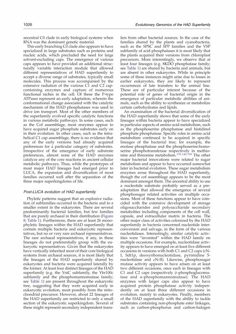
1026 Evolutionary Genomics of the HAD Superfamily
ancestral C0 clade in early biological systems whenRNAwas the dominant genetic material.This early branching C0 clade also appears to have
specialized in large substrates such as proteins andnucleic acids, which precluded the need for largesolvent-excluding caps. The emergence of variouscaps appears to have provided an additional struc-turally variable interaction module that alloweddifferent representatives of HAD superfamily toaccept a diverse range of substrates, typically smallmolecules. This process was accompanied by theextensive radiation of the various C1 and C2 cap-containing enzymes and capture of numerousfunctional niches in the cell. Of these the P-typeATPases represent an early adaptation, wherein theconformational change associated with the catalyticmechanism of the HAD phosphatases was used todrive ion transport. Most of the other members ofthe superfamily evolved specific catalytic functionsin various metabolic pathways. In some cases, suchas the Cof assemblage, most enzymes appear tohave acquired sugar phosphate substrates early onin their evolution. In other cases, such as the tetra-helical C1 cap assemblage, there is no evidence thatany of the early versions had already acquiredpreferences for a particular category of substrates.Irrespective of the emergence of early substratepreferences, almost none of the HAD enzymescatalyze any of the core reactions in ancient cellularmetabolic pathways. Thus, while the prototypes ofmost major HAD lineages had emerged prior toLUCA, the expansion and diversification of mostfamilies occurred well after the separation of thethree major superkingdoms of life.
Post-LUCA evolution of HAD superfamily
Phyletic patterns suggest that an explosive radia-tion of subfamilies occurred in the bacteria and to asmaller extent in the eukaryotes. There are severalpredominantly bacterial families, but few familiesthat are purely archaeal in their distribution (Figure8; Table 1). Furthermore, there are at least 26 mono-phyletic lineages within the HAD superfamily thatcontain multiple bacteria and eukaryotic represen-tatives, but no or very rare archaeal representatives.The rare archaeal representatives, if any, in theselineages do not preferentially group with the eu-karyotic representatives. Given that the eukaryoteshave vertically inherited most of their core biologicalsystems from archaeal sources, it is most likely thatthe lineages of the HAD superfamily shared byeukaryotes and bacteria were acquired laterally bythe former. At least four distinct lineages of the HADsuperfamily (e.g. the YniC subfamily, the Yhr100csubfamily and the phosphomannomutase family;see Table 1) are present throughout the eukaryotictree, suggesting that they were acquired early ineukaryotic evolution, most possibly from the mito-chondrial precursor. However, about 22 lineages ofthe HAD superfamily are restricted to only a smallsection of the eukaryotic superkingdom. Several ofthese might represent secondary independent trans-
fers from other bacterial sources. In the case of thefamilies shared by the plants and cyanobacteria,such as the SPSC and SPP families and the VSPsubfamily of acid phosphatases it is most likely thatthe plants acquired their versions from chloroplastprecursors. More interestingly, we observe that atleast four lineages (e.g. 8KDO phosphatase family;see Table 1) are shared by bacteria and animals, butare absent in other eukaryotes. While in principlesome of these instances might arise due to losses inearlier eukaryotes, they are likely to representoccurrences of late transfers to the animal line.These are of particular interest because of thepotential role of genes of bacterial origin in theemergence of particular metabolic abilities of ani-mals, such as the ability to synthesize or metabolizecertain carbohydrates and lipids.An examination of the bacterial diversification of
the HAD superfamily shows that some of the earlylineages within bacteria appear to have specializedin particular aspects of amino-acid metabolism, suchas the phosphoserine phosphatase and histidinolphosphate phosphatase. Specific roles in amino acidmetabolism continued to be acquired in specificlineages of the bacterial tree; for example, theenolase phosphatase and the phosphoserine:homo-serine phosphotransferase respectively in methio-nine and threonine metabolism.139,140,150 The othermajor bacterial innovations were related to sugarmetabolism and appear to have occurred somewhatlater in bacterial evolution. These sugar metabolismenzymes arose throughout the HAD superfamily,though the cof assemblage appears to be the mostdominant amongst them. The ancestral ability to usea nucleotide substrate probably served as a pre-adaptation that allowed the emergence of severalphosphosugar related activities on multiple occa-sions. Most of these functions appear to have coin-cided with the extensive development of storageoligosaccharides and polysaccharide secondarymetabolites including components of the cell wall,capsule, and extracellular matrix in bacteria. Theother major class of activities colonized by the HADsuperfamily in bacteria concerned nucleotide inter-conversion and salvage, in the form of the variousnucleotidases. Interestingly, similar catalytic activ-ities were “invented” within the HAD family onmultiple occasions. For example, nucleotidase activ-ity appears to have emerged on at least five differentoccasions in versions with both C1 and C2 caps (cN-I, Sdt1p, deoxyribonucleotidase, pyrimidine 5-nucleotidase and cN-II). Likewise, phosphosugarmutase activity appears to have arisen on at leasttwo different occasions, once each in lineages withC1 and C2 caps (respectively β-phosphoglucomu-tase and α-phosphomannomutase). The HADenzymes with larger caps also appear to haveacquired protein phosphatase activity indepen-dently on at least three different occasions inevolution, mainly in eukaryotes. Finally, membersof the HAD superfamily with the ability to tacklesubstrates containing non-phosphate ester linkages,such as carbon-phosphorus and carbon-halogen
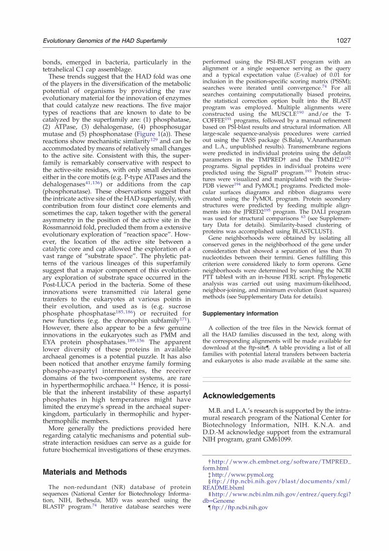
1027Evolutionary Genomics of the HAD Superfamily
bonds, emerged in bacteria, particularly in thetetrahelical C1 cap assemblage.These trends suggest that the HAD fold was one
of the players in the diversification of the metabolicpotential of organisms by providing the rawevolutionary material for the innovation of enzymesthat could catalyze new reactions. The five majortypes of reactions that are known to date to becatalyzed by the superfamily are: (1) phosphatase,(2) ATPase, (3) dehalogenase, (4) phosphosugarmutase and (5) phosphonatase (Figure 1(a)). Thesereactions show mechanistic similarity129 and can beaccommodated bymeans of relatively small changesto the active site. Consistent with this, the super-family is remarkably conservative with respect tothe active-site residues, with only small deviationseither in the core motifs (e.g. P-type ATPases and thedehalogenases41,136) or additions from the cap(phosphonatase). These observations suggest thatthe intricate active site of the HAD superfamily, withcontribution from four distinct core elements andsometimes the cap, taken together with the generalasymmetry in the position of the active site in theRossmannoid fold, precluded them from a extensiveevolutionary exploration of “reaction space”. How-ever, the location of the active site between acatalytic core and cap allowed the exploration of avast range of “substrate space”. The phyletic pat-terns of the various lineages of this superfamilysuggest that a major component of this evolution-ary exploration of substrate space occurred in thePost-LUCA period in the bacteria. Some of theseinnovations were transmitted via lateral genetransfers to the eukaryotes at various points intheir evolution, and used as is (e.g. sucrosephosphate phosphatase185,186) or recruited fornew functions (e.g. the chronophin subfamily171).However, there also appear to be a few genuineinnovations in the eukaryotes such as PMM andEYA protein phosphatases.189,156 The apparentlower diversity of these proteins in availablearchaeal genomes is a potential puzzle. It has alsobeen noticed that another enzyme family formingphospho-aspartyl intermediates, the receiverdomains of the two-component systems, are rarein hyperthermophilic archaea.14 Hence, it is possi-ble that the inherent instability of these aspartylphosphates in high temperatures might havelimited the enzyme's spread in the archaeal super-kingdom, particularly in thermophilic and hyper-thermophilic members.More generally the predictions provided here
regarding catalytic mechanisms and potential sub-strate interaction residues can serve as a guide forfuture biochemical investigations of these enzymes.
†http://www.ch.embnet.org/software/TMPRED_form.html‡http://www.pymol.org§ ftp://ftp.ncbi.nih.gov/blast/documents/xml/
README.blxml∥http://www.ncbi.nlm.nih.gov/entrez/query.fcgi?
db=Genome¶ ftp://ftp.ncbi.nih.gov
Materials and Methods
The non-redundant (NR) database of proteinsequences (National Center for Biotechnology Informa-tion, NIH, Bethesda, MD) was searched using theBLASTP program.74 Iterative database searches were
performed using the PSI-BLAST program with analignment or a single sequence serving as the queryand a typical expectation value (E-value) of 0.01 forinclusion in the position-specific scoring matrix (PSSM);searches were iterated until convergence.74 For allsearches containing computationally biased proteins,the statistical correction option built into the BLASTprogram was employed. Multiple alignments wereconstructed using the MUSCLE190 and/or the T-COFFEE191 programs, followed by a manual refinementbased on PSI-blast results and structural information. Alllarge-scale sequence-analysis procedures were carriedout using the TASS package (S.Balaji, V.Anantharamanand L.A., unpublished results). Transmembrane regionswere predicted in individual proteins using the defaultparameters in the TMPRED† and the TMMH2.0192programs. Signal peptides in individual proteins werepredicted using the SignalP program.193 Protein struc-tures were visualized and manipulated with the Swiss-PDB viewer194 and PyMOL‡ programs. Predicted mole-cular surfaces diagrams and ribbon diagrams werecreated using the PyMOL program. Protein secondarystructures were predicted by feeding multiple align-ments into the JPRED2195 program. The DALI programwas used for structural comparisons 63 (see Supplemen-tary Data for details). Similarity-based clustering ofproteins was accomplished using BLASTCLUST§.Gene neighborhoods were obtained by isolating all
conserved genes in the neighborhood of the gene underconsideration that showed a separation of less than 70nucleotides between their termini. Genes fulfilling thiscriterion were considered likely to form operons. Geneneighborhoods were determined by searching the NCBIPTT tables∥ with an in-house PERL script. Phylogeneticanalysis was carried out using maximum-likelihood,neighbor-joining, and minimum evolution (least squares)methods (see Supplementary Data for details).
Supplementary information
A collection of the tree files in the Newick format ofall the HAD families discussed in the text, along withthe corresponding alignments will be made available fordownload at the ftp-site¶. A table providing a list of allfamilies with potential lateral transfers between bacteriaand eukaryotes is also made available at the same site.
Acknowledgements
M.B. and L.A.'s research is supported by the intra-mural research program of the National Center forBiotechnology Information, NIH. K.N.A. andD.D.-M acknowledge support from the extramuralNIH program, grant GM61099.

1028 Evolutionary Genomics of the HAD Superfamily
Supplementary Data
Supplementary data associated with this articlecan be found, in the online version, at doi:10.1016/j.jmb.2006.06.049
References
1. Vincent, J. B., Crowder, M. W. & Averill, B. A. (1992).Hydrolysis of phosphate monoesters: a biologicalproblem with multiple chemical solutions. TrendsBiochem. Sci. 17, 105–110.
2. Vetter, I. R. & Wittinghofer, A. (1999). Nucleosidetriphosphate-binding proteins: different scaffolds toachieve phosphoryl transfer. Quart. Rev. Biophys. 32,1–56.
3. Iyer, L. M., Leipe, D. D., Koonin, E. V. & Aravind, L.(2004). Evolutionary history and higher orderclassification of AAA+ ATPases. J. Struct. Biol. 146,11–31.
4. Bork, P., Sander, C. & Valencia, A. (1992). An ATPasedomain common to prokaryotic cell cycle proteins,sugar kinases, actin, and hsp70 heat shock proteins.Proc. Natl Acad. Sci. USA, 89, 7290–7294.
5. Haren, L., Ton-Hoang, B. & Chandler, M. (1999).Integrating DNA: transposases and retroviral inte-grases. Annu. Rev. Microbiol. 53, 245–281.
6. Aravind, L. & Koonin, E. V. (1998). A novel family ofpredicted phosphoesterases includes Drosophilaprune protein and bacterial RecJ exonuclease. TrendsBiochem. Sci. 23, 17–19.
7. Aravind, L. & Koonin, E. V. (1998). The HD domaindefines a new superfamily of metal-dependentphosphohydrolases. Trends Biochem. Sci. 23, 469–472.
8. Aravind, L. & Koonin, E. V. (1998). Phosphoesterasedomains associated with DNA polymerases ofdiverse origins. Nucl. Acids Res. 26, 3746–3752.
9. Koonin, E. V. & Tatusov, R. L. (1994). Computeranalysis of bacterial haloacid dehalogenases defines alarge superfamily of hydrolases with diverse speci-ficity. Application of an iterative approach todatabase search. J. Mol. Biol. 244, 125–132.
10. Aravind, L., Galperin, M. Y. & Koonin, E. V. (1998).The catalytic domain of the P-type ATPase has thehaloacid dehalogenase fold. Trends Biochem. Sci. 23,127–129.
11. Goldberg, J., Huang, H. B., Kwon, Y. G., Greengard, P.,Nairn, A. C. & Kuriyan, J. (1995). Three-dimensionalstructure of the catalytic subunit of protein serine/threonine phosphatase-1. Nature, 376, 745–753.
12. Whisstock, J. C., Romero, S., Gurung, R., Nandurkar,H., Ooms, L. M., Bottomley, S. P. & Mitchell, C. A.(2000). The inositol polyphosphate 5-phosphatasesand the apurinic/apyrimidinic base excision repairendonucleases share a common mechanism forcatalysis. J. Biol. Chem. 275, 37055–37061.
13. Grebe, T. W. & Stock, J. B. (1999). The histidineprotein kinase superfamily. Adv. Microb. Physiol. 41,139–227.
14. Koretke, K. K., Lupas, A. N., Warren, P. V., Rosen-berg, M. & Brown, J. R. (2000). Evolution of two-component signal transduction. Mol. Biol. Evol. 17,1956–1970.
15. Hogg, T., Mechold, U., Malke, H., Cashel, M. &Hilgenfeld, R. (2004). Conformational antagonismbetween opposing active sites in a bifunctionalRelA/SpoT homolog modulates (p)ppGpp metabo-
lism during the stringent response (corrected). Cell,117, 57–68.
16. Iyer, L. M. & Aravind, L. (2002). The catalyticdomains of thiamine triphosphatase and CyaB-likeadenylyl cyclase define a novel superfamily ofdomains that bind organic phosphates. BMC Geno-mics, 3, 33.
17. Allen, K. N. & Dunaway-Mariano, D. (2004).Phosphoryl group transfer: evolution of a catalyticscaffold. Trends Biochem. Sci. 29, 495–503.
18. Yamagata, A., Kakuta, Y., Masui, R. & Fukuyama, K.(2002). The crystal structure of exonuclease RecJbound to Mn2+ ion suggests how its characteristicmotifs are involved in exonuclease activity. Proc. NatlAcad. Sci. USA, 99, 5908–5912.
19. Ahn, S., Milner, A. J., Futterer, K., Konopka, M., Ilias,M., Young, T. W. & White, S. A. (2001). The “open”and “closed” structures of the type-C inorganicpyrophosphatases from Bacillus subtilis and Strepto-coccus gordonii. J. Mol. Biol. 313, 797–811.
20. Teplyakov, A., Obmolova, G., Khil, P. P., Howard,A. J., Camerini-Otero, R. D. & Gilliland, G. L. (2003).Crystal structure of the Escherichia coli YcdX proteinreveals a trinuclear zinc active site. Proteins: Struct.Funct. Genet. 51, 315–318.
21. Knofel, T. & Strater, N. (1999). X-ray structure of theEscherichia coli periplasmic 5′-nucleotidase contain-ing a dimetal catalytic site. Nature Struct. Biol. 6,448–453.
22. Mol, C. D., Kuo, C. F., Thayer, M. M., Cunningham,R. P. & Tainer, J. A. (1995). Structure and function ofthe multifunctional DNA-repair enzyme exonucleaseIII. Nature, 374, 381–386.
23. Collet, J. F., Stroobant, V., Pirard, M., Delpierre, G. &Van Schaftingen, E. (1998). A new class of phospho-transferases phosphorylated on an aspartate residuein an amino-terminal DXDX(T/V) motif. J. Biol.Chem. 273, 14107–14112.
24. Anantharaman, V., Aravind, L. & Koonin, E. V.(2003). Emergence of diverse biochemical activities inevolutionarily conserved structural scaffolds ofproteins. Curr. Opin. Chem. Biol. 7, 12–20.
25. Aravind, L. & Koonin, E. V. (1999). DNA polymerasebeta-like nucleotidyltransferase superfamily: identi-fication of three new families, classification andevolutionary history. Nucl. Acids Res. 27, 1609–1618.
26. Shin, D. H., Roberts, A., Jancarik, J., Yokota, H., Kim,R., Wemmer, D. E. & Kim, S. H. (2003). Crystalstructure of a phosphatase with a unique substratebinding domain from Thermotoga maritima. ProteinSci. 12, 1464–1472.
27. Morais, M. C., Zhang, W., Baker, A. S., Zhang, G.,Dunaway-Mariano, D. & Allen, K. N. (2000). Thecrystal structure of Bacillus cereus phosphonoacetal-dehyde hydrolase: insight into catalysis of phos-phorus bond cleavage and catalytic diversificationwithin the HAD enzyme superfamily. Biochemistry,39, 10385–10396.
28. Baker, A. S., Ciocci, M. J., Metcalf, W. W., Kim, J.,Babbitt, P. C., Wanner, B. L. et al. (1998). Insights intothe mechanism of catalysis by the P-C bond-cleavingenzyme phosphonoacetaldehyde hydrolase derivedfrom gene sequence analysis and mutagenesis.Biochemistry, 37, 9305–9315.
29. Qian, N., Stanley, G. A., Hahn-Hagerdal, B. &Radstrom, P. (1994). Purification and characterizationof two phosphoglucomutases from Lactococcus lactissubsp. lactis and their regulation in maltose- andglucose-utilizing cells. J. Bacteriol. 176, 5304–5311.

1029Evolutionary Genomics of the HAD Superfamily
30. Collet, J. F., Gerin, I., Rider, M. H., Veiga-da-Cunha,M. & Van Schaftingen, E. (1997). Human L-3-phosphoserine phosphatase: sequence, expressionand evidence for a phosphoenzyme intermediate.FEBS Letters, 408, 281–284.
31. Seal, S. N. & Rose, Z. B. (1987). Characterization of aphosphoenzyme intermediate in the reaction ofphosphoglycolate phosphatase. J. Biol. Chem. 262,13496–13500.
32. Lahiri, S. D., Zhang, G., Dunaway-Mariano, D. &Allen, K. N. (2002). Caught in the act: the structure ofphosphorylated beta-phosphoglucomutase from Lac-tococcus lactis. Biochemistry, 41, 8351–8359.
33. Ahmadian, M. R., Stege, P., Scheffzek, K. &Wittinghofer, A. (1997). Confirmation of the argi-nine-finger hypothesis for the GAP-stimulatedGTP-hydrolysis reaction of Ras. Nature Struct. Biol.4, 686–689.
34. Peisach, E., Selengut, J. D., Dunaway-Mariano, D. &Allen, K. N. (2004). X-ray crystal structure of thehypothetical phosphotyrosine phosphatase MDP-1of the haloacid dehalogenase superfamily. Biochem-istry, 43, 12770–12779.
35. Hisano, T., Hata, Y., Fujii, T., Liu, J. Q., Kurihara, T.,Esaki, N. & Soda, K. (1996). Crystal structure of L-2-haloacid dehalogenase from Pseudomonas sp. YL. Analpha/beta hydrolase structure that is different fromthe alpha/beta hydrolase fold. J. Biol. Chem. 271,20322–20330.
36. Toyoshima, C., Nakasako, M., Nomura, H. & Ogawa,H. (2000). Crystal structure of the calcium pump ofsarcoplasmic reticulum at 2.6 Å resolution. Nature,405, 647–655.
37. Wang,W., Kim, R., Jancarik, J., Yokota, H. &Kim, S. H.(2001). Crystal structure of phosphoserine phospha-tase from Methanococcus jannaschii, a hyperther-mophile, at 1.8 Å resolution. Structure (Camb), 9,65–71.
38. Lahiri, S. D., Zhang, G., Dunaway-Mariano, D. &Allen, K. N. (2003). The pentacovalent phosphorusintermediate of a phosphoryl transfer reaction.Science, 299, 2067–2071.
39. Rinaldo-Matthis, A., Rampazzo, C., Reichard, P.,Bianchi, V. &Nordlund, P. (2002). Crystal structure ofa human mitochondrial deoxyribonucleotidase. Nat-ure Struct. Biol. 9, 779–787.
40. Olsen, D. B., Hepburn, T. W., Moos, M., Mariano,P. S. & Dunaway-Mariano, D. (1988). Investigationof the Bacillus cereus phosphonoacetaldehydehydrolase. Evidence for a Schiff base mechanismand sequence analysis of an active-site peptidecontaining the catalytic lysine residue. Biochemistry,27, 2229–2234.
41. Kurihara, T., Liu, J. Q., Nardi-Dei, V., Koshikawa, H.,Esaki, N. & Soda, K. (1995). Comprehensive site-directed mutagenesis of L-2-halo acid dehalogenaseto probe catalytic amino acid residues. J. Biochem.(Tokyo), 117, 1317–1322.
42. Aravind, L., Anantharaman, V. & Koonin, E. V.(2002). Monophyly of class I aminoacyl tRNAsynthetase, USPA, ETFP, photolyase, and PP-ATPasenucleotide-binding domains: implications for proteinevolution in the RNA. Proteins: Struct. Funct. Genet.48, 1–14.
43. Rossmann, M. G., Moras, D. & Olsen, K. W. (1974).Chemical and biological evolution of nucleotide-binding protein. Nature, 250, 194–199.
44. Zhao, K., Chai, X. & Marmorstein, R. (2003).Structure of the yeast Hst2 protein deacetylase in
ternary complex with 2′-O-acetyl ADP ribose andhistone peptide. Structure, 11, 1403–1411.
45. Martin, J. L. & McMillan, F. M. (2002). SAM(dependent) I AM: the S-adenosylmethionine-depen-dent methyltransferase fold. Curr. Opin. Struct. Biol.12, 783–793.
46. Schubert, H. L., Blumenthal, R. M. & Cheng, X.(2003). Many paths to methyltransfer: a chronicle ofconvergence. Trends Biochem. Sci. 28, 329–335.
47. Sistla, S. & Rao, D. N. (2004). S-Adenosyl-L-methio-nine-dependent restriction enzymes. Crit. Rev. Bio-chem. Mol. Biol. 39, 1–19.
48. Lowe, J. & Amos, L. A. (1998). Crystal structure of thebacterial cell-division protein FtsZ. Nature, 391,203–206.
49. Anantharaman, V. & Aravind, L. (2006). Diversi-fication of catalytic activities and ligand interac-tions in the protein fold shared by the sugarisomerases, eIF2B, DeoR transcription factors, acyl-CoA transferases and methenyltetrahydrofolatesynthetase. J. Mol. Biol. 356, 823–842.
50. Aravind, L., Leipe, D. D. & Koonin, E. V. (1998).Toprim–a conserved catalytic domain in type IA andII topoisomerases, DnaG-type primases, OLD familynucleases and RecR proteins. Nucl. Acids Res. 26,4205–4213.
51. Clissold, P. M. & Ponting, C. P. (2000). PIN domainsin nonsense-mediated mRNA decay and RNAi. Curr.Biol. 10, R888–R890.
52. Finnin, M. S., Donigian, J. R., Cohen, A., Richon,V. M., Rifkind, R. A., Marks, P. A. et al. (1999).Structures of a histone deacetylase homologuebound to the TSA and SAHA inhibitors. Nature,401, 188–193.
53. Whittaker, C. A. & Hynes, R. O. (2002). Distributionand evolution of von Willebrand/integrin A domains:widely dispersed domains with roles in cell adhesionand elsewhere. Mol. Biol. Cell. 13, 3369–3387.
54. Robinson, V. L., Buckler, D. R. & Stock, A. M. (2000).A tale of two components: a novel kinase and aregulatory switch. Nauret Struct. Biol. 7, 626–633.
55. Wolanin, P. M., Thomason, P. A. & Stock, J. B.(2002). Histidine protein kinases: key signal trans-ducers outside the animal kingdom. Genome Biol. 3,REVIEWS3013.1–3013.8.
56. West, A. H. & Stock, A. M. (2001). Histidinekinases and response regulator proteins in two-component signaling systems. Trends Biochem. Sci.26, 369–376.
57. Ridder, I. S. & Dijkstra, B. W. (1999). Identification ofthe Mg2+-binding site in the P-type ATPase andphosphatase members of the HAD (haloacid deha-logenase) superfamily by structural similarity to theresponse regulator protein CheY. Biochem. J. 339,223–226.
58. Meng, E. C., Polacco, B. J. & Babbitt, P. C. (2004).Superfamily active site templates. Proteins, 55,962–976.
59. Merckel, M. C., Fabrichniy, I. P., Salminen, A.,Kalkkinen, N., Baykov, A. A., Lahti, R. & Goldman,A. (2001). Crystal structure of Streptococcus mutanspyrophosphatase: a new fold for an old mechanism.Structure (Camb), 9, 289–297.
60. Fabrichniy, I. P., Lehtio, L., Salminen, A., Zyryanov,A. B., Baykov, A. A., Lahti, R. & Goldman, A. (2004).Structural studies of metal ions in family II pyropho-sphatases: the requirement for a Janus ion. Biochem-istry, 43, 14403–14411.
61. Chen, S. J. & Wang, J. C. (1998). Identification of

1030 Evolutionary Genomics of the HAD Superfamily
active site residues in Escherichia coli DNA topoi-somerase I. J. Biol. Chem. 273, 6050–6056.
62. Lee, J. O., Rieu, P., Arnaout, M. A. & Liddington, R.(1995). Crystal structure of the A domain from thealpha subunit of integrin CR3 (CD11b/CD18). Cell,80, 631–638.
63. Holm, L. & Sander, C. (1996). The FSSP database: foldclassification based on structure-structure alignmentof proteins. Nucl. Acids Res. 24, 206–209.
64. Holm, L. & Sander, C. (1995). Dali: a network tool forprotein structure comparison. Trends Biochem. Sci. 20,478–480.
65. Kim, Y., Yakunin, A. F., Kuznetsova, E., Xu, X.,Pennycooke, M., Gu, J. et al. (2004). Structure- andfunction-based characterization of a new phospho-glycolate phosphatase from Thermoplasma acidophi-lum. J. Biol. Chem. 279, 517–526.
66. Li, Y. F., Hata, Y., Fujii, T., Hisano, T., Nishihara, M.,Kurihara, T. & Esaki, N. (1998). Crystal structures ofreaction intermediates of L-2-haloacid dehalogenaseand implications for the reaction mechanism. J. Biol.Chem. 273, 15035–15044.
67. Ridder, I. S., Rozeboom, H. J., Kalk, K. H., Janssen,D. B. & Dijkstra, B. W. (1997). Three-dimensionalstructure of L-2-haloacid dehalogenase fromXanthobacter autotrophicus GJ10 complexed withthe substrate-analogue formate. J. Biol. Chem. 272,33015–33022.
68. Calderone, V., Forleo, C., Benvenuti, M., CristinaThaller, M., Maria Rossolini, G. &Mangani, S. (2004).The first structure of a bacterial class B Acidphosphatase reveals further structural heterogeneityamong phosphatases of the haloacid dehalogenasefold. J. Mol. Biol. 335, 761–773.
69. Ridder, I. S., Rozeboom, H. J., Kalk, K. H. & Dijkstra,B. W. (1999). Crystal structures of intermediates inthe dehalogenation of haloalkanoates by L-2-haloa-cid dehalogenase. J. Biol. Chem. 274, 30672–30678.
70. Zhang, G., Mazurkie, A. S., Dunaway-Mariano, D. &Allen, K. N. (2002). Kinetic evidence for a substrate-induced fit in phosphonoacetaldehyde hydrolasecatalysis. Biochemistry, 41, 13370–13377.
71. Morais, M. C., Zhang, G., Zhang, W., Olsen, D. B.,Dunaway-Mariano, D. & Allen, K. N. (2004). X-raycrystallographic and site-directed mutagenesis ana-lysis of the mechanism of Schiff-base formation inphosphonoacetaldehyde hydrolase catalysis. J. Biol.Chem. 279, 9353–9361.
72. Zhang, G., Dai, J., Wang, L., Dunaway-Mariano, D.,Tremblay, L. W. & Allen, K. N. (2005). Catalyticcycling in beta-phosphoglucomutase: a kinetic andstructural analysis. Biochemistry, 44, 9404–9416.
73. Wang, W., Cho, H. S., Kim, R., Jancarik, J., Yokota, H.,Nguyen, H. H. et al. (2002). Structural characteriza-tion of the reaction pathway in phosphoserinephosphatase: crystallographic “snapshots” of inter-mediate states. J. Mol. Biol. 319, 421–431.
74. Altschul, S. F., Madden, T. L., Schaffer, A. A., Zhang,J., Zhang, Z., Miller, W. & Lipman, D. J. (1997).Gapped BLAST and PSI-BLAST: a new generation ofprotein database search programs. Nucl. Acids Res.25, 3389–3402.
75. Selengut, J. D. & Levine, R. L. (2000). MDP-1: a noveleukaryotic magnesium-dependent phosphatase. Bio-chemistry, 39, 8315–8324.
76. Selengut, J. D. (2001). MDP-1 is a new and distinctmember of the haloacid dehalogenase family ofaspartate-dependent phosphohydrolases. Biochemis-try, 40, 12704–12711.
77. Wu, K., Chung, L., Revill, W. P., Katz, L. & Reeves,C. D. (2000). The FK520 gene cluster of Streptomyceshygroscopicus var. ascomyceticus (ATCC 14891)contains genes for biosynthesis of unusual polyke-tide extender units. Gene, 251, 81–90.
78. Hildebrand, M., Waggoner, L. E., Liu, H., Sudek, S.,Allen, S., Anderson, C. et al. (2004). bryA: an unusualmodular polyketide synthase gene from the unculti-vated bacterial symbiont of the marine bryozoanBugula neritina. Chem. Biol. 11, 1543–1552.
79. Archambault, J., Chambers, R. S., Kobor, M. S., Ho,Y., Cartier, M., Bolotin, D. et al. (1997). An essentialcomponent of a C-terminal domain phosphatasethat interacts with transcription factor IIF inSaccharomyces cerevisiae. Proc. Natl Acad. Sci. USA,94, 14300–14305.
80. Archambault, J., Pan, G., Dahmus, G. K., Cartier,M., Marshall, N., Zhang, S. et al. (1998). FCP1, theRAP74-interacting subunit of a human proteinphosphatase that dephosphorylates the carboxyl-terminal domain of RNA polymerase IIO. J. Biol.Chem. 273, 27593–27601.
81. Chambers, R. S. & Dahmus, M. E. (1994). Purifica-tion and characterization of a phosphatase fromHeLa cells which dephosphorylates the C-terminaldomain of RNA polymerase II. J. Biol. Chem. 269,26243–26248.
82. Chambers, R. S. & Kane, C. M. (1996). Purificationand characterization of an RNA polymerase II phos-phatase from yeast. J. Biol. Chem. 271, 24498–24504.
83. Cho, H., Kim, T. K., Mancebo, H., Lane, W. S., Flores,O. & Reinberg, D. (1999). A protein phosphatasefunctions to recycle RNA polymerase II. Genes Dev.13, 1540–1552.
84. Kobor, M. S., Archambault, J., Lester, W., Holstege,F. C., Gileadi, O., Jansma, D. B. et al. (1999). Anunusual eukaryotic protein phosphatase requiredfor transcription by RNA polymerase II and CTDdephosphorylation in S. cerevisiae. Mol. Cell, 4,55–62.
85. Lin, P. S., Marshall, N. F. & Dahmus, M. E. (2002).CTD phosphatase: role in RNA polymerase II cyclingand the regulation of transcript elongation. ProgNucl. Acid Res. Mol. Biol. 72, 333–365.
86. Orphanides, G. & Reinberg, D. (2002). A unifiedtheory of gene expression. Cell, 108, 439–451.
87. Siniossoglou, S., Hurt, E. C. & Pelham, H. R. (2000).Psr1p/Psr2p, two plasma membrane phosphataseswith an essential DXDX(T/V) motif required forsodium stress response in yeast. J. Biol. Chem. 275,19352–19360.
88. Yeo, M., Lin, P. S., Dahmus, M. E. & Gill, G. N. (2003).A novel RNA polymerase II C-terminal domainphosphatase that preferentially dephosphorylatesserine 5. J. Biol. Chem. 278, 26078–26085.
89. Siniossoglou, S., Santos-Rosa, H., Rappsilber, J.,Mann, M. & Hurt, E. (1998). A novel complex ofmembrane proteins required for formation of aspherical nucleus. EMBO J. 17, 6449–6464.
90. Guo, Y., Cheong, N., Zhang, Z., De Rose, R., Deng,Y., Farber, S. A. et al. (2004). Tim50, a component ofthe mitochondrial translocator, regulates mitochon-drial integrity and cell death. J. Biol. Chem. 279,24813–24825.
91. Xu, H., Somers, Z. B., Robinson, M. L., 2nd & M.D.(2005). Tim50a, a nuclear isoform of the mitochon-drial Tim50, interacts with proteins involved insnRNP biogenesis. BMC Cell. Biol. 6, 29.
92. Yu, X., Chini, C. C., He, M., Mer, G. & Chen, J. (2003).

1031Evolutionary Genomics of the HAD Superfamily
The BRCT domain is a phospho-protein bindingdomain. Science, 302, 639–642.
93. Hugouvieux, V., Kwak, J. M. & Schroeder, J. I. (2001).An mRNA cap binding protein, ABH1, modulatesearly abscisic acid signal transduction in Arabidopsis.Cell, 106, 477–487.
94. Xiong, L., Lee, H., Ishitani, M., Tanaka, Y., Stevenson,B., Koiwa, H. et al. (2002). Repression of stress-responsive genes by FIERY2, a novel transcriptionalregulator in Arabidopsis. Proc. Natl Acad. Sci. USA, 99,10899–10904.
95. Zheng, H., Ji, C., Gu, S., Shi, B., Wang, J., Xie, Y. &Mao, Y. (2005). Cloning and characterization of anovel RNA polymerase II C-terminal domainphosphatase. Biochem. Biophys. Res. Commun. 331,1401–1407.
96. Wu, X., Chang, A., Sudol, M. & Hanes, S. D. (2001).Genetic interactions between the ESS1 prolyl-isomer-ase and the RSP5 ubiquitin ligase reveal opposingeffects on RNA polymerase II function. Curr. Genet.40, 234–242.
97. Reichmann, G., Dlugonska, H. & Fischer, H. G.(2002). Characterization of TgROP9 (p36), a novelrhoptry protein of Toxoplasma gondii tachyzoitesidentified by T cell clone. Mol. Biochem. Parasitol.119, 43–54.
98. Boyce, J. D., Chung, J. Y. & Adler, B. (2000). Geneticorganisation of the capsule biosynthetic locus ofPasteurella multocida M1404 (B:2). Vet. Microbiol. 72,121–134.
99. Jilani, A., Ramotar, D., Slack, C., Ong, C., Yang, X. M.,Scherer, S. W. & Lasko, D. D. (1999). Molecularcloning of the human gene, PNKP, encoding apolynucleotide kinase 3′-phosphatase and evidencefor its role in repair of DNA strand breaks caused byoxidative damage. J. Biol. Chem. 274, 24176–24186.
100. Soltis, D. A. & Uhlenbeck, O. C. (1982). Isolation andcharacterization of two mutant forms of T4 poly-nucleotide kinase. J. Biol. Chem. 257, 11332–11339.
101. Petrucco, S., Volpi, G., Bolchi, A., Rivetti, C. &Ottonello, S. (2002). A nick-sensing DNA 3′-repairenzyme from Arabidopsis. J. Biol. Chem. 277,23675–23683.
102. Betti, M., Petrucco, S., Bolchi, A., Dieci, G. &Ottonello, S. (2001). A plant 3′-phosphoesteraseinvolved in the repair of DNA strand breaksgenerated by oxidative damage. J .Biol. Chem. 276,18038–18045.
103. Parsons, J. F., Lim, K., Tempczyk, A., Krajewski, W.,Eisenstein, E. & Herzberg, O. (2002). From struc-ture to function: YrbI from Haemophilus influenzae(HI1679) is a phosphatase. Proteins: Struct. Funct.Genet. 46, 393–404.
104. Wu, J. &Woodard, R.W. (2003). Escherichia coliYrbI is3-deoxy-D-manno-octulosonate 8-phosphate phos-phatase. J. Biol. Chem. 278, 18117–18123.
105. Tzeng, Y. L., Datta, A., Strole, C., Kolli, V. S., Birck,M. R., Taylor, W. P. et al. (2002). KpsF is the arab-inose-5-phosphate isomerase required for 3-deoxy-D-manno-octulosonic acid biosynthesis and for bothlipooligosaccharide assembly and capsular polysac-charide expression in Neisseria meningitidis. J. Biol.Chem. 277, 24103–24113.
106. Leelapon, O., Sarath, G. & Staswick, P. E. (2004). Asingle amino acid substitution in soybean VSPalphaincreases its acid phosphatase activity nearly 20-fold.Planta, 219, 1071–1079.
107. Utsugi, S., Sakamoto,W., Murata, M. &Motoyoshi, F.(1998). Arabidopsis thaliana vegetative storage protein
(VSP) genes: gene organization and tissue-specificexpression. Plant Mol. Biol. 38, 565–576.
108. Gomez, L. & Faurobert, M. (2002). Contribution ofvegetative storage proteins to seasonal nitrogenvariations in the young shoots of peach trees (Prunuspersica L. Batsch). J Exp Bot, 53, 2431–2439.
109. Hunsucker, S. A., Spychala, J. &Mitchell, B. S. (2001).Human cytosolic 5′-nucleotidase I: characterizationand role in nucleoside analog resistance. J. Biol. Chem.276, 10498–10504.
110. Sala-Newby, G. B., Skladanowski, A. C. & Newby,A. C. (1999). The mechanism of adenosine forma-tion in cells. Cloning of cytosolic 5′-nucleotidase-I.J. Biol. Chem. 274, 17789–17793.
111. La Nauze, J. M. & Rosenberg, H. (1968). Theidentification of 2-phosphonoacetaldehyde as anintermediate in the degradation of 2-aminoethylpho-sphonate by Bacillus cereus. Biochim. Biophys. Acta,165, 438–447.
112. Dumora, C., Lacoste, A. M. & Cassaigne, A. (1989).Phosphonoacetaldehyde hydrolase from Pseudomo-nas aeruginosa: purification properties and compar-ison with Bacillus cereus enzyme. Biochim. Biophys.Acta, 997, 193–198.
113. Lee, K. S., Metcalf, W. W. & Wanner, B. L. (1992).Evidence for two phosphonate degradative path-ways in Enterobacter aerogenes. J. Bacteriol. 174,2501–2510.
114. Jiang, W., Metcalf, W. W., Lee, K. S. & Wanner, B. L.(1995). Molecular cloning, mapping, and regulationof Pho regulon genes for phosphonate breakdown bythe phosphonatase pathway of Salmonella typhimur-ium LT2. J Bacteriol, 177, 6411–6421.
115. Ternan, N. G. & Quinn, J. P. (1998). In vitro cleavageof the carbon-phosphorus bond of phosphonopyr-uvate by cell extracts of an environmental Burkhol-deria cepacia isolate. Biochem. Biophys. Res. Commun.248, 378–381.
116. Parker, G. F., Higgins, T. P., Hawkes, T. &Robson, R. L.(1999). Rhizobium (Sinorhizobium) meliloti phn genes:characterization and identification of their proteinproducts. J. Bacteriol. 181, 389–395.
117. Imig, J. D., Zhao, X., Capdevila, J. H., Morisseau, C. &Hammock, B. D. (2002). Soluble epoxide hydrolaseinhibition lowers arterial blood pressure in angio-tensin II hypertension. Hypertension, 39, 690–694.
118. Fang, X., Kaduce, T. L., Weintraub, N. L., Harmon, S.,Teesch, L. M., Morisseau, C. et al. (2001). Pathways ofepoxyeicosatrienoic acid metabolism in endothelialcells. Implications for the vascular effects of solubleepoxide hydrolase inhibition. J. Biol. Chem. 276,14867–14874.
119. Newman, J. W., Morisseau, C., Harris, T. R. &Hammock, B. D. (2003). The soluble epoxide hydro-lase encoded by EPXH2 is a bifunctional enzymewith novel lipid phosphate phosphatase activity.Proc. Natl Acad. Sci. USA, 100, 1558–1563.
120. Ogawa, N., DeRisi, J. & Brown, P. O. (2000). Newcomponents of a system for phosphate accumulationand polyphosphate metabolism in Saccharomycescerevisiae revealed by genomic expression analysis.Mol. Biol. Cell, 11, 4309–4321.
121. Nakanishi, T. & Sekimizu, K. (2002). SDT1/SSM1, amulticopy suppressor of S-II null mutant, encodes anovel pyrimidine 5′-nucleotidase. J. Biol. Chem. 277,22103–22106.
122. Fritzson, P. & Smith, I. (1971). A new nucleotidase ofrat liver with activity toward 3′-and 5′-nucleotides.Biochim. Biophys. Acta, 235, 128–141.

1032 Evolutionary Genomics of the HAD Superfamily
123. Rampazzo, C., Johansson, M., Gallinaro, L., Ferraro,P., Hellman, U., Karlsson, A. et al. (2000). Mammalian5′(3′)-deoxyribonucleotidase, cDNA cloning, andoverexpression of the enzyme in Escherichia coli andmammalian cells. J. Biol. Chem. 275, 5409–5415.
124. Rampazzo, C., Gallinaro, L., Milanesi, E., Frigimelica,E., Reichard, P. & Bianchi, V. (2000). A deoxyribonu-cleotidase in mitochondria: involvement in regulationof dNTP pools and possible link to genetic disease.Proc. Natl Acad. Sci. USA, 97, 8239–8244.
125. Leipe,D.D., Aravind, L., Grishin,N.V.&Koonin, E. V.(2000). The bacterial replicative helicase DnaB evolvedfrom a RecA duplication. Genome Res. 10, 5–16.
126. Iyer, L. M., Aravind, L. & Koonin, E. V. (2001).Common origin of four diverse families of largeeukaryotic DNA viruses. J. Virol. 75, 11720–11734.
127. Iyer, L. M., Makarova, K. S., Koonin, E. V. & Aravind,L. (2004). Comparative genomics of the FtsK-HerAsuperfamily of pumping ATPases: implications forthe origins of chromosome segregation, cell divisionand viral capsid packaging. Nucl. Acids Res. 32,5260–5279.
128. Levy, H. R. (1979). Glucose-6-phosphate dehydro-genases. Adv. Enzymol. Relat. Areas Mol. Biol. 48,97–192.
129. Lahiri, S. D., Zhang, G., Dai, J., Dunaway-Mariano,D. & Allen, K. N. (2004). Analysis of the substratespecificity loop of the HAD superfamily cap domain.Biochemistry, 43, 2812–2820.
130. Gibson, J. L. & Tabita, F. R. (1997). Analysis of thecbbXYZ operon in Rhodobacter sphaeroides. J. Bacteriol.179, 663–669.
131. Sanz, P., Randez-Gil, F. & Prieto, J. A. (1994). Mo-lecular characterization of a gene that confers 2-deoxyglucose resistance in yeast.Yeast, 10, 1195–1202.
132. Randez-Gil, F., Blasco, A., Prieto, J. A. & Sanz, P.(1995). DOGR1 and DOGR2: two genes fromSaccharomyces cerevisiae that confer 2-deoxyglucoseresistance when overexpressed. Yeast, 11, 1233–1240.
133. Norbeck, J., Pahlman, A. K., Akhtar, N., Blomberg,A. & Adler, L. (1996). Purification and characteriza-tion of two isoenzymes of DL-glycerol-3-phospha-tase from Saccharomyces cerevisiae. Identification ofthe corresponding GPP1 and GPP2 genes andevidence for osmotic regulation of Gpp2p expressionby the osmosensing mitogen-activated proteinkinase signal transduction pathway. J. Biol. Chem.271, 13875–13881.
134. Coquard, D., Huecas, M., Ott, M., van Dijl, J. M., vanLoon, A. P. & Hohmann, H. P. (1997). Molecularcloning and characterisation of the ribC gene fromBacillus subtilis: a point mutation in ribC results inriboflavin overproduction. Mol. Gen. Genet. 254,81–84.
135. Mack, M., van Loon, A. P. & Hohmann, H. P. (1998).Regulation of riboflavin biosynthesis in Bacillussubtilis is affected by the activity of the flavoki-nase/flavin adenine dinucleotide synthetase en-coded by ribC. J. Bacteriol. 180, 950–955.
136. Hill, K. E., Marchesi, J. R. & Weightman, A. J. (1999).Investigation of two evolutionarily unrelated halo-carboxylic acid dehalogenase gene families. J. Bacter-iol. 181, 2535–2547.
137. Murdiyatmo, U., Asmara,W., Tsang, J. S., Baines, A. J.,Bull, A. T. & Hardman, D. J. (1992). Molecular biologyof the 2-haloacid halidohydrolase IVa from Pseudomo-nas cepacia MBA4. Biochem. J. 284, 87–93.
138. Tsang, J. S. & Pang, B. C. (2000). Identification of thedimerization domain of dehalogenase IVa of Bur-
kholderia cepacia MBA4. Appl. Environ. Microbiol. 66,3180–3186.
139. Myers, R. W., Wray, J. W., Fish, S. & Abeles, R. H.(1993). Purification and characterization of anenzyme involved in oxidative carbon-carbon bondcleavage reactions in the methionine salvage path-way of Klebsiella pneumoniae. J. Biol. Chem. 268,24785–24791.
140. Balakrishnan, R., Frohlich, M., Rahaim, P. T., Back-man, K. & Yocum, R. R. (1993). Appendix. Cloningand sequence of the gene encoding enzyme E-1 fromthe methionine salvage pathway of Klebsiella oxytoca.J. Biol. Chem. 268, 24792–24795.
141. Satola, S. W., Schirmer, P. L. & Farley, M. M. (2003).Complete sequence of the cap locus of Haemophilusinfluenzae serotype b and non-encapsulated bcapsule-negative variants. Infect. Immun. 71,3639–3644.
142. Valentine, W. N., Fink, K., Paglia, D. E., Harris, S. R.& Adams, W. S. (1974). Hereditary hemolytic anemiawith human erythrocyte pyrimidine 5′-nucleotidasedeficiency. J. Clin. Invest. 54, 866–879.
143. Paglia, D. E. & Valentine, W. N. (1975). Character-istics of a pyrimidine-specific 5(-nucleotidase inhuman erythrocytes. J. Biol. Chem. 250, 7973–7979.
144. Borkenhagen, L. F. & Kennedy, E. P. (1958). Theenzymic equilibration of L-serine with O-phospho-L-serine. Biochim. Biophys. Acta, 28, 222–223.
145. Neuhaus, F. C. & Byrne, W. L. (1958). O-Phosphoser-ine phosphatase. Biochim. Biophys. Acta, 28, 223–224.
146. Schirch, L. & Gross, T. (1968). Serine transhydroxy-methylase. Identification as the threonine andallothreonine aldolases. J. Biol. Chem. 243, 565–5651.
147. Ulevitch, R. J. & Kallen, R. G. (1977). Purification andcharacterization of pyridoxal 5′-phosphate depen-dent serine hydroxymethylase from lamb liver andits action upon beta-phenylserines. Biochemistry, 16,5342–5350.
148. Szebenyi, D. M., Musayev, F. N., di Salvo, M. L., Safo,M. K. & Schirch, V. (2004). Serine hydroxymethyl-transferase: role of glu75 and evidence that serine iscleaved by a retroaldol mechanism. Biochemistry, 43,6865–6876.
149. Patte, J. C., Clepet, C., Bally, M., Borne, F., Mejean, V.& Foglino, M. (1999). ThrH, a homoserine kinaseisozyme with in vivo phosphoserine phosphataseactivity in Pseudomonas aeruginosa. Microbiology, 145,845–853.
150. Singh, S. K., Yang, K., Karthikeyan, S., Huynh, T.,Zhang, X., Phillips, M. A. & Zhang, H. (2004). ThethrH gene product of Pseudomonas aeruginosa is a dualactivity enzyme with a novel phosphoserine: homo-serine phosphotransferase activity. J. Biol. Chem. 279,13166–13173.
151. Houston, B., Seawright, E., Jefferies, D., Hoogland,E., Lester, D., Whitehead, C. & Farquharson, C.(1999). Identification and cloning of a novel phos-phatase expressed at high levels in differentiatinggrowth plate chondrocytes. Biochim. Biophys. Acta,1448, 500–506.
152. Houston, B., Paton, I. R., Burt, D. W. & Farquharson,C. (2002). Chromosomal localization of the chickenand mammalian orthologues of the orphan phos-phatase PHOSPHO1 gene. Anim. Genet. 33, 451–454.
153. Beeston, A. L. & Surette, M. G. (2002). pfs-dependentregulation of autoinducer 2 production in Salmonellaenterica serovar Typhimurium. J. Bacteriol. 184,3450–3456.
154. Allegrini, S., Scaloni, A., Ferrara, L., Pesi, R., Pinna,

1033Evolutionary Genomics of the HAD Superfamily
P., Sgarrella, F. et al. (2001). Bovine cytosolic 5′-nucleotidase acts through the formation of anaspartate 52-phosphoenzyme intermediate. J. Biol.Chem. 276, 33526–33532.
155. Oka, J., Matsumoto, A., Hosokawa, Y. & Inoue, S.(1994). Molecular cloning of human cytosolic purine5′-nucleotidase. Biochem. Biophys. Res. Commun. 205,917–922.
156. Rebay, I., Silver, S. J. & Tootle, T. L. (2005). Newvision from Eyes absent: transcription factors asenzymes. Trends Genet. 21, 163–171.
157. Li, X., Oghi, K. A., Zhang, J., Krones, A., Bush, K. T.,Glass, C. K. et al. (2003). Eya protein phosphatase ac-tivity regulates Six1-Dach-Eya transcriptional effectsin mammalian organogenesis. Nature, 426, 247–254.
158. Tootle, T. L., Silver, S. J., Davies, E. L., Newman, V.,Latek, R. R., Mills, I. A. et al. (2003). The transcriptionfactor Eyes absent is a protein tyrosine phosphatase.Nature, 426, 299–302.
159. Moller, J. V., Juul, B. & le Maire, M. (1996). Structuralorganization, ion transport, and energy transductionof P-type ATPases. Biochim. Biophys. Acta, 1286, 1–51.
160. Axelsen, K. B. & Palmgren, M. G. (1998). Evolution ofsubstrate specificities in the P-type ATPase super-family. J. Mol. Evol. 46, 84–101.
161. Fagan, M. J. & Saier, M. H., Jr (1994). P-type ATPasesof eukaryotes and bacteria: sequence analyses andconstruction of phylogenetic trees. J. Mol. Evol. 38,57–99.
162. Cronin, S. R., Rao, R. & Hampton, R. Y. (2002).Cod1p/Spf1p is a P-type ATPase involved in ERfunction and Ca2+ homeostasis. J. Cell Biol. 157,1017–1028.
163. Ogawa, H., Haga, T. & Toyoshima, C. (2000). SolubleP-type ATPase from an archaeon, Methanococcusjannaschii. FEBS Letters, 471, 99–102.
164. Bramkamp, M., Gassel, M., Herkenhoff-Hesselmann,B., Bertrand, J. & Altendorf, K. (2003). The Methano-caldococcus jannaschii protein Mj0968 is not a P-typeATPase. FEBS Letters, 543, 31–36.
165. le Coq, D., Fillinger, S. & Aymerich, S. (1999). His-tidinol phosphate phosphatase, catalyzing the penul-timate step of the histidine biosynthesis pathway, isencoded by ytvP (hisJ) in Bacillus subtilis. J. Bacteriol.181, 3277–3280.
166. Kneidinger, B., Marolda, C., Graninger, M., Zamya-tina, A., McArthur, F., Kosma, P. et al. (2002).Biosynthesis pathway of ADP-L-glycero-beta-D-manno-heptose in Escherichia coli. J. Bacteriol. 184,363–369.
167. Plumbridge, J. A. (1989). Sequence of the nagBACDoperon in Escherichia coli K12 and pattern oftranscription within the nag regulon. Mol. Microbiol.3, 505–515.
168. Peri, K. G., Goldie, H. & Waygood, E. B. (1990).Cloning and characterization of the N-acetylglucosa-mine operon of Escherichia coli. Biochem. Cell Biol. 68,123–137.
169. Perraud, A. L., Fleig, A., Dunn, C. A., Bagley, L. A.,Launay, P., Schmitz, C. et al. (2001). ADP-ribosegating of the calcium-permeable LTRPC2 channelrevealed by Nudix motif homology. Nature, 411,595–599.
170. Bessman, M. J., Frick, D. N. & O'Handley, S. F. (1996).The MutT proteins or “Nudix” hydrolases, a familyof versatile, widely distributed, “housecleaning” en-zymes. J. Biol. Chem. 271, 25059–25062.
171. Gohla, A., Birkenfeld, J. & Bokoch, G. M. (2005).Chronophin, a novel HAD-type serine protein
phosphatase, regulates cofilin-dependent actindynamics. Nature Cell Biol. 7, 21–29.
172. Hiraishi, H., Ohmagari, T., Otsuka, Y., Yokoi, F. &Kumon, A. (1997). Purification and characterizationof hepatic inorganic pyrophosphatase hydrolyzingimidodiphosphate. Arch. Biochem. Biophys. 341,153–159.
173. Yokoi, F., Hiraishi, H. & Izuhara, K. (2003). Molecularcloning of a cDNA for the human phospholysinephosphohistidine inorganic pyrophosphate phos-phatase. J. Biochem. (Tokyo), 133, 607–614.
174. Vandercammen, A., Francois, J. & Hers, H. G. (1989).Characterization of trehalose-6-phosphate synthaseand trehalose-6-phosphate phosphatase of Sacchar-omyces cerevisiae. Eur. J. Biochem. 182, 613–620.
175. Kaasen, I., Falkenberg, P., Styrvold, O. B. & Strom,A. R. (1992). Molecular cloning and physicalmapping of the otsBA genes, which encode theosmoregulatory trehalose pathway of Escherichiacoli: evidence that transcription is activated by katF(AppR). J. Bacteriol. 174, 889–898.
176. De Smet, K. A., Weston, A., Brown, I. N., Young, D. B.& Robertson, B. D. (2000). Three pathways fortrehalose biosynthesis in mycobacteria. Microbiology,146, 199–208.
177. Wolf, A., Kramer, R. & Morbach, S. (2003). Threepathways for trehalose metabolism in Corynebacter-ium glutamicum ATCC13032 and their significance inresponse to osmotic stress. Mol. Microbiol. 49,1119–11134.
178. Empadinhas, N., Marugg, J. D., Borges, N., Santos,H. & da Costa, M. S. (2001). Pathway for thesynthesis of mannosylglycerate in the hyperthermo-philic archaeon Pyrococcus horikoshii. Biochemicaland genetic characterization of key enzymes. J. Biol.Chem. 276, 43580–43588.
179. Borges, N., Marugg, J. D., Empadinhas, N., da Costa,M. S. & Santos, H. (2004). Specialized roles of the twopathways for the synthesis of mannosylglycerate inosmoadaptation and thermoadaptation of Rhodother-mus marinus. J. Biol. Chem. 279, 9892–9898.
180. Empadinhas, N., Albuquerque, L., Henne, A.,Santos, H. & da Costa, M. S. (2003). The bacteriumThermus thermophilus, like hyperthermophilicarchaea, uses a two-step pathway for the synthesisof mannosylglycerate. Appl. Environ. Microbiol. 69,3272–3279.
181. Tomavo, S., Dubremetz, J. F. & Schwarz, R. T. (1992).Biosynthesis of glycolipid precursors for glycosyl-phosphatidylinositol membrane anchors in a Tox-oplasma gondii cell-free system. J. Biol. Chem. 267,21446–21458.
182. Lunn, J. E. (2002). Evolution of sucrose synthesis.Plant Physiol. 128, 1490–1500.
183. Langenkamper, G., Fung, R. W., Newcomb, R. D.,Atkinson, R. G., Gardner, R. C. & MacRae, E. A.(2002). Sucrose phosphate synthase genes in plantsbelong to three different families. J. Mol. Evol. 54,322–332.
184. Castleden, C. K., Aoki, N., Gillespie, V. J., MacRae, E.A., Quick, W. P., Buchner, P. et al. (2004). Evolutionand function of the sucrose-phosphate synthase genefamilies in wheat and other grasses. Plant Physiol.135, 1753–1764.
185. Hawker, J. S. &Hatch,M.D. (1966). A specific sucrosephosphatase from plant tissues. Biochem. J. 99,102–107.
186. Lunn, J. E. & ap Rees, T. (1990). Apparent equilibriumconstant andmass-action ratio for sucrose-phosphate

1034 Evolutionary Genomics of the HAD Superfamily
synthase in seeds of Pisum sativum. Biochem. J. 267,739–743.
187. Murzin, A. G. (1996). Structural classification ofproteins: new superfamilies. Curr. Opin. Struct. Biol.6, 386–394.
188. Lunn, J. E. (2003). Sucrose-phosphatase gene familiesin plants. Gene, 303, 187–196.
189. Bonini, N. M., Leiserson, W. M. & Benzer, S. (1993).The eyes absent gene: genetic control of cell survivaland differentiation in the developing Drosophila eye.Cell, 72, 379–395.
190. Edgar, R. C. (2004). MUSCLE: multiple sequencealignment with high accuracy and high throughput.Nucl. Acids Res. 32, 1792–1797.
191. Notredame, C., Higgins, D. G. & Heringa, J. (2000).T-Coffee: a novel method for fast and accurate mul-tiple sequence alignment. J. Mol. Biol. 302, 205–217.
192. Krogh, A., Larsson, B., von Heijne, G. & Sonnham-mer, E. L. (2001). Predicting transmembrane proteintopology with a hidden Markov model: applicationto complete genomes. J. Mol. Biol. 305, 567–580.
193. Bendtsen, J. D., Nielsen, H., von Heijne, G. & Brunak,S. (2004). Improved prediction of signal peptides:SignalP 3.0. J. Mol. Biol. 340, 783–795.
194. Guex, N. & Peitsch, M. C. (1997). SWISS-MODEL andthe Swiss-PdbViewer: an environment for compara-tive protein modeling. Electrophoresis, 18, 2714–2723.
195. Cuff, J. A. & Barton, G. J. (2000). Application ofmultiple sequence alignment profiles to improveprotein secondary structure prediction. Proteins:Struct. Funct. Genet. 40, 502–511.
196. Goodstadt, L. & Ponting, C. P. (2001). CHROMA:consensus-based colouring of multiple alignmentsfor publication. Bioinformatics, 17, 845–846.
Edited by B. Honig
(Received 1 December 2005; received in revised form 16 June 2006; accepted 20 June 2006)Available online 7 July 2006
Related Documents