Gene, 125 (199.3)2.5-33 c 1993 Elsevier Science Publishers B.V. All rights reserved. 0378-I I 19~93~~~6.~0 2.5 GENE 06941 Evolutionary divergence of pubA, the structural gene encoding p-hydroxybenzoate hydroxylase in an Acinetobacter cakxmceticus strain well-suited for genetic analysis (Pselddomonus~fbuorescens; monooxygeRase; ~avo~r~te~n~ AUF binding; mutation) Anthony A. DiMarco”, Beate A. Averhoff”*, Eunice E. Kimb and L. Nicholas Omston” Received by A. Nakazawa: 17 August 1992; Revised/Accepted: 5 October,‘16 October 1992; Received at publishers: 17 November 1992 -.. --.--- ---. -. ._- SUMMARY The Fob.4 gene encoding p-hyd~ox~be~z~ate hydroxylase (PobA) from Acinetobacter cuhxeticus has been developed as a genetic tool for the analysis of structure-function reIatio~shjps in this enzyme. By exploiting the favorable genetic system of A. ~u~~o~~et~c~~s strain ADPI, it is possible both to select and to map mutations which disturb PobA activity; characterization and sequence determination of mutants derived in this manner may complement site-directed studies with the homologous Pseudomonas cleruginosa gene. We have determined the nucteotide (nt) sequence of A. ralcoaceticus pob.4 and performed a systematic comparison of the deduced amino acid (aa) sequence with that of the PobA enzyme from Pse~f~~~~~~s .~~~r~scer~s, for which the three-dimensional structure is known. Despite a 25% difference in the C + C content of the homologous genes, constraj~ts against structural divergence of the proteins were revealed by an overatl identity of62.4% in the aligned aa sequences of PobA. Clusters of identical sequence occur at previously identified sites of Iigand binding and at regions associated with subunit-subunit interaction. Based on the conservation of specific residues involved in flavin binding, we have assembled a consensus sequence for nicotinamid~-~avoprotein monooxygen- ases which differs from that of the oxidoreductase class of flavopr~teins. In addition to the conserved regions shared by the two PobA homotogs, there are isolated pockets of divergence. The nt sequence divergence in one such region within the A. e&aucelicus gene can be attributed to the acquisition of short nt sequence repetitions. INTRODUCTION The ~-hydroxy~nzoate hydroxylase (PobA; EC 1.14.13.2) is a Gavin-containing monooxygenase that eat- alyzes the conversion of ~“hydroxybenzoate (POB) to protocatechuate. In this highly specific reaction, enzyme- bound FAD is reduced by NAl3PH and reacts with 0, tu form the C(4a)-hydropero~ide of FAD (Enrseh et al., 19%). This jnterme~ate hydroxy~ates POB, while releas- ing HZ0 and oxidized FAD. Previous elucidation of the three-dimensional structure of PobA from P. $uoreseens (Schreuder et al., 1988a; Wierenga et al., 1979), and subsequent fitting of the aa sequence to the structure (Weijer et ai., 39833, red to the unambiguous assignment of specific residues that interact Correspondence to: Dr. A.A. DiMarco, Department of Biology, Yale IJniversity, P.O. Box 6666, New Haven, CT 065 11, USA. Tel. (203) 432- 350.5; Fax (203) 4X-6161. *Present address: fnstitut fiir Mikrobiologie der ~eorg-August-Un~- tiers&it, G~seba~hst~~~ 8, 34GGGiittingen, Germany. Tel. (49-551 j 39404 1. kb, kilobase or 1000 bp: NAD(P), nicotinamide adenine dinucleotide (phosphate); NAD(P)H. reduced form of NAD(P); nt, nucleotide(s); PaPobA, PobA of P~eud~m~~ius uerugdnosu; PCR, polymerase chain reaction; PfPobA, PohA of ~seudumcmasff~turesce,rr; POB, p-hydroxy- benzoate: P&A. POB b~drox~~ase~#x& gene encodin_e PobA. Abbreviations: A., ,~~iffe~~~f~~~~~; aa, amino acid(s); AcPobA, PobA of A. ~~~~~~~~r~~~s; hp, base pair(s): FAD, flavin adeninc dinucieotide;

Welcome message from author
This document is posted to help you gain knowledge. Please leave a comment to let me know what you think about it! Share it to your friends and learn new things together.
Transcript
Gene, 125 (199.3) 2.5-33 c 1993 Elsevier Science Publishers B.V. All rights reserved. 0378-I I 19~93~~~6.~0 2.5
GENE 06941
Evolutionary divergence of pubA, the structural gene encoding p-hydroxybenzoate hydroxylase in an Acinetobacter cakxmceticus strain well-suited for genetic analysis
(Pselddomonus~fbuorescens; monooxygeRase; ~avo~r~te~n~ AUF binding; mutation)
Anthony A. DiMarco”, Beate A. Averhoff”*, Eunice E. Kimb and L. Nicholas Omston”
Received by A. Nakazawa: 17 August 1992; Revised/Accepted: 5 October,‘16 October 1992; Received at publishers: 17 November 1992
-.. --.--- ---. -. ._-
SUMMARY
The Fob.4 gene encoding p-hyd~ox~be~z~ate hydroxylase (PobA) from Acinetobacter cuhxeticus has been developed as a genetic tool for the analysis of structure-function reIatio~shjps in this enzyme. By exploiting the favorable genetic system of A. ~u~~o~~et~c~~s strain ADPI, it is possible both to select and to map mutations which disturb PobA activity; characterization and sequence determination of mutants derived in this manner may complement site-directed studies with the homologous Pseudomonas cleruginosa gene. We have determined the nucteotide (nt) sequence of A. ralcoaceticus pob.4 and performed a systematic comparison of the deduced amino acid (aa) sequence with that of the PobA enzyme from Pse~f~~~~~~s .~~~r~scer~s, for which the three-dimensional structure is known. Despite a 25% difference in the C + C content of the homologous genes, constraj~ts against structural divergence of the proteins were revealed by an overatl identity of62.4% in the aligned aa sequences of PobA. Clusters of identical sequence occur at previously identified sites of Iigand binding and at regions associated with subunit-subunit interaction. Based on the conservation of specific residues involved in flavin binding, we have assembled a consensus sequence for nicotinamid~-~avoprotein monooxygen- ases which differs from that of the oxidoreductase class of flavopr~teins. In addition to the conserved regions shared by the two PobA homotogs, there are isolated pockets of divergence. The nt sequence divergence in one such region within the A. e&aucelicus gene can be attributed to the acquisition of short nt sequence repetitions.
INTRODUCTION
The ~-hydroxy~nzoate hydroxylase (PobA; EC 1.14.13.2) is a Gavin-containing monooxygenase that eat- alyzes the conversion of ~“hydroxybenzoate (POB) to protocatechuate. In this highly specific reaction, enzyme- bound FAD is reduced by NAl3PH and reacts with 0,
tu form the C(4a)-hydropero~ide of FAD (Enrseh et al., 19%). This jnterme~ate hydroxy~ates POB, while releas- ing HZ0 and oxidized FAD.
Previous elucidation of the three-dimensional structure of PobA from P. $uoreseens (Schreuder et al., 1988a; Wierenga et al., 1979), and subsequent fitting of the aa sequence to the structure (Weijer et ai., 39833, red to the unambiguous assignment of specific residues that interact
Correspondence to: Dr. A.A. DiMarco, Department of Biology, Yale IJniversity, P.O. Box 6666, New Haven, CT 065 11, USA. Tel. (203) 432- 350.5; Fax (203) 4X-6161. *Present address: fnstitut fiir Mikrobiologie der ~eorg-August-Un~- tiers&it, G~seba~hst~~~ 8, 34GG Giittingen, Germany. Tel. (49-551 j 39404 1.
kb, kilobase or 1000 bp: NAD(P), nicotinamide adenine dinucleotide (phosphate); NAD(P)H. reduced form of NAD(P); nt, nucleotide(s); PaPobA, PobA of P~eud~m~~ius uerugdnosu; PCR, polymerase chain reaction; PfPobA, PohA of ~seudumcmasff~turesce,rr; POB, p-hydroxy- benzoate: P&A. POB b~drox~~ase~ #x& gene encodin_e PobA.
Abbreviations: A., ,~~iffe~~~f~~~~~; aa, amino acid(s); AcPobA, PobA of A. ~~~~~~~~r~~~s; hp, base pair(s): FAD, flavin adeninc dinucieotide;
26
with FAD, FOB, and the complementary subunit of the
homodimer. One of the most important unresolved yues-
tions concerning the structure and catalysis of this
enzyme is the identification of a binding site for NADPH.
Despite ongoing attempts at efucidation (Shoun and
Beppu, 1982; Van Berkel et al., 1988; Wijnands and
Mullet-, 1983, there is little direct evidence for a specific
structural element that binds NADPH, and no stretch of
sequence conforms to the consensus derived from several
other ni~otinamide~~AD enzymes.
Inferences about the contributions of aa side chains to
enzyme function can be tested by observing the conse-
quences of their genetic substitution. Accordingly, crystal-
lographic data allowed targeting of specific aa residues
for substitution by site-directed mutagenesis, and conclu-
sions concerning their function were validated by analysis
of the mutant PobA (Entsch et al., 199 la,b; W~st~h~l
et al., 1991). The properties of A. calcoaceticus ADPI are
advantageous for genetic analysis, because they allow
direct selection of strains carrying mutations that inacti-
vate PobA (Hartnett et al., 1990). Furthermore, the natu-
ral transformation system of il. ~~~~~~~~~~~~~.~.~ allows
mutant alieles to be mapped within the distance resolved
by a single DNA sequencing gel.
No single investigation can match the number of mut-
ations introduced during evolutionary time into widely
divergent p&A genes. As divergence occurred, demands
for identical enzyme function favored retention of protein
segments that made essential contributions to activity. Ge-
netic divergence between Psez~domonas and Acinetohacter is significant, because representatives of the two genera
differ by about 20% in the G + C content of their DNA.
As we report here, the nt sequence of the A. ~~~~~~~cfjc~~s
p&A corresponds to a G + C content of 429/o, far below
the 68% G + C content of P. cleruginosa p&A. The numer-
ous mutations that accompanied divergence of the p&A
genes led to major variations in codon usage and resulted
in divergence in some portions of the primary structure of
the gene products. The aim of the present study was to
examine the impact of this divergence on the aa sequence
of PobA and the effect of aa replacements at functionally
important sites on the three dimensional structure of the
protein. In addition, based on the advantages of A. cal-
contericus for genetic manipulation, we attempted to de-
velop a system for rapidly analyzing PobA-- mutants in
order to complement ongoing biochemical studies.
RESULTS AND DISCUSSION
(a) The nt sequence of pnbd and deduced aa sequence of PobA
The p&A gene was originally isolated by complemen-
tation of a PobA deficient mutant of A. ~(~~~i~u~~~~~~~~,s
(Averhoff et al., 1992). The genetic source of the enzyme
was localized to a .S.2-kb Sstl-Pstl subclone (pZR404) by
analysis of gene expression in recombinant Escherichin coli strains. To facilitate nt sequence determination of this
region, we generated an ordered set of nested deletions
originating in both directions. Comparison of deduced
aa sequences with published sequences of PobA from P.
ueruginosa (Entsch et al., 1988) and P. jluorescens (Weijer
et al., 1982) confirmed the presence of the gene within a
1.7-kb region jpZR465). The nt and aa sequences are
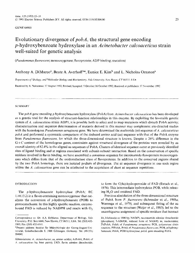
shown in Fig. I.
The p&A gene encodes a 404~aa protein with a caicu-
fated M, of 45 271. There are two potential ATG start
codons. Met” is in perfect agreement with PaPobA, but
Met 1 is more likely to be the first aa based on the quality
and Iocation of the ribosome-binding site (AAGGA)
(Shine and Dalgarno, 1974) just upstream from this start
codon. Following the stop codon (TAA) is a putative
Rho-independent termination structure consisting of a
26nt stem, a loop, and a T, stretch (Fig. 1).
The over&i G + C content of the A, ~~~~~~~~~~t~~u.~ pAA
gene (42%) is much lower than the G +C rich P. raerugi-
nos~t p&A gene (68%) (Entsch et al., 1988) and is consis-
tent with the disparity in the overall G + C content of the
individual organisms. In this case, the difference is largely
reflected in the codon preference. Third residue preference
strongly favors G and C in the P. ~~er~~~~os~~ gene (900/6
G+C), whereas the preference approaches the opposite
extreme in A. culcoucetieu.s (33% G + C).
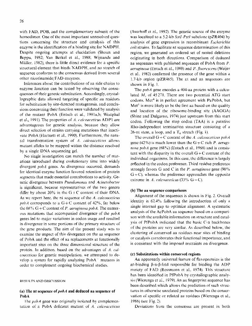
(b) The aa sequence comparisons
Alignment of the sequences is shown in Fig. 2. Overall
identity is 62.4% following the introduction of only a
single internal gap to optimize alignment. A systematic
analysis of the AcPobA aa sequence based on a compari-
son with the available information on structure and catal-
ysis of PfPobA indicated that the basic C-cl backbones
of the proteins are very similar. As described below, the
clustering of conserved as residues near sites of binding
or catalysis corroborates their functional importance, and
is consistent with the imposed restraints on divergence.
(c) substitutions within conserved regious
An apparently universal feature of ~avopruteins is the
nt-binding @-r-P-fold responsible for binding the ADP
moiety of FAD (Rossmann et al., 1974). This structure
has been identified in PfPobA by crystallographic analy-
sis (Wierenga et al., 1979). An aa fingerprint sequence has
been described which allows the prediction of such struc-
tures in otherwise unrelated proteins based on the conser-
vation of specific or related aa residues (Wierenga et al.,
1986) (see Fig. 2).
Deviations from the consensus are present in both
*t*t* TCTTTTTAATGAGGCGGTACTTTRAAAA TAGAAAATAGCAAGGATGATGTTATGCRAACT
N135-> (rbs) n Q T
ATGAAAACCRAAGTTGCAATTATTGGTTCTGGCCCAGCGGCCCAGCGGGA~~AC~AC~CGG~C~C~G MKTKVAIIGSG P A G L L L G Q L
CTTTACAAAGCTGGAATTGACACGTTATTGTGGMCAGCGGTGCCGATTACGTTGCA LYKAGIEHVI "EQRSADYVA
ser A c-N.204
TCACGCATTCGTGCAGGAATTTTAGAGAGTATCGGTCGC*GGA s R I RAGILE QVSVDLLEQAG
GTTGATCAGAACCTCAAAG AAAAAGGATTGCCACATTCGGGCATTGAAATTCTGACCAAT v DQNLKEKGLPHSGIEILTN
GGCCAAAAATTCCGTGTCGATTTATCGGCATTGACTCAAGGTCACGGTATAT GQKFRVDL SALTQGKQVTVY
GGGCAGACCGAAGTTACTAAAGATTTRRTGCAAGCACGTGAGCAGGCTGGTCTTTGCTCA GQTEVTKD LMQAREQACLCS
TTTTATGAATCGAATGATGTTCAAATTCATGATTCATAGTGACTTTT F Y E s N D "QIHDFY N A P K V T F
GAATCCRACGGRACTCACTATCRAATCGAATGTGTGATTTCATTGCAGGATGTGATGGTTAT ESNGTHYQIECDFIAGCDGY
CATGGCGTGTGCCGTGCTAGTGTGCCTCAAGATRAAATTAGGTCTAT H G "CRASV PQDKIKTFE K" Y
CCATTTGGTTGGTTAGGTGTACTTGCCGATGTGCCGCCTGTGGCAGACGAGTT~TTTAT P FGWLGVLADVP PVADELIY
GTTCAATCAGAGCGTGGTTTTGCACTGTGTAGCATGCGCTCAG-CGCG~GCCGATAT "QSERGFA LCSMRS ETRSRY
TACATTCAAGTTCCTTTAACCGATCACGTAGRRRACTGGT YIQ"PLTDH"ENWSDDQFWE
GAGCTTAAGAATCGCCTCGACCCTGAAAGCTGCTGCG~CTCGTTACAGGCCCTTC~TT ELKNRLDPESCE KLVTGPSI
GAGAAAAGTATTGCACCTTTGCGGAGCTTTGTCACAG~CCGATGCGATTTGG-TTA EKSIAPLRS F VT E PM R F G K L
TTCTTAGCTGGTGATGCCGCACATATTGTTCCACCAACGGTCTT F L A G D AAHIVPPTGAKGLNL
GCAGCTTCAGATATTGCATATTTGTCGRGTGTCATTGGGATCT AASOIAYLSSAL IEFYTQGS
GAGCRRGGTATAGATCARTACTCAGAAAAATGCTTGCTTGC~CGTGTATGG-GCAGAGCGT E Q G I DQYSEKCLQRVWKAER
TTTTCATGGTGGATGACCCATTTGTTACATCGCTTTCAAAGCGAGTTTGATCAT F SWWMTHLLHR FETESEFDH
-ATTRAA~AAGCAGAATTGAGCTATATCTTAGGTTCTACGGCAGGTCAGACC*C*C*C K I KQAELSYILG STAGQTTL
GCTGAAAACTATGTGGGTTTACCCTATGAAATCAAATCCCCATGCC A E N YVGLPYEIKS L 0 Y L K H A
AGCTAAAACPiAAAAGAGAGCGATTAG~TATCTCGGCTGTGTTTATTTA s -
CAAGTGAAATTCTCGGCTTTTTCACTGTCACTGTACCGT~A~AACG=GCGA~T~T~GGA TATGGACATAGTGGTCGAGTTCGATTGGCCGACCAGCTCGATGGT~CTCGCTATTGATT TCGCCACTTGCATTACCGACACCGCGTGCAGAAGCGAGAATTTGATCTGGTGCTTGACCA TATTCTACCCAGTTCACCAGCAGTRAGTGCATCAAACTGCCCACCC CGCGAATGATTCATTCCCGGAACGCGCGAT~CGAGC-CTTTGTGCGTCACCC- CTGCTGCTCTTGTATTTTGCCATCAGTTTATCGTACCAGTTTTGTGTGTCATCTACCG~ AATACGCCATCTGCTGTGCCTTGCACCACAATCATTTTACTTTA TCCAGATTGAGCTC
-SstI-
1329 1389 1449 1509 1569 1629 1689 1703
Fig. 1. The nt sequence of the pohA region of A. calcoaceticu.~ and
9 3
69 23
129 43
189 63
249 83
309 103
369 123
429 143
489 163
549 183
609 203
669 223
729 243
789 263
849 283
909 303
969 323
1029 343
1089 363
1149 383
1209 403
1269 404
deduced aa sequence of AcPobA. The putative ribosome-binding site
(rbs) is indicated by asterisks. The sequence of mutation pohA7 (indi-
cated in boldface) was determined following amplification of this region
of chromosomal DNA from strain ADP2309 by PCR using the appro-
priate oligodeoxynucleotide primers. The endpoints of nested deletion
plasmids used for the localization of p&A7 are indicated above the
sequence in italics. Those nt comprising a possible Rho-independent
terminator structure (AC’ ’ = - 19.2 kcalimol) are overlined. The com-
plete sequence has been submitted to GenBank and is available under
the accession No. LO3407. Methods: For nt sequence determination,
plasmid DNA was purified (Holmes and Quigley, 1981), and extracted
once with a solution of Tris-buffered (pH 8.0) phenol:chloroform:isoa-
myl alcohol (24:24:1), and once with a solution of chloroform:isoamyl
alcohol (24:l). Following precipitation with ethanol, the dsDNA sample
was used in dideoxy chain-termination sequencing reactions (Sanger
et al., 1977) with [a-35S]dATP.
AcPobA and PfPobA. In the first case, Gly18 occurs in
place of an acceptable small hydrophobic aa within the
a-helix in both PobA (unless otherwise indicated, residues
are numbered according to the PfPobA sequence to facili-
AC-
Pf-
AC-
Pf-
AC-
Pf-
AC-
Pf-
*c-
Pf-
AC-
Pf-
AC-
Pf-
AC-
Pf-
27
S (AOP2309) ,#..** * .(LOOP). . ! +
MQTMKTK"AIlGSGPAGLLLaQLLYKAG~E~"~"EQRSADY"AsRI~G*LSQ"S -55
III IIIII~Il~IIIIIIlI IIll. II.1 . III IIIl/.III MKTQ"AIIGAGPSGLLLGQLLHKAGIDNVILERQTPDYVLG"LEQG~ -52
00 0000 0000 0 cl 00 +++
VDLLEQAGVDQNLKEKGLPHSGIKILTNGQKFR~DLSALTQGKQVTVYGQTEVTK -110
III IIII II I I.4 II. I.11 I. II II/IIIIIII. VDLLREAGVDRRMARDGLVHEGVEIAFAGQRRR~DLKRLSGGKTVTVYGQTKVTR -107
0
DLMQAREQAGLCSFYESNDVQIHDFYNA-PKVTFESNGTHYQ~KCDFIAGCDGY~ -164
IllIll I .I..I.Il I I/I/ I ..ll.I/lIII.I DLMEAREACGATTVYQAAEVRLHDLQGERPYVTFERDGERLRLDCOYIAGCDGFH -162
0 0 0 00000
GVCRASVPQDKIKTFEKVYPFGWLGVLAOVPPVADELIYVQSERCFALCSMRSET -219
1. I I.1 ...I lI~II/llIl/.III Ill. III1 . IlIIIll II I GISRQSIPAERLKVFER"YPFG~LGLLADTPP"S~~LIYA~~PRGFALCSQRSAT -217 00 = ++ +++
RSRYYIQVPLTDHVENWSDOQFWEELKNRLDPESCEKLVTG~SI~KSIAFLRSFV -274
lIllI~IIlll. II III. II III II I IIIIIIIl./lIIl/III/l RSRYYVQVPLTEKVEDWSDERFWTELKARLPAEVAEKLVTG~SLKKS~AFLRSF" -272
++ =
TEPMRFGKLFLAGDAAHIVPPTGAKGLNLAASDIAYLSSALIEFYTQGSEQGIDQ -329
III I~I/IIII/ll/IllIlIlllllllII~~ I I. I I VEPMQHGRLFLAGOAAHIVPPTGAKGLNLAASDVSTLYRLLLKAYREGRGELLER -327 = 000 0 0 ODCJO
++ +
YSEKCLQRVWKAERFSWWMTHLLHRFETESEFOHKIKQAELSYILGSTAGQTTLA -384
II II l~llIIlllIIII .llII I .I I.11 I Ill II 4.1 YSAICLRRIWKAERFSWWMTSVLHRFPDTOAFSQRIQQTELEYYLGSEAGLATIA -382
= = == == == == == = : =
ENYVGLPYE-IKSLDYLKHAS -404
IllIlIIII I ENYVGLPYEEIE =+
Fig. 2. Alignment of the aa sequences of PobA from A. culcoucericu,s
(AC) and P. ~uorescens (Pf). The PfPobA sequence was taken from
Weijer et al. (1982). Identical aa are connected by vertical lines. Various
marks indicate specific residues: FAD-binding, small circles (Cl); p-hy-
droxybenzoate-binding, +; dimer interface, =, Interacting aa were
determined from published sources (Schreuder et al., 1988a; 1989; Weijer
et al., 1983), and from direct observation of PfPobA from coordinates
IPHH and 2PHH available in the Brookhaven database (Bernstein
et al., 1977). The consensus ADP-binding fold sequence (Wierenga et al.,
1986) is indicated above the residues: a: K,R,H,S,T,Q,N; closed squares
(m): A,I,L.V,M,C; asterisks (*): G only: !: D or E. The aa substitution
in PobA caused by mutation pohA7 in ADP2309 is indicated by an
upward arrow.
tate reference to the three-dimensional structure;
Fig. 3)(Weijer et al., 1983). The second deviation occurs
within the second P-strand at Asnz8 in PfPobA (Weijer
et al., 1983). The replacement of Asnz8 with His in
AcPobA is interesting, because both Asn and His repre-
sent a common departure from the hydrophobic residue
expected at this position in the P-CL-~ fold. The three-
dimensional structure shows Asn28 in contact with Arg113
(Hofsteenge et al., 1980). While it is likely that His in
AcPobA interacts similarly, the difference in volume
(Asn= 135.2 A” and His= 167.3 A”; Richards, 1977), and
the position of this side chain in the interior of the protein
prevented satisfactory packing during attempts to simu-
late this replacement.
Most of the specific residues identified as making con-
tact with the FAD molecule are strictly conserved be-
tween the two proteins (Fig. 2). On the combined basis
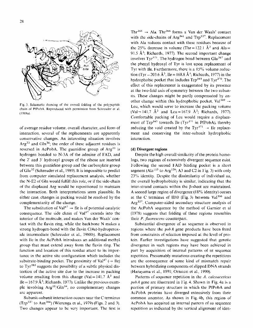
Fig. 3. Schematic drawing of the overall folding of the poiypeptide
chain of PfPobA. Reproduced with permission from Schreuder et al.
(1988a).
of average residue volume, overall character, and form of
interaction, several of the replacements are apparently
conservative changes. An intcrcsting situation involves
Args3 and Gln 34; the order of these adjacent residues is
reversed in AcPobA. The guanidine group of Arg3’ is
hydrogen bonded to N-3A of the adenine of FAD, and
the 2’ and 3’ hydroxyl groups of the ribose are inserted
between this guanidine group and the carboxylate group
of G1u32 (Schreuder et al., 1989). It is impossible to predict
from computer simulated replacement analysis, whether
the N-E2 of Gln would fulfill this role, or if the side-chain
of the displaced Arg would be repositioned to maintain
the interaction. Both interpretations seem plausible. In
either case, changes in packing would be resofved by the
complementarity of the change.
The substitution of Va14’ -+ Be is of potential catalytic
consequence. The side chain of Va14’ extends into the
interior of the molecule, and makes Van der Waals’ con-
tact with the flavin ring, while the backbone N makes a
strong hydrogen-bond with the flavin C(4a)-hydroperox-
ide intermediate (Schreuder et al., 1988b). Replacement
with Be in the AcPobA introduces an additional methyl
group that must extend away from the flavin ring. The
function and location of this residue attest to its impor-
tance in the active site configuration which includes the
substrate-binding pocket. The proximity of Va147 (-+ Be)
to Tyi? suggests the possibility of a subtle physical dis-
tortion of the active site due to the increase in packing
volume resulting from this change (Val= 141.7 A3 and
Ile = 167.9 A”; Richards, 1977). Unlike the previous exam-
ple involving Arg33/Gln34, no complementary changes
are apparent.
Subunit-subunit interaction occurs near the C terminus
(Trp337 to Asn384) (Wierenga et al., 1979) (Figs. 2 and 3).
Two changes appear to be very important. The first is
Thr36h + Ala. Thr”” forms a Van der Waals’ contact
with the side-chains of Argj41 and Trp337. Replacement
with Ala reduces contact with these residues because of
the 25% decrease in volume (Thr = 122.1 A” and Ala=
91.5 A3; Richards, 1977). The second important change
involves Tyr . 371 The hydrogen bond between G1u367 and
the phenyl hydroxyl of Tyr is lost upon replacement of
Tyr with Ile. Furthermore, there is a 15% volume reduc-
tion (Tyr = 203.6 A3, Be = 168.8 A3; Richards, 1977) in the
hydrophobic pocket that includes Trp345 and Tyr3”. The
effect of this replacement is exaggerated by its presence
at the two-fold axis of symmetry between the two subun-
its. These changes might be partly compensated by an-
other change within this hydrophobic pocket, Va134y -+
Leu, which would serve to increase the packing volume
(Val= 141.7 A” and Leu = 167.9 A”; Richards, 1977).
Comfortable packing of Leu would require a displace-
ment of Trp345 towards Be (Tyr 371 in PfPobA), thereby
reducing the void created by the Tyr3” -+ Ile replace-
ment and conserving the inter-subunit hydrophobic
interaction.
(d) Divergent regions
Despite the high overall similarity of the protein homo-
logs, two regions of extensively divergent sequence exist.
Following the second FAD binding pocket is a short
segment (Ala”’ to Arg136; A3 and C2 in Fig. 3) with only
23% identity. Despite the dissimilarity of individual aa,
the overall hydrophobicity is similar, indicating that the
inter-strand contacts within the P-sheet are maintained.
A second large region of divergence (I 8% identity) occurs
at the C terminus of HI0 (Fig. 3) between Va13e6 and
Arg3”. Computer-aided secondary structure analysis of
the AcPobA sequence by the method of Garnier et al
(1978) suggests that folding of these regions resembles
their P. fhrescens counterpart.
Substantial divergence of aa sequence is observed in
regions where the p&A gene products have been freed
from constraints of selection imposed at the level of pro-
tein. Earlier investigations have suggested that genetic
divergence in such regions may have been achieved in
part by acquisition of internal patterns of nt sequence
repetition. Presumably mutations creating the repetitions
are the consequence of some kind of mismatch repair
between hybridizing components of slipped DNA strands
(Harayama et al., 1991; Ornston et al., 1990).
Patterns of sequence repetition in the A. calcoaceticus
p&A gene are illustrated in Fig. 4. Shown in Fig. 4a is a
portion of primary structure in which the PfPobA and
AcPobA proteins have diverged extensively from their
common ancestor. As shown in Fig. 4b, this region of
AcPobA has acquired an internal pattern of aa sequence
repetition as indicated by the vertical alignment of iden-
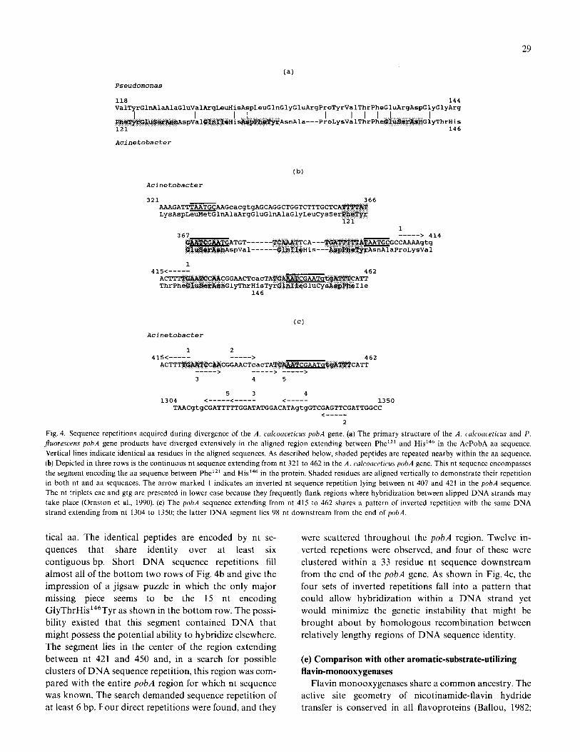
29
Pseudomonas
118 144 ValTyrGlnAlaAlaGluValArgLeuHisAspLeuGlnGlyGluArgProTyrValThrPheGluArgAspGlyGlyArg
I I I I I I I I J. I Phra'fyrOlUSlarAs~spVa1011I~eHis~~he*yrAsnnla---ProLysValThrPheG UScarAsmGlyThrHis 121 146
Acinetobacter
(b)
Acinetobacter
321 366 AAAGATTTAATGCAAGcacgtgAGCAGGCTGGTCTTTGCTCATTT%%T LysAspLeuMetGlnAlaArgGluGlnAlaGlyLeuCysSerpheTyr
121 1
367 -----> 414 TGT------TCA,A?$TTCA---'XYSA%"fW%%$!~TGCGCCAAAAgtg pVal------GlhIleHis---AspPhe!Q?zAsnAlaProLysVal
1 415<----- 462
ACTTT%WW!X!CMCGGAACTcacTATCA&&WGAATq~gA~CATT ThrPheG~uSe7&w#GlyThrHisTyrGknI~eGluCysAspPheIle
146
Acinetobacter
415<____1 L _-_-_--_-> 462 ACTTTTGi&TCCMCGGAACTcacTATCP&&TCGAATqiQXPTTCATT
--_-_-_-_> --_--_-> _-_-_-__>
3 4 5
5 3 4 1304 <_-_-_-_-_-<_-_-_-_- <----- 1350
TAACgtgCGATTTTTGGATATGGACATAgtgGTCGAGTCGAGTTCGATTGGCC <--_-_-_-_
2
Fig. 4. Sequence repetitions acquired during divergence of the A. calcoaceticus pobA gene. (a) The primary structure of the A. calcoaceticus and P. fluoresceas pobA gene products have diverged extensively in the aligned region extending between Phe”’ and His 146 in the AcPobA aa sequence.
Vertical lines indicate identical aa residues in the aligned sequences. As described below, shaded peptides are repeated nearby within the aa sequence.
(b) Depicted in three rows is the continuous nt sequence extending from nt 321 to 462 in the A. calcoaceticus pobA gene. This nt sequence encompasses
the segment encoding the aa sequence between Phe’*r and His I46 in the protein. Shaded residues are aligned vertically to demonstrate their repetition
in both nt and aa sequences. The arrow marked I indicates an inverted nt sequence repetition lying between nt 407 and 421 in the pobA sequence.
The nt triplets cat and gtg are presented in lower case because they frequently flank regions where hybridization between slipped DNA strands may
take place (Ornston et al., 1990). (c) The pobA sequence extending from nt 415 to 462 shares a pattern of inverted repetition with the same DNA
strand extending from nt 1304 to 1350; the latter DNA segment lies 98 nt downstream from the end of pobA.
tical aa. The identical peptides are encoded by nt se-
quences that share identity over at least six
contiguous bp. Short DNA sequence repetitions fill
almost all of the bottom two rows of Fig. 4b and give the
impression of a jigsaw puzzle in which the only major
missing piece seems to be the 15 nt encoding
GlyThrHis 146Tyr as shown in the bottom row. The possi-
bility existed that this segment contained DNA that
might possess the potential ability to hybridize elsewhere.
The segment lies in the center of the region extending
between nt 421 and 450 and, in a search for possible
clusters of DNA sequence repetition, this region was com-
pared with the entire pobA region for which nt sequence
was known. The search demanded sequence repetition of
at least 6 bp. Four direct repetitions were found, and they
were scattered throughout the pobA region. Twelve in-
verted repetions were observed, and four of these were
clustered within a 33 residue nt sequence downstream
from the end of the pobA gene. As shown in Fig. 4c, the
four sets of inverted repetitions fall into a pattern that
could allow hybridization within a DNA strand yet
would minimize the genetic instability that might be
brought about by homologous recombination between
relatively lengthy regions of DNA sequence identity.
(e) Comparison with other aromatic-substrate-utilizing
flavin-monooxygenases
Flavin monooxygenases share a common ancestry. The
active site geometry of nicotinamide-flavin hydride
transfer is conserved in all flavoproteins (Ballou, 1982;
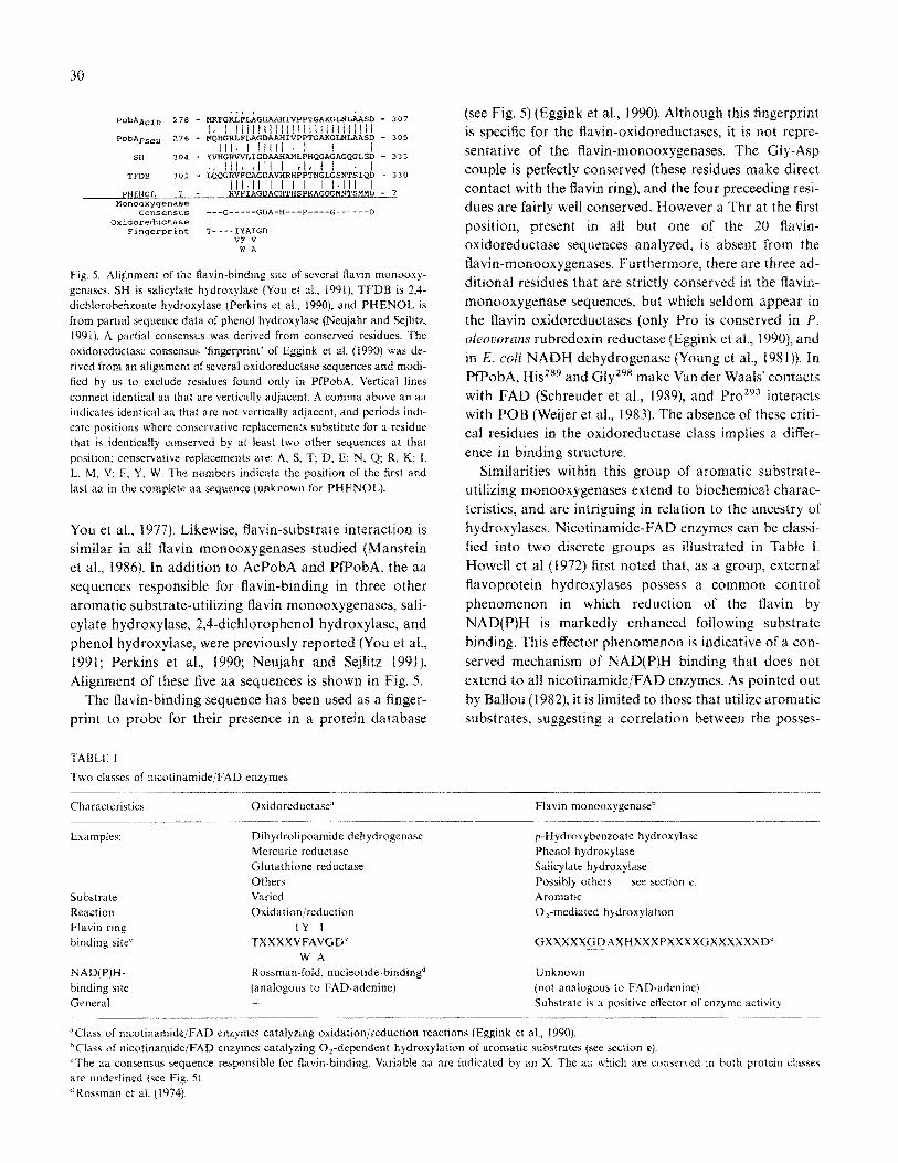
30
..I I
Pob&cin 278 - MXfGXLFLAGD~HIVPPTG~G~~SD I 357
1, I IlJlittillillllllI1llliil POb+seu 276 - ~QHGRLF~GD~H~VP~G~GL~~SD - 355
lll”liiili-I t i SX 354 - Y~nGRWLIGD~~L~HQGAGAGQGLE~ - 333
.Il/lt~lI/I rlrll . I TFDB 301 - LQQGRVFCAGDAVHRHPPTNGLGSNTSIQD - 330
III.IIII I I I I I.111 I PHENOL ? I RVFIAGD_ACHTHSPKAGQGBt4TSftMD - ?
M0”0C%ypXl&=462 ccmsensus ___G-____GDA_H___P____G_____-D
Oxidoreductase Fingerprint T----IYAIGD
VF V WA
Fig, 5. Alignment of the Gavin-binding site of severai fiavm monooxy-
genases. SW is salicylate hydroxylast- (You et al., 1991), TF’DB is 2,4-
dichlorobenzoate hydroxylase (Perkins et al., 19901, and PHENOL is
from partial sequence data of phenol hydroxylase (Neujahr and Sejlitz,
1991). A partiaf consensus was derived from conserved residues. The
oxidoreductase consensus fingerprint’ of Eggink et al. (1990) was de-
rived from an alignment of several oxidoreductase sequences and modi-
fied by us to exclude residues found only in PfPobA. Vertical lines
connect identical aa that are vertically adjacent. A comma above an aa
indicates identical aa that are not vertically adjacent, and periods indi-
cate positions where conservative replacements substitute for a residue
that is identicdIIy conserved by at least two other sequences at that
position; conservative replacements are: A, S, T: D, E; N, Q; R, K; I.
L. M, V; F, Y, W. The numbers indicate the position of the iirst and
last aa in the complete aa sequence (unknown for PHENOL).
You et al., 1977). Likewise, Gavin-substrate interaction is
similar in all ilavin m~)nooxygenases studied (~anstein
et al.. 1986). In addition to AcPobA and PfPobA, the aa
sequences responsible for flavin-binding in three other
aromatic substrate-utilizing flavin monooxygenases, sali-
cylate hydroxylase, 2,4-dichlorophenol hydroxylase, and
phenol hydroxylase, were previously reported (You et al.,
I99 I; Perkins et al., 1990; Neujahr and Sejiitz 1991 j. Alignment of these five aa sequences is shown in Fig. 5.
The flavin-binding sequence has been used as a finger-
print to probe for their presence in a protein database
TABLE I
Two classes of nicotinamide/FAD enzymes
(see Fig. 5) (Eggink et al., 1990). Although this fingerprint
is specific for the Gavin-oxidoreductases~ it is not repre-
sentative of the Gavin-monooxygenases. The GIy-Asp
couple is perfectly conserved (these residues make direct
contact with the Ravin ring), and the four preceeding resi-
dues are fairly well conserved. However a Thr at the first
position, present in all but one of the 20 flavin-
oxidoreductase sequences analyzed, is absent from the
Gavin-monoaxygenases. Furthermore, there are three ad-
ditional residues that are strictly conserved in the ftavin-
monooxygenasc sequences, but which seldom appear in
the flavin oxidoreductases (only Pro is conserved in P.
~~~~~#~~~s rubredoxin reductase (Eggink et al., 1990), and
in E. cvfi NADH dehydrogenase (Young et al., 1981)). In
PfPobA, His”” and Glyz9’ make Van der Waals’ contacts
with FAD (Schreuder et al., 1989), and Pro’“” interacts
with POB (Weijer et al., 1983). The absence of these criti-
cal residues in the oxidoreductase class implies a differ-
ence in binding structure.
Similarities within this group of aromatic substrate-
utilizing monooxygenases extend to biochemical charac-
teristics, and are intriguing in relation to the ancestry of
hydroxylases. Nicotinamide-FAD enzymes can be classi-
fied into two discrete groups as illustrated in Table I.
Howell et al (1972) first noted that, as a group, external
flavoprotein hydroxylases possess a common control
phenomenon in which reduction of the tlavin by
NAD(P)H is markedly enhanced following substrate
binding. This effector phenomenon is indicative of a con-
served mechanism of NAD(P)H binding that does not
extend to all nicotinamide/FAD enzymes. As pointed out
by Ballou (1982), it is limited to those that utilize aromatic
substrates, suggesting a correlation between the posses-
Characteristics Oxidoreductase” Flavin monooxygenas@
Examples:
Substrate
Reaction
Flavin ring
binding site’
NAD(P)H-
binding site
General
Dihydrolipoamide dehydrogenase
Mercuric reductase
Glutathione reductase
Others
Varied
O~id~ition~reduction
IY 1
TXXXXVFAVGD”
WA
Rossman-fold, nucleottdz-binding”
(analogous to FAD-adenine)
p-Hydroxybenzoate hydrorylase
Phenol hydroxylase
Salicylate hydroxylase
Possibly others - see sectlon e.
Aromatic
O,-mediated hydr(~xylation
-
Unknown
(not analogous to FAD-adenine)
Substrate is a positive effector of enzyme activity
“Class of nicotinamide/FAD enzymes catalyzing oxidation/reduction reactions (Eggink et al., 1990).
bClass of nicotin~mide~FAD enzymes catalyzing O,-dependent hydro~~lation of aromatic substrates (see section e),
“The aa consensus sequence responsible for flavin-binding. Variable aa are indicated by an X. The aa which are conserved in both protein ciasses
are underlined (see Fig. Sj.
’ Rossmdn et a). ( 19%).
31
sion an atypical flavin ring/NAD(P)H binding site and the ability of the substrate molecule to dramatically increase the affinity for NAD(P)H binding. These proper- ties segregate with the more obvious distinction of reac- tion mechanism (i.e., O,-mediated hydroxylation of an aromatic substrate). The effector phenomenon has been observed in several hydroxylase enzymes: PobA (Howell et al., 1972; Spector and Massey, 1972), salicylate hydrox- ylase (White-Stevens et al., 1972), orcinol hydroxylase (Otha and Ribbons, 1970), melifotate hydroxylase (Strick- land and Massey, 19731, phenol hydroxylase (Massey and Hemmerich, 1975; Neujahr and Gaal, 1973), and m-hy- droxybenzoate-6-hydroxylase (Massey and Hemmerich, 1975).
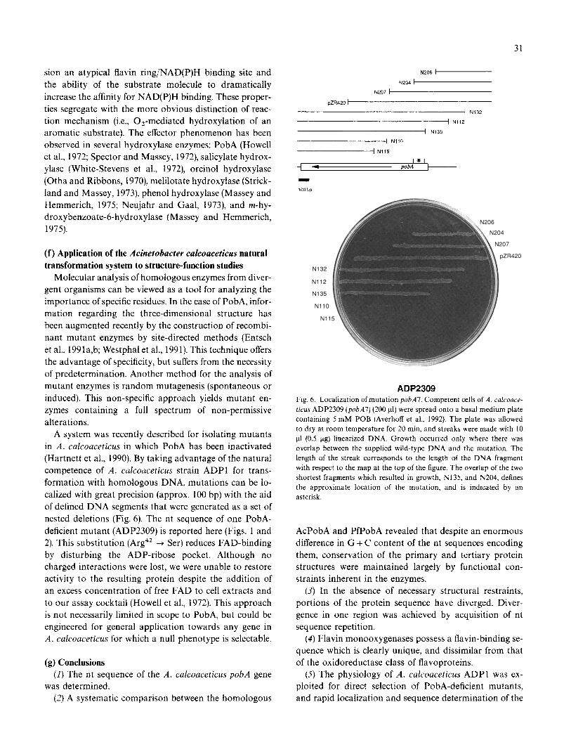
(f) Application of the Acinctobacter cafcoaceticus natural
transformation system to structure-function studies
Molecular analysis of homologous enzymes from diver- gent organisms can be viewed as a tool for analyzing the importance of specific residues. In the case of PobA, infor- mation regarding the three-dimensional structure has been augmented recently by the construction of recombi- nant mutant enzymes by site-directed methods (Entsch et al., 1991a,b; Westphal et al., 1991). This technique offers the advantage of specificity, but suffers from the necessity of predetermination. Another method for the analysis of mutant enzymes is random mutagenesis (spontaneous or induced). This non-specific approach yields mutant en- zymes containing a full spectrum of non-permissive alterations.
A system was recently described for isolating mutants in A. c~~couceticus in which PobA has been inactivated (Hartnett et al., 1990). By taking advantage of the natural competence of A. calcoaceticus strain ADPI for trans- formation with homologous DNA, mutations can be lo- calized with great precision (approx. 100 bp) with the aid of defined DNA segments that were generated as a set of nested deletions (Fig. 6). The nt sequence of one PobA- deficient mutant (ADP2309) is reported here (Figs. 1 and 2). This substitution (Arg4’ -+ Ser) reduces FAD-binding by disturbing the ADP-ribose pocket. Although no charged interactions were lost, we were unable to restore activity to the resulting protein despite the addition of an excess concentration of free FAD to cell extracts and to our assay cocktail (Howell et al., 1972). This approach is not necessarily limited in scope to PobA, but could be engineered for general application towards any gene in A. calcoaceticus for which a null phenotype is selectable.
(g) Conclusions
(I) The nt sequence of the A. calcoaceticus pobA gene was determined.
(2) A systematic comparison between the homologous
N’2GE a
N204 7
N207 1
pZR420 /
I N132
/ N112
1 N135
-1 NiiO - N115
i 4
N132
N112
Ni35
NllO
Nll
i420
ADP2309 Fig. 6. Localization of mutation pobA7. Competent cells of A. calcoaoe-
ticus ADP2309 (pobA7) (200 pl) were spread onto a basal medium plate
containing 5 mM POB (Averhoff et al., 1992). The plate was allowed
to dry at room temperature for 20 min, and streaks were made with IO
u1 (0.5 pg) linearized DNA. Growth occurred only where there was
overlap between the supplied wild-type DNA and the mutation. The
length of the streak corresponds to the length of the DNA fragment
with respect to the map at the top of the figure. The overlap of the two
shortest fragments which resulted in growth, Ni35, and N204. defines
the approximate location of the mutation, and is indicated by an
asterisk.
AcPobA and PfPobA revealed that despite an enormous difference in G + C content of the nt sequences encoding them, conservation of the primary and tertiary protein structures were maintained largely by functional con- straints inherent in the enzymes.
(3) In the absence of necessary structural restraints, portions of the protein sequence have diverged. Diver- gence in one region was achieved by acquisition of nt sequence repetition.
(4) Flavin monooxygenases possess a flavin-binding se- quence which is clearly unique, and dissimilar from that of the oxidoreductase class of flavoproteins.
(5) The physiology of A. calcoaceticus ADPl was ex- ploited for direct selection of PobA-deficient mutants, and rapid localization and sequence dete~ination of the
32
mutant allele. This technique can provide information
concerning non-obvious, but structurally or functionally
critical residues.
ACKNOWLEDGEMENTS
We wish to thank David Jacobsohn for his assistance
in isolating and localizing pobA mutants, and J. Drenth
(University of Griiningen, The Netherlands) for his kind
permission to reproduce Fig. 3. This work was funded by
the Celgene Corporation, the National Science Founda-
tion (MCB-9004839), the National Institutes of Health,
and the Army Research Office. AAD was supported by
a postdoctoral fellowship (PF-3543) from the American
Cancer Society.
REFERENCES
Averhoff, B.A., Gregg-Jolly, L.A., Elsemore, D.A. and Omston, L.N.:
Genetic analysis of supraoperonic clustering by use of natural trans-
formation in Acinetohacter calcoaceticus. J. Bacterial. 174 (1992)
200&204.
Ballou, D.P.: Flavoprotein monooxygenases. In: Massey, V. and Wil-
liams, C.H. (Eds.), Flavins and Flavoproteins. Elsevier. Amsterdam,
1982, pp. 301-310
Bernstein, F.C., Koetzle, T.F., Williams, G.J.B., Meyer Jr.. E.F., Brice,
M.D., Rodgers, J.R., Kennard, O., Shimanouchi, T. and Tasumi, M.:
The protein data bank: a computer-based archival file for macromo-
lecular structures. J. Mol. Biol. I12 (1977) 535-542.
Eggink, G., Engel, H., Vriend, G., Terpstra, P. and Witholt, B.: Ru-
bredoxin reductase of Pseudomonns oleouorans. J. Mol. Biol. 212
(1990) 135-142.
Entsch, B., Ballou, D.P. and Massey, V.: Flavin-oxygen derivatives in-
volved in hydroxylation by p-hydroxybenzoate hydroxylase. J. Biol.
Chem. 25 I (1976) 2550-2563.
Entsch, B., Nan, Y., Weaich, K. and Scott, K.F.: Sequence and organiza-
tion of pohA, the gene coding for p-hydroxybenzoate hydroxylase,
an inducible enzyme from Pseudomonas aeru,qinosa. Gene 71
(1988) 2799291.
Entsch, B., Palfey, B.A., Ballou, D.P. and Massey, V.: Catalytic function
of tyrosine residues in p-hydroxybenzoate hydroxylase as deter-
mined by the study of site-directed mutants. J. Biol. Chem. 266
(1991a) 17341-17349.
Entsch, B., Palfey, B.A., Ballou, D.P. and Massey, V.: Chemical func-
tions of amino acid residues in the active site of p-hydroxybenzoate
hydroxylase as studied by site-directed mutagenesis and biophysical
techniques. In: Massey, V. and Williams, C.H. (Eds.), Flavins and
Flavoproteins 1990. Walter de Gruyter, Berlin, 1991b, pp. 219-230
Garnier, J., Osguthorpe, D.J. and Robson, B.: Analysis of the accuracy
and implications of simple methods for predicting the secondary
structures of globular proteins. J. Mol. Biol. 120 (1978) 97-120.
Harayama, S., Rekik, M., Bairoch, A., Neidle, E.L. and Ornston, L.N.:
Potential DNA slippage structures acquired during evolutionary
divergence of Acinetohacter calcoaceticus chromosomal henABC
and Pseudomonas putida TOL pWW0 plamid xylXYZ, genes encod-
ing benzoate dioxygenases. J. Bacterial. 173 (I 99 I) 7540-7548.
Hartnett, G.B., Averhoff, B.A. and Ornston, L.N.: Selection of Acineto-
hatter calcouceticus mutants deficient in the p-hydroxybenzoate hy-
droxylase gene (p&A), a member of a supraoperonic cluster.
J. Bacterial. 172 (1990) 6160+6161.
Hofsteenge, J., Vereijken, J.M., Weijer, W.J., Beintema, J.J., Wierenga,
R.K. and Drenth, J.: Primary and tertiary structure studies of p-
hydroxybenzoate hydroxylase from Pseudomonas ,jhorescens Eur.
J. Biochem. I I3 (1980) 141-150.
Holmes, D.S. and Quigley, M.: A rapid boiling method for the prepara-
tion of bacterial plasmids Anal. Biochem. 1 I4 (1981) 193-197.
Howell, L.G., Spector, T. and Massey, V.: Purification and properties
of p-hydroxybenzoate hydroxylase from Pseudomonas ,jluorescens
J. Biol. Chem. 247 (1972) 4340-4350.
Manstein, D.J., Pai, E.F., Schopfer, L.M. and Massey, V.: Absolute
stereochemistry of flavins in enzyme-catalyzed reactions. Biochemis-
try 25 (1986) 6807768 16.
Massey, V. and Hemmerich, P.: Flavin and Pteridine Monooxygenases.
In: Boyer, P.D., (Ed.), The Enzymes, 3rd ed. Academic Press, New
York, 1975, pp. 191-252.
Neujahr, H.Y. and Gaal, A.: Phenol hydroxylase from yeast. Purilica-
tion and properties of the enzyme from Trichosporon cutaneum. Eur.
J. Biochem. 35 (1973) 3866400.
Neujahr, H.Y. and Sejlitz, T.: Partial sequences of phenol hydroxylase
and similarities with other flavoproteins. In: Massey, V. and Wil-
liams, C.H., (Eds.), Flavins and Flavoproteins 1990. Walter de
Gruyter, Berlin, 1991, pp. 2355238
Ornston, L.N., Houghton, J., Neidle, E.L. and Gregg, L.A.: Subtle selec-
tion and novel mutation during evolutionary divergence of the B-
ketoadipate pathway. In: Silver, S. (Ed.), Pseudomonas:
Biotransformations, Pathogenesis, and Evolving Biotechnology.
Am. Sot. Microbial., Washington, DC, 1990, pp. 207-225.
Otha, Y. and Ribbons, O.W.: Crystallization of orcinol hydroxylase
from Pseudomonas putida. FEBS Lett. I I (1970) 1899192.
Perkins, E.J., Gordon, M.P., Caceres, 0. and Lurquin, P.F.: Organiza-
tion and sequence analysis of the 2,4-dichlorophenol hydroxylase
and dichlorocatechol oxidative operons of plasmid pJP4 J. Bacte-
riol. 172 (1990) 2351-2359.
Richards, F.M.: Areas, volumes, packing, and protein structure Ann.
Rev. Biophys. Bioeng. 6 (I 977) 15 I -176.
Rossmann, M.G.. Moras, D. and Olsen, K.W.: Chemical and biological
evolution of a nucleotide-binding protein. Nature 250 (1974)
194- 199.
Sanger, F., Nicklen, S. and Coulson, A.R.: Nucleotide sequencing with
chain-terminating inhibitors. Proc. Natl. Acad. Sci. USA 74 (1977)
546335467.
Schreuder, H.A., Van der Laan, J.M., Hol, W.G.J. and Drenth, J.: Crys-
tal structure of p-hydroxybenzoate hydroxylase complexed with its
reaction product 3,4-dihydroxybenzoate. J. Mol. Biol. 199 (1988a)
6377648.
Schreuder, H.A., HOI, W.G.J. and Drenth, J.: Molecular modeling re-
veals the possible importance of a carbonyl oxygen binding pocket
for the catalytic mechanism of p-hydroxybenzoate hydroxylase.
J. Biol. Chem. 263 (1988b) 313ll3136.
Schreuder, H.A., Prick, P.A.J., Wierenga, R.K., Vriend, G., Wilson, K.S.,
HOI, W.G.J. and Drenth, J.: Crystal structure of the p-hydroxyben-
zoate hydroxylase-substrate complex refined at 1.9 8, resolution,
J. Mol. Biol. 208 (1989) 6799696.
Shine, J. and Dalgarno, L.: The 3’-terminal sequence of Escherichia cali
16s ribosomal RNA. Proc. Natl. Acad. Sci. USA 71 (1974)
I3422 1346.
Shoun, H. and Beppu, T.: A histidine residue in p-hydroxybenzoate
hydroxylase essential for binding of reduced nicotinaminde adenine
dinucleotide phosphate. J. Biol. Chem. 257 (1982) 342223428.
Shoun, H., Higashi, N., Beppu, T., Nakamura, S., Hiromi, K. and Arima,
K.: Studies on the interaction of p-hydroxybenzoate hydroxylase
with NADPH.J. Biol. Chem. 254 (1979) 10944-10951.
Spector, T. and Massey, V.: Studies on the effector specificity of p-
hydroxybenzoate hydroxylase from Pseudomonas,puorescens J. Biol.
Chem. 247 (1972) 467994687.
33
Strickland, S. and Massey, V.: The mechanism of action of the flavopro-
tein melilotate hydroxylase. J. Biol. Chem. 248 (1973) 295332962.
Van Berkel, W.J.H., Miiller, F., Jekel, P.A., Weijer, W.J., Schreuder,
H.A. and Wierenga, R.K.: Chemical modification of tyrosine-38 in
p-hydroxybenzoate hydroxylase from Pseudomonas fluorescens by
5’.p-fluorosulfonylbenzoyladenosine. Eur. J. Biochem. 176 (1988)
4499459.
Weijer, W.J., Hofsteenge, J., Beintema, J.J., Wierenga, R.K. and Drenth,
J.: p-Hydroxybenzoate hydroxylase from Pseudomonas puorescens. Eur. .I. Biochem. 133 (1983) 109-l 18.
Weijer, W.J., Hofsteenge, J., Vereijken, J.M., Jekel, P.A. and Beintema,
J.J.: Primary structure of p-hydroxybenzoate hydroxylase from
Pseudomonas puorescens. Biochim. Biophys. Acta 704 (1982)
385-388.
Westphal, A.H., Eschrich, K., Van Dongen, W.M.A.M., Benen, J.A.E.,
de Kok, A. and Van Berkel, W.J.H.: Site-directed mutagenesis of p- hydroxybenzoate hydroxylase from Pseudomonas ,juorescens. In:
Massey, V. and Williams, C.H., (Eds.), Flavins and Flavoproteins
1990. Walter de Gruyter, Berlin, 1991, pp. 231-234.
White-Stevens, R.H. and Kamin, H.: Studies of a flavoprotein, salicylate
hydroxylase. J. Biol. Chem. 247 (1972) 2358-2370.
White-Stevens, R.H., Kamin, H. and Gibson, Q.H.: Studies of a flavo-
protein, salicylate hydroxylase. J. Biol. Chem. 247 (I 972) 2371-238 1.
Wierenga, R.K., De Jong, R.J., Kalk, K.H., HOI, W.G.J. and Drenth, J.:
Crystal structure of p-hydroxybenzoate hydroxylase J. Mol. Biol.
I31 (1979) 55-73.
Wierenga, R.K., Terpstra, P. and HOI, W.G.J.: Prediction of the occur-
rence of the ADP-binding P-a-B-fold in proteins, using an amino
acid sequence fingerprint. J. Mol. Biol. 187 (1986) 101-107.
Wijnands, R.A. and Miller, F.: A study of p-hydroxybenzoate hydrox-
ylase from Pseudomonas ,fluorescens. Biochemistry 21 (1982)
663996646.
You, L-S., Ghosal, D. and Gunsalus, I.C.: Nucleotide sequence analysis
of the Pseudomonas putida PpG7 salicylate hydroxylase gene (nahG)
and its 3’ flanking region, Biochemistry 30 (1991) 163551641.
You, K., Arnold, L.J. and Kaplan, N.O.: The stereospecificity of bacte-
rial external flavoprotein monooxygenases for nicotinamide adenine
dinucleotide. Arch. Biochem. Biophys. I80 (I 977) 550-554.
Young, J.G., Rogers, B., Campbell, H.D., Jaworowski, A. and Shaw,
D.C.: Nucleotide sequence coding for the respiratory NADH
dehydrogenase of Escherichia cob. Eur. .I. Biochem. I16 (1981)
165-170.
Related Documents