HAL Id: halsde-01022002 https://hal.archives-ouvertes.fr/halsde-01022002 Submitted on 27 May 2020 HAL is a multi-disciplinary open access archive for the deposit and dissemination of sci- entific research documents, whether they are pub- lished or not. The documents may come from teaching and research institutions in France or abroad, or from public or private research centers. L’archive ouverte pluridisciplinaire HAL, est destinée au dépôt et à la diffusion de documents scientifiques de niveau recherche, publiés ou non, émanant des établissements d’enseignement et de recherche français ou étrangers, des laboratoires publics ou privés. Evolutionary and ecological perspectives of Late Paleozoic ferns. Part III. Anachoropterid ferns (including Anachoropteris, Tubicaulis, the Sermayaceae, Kaplanopteridaceae and Psalixochlaenaceae) Jean Galtier, Tom L. Phillips To cite this version: Jean Galtier, Tom L. Phillips. Evolutionary and ecological perspectives of Late Paleozoic ferns. Part III. Anachoropterid ferns (including Anachoropteris, Tubicaulis, the Sermayaceae, Kaplanopteridaceae and Psalixochlaenaceae). Review of Palaeobotany and Palynology, Elsevier, 2014, 205, pp.31-73. 10.1016/j.revpalbo.2014.02.012. halsde-01022002

Welcome message from author
This document is posted to help you gain knowledge. Please leave a comment to let me know what you think about it! Share it to your friends and learn new things together.
Transcript
HAL Id: halsde-01022002https://hal.archives-ouvertes.fr/halsde-01022002
Submitted on 27 May 2020
HAL is a multi-disciplinary open accessarchive for the deposit and dissemination of sci-entific research documents, whether they are pub-lished or not. The documents may come fromteaching and research institutions in France orabroad, or from public or private research centers.
L’archive ouverte pluridisciplinaire HAL, estdestinée au dépôt et à la diffusion de documentsscientifiques de niveau recherche, publiés ou non,émanant des établissements d’enseignement et derecherche français ou étrangers, des laboratoirespublics ou privés.
Evolutionary and ecological perspectives of LatePaleozoic ferns. Part III. Anachoropterid ferns
(including Anachoropteris, Tubicaulis, the Sermayaceae,Kaplanopteridaceae and Psalixochlaenaceae)
Jean Galtier, Tom L. Phillips
To cite this version:Jean Galtier, Tom L. Phillips. Evolutionary and ecological perspectives of Late Paleozoic ferns. PartIII. Anachoropterid ferns (including Anachoropteris, Tubicaulis, the Sermayaceae, Kaplanopteridaceaeand Psalixochlaenaceae). Review of Palaeobotany and Palynology, Elsevier, 2014, 205, pp.31-73.�10.1016/j.revpalbo.2014.02.012�. �halsde-01022002�
Review of Palaeobotany and Palynology 205 (2014) 31–73
Contents lists available at ScienceDirect
Review of Palaeobotany and Palynology
j ourna l homepage: www.e lsev ie r .com/ locate / revpa lbo
Evolutionary and ecological perspectives of Late Paleozoic ferns. Part III.Anachoropterid ferns (including Anachoropteris, Tubicaulis, theSermayaceae, Kaplanopteridaceae and Psalixochlaenaceae)
Jean Galtier a,⁎, Tom L. Phillips b
a UMR AMAP, CIRAD, TA-A51/PS2, Boulevard de la Lironde, 34398 Montpellier cedex 5, Franceb Department of Plant Biology, University of Illinois, 265 Morrill Hall, 505 South Goodwin Ave., Urbana, IL 61801, USA
⁎ Corresponding author.E-mail address: [email protected] (J. Galtier).
http://dx.doi.org/10.1016/j.revpalbo.2014.02.0120034-6667/© 2014 Elsevier B.V. All rights reserved.
a b s t r a c t
a r t i c l e i n f oArticle history:Received 7 October 2013Received in revised form 13 January 2014Accepted 26 February 2014Available online 25 March 2014
Keywords:Filicalean fernEvolutionPaleoecologyPermineralizationCarboniferous–Permian
The anachoropterid ferns, previously assigned to the family Anachoropteridaceae, are a group of anatomicallypreserved late Paleozoic filicalean ferns characterized by a C-shaped foliar xylem with abaxially recurved arms(inversicatenalean anatomy) and two main protoxylem strands. The variously curved to strongly inrolled foliarxylem certainly reflects different evolutionary trends within the morphogenus Anachoropteris. The occurrenceof two groups of Tubicaulis is supported by differences in cauline and foliar anatomy and the presence vs. absenceof precocious pinnae. Tubicaulis with solid protostele bears petioles which are not of the Anachoropteris type.Protostelic, rarely siphonostelic, cauline structures corresponding to several types of epiphyllous shoots arewell documented on rachides of several Anachoropteris species and in the genus Kaplanopteris. These shoots,borne on dominant scrambling fronds, are a common means of vegetative propagation, similar to those knownin the contemporaneous botryopterid ferns. This contrasts with the highly branched rhizomatous cauline systemof Psalixochlaena (a whole plant reconstruction is provided) and the erect stems, of tree-ferns type, known insome Tubicaulis and the probably related Grammatopteris. A hemi-epiphytic habit characterized someAnachoropteris and Tubicaulis. This group of ferns therefore exhibited an important diversity of habits. Informa-tion on the distal regions of fronds, i.e. on pinnule morphology and fertile parts, is unfortunately missing in themajority of taxa. Where known, the pinnules are small and dissected, and sporangia, grouped in sori, have a lat-eral annulus. However, differences in soral and sporangial morphology support the recognition of the familiesSermayaceae, Kaplanopteridaceae and Psalixochlaenaceae. The discovery of new fertile anachoropterid pinnaewith adaxially borne branched soral receptacles will justify the distinction of a new family. Finally, there is nowell supported anatomical evidence of fertile frond compressions belonging to anachoropterid ferns.
© 2014 Elsevier B.V. All rights reserved.
1. Introduction
In thefirst two parts of this reviewwe recognized the Zygopteridalesas an extinct group of true ferns known from the late Devonian to theearly Permian (Phillips and Galtier, 2005) while Ankyropteris andother filicalean ferns of the family Tedeleaceae may have originatedfrom the clepsydroid zygopterid clade (Phillips and Galtier, 2011).These two groups are distinct from all the other ferns by the possessionof a phyllophore-type of petiole and, additionally, by a second kind of(small) leaf, known as vascularized aphlebiae, which cloak the stem.The present review concerns the filicalean anachoropterid ferns,based on anatomically preserved taxa, characterized by a petiolar
xylem strandwith abaxial curvature/concavity and typically two groupsof protoxylemon the adaxial face. These ferns are sometimes referred toas “inversicatenaleans”due to the inverted orientation of their C-shapedfoliar xylem strand in comparison to the commonly adaxially concavefoliar strand of modern filicaleans.
The morphogenus Anachoropteris shows the maximum specificdiversity with several evolutionary trends in curved to strongly inrolledfoliar xylem (Galtier and Phillips, 1996). Our knowledge of Anachoropterishas long been restricted to their foliar structures. The first evidence of acauline member was presented by Delevoryas and Morgan (1954) forAnachoropteris clavata (now transferred to the genus KaplanopterisTomescu et al., 2006). Many anachoropterid ferns exhibited epiphyllousshoots borne either laterally or adaxially, or resulting from dichotomyor trifurcation of the rachis (Phillips, 1974; Holmes, 1979). New dataare provided in the present paper on these different types of shoots.These developmental strategies represent effective means of vegetative
32 J. Galtier, T.L. Phillips / Review of Palaeobotany and Palynology 205 (2014) 31–73
propagationwhich arewell known inbotryopterid ferns but absent in thecontemporaneous zygopterid and ankyropterid ferns.
Tubicaulis was originally established for large erect stems with asolid protostele and C-shaped petiolar xylem which is different fromthe Anachoropteris-type. We document the occurrence of two groupsof Tubicaulis differing in stelar and petiolar anatomy and presence vsabsence of precocious pinnae. However, interconnections of someTubicaulis showing a vitalized protostele with some Anachoropterisare now well established. Related genera included in this study arePsalixochlaena with a well documented dominant role of the dichoto-mous rhizome, the small protostelic to siphonostelic Apotropteris, andGrammatopteris, with erect stem and bar-shaped foliar xylem, rathersimilar to Tubicaulis. The broad diversity in size, branching and habitof anachoropterid ferns is discussed.
A few species are well known as whole plants, including laminatefoliage and fertile parts with small annulate sporangia. Theysupport the recognition of several filicalean families (Sermayaceae,Psalixochlaenaceae and Kaplanopteridaceae) but the radiation ofthe group remains to be documented in more detail. We provide a re-construction of the whole plant Psalixochlaena based on the detailedstudies by Holmes (1977, 1981a). There are no well established com-pression assemblages of plants assignable to anachoropterid ferns.
In this review it is not our intent to taxonomically revise or establishnew taxa but rather to complement the published observations andexplore ecological and evolutionary implications.
2. Materials and methods
Fossil plants from the following collections and institutions havebeen studied and illustrated with these designated abbreviations inthe explanations: MNHNP, Muséum National d'Histoire Naturelle,Paris; MHNA, Muséum d'Histoire Naturelle, Autun; NHM, NaturalHistory Museum, London; NMP, National Museum, Prague; SMNHS,Swedish Museum Natural History, Stockholm; UI, University of Illinois,Champaign-Urbana; ULG, University de Liège; UM2, Université deMontpellier; and USTL, Université de Lille. In addition, materials fromthe Museum für Naturkunde, Berlin and Museum für Naturkunde,Chemnitz have been examined but not illustrated with the exceptionof pictures of Tubicaulis solenites (Plate XII, 1–3) and Grammatopterisfreitasii (Plate XVII, 5–8) kindly provided by R. Rössler.
Additional information has been obtained from new preparations re-alized for the purpose of this study in our laboratories in Champaign-Urbana and Montpellier, using the peel technique (Galtier and Phillips,1999).
3. Ferns with Anachoropteris-type petiole
3.1. Occurrences
The genus Anachoropteris was established by Corda (1845) for ana-tomically preserved, isolated rachides with involuted vascular strand.Subsequently more than ten species have been attributed to this genus,ranging from the Namurian C (Early Pennsylvanian) to the Late Permian.This group of ferns, and the related Tubicaulis, Sermaya, Donnegia,Kaplanopteris, Psalixochlaena, Apotropteris, and Grammatopteris, aretherefore mainly restricted to the Pennsylvanian with their latest repre-sentatives in the Permian (Table 1). A single taxon, represented by isolat-ed petioles of Grammatopteris bertrandii, was attributed to the EarlyCarboniferous but its Visean age (Corsin, 1937) needs to be confirmed.As a result, the anachoropterid ferns have a shorter andmore recent evo-lutionary history than the zygopterids which showed successive radia-tions in early and late Carboniferous times (Phillips and Galtier, 2005).
The oldest undisputable representative is an isolated rachis ofAnachoropteris sp. illustrated by Remy and Remy (1977, fig. 49) fromthe Namurian C of Essen-Werden (Germany). This specimen shows a
C-shaped xylem strand with short abaxial “arms” and two widely sepa-rated adaxial protoxylem strands (Fig. 1A).
During Early Pennsylvanian (Langstettian) time, the anachoropteridsshow an important specific diversity reflected in the shape and degreeof involuteness of the foliar xylem, in transverse section, which iseither: massive with very short arms (Fig. 1B), C-shaped to nearlyclosed (Fig. 1C), involute (Fig. 1D), or revolute (Fig. 1E). However, thisdiversification/rapid radiation may be the result of collecting biasdue to the rich fossil–plant assemblages of this age preserved in earlyWestphalian European coal balls (Galtier, 1997).
During the Late Pennsylvanian and Permian times the samevariability is observable and one may suggest several morphological/evolutionary trends:
1. “gillotii–radnicensis group”with short and thick foliar xylem and veryshort arms (Fig. 1B1 to B3; Plate I, 5);
2. “robusta group” with curved to C-shaped to nearly closed xylem(Fig. 1C1 to C7; Plate I, 3–4);
3. Anachoropteriswilliamsonii andotherwith involute xylem(Fig. 1D1–2;Plate I, 1);
4. “pulchra–involuta group” with revolute xylem (Fig. 1E1 to E7;Plate I, 2).
Even if it is tempting to consider the simple anatomy of the firstgroup as primitive and the most complex one of the last group asderived, we have no evidence of this.
Considering that we have illustrated, in Fig. 1, specimens showingthemaximum size known for each taxon onemay recognize an increasein size within the “robusta” and the “pulchra–involuta” groups both ofwhich extend until the Early Permian. It must be noted that the largestin the recorded data (Fig. 1C4, C7, E5–7) were preserved in cherts(clastic substrates) in contrast tomost of the others from coal-ball peats.
In addition to foliar xylem shape, some precise anatomical charac-ters are of taxonomic and evolutionary interest:
– The general shape of the xylem strand may be expressed in ratios ofradial width/tangential width of the xylem strand.
– The distance between the two protoxylem strands (arrows, Plate I)on the rectilinear median region (“apolar bar”) is variable; as aresult, the ratio of this distance (= apolar length)/tangential widthof the xylem strand also proved to be taxonomically significant(Corsin, 1937).
– The xylem thickness is generally uniform but xylem is sometimesthinner near the protoxylem strands (Plate I, 1, 2, 6). On the otherhand, distal enlargement of the arms is uncommon, either justperceptible (Plate I, 4) or quite marked, resulting in club-shapedarms (Plate I, 6) in Anachoropteris clavata. Interestingly this taxonis now separated, for independent reasons, in a different genus,Kaplanopteris.
– The diameter of metaxylem tracheids is also variable and this iswithout relation to the xylem-strand size.
– The pinna trace is either in the form of a solid oval strand (gillotii androbusta groups) or of a U-shaped strand (williamsonii and pulchra–involuta groups, with very rare exceptions).
– Finally, distinct foliar to cauline types of branching proved to becharacteristic of some groups as demonstrated in this paper.
In the text below, the different “groups” are consideredwith empha-sis on their vegetative morphology, including new data on branching.Information on fertile parts, ecology and habit follows.
3.2. The Anachoropteris pulchra–involuta group
The earliest known representative of this group with very involute/revolute xylem arms is an unpublished specimen, “Anachoropterislaveinei” (Fig. 1E1), from the basal Langsettian/early Pennsylvanian of
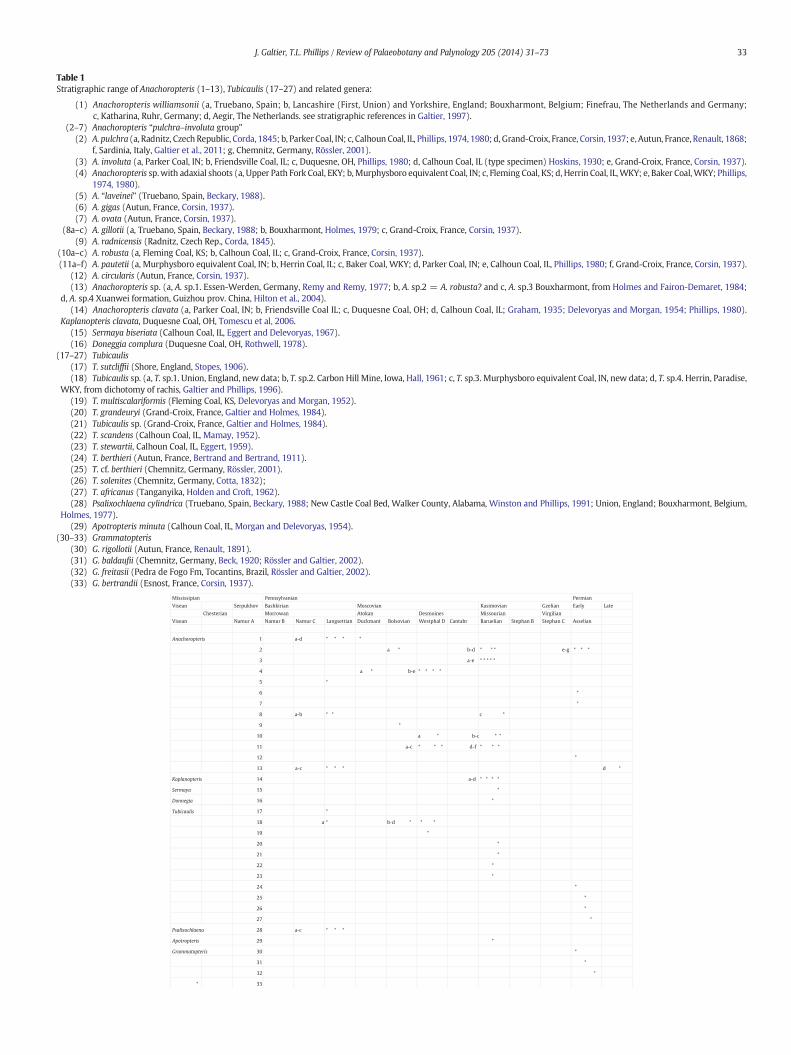
Table 1Stratigraphic range of Anachoropteris (1–13), Tubicaulis (17–27) and related genera:
(1) Anachoropteris williamsonii (a, Truebano, Spain; b, Lancashire (First, Union) and Yorkshire, England; Bouxharmont, Belgium; Finefrau, The Netherlands and Germany;c, Katharina, Ruhr, Germany; d, Aegir, The Netherlands. see stratigraphic references in Galtier, 1997).
(2–7) Anachoropteris “pulchra–involuta group”(2) A. pulchra (a, Radnitz, Czech Republic, Corda, 1845; b, Parker Coal, IN; c, Calhoun Coal, IL, Phillips, 1974, 1980; d, Grand-Croix, France, Corsin, 1937; e, Autun, France, Renault, 1868;
f, Sardinia, Italy, Galtier et al., 2011; g, Chemnitz, Germany, Rössler, 2001).(3) A. involuta (a, Parker Coal, IN; b, Friendsville Coal, IL; c, Duquesne, OH, Phillips, 1980; d, Calhoun Coal, IL (type specimen) Hoskins, 1930; e, Grand-Croix, France, Corsin, 1937).(4) Anachoropteris sp.with adaxial shoots (a, Upper Path Fork Coal, EKY; b,Murphysboro equivalent Coal, IN; c, Fleming Coal, KS; d, Herrin Coal, IL,WKY; e, Baker Coal,WKY; Phillips,
1974, 1980).(5) A. “laveinei” (Truebano, Spain, Beckary, 1988).(6) A. gigas (Autun, France, Corsin, 1937).(7) A. ovata (Autun, France, Corsin, 1937).
(8a–c) A. gillotii (a, Truebano, Spain, Beckary, 1988; b, Bouxharmont, Holmes, 1979; c, Grand-Croix, France, Corsin, 1937).(9) A. radnicensis (Radnitz, Czech Rep., Corda, 1845).
(10a–c) A. robusta (a, Fleming Coal, KS; b, Calhoun Coal, IL; c, Grand-Croix, France, Corsin, 1937).(11a–f) A. pautetii (a, Murphysboro equivalent Coal, IN; b, Herrin Coal, IL; c, Baker Coal, WKY; d, Parker Coal, IN; e, Calhoun Coal, IL, Phillips, 1980; f, Grand-Croix, France, Corsin, 1937).
(12) A. circularis (Autun, France, Corsin, 1937).(13) Anachoropteris sp. (a, A. sp.1. Essen-Werden, Germany, Remy and Remy, 1977; b, A. sp.2 = A. robusta? and c, A. sp.3 Bouxharmont, from Holmes and Fairon-Demaret, 1984;
d, A. sp.4 Xuanwei formation, Guizhou prov. China, Hilton et al., 2004).(14) Anachoropteris clavata (a, Parker Coal, IN; b, Friendsville Coal IL; c, Duquesne Coal, OH; d, Calhoun Coal, IL; Graham, 1935; Delevoryas and Morgan, 1954; Phillips, 1980).
Kaplanopteris clavata, Duquesne Coal, OH, Tomescu et al, 2006.(15) Sermaya biseriata (Calhoun Coal, IL, Eggert and Delevoryas, 1967).(16) Doneggia complura (Duquesne Coal, OH, Rothwell, 1978).
(17–27) Tubicaulis(17) T. sutcliffii (Shore, England, Stopes, 1906).(18) Tubicaulis sp. (a, T. sp.1. Union, England, new data; b, T. sp.2. Carbon Hill Mine, Iowa, Hall, 1961; c, T. sp.3. Murphysboro equivalent Coal, IN, new data; d, T. sp.4. Herrin, Paradise,
WKY, from dichotomy of rachis, Galtier and Phillips, 1996).(19) T. multiscalariformis (Fleming Coal, KS, Delevoryas and Morgan, 1952).(20) T. grandeuryi (Grand-Croix, France, Galtier and Holmes, 1984).(21) Tubicaulis sp. (Grand-Croix, France, Galtier and Holmes, 1984).(22) T. scandens (Calhoun Coal, IL, Mamay, 1952).(23) T. stewartii, Calhoun Coal, IL, Eggert, 1959).(24) T. berthieri (Autun, France, Bertrand and Bertrand, 1911).(25) T. cf. berthieri (Chemnitz, Germany, Rössler, 2001).(26) T. solenites (Chemnitz, Germany, Cotta, 1832);(27) T. africanus (Tanganyika, Holden and Croft, 1962).(28) Psalixochlaena cylindrica (Truebano, Spain, Beckary, 1988; New Castle Coal Bed, Walker County, Alabama, Winston and Phillips, 1991; Union, England; Bouxharmont, Belgium,
Holmes, 1977).(29) Apotropteris minuta (Calhoun Coal, IL, Morgan and Delevoryas, 1954).
(30–33) Grammatopteris(30) G. rigollotii (Autun, France, Renault, 1891).(31) G. baldaufii (Chemnitz, Germany, Beck, 1920; Rössler and Galtier, 2002).(32) G. freitasii (Pedra de Fogo Fm, Tocantins, Brazil, Rössler and Galtier, 2002).(33) G. bertrandii (Esnost, France, Corsin, 1937).
Mississipian Pennsylvanian Permian
Visean Serpukhov Bashkirian Moscovian Kasimovian Gzelian Early Late
Chesterian Morrowan Atokan Desmoines Missourian Virgilian
Visean Namur A Namur B Namur C Langsettian Duckmant Bolsovian Westphal D Cantabr Baruelian Stephan B Stephan C Asselian
Anachoropteris 1 a-d * * * *
2 a * b-d * * * e-g * * *
3 a-e * * * * *
4 a * b-e * * * *
5 *
6 *
7 *
8 a-b * * c *
9 *
10 a * b-c * *
11 a-c * * * d-f * * *
12 *
13 a-c * * * d *
Kaplanopteris 14 a-d * * * *
Sermaya 15 *
Donnegia 16 *
Tubicaulis 17 *
18 a * b-d * * *
19 *
20 *
21 *
22 *
23 *
24 *
25 *
26 *
27 *
Psalixochlaena 28 a-c * * *
Apotropteris 29 *
Grammatopteris 30 *
31 *
32 *
* 33
33J. Galtier, T.L. Phillips / Review of Palaeobotany and Palynology 205 (2014) 31–73
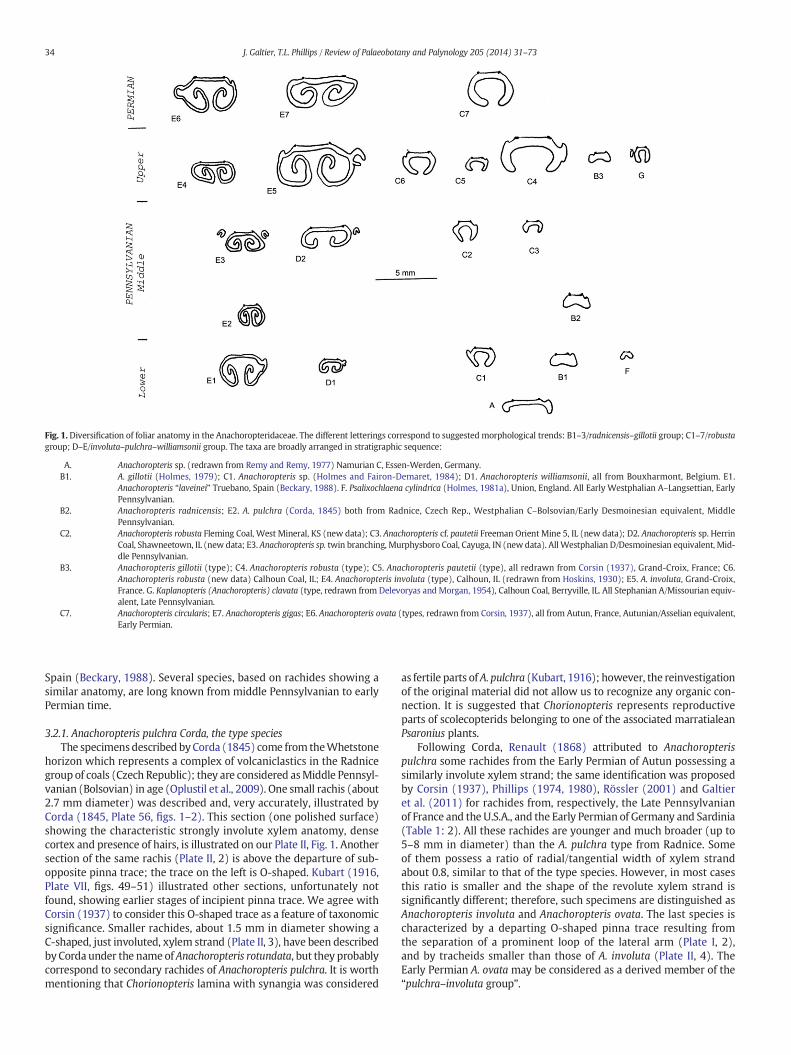
Fig. 1. Diversification of foliar anatomy in the Anachoropteridaceae. The different letterings correspond to suggested morphological trends: B1–3/radnicensis–gillotii group; C1–7/robustagroup; D–E/involuta–pulchra–williamsonii group. The taxa are broadly arranged in stratigraphic sequence:
A. Anachoropteris sp. (redrawn from Remy and Remy, 1977) Namurian C, Essen-Werden, Germany.B1. A. gillotii (Holmes, 1979); C1. Anachoropteris sp. (Holmes and Fairon-Demaret, 1984); D1. Anachoropteris williamsonii, all from Bouxharmont, Belgium. E1.
Anachoropteris “laveinei” Truebano, Spain (Beckary, 1988). F. Psalixochlaena cylindrica (Holmes, 1981a), Union, England. All Early Westphalian A–Langsettian, EarlyPennsylvanian.
B2. Anachoropteris radnicensis; E2. A. pulchra (Corda, 1845) both from Radnice, Czech Rep., Westphalian C–Bolsovian/Early Desmoinesian equivalent, MiddlePennsylvanian.
C2. Anachoropteris robusta Fleming Coal, West Mineral, KS (new data); C3. Anachoropteris cf. pautetii Freeman Orient Mine 5, IL (new data); D2. Anachoropteris sp. HerrinCoal, Shawneetown, IL (new data; E3. Anachoropteris sp. twin branching, Murphysboro Coal, Cayuga, IN (new data). All Westphalian D/Desmoinesian equivalent, Mid-dle Pennsylvanian.
B3. Anachoropteris gillotii (type); C4. Anachoropteris robusta (type); C5. Anachoropteris pautetii (type), all redrawn from Corsin (1937), Grand-Croix, France; C6.Anachoropteris robusta (new data) Calhoun Coal, IL; E4. Anachoropteris involuta (type), Calhoun, IL (redrawn from Hoskins, 1930); E5. A. involuta, Grand-Croix,France. G. Kaplanopteris (Anachoropteris) clavata (type, redrawn from Delevoryas and Morgan, 1954), Calhoun Coal, Berryville, IL. All Stephanian A/Missourian equiv-alent, Late Pennsylvanian.
C7. Anachoropteris circularis; E7. Anachoropteris gigas; E6. Anachoropteris ovata (types, redrawn from Corsin, 1937), all from Autun, France, Autunian/Asselian equivalent,Early Permian.
34 J. Galtier, T.L. Phillips / Review of Palaeobotany and Palynology 205 (2014) 31–73
Spain (Beckary, 1988). Several species, based on rachides showing asimilar anatomy, are long known from middle Pennsylvanian to earlyPermian time.
3.2.1. Anachoropteris pulchra Corda, the type speciesThe specimensdescribed byCorda (1845) come from theWhetstone
horizon which represents a complex of volcaniclastics in the Radnicegroup of coals (Czech Republic); they are considered asMiddle Pennsyl-vanian (Bolsovian) in age (Oplustil et al., 2009). One small rachis (about2.7 mm diameter) was described and, very accurately, illustrated byCorda (1845, Plate 56, figs. 1–2). This section (one polished surface)showing the characteristic strongly involute xylem anatomy, densecortex and presence of hairs, is illustrated on our Plate II, Fig. 1. Anothersection of the same rachis (Plate II, 2) is above the departure of sub-opposite pinna trace; the trace on the left is O-shaped. Kubart (1916,Plate VII, figs. 49–51) illustrated other sections, unfortunately notfound, showing earlier stages of incipient pinna trace. We agree withCorsin (1937) to consider this O-shaped trace as a feature of taxonomicsignificance. Smaller rachides, about 1.5 mm in diameter showing aC-shaped, just involuted, xylem strand (Plate II, 3), have been describedby Corda under the name of Anachoropteris rotundata, but they probablycorrespond to secondary rachides of Anachoropteris pulchra. It is worthmentioning that Chorionopteris lamina with synangia was considered
as fertile parts of A. pulchra (Kubart, 1916); however, the reinvestigationof the original material did not allow us to recognize any organic con-nection. It is suggested that Chorionopteris represents reproductiveparts of scolecopterids belonging to one of the associated marratialeanPsaronius plants.
Following Corda, Renault (1868) attributed to Anachoropterispulchra some rachides from the Early Permian of Autun possessing asimilarly involute xylem strand; the same identification was proposedby Corsin (1937), Phillips (1974, 1980), Rössler (2001) and Galtieret al. (2011) for rachides from, respectively, the Late Pennsylvanianof France and the U.S.A., and the Early Permian of Germany and Sardinia(Table 1: 2). All these rachides are younger and much broader (up to5–8 mm in diameter) than the A. pulchra type from Radnice. Someof them possess a ratio of radial/tangential width of xylem strandabout 0.8, similar to that of the type species. However, in most casesthis ratio is smaller and the shape of the revolute xylem strand issignificantly different; therefore, such specimens are distinguished asAnachoropteris involuta and Anachoropteris ovata. The last species ischaracterized by a departing O-shaped pinna trace resulting fromthe separation of a prominent loop of the lateral arm (Plate I, 2),and by tracheids smaller than those of A. involuta (Plate II, 4). TheEarly Permian A. ovata may be considered as a derived member of the“pulchra–involuta group”.
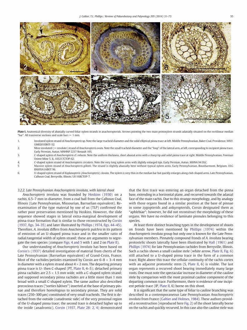
Plate I. Anatomical diversity of abaxially curved foliar xylem strands in anachoropterids. Arrows pointing the two main protoxylem strands adaxially situated on the rectilinear median“bar”. All transverse sections and scale bars = 1 mm.
1. Involuted xylem strand of Anachoropteris sp. Note the large tracheid diameter and the solid elliptical pinna trace at left. Middle Pennsylvanian, Baker Coal, Providence,WKY.UI40201EBOT-52.
2. More involuted (=revolute) strand ofAnachoropteris ovata. Note the small tracheid diameter and the “loop” of the lateral arm, at left, corresponding to incipient pinna trace.Early Permian, Autun. MNHNP 2237 Renault 105.
3. C-shaped xylem of Anachoropteris cf. robusta. Note the uniform thickness, short abaxial arms with a sharp tip and solid pinna trace at right. Middle Pennsylvanian, FreemanOrient Mine 5, IL. UI3217CTOP-62.
4. C-shaped xylem strand of Anachoropteris circularis. Note the very long xylem arms with slightly enlarged tips. Early Permian, Autun. MHNA 94 DLC.5 Massive xylem strand of Anachoropteris gillotii. The strand is slightly abaxially bent without typical xylem arms. Early Pennsylvanian, Bouxharmont, Belgium. ULG
BX695A1ABOT 96.6. U-shaped xylem strand of Kaplanopteris (Anachoropteris) clavata. The xylem is very thin in themedian bar but quickly enlarges along club-shaped arms. Late Pennsylvanian,
Calhoun Coal, Berryville, Illinois. UI1168CTOP-7.
35J. Galtier, T.L. Phillips / Review of Palaeobotany and Palynology 205 (2014) 31–73
3.2.2. Late Pennsylvanian Anachoropteris involuta, with lateral shootAnachoropteris involuta was founded by Hoskins (1930) on a
rachis, 6.5–7 mm in diameter, from a coal ball from the Calhoun Coal,Illinois (Late Pennsylvanian, Missourian, Barruelian equivalent). Re-examination of the type material by one of us (TLP) confirmed therather poor preservation mentioned by Hoskins. However, the slidesequence showed stages in lateral extra-marginal development ofpinna-trace formation that is similar to those reconstructed by Corsin(1937, figs. 34–35) and later illustrated by Phillips (1974, figs. 44–45).Therefore, A. involuta differs from Anachoropteris pulchra in its patternof emission of an U-shaped pinna trace and in the smaller ratio ofradial/tangential width of xylem strand; these are arguments to segre-gate the two species (compare Figs. 4 and 5 with 1 and 2 on Plate II).
Our understanding of Anachoropteris involuta has been based onCorsin's (1937) detailed investigation of material from cherts of theLate Pennsylvanian (Barruelian equivalent) of Grand-Croix, France.Most of the rachides/petioles examined by Corsin are 6–8 × 3–4 mmin diameterwith a xylem strand about 4 × 2mm. The departing primarypinna trace is U- then C-shaped (PT, Plate II, 4–5); detached primarypinna rachides are 2.5 × 1.5 mm wide, with a C-shaped xylem strand;and supposed secondary pinna rachides are a little more than 1 mmbroad with a small C-shaped xylem. The same author also describedprecocious traces (“sorties hâtives”) inserted at the base of primary pin-nae and therefore homologous to secondary pinnae. They are solidtraces (250–300 μm) constituted of very small tracheids. The first is de-tached from the outside (catadromic side) of the very proximal regionof the U-shaped pinna trace; the second trace is detached higher up tothe inside (anadromic). Corsin (1937, Plate 28: 2, 4) demonstrated
that the first trace was entering an organ detached from the pinnabase, extending in a horizontal plane, and recurved towards the adaxialface of themain rachis. Due to this strange morphology, and by analogywith those organs found in a similar position at the base of pinnaein some zygopterids and ankyropterids, Corsin designated them as“aphlebiae”; however, he did not reconstruct the morphology of theseorgans. We have no evidence of laminate pinnules belonging to thisspecies.
At least three distinct branching types in the development of shootson fronds have been mentioned by Phillips (1974) within theAnachoropteris involuta group but only one is known for the Late Penn-sylvanian members. Pinnately compound fronds of A. involuta bearingprotostelic shoots laterally have been illustrated by Hall (1961) andPhillips (1974) for late Pennsylvanian rachides from Berryville, Illinois.Such a rachis shows a small cauline stele (white arrow, Plate II, Fig. 6)still attached to a U-shaped pinna trace in the form of a commontrace. Right above this trace the cellular continuity of the rachis cortexwith an adjacent protostelic stem (S, Plate II, 6) is visible. The lastorgan represents a recurved shoot bearing immediately many largeroots. Onemust note the spectacular increase in diameter of the caulinestele by comparison with the most proximal cauline component of thedeparting common trace. Furthermore, there is evidence of one incipi-ent petiole trace (IP, Plate II, 6) borne on this shoot.
It is significant that the same type of foliar to cauline branching wasdescribed in a contemporaneous Late Pennsylvanian Anachoropterisinvoluta from France (Galtier and Holmes, 1984). These authors provid-ed a reconstruction (reproduced here Fig. 2) of the shoot laterally borneon the rachis and quickly recurved. In this case also the cauline stelewas
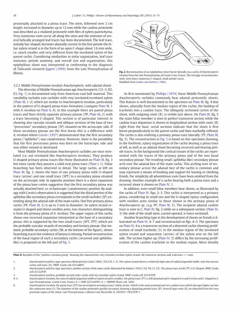
Fig. 2. Reconstruction of an epiphyllous shoot borne laterally on a rachis of Anachoropteris
36 J. Galtier, T.L. Phillips / Review of Palaeobotany and Palynology 205 (2014) 31–73
proximally attached to a pinna trace. The stem, followed over 2 cmlength, increased in diameter up to 12 mmwhile the cauline protostelewas described as a vitalized protostele with files of xylem parenchyma.Very numerous roots occur all along the stem and the emission of sev-eral helically arranged leaf traces has been documented. The leaf trace,initially bar-shaped, becomes abaxially curved. In the free petiole the fo-liar xylem strand is in the form of an open C-shape about 1.6 mmwide,i.e. much smaller and very different from the involuted xylem of theparent rachis. Considering similarities in stelar organization, leaf traceemission, petiole anatomy, and overall size and organization, thisepiphyllous shoot was interpreted as conforming to the diagnosisof Tubicaulis stewartii Eggert (1959) from the Late Pennsylvanian ofIllinois.
involuta from the Late Pennsylvanian of Grand-Croix, France. The strongly recurved proto-stelic stem bears numerous C-shaped, small petiole traces.Modified from Galtier and Holmes (1984).
3.2.3. Middle Pennsylvanian involute Anachoropteris, with adaxial shootsThe diversity ofMiddle Pennsylvanian age Anachoropteris (C2–3, D2,
E3, Fig. 1) is documented only from American coal-ball material. Thisvariability includes rare rachides with very involuted/revoluted xylem(Plate III, 1–2) which are similar to Anachoropteris involuta, particularlyin the pattern of U-shaped pinna-trace formation (compare Plate III, 1with A. involuta on Plate II, 4). In this example there are paired pinnatraces and then strictly opposite primary pinnae (PP, Plate III, 2) witha trace becoming C-shaped. This section is of particular interest inshowing also vascular strands and oblique sections of the base of two“precocious” secondary pinnae (SP) situated on the acroscopic side. Ifthese secondary pinnae are the first borne this is a difference withA. involuta where Corsin (1937) demonstrated that the first secondarypinna (“aphlebia”) was catadromous. However, there is the possibilitythat the first precocious pinna was born on the basiscopic side andwas either missed or destroyed.
Most Middle Pennsylvanian Anachoropteris rachides are once invo-luted (i.e. not revoluted like Anachoropteris involuta). They produceU-shaped primary pinna traces like those illustrated on Plate III, fig. 3but more rarely they possess a solid oval pinna trace (Plate I, 1). Foliarbranching has been observed in detail. The large rachis, at left onPlate III, fig. 3, shows the base of one primary pinna with U-shapedtrace (arrow) and one small trace (SPT) to a secondary pinna situatedon the acroscopic side. In opposite position there is a short expansionof the pinna base cortex suggestive that the first secondary pinna wasactually attached here, i.e. in basiscopic (catadromous) position. By anal-ogywith Corsin's observation it is suggested that the distal portion of theaphlebia-like secondary pinna corresponds to the small rachides (SP) ex-tending along the adaxial side of themain rachis. One free primary pinnarachis (PP, Plate III, 3) is up to 3 mm in diameter; its xylem strand re-mains U-shaped and shows swollen arms, two characters distinguishingit from the primary pinna of A. involuta. The upper region of this rachisshows one recurved expansion interpreted as the base of a secondarypinna; this is supported by the two small traces (SPT, TPT) which arethe probable vascular strands of secondary and tertiary pinnae. One iso-lated, probable secondary rachis (SR, at the bottom of the figure), showsbranching traces but evidence of lamina ismissing. Partial reconstructionof the basal region of such a secondary rachis (recurved and aphlebia-like) is proposed on the left part of Fig. 3.
Plate II. Rachides of the “pulchra–involuta group” showing the characteristic very involute/rev
1. Anachoropteris pulchra, type specimen illustrated by Corda (1845) (Taf. LVI, 1–2)cortex and hairs (H). NMP, Corda coll. E212aBOT.
2. Anachoropteris pulchra, type specimen, another section of the same rachis illustracoll. E212aTOP.
3. Anachoropteris pulchra, probable second order rachis with less involute xylem st4–5. Anachoropteris involuta, the ratio of radial/tangentialwidth of xylemstrand is sma
Late Pennsylvanian, Grand-Croix, France. 4 = UM2 GC514AT01; 6 = SMNHS Flo6. Anachoropteris involuta, the pinna trace (PT) has an incipient secondary trace (wh
the coalescent stem (S). The diameter of the cauline protostele quickly increasedproximal region of the shoot. Late Pennsylvanian, Calhoun Coal, Berryville, IL. UI
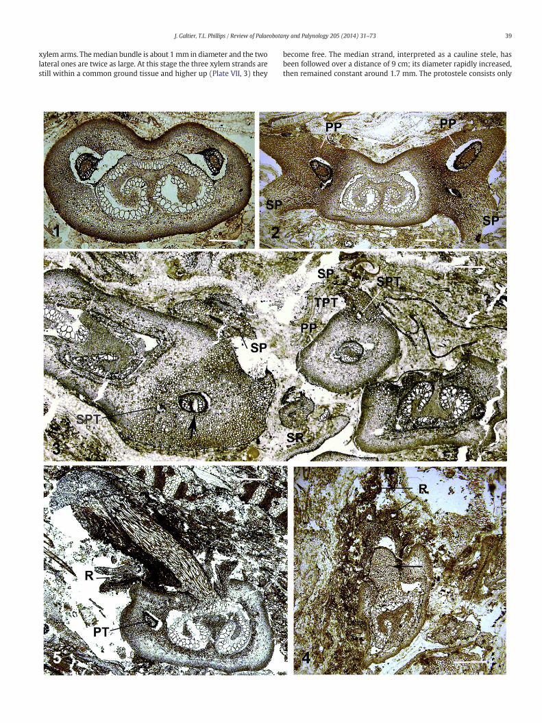
As first mentioned by Phillips (1974) these Middle PennsylvanianAnachoropteris rachides commonly bear adaxial protostelic shoots.This feature is well documented in the specimen on Plate III, fig. 4 thatshows, adaxially from the median region of the rachis, the buildup oftracheids into a cauline trace. The obliquely sectioned cortex of theshoot, with outgoing roots (R), is visible just above. On Plate III, fig. 5the main foliar member is seen in perfect transverse section while thecauline trace departure is shown in longitudinal section with roots (R)right from the base; serial sections indicate that the shoot is firstborne perpendicularly to the parent rachis and then markedly reflexed.The rachis is also emitting a primary pinna trace laterally (PT, Plate III,fig. 5). The reconstruction in Fig. 3 is based on this specimen showing,in the forefront, xylary organization of the rachis bearing a pinna traceat left, as well as an adaxial shoot becoming recurved and bearing peti-ole traces. In the background the cortical tissues of the rachis are repre-sented with the traces of the primary pinna and of the two basalsecondary pinnae. The resulting small (aphlebia-like) secondary pinnaearch over the adaxial face of the main rachis. This arching over of sec-ondary pinnae across the adaxial face of the rachis is common andmay represent a means of hooking and support for leaning or climbingfronds. For simplicity all adventitious roots have been omitted from thedrawing. Another example of a rachis bearing both a pinna trace and arecurved shoot is shown on Plate IV, 1.
In addition, even small foliar members bear shoots, as illustrated bythe rachis of Plate IV, figs. 2–3. This rachis is interpreted as a primarypinna, considering its small size and the U-shaped xylary configurationwith swollen arms similar to those shown in the primary pinna ofAnachoropteris sp. (e.g. PP, Plate III, 3). The incipient adaxial caulinetrace is seen in C, Plate IV, fig. 2 while on a subsequent section (Plate IV,3) the stele of the small stem, curved upward, is twice sectioned.
Another branching type in the development of shoots on fronds is il-lustrated on Plate IV, 4–7 and reconstructed on figs. 4–5. The specimenon Plate IV, 4 is a transverse section of a distorted rachis showing prolif-eration of small tracheids (S) in the median region of the involutedxylem strand and separation (arrow) of the xylem arm on the leftside. The section higher up (Plate IV, 5) differs by the increasing prolif-eration of the cauline tracheids in the median region. More distally
olute xylem strand. All transverse sections and scale bars= 1 mm.
. The xylem strand shows a relatively high ratio of radial/tangential width; note the narrow
ted by Kubart (1916) (Taf. VII, 51, 55). The pinna trace at left (PT) is O-Shaped. NMP, Corda
rand. NMP, Corda coll. E214cTOP.ller; the pinna trace (PT) is still attached andU-shaped in 4, and it is free and C-shaped in 5.rin coll. 3426.ite arrow)which is themost proximal part of a cauline tracewhich diverges higher up into, showing a departing petiole trace (IP). Several large roots (R) are detached from the very22931 1301LTOP-1.

37J. Galtier, T.L. Phillips / Review of Palaeobotany and Palynology 205 (2014) 31–73

Fig. 3. Reconstruction of Anachoropteris sp. foliar member with protostelic adaxial shootfrom the American Middle Pennsylvanian. The rachis is also emitting a lateral pinnatrace which is shown with paired small lateral secondary pinna traces. Note how theresulting small pinna arches over the adaxial face.
38 J. Galtier, T.L. Phillips / Review of Palaeobotany and Palynology 205 (2014) 31–73
(Plate IV, 6) this rachis seems to divide into two lateral foliar members(F1 and F2) and one median cauline siphonostele (S). At this stagethere is still a common cortex cloaking the two foliar organs. The foliarmember F1 was greatly compressed with its xylem obliterated whilethe xylem of foliar member F2 is in the form of a “ring” clearly derivedfrom the right side inrolled arm of the original anachoropterid strand.This circular xylem strand is actually thicker abaxially with a doublerow of large tracheids interpreted as the result of flattening of the pre-viously inrolled tip of the xylem arm; on the opposite adaxial surfacesmall protoxylem tracheids are visible. Another specimen, shown onPlate IV, fig. 7, is interpreted as a little more distal region of the sametype of branching. The siphonostelic cauline trace is departing upwardon the adaxial side and both foliarmembers showwell preserved xylarystrands with bilateral symmetry and protoxylem groups on the adaxialside. However, F1 shows an internal bar separated from the xylem ringwhile in F2 the bar is contiguouswith the lower part of the ring; the lastanatomy is very similar to that of F2 in Plate IV, fig. 6. At this stage bothfoliar members still possess a common cortex. Another example of amost distal section (Plate V, 1) was beyond the recurved cauline stele,and foliar members are fully separated; anatomy of one foliar member(arrow at left) appears unchangedwhile there are two involuted xylarystrands (a dichotomy) at right (double arrows). Finally, this foliar tocauline branching type may be interpreted as a trifurcation (two closebranchings) of the involute foliar member producing one mediansiphonostelic cauline shoot, departing adaxially, and two lateral foliarmembers with initially circular xylem strands. The reconstruction inFig. 4 (not taking in account the first branching, i.e. the separation ofthe compressed foliar member F1) shows the second branching,i.e. the separation of the cauline siphonostele and of foliar member F2.The reconstruction in Fig. 5 tracks all 3 members (2 foliar and themedi-an cauline) in a more distal region of the trifurcation.
The last branching type in the development of shoots on fronds isinterpreted as a dichotomy of the main involuted foliar memberresulting in a cauline siphonostelic trace (Plate V, 2–3). As the caulinestele (S, Plate V, 2) begins to form petiolar traces, the remaining halfof foliar xylem (F, Plate V, 2) progressively recurves toward a C to invo-lute shape. At this stage both xylem strands are within a commoncortex. Higher up (Plate V, 3) the siphonostelic cauline stele enlargesand its pith shows ground tissue similar to that inside the petiolar traces.
3.3. Early Pennsylvanian Anachoropteris williamsonii
This fern was first described by Williamson (1878) under the nameof Rachiopteris gleiche from British coal balls, then as Rachiopterisrotundata by Felix (1886) fromWestphalia. Subsequently, it was attrib-uted to the genus Anachoropteris by Scott (1920) and distinguished as anew species, Anachoropteris williamsonii, by Koopmans (1928). Corsin(1937) provided a detailed specific diagnosis. Holmes (1981b), studyingnew British and Belgianmaterial, first described the evidence of foliar tocauline branching in this species.
Anachoropteris williamsonii is of relatively rare occurrence and re-stricted to Early Pennsylvanian coal balls from west Europe (line 1,Table 1) ranging from the basalmost Langsettian of Spain, England,Belgium, The Netherlands and Germany up to the basal Duckmantianof The Netherlands.
Plate III. Rachides of the “pulchra–involuta group” from the North AmericanMiddle Pennsylvancross sections. Scale bar = 1 mm.
1–2. Anachoropteris sp. petiolewith “twin” foliar branching. Murphysboro Coal, Cayof the same showing the bases of primary pinnae (PP) and of truncated secon
3. Anachoropteris sp. Twomain foliarmembers and two primary pinnae, one attaof secondary pinna (SP) extending over adaxial surface of main rachis at left.Coal, Providence, WKY. UI8222BBOT-52.
4–5. Anachoropteris sp.main foliarmembers showing adaxial shoot origins. Herrin Cand roots (R) above in shoot cortex. UI3123DBOT-30. 5: Shows the cauline tra(PT) laterally. UI slide 4133-7966BTOP-10.
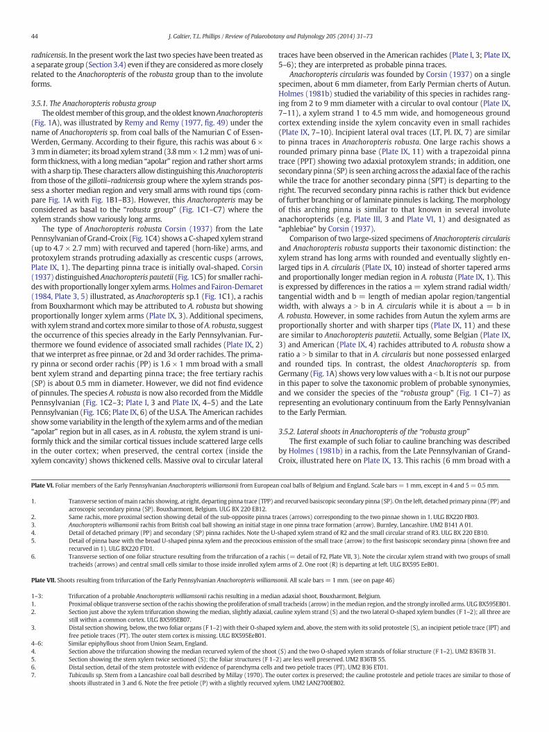
The main rachis is small, about 4–5 × 3 mm in transverse section,and the maximum diameter of 8 mm mentioned by Corsin (1937)seems greatly overestimated. The primary xylem strand, about 2 ×1 mm (Plate VI, 1–3), is in the form of a slender involute arc only 1 or2 tracheids thick. Metaxylem tracheids are up to 150–200 μmdiameter;they exhibit reticulate thickenings to multiseriate scalariform pitting.There are two protoxylem strands on the median rectilinear adaxialface, separated by more than 1 mm. The successive stages of pinna-trace formation may be followed on Plate VI, figs. 3 and 2; they consistof marginal development of U-shaped traces. This is conforming to ob-servation by Holmes (1981b) while Corsin (1937) erroneously de-scribed a massive pinna trace. Primary pinnae are borne alternately(Plate VI, 3) to sub-oppositely (Plate VI, 2). In the pinna base the U-shaped xylem strand is about 300 × 150 μmbroad and it shows the pre-cocious emission of a small cylindrical trace (arrow, Plate VI, 5) less than100 μm diameter; this is the trace of the first second order pinna borneon the outer (catadromic) side which is becoming free (SP, Plate VI, 1)as a structure recurved towards the adaxial side of the main rachis.The primary pinna rachis is about 1mmdiameter at its base and it quick-ly divides (PP, Plate VI, 1 and 4) giving rise, on the inner (anadromic)side, to another secondary pinna rachis (SP, Plate VI, 1 and 4). The lattersmall rachis is cylindrical, about 0.7 mm in diameter, with a broadhomogeneous cortex and a tiny apparently cylindrical xylem strand.We have not obtained information on either more distal branching ofthese secondary pinnae or evidence of lamina, but this is suggestivethat the Anachoropteris williamsonii frond was at least tri-pinnate.
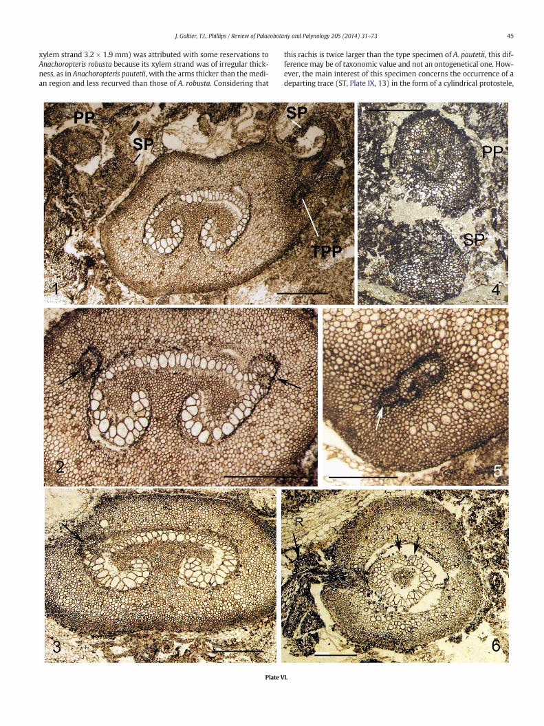
The occurrence of shoots on foliar members of Anachoropteriswilliamsonii was first mentioned by Holmes (1981b) from two speci-mens illustrated here. One Belgian specimen of Anachoropteris showsthe initial enlargement of the xylem strand median region due to theproliferation of small tracheids (arrow, Plate VII, 1); higher up a trifurca-tion results into amiddle trace, in the form of a solid, more or less cylin-drical, protostele (S, Plate VII, 2), and two lateral, circular xylem strands(F1–2, Plate VII, 2), each corresponding to one of the previous enrolled
ian.Main foliar members showing pinna traces and departures and/or adaxial shoots, all in
uga, IN. 1: The two opposite U-shaped pinna traces are just detached. 2:More distal sectiondary pinnae (SP) with their small trace. UI25760DBOT-2 and 25760ETOP-54.ched (U-shaped xylemwith swollen arms, arrowed) and one separate (PP). Note curvatureSR, detached secondary pinna rachis; SPT, TPT, secondary and tertiary pinna traces. Baker
oal, Shawneetown, IL. 4: Shows the buildup of small tracheids (arrow) into a cauline tracece departure in longitudinal section with roots (R), the rachis is also emitting a pinna trace
39J. Galtier, T.L. Phillips / Review of Palaeobotany and Palynology 205 (2014) 31–73
xylem arms. Themedian bundle is about 1mm in diameter and the twolateral ones are twice as large. At this stage the three xylem strands arestill within a common ground tissue and higher up (Plate VII, 3) they
become free. The median strand, interpreted as a cauline stele, hasbeen followed over a distance of 9 cm; its diameter rapidly increased,then remained constant around 1.7 mm. The protostele consists only

40 J. Galtier, T.L. Phillips / Review of Palaeobotany and Palynology 205 (2014) 31–73
of tracheids, about 100 μm in diameterwith some broader in the centralarea. The cauline stele shows the emission of several helically arrangedpetiolar strands which are initially bar-shaped then slightly curvedabaxially (PT, Plate VII, 3). Two protoxylem strands are visible on the ad-axial face of one incipient bar-shaped leaf trace (IPT, Plate VII, 3). Thestem cortex is incompletely preserved but broad cells are commonlydistributed within the middle parenchymatous region. One of the twolateral organs resulting from the trifurcation (F2, Plate VII, 3) is wellpreserved and illustrated in detail on Plate VI, 6. It is circular in cross sec-tion, about 3.5 mm diameter with a cortex of very small cells at the pe-riphery and larger ones in the middle region. The circular xylem strand(1.4 × 1.2 mm) is slightly dorsiventral with two groups of small tra-cheids (probable protoxylem strands) on the adaxial face (arrows,Plate VI, 6). The other tracheids, up to 200 μm broad, are similar tothose of the initial Anachoropteris trace. The central area is occupied bysmall sclerotic cells which correspond histologically to the inner groundtissue seen within the involute arm of Anachoropteris. Interestingly,roots (R, Plate VI, 6) are emitted in the proximal region of these organs.These strange structures, interpreted as foliar, are only preserved for avery short distance. Therefore, this Belgian specimen clearly documentsthe trifurcation of a rachis of A. williamsonii resulting in the productionof a middle epiphyllous shoot in the form of a protostelic caulineorgan arising adaxially as reconstructed in Fig. 6.
A second specimen of the same age, found in a British coal ball(Union Seam, Burnley, Lancashire) confirms this branching. In thisexample the most proximal region is very poorly preserved; however,in sections just beyond the trifurcation (Plate VII, 4 and 5) one can rec-ognize the massive cauline protostele (S) flanked by the two circularxylem strands (F1–2). The cauline stele is recurved (Plate VII, 4) andcut twice in another sectionwith the evidence of a first petiole trace de-parture (Plate VII, 5). The protostele is preserved for more than 12 cm;its diameter increases from 1 to 1.6 mm and parenchyma cells becomeintermixed with tracheids in the distal region (Plate VII, 6). Numerousmassive leaf traces are borne helically on the stem and they soon ac-quire a slight abaxial curvature. Unfortunately, the stemcortex ismostlydestroyed. As in the Belgian specimen, it was not possible to follow, be-yond a few mm, the two organs with circular xylem resulting from thetrifurcation; in one of them (F1 on left, Plate VII, 4) the xylem is 1mm indiameter and thicker on one side with protoxylem strands on the otherside, suggesting a bilateral symmetry of foliar nature. The cortex is rela-tively well preserved with scattered large cells and the free organ isabout 4 mm in diameter, as in the Belgian specimen.
In conclusion, we have the evidence of epiphyllous shoots borne,in both cases, on an Anachoropteris williamsonii rachis. According to
Plate IV. Other examples of shoots borne on North American Middle Pennsylvanian Anachorop
1. Anachoropteris sp. The rachis, in cross section, shows both a pinna trace (PT) andShawneetown, IL. UI9033DTOP-52.
2–3. Anachoropteris sp. Example of small foliar member, here one pinna, bearing shoincipient (C) cauline trace. UI8266ABOT-8. 3: the same showing the recurved ad
4–5. Large foliar member of an involute Anachoropteris, with lateral foliar strand at lefcommon cortical tissue. 5. Shows the increasing proliferation of cauline tracheid
6. More distal section of the same, at this level the xylem is divided into two latercauline trace (S). The foliar xylem F2 obviously derived from the closing of thUI2927ITOP-63.
7. Another specimen, interpreted as amore distal level,with central siphonostele dewithin a common cortex. In foliar xylem F1 there is a “bar” of xylem internal to trepresents only a slight change from the xylem anatomy of F2 in 6. Protoxylem g2524B-7.
Plate V. Other shoots borne on North American Middle Pennsylvanian Anachoropteris rachides
1. Detached foliar member (arrow) with typical xylem ring and internal bar and twNashville, IL. UI Slide 4065, 1578BBOT-6.
2–3. One specimen of involute Anachoropteris sp. with the main foliar member at staMiddle Pennsylvanian, Herrin Coal, Illinois.
2. Section above dichotomy: the remaining half of foliar xylem (F) recurves toward6136-136.
3. A more distal section of the same; the broadened siphonostele shows ground tis
the anatomy of their protostele and of their massive, rectangular thenC-shaped leaf traces, these stems conform to the genus Tubicaulis.They represent a new example of shoots borne on foliar members ofanachoropterid ferns. As reconstructed in Fig. 6, in A. williamsonii theshoot results from a trifurcation of the rachis into two lateral foliarmembers and a median adaxial stem, as in the American MiddlePennsylvanian Anachoropteris described just above; differences concernthe protostelic versus siphonostelic cauline trace and the absenceversus presence of internal bar in the circular xylem of the two resultingfoliar members. This may be an argument to consider the MiddlePennsylvanian anachoropterids as derived members of the samegroup as the Early Pennsylvanian A. williamsonii.
The Tubicaulis-type of stems borne on Anachoropteris williamsoniirachides are comparable to several species of Tubicaulis previouslydescribed from the Pennsylvanian. The stem protostele is solid in theBelgian specimen but it is ranging from solid to mixed in the Englishshoot and this difference may be ontogenetic. As a result, the first onewas considered by Holmes (1981b) as similar to Tubicaulis scandens(Mamay, 1952) while the second was compared to Tubicaulis stewartii(Eggert, 1959) and Tubicaulis multiscalariformis (Delevoryas andMorgan, 1952). One unnamed species of Tubicaulis from the earlyWestphalian of England was mentioned by Millay (1970) who notedsimilarities with T. stewartii and T. multiscalariformis. This stem is illus-trated for the first time on Plate VII, fig. 7. This isolated stem with amixed protostele probably represents the distal part of the same taxonrepresented in Figs. 4 to 6 of the same plate. Of interest, this specimenshows free petioles (P, Plate VII, 7) with a C-shaped but not enrolledxylem strand, therefore distinct from the A. williamsonii rachis anatomy.Isolated rachides with the same anatomy occur in Bouxharmont coalballs and they have been illustrated by Holmes and Fairon-Demaret(1984, Plate 3, fig. 7) under the name of Anachoropteris sp.2; they arereferred as 13c in our Table 1.
3.4. The Anachoropteris gillotii–radnicensis group
As stated above (Section 3.1. and Fig. 1B1–B3) these ferns arecharacterized by a short and thick foliar xylem with very short armsjustifying the erection of the new group based on foliar anatomy. Thisseparation from the “robusta group” is further supported by differencesin cauline branching (see Section 3.5).
Anachoropteris gillotii Corsin (1937) type specimen from Grand-Croix (Late Pennsylvanian), shows a uniformly thick and slightlyrecurved xylem strand. Holmes (1979) attributed older Belgianspecimens, from Early Pennsylvanian coal balls, to the same species
teris rachides. All from Herrin Coal, Illinois. Scale bar = 1 mm.
small cauline tracheids adaxially as well as partial longisection of the recurved shoot (S).
ot. Shawneetown, IL. 2: Primary pinna with U-shaped xylem and swollen arms showingaxial shoot (CX) with expanded twice sectioned xylem. UI8266ABOT.t (arrow) and proliferation of tracheids (S) in themedian region. Note the continuity of thes (S) in the median region. Shawneetown, IL. UI2927ITOP-41 and 46.al foliar strands (F1, very compressed at left, and F2 at right) and a median siphonostelice inrolled right arm of the initial anachoropterid strand shown in 4. Shawneetown, IL.
parting adaxially and better preserved xylemas closed rings in foliarmembers (F1, F2), stillhe closed ring while in F2 the internal bar appears contiguous with the lower region. Thisroups of small tracheids are on the adaxial face of foliar strands. Nashville, IL. UI Slide 3973,
. Scale bar = 1 mm. (see on page 42)
o involute xylary strands (double arrows, upper right corner) resulting from a dichotomy.
ges of dichotomy resulting in an ephemeral siphonostelic trace to a Tubicaulis type stem.
a C- to involute-shape while the cauline strand (S) begins to form petiolar traces. UI slide
sue (arrow) similar to that inside the petiolar traces (P). UI slide 6162-75.
41J. Galtier, T.L. Phillips / Review of Palaeobotany and Palynology 205 (2014) 31–73
considering that no significant differences can be found with Corsin'sfigures. Comparison of sections of one French rachis (Plate VIII, 1) andof the Belgian specimen (Plate VIII, 2) actually supports Holmes' asser-tion. In transverse section the foliar member has a rounded contour,
Plate I
up to 8 mm diameter in the type specimen; xylem strands range from0.4mm×1mm to 0.8mm×2mm. They are slightly concave on the ab-axial side, and forming an arc of about one third of a circle. The centralapolar region is uniformly 3–5 tracheids thick, while adaxial arms are
V.

Plate V (see caption on page 40).
42 J. Galtier, T.L. Phillips / Review of Palaeobotany and Palynology 205 (2014) 31–73
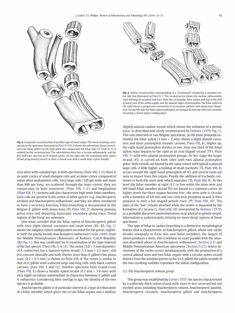
Fig. 5. Xylary reconstruction corresponding to a “trichotomy” cloaked by a common cor-tex, like that illustrated on Plate IV, 7. The reconstruction shows the median siphonostelicstem forming an incipient leaf trace. Note the rectangular foliar xylemwith bar in the midto lower part of the xylem supply and the adaxial ridges of protoxylem. The foliar xylem tothe right shows a progressive conversion to an involute pattern with pinna trace depar-ture. On the left side the foliar xylemundergoes an unequal dichotomywith eachmemberresuming a closed xylary configuration.
Fig. 4. Composite reconstruction of another type of shoot origin. The lower drawing corre-sponds to the specimen illustrated on Plate IV, 4–6. It shows the adventious shoot connect-ed to the foliar xylem on the right while the compressed left foliar side (F1, Plate IV, 5) isomitted on the reconstruction. The adventitious shoot has a circular siphonostele, and thefirst leaf trace also has an O-shaped xylem. On the right side the separating foliar xylemstrand progressively incurls to form a closed oval with a small inner xylem bundle.
43J. Galtier, T.L. Phillips / Review of Palaeobotany and Palynology 205 (2014) 31–73
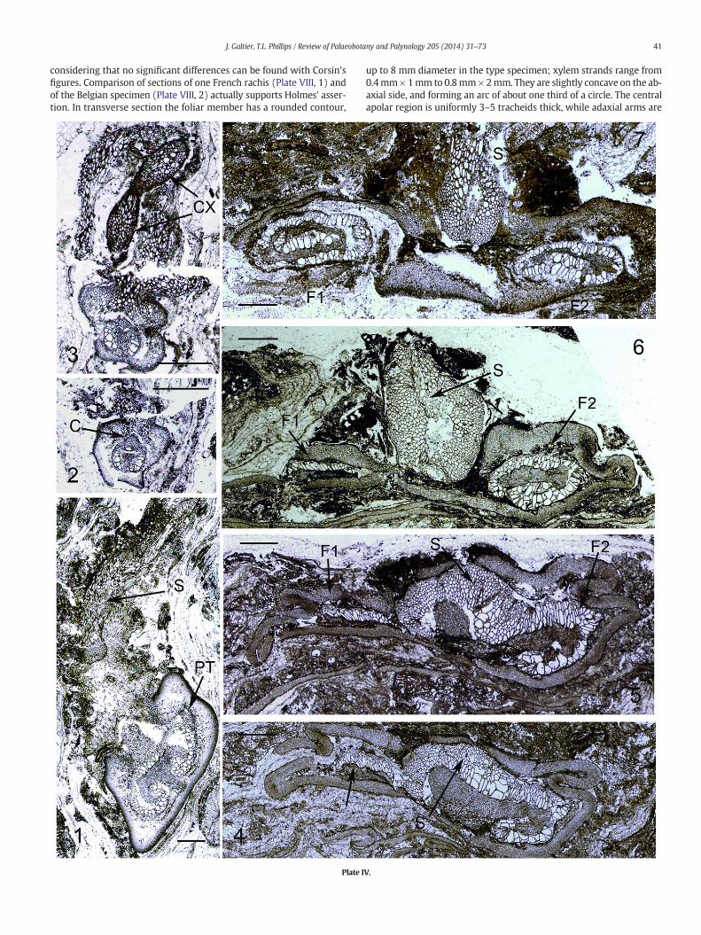
very short with rounded tips. In both specimens (Plate VIII, 1–2) there isan outer cortex of small elongate cells and an inner cortex composed ofwider often isodiametric cells. Very large cells (120 μm wide and morethan 800 μm long) are scattered through the inner cortex; they areconspicuous in both transverse (Plate VIII, 1–2) and longitudinal(Plate VIII, 11) sections and also characterize high order foliarmembers.Such cells are present in the cortex of other species (e.g. Anachoropterisinvoluta and Anachoropteris williamsonii) and they are often consideredto have a secretory function. Foliar branching is documented in theBelgian A. gillotii with pinna base (PI, Plate VIII, 2) showing primarypinna trace and departing basiscopic secondary pinna trace. Distalregions of the frond are unknown.
One must consider that the foliar xylem of Anachoropteris gillotiiwith very slight abaxial curvature and small arms (B1, B3, Fig. 1)shows the simplest xylem configuration recorded for the genus, togeth-er with the poorly known Anachoropteris radnicensis Corda (1845) fromthe Middle Pennsylvanian (Bolsovian) of Radnice, Czech Republic(B2, Fig. 1); this was confirmed by re-examination of the type materialof the last species (Plate VIII, 3, 4, 12). The rachis (3.8 × 5mmdiameter)of A. radnicensis has a massive xylem strand (1.1 mm × 2.2 mm), stillless concave abaxially and with shorter arms than A. gillotii. One pinnatrace (0.2 × 0.3 mm) is shown on Plate VIII, 4. The cortex is similar tothat of A. gillotii with scattered large and long cells with black content(arrows, plate VIII, 3 and 12). Another specimen from Grand-Croix(Plate VIII, 5) shows a slender xylem strand (0.7 mm × 1.8 mm) withvery slight curvature, intermediate in characters between A. gillotii andA. radnicensis. Considering their overlap in age, the identity of the twospecies is probable.
Anachoropteris gillotii is of particular interest in a type of trifurcationof a foliar member which gives rise to two foliar organs and a median,
slightly adaxial cauline strand which shows the initiation of a petioletrace, as described and nicely reconstructed by Holmes (1979, Fig. 1).This was observed in two Belgian specimens; at the most proximal ex-tremity the foliar xylem (1 mm × 2 mm) shows a slight abaxial curva-ture and three protoxylem strands (arrows, Plate VIII, 6). Higher up,the right-hand protoxylem divides in two, then one third of the foliarxylem mass departs to the right as an oval shaped strand (TF1, PlateVIII, 7) with two adaxial protoxylem groups. At this stage the largerstrand, tF2, is curved on both sides with two adaxial protoxylempoles; both strands are bound by the same cortex with typical scatteredlarge cells. A little higher, a buildup of small tracheids (TS, Plate VIII, 8)occurs around the right-hand protoxylem of tF2 and several roots areseen to depart from this region. Finally the addition of tracheids con-tinues to form the stem stele which separates (TS, Plate VIII, 9). At thislevel the foliar member at right (F1) is free while the stem stele andleft-hand foliar member strand TF2 are bound in a common cortex. Athigher levels the three organs become free; the stem stele is circularwith a diameter of 0.8 mm and one centrarch proxylem strand. It thenprepares to emit a bar-shaped petiole trace (PT, Plate VIII, 10). Thesides of the “bar” remain attached while the center is separated by theformation of a lacuna (L, Plate VIII, 10) interpreted by Holmes (1979)as a probable decurrent parenchymatous strip adaxial to petiole strand.Information is unfortunately missing on more distal regions of thesespecimens.
This type of foliar to cauline branching is a distinctivemorphologicalfeature that is characteristic of Anachoropteris gillotii, where one rachisdivides unequally to form two new foliar members, the largest ofwhich produces a stem; this is without an exact parallel with the situa-tion described above in Anachoropteris williamsonii (Section 3.3) andMiddle Pennsylvanian American specimens (Section 3.2.3) where di-chotomy of the rachis occurs simultaneously with the production of acentral adaxial stem and two foliar organs with a circular xylem stranddistinct from the involute parent rachis. In A. gillotii the xylem strands ofthe two resulting rachides reproduce the initial configuration.
3.5. The Anachoropteris robusta group
This groupwas established by Corsin (1937) for species characterizedby a uniformly thick xylem strand with more or less recurved but notinrolled arms, including Anachoropteris robusta, Anachoropteris pautetii,Anachoropteris circularis, Anachoropteris gillotii and Anachoropteris
44 J. Galtier, T.L. Phillips / Review of Palaeobotany and Palynology 205 (2014) 31–73
radnicensis. In the present work the last two species have been treated asa separate group (Section 3.4) even if they are considered asmore closelyrelated to the Anachoropteris of the robusta group than to the involuteforms.
3.5.1. The Anachoropteris robusta groupTheoldestmember of this group, and the oldest knownAnachoropteris
(Fig. 1A), was illustrated by Remy and Remy (1977, fig. 49) under thename of Anachoropteris sp. from coal balls of the Namurian C of Essen-Werden, Germany. According to their figure, this rachis was about 6 ×3mm in diameter; its broad xylem strand (3.8mm×1.2mm)was of uni-form thickness, with a longmedian “apolar” region and rather short armswith a sharp tip. These characters allowdistinguishing this Anachoropterisfrom those of the gillotii–radnicensis group where the xylem strands pos-sess a shorter median region and very small arms with round tips (com-pare Fig. 1A with Fig. 1B1–B3). However, this Anachoropteris may beconsidered as basal to the “robusta group” (Fig. 1C1–C7) where thexylem strands show variously long arms.
The type of Anachoropteris robusta Corsin (1937) from the LatePennsylvanianof Grand-Croix (Fig. 1C4) shows a C-shaped xylem strand(up to 4.7 × 2.7 mm) with recurved and tapered (horn-like) arms, andprotoxylem strands protruding adaxially as crescentic cusps (arrows,Plate IX, 1). The departing pinna trace is initially oval-shaped. Corsin(1937) distinguished Anachoropteris pautetii (Fig. 1C5) for smaller rachi-deswith proportionally longer xylemarms. Holmes and Fairon-Demaret(1984, Plate 3, 5) illustrated, as Anachoropteris sp.1 (Fig. 1C1), a rachisfrom Bouxharmont which may be attributed to A. robusta but showingproportionally longer xylem arms (Plate IX, 3). Additional specimens,with xylem strand and cortexmore similar to those of A. robusta, suggestthe occurrence of this species already in the Early Pennsylvanian. Fur-thermore we found evidence of associated small rachides (Plate IX, 2)that we interpret as free pinnae, or 2d and 3d order rachides. The prima-ry pinna or second order rachis (PP) is 1.6 × 1 mm broad with a smallbent xylem strand and departing pinna trace; the free tertiary rachis(SP) is about 0.5 mm in diameter. However, we did not find evidenceof pinnules. The species A. robusta is now also recorded from theMiddlePennsylvanian (Fig. 1C2–3; Plate I, 3 and Plate IX, 4–5) and the LatePennsylvanian (Fig. 1C6; Plate IX, 6) of the U.S.A. The American rachidesshow some variability in the length of the xylem arms and of themedian“apolar” region but in all cases, as in A. robusta, the xylem strand is uni-formly thick and the similar cortical tissues include scattered large cellsin the outer cortex; when preserved, the central cortex (inside thexylem concavity) shows thickened cells. Massive oval to circular lateral
Plate VI. Foliar members of the Early Pennsylvanian Anachoropteris williamsonii from European
1. Transverse section ofmain rachis showing, at right, departing pinna trace (TPP) aacroscopic secondary pinna (SP). Bouxharmont, Belgium. ULG BX 220 EB12.
2. Same rachis, more proximal section showing detail of the sub-opposite pinna tr3. Anachoropteris williamsonii rachis from British coal ball showing an initial stage4. Detail of detached primary (PP) and secondary (SP) pinna rachides. Note the U-5. Detail of pinna base with the broad U-shaped pinna xylem and the precocious e
recurved in 1). ULG BX220 FT01.6. Transverse section of one foliar structure resulting from the trifurcation of a rac
tracheids (arrows) and central small cells similar to those inside inrolled xylem
Plate VII. Shoots resulting from trifurcation of the Early Pennsylvanian Anachoropteris williams
1–3: Trifurcation of a probable Anachoropteris williamsonii rachis resulting in a media1. Proximal oblique transverse section of the rachis showing the proliferation of sma2. Section just above the xylem trifurcation showing the median, slightly adaxial, c
still within a common cortex. ULG BX595EB07.3. Distal section showing, below, the two foliar organs (F 1–2)with their O-shaped
free petiole traces (PT). The outer stem cortex is missing. ULG BX595EeB01.4–6: Similar epiphyllous shoot from Union Seam, England.4. Section above the trifurcation showing the median recurved xylem of the shoot5. Section showing the stem xylem twice sectioned (S); the foliar structures (F 1–26. Distal section, detail of the stem protostele with evidence of parenchyma cells a7. Tubicaulis sp. Stem from a Lancashire coal ball described by Millay (1970). The
shoots illustrated in 3 and 6. Note the free petiole (P) with a slightly recurved x
traces have been observed in the American rachides (Plate I, 3; Plate IX,5–6); they are interpreted as probable pinna traces.
Anachoropteris circularis was founded by Corsin (1937) on a singlespecimen, about 6 mm diameter, from Early Permian cherts of Autun.Holmes (1981b) studied the variability of this species in rachides rang-ing from 2 to 9 mm diameter with a circular to oval contour (Plate IX,7–11), a xylem strand 1 to 4.5 mm wide, and homogeneous groundcortex extending inside the xylem concavity even in small rachides(Plate IX, 7–10). Incipient lateral oval traces (LT, Pl. IX, 7) are similarto pinna traces in Anachoropteris robusta. One large rachis shows arounded primary pinna base (Plate IX, 11) with a trapezoidal pinnatrace (PPT) showing two adaxial protoxylem strands; in addition, onesecondary pinna (SP) is seen arching across the adaxial face of the rachiswhile the trace for another secondary pinna (SPT) is departing to theright. The recurved secondary pinna rachis is rather thick but evidenceof further branching or of laminate pinnules is lacking. The morphologyof this arching pinna is similar to that known in several involuteanachoropterids (e.g. Plate III, 3 and Plate VI, 1) and designated as“aphlebiae” by Corsin (1937).
Comparison of two large-sized specimens of Anachoropteris circularisand Anachoropteris robusta supports their taxonomic distinction: thexylem strand has long arms with rounded and eventually slightly en-larged tips in A. circularis (Plate IX, 10) instead of shorter tapered armsand proportionally longer median region in A. robusta (Plate IX, 1). Thisis expressed by differences in the ratios a = xylem strand radial width/tangential width and b = length of median apolar region/tangentialwidth, with always a N b in A. circularis while it is about a = b inA. robusta. However, in some rachides from Autun the xylem arms areproportionally shorter and with sharper tips (Plate IX, 11) and theseare similar to Anachoropteris pautetii. Actually, some Belgian (Plate IX,3) and American (Plate IX, 4) rachides attributed to A. robusta show aratio a N b similar to that in A. circularis but none possessed enlargedand rounded tips. In contrast, the oldest Anachoropteris sp. fromGermany (Fig. 1A) shows very low valueswith a b b. It is not our purposein this paper to solve the taxonomic problem of probable synonymies,and we consider the species of the “robusta group” (Fig. 1 C1–7) asrepresenting an evolutionary continuum from the Early Pennsylvanianto the Early Permian.
3.5.2. Lateral shoots in Anachoropteris of the “robusta group”The first example of such foliar to cauline branching was described
by Holmes (1981b) in a rachis, from the Late Pennsylvanian of Grand-Croix, illustrated here on Plate IX, 13. This rachis (6 mm broad with a
coal balls of Belgium and England. Scale bars = 1 mm, except in 4 and 5 = 0.5 mm.
nd recurved basiscopic secondary pinna (SP). On the left, detached primary pinna (PP) and
aces (arrows) corresponding to the two pinnae shown in 1. ULG BX220 FB03.in one pinna trace formation (arrow). Burnley, Lancashire. UM2 B141 A 01.shaped xylem strand of R2 and the small circular strand of R3. ULG BX 220 EB10.mission of the small trace (arrow) to the first basiscopic secondary pinna (shown free and
his (= detail of F2, Plate VII, 3). Note the circular xylem strand with two groups of smallarms of 2. One root (R) is departing at left. ULG BX595 EeB01.
onii. All scale bars = 1 mm. (see on page 46)
n adaxial shoot, Bouxharmont, Belgium.ll tracheids (arrow) in themedian region, and the strongly inrolled arms. ULG BX595EB01.auline xylem strand (S) and the two lateral O-shaped xylem bundles (F 1–2); all three are
xylem and, above, the stemwith its solid protostele (S), an incipient petiole trace (IPT) and
(S) and the two O-shaped xylem strands of foliar structure (F 1–2). UM2 B36TB 31.) are less well preserved. UM2 B36TB 55.nd two petiole traces (PT). UM2 B36 ET01.outer cortex is preserved; the cauline protostele and petiole traces are similar to those ofylem. UM2 LAN2700EB02.
45J. Galtier, T.L. Phillips / Review of Palaeobotany and Palynology 205 (2014) 31–73
xylem strand 3.2 × 1.9 mm) was attributed with some reservations toAnachoropteris robusta because its xylem strand was of irregular thick-ness, as in Anachoropteris pautetii, with the arms thicker than the medi-an region and less recurved than those of A. robusta. Considering that
Plate V
this rachis is twice larger than the type specimen of A. pautetii, this dif-ference may be of taxonomic value and not an ontogenetical one. How-ever, the main interest of this specimen concerns the occurrence of adeparting trace (ST, Plate IX, 13) in the form of a cylindrical protostele,
I.
Plate VII (see caption on page 44).
46 J. Galtier, T.L. Phillips / Review of Palaeobotany and Palynology 205 (2014) 31–73
Fig. 6. Simplified reconstruction of the shoot origin in Anachoropteris williamsonii from thetrifurcation of a large foliarmember, resulting in amedian protostelic stem and two lateralfoliarmemberswith circular xylem. Based on the Belgian specimen illustrated on Plate VII,1–3.
47J. Galtier, T.L. Phillips / Review of Palaeobotany and Palynology 205 (2014) 31–73
about 1mm in diameter, composed of small tracheids of rather uniformsize. The most proximal section was above the level of trace departurefrom the rachis xylem but serial peel sections show the trace departingto the left, enlarging and detaching one root trace before it was broken.The cauline nature of this trace is evident.
Another example of foliar to cauline branching is documented on arachis of the Anachoropteris robusta type from the Early Permian ofAutun; this occurs in two consecutive thin sections of Renault's collec-tion. On the basal section the rachis is circular in transverse sectionwith a xylem strand less than 3 mm broad, with one incipient oval lat-eral trace similar to the example illustrated on Plate IX, 7. However,on the next section (Plate IX, 12) the rachis shows a small shoot longi-tudinally sectioned and attached on the same left flank. This small cau-line structure is morphologically a small bud (6mm long and 2.5mm indiameter) departing laterally, and then perpendicularly with regard tothe parent rachis. The central cauline strand (ST, Plate IX, 12) is sec-tioned longitudinally and shows the emission of several traces to suc-cessive roots (R) in the most proximal region of this small shoot. Thedome-like apical region of the bud is coveredwith hairs. This type of lat-eral shoot appears similar to the previous one illustrated on Plate IX, 13.
Several specimens of Anachoropteris circularis from the Early Perm-ian of Autun correspond to rachides of very different sizes (Plate IX,7–10) bearing lateral shoots. In the first example (Plate IX, 7) the lateraltrace was first interpreted as an incipient pinna trace; however, serialsections reveal that higher up this trace became circular and departedobliquely. A similar situation is observed in another rachis (Plate IX, 8)showing an intermediate stage with a bipartite trace. Higher up, thesame rachis shows the enlarged outgoing trace; its inner part (PT,Plate IX, 9) is interpreted as an oval pinna trace still attached to theouter cauline part (ST). The same type of branching is observed in oneof the largest A. circularis rachis illustrated on Plate IX, 10. In this casealso, the voluminous departing trace is interpreted as a common traceto a pinna and a shoot borne laterally. Roots are observed departingvery proximally from the cauline strand through the rachis cortex. Inall these examples the shoot was broken near its level of attachment.
In conclusion, one must emphasize that the Anachoropteris of the“robusta group” are characterized by the production of lateral shoots,similarly to those of some Anachoropteris of the “involuta group” (seeSection 3.2.2); this pattern is distinct from the foliar to caulinebranching known in the related “Anachoropteris gillotii group” and it isan additional argument to separate the two groups.
3.6. Anachoropteris clavata
Anachoropteris clavata Graham (1935) was founded on small-sizedrachides (averaging 2.5mm in diameter)with a U-shaped xylem strandshowing typically expanded club-like abaxial arms, very distinctive incomparison to other species (Plate I, 6 and Fig. 1G). The type materialof this specieswas from the Late Pennsylvanian of Calhoun Coal, Illinois.From additional specimens of the same origin, Delevoryas and Morgan(1954) provided the first evidence of a stem of Anachoropteris and thefirst report of shoots on foliar members in anachoropterid ferns. In
this important study, the authors demonstrated that lateral traces, aris-ing from a rachis, supply either one pinna or an independent axis whichwas designated as a stem since it possesses a radial symmetry and be-cause petiole-like structures and roots arise from it. The rachis fromwhich the stem originateswas called a “primary petiole” to differentiateit from the “secondary petioles”which arise from the stem. The authorsnoted the obconical construction of the stem but they indicated thatregular phyllotaxy was not detected for the secondary petioles. Finally,they suggested that the main rachides/primary petioles may representscrambling stolon-like structures on which shoots were borne.
Recently Tomescu et al. (2006, 2008) reconstructed thewhole plant,Kaplanopteris clavata, on the basis of vegetative and fertile frond frag-ments and rhizomes. Characteristic anatomy conforming to the mor-phospecies Anachoropteris clavata allowed for integration of previouslydescribedmaterial from the Late Pennsylvanian of Illinoiswith newma-terial from Duquesne Coal, Ohio which was the object of preliminarystudies by Rothwell (1987) and Trivett and Rothwell (1988). In agree-ment with specimens studied by Delevoryas and Morgan (1954), theauthors described the production of an epiphyllous stem (called“rhizome”) originating as an elliptical vascular bundle that divergeslaterally along the frond rachis “at positions of primary pinnae”. The in-crease in diameter and transition of the cauline strand into a circularprotostele was documented, as well as the divergence of the first“stipe” bundle (= “secondary petiole” of Delevoryas and Morgan,1954). Two petioles were observed diverging from the stem preservedfor about 10 mm in length while Delevoryas and Morgan observed upto four fronds. The authors also described the overall morphology oftripinnately dissected fronds with laminate pinnules, as well as latentcroziers that replace primary pinnae. Finally, the authors reconstructedK. clavata as a primarily vining or climbing plant consistingprincipally of“indeterminate vining fronds produced by scanty, small erect rhizomes”(Tomescu et al., 2006) and combining two types of reiterative growth(Tomescu et al., 2008).
One rachis of Anachoropteris clavata bearing a stem, from the LatePennsylvanian of Berryville (Calhoun Coal, Illinois) is illustrated onPlate X. The most proximal section (Plate X, 1) shows, within thesame cortex, the U-shaped xylem strand of the rachis and a smaller lat-eral trace (LT) at right. In the present case, this lateral is interpreted as a“common trace” consisting of the C-shaped strand of a primary pinnatrace (PPT) with the addition of some cauline tracheids (CT), and ofone small departing secondary pinna trace (SPT) as shown on the en-largement (Plate X, 4). The series ofmore than one hundred sections re-veals that differentiation was first in the pinna trace, then adventitiouscauline tracheids. The addition of cauline tracheids continues upwards,resulting in a typically circular protostele as broad as the adjacent pinnatrace (Plate X, 2 and 5). In the interval one small trace, detached fromthe pinna trace from the left side (SPT2, Plate X, 5), is interpreted asthe second secondary pinna trace. At this level, the lateral organ con-taining the bipartite common trace is free cortically (Plate X, 2) but itis only higher up that the shoot and the pinna rachis will separate(Plate X, 3). Root traces have been observed departing from the caulinetrace between the levels of figs. 2 and 3 of Plate X. Preservation does notallow observing any petiole borne on the shoot. We interpret this spec-imen of A. clavata as bearing a shoot at the base of a pinnawhich, in turn,bears proximally two secondary pinnae. This type of foliar to caulinebranching is similar to that described in A. involuta (Plate II, 6) wherea cauline shoot is attached to a pinna trace. If this interpretation is cor-rect, A. clavatawould possess a variability in the position of shoot diver-gence, ranging from the base of a pinna to a direct lateral divergencefrom the rachis, as described by Delevoryas and Morgan (1954) andTomescu et al. (2006).
3.7. Fertile structures
The first report of supposed fertile parts of ferns havingAnachoropteris-type anatomy was by Kubart (1916) who attributed to
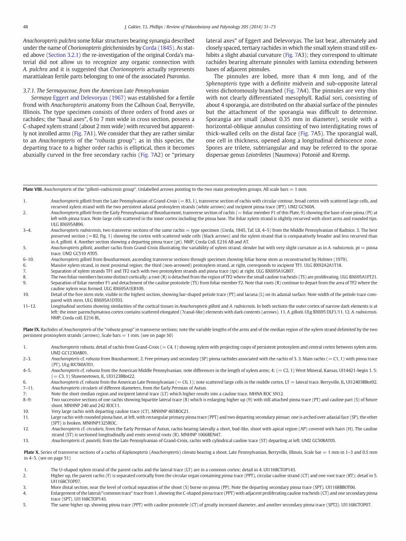
48 J. Galtier, T.L. Phillips / Review of Palaeobotany and Palynology 205 (2014) 31–73
Anachoropteris pulchra some foliar structures bearing synangia describedunder the name of Chorionopteris gleichenioides by Corda (1845). As stat-ed above (Section 3.2.1) the re-investigation of the original Corda's ma-terial did not allow us to recognize any organic connection withA. pulchra and it is suggested that Chorionopteris actually representsmarattialean fertile parts belonging to one of the associated Psaronius.
3.7.1. The Sermayaceae, from the American Late PennsylvanianSermaya Eggert and Delevoryas (1967) was established for a fertile
frond with Anachoropteris anatomy from the Calhoun Coal, Berryville,Illinois. The type specimen consists of three orders of frond axes orrachides; the “basal axes”, 6 to 7 mm wide in cross section, possess aC-shaped xylem strand (about 2mmwide)with recurved but apparent-ly not inrolled arms (Fig. 7A1). We consider that they are rather similarto an Anachoropteris of the “robusta group”; as in this species, thedeparting trace to a higher order rachis is elliptical, then it becomesabaxially curved in the free secondary rachis (Fig. 7A2) or “primary
Plate VIII. Anachoropteris of the “gillotii–radnicensis group”. Unlabelled arrows pointing to the
1. Anachoropteris gillotii from the Late Pennsylvanian of Grand-Croix (= B3, 1), trarecurved xylem strand with the two persistent adaxial protoxylem strands (wh
2. Anachoropteris gillotii from the Early Pennsylvanian of Bouxharmont, transverse sleft with pinna trace. Note large cells scattered in the inner cortex including theULG BX695AB96.
3–4. Anachoropteris radnicensis, two transverse sections of the same rachis = type sppreserved section (=B2, Fig. 1) showing the cortex with scattered wide cells (bin A. gillotii. 4. Another section showing a departing pinna trace (pt). NMP, Cord
5. Anachoropteris gillotii, another rachis from Grand-Croix illustrating the variabilitrace. UM2 GC510 AT05.
6–10. Anachoropteris gillotii from Bouxharmont, ascending transverse sections through6. Massive xylem strand, in most proximal region; the third (non-arrowed) protox7. Separation of xylem strands TF1 and TF2 each with two protoxylem strands and8. The two foliarmembers become distinct cortically; a root (R) is detached from th9. Separation of foliar member F1 and detachment of the cauline protostele (TS) fro
cauline xylem was formed. ULG BX695A1EB109.10. Detail of the free stem stele, visible in the highest section, showing bar-shaped p
pared with stem. ULG BX695A1DT03.11–12. Longitudinal sections showing similarities of the cortical tissues in Anachoropter
left; the inner parenchymatous cortex contains scattered elongated (?canal-like)NMP, Corda coll. E216 BL.
Plate IX. Rachides of Anachoropteris of the “robusta group” in transverse sections; note the variapersistent protoxylem strands (arrows). Scale bars = 1mm. (see on page 50)
1. Anachoropteris robusta, detail of rachis from Grand-Croix (= C4, 1) showing xyleUM2 GC1230AB01.
2–3. Anachoropteris cf. robusta from Bouxharmont; 2. Free primary and secondary (SP(PT). ULg BX780AT01.
4–5. Anachoropteris cf. robusta from the AmericanMiddle Pennsylvanian; note differe(= C3, 1) Shawneetown, IL, UI3123BBot22.
6. Anachoropteris cf. robusta from the American Late Pennsylvanian (= C6, 1); note7–11. Anachoropteris circularis of different diameters, from the Early Permian of Autun7: Note the short median region and incipient lateral trace (LT) which higher resu8–9: Two successive sections of one rachis showing bipartite lateral trace (8) which i
shoot. MNHNP 240 and 242 ROC11.10. Very large rachis with departing cauline trace (CT). MNHNP 465ROC21.11. Large rachiswith rounded pinna base, at left, with rectangular primary pinna trac
(SPT) is broken. MNHNP1325ROC.12. Anachoropteris cf. circularis, from the Early Permian of Autun, rachis bearing lat
strand (ST) is sectioned longitudinally and emits several roots (R). MNHNP 10613. Anachoropteris cf. pautetii, from the Late Pennsylvanian of Grand-Croix, rachis w
Plate X. Series of transverse sections of a rachis of Kaplanopteris (Anachoropteris) clavata bearin 4–5. (see on page 51)
1. The U-shaped xylem strand of the parent rachis and the lateral trace (LT) are in2. Higher up, the parent rachis (F) is separated cortically from the circular organ co
UI1168CTOP07.3. More distal section, near the level of cortical separation of the shoot (S) borne o4. Enlargement of the lateral/“common trace” trace from1, showing theC-shaped p
trace (SPT). UI1168CTOP143.5. The same higher up, showing pinna trace (PPT) with cauline protostele (CT) of
lateral axes” of Eggert and Delevoryas. The last bear, alternately andclosely spaced, tertiary rachides inwhich the small xylem strand still ex-hibits a slight abaxial curvature (Fig. 7A3); they correspond to ultimaterachides bearing alternate pinnules with lamina extending betweenbases of adjacent pinnules.
The pinnules are lobed, more than 4 mm long, and of theSphenopteris type with a definite midvein and sub-opposite lateralveins dichotomously branched (Fig. 7A4). The pinnules are very thinwith not clearly differentiated mesophyll. Radial sori, consisting ofabout 4 sporangia, are distributed on the abaxial surface of the pinnulesbut the attachment of the sporangia was difficult to determine.Sporangia are small (about 0.35 mm in diameter), sessile with ahorizontal-oblique annulus consisting of two interdigitating rows ofthick-walled cells on the distal face (Fig. 7A5). The sporangial wall,one cell in thickness, opened along a longitudinal dehiscence zone.Spores are trilete, subtriangular and may be referred to the sporaedispersae genus Leiotriletes (Naumova) Potonié and Kremp.
two main protoxylem groups. All scale bars = 1 mm.
nsverse section of rachis with circular contour, broad cortex with scattered large cells, andite arrows) and incipient pinna trace (IPT). UM2 GC560A.ection of rachis (= foliar member F1 of this Plate, 9) showing the base of one pinna (PI) atpinna base. The foliar xylem strand is slightly recurved with short arms and rounded tips.
ecimen (Corda, 1845, Taf. LII, 4–5) from the Middle Pennsylvanian of Radnice. 3. The bestlack arrows) and the xylem strand that is comparatively broader and less recurved thana Coll. E216 AB and AT.ty of xylem strand, slender but with very slight curvature as in A. radnicensis. pt = pinna
specimen showing foliar borne stem as reconstructed by Holmes (1979).ylem strand, at right, corresponds to incipient TF1. ULG BX92A2A1T18.pinna trace (tpi) at right. ULG BX695A1GB07.e region of TF2where the small cauline tracheids (TS) are proliferating. ULG BX695A1FT21.m foliar member F2. Note that roots (R) continue to depart from the area of TF2 where the
etiole trace (PT) and lacuna (L) on its adaxial surface. Note width of the petiole trace com-
is gillotii and A. radnicensis. In both sections the outer cortex of narrow dark elements is atelements with dark contents (arrows). 11. A. gillotii. ULg BX695DLF1/11. 12. A. radnicensis.
ble lengths of the arms and of themedian region of the xylem strand delimited by the two
mwith projecting cusps of persistent protoxylem and central cortex between xylem arms.
) pinna rachides associated with the rachis of 3. 3. Main rachis (= C1, 1) with pinna trace
nces in the length of xylem arms; 4: (= C2, 1) West Mineral, Kansas, UI14421-begin 1. 5:
scattered large cells in the middle cortex. LT = lateral trace. Berryville, IL, UI12403BBot92..lts into a cauline trace. MHNA ROC SN12.s enlarging higher up (9) with still attached pinna trace (PT) and cauline part (S) of future
e (PPT) and two departing secondary pinnae; one is archedover adaxial face (SP), the other
erally a short, bud-like, shoot with apical region (AP) covered with hairs (H). The cauline6REN47.ith cylindrical cauline trace (ST) departing at left. UM2 GC508AT05.
ing a shoot. Late Pennsylvanian, Berryville, Illinois. Scale bar = 1 mm in 1–3 and 0.5 mm
a common cortex; detail in 4. UI1168CTOP143.ntaining pinna trace (PPT), circular cauline strand (CT) and one root trace (RT); detail in 5.
n pinna (PP). Note the departing secondary pinna trace (SPT). UI1168BBOT06.inna trace (PPT)with adjacent proliferating cauline tracheids (CT) and one secondary pinna
greatly increased diameter, and another secondary pinna trace (SPT2). UI1168CTOP07.
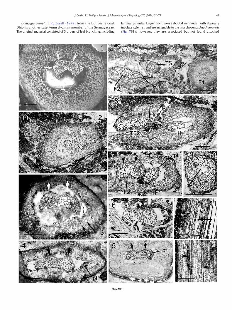
49J. Galtier, T.L. Phillips / Review of Palaeobotany and Palynology 205 (2014) 31–73
Doneggia complura Rothwell (1978) from the Duquesne Coal,Ohio, is another Late Pennsylvanian member of the Sermayaceae.The original material consisted of 3 orders of leaf branching, including
Plate VI
laminar pinnules. Larger frond axes (about 4 mm wide) with abaxiallyinvolute xylem strand are assignable to themorphogenus Anachoropteris(Fig. 7B1); however, they are associated but not found attached
II.
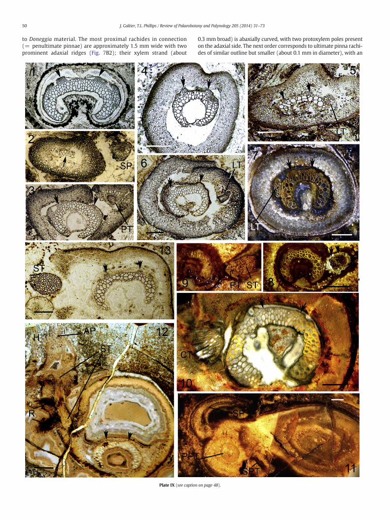
50 J. Galtier, T.L. Phillips / Review of Palaeobotany and Palynology 205 (2014) 31–73
to Doneggia material. The most proximal rachides in connection(= penultimate pinnae) are approximately 1.5 mm wide with twoprominent adaxial ridges (Fig. 7B2); their xylem strand (about
Plate IX (see caption
0.3 mm broad) is abaxially curved, with two protoxylem poles presenton the adaxial side. The next order corresponds to ultimate pinna rachi-des of similar outline but smaller (about 0.1 mm in diameter), with an
on page 48).
Plate X (see caption on page 48).
51J. Galtier, T.L. Phillips / Review of Palaeobotany and Palynology 205 (2014) 31–73
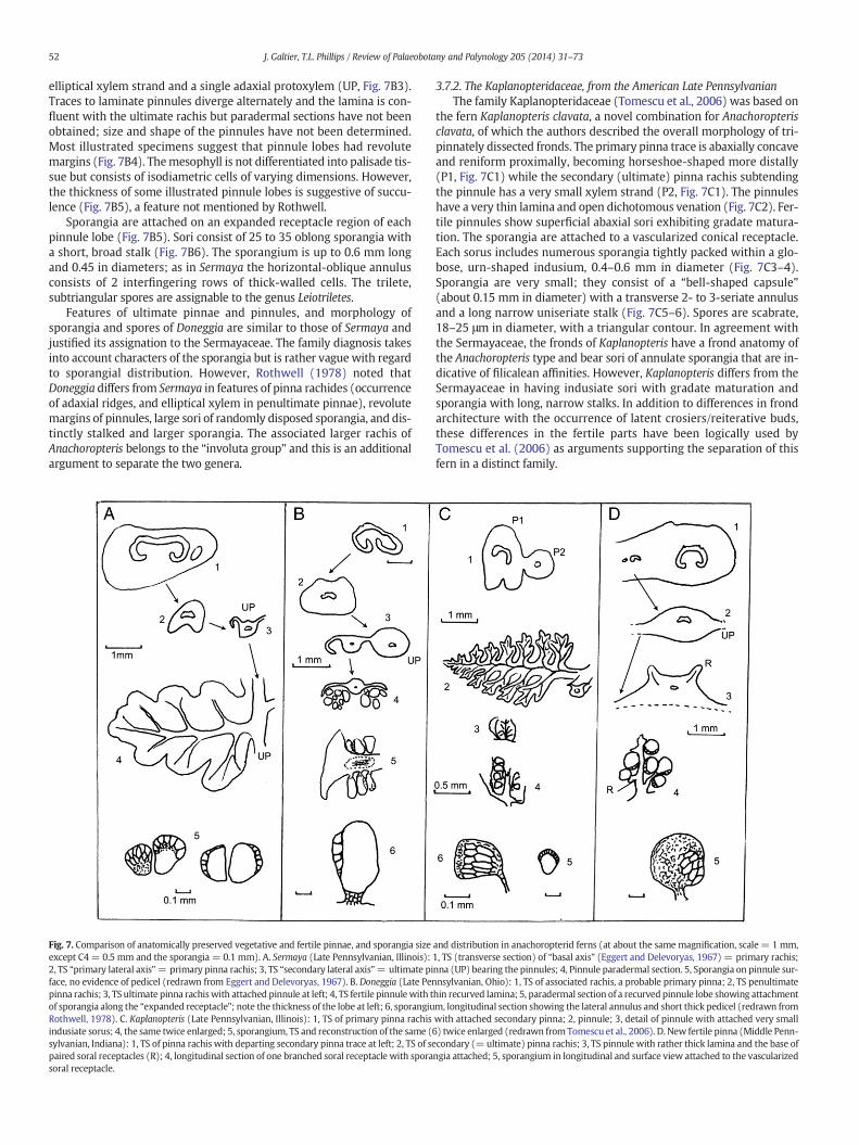
52 J. Galtier, T.L. Phillips / Review of Palaeobotany and Palynology 205 (2014) 31–73
elliptical xylem strand and a single adaxial protoxylem (UP, Fig. 7B3).Traces to laminate pinnules diverge alternately and the lamina is con-fluent with the ultimate rachis but paradermal sections have not beenobtained; size and shape of the pinnules have not been determined.Most illustrated specimens suggest that pinnule lobes had revolutemargins (Fig. 7B4). Themesophyll is not differentiated into palisade tis-sue but consists of isodiametric cells of varying dimensions. However,the thickness of some illustrated pinnule lobes is suggestive of succu-lence (Fig. 7B5), a feature not mentioned by Rothwell.
Sporangia are attached on an expanded receptacle region of eachpinnule lobe (Fig. 7B5). Sori consist of 25 to 35 oblong sporangia witha short, broad stalk (Fig. 7B6). The sporangium is up to 0.6 mm longand 0.45 in diameters; as in Sermaya the horizontal-oblique annulusconsists of 2 interfingering rows of thick-walled cells. The trilete,subtriangular spores are assignable to the genus Leiotriletes.
Features of ultimate pinnae and pinnules, and morphology ofsporangia and spores of Doneggia are similar to those of Sermaya andjustified its assignation to the Sermayaceae. The family diagnosis takesinto account characters of the sporangia but is rather vague with regardto sporangial distribution. However, Rothwell (1978) noted thatDoneggia differs from Sermaya in features of pinna rachides (occurrenceof adaxial ridges, and elliptical xylem in penultimate pinnae), revolutemargins of pinnules, large sori of randomly disposed sporangia, and dis-tinctly stalked and larger sporangia. The associated larger rachis ofAnachoropteris belongs to the “involuta group” and this is an additionalargument to separate the two genera.
Fig. 7. Comparison of anatomically preserved vegetative and fertile pinnae, and sporangia sizeexcept C4= 0.5 mm and the sporangia = 0.1 mm). A. Sermaya (Late Pennsylvanian, Illinois):2, TS “primary lateral axis”= primary pinna rachis; 3, TS “secondary lateral axis”= ultimate piface, no evidence of pedicel (redrawn from Eggert and Delevoryas, 1967). B. Doneggia (Late Penpinna rachis; 3, TS ultimate pinna rachiswith attached pinnule at left; 4, TS fertile pinnulewith tof sporangia along the “expanded receptacle”; note the thickness of the lobe at left; 6, sporangiuRothwell, 1978). C. Kaplanopteris (Late Pennsylvanian, Illinois): 1, TS of primary pinna rachisindusiate sorus; 4, the same twice enlarged; 5, sporangium, TS and reconstruction of the same (sylvanian, Indiana): 1, TS of pinna rachis with departing secondary pinna trace at left; 2, TS of sepaired soral receptacles (R); 4, longitudinal section of one branched soral receptacle with sporasoral receptacle.
3.7.2. The Kaplanopteridaceae, from the American Late PennsylvanianThe family Kaplanopteridaceae (Tomescu et al., 2006) was based on
the fern Kaplanopteris clavata, a novel combination for Anachoropterisclavata, of which the authors described the overall morphology of tri-pinnately dissected fronds. The primary pinna trace is abaxially concaveand reniform proximally, becoming horseshoe-shaped more distally(P1, Fig. 7C1) while the secondary (ultimate) pinna rachis subtendingthe pinnule has a very small xylem strand (P2, Fig. 7C1). The pinnuleshave a very thin lamina and open dichotomous venation (Fig. 7C2). Fer-tile pinnules show superficial abaxial sori exhibiting gradate matura-tion. The sporangia are attached to a vascularized conical receptacle.Each sorus includes numerous sporangia tightly packed within a glo-bose, urn-shaped indusium, 0.4–0.6 mm in diameter (Fig. 7C3–4).Sporangia are very small; they consist of a “bell-shaped capsule”(about 0.15 mm in diameter) with a transverse 2- to 3-seriate annulusand a long narrow uniseriate stalk (Fig. 7C5–6). Spores are scabrate,18–25 μm in diameter, with a triangular contour. In agreement withthe Sermayaceae, the fronds of Kaplanopteris have a frond anatomy ofthe Anachoropteris type and bear sori of annulate sporangia that are in-dicative of filicalean affinities. However, Kaplanopteris differs from theSermayaceae in having indusiate sori with gradate maturation andsporangia with long, narrow stalks. In addition to differences in frondarchitecture with the occurrence of latent crosiers/reiterative buds,these differences in the fertile parts have been logically used byTomescu et al. (2006) as arguments supporting the separation of thisfern in a distinct family.
and distribution in anachoropterid ferns (at about the same magnification, scale = 1 mm,1, TS (transverse section) of “basal axis” (Eggert and Delevoryas, 1967) = primary rachis;nna (UP) bearing the pinnules; 4, Pinnule paradermal section. 5, Sporangia on pinnule sur-nsylvanian, Ohio): 1, TS of associated rachis, a probable primary pinna; 2, TS penultimatehin recurved lamina; 5, paradermal section of a recurved pinnule lobe showing attachmentm, longitudinal section showing the lateral annulus and short thick pedicel (redrawn fromwith attached secondary pinna; 2, pinnule; 3, detail of pinnule with attached very small6) twice enlarged (redrawn from Tomescu et al., 2006). D. New fertile pinna (Middle Penn-condary (= ultimate) pinna rachis; 3, TS pinnule with rather thick lamina and the base ofngia attached; 5, sporangium in longitudinal and surface view attached to the vascularized
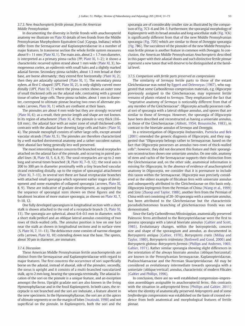
53J. Galtier, T.L. Phillips / Review of Palaeobotany and Palynology 205 (2014) 31–73
3.7.3. New Anachoropteris fertile pinnae, from the AmericanMiddle Pennsylvanian
In documenting the diversity in fertile fronds with anachoropteridanatomy we illustrate on Plate XI details of two fronds from the MiddlePennsylvanian Murphysboro Equivalent Coal (Cayuga, Indiana) whichdiffer from the Sermayaceae and Kaplanopteridaceae in a number ofmajor features. In transverse section the whole fertile system measuresabout 9×11mm(Plate XI, 1). Themain axis, about 2×1.7mmdiameter,is interpreted as a primary pinna rachis (PP, Plate XI, 1–2); it shows acharacteristic recurved xylem strand about 1 mmwide (Plate XI, 3), ho-mogeneous cortex, an epidermis with small hairs and a slightly markedadaxial furrow. Secondary pinna rachides, about 1.3 mm broad at theirbase, are borne alternately; they extend first horizontally (Plate XI, 2),then they are adaxially upturned (Plate XI, 1). The secondary pinnaxylem, at first C-shaped (SPT, Plate XI, 2), is only slightly curved moredistally (UPT, Plate XI, 7) where the pinna cortex shows an outer zoneof small thickened cells on the adaxial side, contrasting with a groundtissue of rather large cells. These pinna rachides, about 1 mm in diame-ter, correspond to ultimate pinnae bearing two rows of alternate pin-nules (arrows, Plate XI, 1) which are confluent at their bases.
The pinnules are about 4 mm wide but they are strongly recurved(Plate XI, 6); as a result, their precise length and shape are not known.In its region of attachment (Plate XI, 4) the pinnule is very thick (0.6–0.8 mm); the adaxial face with an outer zone of small thickened cellscontrasts with the abaxial face showing large cells and hairs (Plate XI,4). The pinnule mesophyll consists of rather large cells, except aroundvascular strands (Plate XI, 5). The pinnules are therefore characterizedby a well marked dorsiventral anatomy and a rather succulent nature,their abaxial face being generally less well preserved.
Themost interesting feature concerns the branched soral receptaclesattached on the adaxial face of the pinnule, and occurring in paired par-allel lines (R, Plate XI, 5, 6, 8, 9). The soral receptacles are up to 2 mmlong and several times branched (R, Plate XI, 7–9, 12); the soral axis is200 to 300 μm in diameter proximally with a tiny branched vascularstrand extending distally, up to the region of sporangial attachment(Plate XI, 7–13). In several sori there are basal receptacular brancheswith attached small sporangia which represent earlier stages of devel-opment and some examples of collapsed sporangia (arrows, Plate XI,8, 9). These are indicative of gradate development, as supported bythe sequence of sporangial sizes shown on these figures and thebasalmost location of more mature sporangia, as shown on Plate XI, 7,9–10, 12.
One fully developed sporangium in longitudinal section with a shortstalk is shown attached to the end of a receptacle branch (S, Plate XI,13). The sporangia are spherical, about 0.4–0.5 mm in diameter, witha short stalk/pedicel and an oblique lateral annulus consisting of tworows of thick-walled cells. The annulus position is low on one facenear the stalk as shown in longitudinal sections and in surface view(S, Plate XI, 7, 11–13). The dehiscence zone consists of narrow elongatecells (arrows, Plate XI, 10) extending down near the base. The spores,about 30 μm in diameter, are immature.
3.7.4. DiscussionThese American Middle Pennsylvanian fertile anachoropterids are
distinct from the Sermayaceae and Kaplanopteridaceae with regard tomajor features. The first concerns the occurrence of sori superficiallyborne on the adaxial, instead of abaxial, side of the pinnules; secondly,the sorus is upright and it consists of a multi-branched vascularizedstalk, up to 2mm long, bearing the sporangia terminally. The adaxial lo-cation of the sori on the pinnule is a unique feature, and an exceptionamongst the ferns. Upright gradate sori are also known in the livingHymenophyllaceae and in the fossil Kaplanopteris. In both cases, the re-ceptacle is not branched and the sori are indusiate, a feature absent inthe new specimens. In the Hymenophyllaceae, the sori are at the apicesof ultimate segments or on themargin of lobes (Iwatzuki, 1990) and notsuperficial on the pinnule. In Kaplanopteris, both the sori and the
sporangia are of considerably smaller size as illustrated by the compar-ison of Fig. 7C3–6 andD4–5. Furthermore, the sporangialmorphology ofKaplanopteriswith its broad annulus and longunicellular stalk (Fig. 7C6)is significantly different from that of the new Middle Pennsylvaniansporangia (Fig. 7D5) which are similar to those of Doneggia sporangia(Fig. 7B6). The succulence of the pinnules of the newMiddle Pennsylva-nian fertile pinnae is another feature in commonwith Doneggia. In con-clusion, the American Middle Pennsylvanian Anachoropteris describedin this paperwith their adaxial shoots and such distinctive fertile pinnaerepresent a new taxon thatwill deserve to be distinguished at the famil-ial level.
3.7.5. Comparison with fertile parts preserved as compressionsThe similarity of Sermaya fertile parts to those of the extant
Gleicheniaceae was noted by Eggert and Delevoryas (1967), who sug-gested that some Carboniferous compression materials, e.g. Oligocarpiapreviously assigned to the Gleicheniaceae, may represent fertileremains of plants related to Sermaya. They emphasized the fact that“vegetative anatomy of Sermaya is noticeably different from that ofanymember of the Gleicheniaceae”. Oligocarpia actually possesses radi-al sori of sporangia with a lateral oblique annulus, and spores that aresimilar to those of Sermaya. However, the sporangia of Oligocarpiahave been described and reconstructed as having a uniseriate annulus,as those of the Gleicheniaceae (Abbott, 1954; Brousmiche, 1983), incontrast to the biseriate annulus of Sermaya and Doneggia.
In a reinvestigation of Oligocarpia lindsaeoides, Psenicka and Bek(2001) emended the generic diagnosis of Oligocarpia, and they sug-gested that “Oligocarpia belongs rather to Sermayaceae, based on thefact that Oligocarpia possesses an annulus two-rows of thick-walledcells”; however, they did not document this feature and their sporangi-um reconstruction shows one uniseriate annulus. On one side, anatomyof stem and rachis of the Sermayaceae supports their distinction fromthe Gleicheniaceae and, on the other side, anatomical information ismissing for Oligocarpia. In the absence of evidence of anachoropteridanatomy in Oligocarpia, we consider that it is premature to includethis taxon within the Sermayaceae. Oligocarpia was precisely consid-ered as the oldest example of filicalean fern with uniseriate annulusand subsequently attributed to the Gleicheniaceae on this basis only.Oligocarpia kepingensis from the Permian of China (Wang et al., 1999)and Szea (Zhaoqi and Taylor, 1988), another fern from the Permian ofChinawith sori consisting of 20–30 sporangiawith a uniseriate annulus,has been attributed to the Gleicheniaceae but the characteristicpseudodichotomous branching of gleicheniaceous fronds was notdemonstrated.
Since the Early Carboniferous/Mississippian, anatomically preservedPaleozoic ferns attributed to the Botryopteridaceae were the first topossess sporangia with a lateral biseriate annulus (Galtier and Scott,1985). Evolutionary changes, within the botryopterids, concernsize and shape of the sporangium and annulus, as documented inBotryopteris antiqua (Galtier, 1970), Botryopteris cratis (Millay andTaylor, 1980), Botryopteris tridentata (Rothwell and Good, 2000) andBotryopteris globosa–Botryopteris forensis (Phillips and Andrews, 1965;Galtier, 1971). Rather similar sporangia showing slight differences inthe orientation of the always biseriate annulus (oblique/horizontal)are known in the Pennsylvanian Sermayaceae, Kaplanopteridaceae,Psalixochlaenaceae and the Permian Skaaripteridaceae. All may beconsidered as evolutionary intermediate towards sporangia with auniseriate (oblique/vertical) annulus, characteristic of modern Filicales(Galtier and Phillips, 1996).
In conclusion, there are no well established compression–impres-sion assemblages assignable to anachoropterid ferns; this contrastswith the situation in ankyropterid ferns (Phillips and Galtier, 2011)where the identity of anatomically preserved Ankyropteris and of someSenftenbergia compressions was established on the basis of crossed evi-dence from both anatomical and morphological features of fertilepinnae.
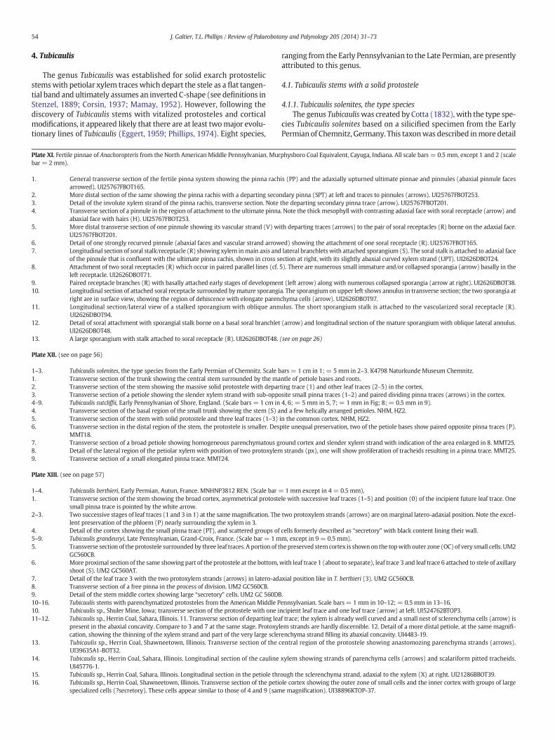
54 J. Galtier, T.L. Phillips / Review of Palaeobotany and Palynology 205 (2014) 31–73
4. Tubicaulis
The genus Tubicaulis was established for solid exarch protostelicstemswith petiolar xylem traceswhich depart the stele as a flat tangen-tial band and ultimately assumes an inverted C-shape (see definitions inStenzel, 1889; Corsin, 1937; Mamay, 1952). However, following thediscovery of Tubicaulis stems with vitalized protosteles and corticalmodifications, it appeared likely that there are at least twomajor evolu-tionary lines of Tubicaulis (Eggert, 1959; Phillips, 1974). Eight species,
Plate XI. Fertile pinnae of Anachoropteris from the North American Middle Pennsylvanian. Murbar = 2 mm).
1. General transverse section of the fertile pinna system showing the pinna racharrowed). UI25767FBOT165.
2. More distal section of the same showing the pinna rachis with a departing secon3. Detail of the involute xylem strand of the pinna rachis, transverse section. Note4. Transverse section of a pinnule in the region of attachment to the ultimate pinna
abaxial face with hairs (H). UI25767FBOT253.5. More distal transverse section of one pinnule showing its vascular strand (V) wi
UI25767FBOT201.6. Detail of one strongly recurved pinnule (abaxial faces and vascular strand arrow7. Longitudinal section of soral stalk/receptacle (R) showing xylem inmain axis and
of the pinnule that is confluent with the ultimate pinna rachis, shown in cross s8. Attachment of two soral receptacles (R) which occur in paired parallel lines (cf.
left receptacle. UI2626DBOT71.9. Paired receptacle branches (R) with basally attached early stages of developmen10. Longitudinal section of attached soral receptacle surrounded bymature sporangi
right are in surface view, showing the region of dehiscence with elongate paren11. Longitudinal section/lateral view of a stalked sporangium with oblique ann
UI2626DBOT94.12. Detail of soral attachment with sporangial stalk borne on a basal soral branchlet
UI2626DBOT48.13. A large sporangium with stalk attached to soral receptacle (R). UI2626DBOT48.
Plate XII. (see on page 56)
1–3. Tubicaulis solenites, the type species from the Early Permian of Chemnitz. Scale b1. Transverse section of the trunk showing the central stem surrounded by the ma2. Transverse section of the stem showing the massive solid protostele with depar3. Transverse section of a petiole showing the slender xylem strand with sub-oppo4–9. Tubicaulis sutcliffii, Early Pennsylvanian of Shore, England. (Scale bars = 1 cm in4. Transverse section of the basal region of the small trunk showing the stem (S) a5. Transverse section of the stem with solid protostele and three leaf traces (1–3)6. Transverse section in the distal region of the stem, the protostele is smaller. Des
MMT18.7. Transverse section of a broad petiole showing homogeneous parenchymatous g8. Detail of the lateral region of the petiolar xylem with position of two protoxylem9. Transverse section of a small elongated pinna trace. MMT24.
Plate XIII. (see on page 57)
1–4. Tubicaulis berthieri, Early Permian, Autun, France. MNHNP3812 REN. (Scale bar =1. Transverse section of the stem showing the broad cortex, asymmetrical protoste
small pinna trace is pointed by the white arrow.2–3. Two successive stages of leaf traces (1 and 3 in 1) at the samemagnification. The
lent preservation of the phloem (P) nearly surrounding the xylem in 3.4. Detail of the cortex showing the small pinna trace (PT), and scattered groups of5–9. Tubicaulis grandeuryi, Late Pennsylvanian, Grand-Croix, France. (Scale bar = 1m5. Transverse section of the protostele surrounded by three leaf traces. A portion of t
GC560CB.6. More proximal section of the same showing part of the protostele at the bottom,
shoot (S). UM2 GC560AT.7. Detail of the leaf trace 3 with the two protoxylem strands (arrows) in latero-ad8. Transverse section of a free pinna in the process of division. UM2 GC560CB.9. Detail of the stem middle cortex showing large “secretory” cells. UM2 GC 560DB10–16. Tubicaulis stems with parenchymatized protosteles from the American Middle P10. Tubicaulis sp., Shuler Mine, Iowa; transverse section of the protostele with one i11–12. Tubicaulis sp., Herrin Coal, Sahara, Illinois. 11. Transverse section of departing lea
present in the abaxial concavity. Compare to 3 and 7 at the same stage. Protoxylcation, showing the thinning of the xylem strand and part of the very large scler
13. Tubicaulis sp., Herrin Coal, Shawneetown, Illinois. Transverse section of the cUI39635A1-BOT32.
14. Tubicaulis sp., Herrin Coal, Sahara, Illinois. Longitudinal section of the caulineUI45776-1.
15. Tubicaulis sp., Herrin Coal, Sahara, Illinois. Longitudinal section in the petiole thr16. Tubicaulis sp., Herrin Coal, Shawneetown, Illinois. Transverse section of the petio
specialized cells (?secretory). These cells appear similar to those of 4 and 9 (sam
ranging from the Early Pennsylvanian to the Late Permian, are presentlyattributed to this genus.
4.1. Tubicaulis stems with a solid protostele
4.1.1. Tubicaulis solenites, the type speciesThe genus Tubicauliswas created by Cotta (1832), with the type spe-
cies Tubicaulis solenites based on a silicified specimen from the EarlyPermian of Chemnitz, Germany. This taxonwas described inmore detail
physboro Coal Equivalent, Cayuga, Indiana. All scale bars = 0.5 mm, except 1 and 2 (scale
is (PP) and the adaxially upturned ultimate pinnae and pinnules (abaxial pinnule faces
dary pinna (SPT) at left and traces to pinnules (arrows). UI25767FBOT253.the departing secondary pinna trace (arrow). UI25767FBOT201.. Note the thick mesophyll with contrasting adaxial face with soral receptacle (arrow) and
th departing traces (arrows) to the pair of soral receptacles (R) borne on the adaxial face.
ed) showing the attachment of one soral receptacle (R). UI25767FBOT165.lateral branchlets with attached sporangium (S). The soral stalk is attached to adaxial face
ection at right, with its slightly abaxial curved xylem strand (UPT). UI2626DBOT24.5). There are numerous small immature and/or collapsed sporangia (arrow) basally in the
t (left arrow) along with numerous collapsed sporangia (arrow at right). UI2626DBOT38.a. The sporangium on upper left shows annulus in transverse section; the two sporangia atchyma cells (arrow). UI2626DBOT97.ulus. The short sporangium stalk is attached to the vascularized soral receptacle (R).
(arrow) and longitudinal section of the mature sporangium with oblique lateral annulus.
(see on page 26)
ars= 1 cm in 1; = 5 mm in 2–3. K4798 Naturkunde Museum Chemnitz.ntle of petiole bases and roots.ting trace (1) and other leaf traces (2–5) in the cortex.site small pinna traces (1–2) and paired dividing pinna traces (arrows) in the cortex.4, 6; = 5 mm in 5, 7; = 1 mm in Fig; 8; = 0.5 mm in 9).nd a few helically arranged petioles. NHM, HZ2.in the common cortex. NHM, HZ2.pite unequal preservation, two of the petiole bases show paired opposite pinna traces (P).
round cortex and slender xylem strand with indication of the area enlarged in 8. MMT25.strands (px), one will show proliferation of tracheids resulting in a pinna trace. MMT25.
1 mm except in 4 = 0.5 mm).le with successive leaf traces (1–5) and position (0) of the incipient future leaf trace. One
two protoxylem strands (arrows) are on marginal latero-adaxial position. Note the excel-
cells formerly described as “secretory” with black content lining their wall.m, except in 9 = 0.5 mm).hepreserved stemcortex is shownon the topwith outer zone (OC)of very small cells. UM2
with leaf trace 1 (about to separate), leaf trace 3 and leaf trace 6 attached to stele of axillary
axial position like in T. berthieri (3). UM2 GC560CB.
.ennsylvanian. Scale bars= 1 mm in 10–12; = 0.5 mm in 13–16.ncipient leaf trace and one leaf trace (arrow) at left. UI524762BTOP3.f trace; the xylem is already well curved and a small nest of sclerenchyma cells (arrow) isem strands are hardly discernible. 12. Detail of a more distal petiole, at the same magnifi-enchyma strand filling its abaxial concavity. UI4483-19.entral region of the protostele showing anastomozing parenchyma strands (arrows).
xylem showing strands of parenchyma cells (arrows) and scalariform pitted tracheids.
ough the sclerenchyma strand, adaxial to the xylem (X) at right. UI21286BBOT39.le cortex showing the outer zone of small cells and the inner cortex with groups of largee magnification). UI38896KTOP-37.
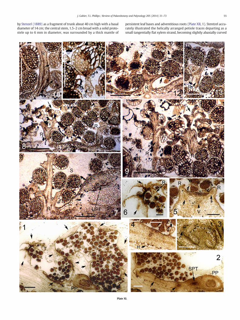
55J. Galtier, T.L. Phillips / Review of Palaeobotany and Palynology 205 (2014) 31–73
by Stenzel (1889) as a fragment of trunk about 40 cm high with a basaldiameter of 14 cm; the central stem, 1.5–2 cm broadwith a solid proto-stele up to 6 mm in diameter, was surrounded by a thick mantle of
Plate X
persistent leaf bases and adventitious roots (Plate XII, 1). Stentzel accu-rately illustrated the helically arranged petiole traces departing as asmall tangentially flat xylem strand, becoming slightly abaxially curved
I.

Plate XII (see caption on page 54).
56 J. Galtier, T.L. Phillips / Review of Palaeobotany and Palynology 205 (2014) 31–73
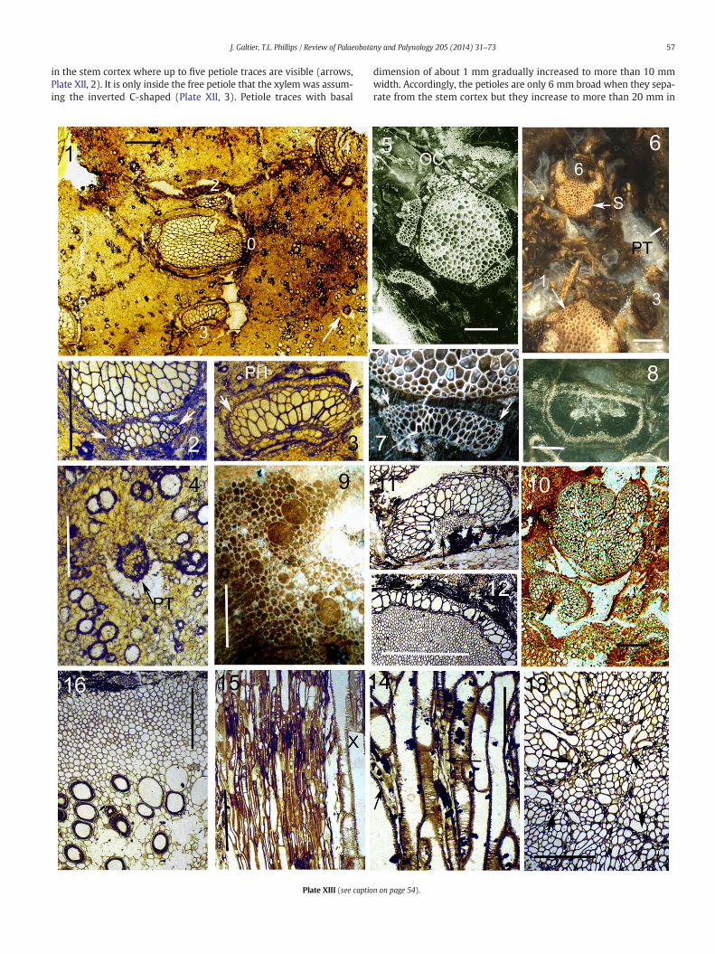
57J. Galtier, T.L. Phillips / Review of Palaeobotany and Palynology 205 (2014) 31–73
in the stem cortex where up to five petiole traces are visible (arrows,Plate XII, 2). It is only inside the free petiole that the xylem was assum-ing the inverted C-shaped (Plate XII, 3). Petiole traces with basal
Plate XIII (see captio
dimension of about 1 mm gradually increased to more than 10 mmwidth. Accordingly, the petioles are only 6 mm broad when they sepa-rate from the stem cortex but they increase to more than 20 mm in
n on page 54).
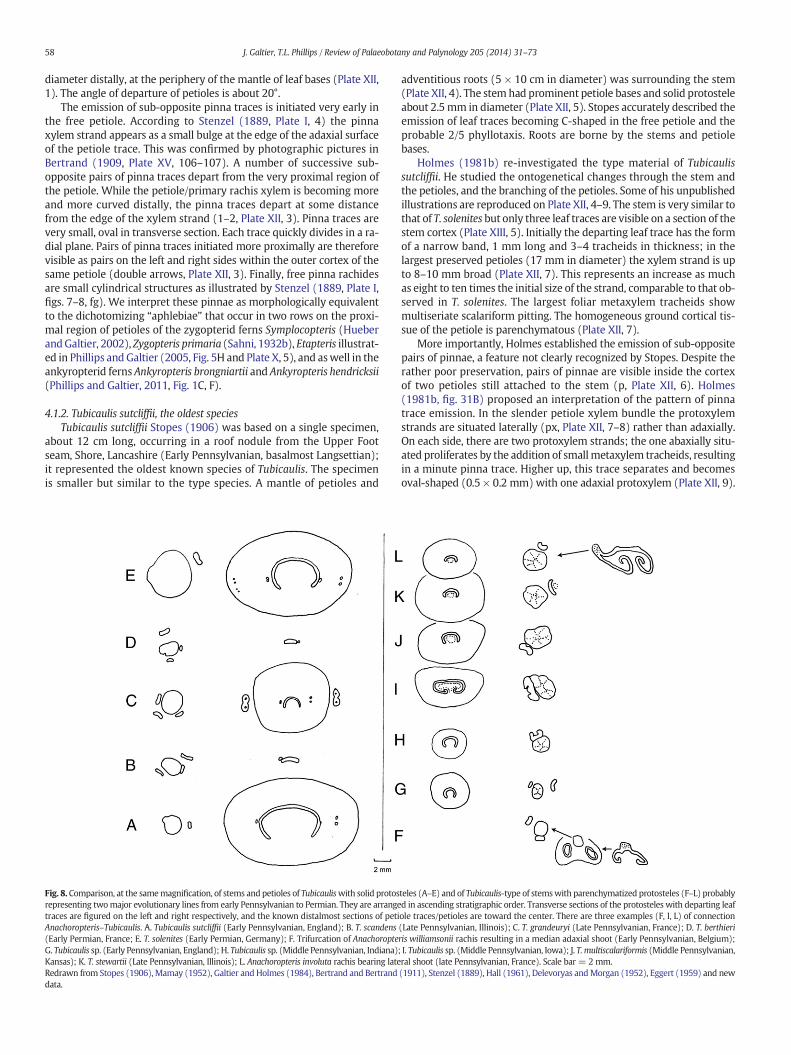
58 J. Galtier, T.L. Phillips / Review of Palaeobotany and Palynology 205 (2014) 31–73
diameter distally, at the periphery of the mantle of leaf bases (Plate XII,1). The angle of departure of petioles is about 20°.
The emission of sub-opposite pinna traces is initiated very early inthe free petiole. According to Stenzel (1889, Plate I, 4) the pinnaxylem strand appears as a small bulge at the edge of the adaxial surfaceof the petiole trace. This was confirmed by photographic pictures inBertrand (1909, Plate XV, 106–107). A number of successive sub-opposite pairs of pinna traces depart from the very proximal region ofthe petiole. While the petiole/primary rachis xylem is becoming moreand more curved distally, the pinna traces depart at some distancefrom the edge of the xylem strand (1–2, Plate XII, 3). Pinna traces arevery small, oval in transverse section. Each trace quickly divides in a ra-dial plane. Pairs of pinna traces initiated more proximally are thereforevisible as pairs on the left and right sides within the outer cortex of thesame petiole (double arrows, Plate XII, 3). Finally, free pinna rachidesare small cylindrical structures as illustrated by Stenzel (1889, Plate I,figs. 7–8, fg). We interpret these pinnae as morphologically equivalentto the dichotomizing “aphlebiae” that occur in two rows on the proxi-mal region of petioles of the zygopterid ferns Symplocopteris (HueberandGaltier, 2002), Zygopteris primaria (Sahni, 1932b), Etapteris illustrat-ed in Phillips andGaltier (2005, Fig. 5H and Plate X, 5), and aswell in theankyropterid ferns Ankyropteris brongniartii and Ankyropteris hendricksii(Phillips and Galtier, 2011, Fig. 1C, F).
4.1.2. Tubicaulis sutcliffii, the oldest speciesTubicaulis sutcliffii Stopes (1906) was based on a single specimen,
about 12 cm long, occurring in a roof nodule from the Upper Footseam, Shore, Lancashire (Early Pennsylvanian, basalmost Langsettian);it represented the oldest known species of Tubicaulis. The specimenis smaller but similar to the type species. A mantle of petioles and
Fig. 8. Comparison, at the samemagnification, of stems and petioles of Tubicauliswith solid protosrepresenting twomajor evolutionary lines from early Pennsylvanian to Permian. They are arrangetraces are figured on the left and right respectively, and the known distalmost sections of petiAnachoropteris–Tubicaulis. A. Tubicaulis sutcliffii (Early Pennsylvanian, England); B. T. scandens(Early Permian, France; E. T. solenites (Early Permian, Germany); F. Trifurcation of AnachoropteG. Tubicaulis sp. (Early Pennsylvanian, England); H. Tubicaulis sp. (Middle Pennsylvanian, Indiana)Kansas); K. T. stewartii (Late Pennsylvanian, Illinois); L. Anachoropteris involuta rachis bearing lateRedrawn from Stopes (1906), Mamay (1952), Galtier and Holmes (1984), Bertrand and Bertranddata.
adventitious roots (5 × 10 cm in diameter) was surrounding the stem(Plate XII, 4). The stemhad prominent petiole bases and solid protosteleabout 2.5 mm in diameter (Plate XII, 5). Stopes accurately described theemission of leaf traces becoming C-shaped in the free petiole and theprobable 2/5 phyllotaxis. Roots are borne by the stems and petiolebases.
Holmes (1981b) re-investigated the type material of Tubicaulissutcliffii. He studied the ontogenetical changes through the stem andthe petioles, and the branching of the petioles. Some of his unpublishedillustrations are reproduced on Plate XII, 4–9. The stem is very similar tothat of T. solenites but only three leaf traces are visible on a section of thestem cortex (Plate XIII, 5). Initially the departing leaf trace has the formof a narrow band, 1 mm long and 3–4 tracheids in thickness; in thelargest preserved petioles (17 mm in diameter) the xylem strand is upto 8–10 mm broad (Plate XII, 7). This represents an increase as muchas eight to ten times the initial size of the strand, comparable to that ob-served in T. solenites. The largest foliar metaxylem tracheids showmultiseriate scalariform pitting. The homogeneous ground cortical tis-sue of the petiole is parenchymatous (Plate XII, 7).
More importantly, Holmes established the emission of sub-oppositepairs of pinnae, a feature not clearly recognized by Stopes. Despite therather poor preservation, pairs of pinnae are visible inside the cortexof two petioles still attached to the stem (p, Plate XII, 6). Holmes(1981b, fig. 31B) proposed an interpretation of the pattern of pinnatrace emission. In the slender petiole xylem bundle the protoxylemstrands are situated laterally (px, Plate XII, 7–8) rather than adaxially.On each side, there are two protoxylem strands; the one abaxially situ-ated proliferates by the addition of smallmetaxylem tracheids, resultingin a minute pinna trace. Higher up, this trace separates and becomesoval-shaped (0.5 × 0.2 mm) with one adaxial protoxylem (Plate XII, 9).
teles (A–E) and of Tubicaulis-type of stemswith parenchymatized protosteles (F–L) probablyd in ascending stratigraphic order. Transverse sections of the protosteles with departing leafole traces/petioles are toward the center. There are three examples (F, I, L) of connection(Late Pennsylvanian, Illinois); C. T. grandeuryi (Late Pennsylvanian, France); D. T. berthieriris williamsonii rachis resulting in a median adaxial shoot (Early Pennsylvanian, Belgium);; I. Tubicaulis sp. (Middle Pennsylvanian, Iowa); J. T.multiscalariformis (Middle Pennsylvanian,ral shoot (late Pennsylvanian, France). Scale bar = 2 mm.(1911), Stenzel (1889), Hall (1961), Delevoryas andMorgan (1952), Eggert (1959) and new
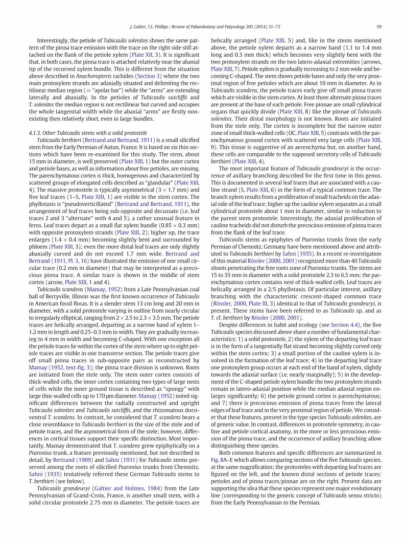
59J. Galtier, T.L. Phillips / Review of Palaeobotany and Palynology 205 (2014) 31–73
Interestingly, the petiole of Tubicaulis solenites shows the same pat-tern of the pinna trace emission with the trace on the right side still at-tached on the flank of the petiole xylem (Plate XII, 3). It is significantthat, in both cases, the pinna trace is attached relatively near the abaxialtip of the recurved xylem bundle. This is different from the situationabove described in Anachoropteris rachides (Section 3) where the twomain protoxylem strands are adaxially situated and delimiting the rec-tilinear median region (= “apolar bar”) while the “arms” are extendinglaterally and abaxially. In the petioles of Tubicaulis sutcliffii andT. solenites the median region is not rectilinear but curved and occupiesthe whole tangential width while the abaxial “arms” are firstly non-existing then relatively short, even in large bundles.
4.1.3. Other Tubicaulis stems with a solid protosteleTubicaulis berthieri (Bertrand and Bertrand, 1911) is a small silicified
stem from the Early Permian of Autun, France. It is based on six thin sec-tions which have been re-examined for this study. The stem, about15mm in diameter, is well preserved (Plate XIII, 1) but the outer cortexand petiole bases, aswell as information about free petioles, aremissing.The parenchymatous cortex is thick, homogenous and characterized byscattered groups of elongated cells described as “glandular” (Plate XIII,4). The massive protostele is typically asymmetrical (3 × 1.7 mm) andfive leaf traces (1–5, Plate XIII, 1) are visible in the stem cortex. Thephyllotaxis is “pseudoverticillated” (Bertrand and Bertrand, 1911), thearrangement of leaf traces being sub-opposite and decussate (i.e. leaftraces 2 and 3 “alternate” with 4 and 5), a rather unusual feature inferns. Leaf traces depart as a small flat xylem bundle (0.85 × 0.3 mm)with opposite protoxylem strands (Plate XIII, 2); higher up, the traceenlarges (1.4 × 0.4 mm) becoming slightly bent and surrounded byphloem (Plate XIII, 3); even the more distal leaf traces are only slightlyabaxially curved and do not exceed 1.7 mm wide. Bertrand andBertrand (1911, Pl. 3, 16) have illustrated the emission of one small cir-cular trace (0.2 mm in diameter) that may be interpreted as a preco-cious pinna trace. A similar trace is shown in the middle of stemcortex (arrow, Plate XIII, 1 and 4).
Tubicaulis scandens (Mamay, 1952) from a Late Pennsylvanian coalball of Berryville, Illinois was the first known occurrence of Tubicaulisin American fossil floras. It is a slender stem 13 cm long and 20 mm indiameter, with a solid protostele varying in outline from nearly circularto irregularly elliptical, ranging from2× 2.5 to 2.3 × 3.5mm. The petioletraces are helically arranged, departing as a narrow band of xylem 1–1.2mm in length and 0.25–0.3mm inwidth. They are gradually increas-ing to 4 mm in width and becoming C-shaped. With one exception allthepetiole traces liewithin the cortex of the stemwhere up to eight pet-iole traces are visible in one transverse section. The petiole traces giveoff small pinna traces in sub-opposite pairs as reconstructed byMamay (1952, text-fig. 3); the pinna trace division is unknown. Rootsare initiated from the stele only. The stem outer cortex consists ofthick-walled cells, the inner cortex containing two types of large nestsof cells while the inner ground tissue is described as “spongy” withlarge thin-walled cells up to 170 μmdiameter.Mamay (1952) noted sig-nificant differences between the radially constructed and uprightTubicaulis solenites and Tubicaulis sutcliffii, and the rhizomatous dorsi-ventral T. scandens. In contrast, he considered that T. scandens bears aclose resemblance to Tubicaulis berthieri in the size of the stele and ofpetiole traces, and the asymmetrical form of the stele; however, differ-ences in cortical tissues support their specific distinction. Most impor-tantly, Mamay demonstrated that T. scandens grew epiphytically on aPsaronius trunk, a feature previously mentioned, but not described indetail, by Bertrand (1909) and Sahni (1931) for Tubicaulis stems pre-served among the roots of silicified Psaronius trunks from Chemnitz.Sahni (1935) tentatively referred these German Tubicaulis stems toT. berthieri (see below).
Tubicaulis grandeuryi (Galtier and Holmes, 1984) from the LatePennsylvanian of Grand-Croix, France, is another small stem, with asolid circular protostele 2.75 mm in diameter. The petiole traces are
helically arranged (Plate XIII, 5) and, like in the stems mentionedabove, the petiole xylem departs as a narrow band (1.1 to 1.4 mmlong and 0.3 mm thick) which becomes very slightly bent with thetwo protoxylem strands on the two latero-adaxial extremities (arrows,Plate XIII, 7). Petiole xylem is gradually increasing to 2mmwide and be-coming C-shaped. The stem shows petiole bases and only the very prox-imal region of free petioles which are about 10 mm in diameter. As inTubicaulis scandens, the petiole traces early give off small pinna traceswhich are visible in the stem cortex. At least three alternate pinna tracesare present at the base of each petiole. Free pinnae are small cylindricalorgans that quickly divide (Plate XIII, 8) like the pinnae of Tubicaulissolenites. Their distal morphology is not known. Roots are initiatedfrom the stele only. The cortex is incomplete but the narrow outerzone of small thick-walled cells (OC, Plate XIII, 5) contrastswith the par-enchymatous ground cortex with scattered very large cells (Plate XIII,9). This tissue is suggestive of an aerenchyma but, on another hand,these cells are comparable to the supposed secretory cells of Tubicaulisberthieri (Plate XIII, 4).
The most important feature of Tubicaulis grandeuryi is the occur-rence of axillary branching described for the first time in this genus.This is documented in several leaf traces that are associated with a cau-line strand (S, Plate XIII, 6) in the form of a typical common trace. Thebranch xylem results froma proliferation of small tracheids on the adax-ial side of the leaf trace; higher up the cauline xylem separates as a smallcylindrical protostele about 1 mm in diameter, similar in reduction tothe parent stem protostele. Interestingly, the adaxial proliferation ofcauline tracheids did not disturb the precocious emission of pinna tracesfrom the flank of the leaf trace.
Tubicaulis stems as epiphytes of Psaronius trunks from the earlyPermian of Chemnitz, Germany have beenmentioned above and attrib-uted to Tubicaulis berthieri by Sahni (1935). In a recent re-investigationof thismaterial Rössler (2000, 2001) recognizedmore than 40 Tubicaulisshoots penetrating the free roots zone of Psaronius trunks. The stems are15 to 35mm in diameter with a solid protostele 2.3 to 6.5 mm; the par-enchymatous cortex contains nest of thick-walled cells. Leaf traces arehelically arranged in a 2/5 phyllotaxis. Of particular interest, axillarybranching with the characteristic crescent-shaped common trace(Rössler, 2000, Plate III, 3) identical to that of Tubicaulis grandeuryi, ispresent. These stems have been referred to as Tubicaulis sp. and asT. cf. berthieri by Rössler (2000, 2001).
Despite differences in habit and ecology (see Section 4.4), the fiveTubicaulis species discussed above share a number of fundamental char-acteristics: 1) a solid protostele, 2) the xylem of the departing leaf traceis in the form of a tangentially flat strand becoming slightly curved onlywithin the stem cortex; 3) a small portion of the cauline xylem is in-volved in the formation of the leaf trace; 4) in the departing leaf traceone protoxylem group occurs at each end of the band of xylem, slightlytowards the adaxial surface (i.e. nearly marginally); 5) in the develop-ment of the C-shaped petiole xylem bundle the two protoxylem strandsremain in latero-adaxial position while the median adaxial region en-larges significantly; 6) the petiole ground cortex is parenchymatous;and 7) there is precocious emission of pinna traces from the lateraledges of leaf trace and in the very proximal region of petiole.We consid-er that these features, present in the type species Tubicaulis solenites, areof generic value. In contrast, differences in protostele symmetry, in cau-line and petiole cortical anatomy, in the more or less precocious emis-sion of the pinna trace, and the occurrence of axillary branching allowdistinguishing these species.
Both common features and specific differences are summarized inFig. 8A–Ewhich allows comparing sections of the five Tubicaulis species,at the samemagnification; the protosteles with departing leaf traces arefigured on the left, and the known distal sections of petiole traces/petioles and of pinna traces/pinnae are on the right. Present data aresupporting the idea that these species represent onemajor evolutionaryline (corresponding to the generic concept of Tubicaulis sensu stricto)from the Early Pennsylvanian to the Permian.
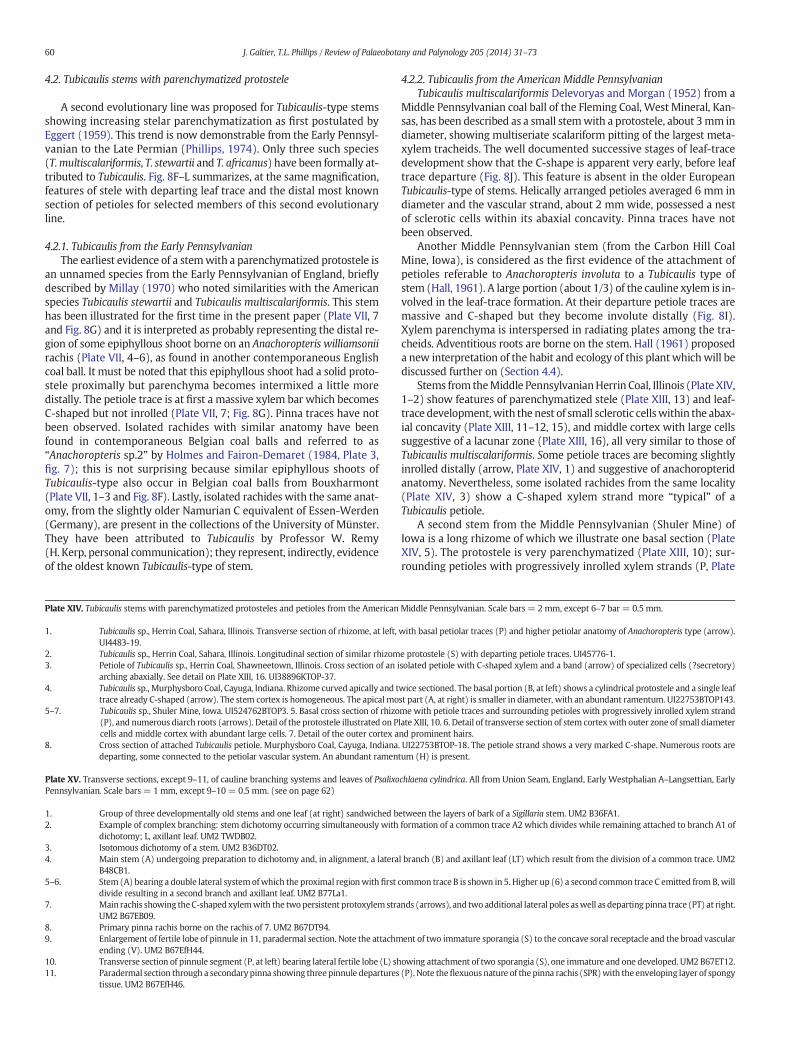
60 J. Galtier, T.L. Phillips / Review of Palaeobotany and Palynology 205 (2014) 31–73
4.2. Tubicaulis stems with parenchymatized protostele
A second evolutionary line was proposed for Tubicaulis-type stemsshowing increasing stelar parenchymatization as first postulated byEggert (1959). This trend is now demonstrable from the Early Pennsyl-vanian to the Late Permian (Phillips, 1974). Only three such species(T.multiscalariformis, T. stewartii and T. africanus) have been formally at-tributed to Tubicaulis. Fig. 8F–L summarizes, at the same magnification,features of stele with departing leaf trace and the distal most knownsection of petioles for selected members of this second evolutionaryline.
4.2.1. Tubicaulis from the Early PennsylvanianThe earliest evidence of a stemwith a parenchymatized protostele is
an unnamed species from the Early Pennsylvanian of England, brieflydescribed by Millay (1970) who noted similarities with the Americanspecies Tubicaulis stewartii and Tubicaulis multiscalariformis. This stemhas been illustrated for the first time in the present paper (Plate VII, 7and Fig. 8G) and it is interpreted as probably representing the distal re-gion of some epiphyllous shoot borne on an Anachoropteris williamsoniirachis (Plate VII, 4–6), as found in another contemporaneous Englishcoal ball. It must be noted that this epiphyllous shoot had a solid proto-stele proximally but parenchyma becomes intermixed a little moredistally. The petiole trace is at first a massive xylem bar which becomesC-shaped but not inrolled (Plate VII, 7; Fig. 8G). Pinna traces have notbeen observed. Isolated rachides with similar anatomy have beenfound in contemporaneous Belgian coal balls and referred to as“Anachoropteris sp.2” by Holmes and Fairon-Demaret (1984, Plate 3,fig. 7); this is not surprising because similar epiphyllous shoots ofTubicaulis-type also occur in Belgian coal balls from Bouxharmont(Plate VII, 1–3 and Fig. 8F). Lastly, isolated rachides with the same anat-omy, from the slightly older Namurian C equivalent of Essen-Werden(Germany), are present in the collections of the University of Münster.They have been attributed to Tubicaulis by Professor W. Remy(H. Kerp, personal communication); they represent, indirectly, evidenceof the oldest known Tubicaulis-type of stem.
Plate XIV. Tubicaulis stems with parenchymatized protosteles and petioles from the American
1. Tubicaulis sp., Herrin Coal, Sahara, Illinois. Transverse section of rhizome, at left,UI4483-19.
2. Tubicaulis sp., Herrin Coal, Sahara, Illinois. Longitudinal section of similar rhizom3. Petiole of Tubicaulis sp., Herrin Coal, Shawneetown, Illinois. Cross section of an i
arching abaxially. See detail on Plate XIII, 16. UI38896KTOP-37.4. Tubicaulis sp., Murphysboro Coal, Cayuga, Indiana. Rhizome curved apically and t
trace already C-shaped (arrow). The stem cortex is homogeneous. The apical mo5–7. Tubicaulis sp., Shuler Mine, Iowa. UI524762BTOP3. 5. Basal cross section of rhizo
(P), and numerous diarch roots (arrows). Detail of the protostele illustrated on Pcells and middle cortex with abundant large cells. 7. Detail of the outer cortex a
8. Cross section of attached Tubicaulis petiole. Murphysboro Coal, Cayuga, Indianadeparting, some connected to the petiolar vascular system. An abundant ramen
Plate XV. Transverse sections, except 9–11, of cauline branching systems and leaves of PsalixoPennsylvanian. Scale bars = 1 mm, except 9–10= 0.5 mm. (see on page 62)
1. Group of three developmentally old stems and one leaf (at right) sandwiched b2. Example of complex branching: stem dichotomy occurring simultaneously with
dichotomy; L, axillant leaf. UM2 TWDB02.3. Isotomous dichotomy of a stem. UM2 B36DT02.4. Main stem (A) undergoing preparation to dichotomy and, in alignment, a latera
B48CB1.5–6. Stem (A) bearing a double lateral systemofwhich the proximal regionwith first c
divide resulting in a second branch and axillant leaf. UM2 B77La1.7. Main rachis showing theC-shapedxylemwith the twopersistent protoxylem stra
UM2 B67EB09.8. Primary pinna rachis borne on the rachis of 7. UM2 B67DT94.9. Enlargement of fertile lobe of pinnule in 11, paradermal section. Note the attachm
ending (V). UM2 B67EfH44.10. Transverse section of pinnule segment (P, at left) bearing lateral fertile lobe (L) sh11. Paradermal section through a secondarypinna showing three pinnule departures
tissue. UM2 B67EfH46.
4.2.2. Tubicaulis from the American Middle PennsylvanianTubicaulis multiscalariformis Delevoryas and Morgan (1952) from a
Middle Pennsylvanian coal ball of the Fleming Coal, West Mineral, Kan-sas, has been described as a small stemwith a protostele, about 3mm indiameter, showing multiseriate scalariform pitting of the largest meta-xylem tracheids. The well documented successive stages of leaf-tracedevelopment show that the C-shape is apparent very early, before leaftrace departure (Fig. 8J). This feature is absent in the older EuropeanTubicaulis-type of stems. Helically arranged petioles averaged 6 mm indiameter and the vascular strand, about 2 mm wide, possessed a nestof sclerotic cells within its abaxial concavity. Pinna traces have notbeen observed.
Another Middle Pennsylvanian stem (from the Carbon Hill CoalMine, Iowa), is considered as the first evidence of the attachment ofpetioles referable to Anachoropteris involuta to a Tubicaulis type ofstem (Hall, 1961). A large portion (about 1/3) of the cauline xylem is in-volved in the leaf-trace formation. At their departure petiole traces aremassive and C-shaped but they become involute distally (Fig. 8I).Xylem parenchyma is interspersed in radiating plates among the tra-cheids. Adventitious roots are borne on the stem. Hall (1961) proposeda new interpretation of the habit and ecology of this plant whichwill bediscussed further on (Section 4.4).
Stems from theMiddle PennsylvanianHerrin Coal, Illinois (Plate XIV,1–2) show features of parenchymatized stele (Plate XIII, 13) and leaf-trace development,with the nest of small sclerotic cellswithin the abax-ial concavity (Plate XIII, 11–12, 15), and middle cortex with large cellssuggestive of a lacunar zone (Plate XIII, 16), all very similar to those ofTubicaulis multiscalariformis. Some petiole traces are becoming slightlyinrolled distally (arrow, Plate XIV, 1) and suggestive of anachoropteridanatomy. Nevertheless, some isolated rachides from the same locality(Plate XIV, 3) show a C-shaped xylem strand more “typical” of aTubicaulis petiole.
A second stem from the Middle Pennsylvanian (Shuler Mine) ofIowa is a long rhizome of which we illustrate one basal section (PlateXIV, 5). The protostele is very parenchymatized (Plate XIII, 10); sur-rounding petioles with progressively inrolled xylem strands (P, Plate
Middle Pennsylvanian. Scale bars= 2 mm, except 6–7 bar = 0.5 mm.
with basal petiolar traces (P) and higher petiolar anatomy of Anachoropteris type (arrow).
e protostele (S) with departing petiole traces. UI45776-1.solated petiole with C-shaped xylem and a band (arrow) of specialized cells (?secretory)
wice sectioned. The basal portion (B, at left) shows a cylindrical protostele and a single leafst part (A, at right) is smaller in diameter, with an abundant ramentum. UI22753BTOP143.me with petiole traces and surrounding petioles with progressively inrolled xylem strandlate XIII, 10. 6. Detail of transverse section of stem cortexwith outer zone of small diameternd prominent hairs.. UI22753BTOP-18. The petiole strand shows a very marked C-shape. Numerous roots aretum (H) is present.
chlaena cylindrica. All from Union Seam, England, Early Westphalian A–Langsettian, Early
etween the layers of bark of a Sigillaria stem. UM2 B36FA1.formation of a common trace A2 which divides while remaining attached to branch A1 of
l branch (B) and axillant leaf (LT) which result from the division of a common trace. UM2
ommon trace B is shown in 5. Higher up (6) a second common trace C emitted from B,will
nds (arrows), and twoadditional lateral poles aswell as departing pinna trace (PT) at right.
ent of two immature sporangia (S) to the concave soral receptacle and the broad vascular
owing attachment of two sporangia (S), one immature and one developed. UM2 B67ET12.(P). Note theflexuous nature of thepinna rachis (SPR)with the enveloping layer of spongy
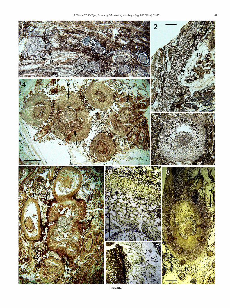
Plate XIV.
61J. Galtier, T.L. Phillips / Review of Palaeobotany and Palynology 205 (2014) 31–73
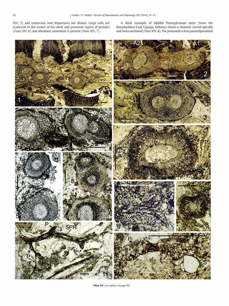
62 J. Galtier, T.L. Phillips / Review of Palaeobotany and Palynology 205 (2014) 31–73
XIV, 5) and numerous root departures are shown. Large cells arescattered in the cortex of the stem and proximal region of petioles(Plate XIV, 6) and abundant ramentum is present (Plate XIV, 7).
Plate XV (see captio
A third example of Middle Pennsylvanian stem (from theMurphysboro Coal, Cayuga, Indiana) shows a rhizome curved apicallyand twice sectioned (Plate XIV, 4). The protostele is less parenchymatized
n on page 60)
63J. Galtier, T.L. Phillips / Review of Palaeobotany and Palynology 205 (2014) 31–73
and smaller than in the previous examples; the departing leaf trace ismassive and already C-shaped as schematized in Fig. 8H. There is a singleleaf trace in the section of the stem (Plate XIV, 4) and the cortex is homo-geneous, apparently devoid of scattered large cells, in the petiole. Rootsare departing either from the protostele or from the petiole trace. A sec-tion of an attached petiole (Plate XIV, 8) shows an abundant ramentumand numerous roots, some connected to the petiole strand.
4.2.3. Tubicaulis from the Late PennsylvanianTubicaulis stewartii Eggert (1959)from a Late Pennsylvanian coal ball
of the Calhoun Coal, Illinois, was characterized as having an abundantxylem parenchyma, a lacunar middle cortex in both stem and petioles,a well developed integumentary system, and decurrent petiole baseswith C-shaped xylem strand about 2 mm wide (Fig. 8K). Roots departfrom the stele and basal portion of leaf traces. This species was consid-ered as closely allied to Tubicaulis multiscalariformis, with similar fea-tures in stem cortex and leaf development; differences mainly concernthemore abundant xylem parenchyma and lacunar cortex (not initiallymentioned in T.multiscalariformis). No instance of the emission of pinnatraces has been found.
The newmaterial of American Tubicaulis, illustrated on Plate XIV, in-dicates that these Middle Pennsylvanian stems already exhibited manyfeatures (abundant xylem parenchyma, possible lacunar middle cortex,well developed ramentum, roots departing from both stele and leaftraces) first recognized in Tubicaulis stewartii.
Two Late Pennsylvanian Tubicaulis stems have been described fromFrance (Galtier and Holmes, 1984) and both have been compared toTubicaulis stewartii. One is an isolated stemwith a subcircular protosteleshowing prominent radiatingfiles of parenchyma cells. The second is anepiphyllous stem laterally borne on a rachis of Anachoropteris involuta(Fig. 8L) mentioned above (Section 3.2.2); the last stem, bearing ninepetioles, was interpreted as conforming to the diagnosis of T. stewartii.
4.2.4. Tubicaulis stem with “mixed pith” from the PermianTubicaulis africanus Holden and Croft (1962) is the only species re-
ported from Gondwana, with a probable Upper Permian age. The stemwas described as upright, surrounded by a thick armor of persistentleaf bases; the cylindrical protostele has a central region (mixed pith)consisting of parenchyma intermixedwith narrow tracheids. Leaf tracesdeveloped in close spiral succession, at first nearly circular in sectionthen quickly developing abaxial curvature to slender C-shape (about5 mmwide) distally where petioles are about 15 mmwide. Distal divi-sion of the petiole trace into three has been illustrated. Roots depart atand near the margins of petiole traces. Among the Tubicaulis with aparenchymatized protostele this species shows a number of uniquefeatures (protostele with a central mixed pith, leaf trace initially nearlycylindrical, petiole trace dividing into three distally, roots departing onlyfrom petiole traces, and thickmantle of leaf bases). To include this plantwithin Tubicaulis, despite these differences, Holden and Croft (1962)proposed an excessively enlarged range of the generic features. Infact, due to its Gondwanan origin and younger age, T. africanus mayrepresent an isolated and divergent taxon.
In conclusion, the stems attributed to this second evolutionary lineshare a number of characteristics (see Fig. 8G–L) which are absent inTubicaulis of the first evolutionary line. These are: 1) a parenchymatizedprotostele; 2) the xylem of the leaf trace is C-shaped very early, beforetrace departure; 3) a relatively large portion of the cauline xylem is in-volved in the leaf trace formation; 4) protoxylem groups of the leaftrace are unclear; 5) the petiole cortex shows a nest of sclerotic cellswithin abaxial xylem concavity; and 6) pinna traces have not been ob-served. Fig. 8 reveals clear differences in stele, leaf trace and petiole or-ganization between the two evolutionary groups. As stated above(Section 4.1.3), the Tubicaulis with a solid protostele (Fig. 8A–E) arecharacterized by features which may be considered as generic. There-fore we consider that the use of the name of Tubicaulis for all these
stems, including those corresponding to the second evolutionary line,is misleading.
4.3. Interconnections between Tubicaulis and Anachoropteris
Present data suggest that there is no proof of connections betweenAnachoropteris rachides/petioles and stems of Tubicaulis with a solidprotostele (= Tubicaulis sensu stricto). Where known (Tubicaulissutcliffii, Tubicaulis grandeuryi, Tubicaulis solenites) the C-shaped petiolexylem anatomy (curved adaxial median region extending to the lateralprotoxylem strands and short arms) does not correspond to the charac-teristics of Anachoropteris as defined in this paper (Section 3.1) wherethe petiole xylem shows a rectilinear adaxial median region delimitedby two prominent adaxial protoxylem strands and generally long arms.
In contrast, Tubicaulis stems with a parenchymatized protostele arethe only ones eventually bearing Anachoropteris fronds. This was firstdemonstrated by Hall (1961) in one stem from Iowa showing the tran-sition from “typical” Tubicaulis petiole to foliar anatomy of theAnachoropteris involuta type (Fig. 8I). This was confirmed by Phillips(1974), and in the present paper on several Middle Pennsylvanianstems from Illinois and Indiana. This transition from a small tubicaulidC-shaped strand to an inrolled xylem corresponds to a more distal re-gion of some attached petioles. However, the transition to large petioleswith a typically involuted-revoluted xylem has not yet been establishedas indicated by the comparison of the largest inrolled petiole (Fig. 8I)with the typical A. involuta rachis (at right in Fig. 8L).
Another type of interconnection is now established: epiphyllousshoots occurring on some Anachoropteris fronds correspond also toTubicaulis-type of stems with parenchymatized protosteles; this isknown from the Early Pennsylvanian, in shoots borne on Anachoropteriswilliamsonii (Fig. 8F), and from the Late Pennsylvanian in shoots borneon Anachoropteris involuta, first mentioned by Hall (1961), and de-scribed in more detail by Galtier and Holmes (1984) (Fig. 8L). In fact,these proved connections raised a nomenclatural problem; Hall(1961) did not describe a new species of Tubicaulis for the stem bearingpetioles showing transition to involute Anachoropteris type, suggestingimplicitly that the name of A. involuta could be used for this plant.
In fact, it must be noted that most epiphyllous shoots borne byAnachoropteris rachides are not of the Tubicaulis-type. Epiphyllousstems borne on Anachoropteris gillotii (Section 3.4; Plate VIII) andAnachoropteris robusta (Section 3.5; Plate IX), like those of Kaplanopterisclavata (Section 3.7; Plate X), have solid protosteles but these are smalland more similar to botryopterid-type protosteles. This has been alreadysuggested byMorgan andDelevoryas (1954)who emphasized the differ-ences between Tubicaulis and these smaller taxa (Anachoropteris clavata,Apotropteris) where a large portion of the stele separates as a leaf trace.Lastly, we have to mention the adaxial shoots borne on Middle Pennsyl-vanian involute Anachoropteris (Section 3.2.3; Plates III–V) whichare either protostelic or siphonostelic but not really conforming to theTubicaulis type.
4.4. Ecology of Anachoropteris and Tubicaulis
Anachoropteris is well known from coal-ball peats; however, withthe notable exceptions of Anachoropteris williamsonii, Anachoropterisclavata and Anachoropteris involuta, the majority of the described spe-cies are associated with clastic, if not volcaniclastic, substrates. This isthe case of the type material of Anachoropteris radnicensis and ofAnachoropteris pulchra (Middle Pennsylvanian, Czech Republic) and ofother specimens attributed to the last species from the Late Pennsylva-nian of France and the Early Permian of Germany and Sardinia. Similar-ly, the types of material of Anachoropteris gillotii, Anachoropteris pautetii,Anachoropteris robusta (Late Pennsylvanian, France), and Anachoropteriscircularis, Anachoropteris gigas, and Anachoropteris ovata (Early Permian,France) come from non-coal swamp environments. This over-representation certainly does not reflect the real diversity but is partly
64 J. Galtier, T.L. Phillips / Review of Palaeobotany and Palynology 205 (2014) 31–73
the result of “splitter” systematic studies of European material. Howev-er, it is significant that we now recognize representatives of all fourmain “anachoropterid groups” (numbered as B to E in Fig. 1) in theolder European coal-ball peats of Early Pennsylvanian time. We previ-ously noted that the largest size within the “pulchra–involuta” and the“robusta” groups was recorded for younger taxa from clastic substrates;this acquisition of a robust stature may reflect higher nutrient environ-ments than those of coal swamps.
In some of the oldest Early Pennsylvanian coal-ball floras fromEurope small ferns (zygopterids, ankyropterids, anachoropterids andbotryopterids) are a common component, occurring on a frequencybasis of 25% in the Bouxharmont coal balls of Belgium and 28% inthose of the Union seam of England. Anachoropterids (includingAnachoropteris and Psalixochlaena) occur in about 6% of the coal ballswith Anachoropteris more common in the Bouxharmont (Holmes andFairon-Demaret, 1984). However, quantitative analyses of these coalballs indicate that these ferns were minor biomass contributors and asimilar pattern is found in coal beds throughout the Pennsylvanian ofNorth America (Phillips et al., 1985; DiMichele and Phillips, 2002).
All the Tubicaulis specimens with a parenchymatized protostele(Fig. 8F–K) come from coal-ball peats with the notable exceptions ofthemuch younger Tubicaulis africanuswhich has divergent characteris-tics, and of one Tubicaulis shoot, identified as Tubicaulis stewartii(Fig. 8L), laterally borne on Tubicaulis involuta rachis from cherts ofthe Late Pennsylvanian of France. In contrast, the majority of Tubicauliswith a solid protostele (Fig. 8C–E) come from clastic environments, andthis is probably also the case of Tubicaulis sutcliffii (Fig. 8A) found in aroof nodule. The only exception is Tubicaulis scandens (Fig. 8B) from aLate Pennsylvanian coal ball. The occurrence of a lacunar cortex in thestems and petioles of the Late Pennsylvanian T. stewartii (Eggert,1959) and the Early Pennsylvanian Tubicaulis sp. from England (PlateVII, 7) may be interpreted as adaptation to the wet coal-swamp envi-ronment. In contrast, the ecological significance of dispersed largecells, often with thickened walls, in the cortex of several Tubicaulis(Plate XIII, 4, 9, 16; Plate XIV, 6) remains unclear.
4.5. Habits of Anachoropteris and Tubicaulis
Different growth architectures corresponding to different categoriesof branching have been recognized in small Paleozoic ferns (Galtier andHolmes, 1982; DiMichele and Phillips, 2002) allowing them to acquirea variety of habits. We previously summarized (Phillips and Galtier,2005, 2011) the very distinct habits observed in the zygopterid andankyropterid ferns.
It is likely that the involute Anachoropteris foliar members wereborne on Tubicaulis-like stems (Hall, 1961; Phillips, 1974) which weresemi erect to erect and rarely branched. The fronds, in turn, played animportant role in vegetative propagation with adventitious shoots de-veloped on several orders of foliar members.
With the exception of Tubicaulis africanus, all the Tubicaulis withparenchymatized protostele correspond to relatively short (knownlength up to 6 cm), unbranched stems with short internodes and bear-ing small petioles. They are covered with roots, sometimes departingalso from the petiole bases, and they are sometimes designated as rhi-zomes. Eggert (1959) considered that Tubicaulis stewartii had an uprightstem with determinate growth; while Hall (1961) considered that theTubicaulis shoot was erect, possibly subterranean, and at least severalcm long.We agree with this interpretationwhich applies also to severalun-named Tubicaulis shoots borne on long scrambling Anachoropterisfrond rachides as discussed above. In this case, the Tubicaulis stem is asmall part of a sort of clonal system of which the scrambling rachidesare the dominant part.
Holmes (1989) recapitulated all known positions of shoots borne onfronds, including the trifurcation of the rachis to give a median stem, sofar only known to occur in the genus Anachoropteris. In the presentpaper, we document the occurrence of epiphyllous shoots in the four
main anachoropterid groups. The petioles of Anachoropteris clavatawere first interpreted (Delevoryas and Morgan, 1954) as having func-tioned as stolons giving rise to new shoots. Hall (1961) suggested thatAnachoropteris petioles were attached to an “embryonically derived pri-mary axis (Tubicaulis) erect or subterranean”, of probable determinategrowth. The extremely long petioles were a more conspicuous part ofthe plant than the stem; they were of indeterminate growth, scram-bling, and, in turn, they were bearing shoots.
This is not contradicted by the new interpretation of the growth ar-chitecture of Kaplanopteris (Anachoropteris) clavata with two types ofreiterative units (Tomescu et al., 2006, 2008); the authors proposedthat short orthotropic epiphyllous plantlets produced fronds withindeterminate growth and scrambling or climbing habit, while frondrachides also bore latent crosiers of arrested fronds. It has been sug-gested that many species of Anachoropteris were much like modern“walking ferns” that produce stem buds on scrambling frond rachidesenabling them to spread rapidly across open substrate (DiMichele andPhillips, 2002). As documented in the present paper, this type of habitoccurs in the Early Pennsylvanian Anachoropteris williamsonii andAnachoropteris gillotii, in several unnamed American Middle Pennsylva-nian involute Anachoropteris species with adaxial shoots, and in the LatePennsylvanian Anachoropteris involuta and Anachoropteris robusta andthe Early Permian Anachoropteris circularis.
In addition to scrambling habit, adaptations to climbing also exist inseveral species showing recurved secondary pinna rachides archingover the adaxial side of the main rachis. This was first described byCorsin (1937) as “aphlebiae” in Anachoropteris involuta, but this featureis also present in someAmerican involute Anachoropteris (Plate III, 3, re-constructed in Fig. 3), in Anachoropteris williamsonii (Plate VI, 1) and inAnachoropteris circularis (Plate IX, 11). The occurrence of A. involuta ra-chis at the periphery of Psaronius root mantle (Galtier, 2008) and fur-thermore, of Anachoropteris pulchra rachides found inside the rootmantle of Psaronius from the Early Permian of Germany (Rössler,2000) and France (JG personal observation), are direct evidence of epi-phytic, or more precisely, of a hemi-epiphytic habit for these plants.
Finally, it is worthmentioning the new AmericanMiddle Pennsylva-nian fertile pinnae associated with foliar members bearing adaxialshoots (cf. Section 3.2.3) which could have aided further propagationin climbing. It is tempting to suggest that such upward facing sori (per-haps the largest branching receptacle recorded) without indusia inviteswind, rain drops or crawling visitors to aid in dispersal. Furthermore,the specimens have abbreviated succulent laminae consistent withavoiding water loss and also consistent with structural strength andwater storage. As a reasonable alternative, if the fertile material isdisplayed adaxially by a fern with scrambling fronds, the face upwould certainly be better for spore dispersal over a prolonged intervalas well as avoiding quick burial by litter if abaxial. Also, in a swamp,the adaxial fertile pinna position would help avoid submergence inwater.
The contrasting sizes within Tubicaulis stems have been emphasized(Galtier and Phillips, 1996, Fig. 6) and correlated with differences inhabit. Unbranched erect stems of Tubicaulis sutcliffii and Tubicaulissolenites, with a thickmantle (about 10 cm in diameter) of large petiolebases and roots, are characteristic of an upright habit. While these twospecies had a solid protostele, the same upright habit also characterizedTubicaulis africanus, the youngest member of the second evolutionaryline (with parenchymatized protosteles). These plants probablyhad the habit of small tree-ferns with very large leaves of which thedistal part is unknown. These fronds, probably not conforming to theAnachoropteris-type anatomy, were bearing precocious, “aphlebia-like”pinnae. The occurrence of these precocious branched pinnae isinterpreted as a convergence with the similar pinnae/“aphlebiae” occur-ring also on the basal region of petioles of the zygopterids, Symplocopterisand Zygopteris primaria, and of the ankyropterid, Anachoropterishendricksii, which have also an erect-stem habit. A protective role fordeveloping frond primordia is suggested for these organs.
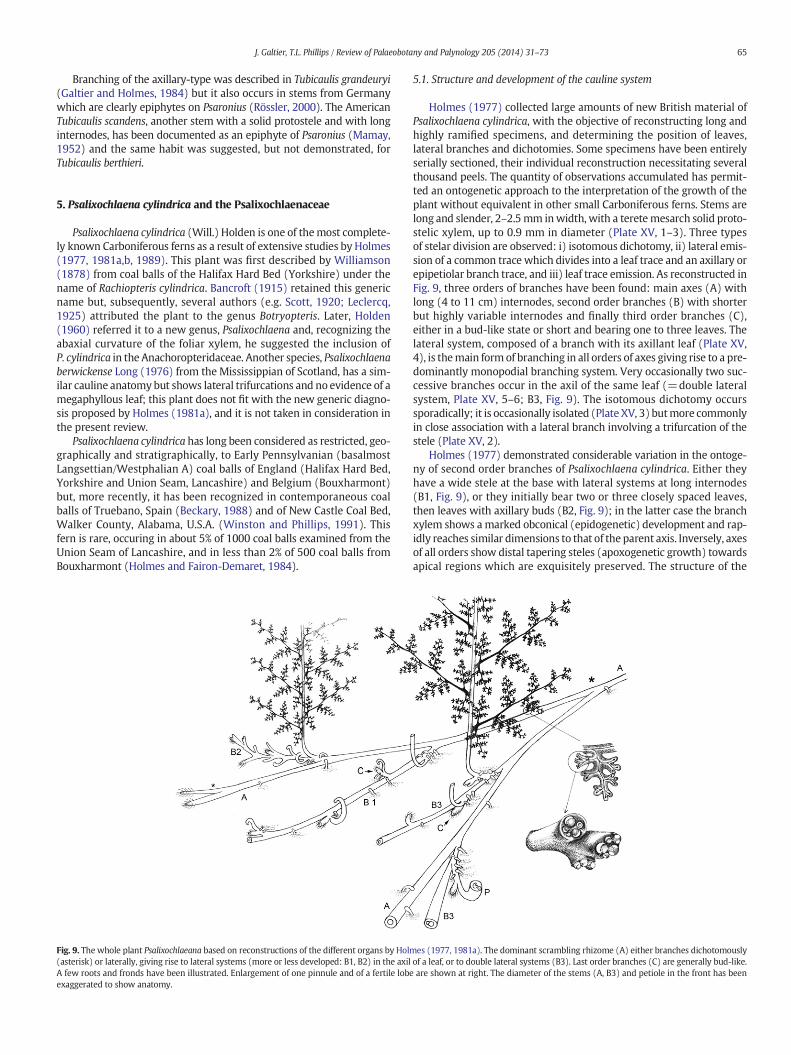
65J. Galtier, T.L. Phillips / Review of Palaeobotany and Palynology 205 (2014) 31–73
Branching of the axillary-type was described in Tubicaulis grandeuryi(Galtier and Holmes, 1984) but it also occurs in stems from Germanywhich are clearly epiphytes on Psaronius (Rössler, 2000). The AmericanTubicaulis scandens, another stem with a solid protostele and with longinternodes, has been documented as an epiphyte of Psaronius (Mamay,1952) and the same habit was suggested, but not demonstrated, forTubicaulis berthieri.
5. Psalixochlaena cylindrica and the Psalixochlaenaceae
Psalixochlaena cylindrica (Will.) Holden is one of themost complete-ly known Carboniferous ferns as a result of extensive studies by Holmes(1977, 1981a,b, 1989). This plant was first described by Williamson(1878) from coal balls of the Halifax Hard Bed (Yorkshire) under thename of Rachiopteris cylindrica. Bancroft (1915) retained this genericname but, subsequently, several authors (e.g. Scott, 1920; Leclercq,1925) attributed the plant to the genus Botryopteris. Later, Holden(1960) referred it to a new genus, Psalixochlaena and, recognizing theabaxial curvature of the foliar xylem, he suggested the inclusion ofP. cylindrica in the Anachoropteridaceae. Another species, Psalixochlaenaberwickense Long (1976) from the Mississippian of Scotland, has a sim-ilar cauline anatomybut shows lateral trifurcations and no evidence of amegaphyllous leaf; this plant does not fit with the new generic diagno-sis proposed by Holmes (1981a), and it is not taken in consideration inthe present review.
Psalixochlaena cylindrica has long been considered as restricted, geo-graphically and stratigraphically, to Early Pennsylvanian (basalmostLangsettian/Westphalian A) coal balls of England (Halifax Hard Bed,Yorkshire and Union Seam, Lancashire) and Belgium (Bouxharmont)but, more recently, it has been recognized in contemporaneous coalballs of Truebano, Spain (Beckary, 1988) and of New Castle Coal Bed,Walker County, Alabama, U.S.A. (Winston and Phillips, 1991). Thisfern is rare, occuring in about 5% of 1000 coal balls examined from theUnion Seam of Lancashire, and in less than 2% of 500 coal balls fromBouxharmont (Holmes and Fairon-Demaret, 1984).
Fig. 9. The whole plant Psalixochlaeana based on reconstructions of the different organs by Holm(asterisk) or laterally, giving rise to lateral systems (more or less developed: B1, B2) in the axilA few roots and fronds have been illustrated. Enlargement of one pinnule and of a fertile lobeexaggerated to show anatomy.
5.1. Structure and development of the cauline system
Holmes (1977) collected large amounts of new British material ofPsalixochlaena cylindrica, with the objective of reconstructing long andhighly ramified specimens, and determining the position of leaves,lateral branches and dichotomies. Some specimens have been entirelyserially sectioned, their individual reconstruction necessitating severalthousand peels. The quantity of observations accumulated has permit-ted an ontogenetic approach to the interpretation of the growth of theplant without equivalent in other small Carboniferous ferns. Stems arelong and slender, 2–2.5mm inwidth, with a teretemesarch solid proto-stelic xylem, up to 0.9 mm in diameter (Plate XV, 1–3). Three typesof stelar division are observed: i) isotomous dichotomy, ii) lateral emis-sion of a common trace which divides into a leaf trace and an axillary orepipetiolar branch trace, and iii) leaf trace emission. As reconstructed inFig. 9, three orders of branches have been found: main axes (A) withlong (4 to 11 cm) internodes, second order branches (B) with shorterbut highly variable internodes and finally third order branches (C),either in a bud-like state or short and bearing one to three leaves. Thelateral system, composed of a branch with its axillant leaf (Plate XV,4), is themain formof branching in all orders of axes giving rise to a pre-dominantly monopodial branching system. Very occasionally two suc-cessive branches occur in the axil of the same leaf (=double lateralsystem, Plate XV, 5–6; B3, Fig. 9). The isotomous dichotomy occurssporadically; it is occasionally isolated (PlateXV, 3) butmore commonlyin close association with a lateral branch involving a trifurcation of thestele (Plate XV, 2).
Holmes (1977) demonstrated considerable variation in the ontoge-ny of second order branches of Psalixochlaena cylindrica. Either theyhave a wide stele at the base with lateral systems at long internodes(B1, Fig. 9), or they initially bear two or three closely spaced leaves,then leaves with axillary buds (B2, Fig. 9); in the latter case the branchxylem shows amarked obconical (epidogenetic) development and rap-idly reaches similar dimensions to that of the parent axis. Inversely, axesof all orders show distal tapering steles (apoxogenetic growth) towardsapical regions which are exquisitely preserved. The structure of the
es (1977, 1981a). The dominant scrambling rhizome (A) either branches dichotomouslyof a leaf, or to double lateral systems (B3). Last order branches (C) are generally bud-like.are shown at right. The diameter of the stems (A, B3) and petiole in the front has been

66 J. Galtier, T.L. Phillips / Review of Palaeobotany and Palynology 205 (2014) 31–73
lateral systemof this fern is similar to some types of ramifications existingwithin the present Hymenophyllaceae and Stromatopteridaceae(Bierhorst, 1974).
The sturdy development of some of the lateral branches suggeststhat the so-called main axes may be the median or distal parts of lateralbranches and that this fern, like the extant Pteridium aquilinum(Webster and Steeves, 1958; Dasanayake, 1960), showed a repetitivegrowth habit where laterals eventually took over the role of mainaxes. Measurements of metaxylem tracheid lengths in short leafy andlong internoded laterals, as well as the occurrence of very long phloemelements in long stems, support the idea of increased growth rate inmore distal regions of Psalixochlaena cylindrica stems.With the possibil-ity of conversion from leaves to lateral systems at the base of branches,P. cylindrica represents the oldest known evidence of a capacityfor changeover from one type of apical functioning to another. Itis also one of the oldest examples of an axillary lateral branch associatedwith a true frond. In the contemporaneous Ankyropteris brongniartii theaxillant leaf is of phyllophore-type. Finally, Holmes (1989) emphasizedthe considerable range of branching types occuring in P. cylindrica,including one exceptional example of adaxial epiphyllous shoot.Psalixochlaena cylindrica, with its highly branched cauline system,strongly contrasts with most of the anachoropterid ferns considered inthis paper where cauline structures are rarely branched and short bycomparison, but with common occurrences of epiphyllous shoots.
5.2. The frond and fertile parts
The frond of Psalixochlaena cylindrica has been reconstructed fromserial peel sections (Holmes, 1981a). Themain rachides have been iden-tified by comparison with petioles (up to 8 cm long) attached to stems.The petioles, up to 2 × 2.5 mm in diameter, are axillant to a branch orclosely spaced in a 2/5 phyllotaxis; they are circinately coiled whenyoung. The petiole xylem basally oval (LT, Plate XV, 4) is changing up-ward to C-shape, up to 0.9 mm wide. The main rachis xylem showstwo persistent protoxylem poles (median arrows, Plate XV, 7) andtwo lateral ones, that represents a typical anachoropterid foliar anatomy(compare to Plate I). Primary pinnae, up 9 cm long, are departing alter-nately at 1 to 3 cm intervals; their rachides, up to 1.4 mmwide, have axylem strand 0.3 mm wide with slight abaxial curvature (Plate XV, 8);like themain rachides they show amarked adaxial groove. They bear al-ternately up to ten secondary pinnae, at least 3 cm long, with a flexuousrachis (SPR, Plate XV, 11) possessing an enveloping layer of spongytissue.
Pinnules are triangular, up to 10 mm long, and composed of multi-lobed segments corresponding to sphenopterid-type foliage (P, PlateXV, 11 and Fig. 9, at right) with narrow finely divided lobes, 0.5 mmwide, with rounded apices. Pinnule veins with a parenchyma sheathterminate in a pyriform swelling (Plate XV, 10); they are surroundedby a spongy tissue which extends down to the outer cortical layer ofpinnae rachides. Sparsely occuring fertile lobes, are possibly restrictedto basal pinnules. Fertile lobes are truncated with a swollen vascularstrand (V, Plate XV, 9) terminating beneath the soral receptacle. The cir-cular soral receptacle is inclined towards the abaxial side (Plate XV, 10).Gradate sori are composed of five annulate sporangia. Most observedsporangia were immature, pyriform, 300 μm long with a bi-triseriateoblique annulus and a vertical zone of dehiscence (S, Plate XV, 10).
The frond, about 30 cm long and 15 cmwide, has been reconstructedby Holmes (1981a) as planated, tripinnate and catadromic with sub-opposite insertion of basal secondary pinnae and pinnules (Fig. 9).This frond resembles a number of Late Carboniferous compression–im-pression fossils showing delicate pinnules with annulate sporangiasituated at lobe endings. The most comparable are Hymenophyllites(Brousmiche, 1983), Boweria minor (Kidston, 1923) and Sphenopterissp. (Leary, 1979) which all correspond to sphenopterid foliage with amuch reduced webbing representing the simplest type of laminate
pinnule. However, Psalixochleana cylindrica does not seem to corre-spond exactly, in all its characters, to any of the above mentioned ferns.
5.3. The Psalixochlaenaceae
Psalixochlaena cylindricawas previously assigned to the coenopteridAnachoropteridaceae but, following the description of its frond andfertile characteristics (terminal circular soral receptacle and gradate soriof five annulate sporangia), it wasmade the type genus of the new familyPsalixochlaenaceae within the Filicales (Holmes (1981a). Psalixochlaenacylindrica is also distinguished from the other anachoropterid fernsby its mesarch protostelic stems which are extensively branched, eitherdichotomously or laterally. It has been emphasized that this fernshows important similarities to the extant Hymenophyllaceae in thestructure of their lateral branching systems and of their marginaland gradate sori. The Sermayaceae (Eggert and Delevoryas, 1967)was, before the Psalixochlaenaceae, thefirst anachoropterid family segre-gated on the basis of fertile characteristics, as was more recently theKaplanopteridaceae (Tomescu et al., 2006). Despite similar sporangia,the Sermayaceae differ from the Psalixochlaenaceae in their superficialsori, in addition to differences concerning cauline and foliar anatomyand morphology. The Kaplanopteridaceae are distinct by the possessionof conical indusiate soral receptacle and small sporangia with long stalks,in addition to differences concerning cauline and foliar anatomy andmorphology.
5.4. Ecology and habit
Psalixochlaena cylindrica is only known from coal-ball peats. As recon-structed in Fig. 9, this fern, with slender stems and long internodes,growing singly or in dense mats, was probably rampant along theswamp surface or slightly subterranean in the manner of someHymenophyllaceae. According to Holmes (1981a) stems have neverbeen observed penetrating any other organ, however some stems havebeen found sandwiched between layers of lycopod bark (Plate XV, 1),as if they had grown through a hollow log lying on the surface. Budsand developed branches always lie parallel to the parent stem and sup-port the idea of the propagation of a horizontal system. Holmes (1977)suggested that stem branching appears admirably suited to colonizationof peat swamps. Dichotomous branching ensures vegetative propagationas does lateral branching but,more importantly, the latter provides a con-tinuity of growth through timeby a relay of axeswhich solve the problemof apical senescence or damage. Perhaps also buds of both delayed andimmediate development offer the possibility of surviving adverse condi-tions in the dormant state.While stemand apices of P. cylindrica are oftenvery well preserved, petioles become decayed over a short distanceand distal frond portions are very rare. This is in agreement with thesuggested habit of rampant stems bearing erect fronds; later the oldfronds decayed leaving only short petiole/rachis bases attached tostems. In conclusion, this specialized habit is different from those de-scribed above in the other anachoropterids which show either dominantscrambling frondswith epiphyllous shoots, or erect stems eithermassiveor epiphytic.
6. Apotropteris minuta
Apotropteris minuta is known only from the Late Pennsylvanian of theU.S.A. This small fernwas originally described byMorgan and Delevoryas(1954) from a coal ball in the Opdyke Coal in the McLeansboro Group,near Dix, Illinois (Phillips, 1980). The original specimen consists of aslender stem (3.8 mm in diameter) bearing petioles and adventitiousroots. The protostele, about 1 mm across, was described as solid withone central group of protoxylem and small groups of tracheids aroundthe circumference of the xylem interpreted as peripheral protoxylem.Metaxylem tracheids have multiseriate scalariform thickenings. Peti-oles arise from stem in no definite sequence. The authors emphasized
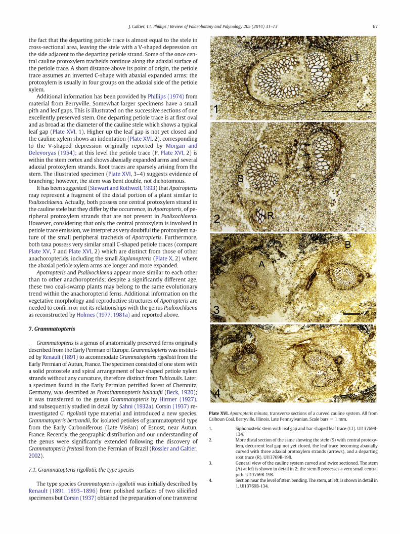
Plate XVI. Apotropteris minuta, transverse sections of a curved cauline system. All fromCalhoun Coal, Berryville, Illinois, Late Pennsylvanian. Scale bars = 1 mm.
1. Siphonostelic stemwith leaf gap and bar-shaped leaf trace (LT). UI13769B-134.
2. More distal section of the same showing the stele (S) with central protoxy-lem, decurrent leaf gap not yet closed, the leaf trace becoming abaxiallycurved with three adaxial protoxylem strands (arrows), and a departingroot trace (R). UI13769B-198.
3. General view of the cauline system curved and twice sectioned. The stem(A) at left is shown in detail in 2; the stem B possesses a very small centralpith. UI13769B-198.
4. Section near the level of stembending. The stem, at left, is shown in detail in1. UI13769B-134.
67J. Galtier, T.L. Phillips / Review of Palaeobotany and Palynology 205 (2014) 31–73
the fact that the departing petiole trace is almost equal to the stele incross-sectional area, leaving the stele with a V-shaped depression onthe side adjacent to the departing petiole strand. Some of the once cen-tral cauline protoxylem tracheids continue along the adaxial surface ofthe petiole trace. A short distance above its point of origin, the petioletrace assumes an inverted C-shape with abaxial expanded arms; theprotoxylem is usually in four groups on the adaxial side of the petiolexylem.
Additional information has been provided by Phillips (1974) frommaterial from Berryville. Somewhat larger specimens have a smallpith and leaf gaps. This is illustrated on the successive sections of oneexcellently preserved stem. One departing petiole trace is at first ovaland as broad as the diameter of the cauline stele which shows a typicalleaf gap (Plate XVI, 1). Higher up the leaf gap is not yet closed andthe cauline xylem shows an indentation (Plate XVI, 2), correspondingto the V-shaped depression originally reported by Morgan andDelevoryas (1954); at this level the petiole trace (P, Plate XVI, 2) iswithin the stem cortex and shows abaxially expanded arms and severaladaxial protoxylem strands. Root traces are sparsely arising from thestem. The illustrated specimen (Plate XVI, 3–4) suggests evidence ofbranching; however, the stem was bent double, not dichotomous.
It has been suggested (Stewart and Rothwell, 1993) that Apotropterismay represent a fragment of the distal portion of a plant similar toPsalixochlaena. Actually, both possess one central protoxylem strand inthe cauline stele but they differ by the occurrence, in Apotropteris, of pe-ripheral protoxylem strands that are not present in Psalixochlaena.However, considering that only the central protoxylem is involved inpetiole trace emission,we interpret as very doubtful the protoxylemna-ture of the small peripheral tracheids of Apotropteris. Furthermore,both taxa possess very similar small C-shaped petiole traces (comparePlate XV, 7 and Plate XVI, 2) which are distinct from those of otheranachoropterids, including the small Kaplanopteris (Plate X, 2) wherethe abaxial petiole xylem arms are longer and more expanded.
Apotropteris and Psalixochlaena appear more similar to each otherthan to other anachoropterids; despite a significantly different age,these two coal-swamp plants may belong to the same evolutionarytrend within the anachoropterid ferns. Additional information on thevegetative morphology and reproductive structures of Apotropteris areneeded to confirm or not its relationships with the genus Psalixochlaenaas reconstructed by Holmes (1977, 1981a) and reported above.
7. Grammatopteris
Grammatopteris is a genus of anatomically preserved ferns originallydescribed from the Early Permian of Europe.Grammatopteriswas institut-ed by Renault (1891) to accommodate Grammatopteris rigollotii from theEarly Permian of Autun, France. The specimen consisted of one stemwitha solid protostele and spiral arrangement of bar-shaped petiole xylemstrands without any curvature, therefore distinct from Tubicaulis. Later,a specimen found in the Early Permian petrified forest of Chemnitz,Germany, was described as Protothamnopteris baldaufii (Beck, 1920);it was transferred to the genus Grammatopteris by Hirmer (1927),and subsequently studied in detail by Sahni (1932a). Corsin (1937) re-investigated G. rigollotii type material and introduced a new species,Grammatopteris bertrandii, for isolated petioles of grammatopterid typefrom the Early Carboniferous (Late Viséan) of Esnost, near Autun,France. Recently, the geographic distribution and our understanding ofthe genus were significantly extended following the discovery ofGrammatopteris freitasii from the Permian of Brazil (Rössler and Galtier,2002).
7.1. Grammatopteris rigollotii, the type species
The type species Grammatopteris rigollotii was initially described byRenault (1891, 1893–1896) from polished surfaces of two silicifiedspecimens but Corsin (1937) obtained the preparation of one transverse
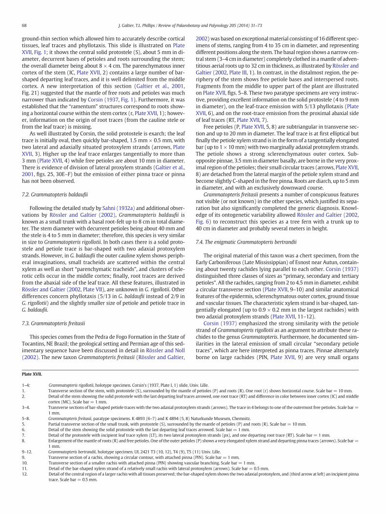
68 J. Galtier, T.L. Phillips / Review of Palaeobotany and Palynology 205 (2014) 31–73
ground-thin section which allowed him to accurately describe corticaltissues, leaf traces and phyllotaxis. This slide is illustrated on PlateXVII, Fig. 1; it shows the central solid protostele (S), about 5 mm in di-ameter, decurrent bases of petioles and roots surrounding the stem;the overall diameter being about 8 × 4 cm. The parenchymatous innercortex of the stem (IC, Plate XVII, 2) contains a large number of bar-shaped departing leaf traces, and it is well delimited from the middlecortex. A new interpretation of this section (Galtier et al., 2001,Fig. 21) suggested that the mantle of free roots and petioles was muchnarrower than indicated by Corsin (1937, Fig. 1). Furthermore, it wasestablished that the “ramentum” structures correspond to roots show-ing a horizontal course within the stem cortex (r, Plate XVII, 1); howev-er, information on the origin of root traces (from the cauline stele orfrom the leaf trace) is missing.
As well illustrated by Corsin, the solid protostele is exarch; the leaftrace is initially oval, then quickly bar-shaped, 1.5 mm × 0.5 mm, withtwo lateral and adaxially situated protoxylem strands (arrows, PlateXVII, 3). Higher up the leaf trace enlarges tangentially to more than3 mm (Plate XVII, 4) while free petioles are about 10 mm in diameter.There is evidence of division of lateral proxylem strands (Galtier et al.,2001, figs. 25, 30E–F) but the emission of either pinna trace or pinnahas not been observed.
7.2. Grammatopteris baldaufii
Following the detailed study by Sahni (1932a) and additional obser-vations by Rössler and Galtier (2002), Grammatopteris baldaufii isknown as a small trunk with a basal root-felt up to 8 cm in total diame-ter. The stem diameter with decurrent petioles being about 40 mm andthe stele is 4 to 5 mm in diameter; therefore, this species is very similarin size to Grammatopteris rigollotii. In both cases there is a solid proto-stele and petiole trace is bar-shaped with two adaxial protoxylemstrands. However, in G. baldaufii the outer cauline xylem shows periph-eral invaginations, small tracheids are scattered within the centralxylem as well as short “parenchymatic tracheids”, and clusters of scle-rotic cells occur in the middle cortex; finally, root traces are derivedfrom the abaxial side of the leaf trace. All these features, illustrated inRössler and Galtier (2002, Plate VII), are unknown in G. rigollotii. Otherdifferences concern phyllotaxis (5/13 in G. baldaufii instead of 2/9 inG. rigollotii) and the slightly smaller size of petiole and petiole trace inG. baldaufii.
7.3. Grammatopteris freitasii
This species comes from the Pedra de Fogo Formation in the State ofTocantins, NE Brazil; the geological setting and Permian age of this sed-imentary sequence have been discussed in detail in Rössler and Noll(2002). The new taxon Grammatopteris freitasii (Rössler and Galtier,
Plate XVII.
1–4: Grammatopteris rigollotii, holotype specimen. Corsin's (1937, Plate I, 1) slide, Un1. Transverse section of the stem, with protostele (S), surrounded by the mantle o2. Detail of the stem showing the solid protostele with the last departing leaf traces
cortex (MC). Scale bar = 1 mm.3–4. Transverse sections of bar-shaped petiole traces with the two adaxial protoxylem
1 mm.5–8. Grammatopteris freitasii, paratype specimens. K 4893 (6–7) and K 4894 (5, 8) Na5. Partial transverse section of the small trunk, with protostele (S), surrounded by6. Detail of the stem showing the solid protostele with the last departing leaf trace7. Detail of the protostele with incipient leaf trace xylem (LT), its two lateral proto8. Enlargement of themantle of roots (R) and free petioles. One of the outer petioles
1 mm.9–12. Grammatopteris bertrandii, holotype specimen. UL 2421 T3 (10, 12), T4 (9), T5 (9. Transverse section of a rachis, showing a circular contour, with attached pinna (10. Transverse section of a smaller rachis with attached pinna (PIN) showing vascu11. Detail of the bar-shaped xylem strand of a relatively small rachis with lateral pr12. Detail of the central region of a larger rachiswith all tissues preserved; the bar-sh
trace. Scale bar = 0.5 mm.
2002)was based on exceptionalmaterial consisting of 16different spec-imens of stems, ranging from 4 to 35 cm in diameter, and representingdifferent positions along the stem. Thebasal region shows a narrow cen-tral stem (3–4 cm in diameter) completely clothed in amantle of adven-titious aerial roots up to 32 cm in thickness, as illustrated by Rössler andGaltier (2002, Plate III, 1). In contrast, in the distalmost region, the pe-riphery of the stem shows free petiole bases and interspersed roots.Fragments from the middle to upper part of the plant are illustratedon Plate XVII, figs. 5–8. These two paratype specimens are very instruc-tive, providing excellent information on the solid protostele (4 to 9 mmin diameter), on the leaf-trace emission with 5/13 phyllotaxis (PlateXVII, 6), and on the root-trace emission from the proximal abaxial sideof leaf traces (RT, Plate XVII, 7).
Free petioles (P, Plate XVII, 5, 8) are subtriangular in transverse sec-tion and up to 20 mm in diameter. The leaf trace is at first elliptical butfinally the petiole xylem strand is in the formof a tangentially elongatedbar (up to 1 × 10mm)with twomarginally adaxial protoxylem strands.The petiole shows a strong sclerenchymatous outer cortex. Sub-opposite pinnae, 3.5mm in diameter basally, are borne in the very prox-imal region of the petioles; their small circular traces (arrows, Plate XVII,8) are detached from the lateral margin of the petiole xylem strand andbecome slightly C-shaped in the free pinna. Roots are diarch, up to 5mmin diameter, and with an exclusively downward course.
Grammatopteris freitasii presents a number of conspicuous featuresnot visible (or not known) in the other species, which justified its sepa-ration but also significantly completed the generic diagnosis. Knowl-edge of its ontogenetic variability allowed Rössler and Galtier (2002,Fig. 6) to reconstruct this species as a tree fern with a trunk up to40 cm in diameter and probably several meters in height.
7.4. The enigmatic Grammatopteris bertrandii
The original material of this taxon was a chert specimen, from theEarly Carboniferous (Late Mississippian) of Esnost near Autun, contain-ing about twenty rachides lying parallel to each other. Corsin (1937)distinguished three classes of sizes as “primary, secondary and tertiarypetioles”. All the rachides, ranging from2 to 4.5mm in diameter, exhibita circular transverse section (Plate XVII, 9–10) and similar anatomicalfeatures of the epidermis, sclerenchymatous outer cortex, ground tissueand vascular tissues. The characteristic xylem strand is bar-shaped, tan-gentially elongated (up to 0.9 × 0.2 mm in the largest rachides) withtwo adaxial protoxylem strands (Plate XVII, 11–12).
Corsin (1937) emphasized the strong similarity with the petiolestrand of Grammatopteris rigollotii as an argument to attribute these ra-chides to the genus Grammatopteris. Furthermore, he documented sim-ilarities in the lateral emission of small circular “secondary petioletraces”, which are here interpreted as pinna traces. Pinnae alternatelyborne on large rachides (PIN, Plate XVII, 9) are very small organs
iv. Lille.f petioles (P) and roots (R). One root (r) shows horizontal course. Scale bar = 10mm.arrowed, one root trace (RT) and difference in color between inner cortex (IC) andmiddle
strands (arrows). The trace in 4 belongs to one of the outermost free petioles. Scale bar =
turkunde Museum, Chemnitz.the mantle of petioles (P) and roots (R). Scale bar = 10mm.s arrowed. Scale bar = 1 mm.xylem strands (px), and one departing root trace (RT). Scale bar = 1 mm.(P) shows a very elongated xylem strand anddeparting pinna traces (arrows). Scale bar =
11) Univ. Lille.PIN). Scale bar = 1 mm.lar branching. Scale bar = 1mm.otoxylem (arrows). Scale bar = 0.5 mm.aped xylem shows the two adaxial protoxylem, and (third arrow at left) an incipient pinna

69J. Galtier, T.L. Phillips / Review of Palaeobotany and Palynology 205 (2014) 31–73
(0.5 mm in diameter proximally and up to 2 mm long) which may bedescribed as “aphleboid”. Pinnae are also borne on smaller rachides(PIN, Plate XVII, 10). They show evidence of vascular branchingand have been interpreted by Corsin as “tertiary petioles” sectioned
longitudinally. In the absence of stem bearing these isolated rachides,it can be speculated that they belong to median region of fronds andthat large rachides (PlateXVII, 9) are proximal to the smaller “secondarypetioles” (Plate XVII, 10) instead of bearing them.
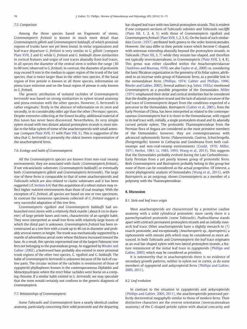
70 J. Galtier, T.L. Phillips / Review of Palaeobotany and Palynology 205 (2014) 31–73
7.5. Comparison
Among the three species based on fragments of stems,Grammatopteris freitasii is known in much more detail thanGrammatopteris gillotii and Grammatopteris baldaufii, of which proximalregions of trunks have not yet been found. In stelar organization andleaf-trace departure G. freitasii is very similar to G. gillotii (comparePlate XVII, 2 and 6) while G. freitasii and G. baldaufii show similaritiesin cortical features and origin of root traces abaxially from leaf traces.In all species the diameter of the central stem is within the range (30to 60mm) observed inG. freitasii; in contrast, the diameter of protostelemay exceed 9mm in themedian to upper region of the trunk of the lastspecies, that is twice larger than in the other two species. If the basalregion of free petiole is known in all three species, information onpinna trace emission and on the basal region of pinnae is only knownin G. freitasii.
The generic attribution of isolated rachides of Grammatopterisbertrandii was based on strong similarities in xylem strand anatomyand pinna emission with the other species. However, G. bertrandii israther enigmatic: firstly in the absence of information on its stem andsecondly, in its considerably older age than the other (Permian) species.Despite extensive collecting at the Esnost locality, additional material ofthis taxon has never been discovered. Nevertheless, its very simplexylem strand with two distant adaxial protoxylem strands is most sim-ilar to the foliar xylem of some of the anachoropteridswith small anten-nae (compare Plate XVII, 11 with Plate VIII, 5). This is suggestive of theidea that G. bertrandii is presently the oldest known representative ofthe anachoropterid ferns.
7.6. Ecology and habit of Grammatopteris
All the Grammatopteris species are known from non-coal swampenvironments; they are associated with clastic (Grammatopteris freitasii),if not volcaniclastic substrates (Grammatopteris baldaufii) or eventuallyboth (Grammatopteris gillotii and Grammatopteris bertrandii). The largesize of these ferns is comparable to that of some anachoropterids andTubicaulis which are also related to clastic substrates and it has beensuggested (cf. Section 4.4) that this acquisition of a robust staturemay re-flect higher nutrient environments than those of coal swamps. With theexception of G. freitasii, all species are based on one or two specimens.In contrast the numerous specimens collected of G. freitasii suggest avery successful adaptation of this tree fern.
Grammatopteris rigollotii and Grammatopteris baldaufii had un-branched erect stemswith a relatively thickmantle (about 8 cm in diam-eter) of large petiole bases and roots, characteristic of an upright habit.They were interpreted as small tree ferns with relatively large leaves ofwhich the distal part is unknown. Grammatopteris freitasii has been re-constructed as a tree fernwith a trunk up to 40 cm in diameter and prob-ably severalmeters in height. The trunkwasmechanically supported by amantle of adventitious aerial roots whose thickness increased toward thebase. As a result, this species represented one of the largest Paleozoic treefernnot belonging to the psaronialean group. As suggested byRössler andGaltier (2002), a buttressed base probably also existed in more proximaltrunk regions of the other two species, G. rigollotii and G. baldaufii. Thehabit ofGrammatopteris bertrandii is unknown because of the lack of cau-line parts. The circular section of the rachides is reminiscent of those ofzygopterid phyllophores known in the contemporaneous Diplolabis andMetaclepsydropsiswhere the erect foliar rachides were borne on a creep-ing rhizome. If a similar habit existed in G. bertrandii, we may speculatethat the stem would certainly not conform to the generic diagnosis ofGrammatopteris.
7.7. Relationships of Grammatopteris
Some Tubicaulis and Grammatopteris have a nearly identical caulineanatomy, particularly concerning their solid protostele and the departing
bar-shaped leaf tracewith two lateral protoxylem strands. This is evidentif one compares sections of Tubicaulis solenites and Tubicaulis sutcliffii(Plate XII, 1, 2, 4, 5) with those of Grammatopteris rigollotii andGrammatopteris freitasii (Plate XVII, 1, 2, 5, 6). On the basis of such similar-ities Corsin (1937) attributed both genera to the order Inversicatenales.However, the taxa differ in their petiole traces which become C-shaped,with antennae extending abaxially beyond the protoxylem strands, inTubicaulis (Plate XII, 3, 7) while they remain bar-shaped, and thereforenot typically inversicatenaleans, in Grammatopteris (Plate XVII, 3, 4, 8).This genus was either classified within the Anachoropteridaceae(Andrews and Boureau, 1970; see also Taylor et al., 2009) or, consideringthe basic filicalean organization in the geometry of its foliar xylem, attrib-uted to an incertae sedis group of Palaeozoic ferns, as a possible link tothe osmundalean ferns (Phillips, 1974; Galtier and Phillips, 1996;Rössler and Galtier, 2002). Several authors (e.g. Sahni, 1932a)mentionedGrammatopteris as a possible progenitor of the Osmundales. Miller(1971) emphasized their stelar and cortical similarities but he consideredthat the double protoxylemstrand and the lack of adaxial curvature of theleaf trace of Grammatopteris depart from the conditions expected of aprecursor to the Osmundales. Rastropteris (Galtier et al., 2001), from theEarly Permian of China, has been interpreted as related to the contempo-raneous Grammatopteris but it is closer to the Osmundaceae, with regardto its leaf trace with, initially, a single protoxylem strand and its adaxiallycurved petiole xylem. The protostelic Thamnopteroideae from thePermian flora of Angara are considered as the most primitive membersof the Osmundales; however, they are contemporaneous withadvanced siphonostelic forms (Palaeosmunda, Guairea, Schuichengella,Zhongmingella) known in Cathaysia and Gondwana from both coal-swamps and non-coal-swamp environments (Gould, 1970; Miller,1971; Herbst, 1981; Li, 1983, 1993; Wang et al., 2013). This suggeststhat the Osmundales probably evolved during the Late Carboniferous–Early Permian from a yet poorly known group of protostelic ferns.Both Grammatopteris and Rastropteris probably belong to this group butnone of them can be considered as the actual progenitor. Of interest, arecent phylogenetic analysis of Osmundales (Wang et al., 2013), withRastropteris as an outgroup, shows Grammatopteris as a member of apolytomy with the Thamnopteroideae.
8. Discussion
8.1. Stele and leaf trace origin
Most anachoropterids are characterized by a primitive caulineanatomy with a solid cylindrical protostele; more rarely there is aparenchymatized protostele (some Tubicaulis). Psalixochlaena standsapart with centrach permanent protoxylem strands and an initiallymon-arch leaf trace. Other anachoropterids have a slightly mesarch to (?)exarch protostele, and exceptionally (Anachoropteris sp., Apotropteris) asiphonostele with minute pith which may be considered as more ad-vanced. In both Tubicaulis and Grammatopteris the leaf trace originatesas an oval bar-shaped xylem with two lateral protoxylem strands, a fea-ture reminiscent of the initial leaf trace in zygopterids (Phillips andGaltier, 2005) which may be considered as primitive.
It is noteworthy that in anachoropterids there is no evidence ofsecondary growth patterns, neither in xylem nor in cortex, as do somemembers of zygopterid and ankyropterid ferns (Phillips and Galtier,2005, 2011).
8.2. Leaf evolution
In contrast to the situation in zygopterids and ankyropterids(Phillips and Galtier, 2005, 2011), the anachoropterids possessed per-fectly dorsiventral megaphylls similar to those of modern ferns. Theirdistinctive characters are the reverse orientation (inversicatenaleananatomy) of the C-shaped petiole xylem with abaxial concavity and
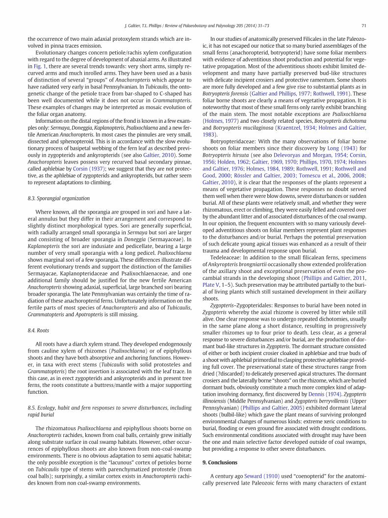
71J. Galtier, T.L. Phillips / Review of Palaeobotany and Palynology 205 (2014) 31–73
the occurrence of two main adaxial protoxylem strands which are in-volved in pinna traces emission.
Evolutionary changes concern petiole/rachis xylem configurationwith regard to the degree of development of abaxial arms. As illustratedin Fig. 1, there are several trends towards: very short arms, simply re-curved arms and much inrolled arms. They have been used as a basisof distinction of several “groups” of Anachoropteris which appear tohave radiated very early in basal Pennsylvanian. In Tubicaulis, the onto-genetic change of the petiole trace from bar-shaped to C-shaped hasbeen well documented while it does not occur in Grammatopteris.These examples of changes may be interpreted as mosaic evolution ofthe foliar organ anatomy.
Information on the distal regions of the frond is known in a fewexam-ples only: Sermaya,Doneggia, Kaplanopteris, Psalixochlaena and a new fer-tile American Anachoropteris. In most cases the pinnules are very small,dissected and sphenopteroid. This is in accordance with the slow evolu-tionary process of basipetal webbing of the fern leaf as described previ-ously in zygopterids and ankyropterids (see also Galtier, 2010). SomeAnachoropteris leaves possess very recurved basal secondary pinnae,called aphlebiae by Corsin (1937); we suggest that they are not protec-tive, as the aphlebiae of zygopterids and ankyropterids, but rather seemto represent adaptations to climbing.
8.3. Sporangial organization
Where known, all the sporangia are grouped in sori and have a lat-eral annulus but they differ in their arrangement and correspond toslightly distinct morphological types. Sori are generally superficial,with radially arranged small sporangia in Sermaya but sori are largerand consisting of broader sporangia in Doneggia (Sermayaceae). InKaplanopteris the sori are indusiate and pedicellate, bearing a largenumber of very small sporangia with a long pedicel. Psalixochlaenashows marginal sori of a few sporangia. These differences illustrate dif-ferent evolutionary trends and support the distinction of the familiesSermayacae, Kaplanopteridaceae and Psalixochlaenaceae, and oneadditional family should be justified for the new fertile AmericanAnachoropteris showing adaxial, superficial, large branched sori bearingbroader sporangia. The late Pennsylvanian was certainly the time of ra-diation of these anachoropterid ferns. Unfortunately information on thefertile parts of most species of Anachoropteris and also of Tubicaulis,Grammatopteris and Apotropteris is still missing.
8.4. Roots
All roots have a diarch xylem strand. They developed endogenouslyfrom cauline xylem of rhizomes (Psalixochlaena) or of epiphyllousshoots and they have both absorptive and anchoring functions. Howev-er, in taxa with erect stems (Tubicaulis with solid protosteles andGrammatopteris) the root insertion is associated with the leaf trace. Inthis case, as in erect zygopterids and ankyropterids and in present treeferns, the roots constitute a buttress/mantle with a major supportingfunction.
8.5. Ecology, habit and fern responses to severe disturbances, includingrapid burial
The rhizomatous Psalixochlaena and epiphyllous shoots borne onAnachoropteris rachides, known from coal balls, certainly grew initiallyalong substrate surface in coal swamp habitats. However, other occur-rences of epiphyllous shoots are also known from non-coal-swampenvironments. There is no obvious adaptation to semi aquatic habitat;the only possible exception is the “lacunous” cortex of petioles borneon Tubicaulis type of stems with parenchymatized protostele (fromcoal balls); surprisingly, a similar cortex exists in Anachoropteris rachi-des known from non coal-swamp environments.
In our studies of anatomically preserved Filicales in the late Paleozo-ic, it has not escaped our notice that somany buried assemblages of thesmall ferns (anachoropterid, botryopterid) have some foliar memberswith evidence of adventitious shoot production and potential for vege-tative propagation. Most of the adventitious shoots exhibit limited de-velopment and many have partially preserved bud-like structureswith delicate incipient crosiers and protective ramentum. Some shootsare more fully developed and a few give rise to substantial plants as inBotryopteris forensis (Galtier and Phillips, 1977; Rothwell, 1991). Thesefoliar borne shoots are clearly a means of vegetative propagation. It isnoteworthy that most of these small ferns only rarely exhibit branchingof the main stem. The most notable exceptions are Psalixochlaena(Holmes, 1977) and two closely related species, Botryopteris dichotomaand Botryopteris mucilaginosa (Kraentzel, 1934; Holmes and Galtier,1983).
Botryopteridaceae: With the many observations of foliar borneshoots on foliar members since their discovery by Long (1943) forBotryopteris hirsuta (see also Delevoryas and Morgan, 1954; Corsin,1956; Holden, 1962; Galtier, 1969, 1970; Phillips, 1970, 1974; Holmesand Galtier, 1976; Holmes, 1984, 1989; Rothwell, 1991; Rothwell andGood, 2000; Rössler and Galtier, 2003; Tomescu et al., 2006, 2008;Galtier, 2010), it is clear that the responses of the plants represent ameans of vegetative propagation. These responses no doubt servedthemwellwhen therewere blowdowns, severe disturbances or suddenburial. All of these plants were relatively small, and whether they wererhizomatous, erect or climbing, theywere easily felled and covered overby the abundant litter and of associated disturbances of the coal swamp.In our opinion, the frequent encounters with so many variously devel-oped adventitious shoots on foliar members represent plant responsesto the disturbances and/or burial. Perhaps the potential preservationof such delicate young apical tissues was enhanced as a result of theirtrauma and developmental response upon burial.
Tedeleaceae: In addition to the small filicalean ferns, specimensof Ankyropteris brongniartii occasionally show extended proliferationof the axillary shoot and exceptional preservation of even the pro-cambial strands in the developing shoot (Phillips and Galtier, 2011,Plate V, 1–5). Such preservationmay be attributed partially to the buri-al of living plants which still sustained development in their axillaryshoots.
Zygopteris–Zygopteridales: Responses to burial have been noted inZygopteris whereby the axial rhizome is covered by litter while stillalive. One clear response was to undergo repeated dichotomies, usuallyin the same plane along a short distance, resulting in progressivelysmaller rhizomes up to four prior to death. Less clear, as a generalresponse to severe disturbances and/or burial, are the production of dor-mant bud-like structures in Zygopteris. The dormant structure consistedof either or both incipient crosier cloaked in aphlebiae and true buds ofa shootwith aphlebial primordial to claspingprotective aphlebiae provid-ing full cover. The preservational state of these structures range fromdried (?discarded) to delicately preserved apical structures. The dormantcrosiers and the laterally borne “shoots” on the rhizome,which are burieddormant buds, obviously constitute a much more complex kind of adap-tation involving dormancy, first discovered by Dennis (1974). Zygopterisillinoiensis (Middle Pennsylvanian) and Zygopteris berryvillensis (UpperPennsylvanian) (Phillips and Galtier, 2005) exhibited dormant lateralshoots (bulbil-like) which gave the plant means of surviving prolongedenvironmental changes of numerous kinds: extreme xeric conditions toburial, flooding or even ground fire associated with drought conditions.Such environmental conditions associated with drought may have beenthe one and main selective factor developed outside of coal swamps,but providing a response to other severe disturbances.
9. Conclusions
A century ago Seward (1910) used “coenopterid” for the anatomi-cally preserved late Paleozoic ferns with many characters of extant
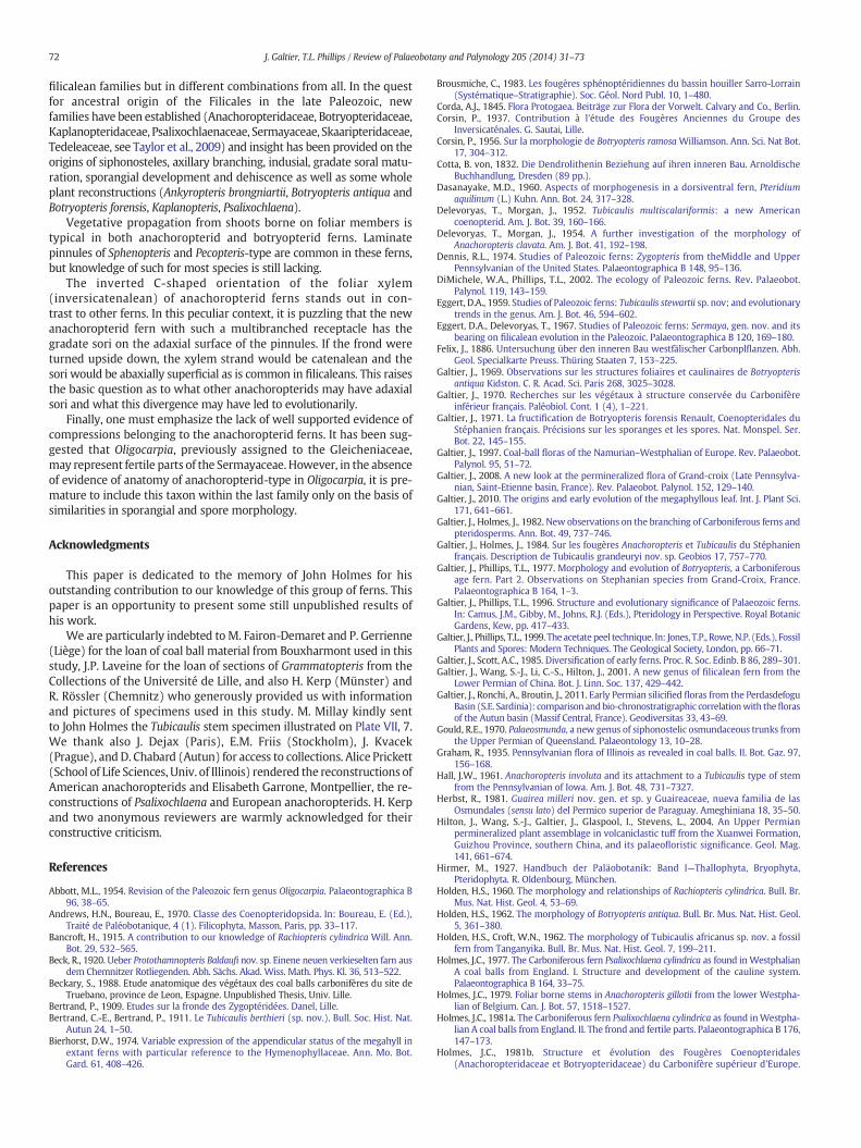
72 J. Galtier, T.L. Phillips / Review of Palaeobotany and Palynology 205 (2014) 31–73
filicalean families but in different combinations from all. In the questfor ancestral origin of the Filicales in the late Paleozoic, newfamilies have been established (Anachoropteridaceae, Botryopteridaceae,Kaplanopteridaceae, Psalixochlaenaceae, Sermayaceae, Skaaripteridaceae,Tedeleaceae, see Taylor et al., 2009) and insight has been provided on theorigins of siphonosteles, axillary branching, indusial, gradate soral matu-ration, sporangial development and dehiscence as well as some wholeplant reconstructions (Ankyropteris brongniartii, Botryopteris antiqua andBotryopteris forensis, Kaplanopteris, Psalixochlaena).
Vegetative propagation from shoots borne on foliar members istypical in both anachoropterid and botryopterid ferns. Laminatepinnules of Sphenopteris and Pecopteris-type are common in these ferns,but knowledge of such for most species is still lacking.
The inverted C-shaped orientation of the foliar xylem(inversicatenalean) of anachoropterid ferns stands out in con-trast to other ferns. In this peculiar context, it is puzzling that the newanachoropterid fern with such a multibranched receptacle has thegradate sori on the adaxial surface of the pinnules. If the frond wereturned upside down, the xylem strand would be catenalean and thesori would be abaxially superficial as is common in filicaleans. This raisesthe basic question as to what other anachoropterids may have adaxialsori and what this divergence may have led to evolutionarily.
Finally, one must emphasize the lack of well supported evidence ofcompressions belonging to the anachoropterid ferns. It has been sug-gested that Oligocarpia, previously assigned to the Gleicheniaceae,may represent fertile parts of the Sermayaceae. However, in the absenceof evidence of anatomy of anachoropterid-type in Oligocarpia, it is pre-mature to include this taxon within the last family only on the basis ofsimilarities in sporangial and spore morphology.
Acknowledgments
This paper is dedicated to the memory of John Holmes for hisoutstanding contribution to our knowledge of this group of ferns. Thispaper is an opportunity to present some still unpublished results ofhis work.
We are particularly indebted toM. Fairon-Demaret and P. Gerrienne(Liège) for the loan of coal ball material from Bouxharmont used in thisstudy, J.P. Laveine for the loan of sections of Grammatopteris from theCollections of the Université de Lille, and also H. Kerp (Münster) andR. Rössler (Chemnitz) who generously provided us with informationand pictures of specimens used in this study. M. Millay kindly sentto John Holmes the Tubicaulis stem specimen illustrated on Plate VII, 7.We thank also J. Dejax (Paris), E.M. Friis (Stockholm), J. Kvacek(Prague), andD. Chabard (Autun) for access to collections. Alice Prickett(School of Life Sciences, Univ. of Illinois) rendered the reconstructions ofAmerican anachoropterids and Elisabeth Garrone, Montpellier, the re-constructions of Psalixochlaena and European anachoropterids. H. Kerpand two anonymous reviewers are warmly acknowledged for theirconstructive criticism.
References
Abbott, M.L., 1954. Revision of the Paleozoic fern genus Oligocarpia. Palaeontographica B96, 38–65.
Andrews, H.N., Boureau, E., 1970. Classe des Coenopteridopsida. In: Boureau, E. (Ed.),Traité de Paléobotanique, 4 (1). Filicophyta, Masson, Paris, pp. 33–117.
Bancroft, H., 1915. A contribution to our knowledge of Rachiopteris cylindrica Will. Ann.Bot. 29, 532–565.
Beck, R., 1920. Ueber Protothamnopteris Baldaufi nov. sp. Einene neuen verkieselten farn ausdem Chemnitzer Rotliegenden. Abh. Sächs. Akad. Wiss. Math. Phys. Kl. 36, 513–522.
Beckary, S., 1988. Etude anatomique des végétaux des coal balls carbonifères du site deTruebano, province de Leon, Espagne. Unpublished Thesis, Univ. Lille.
Bertrand, P., 1909. Etudes sur la fronde des Zygoptéridées. Danel, Lille.Bertrand, C.-E., Bertrand, P., 1911. Le Tubicaulis berthieri (sp. nov.). Bull. Soc. Hist. Nat.
Autun 24, 1–50.Bierhorst, D.W., 1974. Variable expression of the appendicular status of the megahyll in
extant ferns with particular reference to the Hymenophyllaceae. Ann. Mo. Bot.Gard. 61, 408–426.
Brousmiche, C., 1983. Les fougères sphénoptéridiennes du bassin houiller Sarro-Lorrain(Systématique–Stratigraphie). Soc. Géol. Nord Publ. 10, 1–480.
Corda, A.J., 1845. Flora Protogaea. Beiträge zur Flora der Vorwelt. Calvary and Co., Berlin.Corsin, P., 1937. Contribution à l'étude des Fougères Anciennes du Groupe des
Inversicaténales. G. Sautai, Lille.Corsin, P., 1956. Sur la morphologie de Botryopteris ramosa Williamson. Ann. Sci. Nat Bot.
17, 304–312.Cotta, B. von, 1832. Die Dendrolithenin Beziehung auf ihren inneren Bau. Arnoldische
Buchhandlung, Dresden (89 pp.).Dasanayake, M.D., 1960. Aspects of morphogenesis in a dorsiventral fern, Pteridium
aquilinum (L.) Kuhn. Ann. Bot. 24, 317–328.Delevoryas, T., Morgan, J., 1952. Tubicaulis multiscalariformis: a new American
coenopterid. Am. J. Bot. 39, 160–166.Delevoryas, T., Morgan, J., 1954. A further investigation of the morphology of
Anachoropteris clavata. Am. J. Bot. 41, 192–198.Dennis, R.L., 1974. Studies of Paleozoic ferns: Zygopteris from theMiddle and Upper
Pennsylvanian of the United States. Palaeontographica B 148, 95–136.DiMichele, W.A., Phillips, T.L., 2002. The ecology of Paleozoic ferns. Rev. Palaeobot.
Palynol. 119, 143–159.Eggert, D.A., 1959. Studies of Paleozoic ferns: Tubicaulis stewartii sp. nov; and evolutionary
trends in the genus. Am. J. Bot. 46, 594–602.Eggert, D.A., Delevoryas, T., 1967. Studies of Paleozoic ferns: Sermaya, gen. nov. and its
bearing on filicalean evolution in the Paleozoic. Palaeontographica B 120, 169–180.Felix, J., 1886. Untersuchung über den inneren Bau westfälischer Carbonplflanzen. Abh.
Geol. Specialkarte Preuss. Thüring Staaten 7, 153–225.Galtier, J., 1969. Observations sur les structures foliaires et caulinaires de Botryopteris
antiqua Kidston. C. R. Acad. Sci. Paris 268, 3025–3028.Galtier, J., 1970. Recherches sur les végétaux à structure conservée du Carbonifère
inférieur français. Paléobiol. Cont. 1 (4), 1–221.Galtier, J., 1971. La fructification de Botryopteris forensis Renault, Coenopteridales du
Stéphanien français. Précisions sur les sporanges et les spores. Nat. Monspel. Ser.Bot. 22, 145–155.
Galtier, J., 1997. Coal-ball floras of the Namurian–Westphalian of Europe. Rev. Palaeobot.Palynol. 95, 51–72.
Galtier, J., 2008. A new look at the permineralized flora of Grand-croix (Late Pennsylva-nian, Saint-Etienne basin, France). Rev. Palaeobot. Palynol. 152, 129–140.
Galtier, J., 2010. The origins and early evolution of the megaphyllous leaf. Int. J. Plant Sci.171, 641–661.
Galtier, J., Holmes, J., 1982. New observations on the branching of Carboniferous ferns andpteridosperms. Ann. Bot. 49, 737–746.
Galtier, J., Holmes, J., 1984. Sur les fougères Anachoropteris et Tubicaulis du Stéphanienfrançais. Description de Tubicaulis grandeuryi nov. sp. Geobios 17, 757–770.
Galtier, J., Phillips, T.L., 1977. Morphology and evolution of Botryopteris, a Carboniferousage fern. Part 2. Observations on Stephanian species from Grand-Croix, France.Palaeontographica B 164, 1–3.
Galtier, J., Phillips, T.L., 1996. Structure and evolutionary significance of Palaeozoic ferns.In: Camus, J.M., Gibby, M., Johns, R.J. (Eds.), Pteridology in Perspective. Royal BotanicGardens, Kew, pp. 417–433.
Galtier, J., Phillips, T.L., 1999. The acetate peel technique. In: Jones, T.P., Rowe,N.P. (Eds.), FossilPlants and Spores: Modern Techniques. The Geological Society, London, pp. 66–71.
Galtier, J., Scott, A.C., 1985. Diversification of early ferns. Proc. R. Soc. Edinb. B 86, 289–301.Galtier, J., Wang, S.-J., Li, C.-S., Hilton, J., 2001. A new genus of filicalean fern from the
Lower Permian of China. Bot. J. Linn. Soc. 137, 429–442.Galtier, J., Ronchi, A., Broutin, J., 2011. Early Permian silicified floras from the Perdasdefogu
Basin (S.E. Sardinia): comparison andbio-chronostratigraphic correlationwith theflorasof the Autun basin (Massif Central, France). Geodiversitas 33, 43–69.
Gould, R.E., 1970. Palaeosmunda, a new genus of siphonostelic osmundaceous trunks fromthe Upper Permian of Queensland. Palaeontology 13, 10–28.
Graham, R., 1935. Pennsylvanian flora of Illinois as revealed in coal balls. II. Bot. Gaz. 97,156–168.
Hall, J.W., 1961. Anachoropteris involuta and its attachment to a Tubicaulis type of stemfrom the Pennsylvanian of Iowa. Am. J. Bot. 48, 731–7327.
Herbst, R., 1981. Guairea milleri nov. gen. et sp. y Guaireaceae, nueva familia de lasOsmundales (sensu lato) del Permico superior de Paraguay. Ameghiniana 18, 35–50.
Hilton, J., Wang, S.-J., Galtier, J., Glaspool, I., Stevens, L., 2004. An Upper Permianpermineralized plant assemblage in volcaniclastic tuff from the Xuanwei Formation,Guizhou Province, southern China, and its palaeofloristic significance. Geol. Mag.141, 661–674.
Hirmer, M., 1927. Handbuch der Paläobotanik: Band I—Thallophyta, Bryophyta,Pteridophyta. R. Oldenbourg, München.
Holden, H.S., 1960. The morphology and relationships of Rachiopteris cylindrica. Bull. Br.Mus. Nat. Hist. Geol. 4, 53–69.
Holden, H.S., 1962. The morphology of Botryopteris antiqua. Bull. Br. Mus. Nat. Hist. Geol.5, 361–380.
Holden, H.S., Croft, W.N., 1962. The morphology of Tubicaulis africanus sp. nov. a fossilfern from Tanganyika. Bull. Br. Mus. Nat. Hist. Geol. 7, 199–211.
Holmes, J.C., 1977. The Carboniferous fern Psalixochlaena cylindrica as found in WestphalianA coal balls from England. I. Structure and development of the cauline system.Palaeontographica B 164, 33–75.
Holmes, J.C., 1979. Foliar borne stems in Anachoropteris gillotii from the lower Westpha-lian of Belgium. Can. J. Bot. 57, 1518–1527.
Holmes, J.C., 1981a. The Carboniferous fern Psalixochlaena cylindrica as found inWestpha-lian A coal balls from England. II. The frond and fertile parts. Palaeontographica B 176,147–173.
Holmes, J.C., 1981b. Structure et évolution des Fougères Coenopteridales(Anachoropteridaceae et Botryopteridaceae) du Carbonifère supérieur d'Europe.
73J. Galtier, T.L. Phillips / Review of Palaeobotany and Palynology 205 (2014) 31–73
Contribution à l'histoire de la ramification des végétaux supérieurs. (Thèse)Université S. T. L. Montpellier.
Holmes, J.C., 1984. Morphology and evolution of Botryopteris, a Carboniferous age fern. IV.Branching patterns of the European species B. hirsuta and B. ramosa. Description of B.scottii n. sp. Palaeontographica B 191, 1–28.
Holmes, J.C., 1989. Anomalous branching patterns in some fossil Filicales: implications inthe evolution of the megaphyll and the lateral branch, habit and growth pattern.Plant Syst. Evol. 165, 137–158.
Holmes, J.C., Fairon-Demaret, M., 1984. A new look at the flora of the Bouxharmont coalballs from Belgium. Ann. Soc Géol. Belg. 107, 73–87.
Holmes, J., Galtier, J., 1976. Twin shoots borne on a Botryopteris frond from the BritishUpper Carboniferous. Rev. Palaeobot. Palynol. 22, 207–224.
Holmes, J., Galtier, J., 1983. Morphology and evolution of Botryopteris, a Carboniferous agefern. Part III. Botryopteris dichotoma, a new Westphalian species from Belgium withobservations on other species. Palaeontographica B 186, 1–17.
Hoskins, J.H., 1930. Contributions to the coal measure flora of Illinois. Amer. Midl. Nat. 12,154–163.
Hueber, F.M., Galtier, J., 2002. Symplocopteris wyattii n. gen. et n. sp.: a zygopterid fernwith a false trunk from the Tournaisian (Lower Carboniferous) of Queensland,Australia. Rev. Palaeobot. Palynol. 119, 241–273.
Iwatzuki, K., 1990. Hymenophyllaceae, in: Kubitzki, K. (Ed.), The Families and Genera ofVascular Plants. I. Pteridophytes and Gymnosperms (Kramer, K.U., Green, P.S. vol. Ed-itors), Springer Verlag Berlin, pp.157–163.
Kidston, R., 1923. Fossil plants of the Carboniferous rocks of Great Britain. Geol. Surv.Great Brit. Palaeont. 2, 275–376.
Koopmans, R.G., 1928. Researches on the flora of the coal balls from the «Finefrau–Nebenbank» horizon in the province of Limburg (The Netherlands). Geol. Bur. Ned.Mijngebied, Heerlen (53 pp.).
Kraentzel, G., 1934. Etude monographique de Botryopteris mucilaginosa sp. nov. Ann. Soc.Geol. Belg. 56, 51–72.
Kubart, B., 1916. Ein Beitrag zur Kenntnis von Anachoropteris pulchra Corda. Akad. WienDenkschr. 93, 551–584.
Leary, R.L., 1979. One day field trip guidebook, May 27 1979. IX International Congress ofCarboniferous Stratigraphy and Geology.
Leclercq, S., 1925. Introduction à l'étude anatomique des végétaux houillers de Belgique:les coal balls de la couche Bouxharmont des Charbonnages de Werister. Mém. Soc.Géol. Belg. Liège 6, 1–79.
Li, Z.M., 1983. Palaeosmunda emended and two new species. Acta Phytotaxon. Sin. 21,153–160.
Li, Z.M., 1993. The genus Shuichengella gen. nov. and systematic classification of the orderOsmundales. Rev. Palaeobot. Palynol. 77, 51–63.
Long, A.G., 1943. On the occurrence of buds on the leaves of Botryopteris hirsutaWill. Ann.Bot. 7, 133–146.
Long, A.G., 1976. Psalixochlaena berwickense sp. nov., a Lower Carboniferous fern fromBerwickshire. Trans. R. Soc. Edinb. 69, 513–521.
Mamay, S.H., 1952. An epiphytic American species of Tubicaulis Cotta. Ann. Bot. 16,145–163.
Millay, M.A., 1970. A Tubicaulis from the British coal measures. Am. J. Bot. 57, 757.Millay, M.A., Taylor, T.N., 1980. An unusual Botryopterid sporangial aggregation from the
middle Pennsylvanian of North America. Am. J. Bot. 67, 758–773.Miller Jr., C.N., 1971. Evolution of the fern family Osmundaceae based on anatomical
studies. Contrib. Mus. Paleontol. Univ. Mich. 23, 105–169.Morgan, J., Delevoryas, T., 1954. An anatomical study of a new coenopterid petiole. Am. J.
Bot. 41, 198–203.Oplustil, S., Psenicka, J., Libertin, M., Bashforth, A.R., Simunek, Z., Drabkova, J., Daskova, J.,
2009. A Middle Pennsylvanian (Bolsovian) peat-forming forest preserved in situ involcanic ash of the Whetstone Horizon in the Radnice Basin, Czech Republic. Rev.Palaeobot. Palynol. 155, 234–274.
Phillips, T.L., 1970. Morphology and evolution of Botryopteris, a Carboniferous age fern. 1.Observations on some European species. Palaeontographica B 130, 137–172.
Phillips, T.L., 1974. Evolution of vegetativemorphology in coenopterid ferns. Ann.Mo. Bot.Gard. 61, 427–461.
Phillips, T.L., 1980. Stratigraphic and geographic occurrences of permineralized coal-swamp plants—Upper Carboniferous of North America and Europe. In: Dilcher, D.L.,Taylor, T.N. (Eds.), Biostratigraphy of Fossil Plants. Hutchinson and Ross, Stroudsburg,PA, pp. 25–92.
Phillips, T.L., Andrews, H.N., 1965. A fructification of Anachoropteris fromMiddle Pennsyl-vanian of Illinois. Ann. Mo. Bot. Gard. 52, 251–261.
Phillips, T.L., Galtier, J., 2005. Evolutionary and ecological perspectives of Late Paleozoicferns. Part I. Zygopteridales. Rev. Palaeobot. Palynol. 135, 165–203.
Phillips, T.L., Galtier, J., 2011. Evolutionary and ecological perspectives of Late Paleozoicferns. Part II. The genus Ankyropteris and the Tedeleaceae. Rev. Palaeobot. Palynol.164, 1–29.
Phillips, T.L., Peppers, R.A., DiMichele, W.A., 1985. Stratigraphic and interregional changesin Pennsylvanian coal-swamp vegetation: environmental inferences. Int. J. Coal Geol.5, 43–109.
Psenicka, J., Bek, J., 2001. Oligocarpia lindsaeoides (Ettingshausen) Stur and its sporesfrom the Westphalian of central Bohemia (Czech Republic). Acta Mus. Nat. PragueB Hist. Nat. 57, 57–68.
Remy, W., Remy, R., 1977. Die Floren des Erdaltertums. Verlag Glückauf GmbH, Essen.Renault, B., 1868. Note sur un pétiole de fougère fossilede la partie supérieure du terrain
houiller d'Autun. Ann. Sci. Nat. Bot. 9, 282–286.Renault, B., 1891. Note sur la famille des Botryoptéridées. Bull. Soc. Hist. Nat. Autun 4,
498–501.Renault, B., 1893–1896. Bassin Houiller et Permien d'Autun et d'Epinac. Etude des gites
minéraux de la France 4. Flore fossile 2.: Atlas (1893); Texte (1896). ImprimerieNationale, Paris.
Rössler, R., 2000. The late Palaeozoic tree fern Psaronius — an ecosystem unto itself. Rev.Palaeobot. Palynol. 108, 55–74.
Rössler, R., 2001. Der VersteinerteWaldvonChemnitz.Museum fürNaturkundeChemnitz(252 pp.).
Rössler, R., Galtier, J., 2002. First Grammatopteris tree ferns from the Southern Hemisphere—new insights in the evolution of the Osmundaceae from the Permian of Brazil. Rev.Palaeobot. Palynol. 121, 205–230.
Rössler, R., Galtier, J., 2003. The first evidence of the fern Botryopteris from the Permian ofthe Southern Hemisphere reflecting growth form diversity. Rev. Palaeobot. Palynol.127, 99–124.
Rössler, R., Noll, R., 2002. Der permische versteinerteWald von Araguaina/Brasilien-Geologie, Taphonomie und Fossilführung. Veröff. Mus. Naturkunde Chemnitz 25,5–44.
Rothwell, G.W., 1978. Doneggia complura gen. et sp. nov., a filicalean fern from the UpperPennsylvanian of Ohio. Can. J. Bot. 24, 3096–3104.
Rothwell, G.W., 1987. Complex Paleozoic Filicales in the evolutionary radiation of ferns.Am. J. Bot. 74, 458–461.
Rothwell, G.W., 1991. Botryopteris forensis (Botryopteridaceae), a trunk epiphyte of thetreefern Psaronius. Am. J. Bot. 78, 782–788.
Rothwell, G.W., Good, C.W., 2000. Reconstructing the Pennsylvanian-age filicalean fernBotryopteris tridentata (Felix) Scott. Int. J. Plant Sci. 161, 495–507.
Sahni, B., 1931. On certain fossil epiphytic ferns found on stems of the Palaeozoic tree-fernPsaronius. Proc. 18th Indian Sc. Congress, Nagpur, India, p. 270.
Sahni, B., 1932a. On a Palaeozoic tree-fern, Grammatopteris Baldaufi (beck) Hirmer. A linkbetween the Zygopteridaceae and osmundaceae. Ann. Bot. 46.
Sahni, B., 1932b. On the structure of Zygopteris primaria (Cotta) and on the relations be-tween the genera Zygopteris, Etapteris and Botrychioxylon. Philos. Trans. R. Soc.Lond. 222B, 29–45.
Sahni, B., 1935. The roots of Psaronius, intra-cortical or extra-cortical?— a discussion. Curr.Sci. 3, 555–559.
Scott, D.H., 1920. Studies in Fossil Botany, 3rd ed. Adam and Charles Black, London.Seward, A.C., 1910. Fossil Plants. A Text-book for Students of Botany and Geology, vol. II.
University Press, Cambridge.Stenzel, G., 1889. Die Gattung Tubicaulis Cotta. Mitt. Koenigl. Min. Geol. Praehist. Mus.
Dresd. 8, 1–50.Stewart, W.N., Rothwell, G.W., 1993. Paleobotany and the Evolution of Plants, 2nd ed.
Cambridge Univ. Press, New York.Stopes, M.C., 1906. A new fern from the coal measures: Tubicaulis sutcliffii spec. nov. Mem.
Proc. Manch. Lit. Philos. Soc. 50, 1–35.Taylor, T.N., Taylor, E.L., Krings, M., 2009. Paleobotany: The Biology and Evolution of Fossil
Plants, 2nd ed. Academic Press, Amsterdam.Tomescu, A.M.F., Rothwell, G.W., Trivett, M.L., 2006. Kaplanopteridaceae fam. nov., addi-
tional diversity in the initial radiation of filicalean ferns. Int. J. Plant Sci. 167, 615–630.Tomescu, A.M.F., Rothwell, G.W., Trivett, M.L., 2008. Reiterative growth in the complex
adaptive architecture of the Paleozoic (Pennsylvanian) filicalean fern Kaplanopterisclavata. Plant Syst. Evol. 270, 209–216.
Trivett, M.L., Rothwell, G.W., 1988. Modelling the growth architecture of fossil plants: apaleozoic filicalean fern. Evol. Trends Plants 2, 25–29.
Wang, Y., Guignard, G., Barale, G., 1999. Morphological and ultrastructural studies on insitu spores of Oligocarpia (Gleicheniaceae) from the Lower Permian of Xinjiang,China. Int. J. Plant Sci. 160, 1035–1045.
Wang, S.-J., Hilton, J., He, X.-Y., Seyfullah, L.J., Shao, L., 2013. The anatomically preservedstem Zhongmingella gen. nov. from the Upper Permian of China: evaluating theearly evolution and phylogeny of the Osmundales. J. Syst. Palaeontol.. http://dx.doi.org/10.1080/14772019.2012.726658.
Webster, B.D., Steeves, T.A., 1958. Morphogenesis in Pteridium aquilinum (L.) Kuhn. I. Therhizome. New Phytol. 39, 401–422.
Williamson, W.C., 1878. On the organization of the fossil plants from the coal measures.Part IX. Philos. Trans. R. Soc. Lond. 159, 319–364.
Winston, R.B., Phillips, T.L., 1991. A structurally preserved Lower Pennsylvanian flora fromthe New Castle coal bed of Alabama. Geol. Surv. Ala. Circ. 157 (21 pp.).
Zhaoqi, Y., Taylor, T.N., 1988. On a new gleicheniaceous fern from the Permian of SouthChina. Rev. Palaeobot. Palynol. 54, 121–134.
Related Documents