Evolution of Hominin Forelimbs in the Context of Bipedalism Citation Yegian, Andrew Kevork. 2019. Evolution of Hominin Forelimbs in the Context of Bipedalism. Doctoral dissertation, Harvard University, Graduate School of Arts & Sciences. Permanent link http://nrs.harvard.edu/urn-3:HUL.InstRepos:42013061 Terms of Use This article was downloaded from Harvard University’s DASH repository, and is made available under the terms and conditions applicable to Other Posted Material, as set forth at http:// nrs.harvard.edu/urn-3:HUL.InstRepos:dash.current.terms-of-use#LAA Share Your Story The Harvard community has made this article openly available. Please share how this access benefits you. Submit a story . Accessibility

Welcome message from author
This document is posted to help you gain knowledge. Please leave a comment to let me know what you think about it! Share it to your friends and learn new things together.
Transcript
Evolution of Hominin Forelimbs in the Context of Bipedalism
CitationYegian, Andrew Kevork. 2019. Evolution of Hominin Forelimbs in the Context of Bipedalism. Doctoral dissertation, Harvard University, Graduate School of Arts & Sciences.
Permanent linkhttp://nrs.harvard.edu/urn-3:HUL.InstRepos:42013061
Terms of UseThis article was downloaded from Harvard University’s DASH repository, and is made available under the terms and conditions applicable to Other Posted Material, as set forth at http://nrs.harvard.edu/urn-3:HUL.InstRepos:dash.current.terms-of-use#LAA
Share Your StoryThe Harvard community has made this article openly available.Please share how this access benefits you. Submit a story .
Accessibility
Evolution of Hominin Forelimbs in the Context of Bipedalism
A dissertation presented
by
Andrew Kevork Yegian
To
The Department of Human Evolutionary Biology
In partial fulfillment of the requirements
for the degree of
Doctor of Philosophy
in the subject of
Human Evolutionary Biology
Harvard University
Cambridge, Massachusetts
May, 2019

© 2019 Andrew Kevork Yegian
All rights reserved.
Dissertation Adviser: Professor Daniel E. Lieberman Andrew Kevork Yegian
iii
Evolution of Hominin Forelimbs in the Context of Bipedalism
Abstract The evolution of bipedalism in the hominin lineage coincided with a major shift in the
locomotion function of the forelimbs, from producing external forces in contact with the
substrate in the arboreal and quadrupedal last common ancestor with chimpanzees, to
producing no external forces but swinging as angular momentum counterweights to the legs in
striding bipedalism. The shift in forelimb function has been an important topic of study in
human evolution, with fossil forelimbs used to interpret the behavior of extinct species and the
degree to which they relied on terrestrial bipedalism as a locomotion strategy. This thesis uses
biomechanical models and experiments of human walking and running in three studies to
investigate how forelimb variation observed in hominin fossils affect the mechanics and costs of
bipedal locomotion in order to refine interpretations of the evolution of bipedalism in the
hominin lineage.
The first study addressed the question, Why do humans walk with straight arms but run
with bent arms? In order to answer the question an experiment was conducted with a modern
human sample walking and running with both straight and bent forelimbs. The results of the
study indicated that a mechanical tradeoff exists when bending the forelimb at the elbow; bent
forelimbs reduce shoulder muscle torque at the cost of increased elbow muscle torque. Net
metabolic rate results showed that the mechanical tradeoff favors straight forelimbs during
walking, as bent forelimbs increased metabolic rate by 11%. However, the cost of running was
equivalent with straight and bent forelimbs, leaving the question of why humans run with
flexed elbows unanswered.
iv
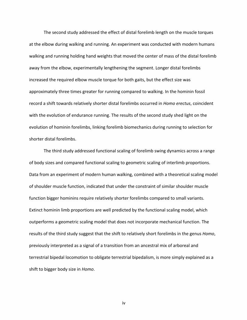
The second study addressed the effect of distal forelimb length on the muscle torques
at the elbow during walking and running. An experiment was conducted with modern humans
walking and running holding hand weights that moved the center of mass of the distal forelimb
away from the elbow, experimentally lengthening the segment. Longer distal forelimbs
increased the required elbow muscle torque for both gaits, but the effect size was
approximately three times greater for running compared to walking. In the hominin fossil
record a shift towards relatively shorter distal forelimbs occurred in Homo erectus, coincident
with the evolution of endurance running. The results of the second study shed light on the
evolution of hominin forelimbs, linking forelimb biomechanics during running to selection for
shorter distal forelimbs.
The third study addressed functional scaling of forelimb swing dynamics across a range
of body sizes and compared functional scaling to geometric scaling of interlimb proportions.
Data from an experiment of modern human walking, combined with a theoretical scaling model
of shoulder muscle function, indicated that under the constraint of similar shoulder muscle
function bigger hominins require relatively shorter forelimbs compared to small variants.
Extinct hominin limb proportions are well predicted by the functional scaling model, which
outperforms a geometric scaling model that does not incorporate mechanical function. The
results of the third study suggest that the shift to relatively short forelimbs in the genus Homo,
previously interpreted as a signal of a transition from an ancestral mix of arboreal and
terrestrial bipedal locomotion to obligate terrestrial bipedalism, is more simply explained as a
shift to bigger body size in Homo.
v
The results of this thesis shed new light on the evolution of human-like walking and
running and the origins of the genus Homo. Previous interpretations of hominin locomotion
behavior that posit a compromised and costly bipedal gait in hominins before Homo lack
biomechanical underpinnings and rely solely on morphological evidence. The results presented
here provide the first mechanistic approach to understanding the evolution of hominin
forelimbs and lead to the conclusion that human-like walking function evolved in
Australopithecus, followed by the coincident evolution of larger body size and endurance
running in the genus Homo.
vi
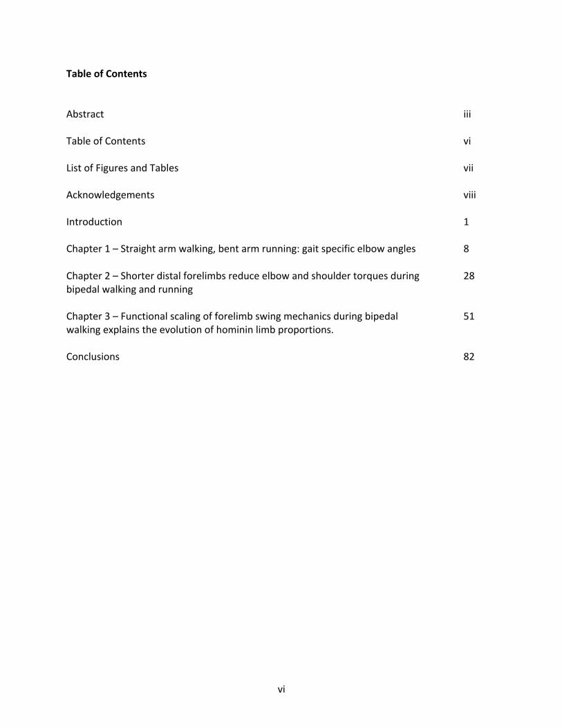
Table of Contents
Abstract iii
Table of Contents vi
List of Figures and Tables vii
Acknowledgements viii
Introduction 1
Chapter 1 – Straight arm walking, bent arm running: gait specific elbow angles 8
Chapter 2 – Shorter distal forelimbs reduce elbow and shoulder torques during 28
bipedal walking and running
Chapter 3 – Functional scaling of forelimb swing mechanics during bipedal 51
walking explains the evolution of hominin limb proportions.
Conclusions 82
vii
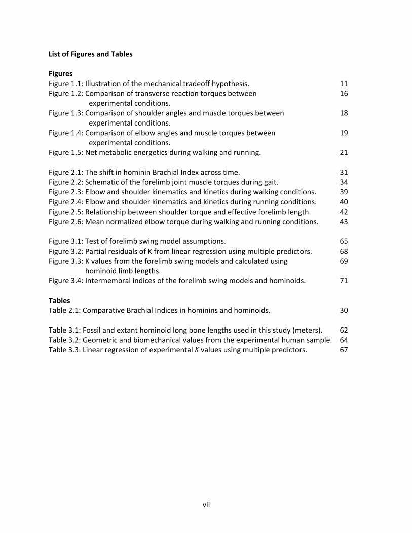
List of Figures and Tables
Figures Figure 1.1: Illustration of the mechanical tradeoff hypothesis. 11
Figure 1.2: Comparison of transverse reaction torques between 16
experimental conditions.
Figure 1.3: Comparison of shoulder angles and muscle torques between 18
experimental conditions.
Figure 1.4: Comparison of elbow angles and muscle torques between 19
experimental conditions.
Figure 1.5: Net metabolic energetics during walking and running. 21
Figure 2.1: The shift in hominin Brachial Index across time. 31
Figure 2.2: Schematic of the forelimb joint muscle torques during gait. 34
Figure 2.3: Elbow and shoulder kinematics and kinetics during walking conditions. 39
Figure 2.4: Elbow and shoulder kinematics and kinetics during running conditions. 40
Figure 2.5: Relationship between shoulder torque and effective forelimb length. 42
Figure 2.6: Mean normalized elbow torque during walking and running conditions. 43
Figure 3.1: Test of forelimb swing model assumptions. 65
Figure 3.2: Partial residuals of K from linear regression using multiple predictors. 68
Figure 3.3: K values from the forelimb swing models and calculated using 69
hominoid limb lengths.
Figure 3.4: Intermembral indices of the forelimb swing models and hominoids. 71
Tables Table 2.1: Comparative Brachial Indices in hominins and hominoids. 30
Table 3.1: Fossil and extant hominoid long bone lengths used in this study (meters). 62
Table 3.2: Geometric and biomechanical values from the experimental human sample. 64
Table 3.3: Linear regression of experimental K values using multiple predictors. 67
viii
Acknowledgments
Pursuing a Ph.D. is a monumental challenge and journey, with ups and downs and twists
and turns throughout years of intense focus on producing new knowledge for the benefit of the
scientific and educational communities. I feel truly blessed to have been able to spend the last
six years at Harvard chasing my intellectual interests and pursuing my own dream of learning
how the world around me works. It’s often said that it takes a village to raise a child, and it truly
takes a village to raise a Ph.D. thesis. Below are thanks to some of the many people who helped
raise me throughout my time at Harvard.
First, the entire HEB community provided a home for me, and will always truly feel like
my academic home. To all the graduate students, postdocs, undergraduates, professors,
administrators, and others who make the HEB community such fertile intellectual ground: you
are the village that raised me and I owe my career to your nurturing.
I thank the HEB administrators, past and present, in particular: Meg Lynch, Meg Jarvi,
Lenia Constantinou, Monica Oyama, Mallory McCoy, and Betty Hughes. You are all the hidden
co-authors of my dissertation, and of all the work produced in HEB. Your endless and
enthusiastic help in navigating the bureaucracy of Harvard, the grant process, and all the steps
that needed to be taken in the past six years smoothed my pathway to my degree so I could
walk it while keeping my eye on the prize. I appreciate all you did for me, and I appreciate most
your patience with me and your dedication to solving every problem big and small.
To the Skeletal Biology and Biomechanics Lab members: you served as my academic
family, and like any loving family you both guided me and challenged me to hone my thinking
ix
and my work, making it the best dissertation it could be. I will always hold the time I had with
Carolyn Eng, Brian Addison, Eric Castillo, Eamon Callison, Tory Tobolski, Tim Kistner, Ben Sibson,
Anna Warrener, Ian Wallace, Nick Holowka, Katie Zink, and Neil Roach dear in my heart.
I owe limitless thanks to my committee members David Pilbeam, Andrew Biewener, and
Madhu Venkadesan for shaping my thesis and providing the constructive feedback and advice
that shaped it over several years. Your guidance was critical in honing my mind from that of
someone interested in science to that of a scientist, able to form questions and hypotheses
with methods to test them.
To my advisor, Dan Lieberman: I can’t use this space to fully explain how much you have
meant to my career and my life. The first time we met you welcomed me into your office to
discuss my intellectual interests, a meeting I thought would be brief. We ended up talking for
over an hour, enthusiastically probing our common interests, and you made me feel in that
moment like I belonged in the academic world. Although the Kenya trip that formed from that
initial discussion did not make it into this thesis, I am forever grateful that we were able to do
an extraordinary project based on that first meeting. You told me my first semester that you
would help make an evolutionary biologist out of me, and by golly you did! Your patience, your
nurturing, your advice, your ability to take my jumbled ideas and see the way to a formulated
plan, are all qualities I will forever appreciate, and you have formed the model for me to be the
best professor and advisor I can be in the future.
None of this would have been possible without the lifetime of encouragement from my
family. Mom, Dad, Patrick, and Elena: you saw my innate curiosity from the beginning and
fostered it at every step of my life. You supported me when I couldn’t stand on my own, both
x
literally and figuratively. Most importantly you gave me the best examples to be a great thinker,
scientist, and person as I grew throughout this process.
Finally, to my loving fiancé, life co-author, and peer review Nesa Wasarhaley: I could
write a dissertation on what you mean to me, and will have a lifetime to tell you. Simply, I love
you and I appreciate you.
1
Introduction
Bipedalism is a defining trait in hominins, with evidence of facultative bipedalism in the
Miocene (Zollikofer et al., 2005) and human-like walking in the Pliocene (Raichlen et al., 2010).
The evolution of bipedalism from an arboreal ancestor with chimpanzees redefined the role of
the forelimb in locomotion. Movement in trees as well as terrestrial quadrupedalism involves
all four limbs contacting the external environment in order to move the center of mass of the
body, while in bipeds the forelimbs produce no contact forces at all. Loss of external contact in
the forelimbs is often thought of as "freeing" the limbs from locomotion, and facilitating
selection for other tasks, such as carrying infants (Wang and Crompton, 2004), digging, food
processing (Zink et al., 2014), tool making (Marzke, 1997), and throwing (Roach et al., 2012).
The forelimbs did not lose all function during gait, however, as they have been shown to play an
important role in walking and running energetics in humans. Despite growing literature on
forelimb locomotion mechanics in modern humans, the connection between bipedal forelimb
dynamics and the evolution of hominin forelimbs has not been quantitatively explored until this
thesis.
The forelimbs play the important role of counterbalancing angular momentum of the
hindlimbs during walking and running in humans (Elftman, 1939; Herr and Popovic, 2008;
Hinrichs, 1987). They swing back and forth once per stride and reciprocal to the hindlimbs,
conserving angular momentum in the body and limiting the free vertical moment about the
center of mass of the body (Collins et al., 2009; Li et al., 2001). This balancing role serves as an
energy saving mechanism in walking and running, as perturbation of normal forelimb swing can
increase metabolic rate by ~10% (e.g. Arellano and Kram, 2014; Umberger, 2008). Though
2
forelimb swing saves net energy, it is likely to have a cost as well; hindlimb swing may account
for up to one-third the total cost of walking (Doke et al., 2007), and muscles are active in the
forelimb during both walking and running (Cappellini, 2006).
Viewed through an evolutionary lens, forelimb anatomy that benefits walking and
running by increasing energy savings or reducing cost should be selected for, unless
counteracted by a tradeoff with another behavior. The fossil record indicates mosaic evolution
of the forelimb in Australopithecus, which has been interpreted as evidence of retained
climbing behavior in these species (Churchill et al., 2013; Jungers, 2009) and a tradeoff between
climbing and walking (e.g.(Jungers, 2009)). The shift to fully modern forelimb anatomy in Homo
also coincided with the evolution of endurance running (Bramble and Lieberman, 2004), and
alternatively may reflect a tradeoff between climbing and running, or non-locomotion
behaviors like tool-making and throwing. In order to assess these tradeoff hypotheses about
forelimb evolution it is necessary to understand how anatomy affects the costs and benefits of
each behavior.
The goal of my thesis was to test hypotheses linking forelimb anatomy to bipedal
function, and to interpret evolution of the hominin forelimb in the context of bipedalism. The
first three chapters focused directly on the link between anatomy, bipedal forelimb mechanics,
and hominin forelimb evolution. In these chapters I addressed two anatomical characters:
forelimb length and distal forelimb length.
Chapter 1 asked the question, why do humans walk with straight forelimbs and run with
flexed forelimbs? Flexing the elbow into a right angle brings the center of mass of the forelimb
closer to the shoulder, effectively shortening forelimb length and reducing the rotational inertia
3
of the limb. I hypothesized that this behavioral mechanism therefore provides a benefit to
walking and running by reducing the cost of swinging the forelimb, but the benefit during
walking does not fit with stereotypical behavior in human walking. Therefore, I also
hypothesized that a tradeoff exists between the cost at the shoulder and the cost at the elbow,
with flexed forelimbs requiring more effort from the elbow muscles, and predicted that
metabolic cost would favor the stereotypical behavior in each gait. I tested my hypotheses and
prediction using an experiment with people walking and running with both forelimb
configurations. The results from the experiment confirm that a tradeoff exists between muscle
torque at the shoulder and elbow, with flexed elbows causing reduced shoulder torque and
increased elbow torque in both walking and running. Walking with flexed elbows was
approximately 11% more costly than with a straight forelimb, as predicted. However, the cost
of running was equivalent between both configurations, leaving the reason for flexed elbows
during running unknown.
Chapter 2 investigated how variation in distal forelimb length affects walking and
running mechanics. Species in the genus Homo including modern humans have relatively short
distal forelimbs, a derived feature compared to Australopithecus. The shift to smaller distal
forelimbs is first evident in Homo erectus (Richmond et al., 2002), and coincides with a shift
towards large day ranges and endurance running (Bramble and Lieberman, 2004). I
hypothesized that shortening of distal forelimb would benefit both walking and running by
reducing muscle torque at the elbow. To test the hypothesis I conducted an experiment with
people walking and running while holding weights in their hands. The addition of mass to the
hands lengthened the distance between the center of mass of the distal forelimb and the
4
elbow, while simultaneously increasing the length of between the center of mass of the entire
forelimb and the shoulder. Artificially increasing distal and overall forelimb lengths increased
muscle torque at both the shoulder and elbow joints, likely increasing the cost of forelimb
swing for both walking and running. However, the effect of relative distal forelimb length on
elbow torque was three times greater during running than during walking. In context of the
greater effect magnitude in running, the shift to shorter distal forelimbs can be explained by
selection for running.
Chapter 3 linked the functions of the forelimbs and hindlimbs during walking in order to
test the hypothesis that hominin forelimb lengths can be predicted by modern human walking
mechanics. Australopiths had relatively long forelimbs compared to Homo erectus and its
descendants (Young et al., 2010), but also had shorter hindlimbs. The same pattern appears in
bipedal theropod dinosaurs, which suggests bipedal mechanics may explain the relationship. In
order to test the hypothesis I combined a model of hindlimb function, the Froude equation,
with a model of forelimb function, the spring-pendulum model, into a new model
encompassing both limbs. In order to compare hominins of different sizes I used the framework
of dynamic similarity, which standardizes gait across geometric lengths (Alexander and Jayes,
1983). I used an experiment to collect walking data and use the model to predict hominin
forelimb lengths across the hindlimb length spectrum. The model prediction could explain the
forelimb lengths of all the hominins but the oldest fossil specimen (Ardipithecus), and similarly
explains theropod dinosaur limb lengths. In light of the results, I hypothesized that bipedalism
links selection on limb lengths in bipeds, leading to a predictable relationship between the
5
limbs that explains why members of the genus Homo like modern humans have relatively short
forelimbs compared to australopiths.
6
References
Alexander, R. and Jayes, A. S. (1983). A dynamic similarity hypothesis for the gaits of quadrupedal mammals. Journal of Zoology.
Arellano, C. J. and Kram, R. (2014). The metabolic cost of human running: is swinging the arms worth it? Journal of Experimental Biology 217, 2456–2461.
Bramble, D. M. and Lieberman, D. E. (2004). Endurance running and the evolution of Homo. Nature 432, 345–352.
Cappellini, G. (2006). Motor Patterns in Human Walking and Running. J. Neurophysiol. 95, 3426–3437.
Churchill, S. E., Holliday, T. W., Carlson, K. J., Jashashvili, T., Macias, M. E., Mathews, S., Sparling, T. L., Schmid, P., de Ruiter, D. J. and Berger, L. R. (2013). The Upper Limb of Australopithecus sediba. Science 340, 1233477–1233477.
Collins, S. H., Adamczyk, P. G. and Kuo, A. D. (2009). Dynamic arm swinging in human walking. Proceedings of the Royal Society B: Biological Sciences 276, 3679–3688.
Doke, J., Donelan, J. M. and Kuo, A. D. (2007). Mechanics and energetics of swinging the human leg. Journal of Experimental Biology 210, 2399–2399.
Elftman, H. (1939). The function of the arms in walking. Human biology.
Herr, H. and Popovic, M. (2008). Angular momentum in human walking. Journal of Experimental Biology 211, 467–481.
Hinrichs, R. N. (1987). Upper Extremity Function in Running. II: Angular Momentum Considerations. Int J Sport Biomech 3, 242–263.
Jungers, W. L. (2009). Interlimb Proportions in Humans and Fossil Hominins: Variability and Scaling. In The First Humans (eds. Grine, F. E., Fleagle, J. G., and Leakey, R. E.), pp. 93–98.
Li, Y., Wang, W., Crompton, R. H. and Günther, M. M. (2001). Free vertical moments and transverse forces in human walking and their role in relation to arm-swing. Journal of Experimental Biology 204, 47–58.
Marzke, M. W. (1997). Precision grips, hand morphology, and tools. Am. J. Phys. Anthropol. 102, 91–110.
Raichlen, D. A., Gordon, A. D., Harcourt-Smith, W. E. H., Foster, A. D. and Haas, W. R. (2010). Laetoli Footprints Preserve Earliest Direct Evidence of Human-Like Bipedal Biomechanics. PLoS ONE 5, e9769–6.
7
Richmond, B. G., Aiello, L. C. and Wood, B. A. (2002). Early hominin limb proportions. Journal of Human Evolution 43, 529–548.
Roach, N. T., Lieberman, D. E., Gill, T. J., IV, Palmer, W. E. and Gill, T. J., III (2012). The effect of humeral torsion on rotational range of motion in the shoulder and throwing performance. Journal of Anatomy 220, 293–301.
Umberger, B. R. (2008). Effects of suppressing arm swing on kinematics, kinetics, and energetics of human walking. J Biomech 41, 2575–2580.
Wang, W. J. and Crompton, R. H. (2004). The role of load-carrying in the evolution of modern body proportions. Journal of Anatomy 204, 417–430.
Young, N. M., Wagner, G. P. and Hallgrimsson, B. (2010). Development and the evolvability of human limbs. Proc. Natl. Acad. Sci. U.S.A. 107, 3400–3405.
Zink, K. D., Lieberman, D. E. and Lucas, P. W. (2014). Food material properties and early hominin processing techniques. Journal of Human Evolution 77, 155–166.
Zollikofer, C. P. E., Ponce de León, M. S., Lieberman, D. E., Guy, F., Pilbeam, D., Likius, A., Mackaye, H. T., Vignaud, P. and Brunet, M. (2005). Virtual cranial reconstruction of Sahelanthropus tchadensis. Nature 434, 755–759.
8
Chapter 1 – Straight arm walking, bent arm running: gait specific elbow angles
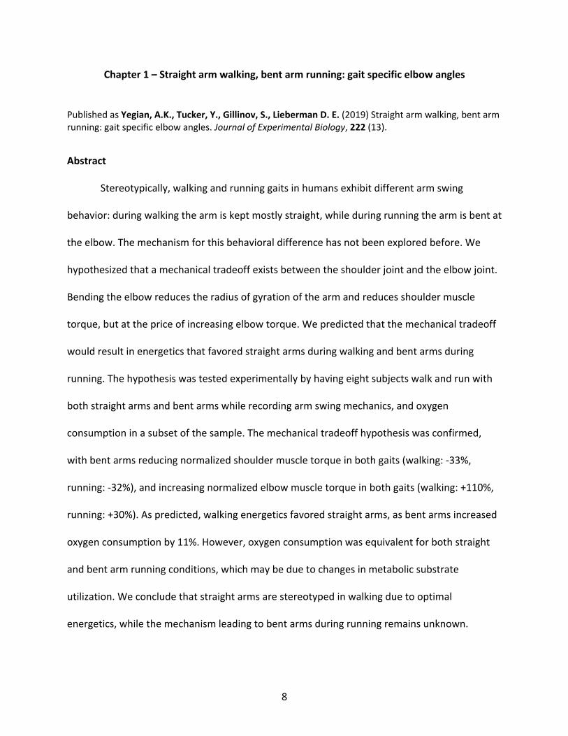
Published as Yegian, A.K., Tucker, Y., Gillinov, S., Lieberman D. E. (2019) Straight arm walking, bent arm running: gait specific elbow angles. Journal of Experimental Biology, 222 (13).
Abstract
Stereotypically, walking and running gaits in humans exhibit different arm swing
behavior: during walking the arm is kept mostly straight, while during running the arm is bent at
the elbow. The mechanism for this behavioral difference has not been explored before. We
hypothesized that a mechanical tradeoff exists between the shoulder joint and the elbow joint.
Bending the elbow reduces the radius of gyration of the arm and reduces shoulder muscle
torque, but at the price of increasing elbow torque. We predicted that the mechanical tradeoff
would result in energetics that favored straight arms during walking and bent arms during
running. The hypothesis was tested experimentally by having eight subjects walk and run with
both straight arms and bent arms while recording arm swing mechanics, and oxygen
consumption in a subset of the sample. The mechanical tradeoff hypothesis was confirmed,
with bent arms reducing normalized shoulder muscle torque in both gaits (walking: -33%,
running: -32%), and increasing normalized elbow muscle torque in both gaits (walking: +110%,
running: +30%). As predicted, walking energetics favored straight arms, as bent arms increased
oxygen consumption by 11%. However, oxygen consumption was equivalent for both straight
and bent arm running conditions, which may be due to changes in metabolic substrate
utilization. We conclude that straight arms are stereotyped in walking due to optimal
energetics, while the mechanism leading to bent arms during running remains unknown.
9
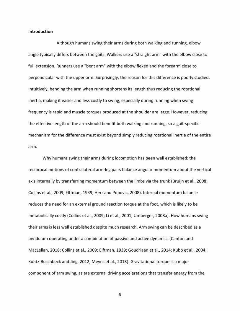
Introduction
Although humans swing their arms during both walking and running, elbow
angle typically differs between the gaits. Walkers use a "straight arm" with the elbow close to
full extension. Runners use a "bent arm" with the elbow flexed and the forearm close to
perpendicular with the upper arm. Surprisingly, the reason for this difference is poorly studied.
Intuitively, bending the arm when running shortens its length thus reducing the rotational
inertia, making it easier and less costly to swing, especially during running when swing
frequency is rapid and muscle torques produced at the shoulder are large. However, reducing
the effective length of the arm should benefit both walking and running, so a gait-specific
mechanism for the difference must exist beyond simply reducing rotational inertia of the entire
arm.
Why humans swing their arms during locomotion has been well established: the
reciprocal motions of contralateral arm-leg pairs balance angular momentum about the vertical
axis internally by transferring momentum between the limbs via the trunk (Bruijn et al., 2008;
Collins et al., 2009; Elftman, 1939; Herr and Popovic, 2008). Internal momentum balance
reduces the need for an external ground reaction torque at the foot, which is likely to be
metabolically costly (Collins et al., 2009; Li et al., 2001; Umberger, 2008a). How humans swing
their arms is less well established despite much research. Arm swing can be described as a
pendulum operating under a combination of passive and active dynamics (Canton and
MacLellan, 2018; Collins et al., 2009; Elftman, 1939; Goudriaan et al., 2014; Kubo et al., 2004;
Kuhtz-Buschbeck and Jing, 2012; Meyns et al., 2013). Gravitational torque is a major
component of arm swing, as are external driving accelerations that transfer energy from the
10
legs to the arms via the trunk (Collins et al., 2009; Kubo et al., 2004; Pontzer et al., 2009). At the
same time, active muscle recruitment develops torques in the trunk, shoulder, and elbow joints
(Ballesteros and Buchthal, 1965; Canton and MacLellan, 2018; Collins et al., 2009; Elftman,
1939; Kuhtz-Buschbeck and Jing, 2012). Neuromuscular control of arm swing is rooted in the
central patterns of human gait (Barthelemy and Nielsen, 2010; Cappellini, 2006; Dietz et al.,
2001), and may be conserved from quadrupedal ancestry (Dietz, 2002).
Arm swing occurs mainly in the parasagittal plane, yet is linked to angular momentum
about the vertical axis. The linkage is partly accomplished by the horizontal joint reaction force
at the shoulder (JRFH) that arises from swing. JRFH causes a transverse plane reaction torque
(τtrv) on the thorax (Figure 1.1A), which is further linked to the lower body by trunk torsion to
transfer momentum between the upper and lower limbs. In the arm, muscle torques occur at
the shoulder (τsho) and the elbow (τelb), generally opposing angular excursion and acting in a
resistive manner (Collins et al., 2009) (Fig. 1.1A). τsho is most simply explained as resembling a
rotational spring and acting on a functionally rigid single pendulum arm. Bending the elbow
moves the center of mass (CoM) of the pendulum closer to the shoulder pivot, reducing the
radius of gyration (RG) and the required τsho (Figure 1.1B).
In order to maintain functional approximation of a single pendulum arm, τelb must resist
external forces that would cause an external torque at the elbow and rotation of the forearm
relative to the upper arm. Gravity is one such external force. Pseudoforces from acceleration of
the thorax also place external torques on the forearm in the reference frame of the upper arm.
Vertical acceleration measured at the shoulder has a much higher magnitude compared to
horizontal acceleration in walking (Kubo et al., 2004). Similarly, measurements of linear
11
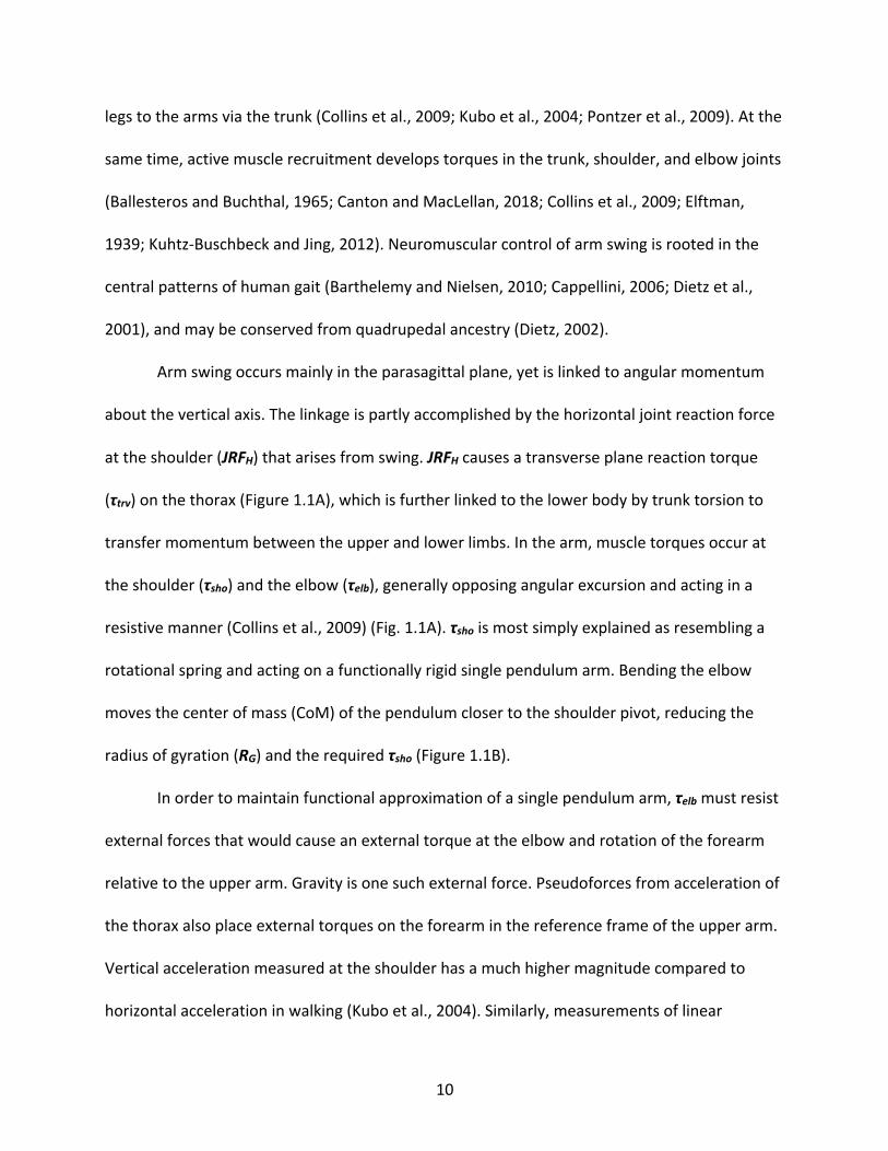
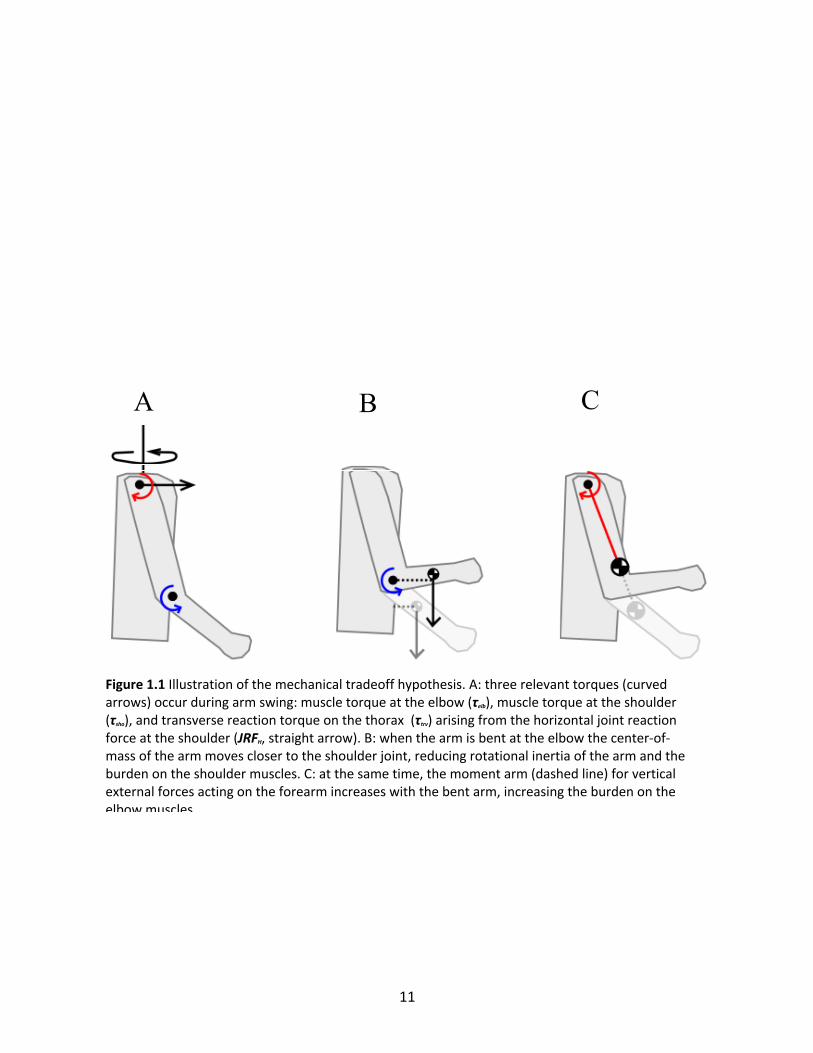
Figure 1.1 Illustration of the mechanical tradeoff hypothesis. A: three relevant torques (curved arrows) occur during arm swing: muscle torque at the elbow (τelb), muscle torque at the shoulder (τsho), and transverse reaction torque on the thorax (τtrv) arising from the horizontal joint reaction force at the shoulder (JRFH, straight arrow). B: when the arm is bent at the elbow the center-of-mass of the arm moves closer to the shoulder joint, reducing rotational inertia of the arm and the burden on the shoulder muscles. C: at the same time, the moment arm (dashed line) for vertical external forces acting on the forearm increases with the bent arm, increasing the burden on the elbow muscles.
12
displacements at C7 vertebral level indicate larger vertical than horizontal accelerations in both
walking and running (Thorstensson et al., 1984). Other forces causing external elbow torques
arise from centripetal and tangential accelerations of the elbow joint center in the arm
reference frame. The net effect of all these forces is likely a large vertical external force
component contributing to the external elbow torque, and a smaller horizontal component.
Bending the elbow to reduce arm RG brings the forearm closer to horizontal, thus increasing the
moment arm of the net vertical external force (Figure 1.1C). Conversely, maintaining a straight
arm places the forearm more parallel with the vertical forces, limiting the external torque they
produce and consequently the resistive τelb.
We propose a mechanical tradeoff hypothesis that posits a tradeoff between muscle
torques at the shoulder and the elbow linked to the average elbow angle. Flexing the elbow,
thus shortening the arm’s moment of inertia, reduces the shoulder muscle torque but at the
cost of increasing the elbow muscle torque. We predict that the energetic consequences of the
mechanical tradeoff favor straight arm walking and bent arm running, and that elbow angle is
determined by energetic cost for each gait. Studies of both walking and running show that
perturbation of normal arm swing, typically by holding or binding the arms to the torso,
increases the net energy cost of locomotion by up to 10% in walking (Collins et al., 2009; Ortega
et al., 2008; Umberger, 2008a) and running (Arellano and Kram, 2014; Egbuonu et al., 1990;
Tseh et al., 2008), indicating that normal arm swing is an important cost-saving mechanism. We
also predict similar non-trivial energy costs to altering normal elbow angle. We tested our
hypothesis and predictions by conducting an experiment with human subjects who walked and
ran with both flexed and extended elbows.
13
Methods and Materials
Eight healthy subjects (four males and four females, age: 26.6 years, s.d. 2.5, mass: 76.6
kg, s.d. 15.9) participated in the experiment. Prior approval was granted by the Harvard
University Institutional Review Board, and all subjects gave informed consent. Subjects walked
and ran on a split-belt treadmill instrumented with force plates (Bertec Corp., Columbus, Ohio).
Four randomized experimental conditions were conducted in random order: straight arm
walking (SW), bent arm walking (BW), straight arm running (SR), and bent arm running (BR). For
SW and BR the subjects were asked to walk and run normally. For BW subjects were instructed
to hold their forearm as they would during running; similarly, the instruction for SR was to hold
the forearm as they would during walking. All walking trials were done at a single dimensionless
speed (Froude = 0.2, range: 1.30 m/s to 1.44 m/s), and running trials were also done at a single
dimensionless speed (Froude = 1, range: 2.90 m/s to 3.22 m/s). Each condition lasted three
minutes, with data collection occurring during the last minute. Six subjects returned within two
weeks for energetic data collection (see below). All analyses used the Igor Pro software
platform (Wavemetrics, Lake Oswega, Oregon).
Kinematic and Kinetic Time Series
Motions of the right forearm, right upper arm, and the thorax were captured with eight
infrared cameras recording at 200 Hz (Qualysis Motion Capture Systems, Gothenburg, Sweden).
Reflective markers were placed on the left and right acromia, right humeral epicondyles, and
right radial and ulnar styloid processes. The right shoulder joint was estimated to be 3.0
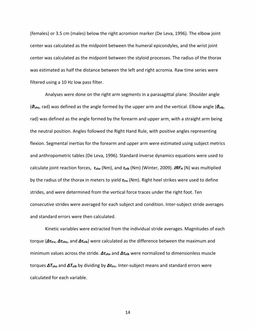
14
(females) or 3.5 cm (males) below the right acromion marker (De Leva, 1996). The elbow joint
center was calculated as the midpoint between the humeral epicondyles, and the wrist joint
center was calculated as the midpoint between the styloid processes. The radius of the thorax
was estimated as half the distance between the left and right acromia. Raw time series were
filtered using a 10 Hz low pass filter.
Analyses were done on the right arm segments in a parasagittal plane. Shoulder angle
(θsho, rad) was defined as the angle formed by the upper arm and the vertical. Elbow angle (θelb,
rad) was defined as the angle formed by the forearm and upper arm, with a straight arm being
the neutral position. Angles followed the Right Hand Rule, with positive angles representing
flexion. Segmental inertias for the forearm and upper arm were estimated using subject metrics
and anthropometric tables (De Leva, 1996). Standard inverse dynamics equations were used to
calculate joint reaction forces, τsho (Nm), and τelb (Nm) (Winter, 2009). JRFH (N) was multiplied
by the radius of the thorax in meters to yield τtrv (Nm). Right heel strikes were used to define
strides, and were determined from the vertical force traces under the right foot. Ten
consecutive strides were averaged for each subject and condition. Inter-subject stride averages
and standard errors were then calculated.
Kinetic variables were extracted from the individual stride averages. Magnitudes of each
torque (Δτtrv, Δτsho, and Δτelb) were calculated as the difference between the maximum and
minimum values across the stride. Δτsho and Δτelb were normalized to dimensionless muscle
torques ΔTsho and ΔTelb by dividing by Δτtrv. Inter-subject means and standard errors were
calculated for each variable.
15
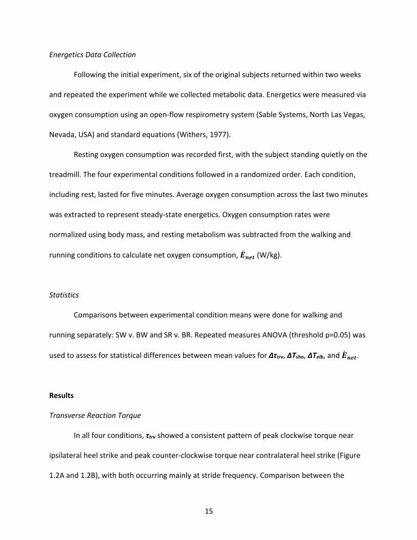
Energetics Data Collection
Following the initial experiment, six of the original subjects returned within two weeks
and repeated the experiment while we collected metabolic data. Energetics were measured via
oxygen consumption using an open-flow respirometry system (Sable Systems, North Las Vegas,
Nevada, USA) and standard equations (Withers, 1977).
Resting oxygen consumption was recorded first, with the subject standing quietly on the
treadmill. The four experimental conditions followed in a randomized order. Each condition,
including rest, lasted for five minutes. Average oxygen consumption across the last two minutes
was extracted to represent steady-state energetics. Oxygen consumption rates were
normalized using body mass, and resting metabolism was subtracted from the walking and
running conditions to calculate net oxygen consumption, !̇#$% (W/kg).
Statistics
Comparisons between experimental condition means were done for walking and
running separately: SW v. BW and SR v. BR. Repeated measures ANOVA (threshold p=0.05) was
used to assess for statistical differences between mean values for Δτtrv, ΔTsho, ΔTelb, and !̇#$%.
Results
Transverse Reaction Torque
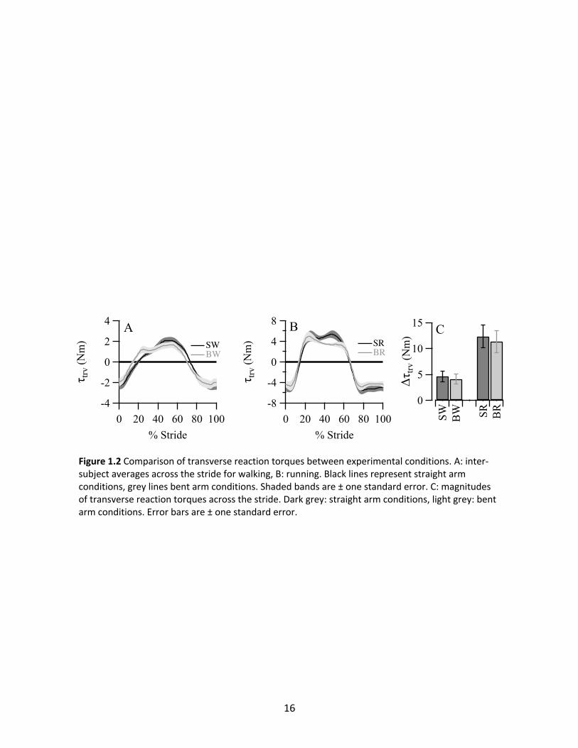
In all four conditions, τtrv showed a consistent pattern of peak clockwise torque near
ipsilateral heel strike and peak counter-clockwise torque near contralateral heel strike (Figure
1.2A and 1.2B), with both occurring mainly at stride frequency. Comparison between the
16
Figure 1.2 Comparison of transverse reaction torques between experimental conditions. A: inter-subject averages across the stride for walking, B: running. Black lines represent straight arm conditions, grey lines bent arm conditions. Shaded bands are ± one standard error. C: magnitudes of transverse reaction torques across the stride. Dark grey: straight arm conditions, light grey: bent arm conditions. Error bars are ± one standard error.
17
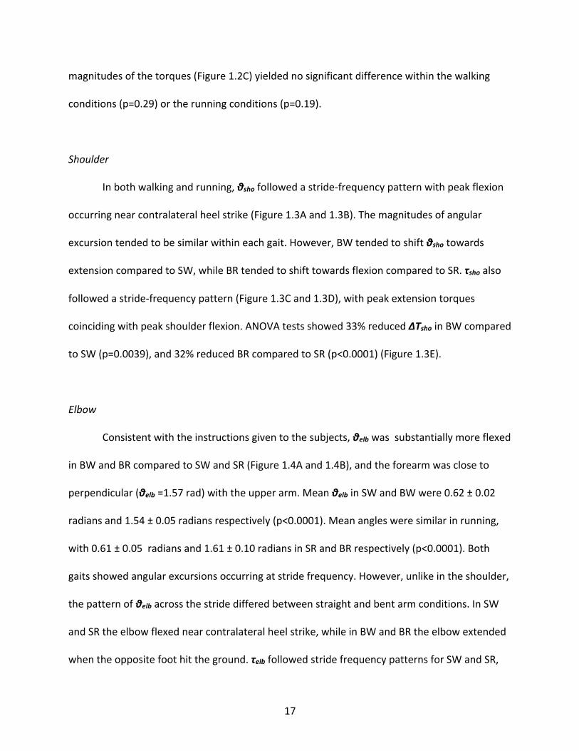
magnitudes of the torques (Figure 1.2C) yielded no significant difference within the walking
conditions (p=0.29) or the running conditions (p=0.19).
Shoulder
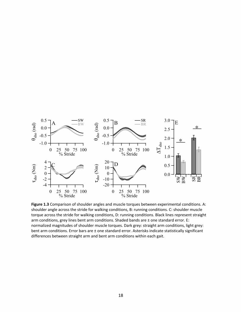
In both walking and running, θsho followed a stride-frequency pattern with peak flexion
occurring near contralateral heel strike (Figure 1.3A and 1.3B). The magnitudes of angular
excursion tended to be similar within each gait. However, BW tended to shift θsho towards
extension compared to SW, while BR tended to shift towards flexion compared to SR. τsho also
followed a stride-frequency pattern (Figure 1.3C and 1.3D), with peak extension torques
coinciding with peak shoulder flexion. ANOVA tests showed 33% reduced ΔTsho in BW compared
to SW (p=0.0039), and 32% reduced BR compared to SR (p<0.0001) (Figure 1.3E).
Elbow
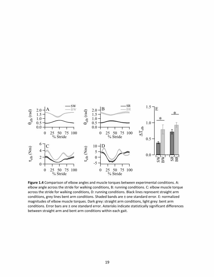
Consistent with the instructions given to the subjects, θelb was substantially more flexed
in BW and BR compared to SW and SR (Figure 1.4A and 1.4B), and the forearm was close to
perpendicular (θelb =1.57 rad) with the upper arm. Mean θelb in SW and BW were 0.62 ± 0.02
radians and 1.54 ± 0.05 radians respectively (p<0.0001). Mean angles were similar in running,
with 0.61 ± 0.05 radians and 1.61 ± 0.10 radians in SR and BR respectively (p<0.0001). Both
gaits showed angular excursions occurring at stride frequency. However, unlike in the shoulder,
the pattern of θelb across the stride differed between straight and bent arm conditions. In SW
and SR the elbow flexed near contralateral heel strike, while in BW and BR the elbow extended
when the opposite foot hit the ground. τelb followed stride frequency patterns for SW and SR,
18
Figure 1.3 Comparison of shoulder angles and muscle torques between experimental conditions. A: shoulder angle across the stride for walking conditions, B: running conditions. C: shoulder muscle torque across the stride for walking conditions, D: running conditions. Black lines represent straight arm conditions, grey lines bent arm conditions. Shaded bands are ± one standard error. E: normalized magnitudes of shoulder muscle torques. Dark grey: straight arm conditions, light grey: bent arm conditions. Error bars are ± one standard error. Asterisks indicate statistically significant differences between straight arm and bent arm conditions within each gait.
19
Figure 1.4 Comparison of elbow angles and muscle torques between experimental conditions. A: elbow angle across the stride for walking conditions, B: running conditions. C: elbow muscle torque across the stride for walking conditions, D: running conditions. Black lines represent straight arm conditions, grey lines bent arm conditions. Shaded bands are ± one standard error. E: normalized magnitudes of elbow muscle torques. Dark grey: straight arm conditions, light grey: bent arm conditions. Error bars are ± one standard error. Asterisks indicate statistically significant differences between straight arm and bent arm conditions within each gait.
20
but step frequency patterns for BW and BR (Figure 1.4C and 1.4D). In addition, mean muscle
torques were substantially shifted towards flexion in the bent arm conditions (on average, 1.21
Nm in walking and 1.60 Nm in running), presumably due to increased gravitational torque.
Comparison of magnitudes yielded significant increases in ΔTelb for the bent arm conditions
compared to the straight arm conditions in both walking (110% increase, p=0.0037) and
running (30% increase, p=0.0096) (Figure 1.4E).
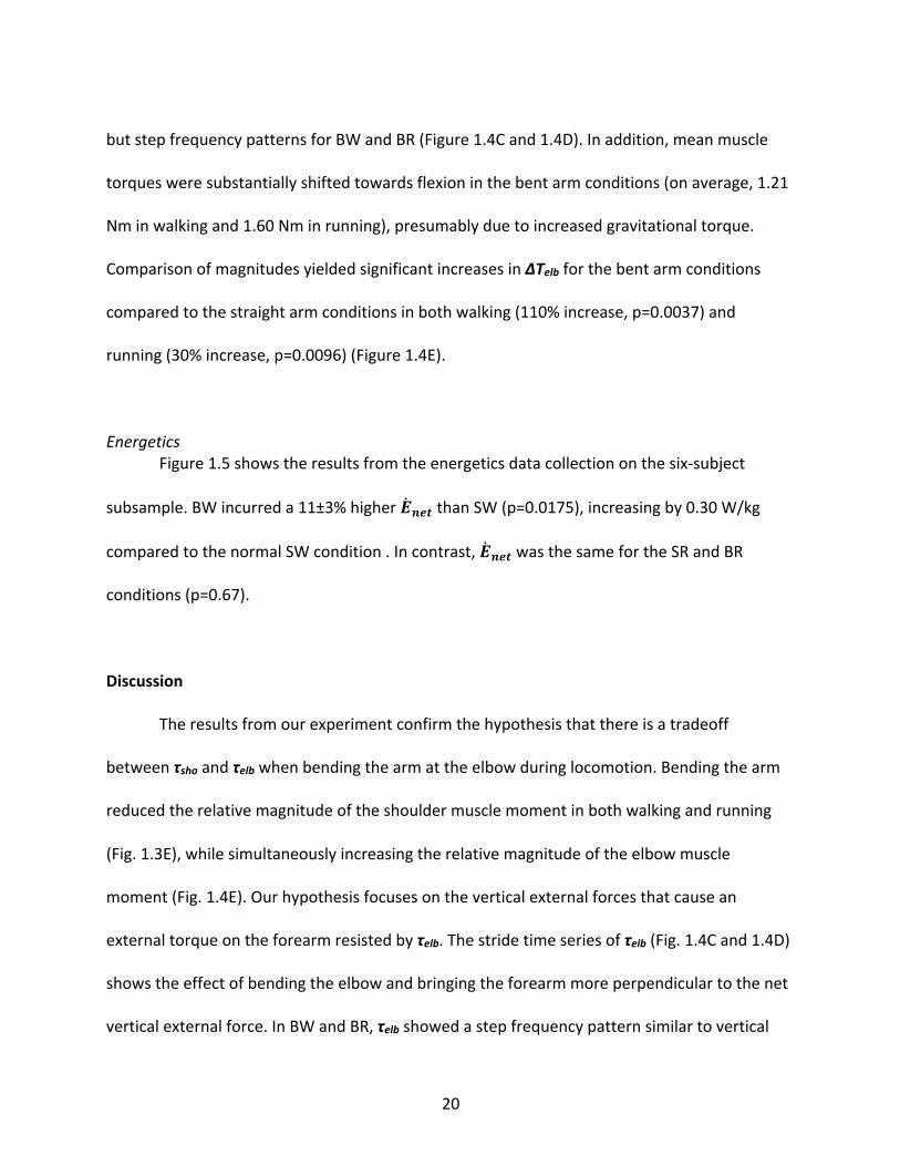
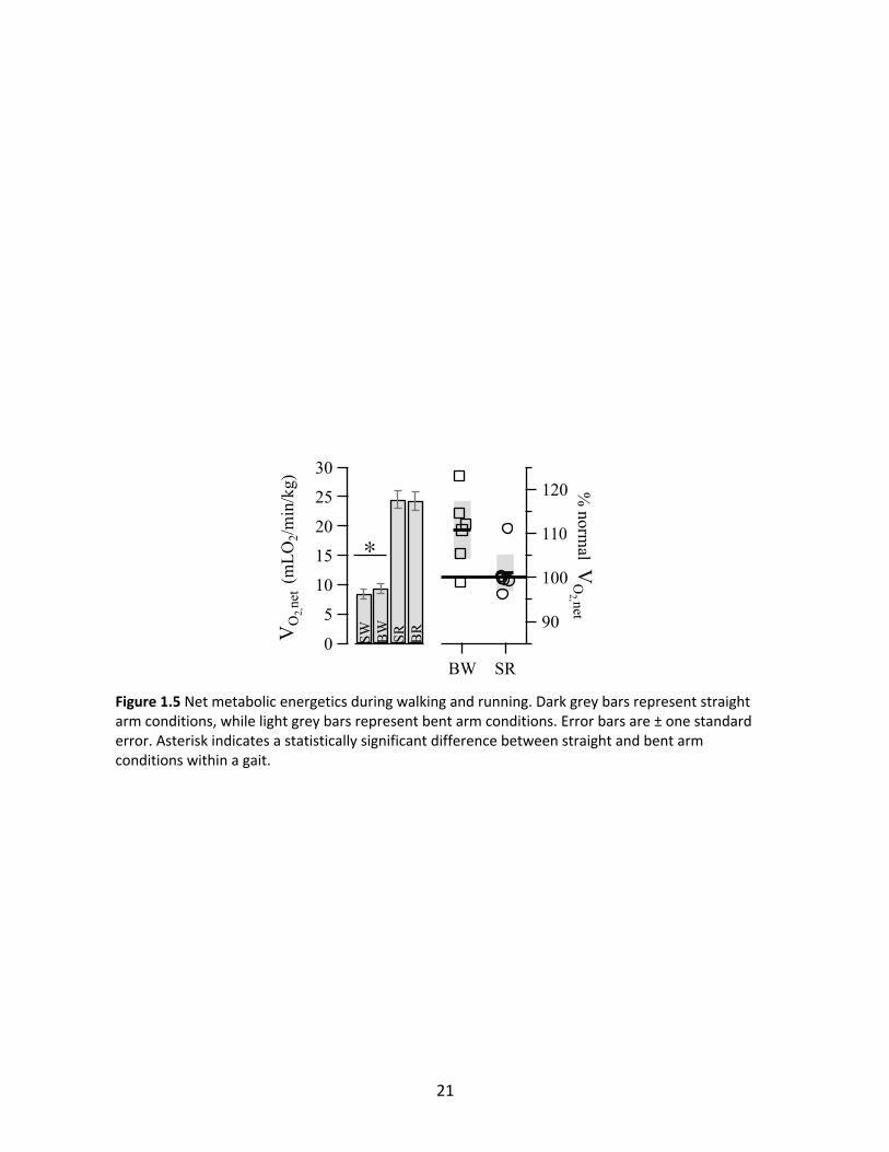
Energetics Figure 1.5 shows the results from the energetics data collection on the six-subject
subsample. BW incurred a 11±3% higher !̇#$% than SW (p=0.0175), increasing by 0.30 W/kg
compared to the normal SW condition . In contrast, !̇#$% was the same for the SR and BR
conditions (p=0.67).
Discussion
The results from our experiment confirm the hypothesis that there is a tradeoff
between τsho and τelb when bending the arm at the elbow during locomotion. Bending the arm
reduced the relative magnitude of the shoulder muscle moment in both walking and running
(Fig. 1.3E), while simultaneously increasing the relative magnitude of the elbow muscle
moment (Fig. 1.4E). Our hypothesis focuses on the vertical external forces that cause an
external torque on the forearm resisted by τelb. The stride time series of τelb (Fig. 1.4C and 1.4D)
shows the effect of bending the elbow and bringing the forearm more perpendicular to the net
vertical external force. In BW and BR, τelb showed a step frequency pattern similar to vertical
21
Figure 1.5 Net metabolic energetics during walking and running. Dark grey bars represent straight arm conditions, while light grey bars represent bent arm conditions. Error bars are ± one standard error. Asterisk indicates a statistically significant difference between straight and bent arm conditions within a gait.
22
accelerations of the trunk (Kubo et al., 2004; Thorstensson et al., 1984). Conversely, τelb
oscillated at stride frequency in SW and SR, likely due to swinging of the upper arm (Fig. 1.3A
and 1.3B) dominating the external torque acting on the forearm.
We predicted that the gait-specific stereotypical behaviors of straight arm walking and
bent arm running are driven by the energetic consequences of the mechanical tradeoff, with
walking favoring straight arms and running favoring bent arms. The first part of our prediction
was supported by our data (Fig. 1.5), as walking with a bent arm increased !̇#$% by 11%, similar
to the magnitude of cost increase caused by restricting arm swing (Bruijn et al., 2008; Collins et
al., 2009; Umberger, 2008b). However, while we predicted !̇#$% would be reduced in bent arm
running, our results show the same metabolic cost between the two elbow angle conditions
(Fig. 1.5).
We surmise three possible reasons the running prediction was not supported. First, we
tested only a single dimensionless speed, and it is possible that running becomes less costly
with bent arms than straight arms at higher speeds than we tested. Although elbow angle did
not affect the net cost of running, higher torques were generated by the shoulder muscles with
straight arms compared to bent, requiring more activated muscle volume. Larger and costlier
motor units tend to be activated as more volume is recruited in muscle contractions
(Duchateau and Enoka, 2011), so it is possible that fiber recruitment order affects the tradeoff
at faster speeds. Second, there may be an independent benefit to bending the arms when
running, such as creating a linkage between the biceps and cleidocraniotrapezius muscles for
the purpose of head stabilization (Lieberman, 2011). Testing for speed effects within each gait
may shed more light on our running energetic results. Third, our analysis was limited to
23
parasagittal arm swing. Bending the elbow affects frontal and transverse plane mechanics;
however, any change in mechanics in those two planes already factor into the net energetics,
so the change would have to provide a non-energetic benefit to be the reason for the typical
running elbow angle.
There is a clear energetic benefit to keeping the arms straight when walking, making
straight arms the "optimal" configuration. Lack of an energetic benefit for either elbow angle in
running means that there is no "optimal" configuration per se. Even though bent arms are
stereotyped in running, exactly how the forearm is carried seems to matter little when it comes
to energetics. To that end, there was much greater variation within our sample in average θelb
for the normal running condition (s.d. of 0.274 rad) than the normal walking condition (s.d.
0.070 rad), matching our anecdotal observation that runners use quite varied forearm
positions.
In light of our results, we hypothesize that bent arms are stereotyped during running in
order to increase endurance running capacity. The evolution of endurance running in the genus
Homo was a major transition in the course of human evolution (Bramble and Lieberman, 2004).
The capacity to run very long distances at speeds that force galloping in prey mammals was a
critical innovation in hunter-gatherer ecology. In our experiment, elbow angle did not affect the
instantaneous metabolic power of running, suggesting the metabolic savings at the shoulder via
bending the arms were balanced by the metabolic costs at the elbows. However, the two
conditions had very different relative burdens between the shoulder and elbow muscles.
Straight arm running requires large shoulder muscle torques and relatively small elbow muscle
torques, while during bent arm running the torque burden is more equitable between the
24
joints. Equitable sharing of the muscular burden between the two joints may reduce the rate of
metabolite buildup and fatigue in the shoulder muscles, and may increase endurance
capabilities. This hypothesis should be tested in a further experiment.
Finally, our results have implications for the evolution of arm proportions in hominins.
Arm length relative to leg length was greater in Australopithecus and in Homo habilis compared
to modern humans (Young et al., 2010), as was forearm length relative to upper arm length
(Churchill et al., 2013; Richmond et al., 2002). Modern arm proportions emerged in Homo
erectus, and coincided with the evolution of endurance running as an important hominin
behavior (Bramble and Lieberman, 2004). Reductions in forearm length and total arm length
should reduce τelb and τsho, respectively, and therefore may be signals of selection for lesser arm
swing costs during endurance running. Selection for running may have been an important
factor shaping the evolution of hominin arms.
Acknowledgments
We thank Andrew Biewener, Nicholas Holowka, Ian Wallace, Eamon Callison, and Victoria
Tobolsky for helpful comments at various stages of the project. We also thank the anonymous
reviewers for their comments and improvements on the manuscript.
Funding
Funding was provided by the Robert A. Chapman Memorial Scholarship for Vertebrate
Locomotion (AKY, Harvard University), and the American School of Prehistoric Research (DEL,
Harvard University).
25
References
Arellano, C. J. and Kram, R. (2014). The metabolic cost of human running: is swinging the arms worth it? J. Exp. Biol. 217, 2456–2461.
Ballesteros, M. and Buchthal, F. (1965). The pattern of muscular activity during the arm swing of natural walking. Acta Physiologica ….
Barthelemy, D. and Nielsen, J. B. (2010). Corticospinal contribution to arm muscle activity during human walking. J. Physiol. (Lond.) 588, 967–979.
Bramble, D. M. and Lieberman, D. E. (2004). Endurance running and the evolution of Homo. Nature 432, 345–352.
Bruijn, S. M., Meijer, O. G., van Dieën, J. H., Kingma, I. and Lamoth, C. J. C. (2008). Coordination of leg swing, thorax rotations, and pelvis rotations during gait: the organisation of total body angular momentum. Gait & Posture 27, 455–462.
Canton, S. and MacLellan, M. J. (2018). Active and passive contributions to arm swing_ Implications of the restriction of pelvis motion during human locomotion. Human Movement Science 57, 314–323.
Cappellini, G. (2006). Motor Patterns in Human Walking and Running. J. Neurophysiol. 95, 3426–3437.
Churchill, S. E., Holliday, T. W., Carlson, K. J., Jashashvili, T., Macias, M. E., Mathews, S., Sparling, T. L., Schmid, P., de Ruiter, D. J. and Berger, L. R. (2013). The Upper Limb of Australopithecus sediba. Science 340, 1233477–1233477.
Collins, S. H., Adamczyk, P. G. and Kuo, A. D. (2009). Dynamic arm swinging in human walking. Proceedings of the Royal Society B: Biological Sciences 276, 3679–3688.
De Leva, P. (1996). Adjustments to Zatsiorsky-Seluyanov's segment inertia parameters. J Biomech 29, 1223–1230.
Dietz, V. (2002). Do human bipeds use quadrupedal coordination? Trends Neurosci. 25, 462–467.
Dietz, V., Fouad, K. and Bastiaanse, C. M. (2001). Neuronal coordination of arm and leg movements during human locomotion. Eur. J. Neurosci. 14, 1906–1914.
Duchateau, J. and Enoka, R. M. (2011). Human motor unit recordings: Origins and insight into the integrated motor system. Brain Research 1409, 42–61.
Egbuonu, M. E., Cavanagh, P. R., and Miller, T. A. (1990). Degradation of running economy through changes in running mechanics. Med. Sci. Sports Exerc. 22, S17.
26
Elftman, H. (1939). The function of the arms in walking. Human biology.
Goudriaan, M., Jonkers, I., van Dieën, J. H. and Bruijn, S. M. (2014). Arm swing in human walking: What is their drive? Gait & Posture 40, 321–326.
Herr, H. and Popovic, M. (2008). Angular momentum in human walking. Journal of Experimental Biology 211, 467–481.
Kubo, M., Wagenaar, R. C., Saltzman, E. and Holt, K. G. (2004). Biomechanical mechanism for transitions in phase and frequency of arm and leg swing during walking. Biol. Cybern. 91, 1–9.
Kuhtz-Buschbeck, J. P. and Jing, B. (2012). Activity of upper limb muscles during human walking. Journal of Electromyography and Kinesiology 22, 199–206.
Li, Y., Wang, W., Crompton, R. H. and Günther, M. M. (2001). Free vertical moments and transverse forces in human walking and their role in relation to arm-swing. Journal of Experimental Biology 204, 47–58.
Lieberman, D. E. (2011). The Evolution of the Human Head. 1st ed. Cambridge, MA: Harvard University Press.
Meyns, P., Bruijn, S. M. and Duysens, J. (2013). The how and why of arm swing during human walking. Gait & Posture 38, 555–562.
Ortega, J. D., Fehlman, L. A. and Farley, C. T. (2008). Effects of aging and arm swing on the metabolic cost of stability in human walking. J Biomech 41, 3303–3308.
Pontzer, H., Holloway, J. H., Raichlen, D. A. and Lieberman, D. E. (2009). Control and function of arm swing in human walking and running. Journal of Experimental Biology 212, 894–894.
Richmond, B. G., Aiello, L. C. and Wood, B. A. (2002). Early hominin limb proportions. Journal of Human Evolution 43, 529–548.
Thorstensson, A. L. F., Nilsson, J., Carlson, H. and Zomlefer, M. R. (1984). Trunk movements in human locomotion. Acta Physio Scandin 121, 9–22.
Tseh, W., Caputo, J. L., and Morgan, D. W. (2008). Influence of gait manipulation on running economy in female distance runners. J. Sports Sci. Med. 7, 91-95.
Umberger, B. R. (2008a). Effects of suppressing arm swing on kinematics, kinetics, and energetics of human walking. J Biomech 41, 2575–2580.
Umberger, B. R. (2008b). Effects of suppressing arm swing on kinematics, kinetics, and energetics of human walking. J Biomech 41, 2575–2580.
27
Winter, D. A. (2009). Biomechanics and Motor Control of Human Movement. 4 ed. Hoboken, NJ: Wiley.
Withers, P. C. (1977). Measurement of VO2, VCO2, and evaporative water loss with a flow-through mask. J Appl Physiol Respir Environ Exerc Physiol 42, 120–123.
Young, N. M., Wagner, G. P. and Hallgrimsson, B. (2010). Development and the evolvability of human limbs. Proc. Natl. Acad. Sci. U.S.A. 107, 3400–3405.f
28
Chapter 2 - Shorter distal forelimbs reduce elbow and shoulder torques during bipedal
walking and running.
Abstract
Early hominins such as australopiths had distal forelimb lengths similar to extant apes,
as measured by the brachial index. A shift to smaller distal forelimbs occurred in Homo erectus,
contemporaneous with evolution of the hunter-gatherer way of life. We hypothesized that
shorter distal forelimbs benefit walking and running, and predicted that the benefit would be
greater in running compared to walking. We tested the hypothesis in modern humans walking
and running while carrying hand weights. The hand weights increased the effective length of
the distal forelimb, simulating a larger brachial index. We found longer distal forelimbs
increased elbow muscle torque by 98% while walking and 70% in running, confirming our
hypothesis that shorter distal forelimbs benefit walking and running. Shoulder muscle torque
similarly increased in both gaits with the addition of hand weights due to elongation of the
effective forelimb length. Normalized elbow torque, which accounted for the effect on shoulder
torque caused by the experimental manipulation, increased by 16% while walking but 52%
while running, indicating that shorter distal forelimbs provide a greater benefit for running by
approximately three-fold. Large day ranges and the evolution of endurance running in Homo
likely contributed to the shift towards relatively smaller distal forelimbs, which were retained in
more recent species including modern humans.
Introduction
29
There has been strong selection on limb structure and function in all vertebrates, but
limb variation is especially interesting in hominins given the evolution of terrestrial bipedalism
from a more arboreal common ancestor with chimpanzees (Gebo, 1996; Richmond et al.,
2002b; Thorpe et al., 2007; Lovejoy et al., 2009; Pilbeam and Lieberman 2017). A longstanding,
common method for categorizing primate limb anatomy is the use of long bone ratios (Schultz
1937), which facilitate body plan comparisons among individuals and species by using size-
normalized indices (Richmond et al., 2002a; Reno et al., 2005; Young et al., 2010). One such
index is the brachial index (BI), defined as the ratio of distal forelimb length (radius length) over
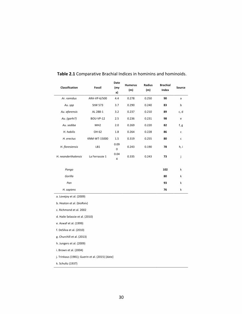
proximal forelimb length (humerus length), indexed to 100. Fossil evidence suggests that for
the first several million years of hominin evolution BI was variable but within the range of
means for extant great apes, between the lower limit of Gorilla (BI=80) and the upper limit of
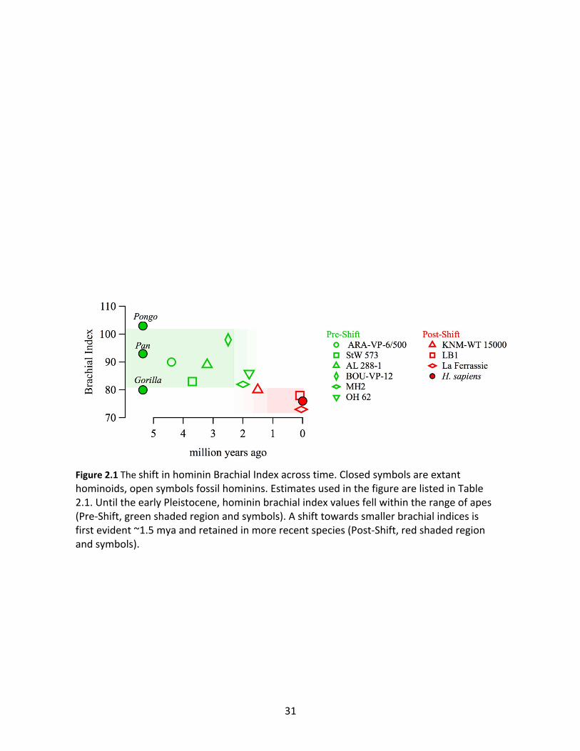
Pongo (BI=101), and mostly clustered between 82-90 (Table 2.1, Figure 2.1). Homo habilis (OH
62) may have had a BI of ~86, but the emergence of Homo erectus in Africa around 1.9 m.y.a.
was accompanied by a shift to a BI of ~80 (represented by KNM-WT 15000), at the edge of the
range of extant great apes (Fig. 1). BIs in the range of approximately 73 to 78 have since
persisted in other more recent species of the genus Homo including Homo sapiens (Fig. 1). This
shift in BI coincided with a suite of additional anatomical changes first evident in H. erectus
linked to the evolution of a hunter-gatherer way of life that included large day ranges,
endurance running, and throwing (Hawkes et al., 1997; Bramble and Lieberman, 2004; Robson
and Wood, 2008; Roach et al., 2014; Hawkes et al., 2018) Because apes have larger BIs than
humans and are generally adapted for arboreal locomotion, there is ongoing debate whether
30
Classification Fossil
Date
(my
a)
Humerus
(m)
Radius
(m)
Brachial
Index Source
Ar. ramidus ARA-VP-6/500 4.4 0.278 0.250 90 a
Au. spp StW 573 3.7 0.290 0.240 83 b
Au. afarensis AL 288-1 3.2 0.237 0.210 89 c, d
Au. (garhi?) BOU-VP-12 2.5 0.236 0.231 98 e
Au. sediba MH2 2.0 0.269 0.220 82 f, g
H. habilis OH 62 1.8 0.264 0.228 86 c
H. erectus KNM-WT-15000 1.5 0.319 0.255 80 c
H. floresiensis LB1 0.09
0 0.243 0.190 78 h, i
H. neanderthalensis La Ferrassie 1 0.04
4 0.335 0.243 73 j
Pongo 102 k
Gorilla 80 k
Pan 93 k
H. sapiens 76 k
a. Lovejoy et al. (2009)
b. Heaton et al. (bioRxiv)
c. Richmond et al. 2002
d. Haile Selassie et al. (2010)
e. Aswaf et al. (1999)
f. DeSilva et al. (2010)
g. Churchill et al. (2013)
h. Jungers et al. (2009)
i. Brown et al. (2004)
j. Trinkaus (1981); Guerin et al. (2015) [date]
k. Schultz (1937)
Table 2.1 Comparative Brachial Indices in hominins and hominoids.
31
Figure 2.1 The shift in hominin Brachial Index across time. Closed symbols are extant hominoids, open symbols fossil hominins. Estimates used in the figure are listed in Table 2.1. Until the early Pleistocene, hominin brachial index values fell within the range of apes (Pre-Shift, green shaded region and symbols). A shift towards smaller brachial indices is first evident ~1.5 mya and retained in more recent species (Post-Shift, red shaded region and symbols).
32
fossil BIs can provide diagnostic information about the behavior of extinct hominins (e.g. see
(Churchill et al., 2013))
There are several potential hypotheses to explain the shift to lower BIs in Homo. One
possible mechanism is developmental integration between the forelimb and hindlimb elements
(Young et al., 2010). If so, selection for relatively shorter distal hindlimbs would lead to shorter
distal forelimbs, and consequently a lower BI. One problem with this hypothesis is lack of
variation and evidence for directional change in the analogous hindlimb skeletal index (Crural
Index: distal hindlimb over proximal hindlimb) (Richmond et al., 2002a; Haile-Selassie et al.,
2010). Selection driven by thermoregulation has previously been hypothesized to contribute to
distal limb evolution (e.g. (Holliday, 1997)). However, in the hot, arid environments of Africa the
thermoregulation hypothesis predicts distal limb elements should get relatively longer, not
shorter as observed in the fossil record. Another potential hypothesis is that selection for
derived manual mechanical tasks such as tool making, and perhaps overhand throwing (Roach
and Lieberman, 2014; Roach et al., 2014), favored higher BIs. However, these and other tasks
that require acceleration of the hand would seemingly benefit from longer distal forelimbs
rather than shorter ones by transferring more momentum to the grasped object, although
quantitative tests of this mechanical hypothesis are lacking. Furthermore, the relationships
between distal forelimb length and the control or accuracy of manual tasks have not been
modeled to date.
Here we explore a final hypothesis for the directional shift towards smaller BIs: that
smaller BI benefits bipedal walking and running mechanics. During walking and running
humans swing their forelimbs in order to counterbalance the angular momentum of the
33
hindlimbs, increasing stability and reducing the energetic cost of locomotion (Elftman, 1939;
Hinrichs, 1987; Herr and Popovic, 2008; Umberger, 2008; Collins et al., 2009). The entire
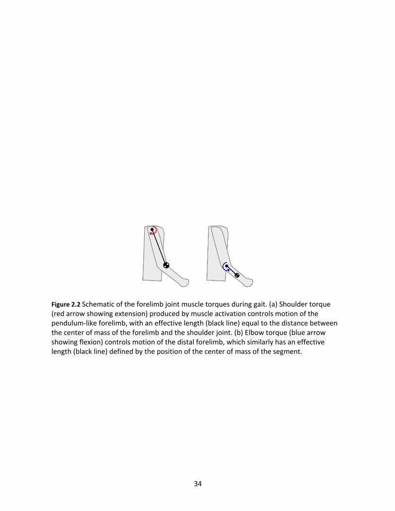
forelimb swings about the shoulder joint like a single pendulum under the control of a shoulder
muscle torque (τsho) produced by the deltoid and other muscles (Yegian et al., prepared). The
effective length of the single pendulum forelimb (Figure 2.2A) is the fundamental determinant
of how much muscular effort at the shoulder is required to swing the limb (Yegian et al.,
prepared). However, the forelimb is not a single pendulum because motion can also occur at
the elbow joint. In order to allow the forelimb to act like a single pendulum during gait the
elbow is kept mostly rigid by muscles (Figure 2.2B), resulting in an elbow muscle torque (τelb)
(Yegian et al., prepared). Muscle contractions needed to develop torques cost metabolic
energy, so both τsho and τelb contribute an unknown, but likely modest, amount to the cost of
locomotion.
Because only τsho contributes to counterbalancing momentum, morphology that reduces
τelb for a given τsho in theory provides an energetic benefit, and we hypothesize that forelimb
variants that produce this outcome might be favored by selection if substantial enough benefits
exist. The obvious candidate for such a mechanism is reduced length of the distal forelimb,
which reduces inertia. Rotational inertia of a segment is defined in the simplest case as mL2,
with m being the mass of the segment and L the length between the joint and the segment
center of mass. For a given angular motion of a segment about a joint, muscle torque and the
resulting energy cost are positively related to the rotational inertia of the segment. Reducing L
and moving the center of mass closer to the joint reduces inertia and consequently reduces the
muscle torque and energy cost of the motion. All else being equal, smaller BI values indicate a
34
Figure 2.2 Schematic of the forelimb joint muscle torques during gait. (a) Shoulder torque (red arrow showing extension) produced by muscle activation controls motion of the pendulum-like forelimb, with an effective length (black line) equal to the distance between the center of mass of the forelimb and the shoulder joint. (b) Elbow torque (blue arrow showing flexion) controls motion of the distal forelimb, which similarly has an effective length (black line) defined by the position of the center of mass of the segment.
35
relatively shorter distal forelimb, and therefore should reduce the relative magnitude and cost
of τelb during locomotion.
In addition to the hypothesis that a smaller BI reduces the joint torques generated
during walking and running, we also hypothesize that the benefit for running is greater than for
walking. While walking, humans tend to keep their elbows mostly straight, but while running
the elbows are usually bent to approximately 90° thus decreasing the forelimb’s effective
length (Yegian et al., Chapter 1). This bent elbow strategy, however, orients the distal forelimb
more horizontally and thus perpendicular to the gravitational force. Gravity acts to push the
elbow toward extension, and must be resisted by elbow muscles and τelb. In addition, the
magnitude of τelb compared to τsho is greater in running than in walking (Yegian et al., Chapter
1). Taken together, relatively shorter distal forelimbs likely provides a greater benefit for
running than walking.
To test the hypothesis that a shorter distal forelimb (i.e. smaller BI) decreases the
external moments generated at the shoulder and elbow in walking and even more so in running
we conducted an experiment using a within-subject design, artificially manipulating the distal
forelimb inertia of the participants by having them hold hand weights. Hand weights shift the
center of mass of the distal forelimb away from the elbow, increasing the effective length of the
segment and its inertia. Within-subjects design controlled for inter-subject variation in other
gait variables, while the inertial manipulation approach allowed for testing greater variation
than possible with a between-subjects comparative approach, increasing the resolution for
detecting a trend. Note that the experiment did not directly alter BIs between treatments
because BI is strictly defined as a skeletal ratio, but instead the hand weight conditions
36
produced an approximate heuristic of a larger BI and the resulting effect on τelb. However, the
hand weights also increased the inertia of the entire forelimb, which affects τsho. We therefore
normalized τelb by dividing by τsho, yielding a dimensionless normalized elbow muscle torque
(Telb) that accounts for the effect of the added mass on the swing dynamics of the whole
forelimb and the control of swing by the shoulder muscles. We then compared the magnitude
of each torque across the stride for normal walking and running to that with added distal
forelimb inertia, and compared the effect size of walking to that of running.
Methods and Materials
Eight humans (four males and four females, age: 26.6 years, s.d. 2.5, mass: 76.6 kg, s.d.
15.9) with no musculoskeletal injuries or illnesses were participants in this experiment. The
Harvard University Institutional Review Board approved the experiment, and all participants
provided informed consent. During the experiment participants walked and ran on a force
plate-instrumented treadmill (Bertec Corp., Columbus, Ohio) at speeds ranging between 1.30
m/s to 1.44 m/s for walking, and 2.90 m/s to 3.22 m/s for running. Treadmill speeds were
calculated individually by using dimensionless speeds (Froude numbers) of 0.2 for walking and
1.0 for running.
In order to test the effects of brachial inertia on walking and running mechanics, we
asked the participants to walk and run normally as well as with three pound (1.36 kg) weights in
each hand. Each participant therefore was measured during four experimental conditions in
random order: normal walking (W), walking with added mass (W+M), normal running (R), and
running with added mass (R+M). Each trial lasted three minutes, and data were collected during
37
the last minute after acclimatization. Modeling and data analysis were conducted using the Igor
Pro software platform (Wavemetrics, Lake Oswega, Oregon).
Data collection consisted of motion capture of the right arm during locomotion. Small
infrared reflective markers were taped to the skin over the following bony landmarks: radial
styloid process, ulnar styloid process, lateral humeral epicondyle, medial humeral epicondyle,
and acromion. Eight infrared cameras tracked the motions of the markers in three-dimensional
space at a sampling frequency of 200 Hz (Qualysis Motion Capture Systems, Gothenburg,
Sweden). When added mass was used, markers were placed on the ends of the hand weights.
The location of the wrist joint was defined as the midpoint between the styloid processes, the
elbow joint was defined as the midpoint between the humeral epicondyles, and the shoulder
joint was estimated to be 3.0 (females) or 3.5 cm (males) below the acromion marker (De Leva,
1996). The location of the added mass was defined as the midpoint of the hand weight. The
data were reduced to only sagittal plane motions, and the raw time series were filtered using a
zero-lag 10 Hz low pass binomial smoothing filter. In addition to the kinematic data, vertical
force traces were obtained to define start and endpoints of individual strides. Ten consecutive
strides were identified and averaged for each subject and condition.
The forelimb was modeled as a two-segment system consisting of the proximal forelimb
and the distal forelimb, with the latter including the hand. Shoulder (θsho) and elbow joint (θelb)
angles were calculated from the joint positions. Inertial properties of the arm segments were
estimated using individual subject measurements and standard anthropometric tables (De
Leva, 1996). In the added mass conditions the mass of the hand weight was included in the
inertia of the distal forelimb. Kinematics and inertia were then combined in a standard inverse
38
dynamics model (Winter, 2005) in order to obtain the muscle torques at the shoulder (τsho) and
elbow (τelb) joints. The magnitudes of the muscle torques (Dτelb and Dτsho, defined as the
difference between maximum and minimum torque during the stride) were extracted, and the
normalized elbow torque, Telb, was calculated as the ratio of Dτelb over Dτsho. In addition, the
effective length of the forelimb was calculated using the positions of the individual segment
masses and the shoulder joint. Inter-subject means of Dτelb, Dτsho, and Telb were compared
between the added mass conditions (W+M and R+M) and the normal conditions (W and R)
using repeated measures ANOVA with significance based on standard α=0.05. Linear regression
was used to confirm that Dτsho was directly related to effective forelimb length.
Results
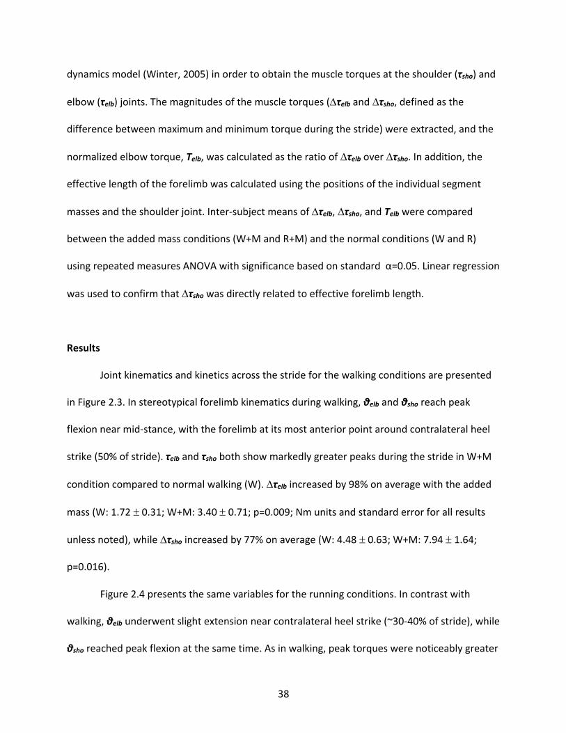
Joint kinematics and kinetics across the stride for the walking conditions are presented
in Figure 2.3. In stereotypical forelimb kinematics during walking, θelb and θsho reach peak
flexion near mid-stance, with the forelimb at its most anterior point around contralateral heel
strike (50% of stride). τelb and τsho both show markedly greater peaks during the stride in W+M
condition compared to normal walking (W). Dτelb increased by 98% on average with the added
mass (W: 1.72 ± 0.31; W+M: 3.40 ± 0.71; p=0.009; Nm units and standard error for all results
unless noted), while Dτsho increased by 77% on average (W: 4.48 ± 0.63; W+M: 7.94 ± 1.64;
p=0.016).
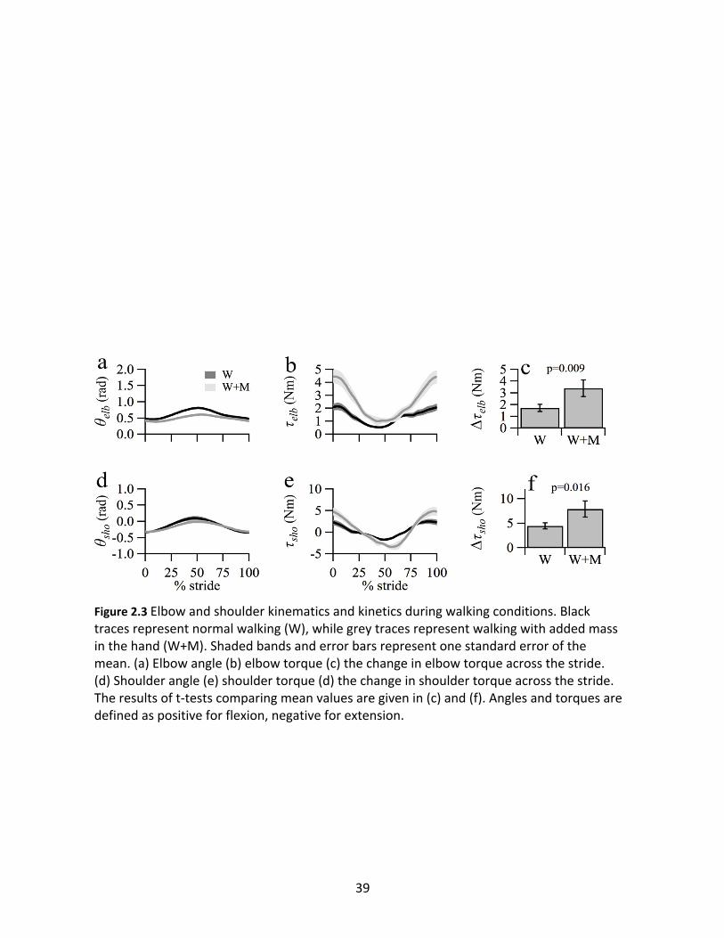
Figure 2.4 presents the same variables for the running conditions. In contrast with
walking, θelb underwent slight extension near contralateral heel strike (~30-40% of stride), while
θsho reached peak flexion at the same time. As in walking, peak torques were noticeably greater
39
Figure 2.3 Elbow and shoulder kinematics and kinetics during walking conditions. Black traces represent normal walking (W), while grey traces represent walking with added mass in the hand (W+M). Shaded bands and error bars represent one standard error of the mean. (a) Elbow angle (b) elbow torque (c) the change in elbow torque across the stride. (d) Shoulder angle (e) shoulder torque (d) the change in shoulder torque across the stride. The results of t-tests comparing mean values are given in (c) and (f). Angles and torques are defined as positive for flexion, negative for extension.
40
Figure 2.4 Elbow and shoulder kinematics and kinetics during running conditions. Black traces represent normal walking (R), while grey traces represent walking with added mass in the hand (R+M). Shaded bands and error bars represent one standard error of the mean. (a) Elbow angle (b) elbow torque (c) the change in elbow torque across the stride. (d) Shoulder angle (e) shoulder torque (d) the change in shoulder torque across the stride. The results of t-tests comparing mean values are given in (c) and (f). Angles and torques are defined as positive for flexion, negative for extension.
41
with added mass. When running with added mass, Dτelb significantly increased by 70%
compared to normal (R: 10.69 ± 1.72; R+M: 18.18 ± 2.75; p=0.004), a similar proportional
increase as walking. In contrast, adding mass to the forearm increased Dτsho by only 10% (R:
16.26 ± 3.28; R+M: 17.91 ± 2.82; p=0.019).
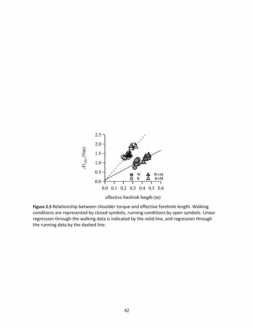
As predicted, linear regressions through individual subject data pooled by gait show
strong correlations between Dτsho and effective forelimb length (Figure 2.5). The trend through
the walking data had a slope of 2.65 Nm/m (p<0.001; r2=0.69), while the trend through the
running data had approximately twice the effect size, with a slope of 5.64 Nm/m (p<0.001;
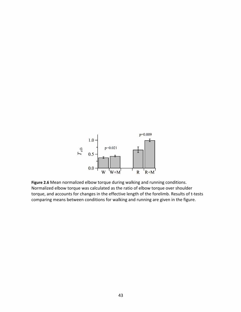
r2=0.74). There were significant increases in relative elbow muscle effort with added inertia for
both gaits (Figure 2.6) as measured by dimensionless elbow torque, Telb, which controlled for
the relationship between Dτsho and effective forelimb length. When walking, the added inertia
increased Telb by 16% (W: 0.38 ± 0.03; W+M: 0.44 ± 0.03; p=0.021). In contrast, added inertia
during running increased Telb by 52% (R: 0.66 ± 0.05, R+M: 1.00 ± 0.11, p=0.009). Therefore, the
same added inertia had ~3x the effect on elbow torque during running compared to walking.
Discussion
The experimental results presented here support the hypothesis that reduced distal
forelimb inertia benefits both walking and running by reducing muscle torque and presumably
effort required by elbow muscles during gait to counteract torques acting on the elbow.
Because distal forelimb inertia is positively related to BI, this provides support for the
hypothesis that reduced BI benefits both walking and running by reducing the effort needed to
stabilize the elbow. While our experiment illustrated the directional effect and mechanical
42
Figure 2.5 Relationship between shoulder torque and effective forelimb length. Walking conditions are represented by closed symbols, running conditions by open symbols. Linear regression through the walking data is indicated by the solid line, and regression through the running data by the dashed line.
43
Figure 2.6 Mean normalized elbow torque during walking and running conditions. Normalized elbow torque was calculated as the ratio of elbow torque over shoulder torque, and accounts for changes in the effective length of the forelimb. Results of t-tests comparing means between conditions for walking and running are given in the figure.
44
benefit of reduced BI, further research is needed to quantify the magnitude of the effect on the
cost of locomotion, or facilitate functional comparisons between hominins with different BIs.
However, by comparing the same inertial manipulation between walking and running in the
same subjects, we were able to observe an approximately three-fold larger benefit for running
compared to walking.
As predicted, we also observed larger τsho with added inertia in the hands. τsho costs
energy via shoulder muscle activation, similar to the elbow, and therefore our results imply that
reduced length of the entire forelimb also benefits walking and running. This finding is
consistent with a spring-pendulum model of forelimb swing during walking in humans, where
the shoulder muscles tune the natural frequency of the forelimb by adjusting the effective
stiffness of the shoulder (Yegian et al., prepared). Longer forelimbs require stiffer shoulders,
and consequently more muscle torque. Therefore, our results suggest that for a given hindlimb
length, longer forelimbs are more costly to swing during bipedal walking. However, the spring-
pendulum model suggests that stiffness is a non-linear function of forelimb length (Yegian et
al., prepared), suggesting that simple skeletal ratios like the intermembral index (forelimb
length divided by hindlimb length) may not be adequate heuristics for comparing walking
mechanics across different body sizes.
The experimental manipulation of adding 1.36 kg to the hands is a substantially larger
inertial change than any variation in BI observed in hominins, yet the manipulation increased
Telb by only 16% in walking and 52% in running. This difference suggests that any BI shift that
occurred in hominins had an even smaller proportional effect on elbow mechanics. Although
the energetic cost of swinging the forelimbs during walking and running is unknown, estimates
45
of hindlimb swing cost range between 10-30% of total cost of locomotion in humans and birds
(Marsh et al., 2004; Gottschall, 2005; Modica and Kram, 2005; Ellerby and Marsh, 2006; Doke et
al., 2007). Forelimb swing cost is likely a smaller portion of the total cost due to smaller torque
magnitudes at the shoulder and elbow compared to the hip and knee.
The benefit of a smaller BI in terms of proportional change to instantaneous locomotion
cost is likely quite small, yet even very small instantaneous energetic benefits can add up over
time and affect selection. For example, the gross daily cost of walking in contemporary Hadza
hunter-gatherers is estimated to be on average approximately 291 kCal for men and 126 kCal
for women (Pontzer et al., 2015), and the total locomotion cost is greater when running is
added to daily activity. If the BI shift that occurred in H. erectus yields a 1% savings in
locomotion energy cost, the savings for contemporary hunter-gatherers is in the order of
approximately 1-5 kCal each day. Over the course of a year, the incremental savings would add
up to roughly 1000 kCal, or several days’ worth of walking energy expenditure. We therefore
hypothesize that walking and running, especially for long distances, would have contributed to
selection for relatively shorter distal forelimbs in hominins.
Evolution of Brachial Index in Hominins
The last common ancestor of chimpanzees and hominins is most likely somewhat
chimpanzee-like in terms of morphology and locomotion, although there is ongoing debate on
this issue (Gebo, 1996; Richmond et al., 2002b; Thorpe et al., 2007; Lovejoy et al., 2009;
Pilbeam and Lieberman 2017). On average, chimpanzees exhibit Bis of approximately 93
(Schultz 1937), while ARA-VP-6/500, the oldest hominin in the dataset used in this study, is
46
estimated to have a similar BI of 90 (Lovejoy et al., 2009). Estimates for the specimens assigned
to Australopithecus and Homo habilis mostly range from 82 (MH2 ) to 88 (AL 288-1), with the
exception of BOU-VP-12 (98, although this large value may be the result of an underestimated
humerus length; Asfaw et al., 1999; Haile-Selassie et al., 2010). If ARA-VP-6/500 approximates
ancestral BI, the somewhat smaller values observed in Australopithecus and H. habilis may
reflect an initial shift driven by selection for human-like walking. There is evidence for modern
hindlimb and center-of-mass walking mechanics in the Laetoli footprints dated to 3.7 m.y.a.,
presumably created by australopiths (Raichlen et al., 2008; 2010; Crompton et al., 2012;
Dingwall et al., 2013). However, several of the early hominin forelimb bones are only preserved
in fragments (particularly AL 288-1, BOU-VP-12, and OH 62) and are prone to large
uncertainties in length estimations. With the lack of a clear ancestral BI value to compare to
and an uncertain fossil record, evidence for an initial shift driven by walking must be considered
weak.
Later fossil specimens assigned to Homo tend to have intact or fully reconstructed
forelimb long bones, and clearly show a shift to smaller BI values when compared to older
specimens with similarly complete long bones (i.e. StW 573 and MH2); later Homo species tend
to exhibit smaller BI than australopiths by approximately 5 to 15 points (Table 2.1). The timing
of the shift coincides with the evolution of the hunter-gatherer lifestyle and associated
locomotion patterns, namely long distance walking and endurance running (Bramble and
Lieberman, 2004), and when combined with the biomechanical evidence from this study
suggests that long distance walking and endurance running drove the shift. Of note, while LB1
shows evolutionary convergence to a body size and limb lengths similar to a small Au. afarensis
47
(AL 288-1), a typical hunter-gatherer BI of 78 was retained in Homo floresiensis (Brown et al.,
2004). This observation suggests that the shift in BI was not driven by scaling effects between
the forelimb elements across body size, and implies that the shift was instead driven by
selection.
The results of our experiment suggest that the derived, stable range of BIs between 70
and 80 in hominin hunter-gatherers are an adaptation to long distance walking and running.
This study provides the first mechanistic explanation for the observed shift in BI in hominins,
and highlights the benefit of using biomechanical experiments with modern humans to predict
directional effects of selection on hominin skeletal anatomy. Future research, however is
needed to estimate the actual energetic savings associated with forelimb shortening to
quantitatively compare performance effects with other activities such as climbing, throwing
and tool-making.
48
References
Asfaw B, White T, Lovejoy O, Latimer B, Simpson S, Suwa G. 1999. Australopithecus garhi: a new species of early hominid from Ethiopia. Science 284:629–635.
Bramble DM, Lieberman DE. 2004. Endurance running and the evolution of Homo. Nature 432:345–352.
Brown P, Sutikna T, Morwood MJ, Soejono RP, Jatmiko, Saptomo EW, Due RA. 2004. A new small-bodied hominin from the Late Pleistocene of Flores, Indonesia. Nature 431:1055–1061.
Churchill SE, Holliday TW, Carlson KJ, Jashashvili T, Macias ME, Mathews S, Sparling TL, Schmid P, de Ruiter DJ, Berger LR. 2013. The Upper Limb of Australopithecus sediba. Science 340:1233477–1233477.
Collins SH, Adamczyk PG, Kuo AD. 2009. Dynamic arm swinging in human walking. Proceedings of the Royal Society B: Biological Sciences 276:3679–3688.
Crompton RH, Pataky TC, Savage R, D'Aout K, Bennett MR, Day MH, Bates K, Morse S, Sellers WI. 2012. Human-like external function of the foot, and fully upright gait, confirmed in the 3.66 million year old Laetoli hominin footprints by topographic statistics, experimental footprint-formation and computer simulation. Journal of The Royal Society Interface 9:707–719.
DeSilva JM, Holt KG, Churchill SE, Carlson KJ, Walker CS, Zipfel B, Berger LR. 2013. The lower limb and mechanics of walking in Australopithecus sediba. Science 340:1232999–1232999.
Dingwall HL, Hatala KG, Wunderlich RE, Richmond BG. 2013. Hominin stature, body mass, and walking speed estimates based on 1.5 million-year-old fossil footprints at Ileret, Kenya. Journal of Human Evolution 64:556–568.
Doke J, Donelan JM, Kuo AD. 2007. Mechanics and energetics of swinging the human leg. Journal of Experimental Biology 210:2399–2399.
Elftman H. 1939. The function of the arms in walking. Human biology.
Ellerby DJ, Marsh RL. 2006. The energetic costs of trunk and distal-limb loading during walking and running in guinea fowl Numida meleagris: II. Muscle energy use as indicated by blood flow. Journal of Experimental Biology 209:2064–2075.
Gebo DL. 1996. Climbing, brachiation, and terrestrial quadrupedalism: historical precursors of hominid bipedalism. Am J Phys Anthropol 101:55–92.
Gottschall JS. 2005. Energy cost and muscular activity required for leg swing during walking. J
49
Appl Physiol 99:23–30.
Guérin G, Frouin M, Talamo S, Aldeias V, Bruxelles L, Chiotti L, Dibble HL, Goldberg P, Hublin J-J, Jain M, Lahaye C, Madelaine S, Maureille B, McPherron SJP, Mercier N, Murray AS, Sandgathe D, Steele TE, Thomsen KJ, Turq A. 2015. A multi-method luminescence dating of the Palaeolithic sequence of La Ferrassie based on new excavations adjacent to the La Ferrassie 1 and 2 skeletons. Journal of Archaeological Science 58:147–166.
Haile-Selassie Y, Latimer BM, Alene M, Deino AL, Gibert L, Melillo SM, Saylor BZ, Scott GR, Lovejoy CO. 2010. An early Australopithecus afarensis postcranium from Woranso-Mille, Ethiopia. Proc Natl Acad Sci USA 107:12121–12126.
Hawkes K, O'Connell J, Blurton Jones N. 2018. Hunter-gatherer studies and human evolution: A very selective review. Am J Phys Anthropol 165:777–800.
Hawkes K, O'Connell JF, Rogers L. 1997. The behavioral ecology of modern hunter-gatherers, and human evolution. Trends Ecol Evol (Amst) 12:29–32.
Herr H, Popovic M. 2008. Angular momentum in human walking. Journal of Experimental Biology 211:467–481.
Hinrichs RN. 1987. Upper Extremity Function in Running. II: Angular Momentum Considerations. Int J Sport Biomech 3:242–263.
Holliday TW. 1997. Postcranial evidence of cold adaptation in European Neandertals. Am J Phys Anthropol 104:245–258.
Jungers WL, Larson SG, Harcourt-Smith W, Morwood MJ, Sutikna T, Awe RD, Djubiantono T. 2009. Descriptions of the lower limb skeleton of Homo floresiensis. Journal of Human Evolution 57:538–554.
Lovejoy CO, Suwa G, Simpson SW, Matternes JH, White TD. 2009. The Great Divides: Ardipithecus ramidus Reveals the Postcrania of Our Last Common Ancestors with African Apes. Science 326:73–73, 100–106.
Marsh RL, Ellerby DJ, Carr JA, Henry HT, Buchanan CI. 2004. Partitioning the energetics of walking and running: swinging the limbs is expensive. Science 303:80–83.
Modica JR, Kram R. 2005. Metabolic energy and muscular activity required for leg swing in running. J Appl Physiol 98:2126–2131.
Pontzer H, Raichlen DA, Wood BM, Emery Thompson M, Racette SB, Mabulla AZP, Marlowe FW. 2015. Energy expenditure and activity among Hadza hunter-gatherers. Am J Hum Biol 27:628–637.
Raichlen DA, Gordon AD, Harcourt-Smith WEH, Foster AD, Haas WR. 2010. Laetoli Footprints
50
Preserve Earliest Direct Evidence of Human-Like Bipedal Biomechanics. PLoS ONE 5:e9769–6.
Raichlen DA, Pontzer H, Sockol MD. 2008. The Laetoli footprints and early hominin locomotor kinematics. Journal of Human Evolution 54:112–117.
Reno PL, De Gusta D, Serrat MA, Meindl RS, White TD, Eckhardt RB, Kuperavage AJ, Galik K, Lovejoy CO. 2005. Plio-Pleistocene Hominid Limb Proportions: Evolutionary Reversals or Estimation Errors? CURR ANTHROPOL 46:575–588.
Richmond BG, Aiello LC, Wood BA. 2002a. Early hominin limb proportions. Journal of Human Evolution 43:529–548.
Richmond BG, Begun DR, Strait DS. 2002b. Origin of human bipedalism: The knuckle-walking hypothesis revisited. Am J Phys Anthropol 116:70–105.
Roach NT, Lieberman DE. 2014. Upper body contributions to power generation during rapid, overhand throwing in humans. Journal of Experimental Biology 217:2139–2149.
Roach NT, Venkadesan M, Rainbow MJ, Lieberman DE. 2014. Elastic energy storage in the shoulder and the evolution of high-speed throwing in Homo. Nature 498:483–486.
Robson SL, Wood B. 2008. Hominin life history: reconstruction and evolution. Journal of Anatomy 212:394–425.
Schultz AH. 1937. Proportions, variability and asymmetries of the long bones of the limbs and the clavicles in man and apes. 9:281–328.
Thorpe SKS, Holder RL, Crompton RH. 2007. Origin of Human Bipedalism As an Adaptation for Locomotion on Flexible Branches. Science 316:1328–1331.
Trinkaus E. 1981. Neanderthal limb proportions and cold adaptation. In: Stringer C, editor. London: Taylor & Francis Ltd. p 187–224.
Umberger BR. 2008. Effects of suppressing arm swing on kinematics, kinetics, and energetics of human walking. J Biomech 41:2575–2580.
Yegian A, Lieberman DE, Gillinov S, Tucker Y. (2019). Straight arm walking, bent arm running: gait-specific elbow angles. Journal of Experimental Biology, 222:13.
Yegian A, Marsh RL, Sternad D, Hasson CJ. (prepared). Active dynamics of arm swing: frequency tuning and the role of muscles. Proceedings of the Royal Society B: Biological Sciences.
Young NM, Wagner GP, Hallgrimsson B. 2010. Development and the evolvability of human limbs. Proc Natl Acad Sci USA 107:3400–3405.
51
Chapter 3 - Functional scaling of forelimb swing mechanics during bipedal walking explains
the evolution of hominin limb proportions.
Abstract
The evolution of terrestrial bipedalism is a fundamental question in hominin evolution.
Australopiths had relatively longer forelimbs than later species in the genus Homo, often
interpreted as evidence for a shift in locomotion behavior from a more primitive mixed
arboreal/terrestrial strategy in Australopithecus to the economical, near-obligate bipedalism of
Homo. However, this interpretation does not take into context how size affects forelimb
function during walking. Using the biomechanical Swing Scaling Model that describes forelimb
swing mechanics during walking, we illustrate that isometrically scaled modern humans are not
functionally similar, as shoulder stiffness controlled by muscles likely increases with increasing
size. When limb proportions are scaled to similar shoulder stiffness, all but the earliest hominin
limb proportions fall within the Swing Scaling Model prediction. The shift to relatively shorter
forelimbs in Homo is mostly explained by an increase in body size, making a transition in
locomotion behavior unnecessary to explain the evolution of hominin limb proportions.
Introduction
Adaptations for terrestrial bipedalism evolved early in the hominin lineage as evidenced
by late Miocene and early Pliocene fossils (Crompton et al., 2008; Lovejoy et al., 2009c;
Zollikofer et al., 2005). The evolutionary pathway to economical, near-obligate terrestrial
bipedalism seen in modern humans is complicated by the observation that while many early
hominin species, such as australopithecines had features suggesting bipedal locomotion in the
52
hindlimb, forelimb features tended to be outside the ranges of human variation. This
observation has led to the common hypothesis that australopiths represent a tradeoff between
terrestrial bipedalism and arboreal locomotion, and longstanding debate as to when human-
like bipedalism evolved (Crompton et al., 2008; Crompton et al., 1998; Hunt, 1994; Jungers,
2009; Kramer and Eck, 2000; Lovejoy, 1988; Raichlen et al., 2010; Stern and Susman, 1983;
Ward, 2002).
Interlimb proportions serve as a rough indicator of locomotion behavior in extant
primates (Fleagle, 2013). Limb proportions are often expressed by the intermembral index (IMI,
(humer+radius)/(femur+tibia)*100), which compares the length of the forelimb to the length of
the hindlimb. Quadrupedal monkeys are generally constrained to values near 100 (i.e. equal
limb lengths), while in hominoids developmental constraints are relaxed allowing for the
evolution of large variation in IMI within the family (Young et al., 2010). Apes, which utilize
climbing/suspensory behavior to some degree, have IMI>100, while bipedal humans have IMI
values ~70 (Schultz, 1937). Within hominins, Ardipithecus and Australopithecus fossils indicate
IMI values above the range of human variation, and modern IMI values do not appear until the
advent of large-bodied Homo erectus in Africa (Holliday et al., 2018).
Relatively long forelimbs in small-bodied australopithecines, intermediate to apes and
humans, are generally interpreted as evidence that the transition to economical near-obligate
bipedalism characteristic of modern humans did not occur until the evolution of Homo (Hunt,
1994; Jungers, 2009; Stern and Susman, 1983). This interpretation rests on the observation that
IMI is independent of size in humans, indicating an isometric scaling relationship between the
limbs (Jungers, 2009). While limb lengths scale isometrically within the human species, it does
53
not necessarily follow that locomotion function and costs also scale isometrically and are
similar across the range of human stature. Although developmental constraints governing
interlimb proportions are relaxed in hominoids compared to quadrupedal monkeys, there are
still strong correlations between limb lengths within species (Young et al., 2010). Therefore,
independence between IMI and size in humans may be the result of underlying isometric
developmental integration between the limbs within a species rather than similar function
during bipedal gait, and scaling of similar function may follow an allometric relationship
between forelimb and hindlimb length.
In order to explore whether isometrically scaled human limbs are equivalent in function
during bipedal walking, and whether functional scaling can explain some of the variation in
hominin IMI, we introduce in this paper a novel biomechanical model of forelimb swing during
walking that incorporates the anatomical lengths of both the forelimbs and the hindlimbs. We
then use the Swing Scaling Model to test for a relationship between shoulder muscle torque
and size within a modern human sample and compare fossil hominin limb lengths to a limb
length scaling relationship based on functional similarity.
Swing Scaling Model
Forelimb swing during walking in humans serves the function of reducing the total
energetic cost of locomotion (Collins et al., 2009; Umberger, 2008). In each stride the forelimbs
swing in opposite timing to the movement of the hindlimbs, resulting in opposing angular
momenta about the vertical axis passing through the body’s center of mass (Collins et al., 2009;
Elftman, 1939; Herr and Popovic, 2008; Li et al., 2001). This counterbalancing mechanism
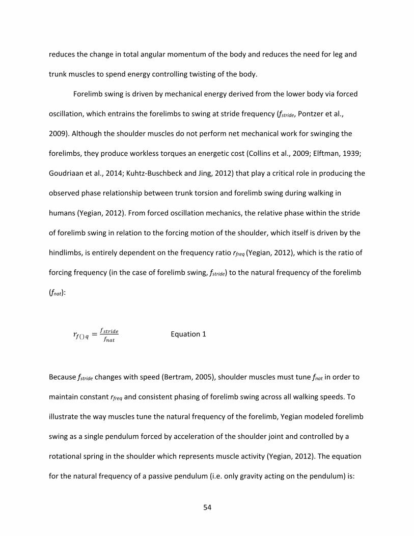
54
reduces the change in total angular momentum of the body and reduces the need for leg and
trunk muscles to spend energy controlling twisting of the body.
Forelimb swing is driven by mechanical energy derived from the lower body via forced
oscillation, which entrains the forelimbs to swing at stride frequency (fstride, Pontzer et al.,
2009). Although the shoulder muscles do not perform net mechanical work for swinging the
forelimbs, they produce workless torques an energetic cost (Collins et al., 2009; Elftman, 1939;
Goudriaan et al., 2014; Kuhtz-Buschbeck and Jing, 2012) that play a critical role in producing the
observed phase relationship between trunk torsion and forelimb swing during walking in
humans (Yegian, 2012). From forced oscillation mechanics, the relative phase within the stride
of forelimb swing in relation to the forcing motion of the shoulder, which itself is driven by the
hindlimbs, is entirely dependent on the frequency ratio rfreq (Yegian, 2012), which is the ratio of
forcing frequency (in the case of forelimb swing, fstride) to the natural frequency of the forelimb
(fnat):
&'()* =',-./01'23-
Equation 1
Because fstride changes with speed (Bertram, 2005), shoulder muscles must tune fnat in order to
maintain constant rfreq and consistent phasing of forelimb swing across all walking speeds. To
illustrate the way muscles tune the natural frequency of the forelimb, Yegian modeled forelimb
swing as a single pendulum forced by acceleration of the shoulder joint and controlled by a
rotational spring in the shoulder which represents muscle activity (Yegian, 2012). The equation
for the natural frequency of a passive pendulum (i.e. only gravity acting on the pendulum) is:
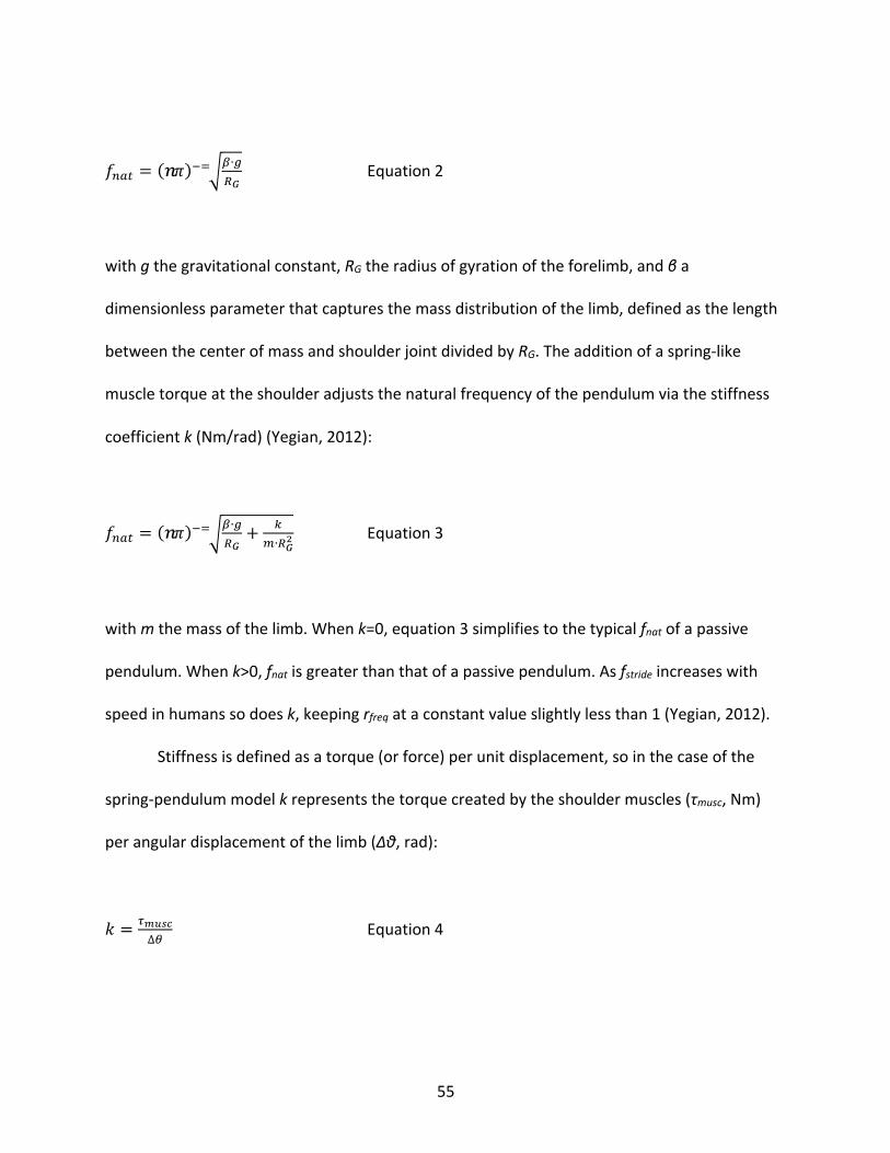
55
4567 = (2:)<=>?∙ABC
Equation 2
with g the gravitational constant, RG the radius of gyration of the forelimb, and β a
dimensionless parameter that captures the mass distribution of the limb, defined as the length
between the center of mass and shoulder joint divided by RG. The addition of a spring-like
muscle torque at the shoulder adjusts the natural frequency of the pendulum via the stiffness
coefficient k (Nm/rad) (Yegian, 2012):
4567 = (2:)<=>?∙ABC+ E
F∙BCG Equation 3
with m the mass of the limb. When k=0, equation 3 simplifies to the typical fnat of a passive
pendulum. When k>0, fnat is greater than that of a passive pendulum. As fstride increases with
speed in humans so does k, keeping rfreq at a constant value slightly less than 1 (Yegian, 2012).
Stiffness is defined as a torque (or force) per unit displacement, so in the case of the
spring-pendulum model k represents the torque created by the shoulder muscles (τmusc, Nm)
per angular displacement of the limb (Δθ, rad):
H = IJK,L∆N
Equation 4
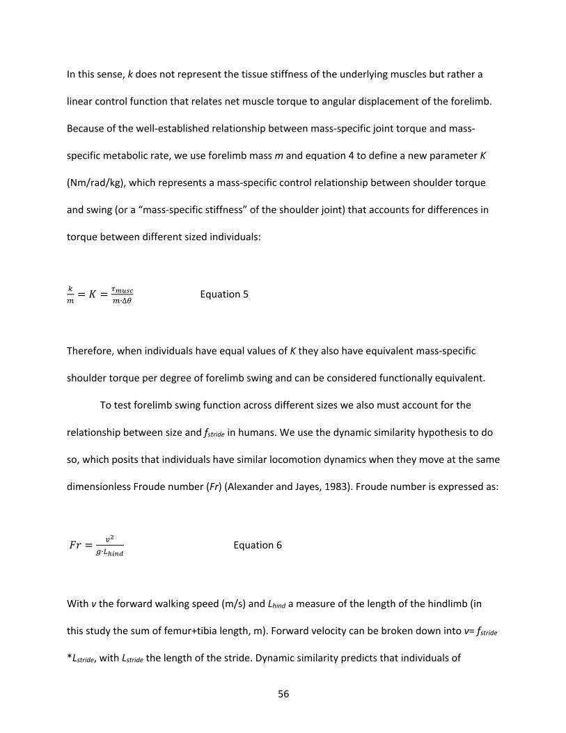
56
In this sense, k does not represent the tissue stiffness of the underlying muscles but rather a
linear control function that relates net muscle torque to angular displacement of the forelimb.
Because of the well-established relationship between mass-specific joint torque and mass-
specific metabolic rate, we use forelimb mass m and equation 4 to define a new parameter K
(Nm/rad/kg), which represents a mass-specific control relationship between shoulder torque
and swing (or a “mass-specific stiffness” of the shoulder joint) that accounts for differences in
torque between different sized individuals:
EF= O = IJK,L
F∙∆N Equation 5
Therefore, when individuals have equal values of K they also have equivalent mass-specific
shoulder torque per degree of forelimb swing and can be considered functionally equivalent.
To test forelimb swing function across different sizes we also must account for the
relationship between size and fstride in humans. We use the dynamic similarity hypothesis to do
so, which posits that individuals have similar locomotion dynamics when they move at the same
dimensionless Froude number (Fr) (Alexander and Jayes, 1983). Froude number is expressed as:
P& = QG
A∙RS/20 Equation 6
With v the forward walking speed (m/s) and Lhind a measure of the length of the hindlimb (in
this study the sum of femur+tibia length, m). Forward velocity can be broken down into v= fstride
*Lstride, with Lstride the length of the stride. Dynamic similarity predicts that individuals of
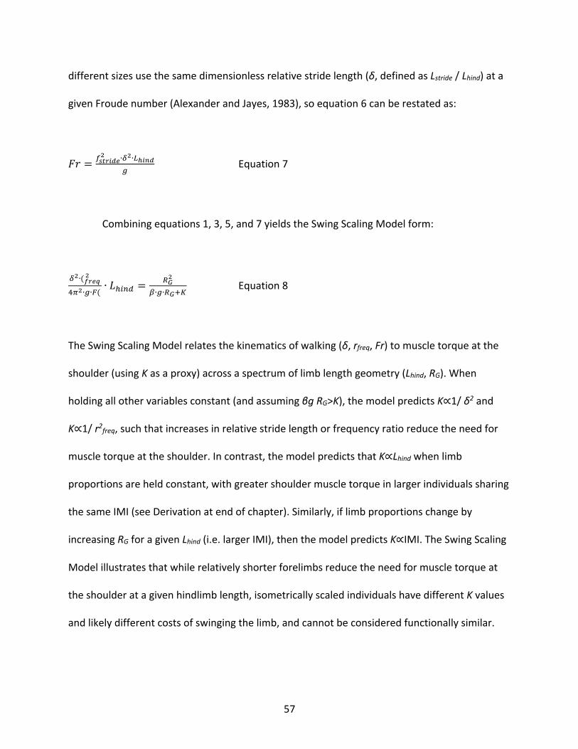
57
different sizes use the same dimensionless relative stride length (δ, defined as Lstride / Lhind) at a
given Froude number (Alexander and Jayes, 1983), so equation 6 can be restated as:
P& = ',-./01G ∙TG∙RS/20
A Equation 7
Combining equations 1, 3, 5, and 7 yields the Swing Scaling Model form:
TG∙(U.1VG
WXG∙A∙Y(∙ Z[\5] =
BCG
?∙A∙BC^_ Equation 8
The Swing Scaling Model relates the kinematics of walking (δ, rfreq, Fr) to muscle torque at the
shoulder (using K as a proxy) across a spectrum of limb length geometry (Lhind, RG). When
holding all other variables constant (and assuming βg RG>K), the model predicts K∝1/ δ2 and
K∝1/ r2freq, such that increases in relative stride length or frequency ratio reduce the need for
muscle torque at the shoulder. In contrast, the model predicts that K∝Lhind when limb
proportions are held constant, with greater shoulder muscle torque in larger individuals sharing
the same IMI (see Derivation at end of chapter). Similarly, if limb proportions change by
increasing RG for a given Lhind (i.e. larger IMI), then the model predicts K∝IMI. The Swing Scaling
Model illustrates that while relatively shorter forelimbs reduce the need for muscle torque at
the shoulder at a given hindlimb length, isometrically scaled individuals have different K values
and likely different costs of swinging the limb, and cannot be considered functionally similar.
58
In this study we utilized two versions of the Swing Scaling Model: an isometric model,
which fixed limb proportions to the IMI of modern humans and allowed K to vary, and a
constant K model, which fixed K to the mean value in modern humans and allowed limb
proportions to vary. The isometric model therefore provides a scaling relationship driven by
similar shape as in humans, while the constant K model provides a scaling relationship driven by
functional similarity to compare fossil hominin limbs to modern humans.
Methods and Materials
Experimental Data Collection
Walking mechanics were recorded in a sample of fifteen humans (6 female 9 male; age:
24±3 years; mass: 74±13 kg; height: 1.77±0.09 m; Lhind: 0.86±0.05 m; IMI=70±2). The
experiment was granted prior approval by the Harvard University IRB, and all participants gave
informed consent prior to participation. Standing hip height was measured unshod, and
hindlimb length Lhind, defined as femur+tibia length, was estimated as 95% of standing hip
height based on published anthropometric data and casts of human hindlimb bones (Winter,
2009). Treadmill speed was calculated for each individual using Fr=0.215 and Lhind using
equation 6. Skeletal forelimb length Lfore, defined as humerus+radius length, was estimated
from external measurements of the participants.
Walking trials were conducted shod on a split-belt treadmill (Bertec Corp., Columbus,
OH, USA) for five minutes. Reflective markers were placed on the right acromion, humeral
epicondyles, and styloid processes of the ulna and radius, and were tracked during the trials
using eight infrared cameras operating at 200Hz (Qualysis Motion Capture Systems, Goteborg,
59
Sweden). The wrist and elbow joint centers were estimated as the midpoints between the
relevant markers, and the shoulder joint center was estimated as 3 (females) or 3.5 cm (males)
below the acromion (De Leva, 1996). Kinematic time series were processed in Igor Pro
(Wavemetrics, Lake Oswega, OR, USA) using a 10 Hz lowpass filter. Anthropometric tables (De
Leva, 1996) and participant metrics were used to estimate the masses of the limb segments and
the positions of the centers of mass. A standard inverse dynamics model consisting of two
forelimb segments was used to calculate τmusc at the shoulder (Winter, 2009).
The two forelimb segments were then combined into a single pendulum arm by
calculating the position of the center-of-mass of the entire forelimb, and then using the
position to calculate the angular displacement of the forelimb Δθ. The total rotational inertia
about the shoulder joint, I (kg*m2), was calculated using the segment mass positions and
inertial distributions determined from the kinematic time series and anthropometric tables,
respectively. RG was calculated from I and the total mass of the limb, m, using the formula:
ab = > cF Equation 9
Shoulder stiffness k was then determined as the slope of τmusc vs. Δθ, with model parameter K
the ratio k/m. rfreq was estimated from equations 1 and 3, while δ was estimated using treadmill
speed, stride frequency, and Lhind.
Swing Scaling Model
60
The Swing Scaling Model is quantitatively expressed in equation 8. Mean values of δ and
rfreq from the experimental sample were used for the model under the assumptions that they
are independent of size (i.e. Lhind). We tested these assumptions by running linear regressions of
the two variables against Lhind in Matlab (Mathworks, Natick, MA, USA), with the hypothesis
that each slope=0. We also assumed that IMI is independent of size in humans (Jungers, 2009),
and similarly tested the assumption using linear regression of the related measure Lfore / Lhind
(equivalent to IMI/100) vs. Lhind.
To test our hypotheses, two versions of the forelimb swing model were constructed.
The “isometric model” was created by fixing the model’s limb proportions based on the
geometry of the human sample (IMI=70) and solving for K across the range of Lhind. In this way
the isometric model illustrates changes in function, measured as K, across size in isometric
humans. The “constant K model” was created by fixing the value of K to the experimental mean
and then solving for Lfore across the range of Lhind. The constant K model allows relative limb
proportions to change with size as long as K remains the same, and represents predicted
human limb proportions under a scaling relationship constrained to a single value of K. In order
to produce a rough prediction interval for the geometry of the constant K model we substituted
the upper and lower 95% prediction interval limits from the experimental sample K into the
model, yielding Lfore length curves that approximately represent the limits of limb length
combinations that are consistent with modern human forelimb swing mechanics.
We also estimated K values for limb lengths observed in other hominoids, specifically
nine fossil hominin specimens and African apes (see next section), and compared the values to
the variation in K observed in the human sample. We note that the hominoid K values are not
61
estimates of the actual values of K during bipedal walking in the specimens, but rather are used
to test whether or not modern humans with those limb lengths would yield similar K values as
actual modern humans.
Hominin Fossil Limb Lengths
We compiled a dataset of nine fossil hominin limb lengths by obtaining estimates from
the literature for four limb bones: humerus, radius, femur, and tibia (Table 3.1). When multiple
estimates for a bone were available in the literature we chose those preferred by the authors,
or the midpoint when narrow ranges were given. For a few specimens tibia lengths were
estimated using femur lengths and an assumed Crural Index (tibia/femur*100) of 83, following
previous studies (Haile-Selassie et al., 2010; Holliday et al., 2018; Richmond et al., 2002). There
is no known radius associated with the adult Dmanisi Homo erectus, so we estimated its length
using a Brachial Index (radius/humerus*100) value of 80 taken from its conspecific, KNM-WT-
15000.
The radius of AL 288-1 has been estimated to be as short as 174 mm (Richmond et al.,
2002) and as long as 215 mm (Asfaw et al., 1999), alongside further estimates of 181 mm
(Holliday et al., 2018) and 206 mm (Kimbel et al., 1994). We chose the midpoint of the short
and long estimates (195 mm) as the value for this study. OH 62 presents the greatest challenge
for estimating limb lengths due to the highly fragmented nature of the femur and tibia. The
commonly cited femur length of 280 mm is almost certainly too short (Reno et al., 2005).
McHenry used a somewhat longer estimate of 315 mm in his study estimating body mass of
fossil specimens (McHenry, 1991), while Haeusler and McHenry speculated a range of lengths
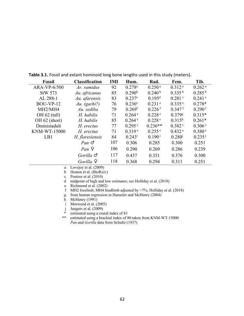
62
Table 3.1. Fossil and extant hominoid long bone lengths used in this study (meters).
Fossil Classification IMI Hum. Rad. Fem. Tib. ARA-VP-6/500 Ar. ramidus 92 0.278a 0.250 a 0.312 a 0.262 a StW 573 Au. africanus 85 0.290b 0.240 b 0.335 b 0.285 b AL 288-1 Au. afarensis 83 0.237c 0.195d 0.281 c 0.241 c BOU-VP-12 Au. (garhi?) 76 0.236e 0.231 e 0.335 e 0.278* MH2/MH4 Au. sediba 79 0.269f 0.226 f 0.347 f 0.290 f OH 62 (tall) H. habilis 71 0.264 e 0.228 e 0.379g 0.315* OH 62 (short) H. habilis 85 0.264 e 0.228 e 0.315h 0.261* Dminisiadult H. erectus 77 0.295 c 0.236** 0.382 c 0.306 c
KNM-WT-15000 H. erectus 71 0.319 e 0.255 e 0.432 e 0.380 e LB1 H. floresiensis 84 0.243i 0.190 i 0.280j 0.235 j Pan ♂ 107 0.306 0.285 0.300 0.251 Pan ♀ 106 0.290 0.269 0.286 0.239 Gorilla ♂ 117 0.437 0.351 0.376 0.300 Gorilla ♀ 118 0.368 0.294 0.311 0.251
a Lovejoy et al. (2009) b Heaton et al. (BioRxiv) c Pontzer et al. (2010) d midpoint of high and low estimates; see Holliday et al. (2018) e Richmond et al. (2002) f MH2 forelimb, MH4 hindlimb adjusted by +7%; Holliday et al. (2018) g from human regression in Haeusler and McHenry (2004) h McHenry (1991) i Morwood et al. (2005) j Jungers et al. (2009) * estimated using a crural index of 83 ** estimated using a brachial index of 80 taken from KNM-WT-15000 Pan and Gorilla data from Schultz (1937)
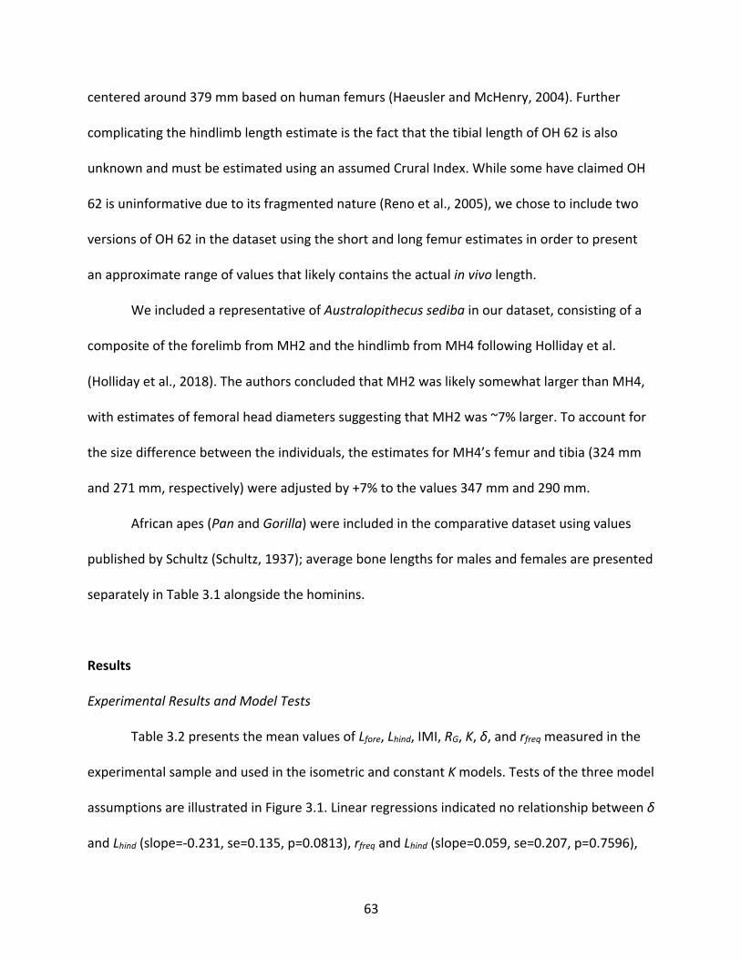
63
centered around 379 mm based on human femurs (Haeusler and McHenry, 2004). Further
complicating the hindlimb length estimate is the fact that the tibial length of OH 62 is also
unknown and must be estimated using an assumed Crural Index. While some have claimed OH
62 is uninformative due to its fragmented nature (Reno et al., 2005), we chose to include two
versions of OH 62 in the dataset using the short and long femur estimates in order to present
an approximate range of values that likely contains the actual in vivo length.
We included a representative of Australopithecus sediba in our dataset, consisting of a
composite of the forelimb from MH2 and the hindlimb from MH4 following Holliday et al.
(Holliday et al., 2018). The authors concluded that MH2 was likely somewhat larger than MH4,
with estimates of femoral head diameters suggesting that MH2 was ~7% larger. To account for
the size difference between the individuals, the estimates for MH4’s femur and tibia (324 mm
and 271 mm, respectively) were adjusted by +7% to the values 347 mm and 290 mm.
African apes (Pan and Gorilla) were included in the comparative dataset using values
published by Schultz (Schultz, 1937); average bone lengths for males and females are presented
separately in Table 3.1 alongside the hominins.
Results
Experimental Results and Model Tests
Table 3.2 presents the mean values of Lfore, Lhind, IMI, RG, K, δ, and rfreq measured in the
experimental sample and used in the isometric and constant K models. Tests of the three model
assumptions are illustrated in Figure 3.1. Linear regressions indicated no relationship between δ
and Lhind (slope=-0.231, se=0.135, p=0.0813), rfreq and Lhind (slope=0.059, se=0.207, p=0.7596),
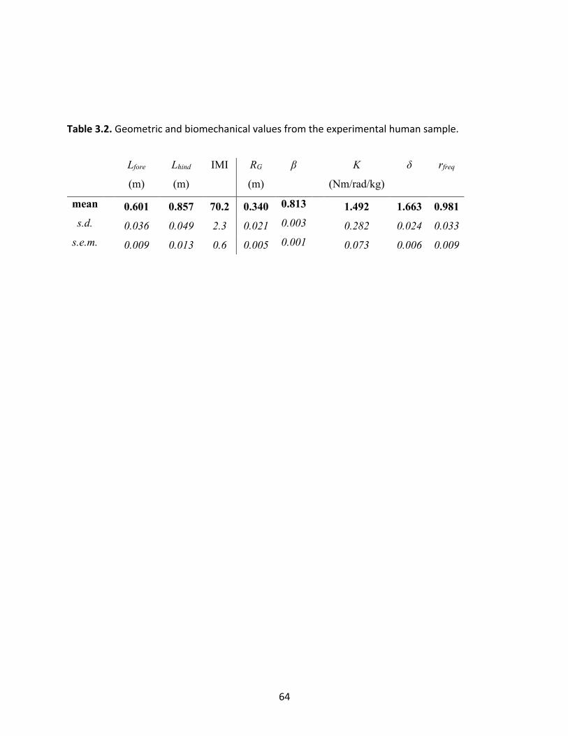
64
Table 3.2. Geometric and biomechanical values from the experimental human sample.
Lfore
(m)
Lhind
(m)
IMI
RG
(m)
β
K
(Nm/rad/kg)
δ
rfreq
mean 0.601 0.857 70.2 0.340 0.813 1.492 1.663 0.981 s.d. 0.036 0.049 2.3 0.021 0.003 0.282 0.024 0.033 s.e.m. 0.009 0.013 0.6 0.005 0.001 0.073 0.006 0.009
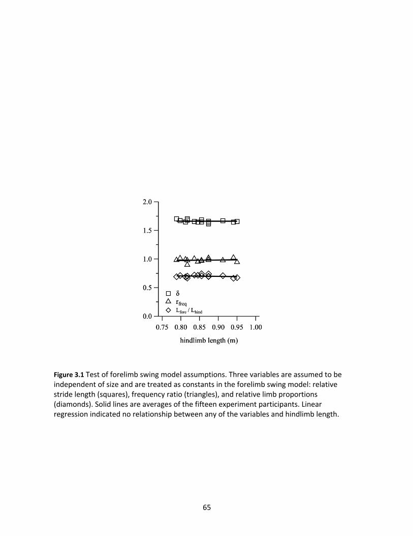
65
Figure 3.1 Test of forelimb swing model assumptions. Three variables are assumed to be independent of size and are treated as constants in the forelimb swing model: relative stride length (squares), frequency ratio (triangles), and relative limb proportions (diamonds). Solid lines are averages of the fifteen experiment participants. Linear regression indicated no relationship between any of the variables and hindlimb length.
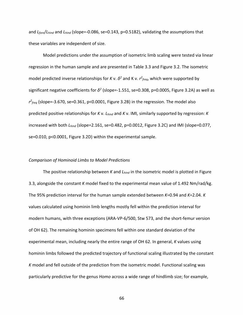
66
and Lfore/Lhind and Lhind (slope=-0.086, se=0.143, p=0.5182), validating the assumptions that
these variables are independent of size.
Model predictions under the assumption of isometric limb scaling were tested via linear
regression in the human sample and are presented in Table 3.3 and Figure 3.2. The isometric
model predicted inverse relationships for K v. δ2 and K v. r2freq, which were supported by
significant negative coefficients for δ2 (slope=-1.551, se=0.308, p=0.0005, Figure 3.2A) as well as
r2freq (slope=-3.670, se=0.361, p<0.0001, Figure 3.2B) in the regression. The model also
predicted positive relationships for K v. Lhind and K v. IMI, similarly supported by regression: K
increased with both Lhind (slope=2.161, se=0.482, p=0.0012, Figure 3.2C) and IMI (slope=0.077,
se=0.010, p<0.0001, Figure 3.2D) within the experimental sample.
Comparison of Hominoid Limbs to Model Predictions
The positive relationship between K and Lhind in the isometric model is plotted in Figure
3.3, alongside the constant K model fixed to the experimental mean value of 1.492 Nm/rad/kg.
The 95% prediction interval for the human sample extended between K=0.94 and K=2.04. K
values calculated using hominin limb lengths mostly fell within the prediction interval for
modern humans, with three exceptions (ARA-VP-6/500, Stw 573, and the short-femur version
of OH 62). The remaining hominin specimens fell within one standard deviation of the
experimental mean, including nearly the entire range of OH 62. In general, K values using
hominin limbs followed the predicted trajectory of functional scaling illustrated by the constant
K model and fell outside of the prediction from the isometric model. Functional scaling was
particularly predictive for the genus Homo across a wide range of hindlimb size; for example,
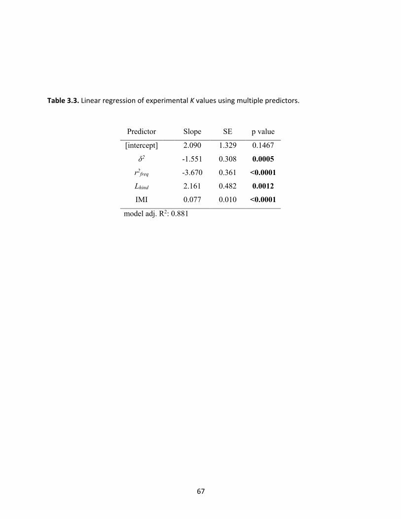
67
Table 3.3. Linear regression of experimental K values using multiple predictors.
Predictor Slope SE p value
[intercept] 2.090 1.329 0.1467
δ2 -1.551 0.308 0.0005 r2freq -3.670 0.361 <0.0001 Lhind 2.161 0.482 0.0012 IMI 0.077 0.010 <0.0001
model adj. R2: 0.881
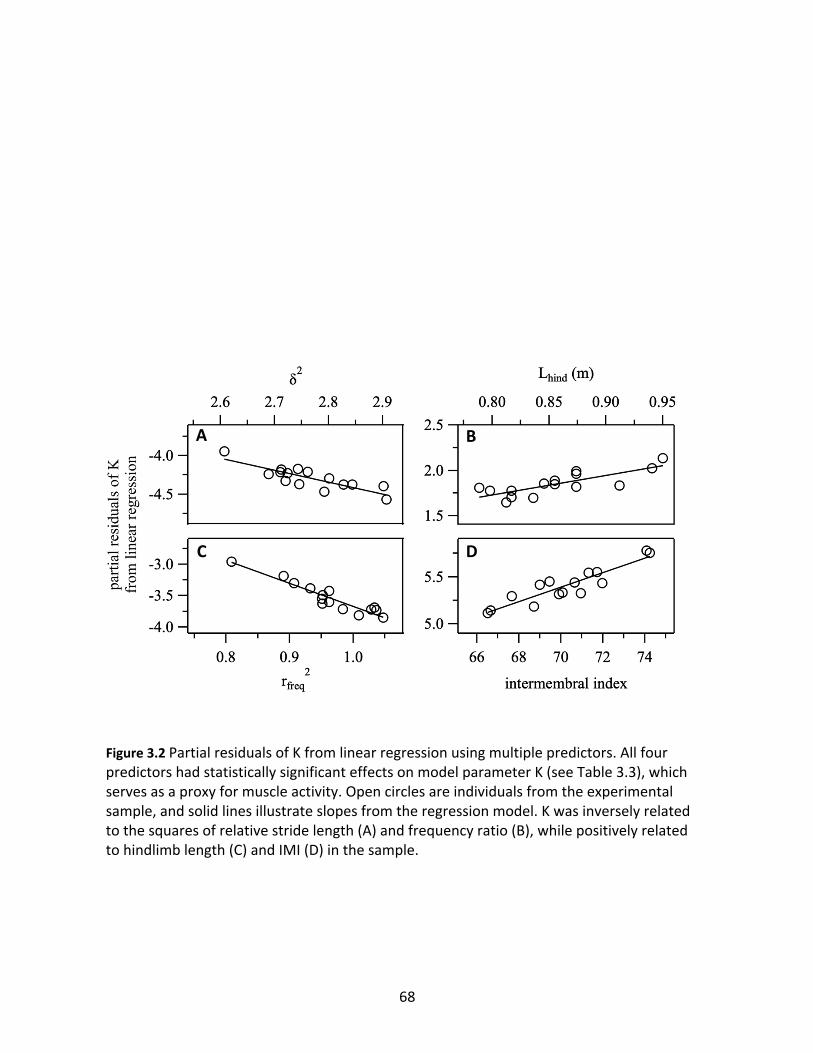
68
Figure 3.2 Partial residuals of K from linear regression using multiple predictors. All four predictors had statistically significant effects on model parameter K (see Table 3.3), which serves as a proxy for muscle activity. Open circles are individuals from the experimental sample, and solid lines illustrate slopes from the regression model. K was inversely related to the squares of relative stride length (A) and frequency ratio (B), while positively related to hindlimb length (C) and IMI (D) in the sample.
A B
C D
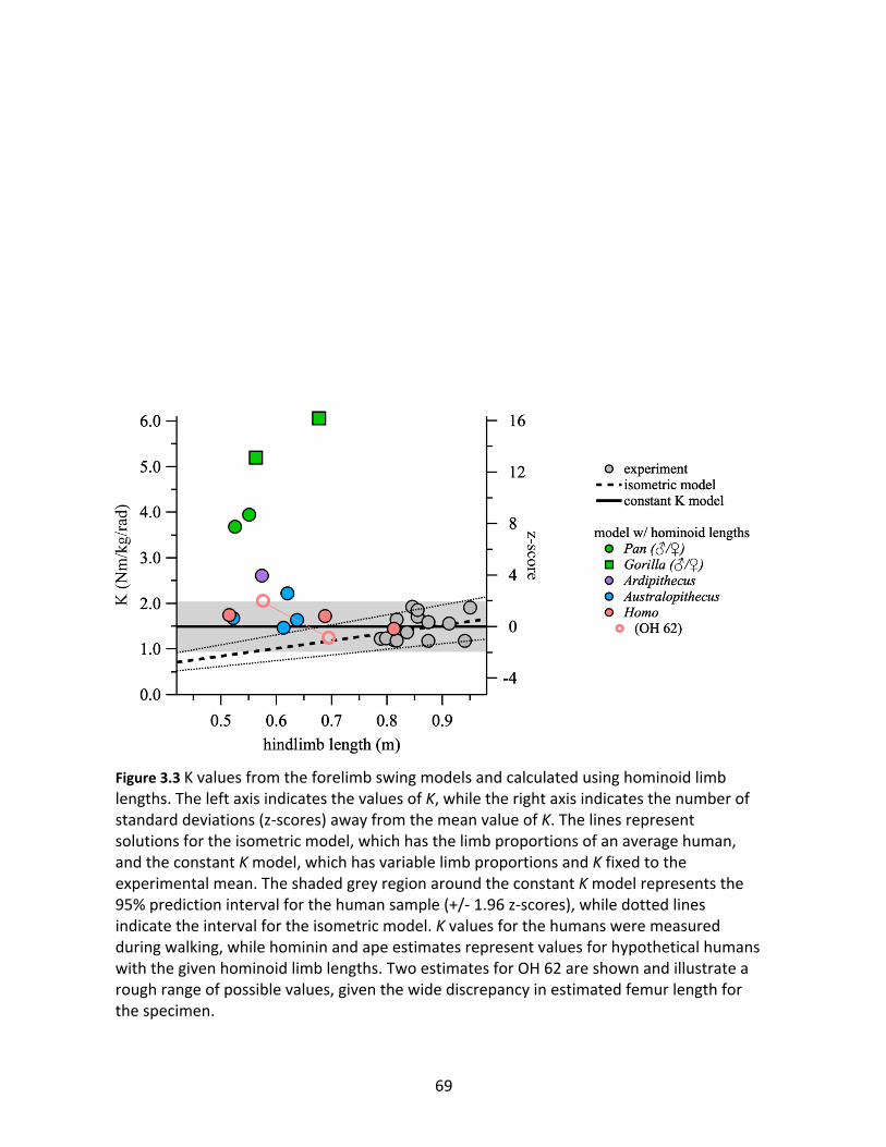
69
Figure 3.3 K values from the forelimb swing models and calculated using hominoid limb lengths. The left axis indicates the values of K, while the right axis indicates the number of standard deviations (z-scores) away from the mean value of K. The lines represent solutions for the isometric model, which has the limb proportions of an average human, and the constant K model, which has variable limb proportions and K fixed to the experimental mean. The shaded grey region around the constant K model represents the 95% prediction interval for the human sample (+/- 1.96 z-scores), while dotted lines indicate the interval for the isometric model. K values for the humans were measured during walking, while hominin and ape estimates represent values for hypothetical humans with the given hominoid limb lengths. Two estimates for OH 62 are shown and illustrate a rough range of possible values, given the wide discrepancy in estimated femur length for the specimen.
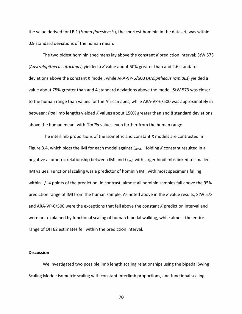
70
the value derived for LB 1 (Homo floresiensis), the shortest hominin in the dataset, was within
0.9 standard deviations of the human mean.
The two oldest hominin specimens lay above the constant K prediction interval; StW 573
(Australopithecus africanus) yielded a K value about 50% greater than and 2.6 standard
deviations above the constant K model, while ARA-VP-6/500 (Ardipithecus ramidus) yielded a
value about 75% greater than and 4 standard deviations above the model. StW 573 was closer
to the human range than values for the African apes, while ARA-VP-6/500 was approximately in
between: Pan limb lengths yielded K values about 150% greater than and 8 standard deviations
above the human mean, with Gorilla values even farther from the human range.
The interlimb proportions of the isometric and constant K models are contrasted in
Figure 3.4, which plots the IMI for each model against Lhind. Holding K constant resulted in a
negative allometric relationship between IMI and Lhind, with larger hindlimbs linked to smaller
IMI values. Functional scaling was a predictor of hominin IMI, with most specimens falling
within +/- 4 points of the prediction. In contrast, almost all hominin samples fall above the 95%
prediction range of IMI from the human sample. As noted above in the K value results, StW 573
and ARA-VP-6/500 were the exceptions that fell above the constant K prediction interval and
were not explained by functional scaling of human bipedal walking, while almost the entire
range of OH 62 estimates fell within the prediction interval.
Discussion
We investigated two possible limb length scaling relationships using the bipedal Swing
Scaling Model: isometric scaling with constant interlimb proportions, and functional scaling
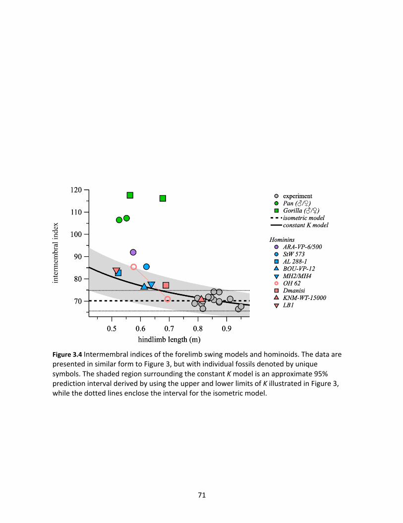
71
Figure 3.4 Intermembral indices of the forelimb swing models and hominoids. The data are presented in similar form to Figure 3, but with individual fossils denoted by unique symbols. The shaded region surrounding the constant K model is an approximate 95% prediction interval derived by using the upper and lower limits of K illustrated in Figure 3, while the dotted lines enclose the interval for the isometric model.
72
with constant K when walking. Experimental data collected from modern humans indicated that
isometric scaling of the limbs yields a relationship between K and size measured by hindlimb
length, with larger individuals requiring larger K values and presumably greater cost of swing
(Figures 3.2 and 3.3). In contrast, functional scaling yields a change in limb proportions across
size, with larger individuals requiring relatively shorter forelimbs in order to produce the same
forelimb swing mechanics as smaller individuals (Figure 3.4).
Our results shed new light on the origins of the genus Homo and the evolution of
modern limb proportions in the Pleistocene. The emergence of Homo coincided with smaller
IMI values compared to Australopithecus, exemplified by the human-like IMI of Homo erectus.
The appearance of modern limb proportions in H. erectus has previously been thought to
represent a transition from mixed locomotion behavior in Australopithecus, including
climbing/suspension, to near-obligate terrestrial bipedalism in Homo (Fleagle, 2013; Hunt,
1994; Jungers, 2009; Stern and Susman, 1983). The observed shift to smaller IMI also coincided
with a shift towards larger body size, and the evolution of endurance running and the hunter-
gatherer way of life (Bramble and Lieberman, 2004). The results of this study indicate that
functional scaling of human bipedal walking mechanics explains most of the IMI shift from
Australopithecus to Homo, suggesting that the relatively short forelimbs of H. erectus were
driven by the evolution of longer hindlimbs and larger body sizes compared to Pliocene
hominins rather than a transition in locomotion behavior.
Homo floresiensis provides an evolutionary test case for the hypothesis that the modern
IMI observed in Homo was driven by evolution of body size rather than a transition in
locomotion behavior. H. floresiensis was a very short-statured and very recent species found on
73
the island of Flores in Indonesia (Brown et al., 2004), which likely descended from a large-
bodied H. erectus population (Kaifu et al., 2011; Kubo et al., 2013) yet had similar IMI as a short-
statured Australopithecus afarensis (Lucy, AL 288-1). There is ongoing debate as to whether
australopith-like features in H. floresiensis, such as the IMI of LB1, are symplesiomorphs
retained from an early Homo ancestor that underwent a previously unknown migration out of
Africa (Argue et al., 2017; Jungers et al., 2016), or evolutionary convergence. The results of this
study suggest that the australopith-like IMI of H. floresiensis and the human like IMI of H.
erectus lie along the same functional scaling relationship shared with modern humans; just as a
shift from the large IMI of Lucy (83) to the human value in KNM-WT-15000 (71) can be linked to
the shift to large body size, a shift back to an ancestral IMI in LB1 (84) can be linked to a reversal
to small body size. Therefore, the limb proportions of H. floresiensis are most simply explained
by a H. erectus ancestor that converged on the limb proportions of Lucy due to functional
scaling of bipedal forelimb swing mechanics.
The fossil specimens assigned to Australopithecus and Ardipithecus that were used in
this study tend to be more fragmentary than the Homo specimens (excluding OH 62), with
more uncertainty in their IMI estimates. Three australopiths fell within the prediction interval of
the Swing Scaling Model using the limb estimates in Table 3.1, while the most complete
australopith postcranium (StW 573, Au. africanus) lay above the prediction interval. These
results suggest that the forelimb swing mechanics of Au. afarensis (Lucy), Au. garhi (BOU-VP-
12/1), and Au. sediba (MH2/MH4 composite) were comparable to modern humans, and that
Au. africanus likely had somewhat greater muscle activation at the shoulder. In general,
comparisons of the hominins to the model prediction shows that the relationship between size
74
and IMI must be accounted for when interpreting forelimb length as a signal of locomotion
behavior in fossils. The postcranial anatomy of Ardipithecus ramidus (ARA-VP-6/500) contains
multiple lines of evidence pointing to a walking gait unlike modern humans (Lovejoy et al.,
2009a; Lovejoy et al., 2009c; Suwa et al., 2009), making it unsurprising that the K value and IMI
for this specimen lay the furthest away from the human prediction interval. Ardipithecus is
likely the best example of a mixed locomotion strategy in hominins (Lovejoy et al., 2009b;
White et al., 2009), with the evolution of a more modern walking gait in Australopithecus. The
Laetoli footprints, dated to 3.6 million years ago, provide some evidence that human-like
walking was present in the middle Pliocene australopiths (Raichlen et al., 2010; Raichlen et al.,
2008), which is further supported in this study by the human-like K estimated for Lucy (3.2
m.y.a.)
Precise comparisons of estimated shoulder muscle torque were limited by the use of K
as a proxy. Doing so assumed that the ratio PCSA* dmusc /m was constant across all individuals,
which is likely true on average in isometrically scaled humans. However, interspecific variation
in musculotendon anatomy and forelimb mass may cause error when using K as the compared
variable. Therefore, this study should be considered a first order comparison, with further
estimations of fossil shoulder anatomy and limb mass distribution necessary for more precise
estimates of muscle torque.
Intriguingly, the Swing Scaling Model may also shed light on bipedal theropod dinosaur
limb scaling. A recent study comparing Lfore and Lhind to snout-vent length (SVL) in non-avian
theropods yielded the scaling relationships Lfore∝SVL0.70 and Lhind∝SVL0.88 (Dececchi and Larsson,
2013), which suggests that Lfore∝Lhind0.80. Although forelimb function during bipedal locomotion
75
in dinosaurs has not been explored before, and the forelimbs may have had no function during
bipedal gait, if muscle torque at the shoulder was a relevant component of the cost of
locomotion the same principles that contributed to negative allometry in hominin limb scaling
may have also contributed to negative allometry in non-avian theropod limb scaling.
We used the Swing Scaling Model to investigate forelimb swing during walking in
hominins in this study, but the model equation describes the dynamics of a forced pendulum in
general, and therefore may be applicable to hindlimb swing. Hip torque during hindlimb swing
has previously been approximated as spring-like in a passive dynamic walking model (Kuo,
2001), analogous to the spring-like torque in the shoulder in this study. It has long been known
that animals with distally distributed hindlimb mass (i.e. long hindlimb RG during swing), such as
non-cursorial species, take longer relative strides compared to animals with the mass
concentrated towards the hip (Alexander and Jayes, 1983; Raichlen et al., 2013). The Swing
Scaling Model shows that while longer RG increases K at the joint (e.g. K∝IMI in Figure 3.2D),
longer relative strides can offset the increase to an extent (e.g. K∝1/ δ2 in Figure 3.2A).
Therefore, longer relative strides in non-cursorial animals may be due to conservation of swing
cost across a diverse phylogeny.
Conclusions
Although human limb proportions scale isometrically, with constant IMI across all body
sizes (Figure 3.1), muscle torque at the shoulder increases with increasing hindlimb length
(Figures 3.2 and 3.3). Humans are therefore not functionally similar when it comes to forelimb
swing mechanics during walking, and the isometric relationship between limb lengths may be
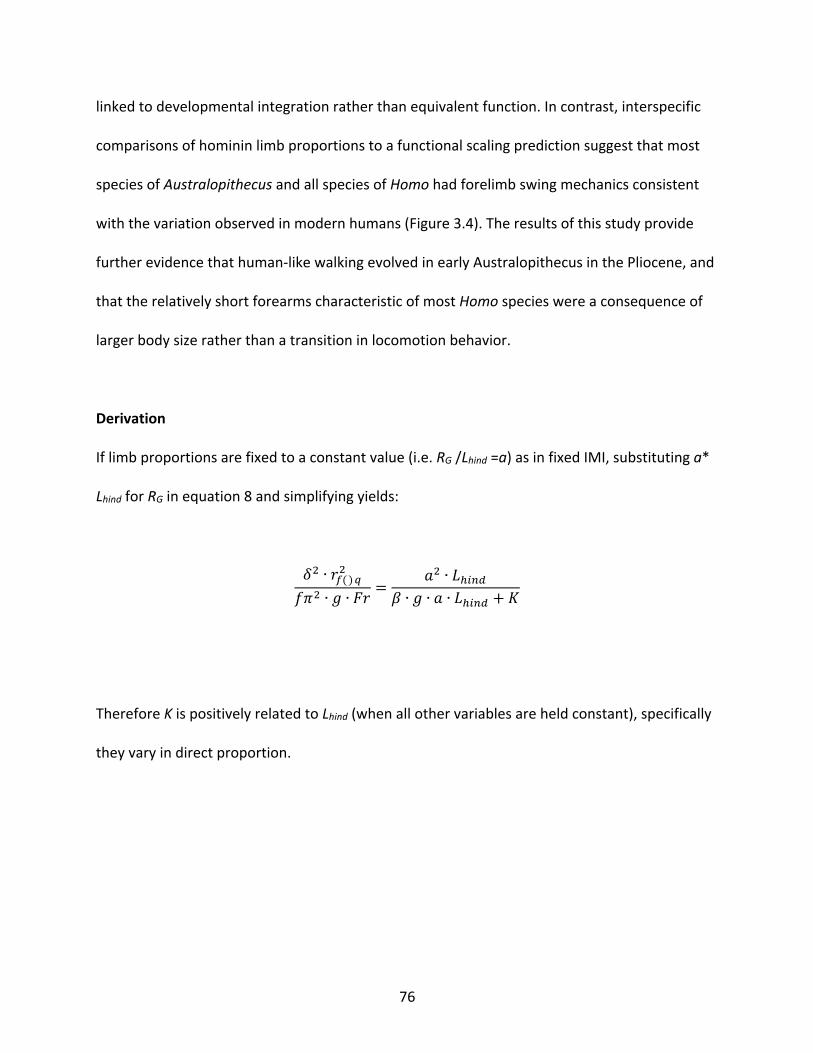
76
linked to developmental integration rather than equivalent function. In contrast, interspecific
comparisons of hominin limb proportions to a functional scaling prediction suggest that most
species of Australopithecus and all species of Homo had forelimb swing mechanics consistent
with the variation observed in modern humans (Figure 3.4). The results of this study provide
further evidence that human-like walking evolved in early Australopithecus in the Pliocene, and
that the relatively short forearms characteristic of most Homo species were a consequence of
larger body size rather than a transition in locomotion behavior.
Derivation
If limb proportions are fixed to a constant value (i.e. RG /Lhind =a) as in fixed IMI, substituting a*
Lhind for RG in equation 8 and simplifying yields:
de ∙ &'()*e
4:e ∙ g ∙ P& =he ∙ Z[\5]
i ∙ g ∙ h ∙ Z[\5] + O
Therefore K is positively related to Lhind (when all other variables are held constant), specifically
they vary in direct proportion.
77
References
Alexander, R. M. and Jayes, A. S. (1983). A dynamic similarity hypothesis for the gaits of quadrupedal mammals. Journal of Zoology.
Argue, D., Groves, C. P., Lee, M. S. Y. and Jungers, W. L. (2017). The affinities of Homo
floresiensis based on phylogenetic analyses of&nbsp;cranial, dental, and postcranial characters. Journal of Human Evolution 107, 107-133.
Asfaw, B., White, T., Lovejoy, O., Latimer, B., Simpson, S. and Suwa, G. (1999).
Australopithecus garhi: a new species of early hominid from Ethiopia. Science (New York, N.Y.) 284, 629-635.
Bertram, J. E. A. (2005). Constrained optimization in human walking: cost minimization and gait
plasticity. Journal of Experimental Biology 208, 979-991. Bramble, D. M. and Lieberman, D. E. (2004). Endurance running and the evolution of Homo.
Nature 432, 345-352. Brown, P., Sutikna, T., Morwood, M. J., Soejono, R. P., Jatmiko, Saptomo, E. W. and Due, R. A.
(2004). A new small-bodied hominin from the Late Pleistocene of Flores, Indonesia. Nature 431, 1055-1061.
Collins, S. H., Adamczyk, P. G. and Kuo, A. D. (2009). Dynamic arm swinging in human walking.
Proceedings of the Royal Society B: Biological Sciences 276, 3679-3688. Crompton, R. H., Vereecke, E. E. and Thorpe, S. K. S. (2008). Locomotion and posture from the
common hominoid ancestor to fully modern hominins, with special reference to the last common panin/hominin ancestor. Journal of anatomy 212, 501-543.
Crompton, R. H., Weijie, L. and Günther, M. (1998). The mechanical effectiveness of erect and
“bent-hip, bent-knee” bipedal walking inAustralopithecus afarensis. Journal of Human Evolution 35, 55-74.
De Leva, P. and de Leva, P. (1996). Adjustments to Zatsiorsky-Seluyanov's segment
inertia parameters. Journal of biomechanics 29, 1223-1230. Dececchi, T. A. and Larsson, H. C. E. (2013). Body and limb size cissociation at the origin of
birds: uncoupling allometric constraints across a macroevolutionary transition. Evolution 67, 2741-2752.
Elftman, H. (1939). The function of the arms in walking. Human biology.
78
Fleagle, J. G. (2013). Primate Adaptation and Evolution. San Diego: Academic Press. Goudriaan, M., Jonkers, I., van Dieën, J. H. and Bruijn, S. M. (2014). Arm swing in human
walking: What is their drive? Gait & Posture 40, 321-326. Haeusler, M. and McHenry, H. M. (2004). Body proportions of Homo habilis reviewed. Journal
of Human Evolution 46, 433-465. Haile-Selassie, Y., Latimer, B. M., Alene, M., Deino, A. L., Gibert, L., Melillo, S. M., Saylor, B. Z.,
Scott, G. R. and Lovejoy, C. O. (2010). An early Australopithecus afarensis postcranium from Woranso-Mille, Ethiopia. Proceedings of the National Academy of Sciences of the United States of America 107, 12121-12126.
Heaton, J. L., Pickering, T. R., Carlson, K. J., Crompton, R. H., Jashashvili, T., Beaudet, A.,
Bruxelles, L., Kuman, K., Heile, A. J., Stratford, D. et al. The Long Limb Bones of the StW 573 Australopithecus Skeleton from Sterkfontein Member 2: Descriptions and Proportions. bioRxiv.
Herr, H. and Popovic, M. (2008). Angular momentum in human walking. Journal of
Experimental Biology 211, 467-481. Holliday, T. W., Churchill, S. E., Carlson, K. J., DeSilva, J. M., Schmid, P., Walker, C. S. and
Berger, L. R. (2018). Special Issue: Australopithecus sediba --- Body size and proportions of Australopithecus sediba. PaleoAnthropology, 406-422.
Hunt, K. D. (1994). The evolution of human bipedality: ecology and functional morphology.
Journal of Human Evolution 26, 183-202. Jungers, W. L. (2009). Interlimb Proportions in Humans and Fossil Hominins: Variability and
Scaling eds. F. E. Grine J. G. Fleagle and R. E. Leakey), pp. 93-98. Jungers, W. L., Grabowski, M., Hatala, K. G. and Richmond, B. G. (2016). The evolution of body
size and shape in the human career. Philosophical Transactions of the Royal Society B: Biological Sciences 371, 20150247-10.
Kaifu, Y., Baba, H., Sutikna, T., Morwood, M. J., Kubo, D., Saptomo, E. W., Jatmiko, Awe, R. D.
and Djubiantono, T. (2011). Craniofacial morphology of Homo floresiensis: Description, taxonomic affinities, and evolutionary implication. Journal of Human Evolution 61, 644-682.
Kimbel, W. H., Johanson, D. C. and Rak, Y. (1994). The first skull and other new discoveries of
Australopithecus afarensis at Hadar, Ethiopia. Nature 368, 449-451.
79
Kramer, P. A. and Eck, G. G. (2000). Locomotor energetics and leg length in hominid bipedality. Journal of Human Evolution 38, 651-666.
Kubo, D., Kono, R. T. and Kaifu, Y. (2013). Brain size of Homo floresiensis and its evolutionary
implications. Proceedings of the Royal Society B: Biological Sciences 280, 20130338. Kuhtz-Buschbeck, J. P. and Jing, B. (2012). Activity of upper limb muscles during human
walking. Journal of Electromyography and Kinesiology 22, 199-206. Kuo, A. D. (2001). A Simple Model of Bipedal Walking Predicts the Preferred Speed–Step Length
Relationship. Journal of Biomechanical Engineering 123, 264-6. Li, Y., Wang, W. and Crompton, R. H. (2001). Free vertical moments and transverse forces in
human walking and their role in relation to arm-swing. Journal of Experimental …. Lovejoy, C. O. (1988). Evolution of human walking. Scientific American 259, 118-125. Lovejoy, C. O., Simpson, S. W., White, T. D., Asfaw, B. and Suwa, G. (2009a). Careful Climbing
in the Miocene: The Forelimbs of Ardipithecus ramidus and Humans Are Primitive. Science (New York, N.Y.) 326, 70.
Lovejoy, C. O., Suwa, G., Simpson, S. W., Matternes, J. H. and White, T. D. (2009b). The Great
Divides: Ardipithecus ramidus Reveals the Postcrania of Our Last Common Ancestors with African Apes. Science (New York, N.Y.) 326, 73.
Lovejoy, C. O., Suwa, G., Spurlock, L., Asfaw, B. and White, T. D. (2009c). The Pelvis and Femur
of Ardipithecus ramidus: The Emergence of Upright Walking. Science (New York, N.Y.) 326, 71.
McHenry, H. M. (1991). Femoral lengths and stature in Plio-Pleistocene hominids. American
journal of physical anthropology 85, 149-158. Morwood, M. J., Brown, P., Jatmiko, Sutikna, T., Saptomo, E. W., Westaway, K. E., Due, R. A.,
Roberts, R. G., Maeda, T., Wasisto, S. et al. (2005). Further evidence for small-bodied hominins from the Late Pleistocene of Flores, Indonesia. Nature 437, 1012-1017.
Pontzer, H., Holloway, J. H., Raichlen, D. A. and Lieberman, D. E. (2009). Control and function
of arm swing in human walking and running. Journal of Experimental Biology 212, 894-894.
Pontzer, H., Rolian, C., Rightmire, G. P., Jashashvili, T., de León, M. S. P., Lordkipanidze, D. and
Zollikofer, C. P. E. (2010). Locomotor anatomy and biomechanics of the Dmanisi hominins. Journal of Human Evolution 58, 492-504.
80
Raichlen, D. A., Gordon, A. D., Harcourt-Smith, W. E. H., Foster, A. D. and Haas, W. R. (2010). Laetoli Footprints Preserve Earliest Direct Evidence of Human-Like Bipedal Biomechanics. PLoS ONE 5, e9769-6.
Raichlen, D. A., Pontzer, H. and Shapiro, L. J. (2013). A new look at the Dynamic Similarity
Hypothesis: the importance of swing phase. Biology Open 2, 1032-1036. Raichlen, D. A., Pontzer, H. and Sockol, M. D. (2008). The Laetoli footprints and early hominin
locomotor kinematics. Journal of Human Evolution 54, 112-117. Reno, P. L., De Gusta, D., Serrat, M. A., Meindl, R. S., White, T. D., Eckhardt, R. B., Kuperavage,
A. J., Galik, K. and Lovejoy, C. O. (2005). Plio-Pleistocene Hominid Limb Proportions: Evolutionary Reversals or Estimation Errors? Current Anthropology 46, 575-588.
Richmond, B. G., Aiello, L. C. and Wood, B. A. (2002). Early hominin limb proportions. Journal
of Human Evolution 43, 529-548. Schultz, A. H. (1937). Proportions, variability and asymmetries of the long bones of the limbs
and the clavicles in man and apes. 9, 281-328. Stern, J. T. and Susman, R. L. (1983). The locomotor anatomy of Australopithecus afarensis.
American journal of physical anthropology 60, 279-317. Suwa, G., Asfaw, B., Kono, R. T., Kubo, D., Lovejoy, C. O. and White, T. D. (2009). The
Ardipithecus ramidus Skull and Its Implications for Hominid Origins. Science (New York, N.Y.) 326, 68.
Umberger, B. R. (2008). Effects of suppressing arm swing on kinematics, kinetics, and
energetics of human walking. Journal of biomechanics 41, 2575-2580. Ward, C. V. (2002). Interpreting the posture and locomotion ofAustralopithecus afarensis:
Where do we stand? American journal of physical anthropology 119, 185-215. White, T. D., Asfaw, B., Beyene, Y., Haile-Selassie, Y., Lovejoy, C. O., Suwa, G. and
WoldeGabriel, G. (2009). Ardipithecus ramidus and the Paleobiology of Early Hominids. Science (New York, N.Y.) 326, 64.
Winter, D. A. (2009). Biomechanics and Motor Control of Human Movement. Hoboken, NJ:
Wiley. Yegian, A. K. (2012). The role of muscles in arm swing and thoracic rotation during walking.
Northeastern University, thesis.
81
Young, N. M., Wagner, G. P. and Hallgrimsson, B. (2010). Development and the evolvability of human limbs. Proceedings of the National Academy of Sciences of the United States of America 107, 3400-3405.
Zollikofer, C. P. E., Ponce de León, M. S., Lieberman, D. E., Guy, F., Pilbeam, D., Likius, A.,
Mackaye, H. T., Vignaud, P. and Brunet, M. (2005). Virtual cranial reconstruction of Sahelanthropus tchadensis. Nature 434, 755-759.
82
Conclusions
The first three chapters of this thesis directly tested hypotheses about how anatomy
affects function during bipedal locomotion in order to better understand the evolution of
hominin forelimbs, focusing on the length of the distal segment and the overall length as the
relevant anatomical characters. In summary, I found that distal forelimb length affects bipedal
gait via elbow mechanics, with longer distal forelimbs increasing elbow muscle torque and
presumably the cost of forelimb swing during walking and running. Notably, the effect on elbow
torque is more pronounced in running compared to walking, by about three-fold. Overall
forelimb length also affects function, as longer limbs increase shoulder muscle torque in
modern humans. However, a human-based mechanical model shows that almost all of the
hominins, including all australopiths, had forelimb and hindlimb lengths that are consistent with
modern human walking dynamics. In the context of bipedalism, the shift to shorter distal
forelimbs and shorter relative forelimbs in Homo erectus can be explained by the evolution of
endurance running and long hindlimbs respectively.
Whether or not tradeoffs existed in the forelimb between bipedalism and other
behaviors like climbing remains unknown, and selection for non-locomotion tasks such as tool
making may have also played a role in the length shifts observed in the fossil record. In order to
test hypotheses about tradeoffs with bipedalism, climbing and tool making will need to be
described in a similar mechanical framework as bipedalism in this thesis. Climbing, tool making,
throwing, etc. can all be considered skilled tasks, with an optimal set of dynamics that can be
learned. Experience or skill at the relevant task must be a critical consideration when designing
experiments on biomechanics of human movement to test hypotheses about human evolution.
83
Implications for Understanding Hominin Evolution
To date, the standard story of hominin evolution is that an intermediate form of bipedal
walking evolved in Ardipithecus and Australopithecus, with the final form of bipedal walking
and running evolving in the genus Homo. This dissertation challenges the standard story by
showing that major changes to the hominin forelimbs, namely the decrease in relative distal
forelimb length and the decrease in relative forelimb length overall, can be explained mostly by
the evolution of endurance running and larger body size in the genus Homo.
In light of the results presented here, I argue for a new paradigm when viewing the
evolution of hominins and the fossil record: that human-like walking evolved early on by the
time of Australopithecus, with endurance running added as an ecologically relevant gait with
the advent of Homo erectus. The previous analytic approach of comparing fossil hominin
specimens to chimpanzees and humans and using analogy to interpret extinct gait has flaws
when function cannot be so easily interpolated from the geometry of fossil bones. Rather, a
functional approach that uses biomechanical modeling to assess how variation found in the
fossil record would affect the costs of bipedal walking and running is the most fruitful approach
to understanding hominin locomotion behavior going forward.
Related Documents