Evolution of Exchangeable Copper and Relative Exchangeable Copper through the Course of Wilson’s Disease in the Long Evans Cinnamon Rat Franc ¸ oise Schmitt 1,2 , Guillaume Podevin 2 , Joe ¨ l Poupon 3 , Je ´ro ˆ me Roux 4 , Pierre Legras 4 , Jean- Marc Trocello 5,6 , France Woimant 5,6 , Olivier Lapre ´ vote 3,7 , Tuan Huy NGuyen 1 , Souleiman El Balkhi 3,6,8 * 1 INSERM UMR1064, Jean Monnet Hospital, Nantes, France, 2 HIFIH - Pediatric Hepatogastroenterology Team, University Hospital of Angers, Angers, France, 3 Laboratory of Biological Toxicology, Lariboisie `re Hospital, APHP, Paris, France, 4 SCAHU, Animal Facility, Medicine University of Angers, Angers, France, 5 Department of Neurology, Lariboisie ` re Hospital, APHP, Paris, France, 6 Centre national de Maladie Rare Wilson, Lariboisie ` re Hospital, APHP, Paris, France, 7 Analytical and Experimental Toxicology (C- TAC), Faculty of Pharmacy, University Paris Descartes, Sorbonne Paris Cite ´, Paris, France, 8 INSERM U1144, Variability of the Response to Psychotropic Drugs, Paris Descartes University, Sorbonne Paris Cite ´, Faculty of Pharmacy, Paris, France Abstract Background: Wilson’s disease (WD) is an inherited disorder of copper metabolism leading to liver failure and/or neurological impairment. Its diagnosis often remains difficult even with genetic testing. Relative exchangeable copper (REC) has recently been described as a reliable serum diagnostic marker for WD. Methodology/Principal Findings: The aim of this study was to validate the use of REC in the Long Evans Cinnamon (LEC) rat, an animal model for WD, and to study its relevance under different conditions in comparison with conventional markers. Two groups of LEC rats and one group of Long-Evans (LE) rats were clinically and biologically monitored from 6 to 28 weeks of age. One group of LEC rats was given copper-free food. The other groups had normal food. Blood samples were collected each month and different serum markers for WD (namely ceruloplasmin oxidase activity, exchangeable copper (CuEXC), total serum copper and REC) and acute liver failure (serum transaminases and bilirubinemia) were tested. Every LEC rat under normal food developed acute liver failure (ALF), with 40% global mortality. Serum transaminases and bilirubinemia along with total serum copper and exchangeable copper levels increased with the onset of acute liver failure. A correlation was observed between CuEXC values and the severity of ALF. Cut-off values were different between young and adult rats and evolved because of age and/or liver failure. Only REC, with values .19%, was able to discriminate LEC groups from the LE control group at every time point in the study. REC sensitivity and specificity reached 100% in adults rats. Conclusions/Significance: REC appears to be independent of demographic or clinical data in LEC rats. It is a very simple and reliable blood test for the diagnosis of copper toxicosis owing to a lack of ATP7B function. CuEXC can be used as an accurate biomarker of copper overload. Citation: Schmitt F, Podevin G, Poupon J, Roux J, Legras P, et al. (2013) Evolution of Exchangeable Copper and Relative Exchangeable Copper through the Course of Wilson’s Disease in the Long Evans Cinnamon Rat. PLoS ONE 8(12): e82323. doi:10.1371/journal.pone.0082323 Editor: Fanis Missirlis, Queen Mary University of London, United Kingdom Received August 20, 2013; Accepted October 31, 2013; Published December 17, 2013 Copyright: ß 2013 Schmitt et al. This is an open-access article distributed under the terms of the Creative Commons Attribution License, which permits unrestricted use, distribution, and reproduction in any medium, provided the original author and source are credited. Funding: This work has been partially funded by grants from the Fondation de l’Avenir. The funders had no role in study design, data collection and analysis, decision to publish, or preparation of the manuscript. No additional external funding was received for this study. Competing Interests: The authors have declared that no competing interests exist. * E-mail: [email protected] Introduction Wilson’s disease (WD) is a rare autosomal recessive disorder of copper metabolism due to loss of function mutations in the gene encoding ATP7B protein (GeneBank: U03464.1) [1]. This protein acts as a copper ATPase transporter, particularly in the liver where it ensures both excretion of copper from the hepatocytes into the bile and incorporation of copper into apoceruloplasmin. This yields to an efficient and stable protein: holoceruloplasmin (Cp) [2]. Defective ATP7B protein leads to progressive accumulation of copper in the liver and other tissues, resulting in hepatic and/or neurological impairment. Once diagnosed, the disease must be treated by lifelong use of copper chelating agents or by zinc salt therapy [3], and can even in some extreme cases require liver transplantation [2,4]. Absence or delay of treatment can lead to irreversible sequelae and even death. The diagnosis of WD is based on a combination of clinical and biological findings and can be confirmed by genetic analysis. Specialized laboratories can identify up to 97% of WD patients [5]. Determination of copper in liver biopsy remains a common practice to confirm WD in some countries. However, liver biopsy is invasive and genetic testing is costly and not available everywhere. In addition, classic biological tests lack specificity to identify some cases of WD patients, especially when extrahepatic signs are not developed (i.e. with no Kayser-Fleischer ring or MRI imaging abnormalities). On the other hand, there is a need to diagnose WD in presymptomatic patients in order to start their PLOS ONE | www.plosone.org 1 December 2013 | Volume 8 | Issue 12 | e82323

Welcome message from author
This document is posted to help you gain knowledge. Please leave a comment to let me know what you think about it! Share it to your friends and learn new things together.
Transcript
Evolution of Exchangeable Copper and RelativeExchangeable Copper through the Course of Wilson’sDisease in the Long Evans Cinnamon RatFrancoise Schmitt1,2, Guillaume Podevin2, Joel Poupon3, Jerome Roux4, Pierre Legras4, Jean-
Marc Trocello5,6, France Woimant5,6, Olivier Laprevote3,7, Tuan Huy NGuyen1, Souleiman El Balkhi3,6,8*
1 INSERM UMR1064, Jean Monnet Hospital, Nantes, France, 2 HIFIH - Pediatric Hepatogastroenterology Team, University Hospital of Angers, Angers, France, 3 Laboratory
of Biological Toxicology, Lariboisiere Hospital, APHP, Paris, France, 4 SCAHU, Animal Facility, Medicine University of Angers, Angers, France, 5 Department of Neurology,
Lariboisiere Hospital, APHP, Paris, France, 6 Centre national de Maladie Rare Wilson, Lariboisiere Hospital, APHP, Paris, France, 7 Analytical and Experimental Toxicology (C-
TAC), Faculty of Pharmacy, University Paris Descartes, Sorbonne Paris Cite, Paris, France, 8 INSERM U1144, Variability of the Response to Psychotropic Drugs, Paris
Descartes University, Sorbonne Paris Cite, Faculty of Pharmacy, Paris, France
Abstract
Background: Wilson’s disease (WD) is an inherited disorder of copper metabolism leading to liver failure and/or neurologicalimpairment. Its diagnosis often remains difficult even with genetic testing. Relative exchangeable copper (REC) has recentlybeen described as a reliable serum diagnostic marker for WD.
Methodology/Principal Findings: The aim of this study was to validate the use of REC in the Long Evans Cinnamon (LEC)rat, an animal model for WD, and to study its relevance under different conditions in comparison with conventional markers.Two groups of LEC rats and one group of Long-Evans (LE) rats were clinically and biologically monitored from 6 to 28 weeksof age. One group of LEC rats was given copper-free food. The other groups had normal food. Blood samples were collectedeach month and different serum markers for WD (namely ceruloplasmin oxidase activity, exchangeable copper (CuEXC),total serum copper and REC) and acute liver failure (serum transaminases and bilirubinemia) were tested. Every LEC ratunder normal food developed acute liver failure (ALF), with 40% global mortality. Serum transaminases and bilirubinemiaalong with total serum copper and exchangeable copper levels increased with the onset of acute liver failure. A correlationwas observed between CuEXC values and the severity of ALF. Cut-off values were different between young and adult ratsand evolved because of age and/or liver failure. Only REC, with values .19%, was able to discriminate LEC groups from theLE control group at every time point in the study. REC sensitivity and specificity reached 100% in adults rats.
Conclusions/Significance: REC appears to be independent of demographic or clinical data in LEC rats. It is a very simple andreliable blood test for the diagnosis of copper toxicosis owing to a lack of ATP7B function. CuEXC can be used as anaccurate biomarker of copper overload.
Citation: Schmitt F, Podevin G, Poupon J, Roux J, Legras P, et al. (2013) Evolution of Exchangeable Copper and Relative Exchangeable Copper through the Courseof Wilson’s Disease in the Long Evans Cinnamon Rat. PLoS ONE 8(12): e82323. doi:10.1371/journal.pone.0082323
Editor: Fanis Missirlis, Queen Mary University of London, United Kingdom
Received August 20, 2013; Accepted October 31, 2013; Published December 17, 2013
Copyright: � 2013 Schmitt et al. This is an open-access article distributed under the terms of the Creative Commons Attribution License, which permitsunrestricted use, distribution, and reproduction in any medium, provided the original author and source are credited.
Funding: This work has been partially funded by grants from the Fondation de l’Avenir. The funders had no role in study design, data collection and analysis,decision to publish, or preparation of the manuscript. No additional external funding was received for this study.
Competing Interests: The authors have declared that no competing interests exist.
* E-mail: [email protected]
Introduction
Wilson’s disease (WD) is a rare autosomal recessive disorder of
copper metabolism due to loss of function mutations in the gene
encoding ATP7B protein (GeneBank: U03464.1) [1]. This protein
acts as a copper ATPase transporter, particularly in the liver where
it ensures both excretion of copper from the hepatocytes into the
bile and incorporation of copper into apoceruloplasmin. This
yields to an efficient and stable protein: holoceruloplasmin (Cp)
[2]. Defective ATP7B protein leads to progressive accumulation of
copper in the liver and other tissues, resulting in hepatic and/or
neurological impairment. Once diagnosed, the disease must be
treated by lifelong use of copper chelating agents or by zinc salt
therapy [3], and can even in some extreme cases require liver
transplantation [2,4]. Absence or delay of treatment can lead to
irreversible sequelae and even death.
The diagnosis of WD is based on a combination of clinical and
biological findings and can be confirmed by genetic analysis.
Specialized laboratories can identify up to 97% of WD patients
[5]. Determination of copper in liver biopsy remains a common
practice to confirm WD in some countries. However, liver biopsy
is invasive and genetic testing is costly and not available
everywhere. In addition, classic biological tests lack specificity to
identify some cases of WD patients, especially when extrahepatic
signs are not developed (i.e. with no Kayser-Fleischer ring or MRI
imaging abnormalities). On the other hand, there is a need to
diagnose WD in presymptomatic patients in order to start their
PLOS ONE | www.plosone.org 1 December 2013 | Volume 8 | Issue 12 | e82323
decoppering therapy before the onset of the disease. A more rapid
and cost-effective test is therefore required.
Exchangeable copper (CuEXC) and its derived Relative
Exchangeable Copper (REC, ratio CuEXC/total copper %) has
recently been proposed as a new biomarker for diagnosing WD in
humans [6]. In that study, REC provided 100% specificity and
sensitivity. CuEXC corresponds to the labile fraction of copper
bond mainly to albumin [7,8,9]. An increase of CuEXC superior
to normal levels is thought to reflect a blood and tissue copper
overload that occurs after saturation of hepatocytes and the
spillage of Cp unbound copper into the blood owing to hepatic
cytolysis. However, in that study [6], almost all of the patients
already had already had neurological and/or hepatic manifesta-
tions related to WD and were compared with healthy subjects,
meaning that the sickest of the sick were compared with the
‘‘healthiest’’ of the healthy. In addition, it is possible that the onset
of disease manifestations (9 months-60 years) [10,11] could vary
according to the nature of the mutation, environmental conditions
(such as daily copper intake) and general health status (not related
to WD hepatic or renal failure). In order to confirm the validity of
REC as a specific and sensitive non-invasive biomarker regardless
of biological status and environmental conditions, we tested it in a
WD animal model and followed it up throughout the progression
of the disease.
Among WD animal models, one of the most studied is the Long-
Evans Cinnamon (LEC) rat [12], a natural mutant strain of Long-
Evans rat discovered in Japan in 1983 [13]. As in WD, LEC rats
suffer from an inherited disorder of copper metabolism due to a
loss of function mutation in the ATP7B gene [14,15], but without
any reported neurological impairment [12,16]. Its hereditary
hepatitis has been correlated with a 900 bp lack at the 39 end of
ATP7B gene [17]. The sensitivity of this animal model to dietary
copper [18] and the natural tendency to develop acute liver failure
(ALF) make it suitable to evaluate the sensitivity and the specificity
of copper related parameters even in extreme conditions. A copper
profile in LEC rats includes reduction of serum level of copper,
reduction of ceruloplasmin and ceruloplasmin oxidase activity
(COA) and copper accumulation in the liver. In the majority of
studies, an elevation of bilirubinemia and liver transaminases starts
between 10 to 14 weeks of age, corresponding to the first acute
liver failure episode [19,20,21].
Hence, the first aim of this study was to assess the ability of the
REC to discriminate LEC rats (with an ATP7B mutation) from
Long-Evans (LE) control rats (without ATP7B mutation), whatever
their copper intake regimens or the progression of the underlying
liver failure. The second aim was to determine the accuracy of
CuEXC as a biomarker able to reflect copper overload before and
after the onset of liver failure.
Materials and Methods
2.1 Ethical statement, animal care and experimentalprocedures
Long-Evans Cinnamon rats were first purchased from IAR
(Institute for Animal Reproduction, Ibaraki, Japan) and then bred
at the animal facilities of Angers, France. Long-Evans (LE) rats
were directly purchased from Janvier, Inc. (Janvier S.A.S, Le
Genest St-Isle, France). The animals were housed at the animal
facilities of Angers University Medical School (SCAHU) and
received human care according to the guidelines of the French
Agriculture Ministry. Long-Evans Cinnamon and Long-Evans rats
were 6 weeks old at the beginning of the study and there were both
males and females. They were maintained in a 12-hour light cycle
and fed ad libitum. Their food was composed of either AltrominH
1320 diet normally dosed in copper (13 ppm) or AltrominH C1041
food containing less than 1 ppm of copper (0.367 mg/kg). Both
were provided by GENESTILH, Royaucourt, France. This
experiment was authorized by the ‘‘Pays de la Loire’’ ethical
committee for animal experimentation (CEEA.2012.12). On
weaning, the animals were all kept on normal food for one week
for acclimatization and then were given either normal or C1041
food. The daily clinical monitoring and blood sampling began at
week 6. Blood was drawn from the retro-orbital sinus every four
weeks or in case of clinical signs of liver failure. This procedure
was performed under isoflurane general anesthesia (3% v/v in air),
without additional use of analgesics.
Altogether, 15 LEC rats received normal food (8 males, 7
females), 9 LEC rats (6 males, 3 females) had C1041 food and 6
Long-Evans rats (3 males, 3 females) served as the control group
which was also given normal food. Hereafter, these three groups
will be referred to as LEC, C1041 LEC and LE, respectively.
Careful clinical observation of the LEC rats resulted in the
gradation of the symptoms developed during acute liver failure
(ALF). These signs included extension of jaundice, behavior of the
rat, and weight variations, in agreement with veterinarian pain
scales [22]. It allowed us to establish a classification of hepatic
failure in 4 grades of severity, going from no clinical disease to
fulminant hepatitis (Table 1). To our knowledge, this has not been
performed elsewhere. Rats scoring up to 6 were humanely
sacrificed under general anesthesia by exsanguination, and blood
samples as well as liver biopsies were processed.
2.2 Liver function testsSerum total bilirubin and alanine (ALT) and aspartate (AST)
aminotransferases were measured in the Department of Biochem-
istry at the Nantes University Hospital.
2.3 Serum ceruloplasmin oxidase activityThe enzymatic oxidase activity of ceruloplasmin is directly
correlated to its functionality in the serum, meaning that copper
has been properly incorporated in apoceruloplasmin by the action
of the ATP7B protein in the liver. Hence, COA is a direct
reflection of the functionality of ATP7B in hepatocytes and is
collapsed in WD patients [23]. Ceruloplasmin oxidase activity was
measured with o-dianisidine dihydrochloride (4, 49-diamino-3,39-
dimethoxy-biphenyl) as substrate (Sigma-Aldrich France, Saint-
Quentin Fallavier, France), as described by Schosinsky in 1974
[24]. Absorbance was measured at 540 nm on a Beckman-Coulter
Table 1. Acute liver failure intensity scale in the LEC rat.
ScoreMucocutaneousappearance Behavior Weight
0 Exclusively pink Hyperactive Gain
1 Yellow ears and/or tail Quiet Stable
2 Yellow ears, tail and snout Response to stimuli Loss
3 Yellow ears, tail,snout and feet
Dying
The sum of the numbers obtained in each column attributes of a grade for theintensity of hepatic failure as follows:0: no hepatic disease;1–3: mild hepatic failure;4–5: moderate hepatic failure;6–8: fulminant hepatitis.doi:10.1371/journal.pone.0082323.t001
Wilson’s Disease Diagnosis and Monitoring
PLOS ONE | www.plosone.org 2 December 2013 | Volume 8 | Issue 12 | e82323
DUH640 spectrophotometer. Ceruloplasmin oxidase activity was
defined as ‘‘625*absorbance’’ and reported in units/liter (U/l).
2.4 Serum Copper concentration, CuEXC and RECThe technical approach for the determination of exchangeable
copper has already been published [6,25]. Briefly, blood samples
were collected and centrifuged at 3,000 rpm for 10 min. Serum
was immediately frozen at 280uC, sent in dry ice to be analyzed in
fewer than 7 days at the laboratory of Biological Toxicology at
Lariboisiere Hospital (Paris). Serum was thawed for 20 minutes at
room temperature. It was then diluted with EDTA 3 g.L21 (1:1)
and incubated for one hour at room temperature before
ultrafiltration on AmiconH Ultra-4H filters with a 30 kDa cutoff
(Millipore, Molsheim, France). The measurements of copper in
ultrafiltrates were performed by Electrothermal Atomic Absorp-
tion Spectrometry (ETAAS) (5100, Perkin Elmer, Les Ulis France).
Total serum copper was measured by the same instrument. REC
was calculated following the formula: REC = CuEXC/Total
serum copper %.
2.5 Statistical analysisStatistical analysis was performed using RH 2.13.1 software,
with a significance level at P,0.05. For quantitative values,
Wilcoxon’s paired test or Mann-Whitney test were used as
comparison tests for small samples, and unpaired t-test for larger
ones.
Received Operating Curves (ROC), sensitivity (SE) specificity
(SP) and likelihood ratio analysis were established using GraphPad
PrimH 5.04 for Windows (GraphPad Software, San Diego, CA),
with confidence intervals (CI) fixed at 95%.
Results
3.1 Liver disease evolution in the LEC ratsAll LEC rats under normal feeding presented clinical signs of
ALF owing to their underlying pathology during the course of the
experiment at a median age of 13 weeks (9–18). There was a broad
range of severity in the development of ALF from moderate
jaundice of ears and tails without behavioral changes to severe
mucocutaneous jaundice with complete exhaustion of the animal
leading to death within 24 hours. As expected, no rat in the C1041
LEC group or in the LE control group showed signs of ALF during
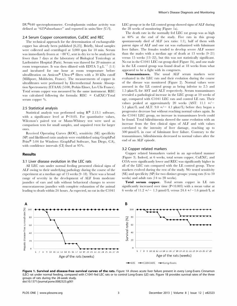
the 28 weeks of monitoring (Figure 1a).
The death rate in the normally fed LEC rat group was as high
as 40% at the end of the study. Five rats in this group
spontaneously died of ALF (sex ratio: 1:1), half of them with
patent signs of ALF and one rat was euthanized with fulminant
liver failure. The females tended to develop severe ALF sooner
than the males with a median age of death at 13 weeks (9–21)
versus 15 weeks (13–22), but this was not statistically significant.
No rat in the C1041 LEC rat group died (Figure 1b), and one male
in the LE control group was found dead at 18 weeks from what
appeared to be a fight with its congeners.
Transaminases. The usual ALF serum markers were
evaluated in the LEC rats and their evolution during the course
of the disease was monitored (Figure 2). Normal values were
assessed in the LE control group as being inferior to 2.5 and
1.5 mkat/L for AST and ALT respectively. Serum transaminases
revealed a pathological increase in the LEC group at 10 weeks of
age, compared with C1041 LEC rats and control LE rats. Their
values peaked at approximately 20 weeks (AST: 11.1 +/2
5.1 mkat/L and ALT: 9.0 +/2 4.1 mkat/L) before they began a
progressive decrease but without reaching normal values again. In
the C1041 LEC group, no increase in transaminases levels could
be found. Total bilirubinemia showed the same evolution with an
increase from the first clinical signs of ALF and with values
correlated to the intensity of liver damage, reaching up to
500 mmol/L in case of fulminant liver failure. Contrary to the
transaminases, bilirubinemia decreased to normal values after the
end of an ALF episode.
3.2 Copper related markersCopper related biomarkers varied in an age-related manner
(Figure 3). Indeed, at 6 weeks, total serum copper, CuEXC, and
COA were significantly lower and REC was significantly higher in
all of the LEC rats compared with the LE control group. These
markers evolved during the rest of the study. We tested sensitivity
(SE) and specificity (SP) for two distinct groups: young rats (6 to 10
weeks) and adult rats (14 to 28 weeks).
Total serum copper. Total serum copper in LE rats
significantly increased over time (P,0.005) with a mean value at
6 weeks of 11.2 +/2 1.3 mmol/L versus 24.4 +/21.6 mmol/L at
Figure 1. Survival and disease-free survival curves of the rats. Figure 1A shows acute liver failure present in every Long-Evans Cinnamon(LEC) rat under normal feeding, compared with C1041-fed LEC rats or to control Long-Evans (LE) rats. Figure 1B provides survival rates of the threegroups of rats during the 28-week study.doi:10.1371/journal.pone.0082323.g001
Wilson’s Disease Diagnosis and Monitoring
PLOS ONE | www.plosone.org 3 December 2013 | Volume 8 | Issue 12 | e82323
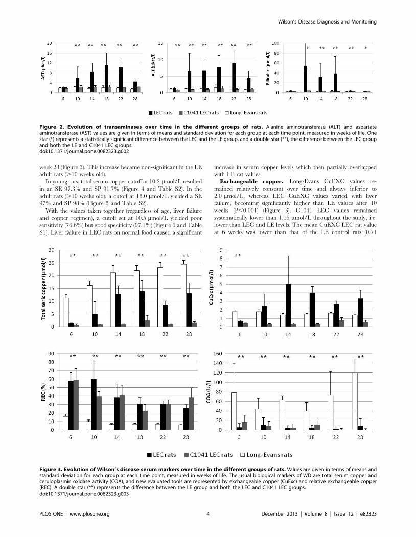
week 28 (Figure 3). This increase became non-significant in the LE
adult rats (.10 weeks old).
In young rats, total serum copper cutoff at 10.2 mmol/L resulted
in an SE 97.3% and SP 91.7% (Figure 4 and Table S2). In the
adult rats (.10 weeks old), a cutoff at 18.0 mmol/L yielded a SE
97% and SP 98% (Figure 5 and Table S2).
With the values taken together (regardless of age, liver failure
and copper regimen), a cutoff set at 10.5 mmol/L yielded poor
sensitivity (76.6%) but good specificity (97.1%) (Figure 6 and Table
S1). Liver failure in LEC rats on normal food caused a significant
increase in serum copper levels which then partially overlapped
with LE rat values.
Exchangeable copper. Long-Evans CuEXC values re-
mained relatively constant over time and always inferior to
2.0 mmol/L, whereas LEC CuEXC values varied with liver
failure, becoming significantly higher than LE values after 10
weeks (P,0.001) (Figure 3). C1041 LEC values remained
systematically lower than 1.15 mmol/L throughout the study, i.e.
lower than LEC and LE levels. The mean CuEXC LEC rat value
at 6 weeks was lower than that of the LE control rats (0.71
Figure 2. Evolution of transaminases over time in the different groups of rats. Alanine aminotransferase (ALT) and aspartateaminotransferase (AST) values are given in terms of means and standard deviation for each group at each time point, measured in weeks of life. Onestar (*) represents a statistically significant difference between the LEC and the LE group, and a double star (**), the difference between the LEC groupand both the LE and C1041 LEC groups.doi:10.1371/journal.pone.0082323.g002
Figure 3. Evolution of Wilson’s disease serum markers over time in the different groups of rats. Values are given in terms of means andstandard deviation for each group at each time point, measured in weeks of life. The usual biological markers of WD are total serum copper andceruloplasmin oxidase activity (COA), and new evaluated tools are represented by exchangeable copper (CuExc) and relative exchangeable copper(REC). A double star (**) represents the difference between the LE group and both the LEC and C1041 LEC groups.doi:10.1371/journal.pone.0082323.g003
Wilson’s Disease Diagnosis and Monitoring
PLOS ONE | www.plosone.org 4 December 2013 | Volume 8 | Issue 12 | e82323
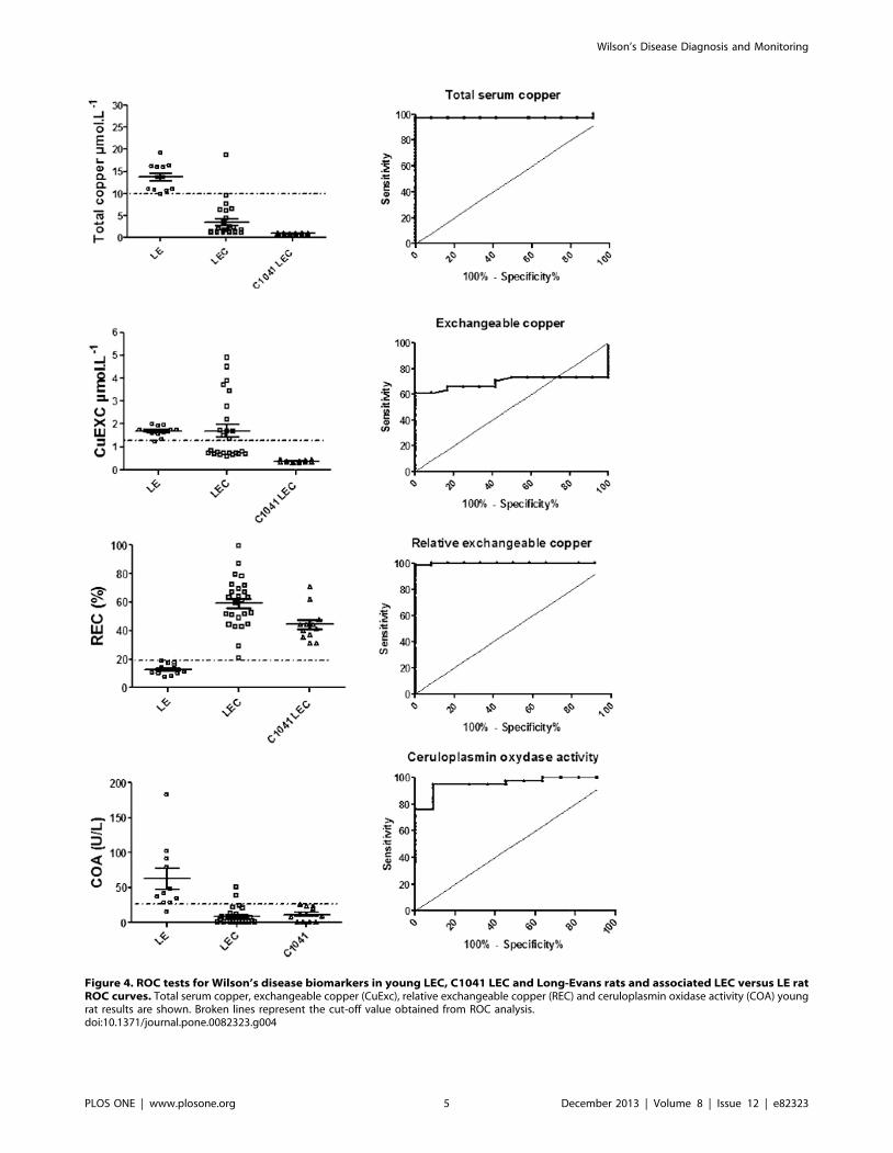
Figure 4. ROC tests for Wilson’s disease biomarkers in young LEC, C1041 LEC and Long-Evans rats and associated LEC versus LE ratROC curves. Total serum copper, exchangeable copper (CuExc), relative exchangeable copper (REC) and ceruloplasmin oxidase activity (COA) youngrat results are shown. Broken lines represent the cut-off value obtained from ROC analysis.doi:10.1371/journal.pone.0082323.g004
Wilson’s Disease Diagnosis and Monitoring
PLOS ONE | www.plosone.org 5 December 2013 | Volume 8 | Issue 12 | e82323
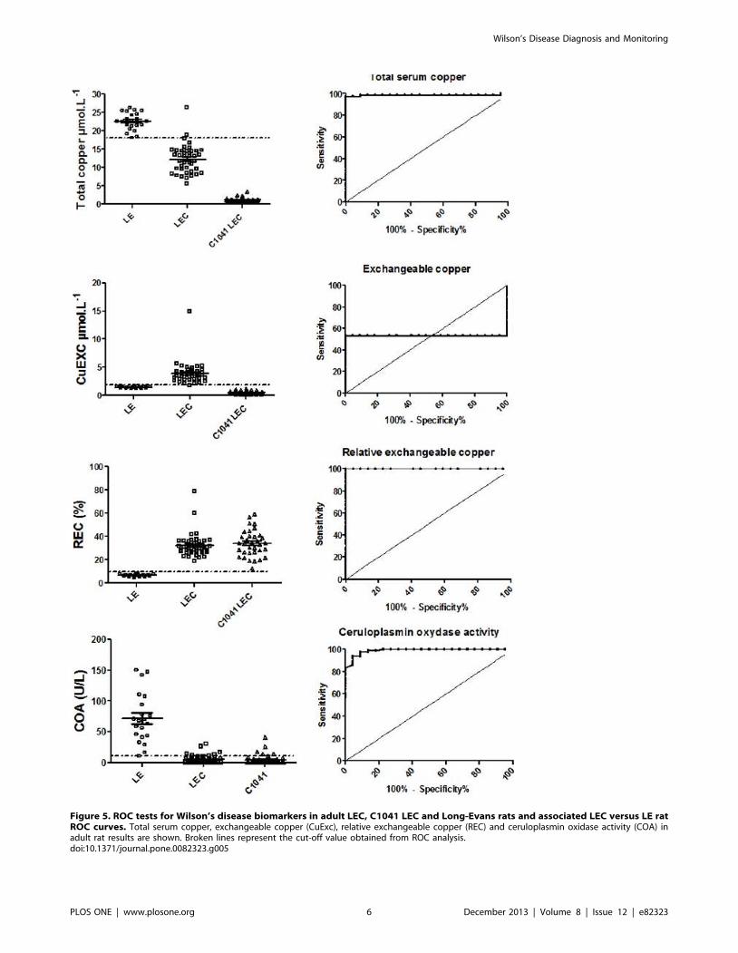
Figure 5. ROC tests for Wilson’s disease biomarkers in adult LEC, C1041 LEC and Long-Evans rats and associated LEC versus LE ratROC curves. Total serum copper, exchangeable copper (CuExc), relative exchangeable copper (REC) and ceruloplasmin oxidase activity (COA) inadult rat results are shown. Broken lines represent the cut-off value obtained from ROC analysis.doi:10.1371/journal.pone.0082323.g005
Wilson’s Disease Diagnosis and Monitoring
PLOS ONE | www.plosone.org 6 December 2013 | Volume 8 | Issue 12 | e82323

Figure 6. Global ROC tests for Wilson’s disease biomarkers in LEC, C1041 LEC and Long-Evans rats and associated LEC versus LE ratROC curves. Total serum copper, exchangeable copper (CuExc), relative exchangeable copper (REC) and ceruloplasmin oxidase activity (COA) inadult rats results are shown. Broken lines represent the cut-off value obtained from ROC analysis.doi:10.1371/journal.pone.0082323.g006
Wilson’s Disease Diagnosis and Monitoring
PLOS ONE | www.plosone.org 7 December 2013 | Volume 8 | Issue 12 | e82323
+/2 0.07 mmol/l vs. 1.8 +/2 0.2 mmol/l, P = 0.001) but like total
serum copper, CuEXC values were also dependent on copper
intakes and ALF. Thus, CuEXC was unable to discriminate all of
the LEC from the LE control group and yielded very low
sensitivity and specificity (Figures 4 and 5).
Relative exchangeable copper. In both the LEC and
C1041 LEC groups, REC was significantly higher than in the
LE control group (P,0.01) at every time point of the study. Mean
values were at 49.5% +/2 16.6% in the LEC group, 36.5% +/2
3.8% in the C1041 LEC group, versus 8.9% +/2 1.2% in the LE
control group. A cutoff set at 18.2% offered SE and SP at 98% and
100%, respectively, in young rats. A cutoff set at 10.5% offered
100% SE and 100% SP for adult rats (Figures 4 and 5, Table S2).
Taking the young and adult values together, a cutoff set at 19.0%
enabled the marker to discriminate both LEC groups from the LE
control group with 97.3% sensitivity and 100% specificity (Figure 6
and Table S1). REC values were not significantly different
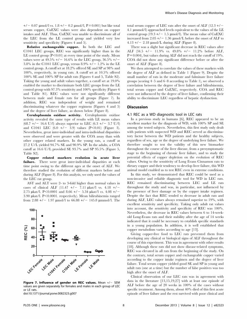
between male and female rats for all groups (Figure 7). In
addition, REC was independent of weight and remained
discriminating whatever the copper regimens (Figures 4 and 5)
and the degree of liver failure, as shown below (Figure 8).
Ceruloplasmin oxidase activity. Ceruloplasmin oxidase
activity revealed the same type of results with LE mean values
(68.7 +/2 16.6 U/l) always superior to LEC (6.3 +/2 3.7 U/l)
and C1041 LEC (6.0 +/2 3.9) values (P,0.01) (Figure 3).
Nevertheless, great inter-individual and intra-individual disparities
were observed and were greater with the COA assay than with
other copper related markers. In the young rats, a cutoff at
27.2 U/L yielded 94.7% SE and 90.9% SP. In the adults, a COA
cutoff at 16.6 U/L provided SE 93.7% and SP 95.5% (Figure 5,
Table S2).
Copper related markers evolution in acute liver
failure. There were great inter-individual disparities at each
time point owing to the different ages at the onset of ALF. We
therefore studied the evolution of different markers before and
during ALF (Figure 8). For this analysis, we only used the values of
the LEC rat group.
AST and ALT were 2- to 3-fold higher than normal values in
cases of clinical ALF (11.47 +/2 7.11 mkat/l vs. 4.18 +/2
2.75 mkat/l. P,0.0001 and 8.66 +/2 5.34 mkat/l vs. 4.08 +/2
3.90 mkat/l. P,0.0001. respectively). Mean bilirubinemia ranged
from 2.88 +/2 1.97 mmol/l to 66.88 +/2 145.8 mmmol/l. The
total serum copper of LEC rats after the onset of ALF (12.3 +/2
4.1 mmmol/l) approached levels equivalent to the values of the LE
control group (19.3 +/2 1.5 mmol/l). The mean value of CuEXC
increased from 2.05 +/2 1.36 mmol/L before the onset of ALF to
4.13 +/2 2.18 mmol/L during ALF (Figure 8).
There was a slight but significant decrease in REC values after
ALF (34.5 +/2 11.9% vs. 49.8% +/2 11.2% before ALF,
P = 0.004), but values during ALF did not reach the cutoff of 19%.
COA did not show any significant difference before or after the
onset of ALF (Figure 8).
We then attempted to correlate the values of these markers with
the degree of ALF as defined in Table 1 (Figure 9). Despite the
small number of rats in the moderate and fulminate liver failure
groups (scoring 4–5 and 6–8 according to Table 1), we observed a
correlation between the degree of liver failure and bilirubinemia,
total serum copper and CuEXC, respectively. COA and REC
were not influenced by the degree of liver failure, confirming their
ability to discriminate LEC regardless of hepatic dysfunction.
Discussion
4.1 REC as a WD diagnostic tool in LEC ratsIn a previous study in humans [6], REC appeared to be an
excellent marker for the diagnosis of WD, with 100% SP and SE
among the tested subjects. Nevertheless, this first study only dealt
with patients with suspected WD and REC served as discrimina-
tory factor between the WD patients and the healthy subjects,
regardless of sex, age or the degree of underlying liver failure. We
therefore sought to test the validity of this new biomarker
throughout the course of the liver disease, from a presymptomatic
stage to the beginning of chronic liver failure, and to study the
potential effects of copper depletion on the evolution of REC
values. Owing to the sensitivity of Long Evans Cinnamon rats to
dietary copper and their tendency to develop liver failure, this WD
animal model enabled us to test REC even in extreme conditions.
In this study, we demonstrated that REC could be used as a
non-invasive and reliable diagnostic tool for WD in LEC rats.
REC remained discriminating between LEC and LE rats
throughout the study and was, in particular, not influenced by
the presence of liver damage or by the copper intake regimen.
Despite the fact that REC tended to decrease in adult rats and
during ALF, LEC values always remained superior to 19%, with
excellent sensitivity and specificity. Taking only adult rat values
into account, the sensitivity and specificity of REC was 100%.
Nevertheless, the decrease in REC values between 6 to 14-week-
old Long-Evans rats and their stability after the age of 14 weeks
indicated that it could be necessary to establish specific standards
in a young population. In addition, it is well established that
copper metabolism varies according to age [13].
Giving copper-free food to LEC rats prevented them from
developing any clinical or biological signs of ALF throughout the
course of this experiment. This was in agreement with other results
[18]. Although these rats did not show disease-related symptoms,
REC was elevated in all rats from the beginning of the study. On
the contrary, total serum copper and exchangeable copper varied
according to the copper intake regimen and the degree of liver
failure. Total serum copper yielded good SE and SP in young and
adult rats (one at a time) but the number of false positives was too
high after the onset of ALF.
Clinical observation of our LEC rats was in agreement with
data in the literature [13,15,19,27] with at least one episode of
ALF before the age of 20 weeks in 100% of the cases without
specific treatment. Among them, about 40% died of this first acute
episode of liver failure and the rest survived with poor clinical and
Figure 7. Influence of gender on REC values. Mean +/2 SEMvalues are given separately for females and males in each group of LECor LE rats.doi:10.1371/journal.pone.0082323.g007
Wilson’s Disease Diagnosis and Monitoring
PLOS ONE | www.plosone.org 8 December 2013 | Volume 8 | Issue 12 | e82323
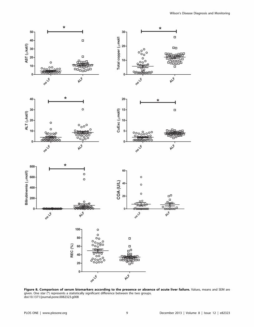
Figure 8. Comparison of serum biomarkers according to the presence or absence of acute liver failure. Values, means and SEM aregiven. One star (*) represents a statistically significant difference between the two groups.doi:10.1371/journal.pone.0082323.g008
Wilson’s Disease Diagnosis and Monitoring
PLOS ONE | www.plosone.org 9 December 2013 | Volume 8 | Issue 12 | e82323

Figure 9. Evolution of serum biomarkers according to the grade of severity of acute hepatic failure. Values, means and SEM are given.One star (*) represents a statistically significant increase in serum marker value between the two grades of ALF.doi:10.1371/journal.pone.0082323.g009
Wilson’s Disease Diagnosis and Monitoring
PLOS ONE | www.plosone.org 10 December 2013 | Volume 8 | Issue 12 | e82323
biological signs of chronic liver disease such as chronic jaundice
and mild elevation of ALT and AST. In this group, transminases
were within normal ranges until the age of 10 weeks and
bilirubinemia levels were normal after ALF had stopped. On the
contrary, total serum copper increased during ALF to the values
found in the LE control group. The only markers that could
discriminate LEC rats throughout the study were COA (in
agreement with Merle et al. [23]) and REC. However, COA has
been reported to be almost absent in the LEC strain in some
studies [26] but has been detected in others [19]. In our LEC rats,
COA had great inter and intra-individual disparities.
Further studies are required in order to guarantee the specificity
of REC in differentiating WD from other kinds of liver disease.
Preliminary results using a model of cholestasis in rats showed that
REC values were considerably lower than REC values in our
animal model (data not shown).
4.2 CuEXC evolution during liver failureWithin the population of LEC rats, our results indicate that total
serum copper and CuEXC levels were correlated to ALF. CuEXC
in LEC rats started to increase with elevation of the hepatic
cytolysis markers (ALT and AST) and its values remained high as
the ALF settled.
As suggested in previous studies [18,28,29], copper is supposed
to progressively accumulate in LEC hepatocytes during the first
weeks of life until cytosolic storage capacities are overwhelmed.
This could be accelerated by a massive burden of dietary copper as
recently reported by Siaj et al. [18]. Liver accumulated copper
induces liver deterioration through oxidative stress and cell cycle
activation and is finally released in the blood – not bound to
ceruloplasmin – then eliminated through kidney ultrafiltration.
This leads to an elevation of urinary copper. CuEXC has been
described as the labile fraction of copper bound to different
molecules such as albumin and transcuprein [8]. Hence, an
increase in CuEXC serum levels could indicate the presence of
abnormally high amount of toxic extrahepatic copper.
Another potential use of CuEXC could be as a marker of
decoppering therapy efficiency. In fact, we have observed that
CuEXC levels did not vary with time in the group of C1041 LEC
rats whose copper intakes were quasi-absent and always remained
under 1.0 mmol/l. Zinc-salt therapy, for instance, acts by inducing
metallothionein synthesis in the enterocytes [30], which seques-
trate copper and eliminate it through feces [31]. Overtime, it
depletes copper from the organism by a negative imbalance
between intakes and outtakes. It is for this reason that CuEXC
could be a good and simple marker for long-term monitoring with
a persistent low value being the sign of good observance. Further
studies will therefore be needed to assess the potential use of
CuEXC as a monitoring marker in rats under chelating or zinc salt
therapy.
Since CuEXC corresponds to the labile fraction of copper not
bound to ceruloplasmin, the only factor that could a priori cause
REC values to evolve toward normal values should be the
restoration of holoceruloplasmin levels in the serum which is
controlled by a functional ATP7B protein in the liver [32,33]. This
hypothesis must be validated in a WD patient with a liver
transplantation and could have a future application in pre-clinical
trials in LEC rats by helping the following up of WD gene therapy.
Conclusion
In summary, our results indicate that REC could be used as a
reliable WD diagnostic marker with 100% sensitivity and 100%
specificity. REC is at least as good as COA to discriminate LEC
(ATP7B-) from LE (ATP7B+) rats. In addition, it does not depend
on the existence of underlying liver damage or on the copper
intake regimen. On the other hand, we propose the use of
exchangeable copper as a marker which able to reflect copper
overload before and during ALF. Finally, in comparison with
normal Long-Evans rats, we can propose cut-off values for each
serum marker tested in this WD animal model.
Supporting Information
Table S1 Results of ROC curve analysis of serummarkers for Wilson’s disease.(DOC)
Table S2 Detailed ROC curve analysis of Wilson’sdisease markers in young and adult LEC rats.(DOC)
Author Contributions
Conceived and designed the experiments: FS SE GP. Performed the
experiments: FS SE. Analyzed the data: FS SE JMT FW JP. Contributed
reagents/materials/analysis tools: OL THN SE FS. Wrote the paper: FS
SE. Animal care and sampling: JR PL.
References
1. Lalioti V, Sandoval I, Cassio D, Duclos-Vallee JC (2010). Molecular pathology
of Wilson’s disease: a brief. J Hepatol 53: 1151–1153.
2. Schilsky ML (2009) Wilson disease: current status and the future. Biochimie 91:
1278–1281.
3. Merle U, Stremmel W, Encke J (2007) Perspectives for gene therapy of Wilson
disease. Curr Gene Ther 7: 217–220.
4. Medici V, Mirante VG, Fassati LR, Pompili M, Forti D, et al. (2005) Liver
transplantation for Wilson’s disease: The burden of neurological and psychiatric
disorders. Liver Transpl 11: 1056–1063.
5. Kenney SM, Cox DW (2007) Sequence variation database for the Wilson
disease copper transporter, ATP7B. Hum Mutat 28: 1171–1177.
6. El Balkhi S, Trocello JM, Poupon J, Chappuis P, Massicot F, et al. (2011)
Relative exchangeable copper: a new highly sensitive and highly specific
biomarker for Wilson’s disease diagnosis. Clin Chim Acta 412: 2254–2260.
7. Venelinov TI, Beattie JH, Dainty JR, Hollands WJ, Fairweather-Tait SJ, et al.
(2007) Stable isotope pilot study of exchangeable copper kinetics in human blood
plasma. J Trace Elem Med Biol 21: 138–140.
8. Buckley WT, Vanderpool RA (2008) Analytical variables affecting exchangeable
copper determination in blood plasma. Biometals 21: 601–612.
9. El Balkhi S, Poupon J, Trocello JM, Massicot F, Woimant F, et al. (2010)
Human plasma copper proteins speciation by size exclusion chromatography
coupled to inductively coupled plasma mass spectrometry. Solutions for columns
calibration by sulfur detection. Anal Chem 82: 6904–6910.
10. Kim JW, Kim JH, Seo JK, Ko JS, Chang JY, et al. (2013) Genetically confirmed
Wilson disease in a 9-month old boy with elevations of aminotransferases.
World J Hepatol 5: 156–159.
11. Chappuis P, Callebert J, Quignon V, Woimant F, Laplanche JL (2007) Late
neurological presentations of Wilson disease patients in French population and
identification of 8 novel mutations in the ATP7B gene. J Trace Elem Med Biol
21: 37–42.
12. Vonk WI, Wijmenga C, van de Sluis B (2008) Relevance of animal models for
understanding mammalian copper homeostasis. Am J Clin Nutr 88: 840S–845S.
13. Yoshida MC, Masuda R, Sasaki M, Takeichi N, Kobayashi H, et al. (1987) New
mutation causing hereditary hepatitis in the laboratory rat. J Hered 78: 361–365.
14. Suzuki KT, Kanno S, Misawa S, Aoki Y (1995) Copper metabolism leading to
and following acute hepatitis in LEC rats. Toxicology 97: 81–92.
15. Masuda R, Yoshida MC, Sasaki M, Dempo K, Mori M (1988) Hereditary
hepatitis of LEC rats is controlled by a single autosomal recessive gene. Lab
Anim 22: 166–169.
16. Hayashi M, Fuse S, Endoh D, Horiguchi N, Nakayama K, et al. (2006)
Accumulation of copper induces DNA strand breaks in brain cells of Long-Evans
Cinnamon (LEC) rats, an animal model for human Wilson Disease. Exp Anim
55: 419–426.
17. Wu J, Forbes JR, Chen HS, Cox DW (1994) The LEC rat has a deletion in the
copper transporting ATPase gene homologous to the Wilson disease gene. Nat
Genet 7: 541–545.
Wilson’s Disease Diagnosis and Monitoring
PLOS ONE | www.plosone.org 11 December 2013 | Volume 8 | Issue 12 | e82323
18. Siaj R, Sauer V, Stoppeler S, Spiegel HU, Kohler G, et al. (2013) Dietary
copper triggers onset of fulminant hepatitis in the Long-Evans cinnamon ratmodel. World J Gastroenterol 18: 5542–5550.
19. Li Y, Togashi Y, Sato S, Emoto T, Kang JH, et al. (1991) Spontaneous hepatic
copper accumulation in Long-Evans Cinnamon rats with hereditary hepatitis. Amodel of Wilson’s disease. J Clin Invest 87: 1858–1861.
20. Sone K, Maeda M, Wakabayashi K, Takeichi N, Mori M, et al. (1996)Inhibition of hereditary hepatitis and liver tumor development in Long-Evans
cinnamon rats by the copper-chelating agent trientine dihydrochloride.
Hepatology 23: 764–770.21. Fong RN, Gonzalez BP, Fuentealba IC, Cherian MG (2004) Role of tumor
necrosis factor-alpha in the development of spontaneous hepatic toxicity inLong-Evans Cinnamon rats. Toxicol Appl Pharmacol 200: 121–130.
22. Carstens E, Moberg GP (2000) Recognizing pain and distress in laboratoryanimals. ILAR J 41: 62–71.
23. Merle U, Eisenbach C, Weiss KH, Tuma S, Stremmel W (2009) Serum
ceruloplasmin oxidase activity is a sensitive and highly specific diagnostic markerfor Wilson’s disease. J Hepatol 51: 925–930.
24. Schosinsky KH, Lehmann HP, Beeler MF (1974) Measurement of ceruloplasminfrom its oxidase activity in serum by use of o-dianisidine dihydrochloride. Clin
Chem 20: 1556–1563.
25. El Balkhi S, Poupon J, Trocello JM, Leyendecker A, Massicot F, et al. (2009)Determination of ultrafiltrable and exchangeable copper in plasma: stability and
reference values in healthy subjects. Anal Bioanal Chem 394: 1477–1484.
26. Terada K, Nakako T, Yang XL, Iida M, Aiba N, et al. (1998) Restoration of
holoceruloplasmin synthesis in LEC rat after infusion of recombinant adenovirusbearing WND cDNA. J Biol Chem 273: 1815–1820.
27. Kasai N, Osanai T, Miyoshi I, Kamimura E, Yoshida MC, et al. (1990) Clinico-
pathological studies of LEC rats with hereditary hepatitis and hepatoma in theacute phase of hepatitis. Lab Anim Sci 40: 502–505.
28. Bartee MY, Lutsenko S (2007) Hepatic copper-transporting ATPase ATP7B:function and inactivation at the molecular and cellular level. Biometals 20: 627–
637.
29. Gray LW, Peng F, Molloy SA, Pendyala VS, Muchenditsi A, et al. (2012)Urinary copper elevation in a mouse model of Wilson’s disease is a regulated
process to specifically decrease the hepatic copper load. PLoS One 7: e38327.30. Yuzbasiyan-Gurkan V, Grider A, Nostrant T, Cousins RJ, Brewer GJ (1992)
Treatment of Wilson’s disease with zinc: X. Intestinal metallothionein induction.J Lab Clin Med 120: 380–386.
31. Anderson LA, Hakojarvi SL, Boudreaux SK (1998) Zinc acetate treatment in
Wilson’s disease. Ann Pharmacother 32: 78–87.32. Bahde R, Kapoor S, Bhargava KK, Schilsky ML, Palestro CJ, et al. (2012) PET
with 64Cu-histidine for noninvasive diagnosis of biliary copper excretion inLong-Evans cinnamon rat model of Wilson disease. J Nucl Med 53: 961–968.
33. Meng Y, Miyoshi I, Hirabayashi M, Su M, Mototani Y, et al. (2004) Restoration
of copper metabolism and rescue of hepatic abnormalities in LEC rats, ananimal model of Wilson disease, by expression of human ATP7B gene. Biochim
Biophys Acta 1690: 208–219.
Wilson’s Disease Diagnosis and Monitoring
PLOS ONE | www.plosone.org 12 December 2013 | Volume 8 | Issue 12 | e82323
Related Documents