Pavlicek et al. BMC Evolutionary Biology 2010, 10:154 http://www.biomedcentral.com/1471-2148/10/154 Open Access RESEARCH ARTICLE © 2010 Pavlicek et al; licensee BioMed Central Ltd. This is an Open Access article distributed under the terms of the Creative Commons Attribution License (http://creativecommons.org/licenses/by/2.0), which permits unrestricted use, distribution, and reproduction in any medium, provided the original work is properly cited. Research article Evolution of AANAT: expansion of the gene family in the cephalochordate amphioxus Jiri Pavlicek 1 , Sandrine Sauzet 2 , Laurence Besseau 2 , Steven L Coon 1 , Joan L Weller 1 , Gilles Boeuf 2,3 , Pascaline Gaildrat 1 , Marina V Omelchenko 4 , Eugene V Koonin 4 , Jack Falcón 2 and David C Klein* 1 Abstract Background: The arylalkylamine N-acetyltransferase (AANAT) family is divided into structurally distinct vertebrate and non-vertebrate groups. Expression of vertebrate AANATs is limited primarily to the pineal gland and retina, where it plays a role in controlling the circadian rhythm in melatonin synthesis. Based on the role melatonin plays in biological timing, AANAT has been given the moniker "the Timezyme". Non-vertebrate AANATs, which occur in fungi and protists, are thought to play a role in detoxification and are not known to be associated with a specific tissue. Results: We have found that the amphioxus genome contains seven AANATs, all having non-vertebrate type features. This and the absence of AANATs from the genomes of Hemichordates and Urochordates support the view that a major transition in the evolution of the AANATs may have occurred at the onset of vertebrate evolution. Analysis of the expression pattern of the two most structurally divergent AANATs in Branchiostoma lanceolatum (bl) revealed that they are expressed early in development and also in the adult at low levels throughout the body, possibly associated with the neural tube. Expression is clearly not exclusively associated with the proposed analogs of the pineal gland and retina. blAANAT activity is influenced by environmental lighting, but light/dark differences do not persist under constant light or constant dark conditions, indicating they are not circadian in nature. bfAANATα and bfAANATδ' have unusually alkaline (> 9.0) optimal pH, more than two pH units higher than that of vertebrate AANATs. Conclusions: The substrate selectivity profiles of bfAANATα and δ' are relatively broad, including alkylamines, arylalkylamines and diamines, in contrast to vertebrate forms, which selectively acetylate serotonin and other arylalkylamines. Based on these features, it appears that amphioxus AANATs could play several roles, including detoxification and biogenic amine inactivation. The presence of seven AANATs in amphioxus genome supports the view that arylalkylamine and polyamine acetylation is important to the biology of this organism and that these genes evolved in response to specific pressures related to requirements for amine acetylation. Background The AANAT family is part of the large and diverse super- family of GCN5-like acetyltransferases which use AcCoA as the acetyl donor and share a common AcCoA binding fold [1]. Members of the AANAT family share limited sequence identity and are divided into two groups: verte- brate AANATs; and, the non-vertebrate AANATs. The latter are found in fungi, protists, and bacteria and lack defining physical characteristics of vertebrate AANAT [1- 3]. The biological role of vertebrate AANAT is to acety- late serotonin in the synthesis of melatonin (tryptophan → hydroxytryptophan → serotonin → N-acetylserotonin → melatonin) [4,5]. Vertebrate AANAT is associated with biological timing: daily changes in the activity of this enzyme regulate the daily rhythm in melatonin synthesis, which is essential for optimal temporal coordination of biological functions with night/day and seasonal changes and for photic entrainment [6]. The central role of verte- brate AANAT in biological timing has earned it the mon- iker 'the Timezyme' [7]. Non-vertebrate AANATs are thought to play a detoxifying role by neutralizing arylal- kylamines [8] and a role in DNA biology by acetylating polyamines [9]. * Correspondence: [email protected] 1 Section on Neuroendocrinology, Program in Developmental Endocrinology and Genetics, The Eunice Kennedy Shriver National Institute of Child Health and Human Development, National Institutes of Health, Bethesda, 20892, MD, USA Full list of author information is available at the end of the article

Welcome message from author
This document is posted to help you gain knowledge. Please leave a comment to let me know what you think about it! Share it to your friends and learn new things together.
Transcript
Pavlicek et al. BMC Evolutionary Biology 2010, 10:154http://www.biomedcentral.com/1471-2148/10/154
Open AccessR E S E A R C H A R T I C L E
Research articleEvolution of AANAT: expansion of the gene family in the cephalochordate amphioxusJiri Pavlicek1, Sandrine Sauzet2, Laurence Besseau2, Steven L Coon1, Joan L Weller1, Gilles Boeuf2,3, Pascaline Gaildrat1, Marina V Omelchenko4, Eugene V Koonin4, Jack Falcón2 and David C Klein*1
AbstractBackground: The arylalkylamine N-acetyltransferase (AANAT) family is divided into structurally distinct vertebrate and non-vertebrate groups. Expression of vertebrate AANATs is limited primarily to the pineal gland and retina, where it plays a role in controlling the circadian rhythm in melatonin synthesis. Based on the role melatonin plays in biological timing, AANAT has been given the moniker "the Timezyme". Non-vertebrate AANATs, which occur in fungi and protists, are thought to play a role in detoxification and are not known to be associated with a specific tissue.
Results: We have found that the amphioxus genome contains seven AANATs, all having non-vertebrate type features. This and the absence of AANATs from the genomes of Hemichordates and Urochordates support the view that a major transition in the evolution of the AANATs may have occurred at the onset of vertebrate evolution. Analysis of the expression pattern of the two most structurally divergent AANATs in Branchiostoma lanceolatum (bl) revealed that they are expressed early in development and also in the adult at low levels throughout the body, possibly associated with the neural tube. Expression is clearly not exclusively associated with the proposed analogs of the pineal gland and retina. blAANAT activity is influenced by environmental lighting, but light/dark differences do not persist under constant light or constant dark conditions, indicating they are not circadian in nature. bfAANATα and bfAANATδ' have unusually alkaline (> 9.0) optimal pH, more than two pH units higher than that of vertebrate AANATs.
Conclusions: The substrate selectivity profiles of bfAANATα and δ' are relatively broad, including alkylamines, arylalkylamines and diamines, in contrast to vertebrate forms, which selectively acetylate serotonin and other arylalkylamines. Based on these features, it appears that amphioxus AANATs could play several roles, including detoxification and biogenic amine inactivation. The presence of seven AANATs in amphioxus genome supports the view that arylalkylamine and polyamine acetylation is important to the biology of this organism and that these genes evolved in response to specific pressures related to requirements for amine acetylation.
BackgroundThe AANAT family is part of the large and diverse super-family of GCN5-like acetyltransferases which use AcCoAas the acetyl donor and share a common AcCoA bindingfold [1]. Members of the AANAT family share limitedsequence identity and are divided into two groups: verte-brate AANATs; and, the non-vertebrate AANATs. Thelatter are found in fungi, protists, and bacteria and lackdefining physical characteristics of vertebrate AANAT [1-
3]. The biological role of vertebrate AANAT is to acety-late serotonin in the synthesis of melatonin (tryptophan→ hydroxytryptophan → serotonin → N-acetylserotonin→ melatonin) [4,5]. Vertebrate AANAT is associated withbiological timing: daily changes in the activity of thisenzyme regulate the daily rhythm in melatonin synthesis,which is essential for optimal temporal coordination ofbiological functions with night/day and seasonal changesand for photic entrainment [6]. The central role of verte-brate AANAT in biological timing has earned it the mon-iker 'the Timezyme' [7]. Non-vertebrate AANATs arethought to play a detoxifying role by neutralizing arylal-kylamines [8] and a role in DNA biology by acetylatingpolyamines [9].
* Correspondence: [email protected] Section on Neuroendocrinology, Program in Developmental Endocrinology and Genetics, The Eunice Kennedy Shriver National Institute of Child Health and Human Development, National Institutes of Health, Bethesda, 20892, MD, USAFull list of author information is available at the end of the article
© 2010 Pavlicek et al; licensee BioMed Central Ltd. This is an Open Access article distributed under the terms of the Creative CommonsAttribution License (http://creativecommons.org/licenses/by/2.0), which permits unrestricted use, distribution, and reproduction inany medium, provided the original work is properly cited.
Pavlicek et al. BMC Evolutionary Biology 2010, 10:154http://www.biomedcentral.com/1471-2148/10/154
Page 2 of 15
Genomes of vertebrates contain a single copy of theAANAT gene, except for teleost fish, some of which pos-sess up to three paralogs [2], and cows which possess twoparalogs (unpublished; NCBI, NIH, http://www.ncbi.nlm.nih.gov). Members of the AANAT familyare not in the available genomes of Hemichordates andUrochordates, which leaves open the question of whenvertebrate AANAT first appeared in chordates. A strikingcharacteristic of vertebrate AANAT is that it is consis-tently expressed at significant levels in only two tissues,both of which are photosensitive organs, the pineal glandand retina. This fits with the evidence that pinealocytesand retinal photoreceptors evolved from a commonancestral photodetector [10-13]. The vertebrate AANAThas a neutral pH optimum and exhibits high selectivityfor arylalkylamines [7].
Vertebrate AANATs encode proteins that have severalhighly conserved structural characteristics [2,8], whichfacilitate arylalkylamine acetylation and regulation. Thesefeatures include flanking regulatory regions, which medi-ate rapid changes in enzyme activity; a pair of histidineswhich facilitate catalysis [14]; and, a proline-containingtripeptide in a floppy loop, which confers a high catalyticrate through an effect on substrate binding [15]. Verte-brate AANATs also have high selectivity for arylalkylam-ines conferred by the binding pocket.
Non-vertebrate type AANATs are found in thegenomes of most fungi, many unicellular eukaryotes, anda variety of bacteria [16]. The proteins encoded by thesegenes do not contain the characteristic structural featuresof vertebrate AANATs. Genes similar to non-vertebrateAANATs are not found in vertebrates.
Amphioxus AANATs are of interest in understandingthe evolution of vertebrate AANAT because cephalochor-dates and vertebrates form a clade [17]. Accordingly, thestudy of the AANAT from this organism might providenew insights into the transition from the non-vertebratemembers of the AANAT family to the vertebrate forms.Sequence in the trace archives has indicated that a non-vertebrate type member of the AANAT family is presentin amphioxus [2]. The subsequent availability of the nearcomplete amphioxus genome [18] has allowed us to com-prehensively analyze the AANAT family in amphioxus, aspresented here. The findings of this study indicate thatrapid expansion of non-vertebrate forms occurred inamphioxus and that a vertebrate form is absent. The sub-strate preference profile of the encoded enzymes suggeststo us that they are likely to play a role in detoxification orin biogenic amine inactivation, or both.
MethodsAnimalsBranchiostoma floridae (B. floridae, bf ) and Branchios-toma lanceolatum (B. lanceolatum, bl) were used; the for-
mer were obtained from Gulf Specimen Marine Lab(Panacea, FL, USA) and the latter from the Bay of Banyulssur Mer (France) [17]. B. floridae heads were used to gen-erate the cDNA used to clone full length bfAANATα andbfAANATδ' that were used for enzyme characterization.B. lanceolatum were used for in vivo studies of geneexpression and enzyme activity. They were cultured insand-filtered seawater under a natural lighting cycle andtemperature, as indicated elsewhere [17]. Intensity of thelight was 136 lux. For the experiment with constant con-ditions, animals were housed for 14 days in constant lightand/or darkness and then were used for the activity assay.
Identification of amphioxus AANATsbfAANAT homologs were identified through a tBLASTnsearch of genomic sequences available at: http://genome.jgi-psf.org[18]. Seven genes for AANAThomologs (α - η were identified in genome assembly v2.0.A second allele for five of the genes was found in v1.0.Based on these sequences, primers were designed (Addi-tional file 1) for cloning of blAANAT cDNAs. qPCR prim-ers for specific gene products were based on the B.lanceolatum clones.
Sequence similarity search and phylogenetic analysisThe AANAT homologs were identified by searching thenon-redundant database of protein sequences (NCBI,NIH, http://www.ncbi.nlm.nih.gov) using the PSI BLASTprogram [19] and by searching the genomes and pro-teomes available at JGI http://genome.jgi-psf.org/euk_cur1.html using the tBLASTn and BLASTp pro-grams, respectively. Additionally, AANAT homologs weredetected in the genomes of the red algae Gracilariachangii, segmented worm Alvinella pompejana, Mediter-ranean mussel Mytilus galloprovincialis (NCBI, ESTdatabase) and the basal fungus Rhizopus oryzae http://www.broadinstitute.org/annotation/genome/rhizopus_oryzae/MultiHome.html.
Representatives from different groups including bacte-ria, fungi, protists, and animals were selected for phyloge-netic analysis, and a multiple alignment of the respectiveprotein sequences was constructed using the MUSCLEprogram [20].
A maximum likelihood phylogenetic tree was con-structed using the TreeFinder program [21] by optimiz-ing a default starting tree constructed using theNeighbor-joining method with the Whelan and Goldman(WAG) empirical model of substitutions [22] To selectthe best substitution model, we compared the likelihoodsof trees constructed using 8 models: WAG (21), JTT [23],VT [24], BLOSUM [25], Dayhoff [26], cpREV [27], rtREV[28], PMB [29] by running the TreeFinder program [21];this test showed the best fit with the data for the WAGmodel. The reliability of the internal tree branches was
Pavlicek et al. BMC Evolutionary Biology 2010, 10:154http://www.biomedcentral.com/1471-2148/10/154
Page 3 of 15
estimated using the LR-ELW bootstrap method [30]. Aconstrained large scale tree topology was created usingthe TreeView program [31]. Alternative tree topologieswere compared with the Approximately Unbiased test[32] using the TreeFinder program.
Evolutionary distances were estimated using the dis-tance matrix created by the PROTDIST program ofPHYLIP package [33] based on a truncated alignment ofAANATs and using the Jones-Taylor-Thornton (JTT)substitution model [23]. The mean differences (p-dis-tance) and identities between the major taxonomicgroups of AANATs were calculated using the MEGA pro-gram [34].
Prokaryotic expression of recombinant proteinspGEX 4T-1 (GE Healthcare, Piscataway, NJ) was used forthe recombinant expression of full-length bfAANATαand bfAANATδ'. The constructed plasmid wassequenced to confirm identity. The construct, which pro-duces a GST fusion protein, was transformed into E. colistrain BL21Gold (DE3) pLysS (Novagen, Madison, WI).The cells were grown at 37°C; when the OD600 = 0.6, thecultures were cooled to 25°C and isopropyl β-D-1-thioga-lactopyranoside was added (final concentration = 0.2mM). The cells were harvested by centrifugation after 12hours of culturing (5000 × g, 30 min, 4°C) and resus-pended in 2× PBS, pH 7.5, containing 10 mM DTT (Buf-fer A) and a mixture of protease inhibitors (Complete®,Roche, Indianapolis, IN). The cells were then lysed bysonication and the resulting lysate was centrifuged (8500× g, 25 min, 4°C). The supernatant was mixed with Gluta-thione-Sepharose 4B affinity matrix (GE Healthcare) pre-equilibrated with Buffer A. The suspension was agitatedfor 1 h and then packed into a glass column. The columnwas washed with 5 column volumes of Buffer A, followedby 5 column volumes of buffer containing 50 mM Tris-HCl, 0.1 M sodium citrate, 5 mM DTT and 10% glycerol,pH 7.8 (Buffer B). GST fusion protein was then elutedwith 5 column volumes of Buffer B containing 10 mMglutathione. Protein was concentrated and dialyzedagainst buffer containing 0.1 M ammonium acetate, 25mM NaCl and 1 mM TCEP; the resulting preparation wasstored at -80°C. Where indicated, purified GST-freebfAANATα was used; otherwise, the GST-bfAANATα orGST-AANATδ' fusion product was used, consistent withprevious studies [15,35,36].
SDS-PAGEProteins were resolved on preformed 14% Tris-glycine (1mm) gels, using the manufacturer's protocol (NOVEX,Invitrogen) [37]. The molecular mass of the proteins wasestimated using Rainbow™ markers (GE Healthcare).
Fluorescence-based protein studiesFluorescence measurements were done on an ISS PC1photon-counting spectrofluorimeter (ISS, Inc., Cham-paign, IL) at 22°C. To study binding properties ofbfAANATα protein, the interaction between bfAANATαand CoA-S-N-acetyl-7-hydroxynaphthylethylamine(CoA-HNE), a fluorescent bisubstrate inhibitor of verte-brate AANAT, was analyzed [36]. This was done by mea-suring CoA-HNE (1 μM)-dependent quenching oftryptophan fluorescence in 0.1 M ammonium acetate, 25mM NaCl and 1 mM TCEP, pH 6.8 (excitation = 290 nm;emission = 310 nm). Off-rates of CoA-HNE were moni-tored as described [15] using protein preparations thatwere 90% saturated with CoA-HNE. Following a controlincubation period, the probe was displaced with a 100-fold excess of another bisubstrate inhibitor, CoA-S-N-acetyltryptamine (CoA-T), that is optically inactive in thisanalysis [36]. The limiting value of koff was calculated bynon-linear fitting (SigmaPlot Version 10.0, Systat Soft-ware, Inc., Point Richmond, CA) using the equation:
where A(t) is the observed fluorescence anisotropy attime 't' after the initial addition of CoA-T and koff is therate constant.
Determination of N-acetyltransferase activityRecombinant bfAANATα activity was measuredradiochemically by measuring [3H]acetyl product formedfrom [3H]acetyl-coenzyme A ([3H]AcCoA; GE Health-care, UK) and an amine substrate as described [15]; or,colorimetrically by measuring CoASH formed by incuba-tion with AcCoA and an amine substrate. CoASH isdetected as the colored product formed by reaction with5,5'-dithio-bis(2-nitrobenzoic acid) (DTNB) [35]. In thecolorimetric assay, standard reactions were performed ina total volume of 100 μL containing: the substrate at indi-cated concentrations, 0.5 mM AcCoA, 0.05 mg/ml bovineserum albumin (BSA), and 2 mM ethylene diamine tet-raacetic acid (EDTA). For analysis of GST-bfAANATα,0.25 μg of protein was included in 0.1 M Tris buffer pH9.5; the activity of ovine GST-AANAT, which was used asa reference standard [15], was determined by incubating10 ng of protein in 0.1 M phosphate buffer, pH 6.8. Incu-bation conditions were either 80 min at 37°C foramphioxus or 30 min at 37°C for ovine AANAT. Enzymeswere then inactivated by the addition of 150 μl stop solu-tion (1 mM DTNB, 10 mM EDTA, and 3 M guanidinehydrochloride); absorbance (405 nm) was measured after5 min incubation at room temperature. The Vmax and Km
A t A A A exp k toff( ) = ∞( ) + ( ) ∞( )( ) − ×( )0 – ,
Pavlicek et al. BMC Evolutionary Biology 2010, 10:154http://www.biomedcentral.com/1471-2148/10/154
Page 4 of 15
were calculated by non-linear fitting (Prism5 fromGraphPad) using the equation:
where V is the observed enzyme velocity (activity), Vmaxis the maximum enzyme velocity, S is the substrate con-centration, and Km is the Michaelis-Menten constant.
For the measurement of enzyme activity in amphioxushead tissue, the colorimetric assay (see above) was usedwith 10 mM β-phenylethylamine (PEA) as a substrate and10 μg of protein from tissue homogenized in assay buffer.For each measurement a corresponding assay was per-formed in the presence of 1 mM CoA-T [15], and anotherin the absence of PEA. The controls used included anassay without tissue homogenate and another with aheat-inactivated (65°C, 5 min) homogenate. Samples werecollected at noon and midnight in the experiments study-ing the light/dark variations, and at noon in all otherexperiments.
Quantitative measurement of B. lanceolatum AANAT transcripts (qPCR)Total RNA was extracted from head and from body usinga Trizol extraction kit (Invitrogen, Cergy Pontoise,France). 1 μg was incubated with 1 unit of DNase I(Roche; Meylan, France) for 20 min at 37°C. FollowingDNase inactivation (65°C, 10 min), the sequence wasreverse-transcribed using Powerscript Reverse Tran-scriptase (Clontech, Mountain View, CA). qPCR of theresulting cDNAs was performed with the primer setsqAANAT (Additional file 1) using the 'Light Cycler FastStart DNA Master SYBR Green I kit' (Roche MolecularBiochemicals, USA) in a total volume of 20 μl as follows:95°C (10 min), followed by 35 cycles of denaturation at95°C (10 sec), annealing at 60°C (15 sec) and extension at72°C (15 sec). qPCR was done using a Light Cycler 1.5(Roche Diagnostics, USA). Amplification efficiency wasmeasured using serial dilutions of plasmids containing afragment of the corresponding blAANAT gene. Datawere normalized to the average value obtained with threehousekeeping genes from B. lanceolatum: L17(AY130354), L18 (AY130454) and actin (Y13663). ThePCR conditions for the housekeeping genes were as indi-cated above except that the annealing temperature was66°C for actin. Primers for the housekeeping genes aregiven in Additional file 1.
Tissue distribution of amphioxus AANATα studied by RT-PCRThe tissue distribution of amphioxus AANATα wasdetermined by RT-PCR. Total RNA from different partsof B. lanceolatum was extracted using the Trizol method(Invitrogen); 1 μg was incubated with 1 unit of DNAse I
(Roche) for 20 min at 37°C. Following DNAse inactiva-tion (65°C, 10 min), the RNA was reverse-transcribedusing Powerscript Reverse Transcriptase (Clontech) PCRamplifications of the resulting cDNAs using AdvantagecDNA Polymerase (Clontech) were performed with theprimers for blAANATα (Additional file 1), and the follow-ing conditions: 95°C (1 min) followed by 30 cycles ofdenaturation at 94°C (20 sec), annealing at 66°C (1 min)and extension at 68°C (30 sec). In the controls, the tem-plate cDNA was replaced by either water or RNA thatwas not reverse-transcribed. The PCR products wereloaded in a 1% agarose gel, in the presence of DNA/Hinf Imarker (Promega, Madison, WI). Fragments of theexpected size were extracted, subcloned in pGEM-T Easy(Promega) and sequenced.
Tissue distribution of amphioxus AANATα studied by in toto hybridizationB. lanceolatum were collected at Argelès-sur-Mer(France) in July 2009; gametes were obtained by heatstimulation [17,38]. Eggs and embryos were fixed in 4%paraformaldehyde (PFA) in MOPS-EGTA buffer and pro-cessed for in toto hybridization as detailed elsewhere [39],except that the chromogenic reaction was performedusing BM Purple (Roche) [40]. Two digoxigenin labeledprobes corresponding to the α or δζ subtypes weredesigned. The former was obtained using the cloningprimers cAANATαF2 and cAANATαR2 (Additional file1) and yielded a probe of 453 bp. The latter was obtainedusing the cloning primers cAANATδζF2 andcAANATδζR2 (Additional file 1) and yielded a probe of308 bp. Hybridization treatments with the different senseand antisense probes were performed simultaneously.
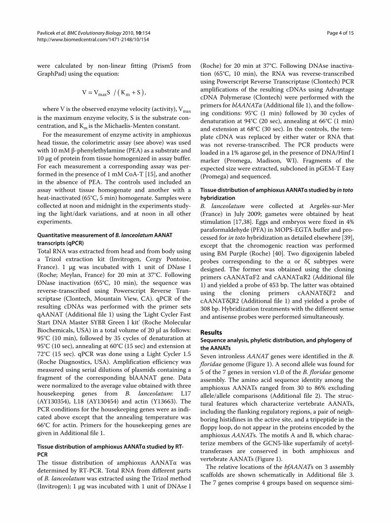
ResultsSequence analysis, phyletic distribution, and phylogeny of the AANATsSeven intronless AANAT genes were identified in the B.floridae genome (Figure 1). A second allele was found for5 of the 7 genes in version v1.0 of the B. floridae genomeassembly. The amino acid sequence identity among theamphioxus AANATs ranged from 30 to 86% excludingallele/allele comparisons (Additional file 2). The struc-tural features which characterize vertebrate AANATs,including the flanking regulatory regions, a pair of neigh-boring histidines in the active site, and a tripeptide in thefloppy loop, do not appear in the proteins encoded by theamphioxus AANATs. The motifs A and B, which charac-terize members of the GCN5-like superfamily of acetyl-transferases are conserved in both amphioxus andvertebrate AANATs (Figure 1).
The relative locations of the bfAANATs on 3 assemblyscaffolds are shown schematically in Additional file 3.The 7 genes comprise 4 groups based on sequence simi-
V V S K Smax m= +( )/ ,
Pavlicek et al. BMC Evolutionary Biology 2010, 10:154http://www.biomedcentral.com/1471-2148/10/154
Page 5 of 15
larity - group 1: αη group 2: β group 3: γε group 4: δζ.Primers were prepared to amplify members of each of thefour groups in B. lanceolatum; low sequence conserva-tion prevented amplification of all seven. The resultingPCR products were sequenced and designatedblAANATα, blAANATα' blAANATγ, and blAANATγδ(GenBank accession numbers FJ668653, FJ668656,FJ668654, FJ668655, respectively; α' is used to identify theα allele), according to the homology with individualbfAANAT genes; representatives of the other genes were
not obtained in this screen. The clone blAANATδζ wasdesignated this way because it could not be reliablyascribed to either the δ or ζ gene based on similarity tothe B. floridae counterparts. The amino acid sequenceidentity between the cloned PCR products for the B. lan-ceolatum α, α', γ and δζ isoforms and the correspondingB. floridae genes was 94%, 86%, 76%, and 73%, respec-tively. Comparisons of the amino acid sequences of thecloned blAANATα and blAANATδζ fragments to their B.floridae counterparts are shown in Additional file 4.
Examination of the phyletic distribution of the detectedAANAT family members shows that the family is repre-sented not only in chordates and cephalochordates butalso in some invertebrates such as the primitive metazoanTrichoplax (four paralogs), the segmented worms Capi-tella and Alvinella, and the mollusk Mytilus. Among non-metazoan eukaryotes, AANAT homologs were detectedin different fungal lineages including basidiomycetes,ascomycetes, and the basal fungi Rhizopus oryzae andPhycomyces blakesleeanus, and in many unicellulareukaryotes (protists). The protists in which AANAThomologs were detected include representatives fromthree supergroups of eukaryotes, namely, plantae,chromalveolates and excavates [41]. In plantae, AANAThomologs were found in green and red algae; amongchromalveolates, members of the AANAT family werepresent in diatoms, oomycetes, and a haptophyte Emili-ania huxleyi; and, among excavates, an AANAT homologwas identified in the heterolobosean Naegleria gruberi.AANAT homologs are also present in a number of bacte-rial lineages including firmicutes, actinobacteria, pro-teobacteria and the bacteroidetes/chlorobi group (seeAdditional files 5 and 6).
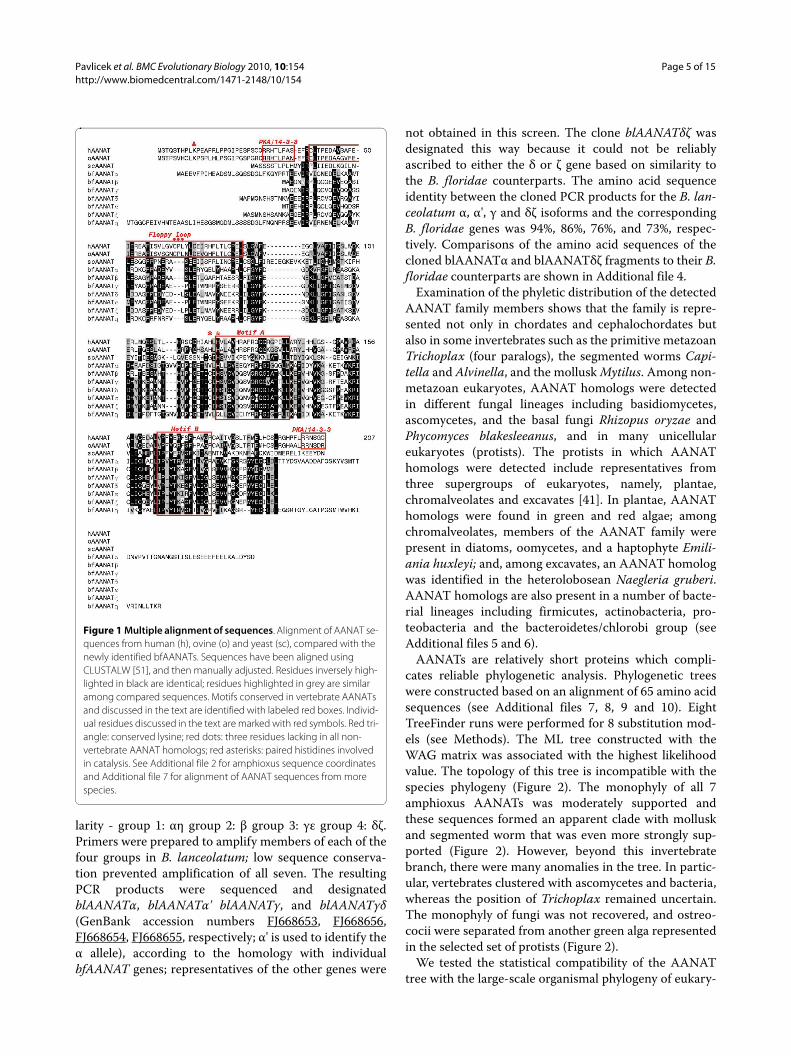
AANATs are relatively short proteins which compli-cates reliable phylogenetic analysis. Phylogenetic treeswere constructed based on an alignment of 65 amino acidsequences (see Additional files 7, 8, 9 and 10). EightTreeFinder runs were performed for 8 substitution mod-els (see Methods). The ML tree constructed with theWAG matrix was associated with the highest likelihoodvalue. The topology of this tree is incompatible with thespecies phylogeny (Figure 2). The monophyly of all 7amphioxus AANATs was moderately supported andthese sequences formed an apparent clade with molluskand segmented worm that was even more strongly sup-ported (Figure 2). However, beyond this invertebratebranch, there were many anomalies in the tree. In partic-ular, vertebrates clustered with ascomycetes and bacteria,whereas the position of Trichoplax remained uncertain.The monophyly of fungi was not recovered, and ostreo-cocii were separated from another green alga representedin the selected set of protists (Figure 2).
We tested the statistical compatibility of the AANATtree with the large-scale organismal phylogeny of eukary-
Figure 1 Multiple alignment of sequences. Alignment of AANAT se-quences from human (h), ovine (o) and yeast (sc), compared with the newly identified bfAANATs. Sequences have been aligned using CLUSTALW [51], and then manually adjusted. Residues inversely high-lighted in black are identical; residues highlighted in grey are similar among compared sequences. Motifs conserved in vertebrate AANATs and discussed in the text are identified with labeled red boxes. Individ-ual residues discussed in the text are marked with red symbols. Red tri-angle: conserved lysine; red dots: three residues lacking in all non-vertebrate AANAT homologs; red asterisks: paired histidines involved in catalysis. See Additional file 2 for amphioxus sequence coordinates and Additional file 7 for alignment of AANAT sequences from more species.
Pavlicek et al. BMC Evolutionary Biology 2010, 10:154http://www.biomedcentral.com/1471-2148/10/154
Page 6 of 15
otes using the constrained tree approach (see Methodsfor details). A constrained tree was created (Additionalfile 11) in which all species were combined into fourmajor clades in accord with the overall organismal phy-logeny, namely, bacteria, protists, fungi and animals.Inside the animal branch, with Trichoplax at the root, theamphioxus branch was placed as the sister group of verte-brates, and the segmented worms and mussel were posi-tioned between chordates and Trichoplax. The fungalbranch was placed as the sister group of animals. TheApproximately Unbiased test result (p-value 0.148) forthe tree generated with this approach (Additional files 12and 13) shows that the "classical" constrained tree topol-ogy could not be rejected with confidence although thelikelihood of that topology is much lower than that of thebest tree shown in Figure 2.
The results of the phylogenetic analysis of the AANATsemphasize the major distinction between the vertebrateforms and those from other animals, presumably, owingto a dramatic acceleration of evolution at the base of thevertebrate branch. Beyond that conclusion, however, theresults were quite uncertain due to the low informationcontent of the alignment, between-branch differences inthe evolutionary rates, and uneven sampling of differentgroups.
Expression and characterization of bfAANATα proteinA comparison of the predicted amino acid sequencesderived from the published genomic sequence and from
our cloned bfAANATα (GenBank accession numberEU380676) revealed differences in five amino acid resi-dues scattered throughout (Additional file 4). These dif-ferences, most likely, reflect the high degree ofpolymorphism in B. floridae [18]. The effects of these dif-ferences are unknown.
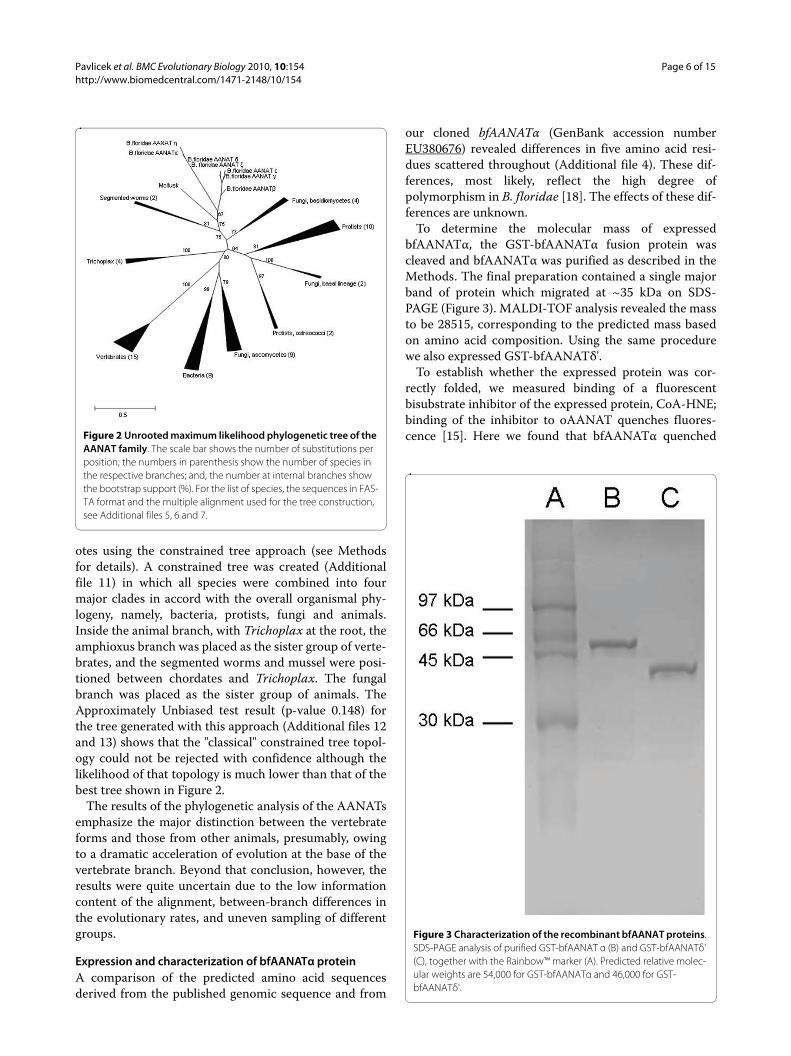
To determine the molecular mass of expressedbfAANATα, the GST-bfAANATα fusion protein wascleaved and bfAANATα was purified as described in theMethods. The final preparation contained a single majorband of protein which migrated at ~35 kDa on SDS-PAGE (Figure 3). MALDI-TOF analysis revealed the massto be 28515, corresponding to the predicted mass basedon amino acid composition. Using the same procedurewe also expressed GST-bfAANATδ'.
To establish whether the expressed protein was cor-rectly folded, we measured binding of a fluorescentbisubstrate inhibitor of the expressed protein, CoA-HNE;binding of the inhibitor to oAANAT quenches fluores-cence [15]. Here we found that bfAANATα quenched
Figure 3 Characterization of the recombinant bfAANAT proteins. SDS-PAGE analysis of purified GST-bfAANAT α (B) and GST-bfAANATδ' (C), together with the Rainbow™ marker (A). Predicted relative molec-ular weights are 54,000 for GST-bfAANATα and 46,000 for GST-bfAANATδ'.
Figure 2 Unrooted maximum likelihood phylogenetic tree of the AANAT family. The scale bar shows the number of substitutions per position; the numbers in parenthesis show the number of species in the respective branches; and, the number at internal branches show the bootstrap support (%). For the list of species, the sequences in FAS-TA format and the multiple alignment used for the tree construction, see Additional files 5, 6 and 7.
Pavlicek et al. BMC Evolutionary Biology 2010, 10:154http://www.biomedcentral.com/1471-2148/10/154
Page 7 of 15
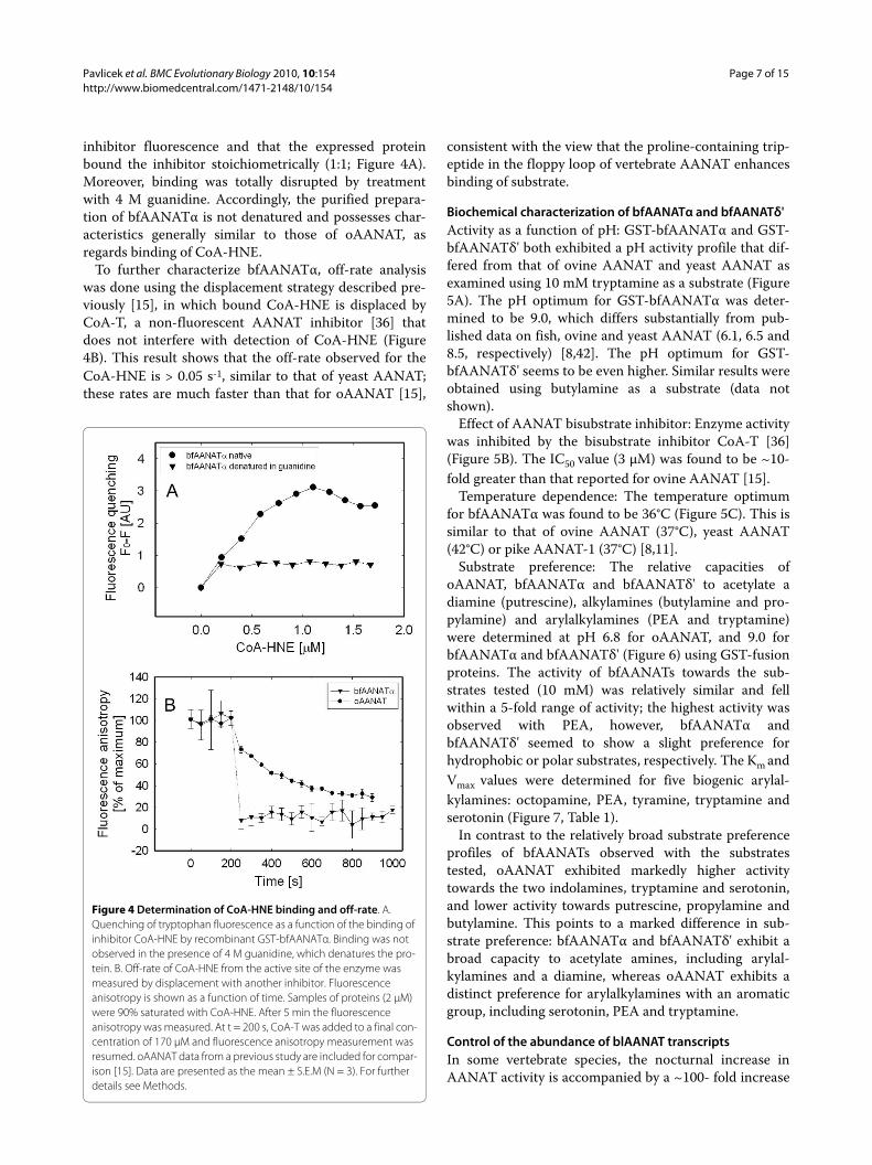
inhibitor fluorescence and that the expressed proteinbound the inhibitor stoichiometrically (1:1; Figure 4A).Moreover, binding was totally disrupted by treatmentwith 4 M guanidine. Accordingly, the purified prepara-tion of bfAANATα is not denatured and possesses char-acteristics generally similar to those of oAANAT, asregards binding of CoA-HNE.
To further characterize bfAANATα, off-rate analysiswas done using the displacement strategy described pre-viously [15], in which bound CoA-HNE is displaced byCoA-T, a non-fluorescent AANAT inhibitor [36] thatdoes not interfere with detection of CoA-HNE (Figure4B). This result shows that the off-rate observed for theCoA-HNE is > 0.05 s-1, similar to that of yeast AANAT;these rates are much faster than that for oAANAT [15],
consistent with the view that the proline-containing trip-eptide in the floppy loop of vertebrate AANAT enhancesbinding of substrate.
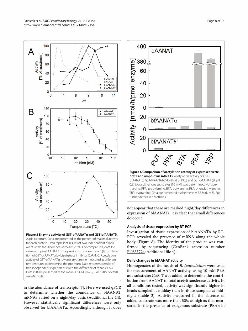
Biochemical characterization of bfAANATα and bfAANATδ'Activity as a function of pH: GST-bfAANATα and GST-bfAANATδ' both exhibited a pH activity profile that dif-fered from that of ovine AANAT and yeast AANAT asexamined using 10 mM tryptamine as a substrate (Figure5A). The pH optimum for GST-bfAANATα was deter-mined to be 9.0, which differs substantially from pub-lished data on fish, ovine and yeast AANAT (6.1, 6.5 and8.5, respectively) [8,42]. The pH optimum for GST-bfAANATδ' seems to be even higher. Similar results wereobtained using butylamine as a substrate (data notshown).
Effect of AANAT bisubstrate inhibitor: Enzyme activitywas inhibited by the bisubstrate inhibitor CoA-T [36](Figure 5B). The IC50 value (3 μM) was found to be ~10-fold greater than that reported for ovine AANAT [15].
Temperature dependence: The temperature optimumfor bfAANATα was found to be 36°C (Figure 5C). This issimilar to that of ovine AANAT (37°C), yeast AANAT(42°C) or pike AANAT-1 (37°C) [8,11].
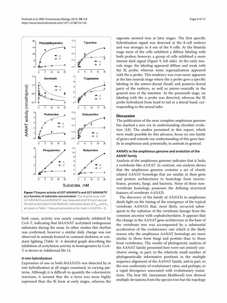
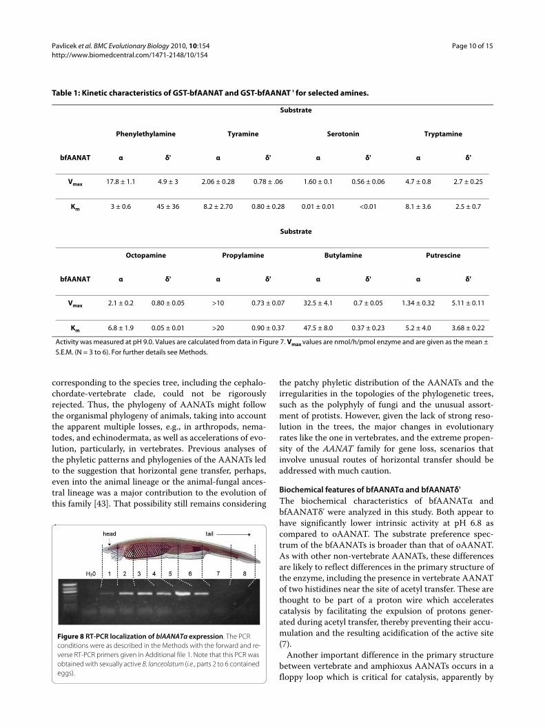
Substrate preference: The relative capacities ofoAANAT, bfAANATα and bfAANATδ' to acetylate adiamine (putrescine), alkylamines (butylamine and pro-pylamine) and arylalkylamines (PEA and tryptamine)were determined at pH 6.8 for oAANAT, and 9.0 forbfAANATα and bfAANATδ' (Figure 6) using GST-fusionproteins. The activity of bfAANATs towards the sub-strates tested (10 mM) was relatively similar and fellwithin a 5-fold range of activity; the highest activity wasobserved with PEA, however, bfAANATα andbfAANATδ' seemed to show a slight preference forhydrophobic or polar substrates, respectively. The Km andVmax values were determined for five biogenic arylal-kylamines: octopamine, PEA, tyramine, tryptamine andserotonin (Figure 7, Table 1).
In contrast to the relatively broad substrate preferenceprofiles of bfAANATs observed with the substratestested, oAANAT exhibited markedly higher activitytowards the two indolamines, tryptamine and serotonin,and lower activity towards putrescine, propylamine andbutylamine. This points to a marked difference in sub-strate preference: bfAANATα and bfAANATδ' exhibit abroad capacity to acetylate amines, including arylal-kylamines and a diamine, whereas oAANAT exhibits adistinct preference for arylalkylamines with an aromaticgroup, including serotonin, PEA and tryptamine.
Control of the abundance of blAANAT transcriptsIn some vertebrate species, the nocturnal increase inAANAT activity is accompanied by a ~100- fold increase
Figure 4 Determination of CoA-HNE binding and off-rate. A. Quenching of tryptophan fluorescence as a function of the binding of inhibitor CoA-HNE by recombinant GST-bfAANATα. Binding was not observed in the presence of 4 M guanidine, which denatures the pro-tein. B. Off-rate of CoA-HNE from the active site of the enzyme was measured by displacement with another inhibitor. Fluorescence anisotropy is shown as a function of time. Samples of proteins (2 μM) were 90% saturated with CoA-HNE. After 5 min the fluorescence anisotropy was measured. At t = 200 s, CoA-T was added to a final con-centration of 170 μM and fluorescence anisotropy measurement was resumed. oAANAT data from a previous study are included for compar-ison [15]. Data are presented as the mean ± S.E.M (N = 3). For further details see Methods.
Pavlicek et al. BMC Evolutionary Biology 2010, 10:154http://www.biomedcentral.com/1471-2148/10/154
Page 8 of 15
in the abundance of transcripts [7]. Here we used qPCRto determine whether the abundance of blAANATmRNAs varied on a night/day basis (Additional file 14).However statistically significant differences were onlyobserved for blAANATα. Accordingly, although it does
not appear that there are marked night/day differences inexpression of blAANATs, it is clear that small differencesdo occur.
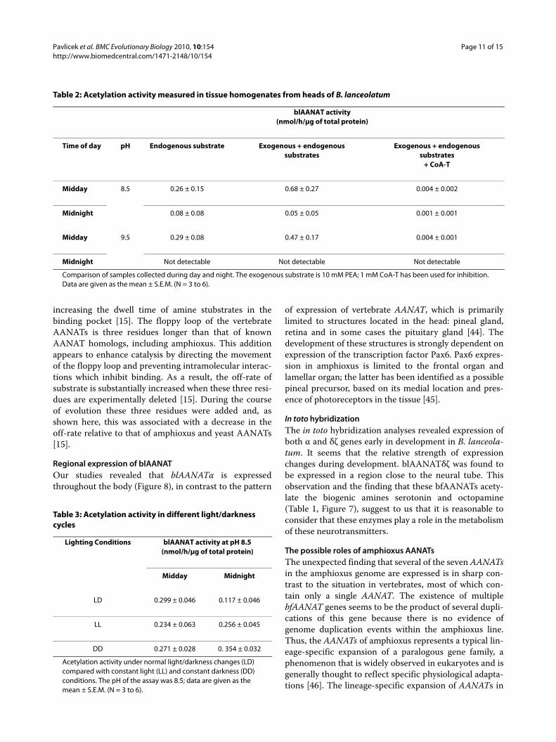
Analysis of tissue expression by RT-PCRInvestigation of tissue expression of blAANATα by RT-PCR revealed the presence of mRNA along the wholebody (Figure 8). The identity of the product was con-firmed by sequencing (GenBank accession numberEU635724; Additional file 4).
Daily changes in blAANAT activityHomogenates of the heads of B. lanceolatum were usedfor measurement of AANAT activity, using 10 mM PEAas a substrate; CoA-T was added to determine the contri-bution from AANAT to total acetyltransferase activity. Inall conditions tested, activity was significantly higher inheads sampled at midday than in those sampled at mid-night (Table 2). Activity measured in the absence ofadded substrate was more than 50% as high as that mea-sured in the presence of exogenous substrate (PEA); in
Figure 5 Enzyme activity of GST-bfAANATα and GST-bfAANATδ'. A. pH optimum. Data are presented as the percent of maximal activity for each protein. Data represent results of two independent experi-ments with the difference of means < 5%. For comparison, data for ovine and yeast AANAT from a previous study are shown [8]. B. Inhibi-tion of GST-bfAANATα by bisubstrate inhibitor CoA-T. C. Acetylation activity of GST-bfAANATα towards tryptamine measured at different temperatures to determine the optimum. Data represent results of two independent experiments with the difference of means < 5%. Data in B are presented as the mean ± S.E.M (N = 3). For further details see Methods.
Figure 6 Comparison of acetylation activity of expressed verte-brate and amphioxus AANATs. Acetylation activity of GST-bfAANATα, GST-bfAANATδ' (both at pH 9.0) and GST-oAANAT (at pH 6.8) towards various substrates (10 mM) was determined. PUT: pu-trescine, PPA: propylamine, BTA: butylamine, PEA: phenylethylamine, TRP: tryptamine. Data are presented as the mean ± S.E.M (N = 3). For further details see Methods.
Pavlicek et al. BMC Evolutionary Biology 2010, 10:154http://www.biomedcentral.com/1471-2148/10/154
Page 9 of 15
both cases, activity was nearly completely inhibited byCoA-T, indicating that blAANAT acetylated endogenoussubstrates during the assay. In other studies this rhythmwas confirmed, however a similar daily change was notobserved in animals housed in constant darkness or con-stant lighting (Table 3). A detailed graph describing theinhibition of acetylation activity in homogenates by CoA-T is shown in Additional file 15.
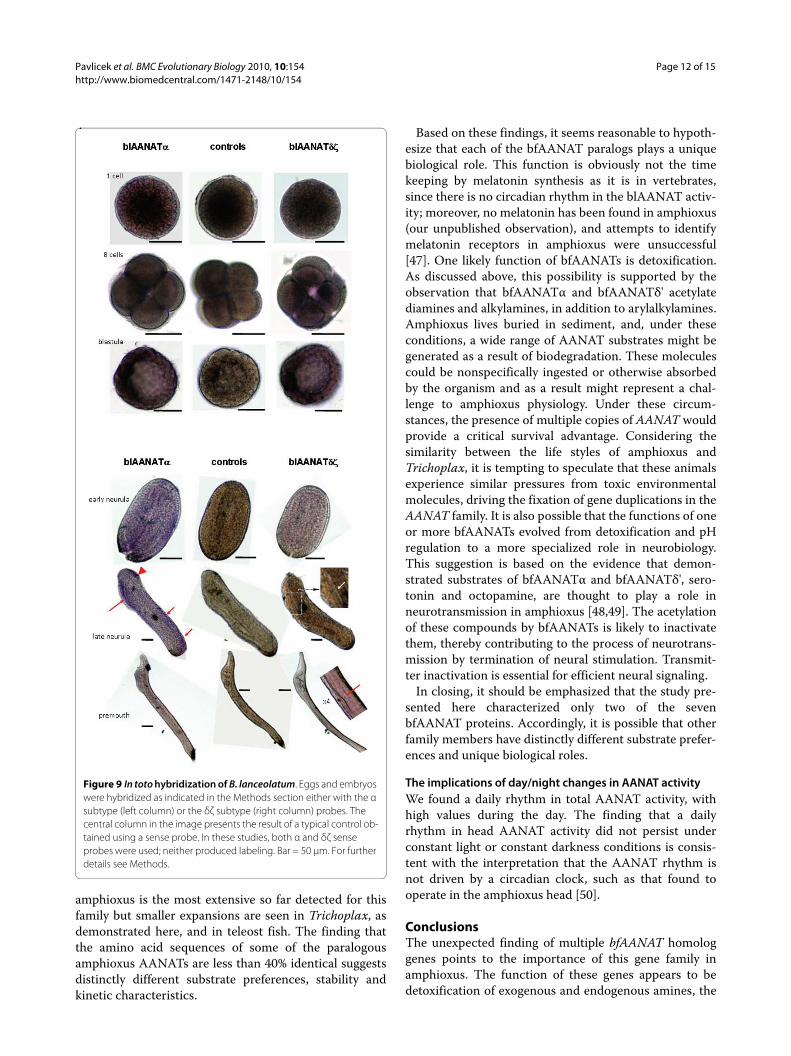
In toto hybridizationExpression of one or both blAANATs was detected by intoto hybridization at all stages examined, in varying pat-terns. Although it is difficult to quantify the colorimetricreactions, it seemed that the α form was more highlyexpressed than the δζ form at early stages, whereas the
opposite seemed true at later stages. The first specifichybridization signal was detected at the 8-cell embryoand was stronger in 4 out of the 8 cells. At the blastulastage most of the cells exhibited a diffuse labeling withboth probes; however, a group of cells exhibited a moreintense dark signal (Figure 9, left side). At the early neu-rula stage, the labeling appeared diffuse and weak withthe δζ probe, whereas some regionalization appearedwith the α probe. This tendency was even more apparentat the late neurula stage where the α probe gave a specificlabeling in the antero-dorsal (head) and posterio-dorsalparts of the embryo, as well as antero-ventrally in thegeneral area of the intestine. At the premouth stage, nolabeling with the α probe was detected, whereas the δζprobe hybridized from head to tail in a dorsal band, cor-responding to the neural tube.
DiscussionThe publication of the near complete amphioxus genomehas marked a new era in understanding chordate evolu-tion [18]. The studies presented in this report, whichwere made possible by this advance, focus on one familyof genes and extends our understanding of this gene fam-ily in amphioxus and, potentially, in animals in general.
AANATs in the amphioxus genome and evolution of the AANAT familyAnalysis of the amphioxus genome indicates that it lacksa vertebrate-like AANAT. In contrast, our analysis showsthat the amphioxus genome contains a set of closelyrelated AANAT homologs that are similar in their geneand protein architectures to homologs from inverte-brates, protists, fungi, and bacteria. None of these non-vertebrate homologs possesses the defining structuralfeatures of vertebrate AANATs.
The discovery of the family of AANATs in amphioxussheds light on the timing of the emergence of the typicalvertebrate AANATs that, most likely, occurred subse-quent to the radiation of the vertebrate lineage from thecommon ancestor with cephalochordates. It appears thatthe change in the AANAT gene architecture at the base ofthe vertebrate tree was accompanied by a substantialacceleration of the evolutionary rate which is the likelyreason why the amphioxus AANAT homologs are moresimilar to those from fungi and protists than to thosefrom vertebrates. The results of phylogenetic analysis ofthe AANAT family presented here were not entirely con-clusive owing, in part, to the relatively small number ofphylogenetically informative positions in the multiplesequence alignment of the AANAT family, and in part, tothe non-uniformity of evolutionary rates, and perhaps, toa rapid divergence associated with evolutionary transi-tions. The best ML (maximum likelihood) tree showedmultiple deviations from the species tree but the topology
Figure 7 Enzyme activity of GST-bfAANATα and GST-bfAANATδ' as a function of substrate concentration. The enzyme assay with GST-bfAANATα and bfAANATδ' was measured at pH 9.0 and was per-formed as described in the Methods. Calculated values of Vmax and Km
are given in Table 1. Data are presented as the mean ± S.E.M (N = 3).
Pavlicek et al. BMC Evolutionary Biology 2010, 10:154http://www.biomedcentral.com/1471-2148/10/154
Page 10 of 15
corresponding to the species tree, including the cephalo-chordate-vertebrate clade, could not be rigorouslyrejected. Thus, the phylogeny of AANATs might followthe organismal phylogeny of animals, taking into accountthe apparent multiple losses, e.g., in arthropods, nema-todes, and echinodermata, as well as accelerations of evo-lution, particularly, in vertebrates. Previous analyses ofthe phyletic patterns and phylogenies of the AANATs ledto the suggestion that horizontal gene transfer, perhaps,even into the animal lineage or the animal-fungal ances-tral lineage was a major contribution to the evolution ofthis family [43]. That possibility still remains considering
the patchy phyletic distribution of the AANATs and theirregularities in the topologies of the phylogenetic trees,such as the polyphyly of fungi and the unusual assort-ment of protists. However, given the lack of strong reso-lution in the trees, the major changes in evolutionaryrates like the one in vertebrates, and the extreme propen-sity of the AANAT family for gene loss, scenarios thatinvolve unusual routes of horizontal transfer should beaddressed with much caution.
Biochemical features of bfAANATα and bfAANATδ'The biochemical characteristics of bfAANATα andbfAANATδ' were analyzed in this study. Both appear tohave significantly lower intrinsic activity at pH 6.8 ascompared to oAANAT. The substrate preference spec-trum of the bfAANATs is broader than that of oAANAT.As with other non-vertebrate AANATs, these differencesare likely to reflect differences in the primary structure ofthe enzyme, including the presence in vertebrate AANATof two histidines near the site of acetyl transfer. These arethought to be part of a proton wire which acceleratescatalysis by facilitating the expulsion of protons gener-ated during acetyl transfer, thereby preventing their accu-mulation and the resulting acidification of the active site(7).
Another important difference in the primary structurebetween vertebrate and amphioxus AANATs occurs in afloppy loop which is critical for catalysis, apparently by
Table 1: Kinetic characteristics of GST-bfAANAT and GST-bfAANAT ' for selected amines.
Substrate
Phenylethylamine Tyramine Serotonin Tryptamine
bfAANAT α δ' α δ' α δ' α δ'
Vmax 17.8 ± 1.1 4.9 ± 3 2.06 ± 0.28 0.78 ± .06 1.60 ± 0.1 0.56 ± 0.06 4.7 ± 0.8 2.7 ± 0.25
Km 3 ± 0.6 45 ± 36 8.2 ± 2.70 0.80 ± 0.28 0.01 ± 0.01 <0.01 8.1 ± 3.6 2.5 ± 0.7
Substrate
Octopamine Propylamine Butylamine Putrescine
bfAANAT α δ' α δ' α δ' α δ'
Vmax 2.1 ± 0.2 0.80 ± 0.05 >10 0.73 ± 0.07 32.5 ± 4.1 0.7 ± 0.05 1.34 ± 0.32 5.11 ± 0.11
Km 6.8 ± 1.9 0.05 ± 0.01 >20 0.90 ± 0.37 47.5 ± 8.0 0.37 ± 0.23 5.2 ± 4.0 3.68 ± 0.22
Activity was measured at pH 9.0. Values are calculated from data in Figure 7. Vmax values are nmol/h/pmol enzyme and are given as the mean ± S.E.M. (N = 3 to 6). For further details see Methods.
Figure 8 RT-PCR localization of blAANATα expression. The PCR conditions were as described in the Methods with the forward and re-verse RT-PCR primers given in Additional file 1. Note that this PCR was obtained with sexually active B. lanceolatum (i.e., parts 2 to 6 contained eggs).
Pavlicek et al. BMC Evolutionary Biology 2010, 10:154http://www.biomedcentral.com/1471-2148/10/154
Page 11 of 15
increasing the dwell time of amine stubstrates in thebinding pocket [15]. The floppy loop of the vertebrateAANATs is three residues longer than that of knownAANAT homologs, including amphioxus. This additionappears to enhance catalysis by directing the movementof the floppy loop and preventing intramolecular interac-tions which inhibit binding. As a result, the off-rate ofsubstrate is substantially increased when these three resi-dues are experimentally deleted [15]. During the courseof evolution these three residues were added and, asshown here, this was associated with a decrease in theoff-rate relative to that of amphioxus and yeast AANATs[15].
Regional expression of blAANATOur studies revealed that blAANATα is expressedthroughout the body (Figure 8), in contrast to the pattern
of expression of vertebrate AANAT, which is primarilylimited to structures located in the head: pineal gland,retina and in some cases the pituitary gland [44]. Thedevelopment of these structures is strongly dependent onexpression of the transcription factor Pax6. Pax6 expres-sion in amphioxus is limited to the frontal organ andlamellar organ; the latter has been identified as a possiblepineal precursor, based on its medial location and pres-ence of photoreceptors in the tissue [45].
In toto hybridizationThe in toto hybridization analyses revealed expression ofboth α and δζ genes early in development in B. lanceola-tum. It seems that the relative strength of expressionchanges during development. blAANATδζ was found tobe expressed in a region close to the neural tube. Thisobservation and the finding that these bfAANATs acety-late the biogenic amines serotonin and octopamine(Table 1, Figure 7), suggest to us that it is reasonable toconsider that these enzymes play a role in the metabolismof these neurotransmitters.
The possible roles of amphioxus AANATsThe unexpected finding that several of the seven AANATsin the amphioxus genome are expressed is in sharp con-trast to the situation in vertebrates, most of which con-tain only a single AANAT. The existence of multiplebfAANAT genes seems to be the product of several dupli-cations of this gene because there is no evidence ofgenome duplication events within the amphioxus line.Thus, the AANATs of amphioxus represents a typical lin-eage-specific expansion of a paralogous gene family, aphenomenon that is widely observed in eukaryotes and isgenerally thought to reflect specific physiological adapta-tions [46]. The lineage-specific expansion of AANATs in
Table 2: Acetylation activity measured in tissue homogenates from heads of B. lanceolatum
blAANAT activity(nmol/h/μg of total protein)
Time of day pH Endogenous substrate Exogenous + endogenoussubstrates
Exogenous + endogenoussubstrates
+ CoA-T
Midday 8.5 0.26 ± 0.15 0.68 ± 0.27 0.004 ± 0.002
Midnight 0.08 ± 0.08 0.05 ± 0.05 0.001 ± 0.001
Midday 9.5 0.29 ± 0.08 0.47 ± 0.17 0.004 ± 0.001
Midnight Not detectable Not detectable Not detectable
Comparison of samples collected during day and night. The exogenous substrate is 10 mM PEA; 1 mM CoA-T has been used for inhibition. Data are given as the mean ± S.E.M. (N = 3 to 6).
Table 3: Acetylation activity in different light/darkness cycles
Lighting Conditions blAANAT activity at pH 8.5(nmol/h/μg of total protein)
Midday Midnight
LD 0.299 ± 0.046 0.117 ± 0.046
LL 0.234 ± 0.063 0.256 ± 0.045
DD 0.271 ± 0.028 0. 354 ± 0.032
Acetylation activity under normal light/darkness changes (LD) compared with constant light (LL) and constant darkness (DD) conditions. The pH of the assay was 8.5; data are given as the mean ± S.E.M. (N = 3 to 6).
Pavlicek et al. BMC Evolutionary Biology 2010, 10:154http://www.biomedcentral.com/1471-2148/10/154
Page 12 of 15
amphioxus is the most extensive so far detected for thisfamily but smaller expansions are seen in Trichoplax, asdemonstrated here, and in teleost fish. The finding thatthe amino acid sequences of some of the paralogousamphioxus AANATs are less than 40% identical suggestsdistinctly different substrate preferences, stability andkinetic characteristics.
Based on these findings, it seems reasonable to hypoth-esize that each of the bfAANAT paralogs plays a uniquebiological role. This function is obviously not the timekeeping by melatonin synthesis as it is in vertebrates,since there is no circadian rhythm in the blAANAT activ-ity; moreover, no melatonin has been found in amphioxus(our unpublished observation), and attempts to identifymelatonin receptors in amphioxus were unsuccessful[47]. One likely function of bfAANATs is detoxification.As discussed above, this possibility is supported by theobservation that bfAANATα and bfAANATδ' acetylatediamines and alkylamines, in addition to arylalkylamines.Amphioxus lives buried in sediment, and, under theseconditions, a wide range of AANAT substrates might begenerated as a result of biodegradation. These moleculescould be nonspecifically ingested or otherwise absorbedby the organism and as a result might represent a chal-lenge to amphioxus physiology. Under these circum-stances, the presence of multiple copies of AANAT wouldprovide a critical survival advantage. Considering thesimilarity between the life styles of amphioxus andTrichoplax, it is tempting to speculate that these animalsexperience similar pressures from toxic environmentalmolecules, driving the fixation of gene duplications in theAANAT family. It is also possible that the functions of oneor more bfAANATs evolved from detoxification and pHregulation to a more specialized role in neurobiology.This suggestion is based on the evidence that demon-strated substrates of bfAANATα and bfAANATδ', sero-tonin and octopamine, are thought to play a role inneurotransmission in amphioxus [48,49]. The acetylationof these compounds by bfAANATs is likely to inactivatethem, thereby contributing to the process of neurotrans-mission by termination of neural stimulation. Transmit-ter inactivation is essential for efficient neural signaling.
In closing, it should be emphasized that the study pre-sented here characterized only two of the sevenbfAANAT proteins. Accordingly, it is possible that otherfamily members have distinctly different substrate prefer-ences and unique biological roles.
The implications of day/night changes in AANAT activityWe found a daily rhythm in total AANAT activity, withhigh values during the day. The finding that a dailyrhythm in head AANAT activity did not persist underconstant light or constant darkness conditions is consis-tent with the interpretation that the AANAT rhythm isnot driven by a circadian clock, such as that found tooperate in the amphioxus head [50].
ConclusionsThe unexpected finding of multiple bfAANAT homologgenes points to the importance of this gene family inamphioxus. The function of these genes appears to bedetoxification of exogenous and endogenous amines, the
Figure 9 In toto hybridization of B. lanceolatum. Eggs and embryos were hybridized as indicated in the Methods section either with the α subtype (left column) or the δζ subtype (right column) probes. The central column in the image presents the result of a typical control ob-tained using a sense probe. In these studies, both α and δζ sense probes were used; neither produced labeling. Bar = 50 μm. For further details see Methods.
Pavlicek et al. BMC Evolutionary Biology 2010, 10:154http://www.biomedcentral.com/1471-2148/10/154
Page 13 of 15
latter including neurotransmitters. It will be of interest todetermine the precise distribution of each gene, howtheir expression is regulated and how each gene contrib-utes to amphioxus biology.
Additional material
AbbreviationsAANAT: arylalkylamine N-acetyltransferase; oAANAT: ovine AANAT; scAANAT:Saccharomyces cerevisiae AANAT; bfAANAT: B. floridae AANAT; blAANAT: B. lance-olatum AANAT; AcCoA: acetyl coenzyme A; AU: arbitrary unit; CoA-HNE:hydroxynaphthylethylamine coenzyme A; CoA-T: tryptamine coenzyme A;DTT: dithiothreitol; GST: glutathione S-transferase; PBS: phosphate buffer saline;PEA: phenylethylamine; TCEP: tris[2-carboxyethyl]phosphine.
Authors' contributionsJP performed biochemical, physico-chemical, molecular biological studies anddrafted the manuscript, SS participated in biochemical studies, LB participatedin biological studies, SLC participated in molecular genetic studies and helpedto draft the manuscript, JLW participated in biochemical studies, GB, PG and JFparticipated in biochemical and biological studies, MVO and EVK performedsequence and phylogenetic analysis and DCK conceived of the study, partici-pated in its coordination and helped to draft the manuscript. All authors readand approved the final manuscript.
AcknowledgementsThis work was supported by the NIH Intramural Research Program through the NICHD, National Institutes of Health. The costs of publication of this article were defrayed in part by the payment of page charges. This article must there-fore be hereby marked "advertisement" in accordance with 18 U.S.C. Section 1734 solely to indicate this fact. Work was also supported by the CNRS, UPMC and the French National Research Agency (project TEMPANAT, ANR-07-BLAN-0097). We are grateful to Dr. Dan L. Sackett for help with fluorescence measure-ment and Drs. Howard Jaffe and Sam J. Clokie for help with the MALDI-TOF analysis of protein samples.
Author Details1Section on Neuroendocrinology, Program in Developmental Endocrinology and Genetics, The Eunice Kennedy Shriver National Institute of Child Health and Human Development, National Institutes of Health, Bethesda, 20892, MD, USA, 2Laboratoire Arago, UPMC-Paris 6 and CNRS, FRE 3247 & GDR 2821, Facteurs du Milieu & Mécanismes Adaptatifs, Banyuls/Mer, 66651, France, 3Museum National d'Histoire Naturelle (MNHN), 43 rue Cuvier, Paris, 75005, France and 4National Center for Biotechnology Information, National Library of Medicine, National Institutes of Health, Bethesda, 20894, MD, USA
References1. Dyda F, Klein DC, Hickman AB: GCN5-related N-acetyltransferases: a
structural overview. Annu Rev Biophys Biomol Struct 2000, 29:81-103.2. Coon SL, Klein DC: Evolution of arylalkylamine N-acetyltransferase:
emergence and divergenceMol Cell Endocrinol. Mol Cell Endocrinol 2006, 252:2-10.
Additional file 1 Primers used for cloning PCR, RT-PCR localization of blAANAT expression and qPCR experiments. Cloning primers were based on B. floridae genomic sequence and used to amplify the indicated blAANATs.Additional file 2 Pairwise comparison scores of protein sequences. Pairwise comparison scores based on CLUSTALW multiple sequence align-ment of deduced peptide sequences of all 7 amphioxus AANAT homologs and their known alleles. Shaded boxes designate comparisons between alleles. The coordinates of each gene and allele (') in genomic assembly v1.0 are (scaffold number:sequence location): bfAANATα, 54:2,458,444-2,459,187; bfAANATα', 56:737,209-736,466; bfAANATβ, 54:2,461,188-2,461,679; bfAANATβ', 56:734,493-734,002; bfAANATγ, 54:2,488,683-2,489,174; bfAANATγ', 56:702,576-702,085; bfAANATδ, 54:2,491,399-2,491,929; bfAANATδ', 54:699,628-699,095; bfAANATε, 200:1,410,854-1,410,362 (includes deletion of 1 of 6 consecutive A's at bp 133 to maintain reading frame); bfAANATζ, 200:1,409,115-1,408,588; bfAANATη, 234:825,177-825,883 (includes insertion of 1 base [4th A] at position 442 to maintain reading frame); bfAANATη', 81:2,115,130-2,115,834 (includes insertion of 1 base [4th
T] at position 106 to maintain reading frame; and deletion of 1 base [4th G] at position 652 to maintain reading frame).Additional file 3 Positioning of the 7 genes for bfAANAT on the assem-bly scaffolds as given in B. floridae genome assembly v2.0.Additional file 4 Comparison of deduced amino acid sequences of characterized AANATs to reference genomic sequences. A. AANATα. B. AANATδ'. (g) denotes derived from the published B. floridae genomic sequence. (c) denotes cloned from B. floridae head cDNA, and are the full length recombinant proteins used for enzyme characterization. (f ) denotes a partial fragment cloned from B. lanceolatum head cDNA and used as a probe for in toto hybridization. Highlighted residues are those that differ from the published B. floridae genomic sequence. "--", gap inserted to pre-serve alignment; "...", missing sequence.Additional file 5 List of species used for construction of phylogenic tree. List of species containing AANAT homologs used for the construction of the phylogenetic tree shown in Figure 2. "gi" numbers precede the spe-cies name, where available; the source of the sequence is given in paren-theses. Sequences for which "gi" numbers are not available are given in Additional file 6.Additional file 6 FASTA sequences of AANAT homologs. FASTA sequences of those of AANAT homologs used for the construction of the phylogenetic tree shown in Figure 2, but not having "gi" numbers, (as listed in Additional file 5).Additional file 7 Multiple alignment used for construction of the phy-logenic tree. Multiple sequence alignment of the conserved regions of selected AANAT sequences from various species used for the construction of the phylogenetic tree in Figure 2. Species included in this alignment are given in Additional file 5.Additional file 8 Average evolutionary distance (substitution per position) between major taxonomic groups for AANAT proteins based on the truncated alignment shown in Additional file 7 (JTT substitu-tion model, gamma distribution 1).Additional file 9 Average differences (p-distance) between major tax-onomic groups for AANAT proteins, based on the truncated align-ment shown in Additional file7, calculated using the MEGA program34[34].Additional file 10 Average percent identity between major taxo-nomic groups for AANAT proteins based on the truncated alignment shown in Additional file7, calculated from Additional file9.Additional file 11 Model of constrained large-scale taxonomic topol-ogy tree of selected AANAT sequences.
Additional file 12 Topology tree - normal view. Reconstructed con-strained large-scale taxonomic topology tree of selected AANAT sequences - normal view based on model shown on Additional file 11. The scale bar represents the number of substitutions per position; numbers in parenthe-sis show number of species. The number at internal branches shows the bootstrap support (%).Additional file 13 Topology tree - radiation view. Reconstructed con-strained large-scale taxonomic topology tree of selected AANAT sequences - radiation view based on model shown on Additional file 11. The scale bar represents the number of substitutions per position; numbers in parenthe-sis show number of species. The number at internal branches shows the bootstrap support (%).Additional file 14 Comparison of day/night expression of three blAANATs detected by qPCR in the head and body of amphioxus. Prim-ers used are given in Additional file 1. Asterisk indicates statistical signifi-cance of day/night differences. Data are presented as the mean ± S.E.M. (N = 3). For further details see Methods.Additional file 15 Inhibition of acetylation activity. Inhibition of acety-lation activity in homogenate from amphioxus heads at pH 8.5 using 10 mM PEA and 0.5 mM AcCoA as a substrate and various concentrations of CoA-T as an inhibitor. Data are presented as the mean ± S.E.M. (N = 3).
Received: 30 July 2009 Accepted: 25 May 2010 Published: 25 May 2010This article is available from: http://www.biomedcentral.com/1471-2148/10/154© 2010 Pavlicek et al; licensee BioMed Central Ltd. This is an Open Access article distributed under the terms of the Creative Commons Attribution License (http://creativecommons.org/licenses/by/2.0), which permits unrestricted use, distribution, and reproduction in any medium, provided the original work is properly cited.BMC Evolutionary Biology 2010, 10:154
Pavlicek et al. BMC Evolutionary Biology 2010, 10:154http://www.biomedcentral.com/1471-2148/10/154
Page 14 of 15
3. Vetting MWS, de Carvalho LP, Yu M, Hegde SS, Magnet S, Roderick SL, Blanchard JS: Structure and functions of the GNAT superfamily of acetyltransferases. Arch Biochem Biophys 2005, 433:212-226.
4. Klein DC, Coon SL, Roseboom PH, Weller JL, Bernard M, Gastel JA, Zatz M, Iuvone PM, Rodriguez IR, Begay V, Falcon J, Cahill GM, Cassone VM, Baler R: The melatonin rhythm-generating enzyme: molecular regulation of serotonin N-acetyltransferase in the pineal gland. Recent Prog Horm Res 1997, 52:307-357.
5. Coon SL, Roseboom PH, Baler R, Weller JL, Namboodiri MA, Koonin EV, Klein DC: Pineal serotonin N-acetyltransferase: expression cloning and molecular analysis. Science 1995, 270:1681-1683.
6. Arendt J: Melatonin and the Mammalian Pineal Gland. London: Chapman and Hall; 1995.
7. Klein DC: Arylalkylamine N-acetyltransferase: "the Timezyme". J Biol Chem 2007, 282:4233-4237.
8. Ganguly S, Mummaneni P, Steinbach PJ, Klein DC, Coon SL: Characterization of the Saccharomyces cerevisiae homolog of the melatonin rhythm enzyme arylalkylamine N-acetyltransferase (EC 2.3.1.87). J Biol Chem 2001, 276:47239-47247.
9. Liu B, Sutton A, Sternglanz R: A yeast polyamine acetyltransferase. J Biol Chem 2005, 280:16659-16664.
10. Begay V, Falcon J, Cahill GM, Klein DC, Coon SL: Transcripts encoding two melatonin synthesis enzymes in the teleost pineal organ: circadian regulation in pike and zebrafish, but not in trout. Endocrinology 1998, 139:905-912.
11. Coon SL, Begay V, Deurloo D, Falcon J, Klein DC: Two arylalkylamine N-acetyltransferase genes mediate melatonin synthesis in fish. J Biol Chem 1999, 274:9076-9082.
12. Falcon J, Gothilf Y, Coon SL, Boeuf G, Klein DC: Genetic, temporal and developmental differences between melatonin rhythm generating systems in the teleost fish pineal organ and retina. J Neuroendocrinol 2003, 14:378-382.
13. Zilberman-Peled B, Benhar I, Coon SL, Ron B, Gothilf Y: Duality of serotonin-N-acetyltransferase in the gilthead seabream (Sparus aurata): molecular cloning and characterization of recombinant enzymes. Gen Comp Endocrinol 2004, 138:139-147.
14. Scheibner KA, De Angelis AJ, Burley SK, Cole PA: Investigation of the roles of catalytic residues in serotonin N-acetyltransferase. J Biol Chem 2002, 277:18118-18126.
15. Pavlicek J, Coon SL, Ganguly S, Weller JL, Hassan SA, Sackett DL, Klein DC: Evidence that proline focuses movement of the floppy loop of arylalkylamine N-acetyltransferase (EC 2.3.1.87). J Biol Chem 2008, 283:14552-14558.
16. Iyer LM, Aravind L, Coon SL, Klein DC, Koonin EV: Evolution of cell-cell signaling in animals: did late horizontal gene transfer from bacteria have a role? Trends Genet 2004, 20:292-299.
17. Fuentes M, Schubert M, Dalfo D, Candiani S, Benito E, Gardenyes J, Godoy L, Moret F, Illas M, Patten I, Permanyer J, Oliveri D, Boeuf G, Falcon J, Pestarino M, Fernandez JG, Albalat R, Laudet V, Vernier P, Escriva H: Preliminary observations on the spawning conditions of the European amphioxus (Branchiostoma lanceolatum) in captivity. J Exp Zoolog B Mol Dev Evol 2004, 302:384-391.
18. Putnam NH, Butts T, Ferrier DE, Furlong RF, Hellsten U, Kawashima T, Robinson-Rechavi M, Shoguchi E, Terry A, Yu JK, Benito-Gutiérrez EL, Dubchak I, Garcia-Fernàndez J, Gibson-Brown JJ, Grigoriev IV, Horton AC, de Jong PJ, Jurka J, Kapitonov VV, Kohara Y, Kuroki Y, Lindquist E, Lucas S, Osoegawa K, Pennacchio LA, Salamov AA, Satou Y, Sauka-Spengler T, Schmutz J, Shin-I T, Toyoda A, Bronner-Fraser M, Fujiyama A, Holland LZ, Holland PW, Satoh N, Rokhsar DS: The amphioxus genome and the evolution of the chordate karyotype. Nature 2008, 453:1064-1071.
19. Altschul SF, Madden TL, Schäffer AA, Zhang J, Zhang Z, Miller W, Lipman DJ: Gapped BLAST and PSI-BLAST: a new generation of protein database search programs. Nucleic Acids Res 1997, 25:3389-3402.
20. Edgar RC: MUSCLE: multiple sequence alignment with high accuracy and high throughput. Nucleic Acids Res 2004, 32:1792-1797.
21. Jobb G, von Haeseler A, Strimmer K: TREEFINDER: a powerful graphical analysis environment for molecular phylogenetics. BMC Evol Biol 2004, 4:18.
22. Whelan S, Goldman N: A general empirical model of protein evolution derived from multiple protein families using a maximum-likelihood approach. Mol Biol Evol 2001, 18:691-699.
23. Jones DT, Taylor WR, Thornton JM: The rapid generation of mutation data matrices from protein sequences. Comput Appl Biosci 1992, 8:275-282.
24. Müller T, Vingron M: Modeling amino acid replacement. J Comput Biol 2000, 7:761-776.
25. Henikoff S, Henikoff JG: Amino acid substitution matrices from protein blocks. Proc Natl Acad Sci USA 1992, 89:10915-10919.
26. Kosiol C, Goldman N: Different versions of the Dayhoff rate matrix. Mol Biol Evol 2005, 22:193-199.
27. Adachi J, Waddell PJ, Martin W, Hasegawa M: Plastid genome phylogeny and a model of amino acid substitution for proteins encoded by chloroplast DNA. J Mol Evol 2000, 50:348-358.
28. Dimmic MW, Rest JS, Mindell DP, Goldstein RA: rtREV: an amino acid substitution matrix for inference of retrovirus and reverse transcriptase phylogeny. J Mol Evol 2002, 55:65-73.
29. Veerassamy S, Smith A, Tillier ER: A transition probability model for amino acid substitutions from blocks. J Comput Biol 2003, 10:997-1010.
30. Strimmer K, Rambaut A: Inferring confidence sets of possibly misspecified trees. Proc Biol Sci 2002, 269:137-142.
31. Page RD: TreeView: an application to display phylogenetic trees on personal computers. Comput Appl Biosci 1996, 12:357-358.
32. Shimodaira H: An approximately unbiased test of phylogenetic tree selection. Syst Biol 2002, 51:492-508.
33. Felsenstein J: Inferring phylogenies from protein sequences by parsimony, distance, and likelihood methods. Methods Enzymol 1996, 266:418-427.
34. Kumar S, Tamura K, Nei M: MEGA3: Integrated software for Molecular Evolutionary Genetics Analysis and sequence alignment. Briefings in Bioinformatics 2004, 2:150-163.
35. De Angelis J, Gastel J, Klein DC, Cole PA: Kinetic analysis of the catalytic mechanism of serotonin N-acetyltransferase (EC 2.3.1.87). J Biol Chem 1998, 273:3045-3050.
36. Khalil EM, Cole PA: A potent inhibitor of the melatonin rhythm enzyme. J Am Chem Soc 1998, 120:6195-6196.
37. Laemmli UK: Cleavage of structural proteins during the assembly of the head of bacteriophage T4. Nature 1970, 227:680-685.
38. Fuentès M, Benito E, Bertrand S, Paris M, Mignardot A, Godoy L, Jimenez-Delgad S, Oliveri D, Candiani S, Hirsinger E, D'Aniello S, Pascual-Anaya J, Maeso J, Pestarino M, Vernier P, Nicolas JF, Schubert M, Laudet V, Geneviere AM, Albalat R, Fernandez JG, Holland ND, Escriva H: Insights into spawning behavior and development of the European amphioxus (Branchiostoma lanceolatum). J Exp Zool B-Mol Dev Evol 2007, 308B:484-494.
39. Holland LZ, Holland PWH, Holland ND: Revealing homologies between body parts of distantly related animals by in situ hybridization to developmental genes: Amphioxus vs. vertebrates. In Molecular Zoology: Advances, Strategies, and Protocols Edited by: Ferraris JD, Palumbi SR. New York: Wiley-Liss; 1996:267-282.
40. Besseau L, Benyassi A, Moller M, Coon SL, Weller JL, Boeuf G, Klein DC, Falcón J: Melatonin pathway: breaking the 'high-at-night' rule in trout retina. Exp Eye Res 2006, 82:620-627.
41. Keeling PJ, Burger G, Durnford DG, Lang BF, Lee RW, Pearlman RE, Roger AJ, Gray MW: The tree of eukaryotes. Trends Ecol Evol 2005, 20:670-676.
42. Benyassi A, Schwartz C, Coon SL, Klein DC, Falcon J: Melatonin synthesis: arylalkylamine N-acetyltransferases in trout retina and pineal organ are different. Neuroreport 2000, 11:255-258.
43. Iyer LM, Aravind L, Coon SL, Klein DC, Koonin EV: Evolution of cell-cell signaling in animals: did late horizontal gene transfer from bacteria have a role? Trends Genet 2004, 20:292-299.
44. Fleming JV, Barrett P, Coon SL, Klein DC, Morgan PJ: Ovine arylalkylamine N-acetyltransferase in the pineal and pituitary glands: differences in function and regulation. Endocrinology 1999, 140:972-978.
45. Glardon S, Holland LZ, Gehring WJ, Holland ND: Isolation and developmental expression of the amphioxus Pax-6 gene (AmphiPax-6): insights into eye and photoreceptor evolution. Development 1998, 125:2701-2710.
46. Lespinet O, Wolf YI, Koonin EV, Aravind L: The role of lineage-specific gene family expansion in the evolution of eukaryotes. Genome Res 2002, 12:1048-1059.
47. Vernadakis AJ, Bemis WE, Bittman EL: Localization and partial characterization of melatonin receptors in amphioxus, hagfish, lamprey, and skate. Gen Comp Endocrinol 1998, 110:67-78.
http://www.ncbi.nlm.nih.gov/entrez/query.fcgi?cmd=Retrieve&db=PubMed&dopt=Abstract&list_uids=9238858
http://www.ncbi.nlm.nih.gov/entrez/query.fcgi?cmd=Retrieve&db=PubMed&dopt=Abstract&list_uids=7502081
http://www.ncbi.nlm.nih.gov/entrez/query.fcgi?cmd=Retrieve&db=PubMed&dopt=Abstract&list_uids=9492019
http://www.ncbi.nlm.nih.gov/entrez/query.fcgi?cmd=Retrieve&db=PubMed&dopt=Abstract&list_uids=9254694
http://www.ncbi.nlm.nih.gov/entrez/query.fcgi?cmd=Retrieve&db=PubMed&dopt=Abstract&list_uids=1633570
http://www.ncbi.nlm.nih.gov/entrez/query.fcgi?cmd=Retrieve&db=PubMed&dopt=Abstract&list_uids=1438297
http://www.ncbi.nlm.nih.gov/entrez/query.fcgi?cmd=Retrieve&db=PubMed&dopt=Abstract&list_uids=8902363
http://www.ncbi.nlm.nih.gov/entrez/query.fcgi?cmd=Retrieve&db=PubMed&dopt=Abstract&list_uids=8743697
http://www.ncbi.nlm.nih.gov/entrez/query.fcgi?cmd=Retrieve&db=PubMed&dopt=Abstract&list_uids=9446620
http://www.ncbi.nlm.nih.gov/entrez/query.fcgi?cmd=Retrieve&db=PubMed&dopt=Abstract&list_uids=5432063
http://www.ncbi.nlm.nih.gov/entrez/query.fcgi?cmd=Retrieve&db=PubMed&dopt=Abstract&list_uids=9927331
Pavlicek et al. BMC Evolutionary Biology 2010, 10:154http://www.biomedcentral.com/1471-2148/10/154
Page 15 of 15
48. Moret F, Guilland JC, Coudouel S, Rochette L, Vernier P: Distribution of tyrosine hydroxylase, dopamine, and serotonin in the central nervous system of amphioxus (Branchiostoma lanceolatum): implications for the evolution of catecholamine systems in vertebrates. J Comp Neurol 2004, 468:135-150.
49. Burman C, Maqueira B, Coadwell J, Evans PD: Eleven new putative aminergic G-protein coupled receptors from Amphioxus (Branchiostoma floridae): identification, sequence analysis and phylogenetic relationship. Invert Neurosci 2007, 7:87-98.
50. Schomerus C, Korf HW, Laedtke E, Moret F, Zhang Q, Wicht y: Nocturnal behavior and rhythmic period gene expression in a lancelet, Branchiostoma lanceolatum. J Biol Rhythms 2008, 23:170-181.
51. Chenna R, Sugawara H, Koike T, Lopez R, Gibson TJ, Higgins DG, Thompson JD: Multiple sequence alignment with the Clustal series of programs. Nucleic Acids Res 2003, 31:3497-3500.
doi: 10.1186/1471-2148-10-154Cite this article as: Pavlicek et al., Evolution of AANAT: expansion of the gene family in the cephalochordate amphioxus BMC Evolutionary Biology 2010, 10:154
Related Documents