RESEARCH ARTICLE 3817 Development 139, 3817-3826 (2012) doi:10.1242/dev.084053 © 2012. Published by The Company of Biologists Ltd INTRODUCTION Variation in cell ploidy levels, termed somatic endopolyploidy, is part of normal development and physiology in many eukaryotic organisms, ranging from insects to mammals and plants (Brodsky and Uryvaeva, 1977). As a widespread phenomenon, endopolyploidy can result from several mechanisms, such as the generation of multinucleate cells originating from acytokinetic mitosis, nuclear fusion, endomitosis or from endoreduplication. The last two mechanisms represent the most frequently encountered modes of polyploidisation in eukaryotes (Lee et al., 2010). During endomitosis, chromosomes double and condense, sister chromatids separate normally, but chromosomes return to the interphase state within a single nucleus, thus doubling the chromosome number. Although present in various phyla, endomitosis is rare in the plant kingdom (D’Amato, 1984). Rather, endoreduplication is the major source of endopolyploidy in higher plants, particularly in angiosperms in which it arises in most species and in many different tissues (Nagl, 1976; D’Amato, 1984). During endoreduplication, endonuclear DNA duplication occurs in the absence of any obvious condensation and decondensation steps, thus producing chromosomes with multivalent (2, 4, 8, 16, etc.) chromatids without any change in chromosome number (Joubès and Chevalier, 2000). The adaptive value of endoreduplication during plant development is not fully understood, and various functional hypotheses have arisen in the literature. As an inducer of nuclear DNA amplification, endoreduplication has been proposed to provide a means to sustain growth under adverse environmental conditions, such as genotoxic stress (Hase et al., 2006; Adachi et al., 2011), saline stress (Ceccarelli et al., 2006), water deficit (Cookson et al., 2006) and low temperature (Barow, 2006). The most obvious role for endoreduplication during plant development has been ascribed to the determination of cell size, as positive correlations between endoreduplication and cell size are classically observed in many plant species, organs and cell types (Chevalier et al., 2011). According to the ‘karyoplasmic ratio’ theory, successive endocycles influence the final size of the cell, which tends to adjust its cytoplasmic volume with respect to the nuclear DNA content (for a review, see Sugimoto-Shirasu and Roberts, 2003). As far as morphogenesis is concerned, endoreduplication may impact not only the final size of the cell but also its differentiation. In Arabidopsis, four rounds of endoreduplication are required during the developmental programme of epidermal hair cells called trichomes in order to produce mature many-branched single cells with an average DNA content of 32C (Hülskamp et al., 1999), and a direct correlation between the ploidy level and the number of branches does occur: mutations that affect the number of endocycles lead to trichomes with supernumerary or reduced numbers of branches (Ishida et al., 2008). As an important determinant of cell identity, endoreduplication was recently demonstrated to be crucial for cell fate acquisition and maintenance, thus contributing to the formation of specialised cell type patterns (Bramsiepe et al., 2010; Roeder et al., 2010). Related 1 University of Bordeaux, UMR1332 Biologie du Fruit et Pathologie, BP 81, F-33140 Villenave d’Ornon, France. 2 INRA, Unité Mixte de Recherche, 1332 Biologie du Fruit et Pathologie, BP 81, F-33140 Villenave d’Ornon, France. 3 INRA, UMR1349 Institut de Génétique, Environnement et Protection des Plantes (IGEPP), BP 35327, F-35653 Le Rheu, France. 4 CNRS, IFR 87, La Plante et son Environnement, Imagif, F-91198 Gif-sur-Yvette, France. 5 CNRS, Institut des Sciences du Végétal, UPR 2355 and FRC 3115, F-91198 Gif-sur-Yvette, France. 6 CNRS, Bordeaux Imaging Center, UMS 3420, F-33000 Bordeaux, France. *These authors contributed equally to this work ‡ Author for correspondence ([email protected]) Accepted 31 July 2012 SUMMARY Endopolyploidy is a widespread process that corresponds to the amplification of the genome in the absence of mitosis. In tomato, very high ploidy levels (up to 256C) are reached during fruit development, concomitant with very large cell sizes. Using cellular approaches (fluorescence and electron microscopy) we provide a structural analysis of endoreduplicated nuclei at the level of chromatin and nucleolar organisation, nuclear shape and relationship with other cellular organelles such as mitochondria. We demonstrate that endopolyploidy in pericarp leads to the formation of polytene chromosomes and markedly affects nuclear structure. Nuclei manifest a complex shape, with numerous deep grooves that are filled with mitochondria, affording a fairly constant ratio between nuclear surface and nuclear volume. We provide the first direct evidence that endopolyploidy plays a role in increased transcription of rRNA and mRNA on a per-nucleus basis. Overall, our results provide quantitative evidence in favour of the karyoplasmic theory and show that endoreduplication is associated with complex cellular organisation during tomato fruit development. KEY WORDS: Endoreduplication, Nuclear structure, Tomato, Fruit development, Transcription Evidence for karyoplasmic homeostasis during endoreduplication and a ploidy-dependent increase in gene transcription during tomato fruit growth Matthieu Bourdon 1,2, *, Julien Pirrello 2, *, Catherine Cheniclet 2,6 , Olivier Coriton 3 , Mickaël Bourge 4 , Spencer Brown 5 , Adeline Moïse 2 , Martine Peypelut 2,6 , Valérie Rouyère 1 , Jean-Pierre Renaudin 1 , Christian Chevalier 2,‡ and Nathalie Frangne 1 DEVELOPMENT

Welcome message from author
This document is posted to help you gain knowledge. Please leave a comment to let me know what you think about it! Share it to your friends and learn new things together.
Transcript

RESEARCH ARTICLE 3817
Development 139, 3817-3826 (2012) doi:10.1242/dev.084053© 2012. Published by The Company of Biologists Ltd
INTRODUCTIONVariation in cell ploidy levels, termed somatic endopolyploidy, ispart of normal development and physiology in many eukaryoticorganisms, ranging from insects to mammals and plants (Brodskyand Uryvaeva, 1977). As a widespread phenomenon,endopolyploidy can result from several mechanisms, such as thegeneration of multinucleate cells originating from acytokineticmitosis, nuclear fusion, endomitosis or from endoreduplication.The last two mechanisms represent the most frequentlyencountered modes of polyploidisation in eukaryotes (Lee et al.,2010). During endomitosis, chromosomes double and condense,sister chromatids separate normally, but chromosomes return to theinterphase state within a single nucleus, thus doubling thechromosome number. Although present in various phyla,endomitosis is rare in the plant kingdom (D’Amato, 1984). Rather,endoreduplication is the major source of endopolyploidy in higherplants, particularly in angiosperms in which it arises in mostspecies and in many different tissues (Nagl, 1976; D’Amato, 1984).During endoreduplication, endonuclear DNA duplication occurs inthe absence of any obvious condensation and decondensation steps,thus producing chromosomes with multivalent (2, 4, 8, 16, etc.)
chromatids without any change in chromosome number (Joubèsand Chevalier, 2000).
The adaptive value of endoreduplication during plantdevelopment is not fully understood, and various functionalhypotheses have arisen in the literature. As an inducer of nuclearDNA amplification, endoreduplication has been proposed toprovide a means to sustain growth under adverse environmentalconditions, such as genotoxic stress (Hase et al., 2006; Adachi etal., 2011), saline stress (Ceccarelli et al., 2006), water deficit(Cookson et al., 2006) and low temperature (Barow, 2006).
The most obvious role for endoreduplication during plantdevelopment has been ascribed to the determination of cell size, aspositive correlations between endoreduplication and cell size areclassically observed in many plant species, organs and cell types(Chevalier et al., 2011). According to the ‘karyoplasmic ratio’theory, successive endocycles influence the final size of the cell,which tends to adjust its cytoplasmic volume with respect to thenuclear DNA content (for a review, see Sugimoto-Shirasu andRoberts, 2003).
As far as morphogenesis is concerned, endoreduplication mayimpact not only the final size of the cell but also its differentiation.In Arabidopsis, four rounds of endoreduplication are requiredduring the developmental programme of epidermal hair cells calledtrichomes in order to produce mature many-branched single cellswith an average DNA content of 32C (Hülskamp et al., 1999), anda direct correlation between the ploidy level and the number ofbranches does occur: mutations that affect the number ofendocycles lead to trichomes with supernumerary or reducednumbers of branches (Ishida et al., 2008). As an importantdeterminant of cell identity, endoreduplication was recentlydemonstrated to be crucial for cell fate acquisition andmaintenance, thus contributing to the formation of specialised celltype patterns (Bramsiepe et al., 2010; Roeder et al., 2010). Related
1University of Bordeaux, UMR1332 Biologie du Fruit et Pathologie, BP 81, F-33140Villenave d’Ornon, France. 2INRA, Unité Mixte de Recherche, 1332 Biologie du Fruitet Pathologie, BP 81, F-33140 Villenave d’Ornon, France. 3INRA, UMR1349 Institutde Génétique, Environnement et Protection des Plantes (IGEPP), BP 35327, F-35653Le Rheu, France. 4CNRS, IFR 87, La Plante et son Environnement, Imagif, F-91198Gif-sur-Yvette, France. 5CNRS, Institut des Sciences du Végétal, UPR 2355 and FRC3115, F-91198 Gif-sur-Yvette, France. 6CNRS, Bordeaux Imaging Center, UMS 3420,F-33000 Bordeaux, France.
*These authors contributed equally to this work‡Author for correspondence ([email protected])
Accepted 31 July 2012
SUMMARYEndopolyploidy is a widespread process that corresponds to the amplification of the genome in the absence of mitosis. In tomato,very high ploidy levels (up to 256C) are reached during fruit development, concomitant with very large cell sizes. Using cellularapproaches (fluorescence and electron microscopy) we provide a structural analysis of endoreduplicated nuclei at the level ofchromatin and nucleolar organisation, nuclear shape and relationship with other cellular organelles such as mitochondria. Wedemonstrate that endopolyploidy in pericarp leads to the formation of polytene chromosomes and markedly affects nuclearstructure. Nuclei manifest a complex shape, with numerous deep grooves that are filled with mitochondria, affording a fairlyconstant ratio between nuclear surface and nuclear volume. We provide the first direct evidence that endopolyploidy plays a rolein increased transcription of rRNA and mRNA on a per-nucleus basis. Overall, our results provide quantitative evidence in favour ofthe karyoplasmic theory and show that endoreduplication is associated with complex cellular organisation during tomato fruitdevelopment.
KEY WORDS: Endoreduplication, Nuclear structure, Tomato, Fruit development, Transcription
Evidence for karyoplasmic homeostasis duringendoreduplication and a ploidy-dependent increase in genetranscription during tomato fruit growthMatthieu Bourdon1,2,*, Julien Pirrello2,*, Catherine Cheniclet2,6, Olivier Coriton3, Mickaël Bourge4, Spencer Brown5, Adeline Moïse2, Martine Peypelut2,6, Valérie Rouyère1, Jean-Pierre Renaudin1, Christian Chevalier2,‡ and Nathalie Frangne1
DEVELO
PMENT

3818
to cell specialisation, numerous correlative examples from theanimal and plant kingdoms have led to the popular proposal thatwhen associated with highly metabolically active cells,endoreduplication would support an increase in transcriptional andmetabolic activities (Lee et al., 2010). However, this proposal hasnever been clearly demonstrated.
Investigating endoreduplication in tomato (Solanumlycopersicum Mill.) is pertinent, not only because elevated levelsof endopolyploidy occur in the course of fruit development whencompared with other plant organs and species, but also because itdoes contribute to cell and fruit growth in a developmentally andgenetically regulated manner (reviewed by Chevalier et al., 2011).Recently, we developed a fluorescent in situ hybridisation methodthat allowed us to establish a ploidy map of tomato pericarp at themature green stage (Bourdon et al., 2011), thereby overcoming thedifficulty of determining ploidy at the cellular level in a tissuecontext. To pave the way towards an understanding of thefunctional role of endoreduplication during tomato fruitdevelopment, we have performed a structural and molecularanalysis of endoreduplicated nuclei in fruit pericarp and provide thefirst direct evidence for a ploidy-dependent increase in genetranscription.
MATERIALS AND METHODSPlant material and growth conditionsCherry tomato (Solanum lycopersicum Mill. cv Wva106) plants weregrown in a greenhouse under a thermoperiod of 25°C/20°C and aphotoperiod of 14/10 hours (day/night). Fruits were harvested at the maturegreen stage [29-32 days post-anthesis (dpa)].
Nuclei sortingTomato pericarp nuclei were prepared, sorted by flow cytometry and fixedon microscope slides as described (Bourdon et al., 2011).
Fluorescence techniquesFor Chromomycin A3 labelling experiments, three fruits were used toprepare slides containing 500 nuclei of each ploidy level (2C to 128C)sorted directly onto three-well microscope glass slides (Superfrost, CML,France) prepared with a cushion of 20 l 500 mM sucrose, 22 mM MgCl2,15 mM sodium citrate, 10 mM MOPS pH 7, 2% formaldehyde and 50g/ml Chromomycin A3. Image acquisition was performed promptly aftersorting using a Leica DMI6000 microscope with a 20� objective andexcitation (Ex) and emission (Em) wavelengths as follow: Ex 450-490 nm,Em long-pass 515 nm.
For 3,3�-dihexyloxacarbocyanine iodide [DiOC6(3)] labellingexperiments, equatorial fragments of 30 dpa fruit pericarp were fixed undervacuum infiltration at 20 kPa for 1 hour at 20°C in 4% paraformaldehydein 0.1 M phosphate buffer pH 7.2. Fixed tissues were left in the fixativesolution overnight then washed for 10 minutes in 0.1 M phosphate buffer.A vibrating blade microtome was used to obtain 150 m sections asdescribed (Bourdon et al., 2011). Staining was performed for 10 minutesin a 1.75 M DiOC6(3) solution in 0.1 M phosphate buffer pH 7.2. ForRhodamine-123 labelling experiments (Emaus et al., 1986), handmadeequatorial slices were prepared. Staining was performed for 1 minute in 0.5M Rhodamine-123, 0.1 M phosphate buffer pH 7.2 supplemented with0.3 M sorbitol. For both labelling experiments the sections were washedfor 1 minute in their respective buffers, and stained for 1 minute with 10g/ml DAPI. Sections were then mounted on microscope slides in the samerespective buffer and immediately observed by confocal microscopy usingthe following wavelengths: for DiOC6(3), Ex 488 nm and Em 505-555; forRhodamine-123, Ex 496 nm and Em 500-560 nm.
Two anti-RNA polymerase II (RNA pol II) antibodies (S2P and NP)were used for dual immunolabelling of nuclear suspensions supplementedwith 5% BSA and 0.3% Triton X-100 (to eliminate plastid fluorescence).Nuclei were washed in 1.5 ml tubes by centrifugation (800 g, 4 minutes)through this buffer supplemented with 0.5 M sucrose, onto a 2 M sucrosecushion. For each of 5000 nuclei classified for ploidy by simultaneous
DAPI staining, the immunolabelling intensities of each antibody (S2P andNP; secondary antibodies labelled with Alexa Fluor 633 and Alexa Fluor488, respectively) and their ratio (S2P/NP) was determined by flowcytometry. Fluorescent images of the nuclei were recorded as z-series on aconfocal laser-scanning microscope (Leica, Scanhead TCS-SP2 on aDMRXA2) with a 63�/NA1.4 HCX PL APO objective, using thefollowing wavelengths: Ex 488 nm, Em 507-594 nm. For image acquisitionof DAPI/transmission pericarp slices, a Nikon DM5000 epifluorescencemicroscope equipped with a CCD CoolSNAP HQ2 (Photometrics) wasused and the fluorescence was recovered with a 450-490 nm band passfilter under Ex 340-380 nm.
DNA and RNA fluorescent in situ hybridisation (FISH)For FISH experiments, three fruits were used for preparing slidescontaining up to 2000 nuclei of a given ploidy level (2C to 128C). Nucleiwere sorted on slides covered with a 10 l 500 mM sucrose cushion,allowed to dry at room temperature and stored at –20°C.
DNA FISH was performed as described (Bourdon et al., 2011) using a45S rDNA ribosomal probe corresponding to a 9 kb EcoRI fragmentencoding the Triticum aestivum 45S rDNA repeat unit (18S-5.8S-26S genesand spacers) (Gerlach and Bedbrook, 1979).
For RNA FISH, antisense and sense (negative control) probes for the5.8S sequence of the tomato 45S rDNA transcription unit were amplifiedby PCR using tomato (cv Microtom) genomic DNA and the set of primersdescribed in supplementary material Table S1. Amplicons were purified onan agarose gel using the Wizard PCR Preps DNA Purification System(Promega). Digoxigenin (DIG)-labelled RNA probes were synthesized byin vitro transcription using T7 RNA polymerase and the SP6/T7 DIG RNALabelling Kit (Roche Diagnostics) according to the manufacturer’sinstructions. RNA FISH was performed directly after nuclei isolation asdescribed (Jolly et al., 1997). Nuclei were prepared on ice from ~2 mmthick sections of 30 dpa fruit pericarp by gentle chopping in 1 ml CyStainUV ploidy buffer (Partec). The suspension was filtered through a 30 mnylon mesh directly onto a 400 l 2 M sucrose cushion in the same bufferand centrifuged at 1200 g for 10 minutes at 4°C in order to pelletamyloplasts. Then, 150 l of this suspension were recovered just over thecushion interface and gently homogenised by pipetting. Finally, 10 l ofthis homogenate were spread on pasteurised glass microscope slides andallowed to dry for 10-15 minutes. The prepared nuclei were then subjectedto fixation, permeabilisation and dehydration steps as described in protocol3 of Jolly et al. (Jolly et al., 1997). Hybridisation conditions and post-hybridisation washes were as used for DNA FISH on isolated interphasenuclei as described (Bourdon et al., 2011). Signal detection of DIG-labelledprobes was carried out with anti-DIG-fluorescein (Roche Diagnostics) for1 hour in 4� SSC, 0.2% Tween 20 solution according to themanufacturer’s instructions. The nuclei were finally mounted andcounterstained in Vectashield (Vector Laboratories) containing 1.5 g/mlDAPI. Three negative controls were performed to confirm the specificityof this protocol: (1) hybridisation with the T7 transcribed 5.8S sense probedescribed above; (2) a 1 hour treatment with RNase immediately after thenuclei isolation step; (3) hybridisation with a mock treatment (DEPC-treated water instead of the DIG-labelled probe).
Fluorescent images of nuclei were recorded as described above using aconfocal laser-scanning microscope and the following wavelengths: for45S and 5S rDNA FISH (Texas Red), Ex 543 nm and Em 570-675 nm; for5.8S rRNA FISH (fluorescein), Ex 488 nm and Em 500-550 nm; for DAPI,Ex 405 nm and Em 430-490 nm.
Quantitative (q) PCRTomato pericarp fragments were chopped in 3 ml Galbraith’s extractionbuffer (Galbraith et al., 2011) plus 0.1% Triton X-100 and 30 units RNasinPlus RNase inhibitor (Promega) and nuclei were sorted as described(Bourdon et al., 2011). Total nuclear RNA was extracted from 30,000sorted nuclei using Trizol reagent (Invitrogen), purified using the RNeasyMini Kit (Qiagen), and then treated with 0.06 units/l DNA-free TURBODNase (Ambion) to remove genomic DNA. The reverse transcription oftotal nuclear RNA was performed using I-Script (Bio-Rad) in the presenceof random primers. The cDNA obtained was diluted 1/10 in distilled water.For real-time PCR, reaction mixes (20 l) were prepared with 10 l GoTaq
RESEARCH ARTICLE Development 139 (20)
DEVELO
PMENT
qPCR Master Mix (Promega), 0.4 M each primer and 5 l diluted cDNA.To monitor gene amplification on genomic DNA, 100 sorted nucleipreviously boiled for 5 minutes were added to the reaction mix instead ofdiluted cDNA. PCR amplifications were carried out using a Q-PCR iCycler(Bio-Rad) as: an initial denaturation step of 5 minutes at 95°C, followedby 40 cycles of 15 seconds at 95°C and 25 seconds at 58°C. Data wereexpressed as relative gene expression normalised to the Ct value obtainedfor the 32C sample. Primers used in qPCR and RT-PCR experiments arelisted in supplementary material Table S2.
RNA pol II immunocytolocalisationPericarp slices were fixed for 1 hour in 4% formaldehyde in Tris bufferedsaline (TBS; 10 mM Tris pH 7.5, 154 mM NaCl) under vacuum infiltrationat 20 kPa. Fixed tissues were then washed twice for 5 minutes each in TBSand 150 m sections were obtained using a vibrating blade microtome. Thesections were incubated in blocking solution (5% BSA in TBS) for 30minutes at 37°C, and washed in TBS for 5 minutes. The polyclonal rabbitanti-RNA pol II S2P (phosphorylated on Ser2; Abcam 5095) or NP (non-phosphorylated; Abcam 817) primary antibodies were diluted 1:200 in TBScontaining 0.1% Tween 20 and 5% BSA, and sections were incubatedovernight at 4°C. Samples were washed three times for 10 minutes each inTBS and incubated overnight at 4°C with goat anti-rabbit IgG-Alexa 488(Invitrogen) diluted 1:100 in TBS containing 0.1% Tween 20 and 5% BSA.After three washes in TBS for 5 minutes each, pericarp slices weremounted and counterstained as described above.
Transmission electron microscopy (TEM)Pericarp fragments were fixed under partial vacuum (20 kPa) in 2.5%glutaraldehyde in 150 mM phosphate buffer pH 7.2 for 4-6 hours at 4°C.After three washes in phosphate buffer (successively 200 mM, 150 mMand 100 mM), the samples were post-fixed in 1% osmium tetroxide in 100mM phosphate buffer for 3 hours at 4°C, rinsed with phosphate buffer (100mM then 50 mM) and finally water. An additional fixation step wasperformed in 1% tannic acid (BDH) in water for 30 minutes at roomtemperature. Tissues were dehydrated at 4°C through an ethanol series andembedded in Epon according to Carde (Carde, 1987).
Ultrathin sections (50 nm) were stained with 7% uranyl acetate in 50%ethanol and optionally with lead citrate. Digital images were acquired ona CM10 (FEI) transmission electron microscope with an AMT side-portcamera (Elexience).
Image analysisAll measurements and counting were with Image-Pro Plus 6.2 software(Media Cybernetics), except for nuclear volumes which were measuredwith ImageJ software (NIH).
On TEM images, the nuclear radius was calculated as the mean averageof Feret min and Feret max, being respectively the width and length of thesmallest rectangle enclosing the nucleus. The nuclear perimeter wasmeasured as the length of the nuclear envelope, including that of enclosedinvaginations. Counting of mitochondria on TEM images was performedby tagging the mitochondria close to the nucleus, i.e. within ~1 m of thenuclear envelope (the mean size of mitochondria in this tissue being 1 m� 0.5 m).
For counting mitochondria (stained with Rhodamine-123), the z-serieswere treated by blind deconvolution using AutoQuant X software (RoperScientific). On each z-plane the mitochondria located within the DAPI-stained area or within ~1 m of this area were manually tagged usingImage-Pro Plus, the tags being cumulated for the whole z-series. Thismethod ensures that mitochondria that are visible on two successive planesdue to poor resolution in the z-axis (z-step of 1 m) are not counted twice.The nuclear volumes were estimated after segmentation of each plane ofthe DAPI z-series using the Object Counter 3D plug-in of ImageJ.
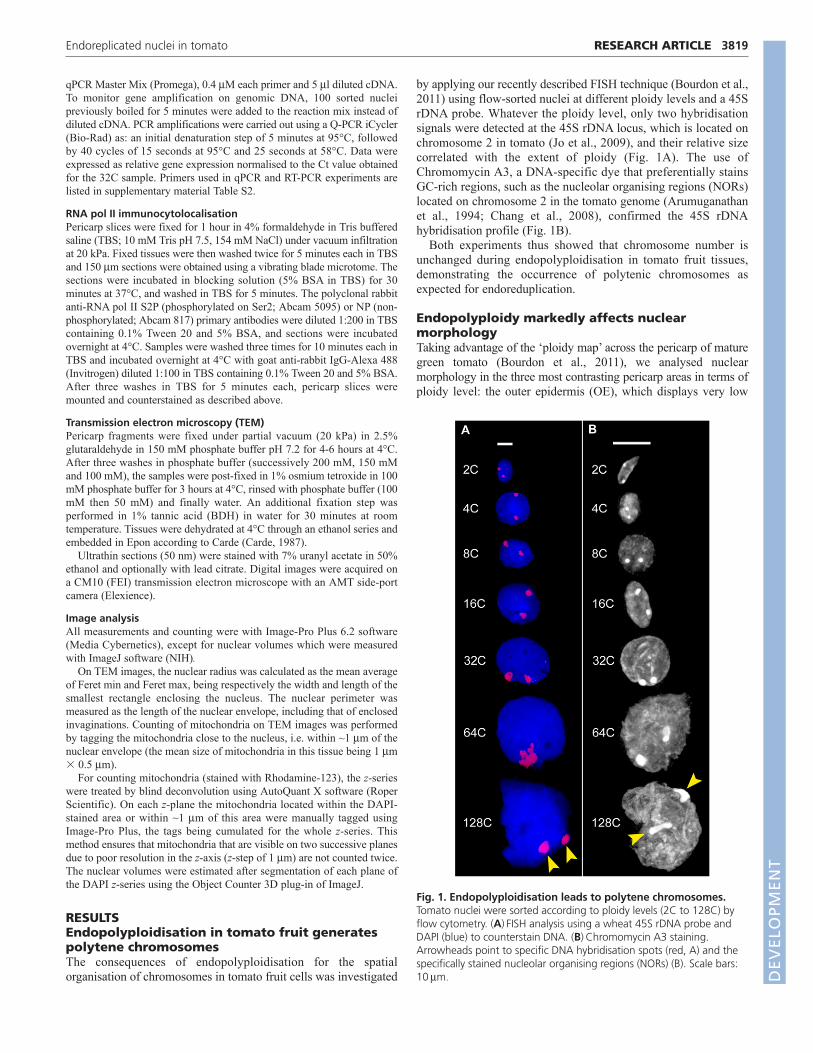
RESULTSEndopolyploidisation in tomato fruit generatespolytene chromosomesThe consequences of endopolyploidisation for the spatialorganisation of chromosomes in tomato fruit cells was investigated
by applying our recently described FISH technique (Bourdon et al.,2011) using flow-sorted nuclei at different ploidy levels and a 45SrDNA probe. Whatever the ploidy level, only two hybridisationsignals were detected at the 45S rDNA locus, which is located onchromosome 2 in tomato (Jo et al., 2009), and their relative sizecorrelated with the extent of ploidy (Fig. 1A). The use ofChromomycin A3, a DNA-specific dye that preferentially stainsGC-rich regions, such as the nucleolar organising regions (NORs)located on chromosome 2 in the tomato genome (Arumuganathanet al., 1994; Chang et al., 2008), confirmed the 45S rDNAhybridisation profile (Fig. 1B).
Both experiments thus showed that chromosome number isunchanged during endopolyploidisation in tomato fruit tissues,demonstrating the occurrence of polytenic chromosomes asexpected for endoreduplication.
Endopolyploidy markedly affects nuclearmorphologyTaking advantage of the ‘ploidy map’ across the pericarp of maturegreen tomato (Bourdon et al., 2011), we analysed nuclearmorphology in the three most contrasting pericarp areas in terms ofploidy level: the outer epidermis (OE), which displays very low
3819RESEARCH ARTICLEEndoreplicated nuclei in tomato
Fig. 1. Endopolyploidisation leads to polytene chromosomes.Tomato nuclei were sorted according to ploidy levels (2C to 128C) byflow cytometry. (A)FISH analysis using a wheat 45S rDNA probe andDAPI (blue) to counterstain DNA. (B)Chromomycin A3 staining.Arrowheads point to specific DNA hybridisation spots (red, A) and thespecifically stained nucleolar organising regions (NORs) (B). Scale bars:10m. D
EVELO
PMENT
3820
ploidy levels of 2C and 4C; the inner epidermis (IE), which displaysmid-ploidy levels ranging from 8C to 32C; and the mid- to innermesocarp (M), which displays high ploidy levels of 32C to 128C.
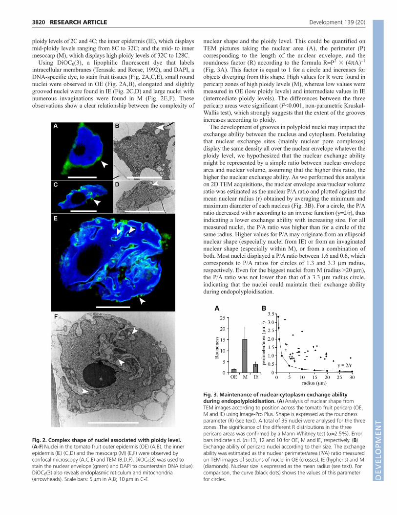
Using DiOC6(3), a lipophilic fluorescent dye that labelsintracellular membranes (Terasaki and Reese, 1992), and DAPI, aDNA-specific dye, to stain fruit tissues (Fig. 2A,C,E), small roundnuclei were observed in OE (Fig. 2A,B), elongated and slightlygrooved nuclei were found in IE (Fig. 2C,D) and large nuclei withnumerous invaginations were found in M (Fig. 2E,F). Theseobservations show a clear relationship between the complexity of
nuclear shape and the ploidy level. This could be quantified onTEM pictures taking the nuclear area (A), the perimeter (P)corresponding to the length of the nuclear envelope, and theroundness factor (R) according to the formula RP2 � (4A)–1
(Fig. 3A). This factor is equal to 1 for a circle and increases forobjects diverging from this shape. High values for R were found inpericarp zones of high ploidy levels (M), whereas low values weremeasured in OE (low ploidy levels) and intermediate values in IE(intermediate ploidy levels). The differences between the threepericarp areas were significant (P<0.001, non-parametric Kruskal-Wallis test), which strongly suggests that the extent of the groovesincreases according to ploidy.
The development of grooves in polyploid nuclei may impact theexchange ability between the nucleus and cytoplasm. Postulatingthat nuclear exchange sites (mainly nuclear pore complexes)display the same density all over the nuclear envelope whatever theploidy level, we hypothesized that the nuclear exchange abilitymight be represented by a simple ratio between nuclear envelopearea and nuclear volume, assuming that the higher this ratio, thehigher the nuclear exchange ability. As we performed this analysison 2D TEM acquisitions, the nuclear envelope area/nuclear volumeratio was estimated as the nuclear P/A ratio and plotted against themean nuclear radius (r) obtained by averaging the minimum andmaximum diameter of each nucleus (Fig. 3B). For a circle, the P/Aratio decreased with r according to an inverse function (y2/r), thusindicating a lower exchange ability with increasing size. For allmeasured nuclei, the P/A ratio was higher than for a circle of thesame radius. Higher values for P/A may originate from an ellipsoidnuclear shape (especially nuclei from IE) or from an invaginatednuclear shape (especially within M), or from a combination ofboth. Most nuclei displayed a P/A ratio between 1.6 and 0.6, whichcorresponds to P/A ratios for circles of 1.3 and 3.3 m radius,respectively. Even for the biggest nuclei from M (radius >20 m),the P/A ratio was not lower than that of a 3.3 m radius circle,indicating that the nuclei could maintain their exchange abilityduring endopolyploidisation.
RESEARCH ARTICLE Development 139 (20)
Fig. 2. Complex shape of nuclei associated with ploidy level. (A-F)Nuclei in the tomato fruit outer epidermis (OE) (A,B), the innerepidermis (IE) (C,D) and the mesocarp (M) (E,F) were observed byconfocal microscopy (A,C,E) and TEM (B,D,F). DiOC6(3) was used tostain the nuclear envelope (green) and DAPI to counterstain DNA (blue).DiOC6(3) also reveals endoplasmic reticulum and mitochondria(arrowheads). Scale bars: 5m in A,B; 10m in C-F.
Fig. 3. Maintenance of nuclear-cytoplasm exchange abilityduring endopolyploidisation. (A)Analysis of nuclear shape fromTEM images according to position across the tomato fruit pericarp (OE,M and IE) using Image-Pro Plus. Shape is expressed as the roundnessparameter (R) (see text). A total of 35 nuclei were analysed for the threezones. The significance of the different R distributions in the threepericarp areas was confirmed by a Mann-Whitney test (2.5%). Errorbars indicate s.d. (n13, 12 and 10 for OE, M and IE, respectively. (B)Exchange ability of pericarp nuclei according to their size. The exchangeability was estimated as the nuclear perimeter/area (P/A) ratio measuredon TEM images of sections of nuclei in OE (crosses), IE (hyphens) and M(diamonds). Nuclear size is expressed as the mean radius (see text). Forcomparison, the curve (black dots) shows the values of this parameterfor circles. D
EVELO
PMENT
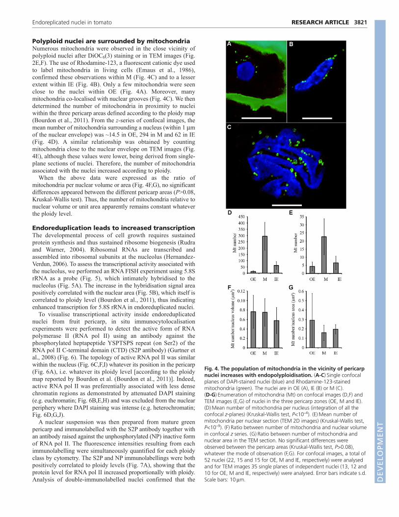
Polyploid nuclei are surrounded by mitochondriaNumerous mitochondria were observed in the close vicinity ofpolyploid nuclei after DiOC6(3) staining or in TEM images (Fig.2E,F). The use of Rhodamine-123, a fluorescent cationic dye usedto label mitochondria in living cells (Emaus et al., 1986),confirmed these observations within M (Fig. 4C) and to a lesserextent within IE (Fig. 4B). Only a few mitochondria were seenclose to the nuclei within OE (Fig. 4A). Moreover, manymitochondria co-localised with nuclear grooves (Fig. 4C). We thendetermined the number of mitochondria in proximity to nucleiwithin the three pericarp areas defined according to the ploidy map(Bourdon et al., 2011). From the z-series of confocal images, themean number of mitochondria surrounding a nucleus (within 1 mof the nuclear envelope) was ~14.5 in OE, 294 in M and 62 in IE(Fig. 4D). A similar relationship was obtained by countingmitochondria close to the nuclear envelope on TEM images (Fig.4E), although these values were lower, being derived from single-plane sections of nuclei. Therefore, the number of mitochondriaassociated with the nuclei increased according to ploidy.
When the above data were expressed as the ratio ofmitochondria per nuclear volume or area (Fig. 4F,G), no significantdifferences appeared between the different pericarp areas (P>0.08,Kruskal-Wallis test). Thus, the number of mitochondria relative tonuclear volume or unit area apparently remains constant whateverthe ploidy level.
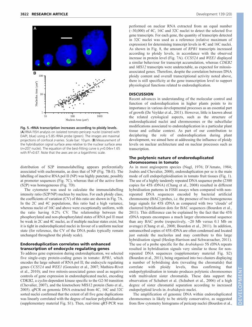
Endoreduplication leads to increased transcriptionThe developmental process of cell growth requires sustainedprotein synthesis and thus sustained ribosome biogenesis (Rudraand Warner, 2004). Ribosomal RNAs are transcribed andassembled into ribosomal subunits at the nucleolus (Hernandez-Verdun, 2006). To assess the transcriptional activity associated withthe nucleolus, we performed an RNA FISH experiment using 5.8SrRNA as a probe (Fig. 5), which intimately hybridised to thenucleolus (Fig. 5A). The increase in the hybridisation signal areapositively correlated with the nuclear area (Fig. 5B), which itself iscorrelated to ploidy level (Bourdon et al., 2011), thus indicatingenhanced transcription for 5.8S rRNA in endoreduplicated nuclei.
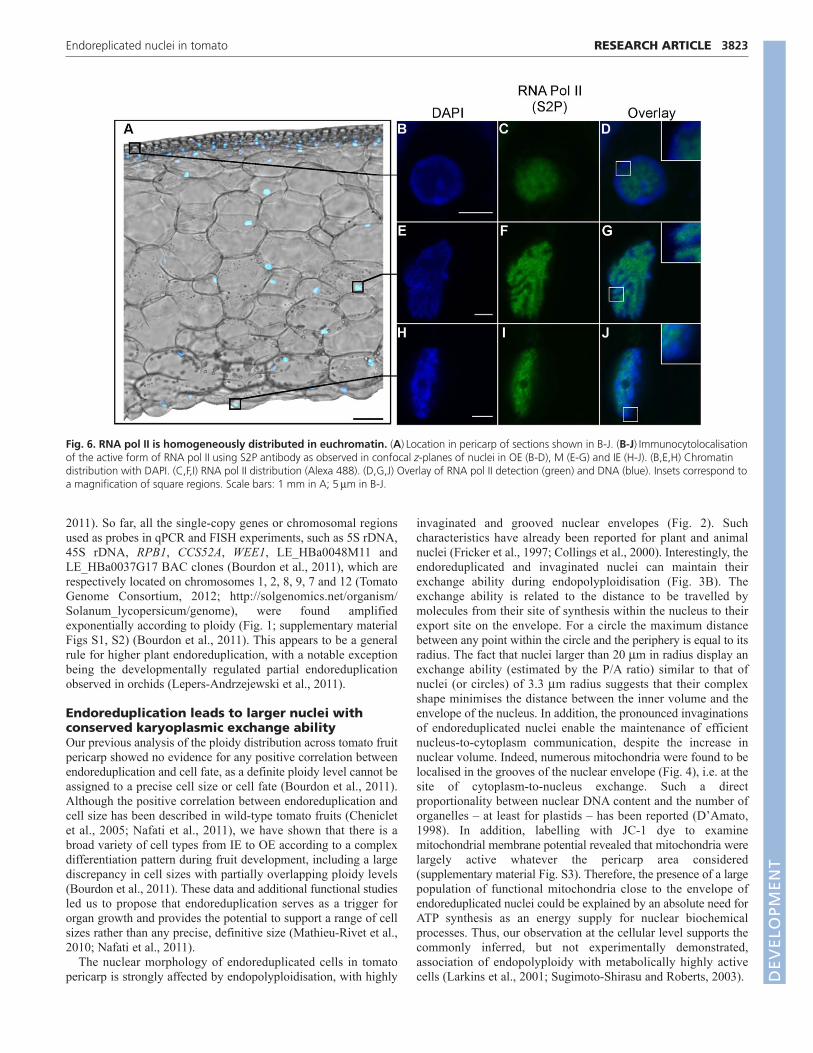
To visualise transcriptional activity inside endoreduplicatednuclei from fruit pericarp, in situ immunocytolocalisationexperiments were performed to detect the active form of RNApolymerase II (RNA pol II) using an antibody against thephosphorylated heptapeptide YSPTSPS repeat (on Ser2) of theRNA pol II C-terminal domain (CTD) (S2P antibody) (Gurtner etal., 2008) (Fig. 6). The topology of active RNA pol II was similarwithin the nucleus (Fig. 6C,F,I) whatever its position in the pericarp(Fig. 6A), i.e. whatever its ploidy level [according to the ploidymap reported by Bourdon et al. (Bourdon et al., 2011)]. Indeed,active RNA pol II was preferentially associated with less densechromatin regions as demonstrated by attenuated DAPI staining(e.g. euchromatin; Fig. 6B,E,H) and was excluded from the nuclearperiphery where DAPI staining was intense (e.g. heterochromatin;Fig. 6D,G,J).
A nuclear suspension was then prepared from mature greenpericarp and immunolabelled with the S2P antibody together withan antibody raised against the unphosphorylated (NP) inactive formof RNA pol II. The fluorescence intensities resulting from eachimmunolabelling were simultaneously quantified for each ploidyclass by cytometry. The S2P and NP immunolabellings were bothpositively correlated to ploidy levels (Fig. 7A), showing that theprotein level for RNA pol II increased proportionally with ploidy.Analysis of double-immunolabelled nuclei confirmed that the
3821RESEARCH ARTICLEEndoreplicated nuclei in tomato
Fig. 4. The population of mitochondria in the vicinity of pericarpnuclei increases with endopolyploidisation. (A-C)Single confocalplanes of DAPI-stained nuclei (blue) and Rhodamine-123-stainedmitochondria (green). The nuclei are in OE (A), IE (B) or M (C). (D-G)Enumeration of mitochondria (Mt) on confocal images (D,F) andTEM images (E,G) of nuclei in the three pericarp zones (OE, M and IE).(D)Mean number of mitochondria per nucleus (integration of all theconfocal z-planes) (Kruskal-Wallis test, P<10–4). (E)Mean number ofmitochondria per nuclear section (TEM 2D images) (Kruskal-Wallis test,P<10–9). (F)Ratio between number of mitochondria and nuclear volumein confocal z series. (G)Ratio between number of mitochondria andnuclear area in the TEM section. No significant differences wereobserved between the pericarp areas (Kruskal-Wallis test, P>0.08),whatever the mode of observation (F,G). For confocal images, a total of52 nuclei (22, 15 and 15 for OE, M and IE, respectively) were analysedand for TEM images 35 single planes of independent nuclei (13, 12 and10 for OE, M and IE, respectively) were analysed. Error bars indicate s.d.Scale bars: 10m. D
EVELO
PMENT
3822
distribution of S2P immunolabelling appears preferentiallyassociated with euchromatin, as does that of NP (Fig. 7B-E). Thelabelling of inactive RNA pol II (NP) was highly punctate, possiblyat promoter sequences (Fig. 7C), whereas that of the active form(S2P) was homogeneous (Fig. 7D).
The cytometer was used to calculate the immunolabellingintensity ratio (S2P/NP) nucleus by nucleus. For each ploidy class,the coefficients of variation (CV) of this ratio are shown in Fig. 7A.In the 2C and 4C populations, this ratio had a high variance,whereas nuclei of 16C and above were exceptionally uniform, withthe ratio having 0.2% CV. The relationship between thephosphorylated and non-phosphorylated states of RNA pol II mustbe weak in 2C and 4C nuclei, as if multiple nuclear states exist, butit is tight in endoreduplicated nuclei in favour of a uniform nuclearstate (for reference, the CV of the DNA peaks typically remainunchanged throughout the ploidy scale).
Endoreduplication correlates with enhancedtranscription of endocycle regulating genesTo address gene expression during endoreduplication, we selectedfive single-copy protein-coding genes in tomato: RPB1, whichencodes the large subunit of RNA pol II; the endocycle regulatinggenes CCS52A and WEE1 (Gonzalez et al., 2007; Mathieu-Rivetet al., 2010); and two mitosis-associated genes used as negativecontrols of gene expression in endoreduplicated nuclei, encodingCDKB2, a cyclin-dependent kinase specific to the G2-M transition(Chevalier, 2007), and the kinetochore MIS12 protein (Sato et al.,2005). qPCR on genomic DNA extracted from 4C, 16C and 32Csorted nuclei confirmed that the extent of all five gene duplicationswas linearly correlated with the degree of nuclear polyploidisation(supplementary material Fig. S1). Then, real-time qRT-PCR was
performed on nuclear RNA extracted from an equal number(~30,000) of 4C, 16C and 32C nuclei to detect the selected fivegene transcripts. For each gene, the quantity of transcripts detectedin 32C nuclei was used as a reference (relative maximum ofexpression) for determining transcript levels in 4C and 16C nuclei.As shown in Fig. 8, the amount of RPB1 transcripts increasedaccording to ploidy levels, in accordance with the observedincrease in protein level (Fig. 7A). CCS52A and WEE1 displayeda similar behaviour for transcript accumulation, whereas CDKB2and MIS12 transcripts were undetectable, as expected for mitosis-associated genes. Therefore, despite the correlation between DNAploidy content and overall transcriptional activity noted above,there is still specificity at the gene transcription level to supportphysiological functions related to endoreduplication.
DISCUSSIONRecent advances in understanding of the molecular control andfunction of endoreduplication in higher plants points to itsimportance in various developmental processes as an essential partof growth (De Veylder et al., 2011). However, little is known aboutthe related cytological aspects, such as the structure ofendoreduplicated nuclei and chromosomes or the subcellularmodifications associated to endoreduplication in a particular planttissue and cellular context. As part of our contribution todeciphering the role of endoreduplication during plantdevelopment, we aimed here at addressing the influence of ploidylevels on nuclear architecture and on nuclear processes such astranscription.
The polytenic nature of endoreduplicatedchromosomes in tomatoAs in most angiosperm species (Nagl, 1976; D’Amato, 1984;Joubès and Chevalier, 2000), endoreduplication per se is the mainmode of cell endopolyploidisation in tomato fruit tissues (Fig. 1).The use of a highly tandemly repeated DNA sequence probe (2300copies for 45S rDNA) (Chang et al., 2008) resulted in differenthybridisation patterns in FISH assays when compared with non-repeated DNA sequences encoded by bacterial artificialchromosome (BAC) probes, i.e. the presence of two homogeneouslarge signals for 45S rDNA as compared with two ‘clouds’ ofclustered hybridisation dots for BAC sequences (Bourdon et al.,2011). This difference can be explained by the fact that the 45SrDNA repeats encompass a much larger chromosomal sequencethan any BAC probe (respectively, ~20 Mb versus 0.1 Mb onaverage) (Chang et al., 2008; Bourdon et al., 2011). In addition,untranscribed copies of 45S rDNA are often condensed and locatedjust outside the nucleolus and may contribute to this largehybridisation signal (Heslop-Harrison and Schwarzacher, 2011).The use of a probe specific for the Arabidopsis 5S rDNA repeatsresulted in hybridisation signals very similar to those for non-repeated DNA sequences (supplementary material Fig. S2)(Bourdon et al., 2011), being organised into two clusters displayinga number of hybridising dots (revealing the chromatids) thatcorrelate with ploidy levels, thus confirming thatendopolyploidisation in tomato produces polytenic chromosomeswith multivalent sister chromatids. These data support theobservation of Schubert et al. (Schubert et al., 2006) of a highdegree of sister chromatid separation according to increasedendopolyploid levels in Arabidopsis nuclei.
The amplification of genomic DNA within endoreduplicatedchromosomes is likely to be strictly conservative, as suggestedfrom flow cytometry histograms of pericarp nuclei (Bourdon et al.,
RESEARCH ARTICLE Development 139 (20)
Fig. 5. rRNA transcription increases according to ploidy levels.(A)RNA FISH analysis on isolated tomato pericarp nuclei (stained withDAPI, blue) using a 5.8S rRNA probe (green). The images are maximalprojections of confocal z-series. Scale bar: 10m. (B)Measurement ofthe hybridisation signal surface area relative to the nuclear surface area(n207 nuclei). The equation of the best-fitting curve is y0.04x+1.65with R20.67. Note that the axes are on a logarithmic scale.
DEVELO
PMENT
2011). So far, all the single-copy genes or chromosomal regionsused as probes in qPCR and FISH experiments, such as 5S rDNA,45S rDNA, RPB1, CCS52A, WEE1, LE_HBa0048M11 andLE_HBa0037G17 BAC clones (Bourdon et al., 2011), which arerespectively located on chromosomes 1, 2, 8, 9, 7 and 12 (TomatoGenome Consortium, 2012; http://solgenomics.net/organism/Solanum_lycopersicum/genome), were found amplifiedexponentially according to ploidy (Fig. 1; supplementary materialFigs S1, S2) (Bourdon et al., 2011). This appears to be a generalrule for higher plant endoreduplication, with a notable exceptionbeing the developmentally regulated partial endoreduplicationobserved in orchids (Lepers-Andrzejewski et al., 2011).
Endoreduplication leads to larger nuclei withconserved karyoplasmic exchange abilityOur previous analysis of the ploidy distribution across tomato fruitpericarp showed no evidence for any positive correlation betweenendoreduplication and cell fate, as a definite ploidy level cannot beassigned to a precise cell size or cell fate (Bourdon et al., 2011).Although the positive correlation between endoreduplication andcell size has been described in wild-type tomato fruits (Chenicletet al., 2005; Nafati et al., 2011), we have shown that there is abroad variety of cell types from IE to OE according to a complexdifferentiation pattern during fruit development, including a largediscrepancy in cell sizes with partially overlapping ploidy levels(Bourdon et al., 2011). These data and additional functional studiesled us to propose that endoreduplication serves as a trigger fororgan growth and provides the potential to support a range of cellsizes rather than any precise, definitive size (Mathieu-Rivet et al.,2010; Nafati et al., 2011).
The nuclear morphology of endoreduplicated cells in tomatopericarp is strongly affected by endopolyploidisation, with highly
invaginated and grooved nuclear envelopes (Fig. 2). Suchcharacteristics have already been reported for plant and animalnuclei (Fricker et al., 1997; Collings et al., 2000). Interestingly, theendoreduplicated and invaginated nuclei can maintain theirexchange ability during endopolyploidisation (Fig. 3B). Theexchange ability is related to the distance to be travelled bymolecules from their site of synthesis within the nucleus to theirexport site on the envelope. For a circle the maximum distancebetween any point within the circle and the periphery is equal to itsradius. The fact that nuclei larger than 20 m in radius display anexchange ability (estimated by the P/A ratio) similar to that ofnuclei (or circles) of 3.3 m radius suggests that their complexshape minimises the distance between the inner volume and theenvelope of the nucleus. In addition, the pronounced invaginationsof endoreduplicated nuclei enable the maintenance of efficientnucleus-to-cytoplasm communication, despite the increase innuclear volume. Indeed, numerous mitochondria were found to belocalised in the grooves of the nuclear envelope (Fig. 4), i.e. at thesite of cytoplasm-to-nucleus exchange. Such a directproportionality between nuclear DNA content and the number oforganelles – at least for plastids – has been reported (D’Amato,1998). In addition, labelling with JC-1 dye to examinemitochondrial membrane potential revealed that mitochondria werelargely active whatever the pericarp area considered(supplementary material Fig. S3). Therefore, the presence of a largepopulation of functional mitochondria close to the envelope ofendoreduplicated nuclei could be explained by an absolute need forATP synthesis as an energy supply for nuclear biochemicalprocesses. Thus, our observation at the cellular level supports thecommonly inferred, but not experimentally demonstrated,association of endopolyploidy with metabolically highly activecells (Larkins et al., 2001; Sugimoto-Shirasu and Roberts, 2003).
3823RESEARCH ARTICLEEndoreplicated nuclei in tomato
Fig. 6. RNA pol II is homogeneously distributed in euchromatin. (A)Location in pericarp of sections shown in B-J. (B-J)Immunocytolocalisationof the active form of RNA pol II using S2P antibody as observed in confocal z-planes of nuclei in OE (B-D), M (E-G) and IE (H-J). (B,E,H) Chromatindistribution with DAPI. (C,F,I) RNA pol II distribution (Alexa 488). (D,G,J) Overlay of RNA pol II detection (green) and DNA (blue). Insets correspond toa magnification of square regions. Scale bars: 1 mm in A; 5m in B-J.
DEVELO
PMENT

3824
Endoreduplication triggers enhancedtranscriptional activityThe highly polyploid endosperm cells of maize kernels, whichaccumulate large amounts of starch and storage proteins (Sabelliand Larkins, 2009), and the nitrogen-fixing symbiotic root nodulecells in legumes (Cebolla et al., 1999) are quoted as metabolicallyspecialised and highly endoreduplicated cells. Interestingly, animportant remodelling of the transcriptome is tightly linked toendoreduplication in nitrogen-fixing nodules (Mergaert et al.,2003). As far as tomato fruit is concerned, only descriptiveanalyses for transcriptional and metabolic profiling inendoreduplicated tissues have been provided (Lemaire-Chamley etal., 2005; Mounet et al., 2009; Schauer et al., 2006; Steinhauser etal., 2010), without any direct link with the extent ofendoreduplication.
Using RNA FISH we demonstrated that the transcription of 5.8SrDNA within the nucleolus is enhanced during endoreduplication(Fig. 5). In addition, the 5.8S rRNA hybridisation signal thatreflects the size of the nucleolus was positively correlated withnuclear size and consequently with ploidy levels. These data are
consistent with our observation of NOR enlargement according toploidy (Fig. 1B). Such a nucleolar enlargement has been reportedin rodent trophoblasts (Zybina and Zybina, 1996) and inDrosophila (Pierce et al., 2004) and associated with increasedtranscriptional and translational activities in the context ofendopolyploidisation. This feature has also been associated withthe rate of proliferation in cancer cells (Derenzini et al., 2000). Itis therefore tempting to draw a parallel with the increased rate ofgrowth that is often associated with endoreduplication. We foundthat the cytoplasmic area of cells within the three most contrastingpericarp zones in terms of ploidy levels (OE, IE and M, where themost common nuclear classes are 2C, 16C and 64C, respectively)indeed increases with ploidy levels (supplementary material Fig.S4A), as does the estimated cytoplasmic volume (supplementarymaterial Fig. S4B). This demonstrates the maintenance of thenuclear to cytoplasmic ratio to drive cell growth, consistent withthe karyoplasmic ratio theory. Ribosomal RNA, protein synthesisand exchange must be proportionally enhanced to increaseribosome biogenesis and hence maintain homeostasis ofcytoplasmic components essential for sustained cell growth.
Previous functional analyses of genes regulating the endocycle(Gonzalez et al., 2007; Mathieu-Rivet et al., 2010; Nafati et al.,2011) demonstrated the biological significance ofendoreduplication as a morphogenetic factor for cell and fruitgrowth in tomato. In addition, enhancing endoreduplication andconsequently fruit growth also strongly impacted nitrogenmetabolism in developing fruits (Mathieu-Rivet et al., 2010),establishing a link between endoreduplication-induced growth andprimary metabolism, as reported in maize endosperm (Leiva-Netoet al., 2004). Interestingly, a causal relationship between cell size,polyploidy and transcription in yeast has been demonstrated,suggesting that cells adjust their transcriptional capacities to theirgeometric properties, including cell size (Wu et al., 2010).
Here, we showed that transcription of the WEE1 and CCS52Agenes that control endoreduplication (Gonzalez et al., 2007;Mathieu-Rivet et al., 2010) is increased on a per-nucleus basisaccording to ploidy (Fig. 8). This observation was also true forRPB1, which encodes a subunit of RNA pol II. The use ofpolyclonal antibodies targeting the YSPTSPS repeat of the RNApol II CTD allowed us to discriminate between the active andinactive status of RNA pol II. The RNA pol II CTD repeat isdifferentially phosphorylated according to its position on a gene:when located on a promoter sequence, it is largelyunphosphorylated (NP form), whereas the active form of RNA polII requires phosphorylation at Ser2 and Ser5 (Hirose and Ohkuma,
RESEARCH ARTICLE Development 139 (20)
Fig. 7. The amount of RNA pol II increases according to ploidylevels. (A)Fluorescence intensity from cytometry of double-immunolabelled nuclei using the S2P and NP RNA pol II antibodies. Foreach ploidy class, the coefficient of variation (CV%) of the parameter‘S2P/NP ratio of each nucleus’ is indicated (see text for interpretation).Error bars indicate s.d. (B-E) Phosphorylated and non-phosphorylatedRNA pol II in confocal z-plane of an isolated nucleus. (B)ChromatinDNA revealed by DAPI. (C)Distribution of NP RNA pol II (Alexa 488).(D)Distribution of S2P RNA pol II (Alexa 633). (E)Overlay of B-D. Scalebar: 10m.
Fig. 8. Transcript levels of RPB1, WEE1 and CCS52A increaseaccording to ploidy levels. The expression of RPB1, WEE1 andCCS52A in nuclei sorted at different ploidy levels (4C, 16C and 32C)was assessed by qRT-PCR. Data are the mean of three biologicalreplicates. Error bars indicate s.d.
DEVELO
PMENT
2007). In addition, phosphorylation at Ser5 is mainly associatedwith the initiation of transcription (Hengartner et al., 1998;Komarnitsky et al., 2000), whereas phosphorylation at Ser2 (S2Pform) is predominantly present during the elongation step oftranscription (Ni et al., 2004). Endoreduplication is linked to thesustained transcription/translation and activity of RPB1 (Figs 6, 7).Furthermore, the results obtained with the S2P antibody suggestthat transcriptional elongation is excluded from the perinuclearheterochromatic region and occurs within euchromatin in ahomogeneous manner (Fig. 6). Subnuclear compartments were notevident, suggesting that the euchromatin regions in polyploidnuclei are apparently indiscriminately transcribed, as if all copiesof amplified genes are equally functional in the tomatoendoreduplicated genome. Moreover, the exceptionally tight CVobserved for the S2P/NP ratio in polyploid nuclei (Fig. 7) revealsa uniformity of transcriptional activity when compared with thatobserved in 2C and 4C nuclei. Altogether, these results demonstratea strong relationship between endoreduplication and transcription.
ConclusionsThe role of endoreduplication in organ growth is still a matter ofdebate. An organismal regulation of growth in which organ growthitself acts as a ‘hub’ driving cell division and expansion, as well asendoreduplication, with compensation if one of the underlyingcellular processes is limiting (John and Qi, 2008; Massonnet et al.,2011), is opposed to a cell-based (autonomous) mode of growthcontrol that relies on the balance of cellular processes (division,expansion and endoreduplication) (Sugimoto-Shirasu and Roberts,2003).
The results presented here provide quantitative evidence infavour of the karyoplasmic ratio theory. Different levels of specificstructural and functional organisation in endoreduplicated cellswere revealed, shedding light on the contribution ofendoreduplication to the establishment of a highly structuredcellular system that integrates multiple physiological functions,including transcription, and acts as a morphogenetic factorsupporting cell growth during tomato fruit development. Ourperspective is that karyoplasmic homeostasis ensures function notonly by geometry, but also by cooperative adaptation.
AcknowledgementsImage acquisition and analysis were performed at the Plant Imaging Pole ofthe Bordeaux Imaging Center. Flow cytometry was performed at the ImagifPlatform (IFR87 and ASTRE, CNRS, Gif-sur-Yvette) with Marie-Noëlle Soler,whom we thank for help with imaging. We thank Norbert Bollier forcontribution to the results obtained on mitochondria.
FundingThis work was carried out with the financial support of the French AgenceNationale de la Recherche [grant ANR-09-GENM-105].
Competing interests statementThe authors declare no competing financial interests.
Supplementary materialSupplementary material available online athttp://dev.biologists.org/lookup/suppl/doi:10.1242/dev.084053/-/DC1
ReferencesAdachi, S., Minamisawa, K., Okushima, Y., Inagaki, S., Yoshiyama, K.,
Kondou, Y., Kaminuma, E., Kawashima, M., Toyoda, T., Matsui et al.(2011). Programmed induction of endoreduplication by DNA double-strandbreaks in Arabidopsis. Proc. Natl. Acad. Sci. USA 108, 10004-10009.
Arumuganathan, K., Martin, G. B., Telenius, H., Tanksley, S. D. and Earle, E.D. (1994). Chromosome 2-specific DNA clones from flow-sorted chromosomesof tomato. Mol. Gen. Genet. 242, 551-558.
Barow, M. (2006). Endopolyploidy in seed plants. BioEssays 28, 271-281.
Bourdon, M., Coriton, O., Cheniclet, C., Brown, S., Poujol, C., Chevalier, C.,Renaudin, J.-P., Frangne, N. (2011). In planta quantification of endopolyploidyusing fluorescent in situ hybridization (FISH). Plant J. 66, 1089-1099.
Bramsiepe, J., Wester, K., Weinl, C., Roodbarkelari, F., Kasili, R., Larkin, J. C.,Hülskamp, M. and Schnittger, A. (2010). Endoreplication controls cell fatemaintenance. PLoS Genet. 6, e1000996.
Brodsky, W. Y. and Uryvaeva, I. V. (1977). Cell polyploidy: its relation to tissuegrowth and function. Int. Rev. Cytol. 50, 275-332.
Carde, J. P. (1987). Electron microscopy of plant cell membranes. MethodsEnzymol. 148, 599-622.
Cebolla, A., Maria Vinardell, J., Kiss, E., Olah, B., Roudier, F., Kondorosi, A.and Kondorosi, E. (1999). The mitotic inhibitor ccs52 is required forendoreduplication and ploidy-dependent cell enlargement in plants. EMBO J. 18,4476-4484.
Ceccarelli, M., Sanantonio, E., Marmottini, F., Amzallag, G. N. and Cionini, P.G. (2006). Chromosome endoreduplication as a factor of salt adaptation inSorghum bicolor. Protoplasma 227, 113-118.
Chang, S.-B., Yang, T.-J., Datema, E., van Vugt, J., Vosman, B., Kuipers, A.,Meznikova, M., Szinay, D., Lankhorst, R., Jacobsen, E. and de Jong, H.(2008). FISH mapping and molecular organization of the major repetitivesequences of tomato. Chromosome Res. 16, 919-933.
Cheniclet, C., Rong, W. Y., Causse, M., Frangne, N., Bolling, L., Carde, J.-P.and Renaudin, J.-P. (2005). Cell expansion and endoreduplication show a largegenetic variability in pericarp and contribute strongly to tomato fruit growth.Plant Physiol. 139, 1984-1994.
Chevalier, C. (2007). Cell cycle control and fruit development. In Annual PlantReviews Volume 32: Cell Cycle Control and Plant Development (ed. D. Inzé), pp.269-293. Oxford, UK: Blackwell Publishing.
Chevalier, C., Nafati, M., Mathieu-Rivet, E., Bourdon, M., Frangne, N.,Cheniclet, C., Renaudin, J.-P., Gévaudant, F., Hernould, M. (2011).Elucidating the functional role of endoreduplication in tomato fruitdevelopment. Ann. Bot. 107, 1159-1169.
Collings, D. A., Carter, C. N., Rink, J. C., Scott, A. C., Wyatt, S. E. and Allen,N. S. (2000). Plant nuclei can contain extensive grooves and invaginations. PlantCell 12, 2425-2440.
Cookson, S. J., Radziejwoski, A. and Granier, C. (2006). Cell and leaf sizeplasticity in Arabidopsis: what is the role of endoreduplication? Plant CellEnviron. 29, 1273-1283.
D’Amato, F. (1984). Role of polyploidy in reproductive organs and tissues. InEmbryology of Angiosperms (ed. B. M. Johri), pp. 519-566. New York: Springer.
D’Amato, F. (1998). Chromosome endoreduplication in plant tissue developmentand function. In Plant Cell Proliferation and its Regulation in Growth andDevelopment (ed. J. A. Bryant and D. Chiatante), pp. 153-166. Chichester: J.Wiley & Sons.
De Veylder, L., Larkin, J. C. and Schnittger, A. (2011). Molecular control andfunction of endoreduplication in development and physiology. Trends Plant Sci.16, 624-634.
Derenzini, M., Trere, D., Pession, A., Govoni, M., Sirri, V. and Chieco, P.(2000). Nucleolar size indicates the rapidity of cell proliferation in cancer tissues.J. Pathol. 191, 181-186.
Emaus, R. K., Grunwald, R. and Lemasters, J. J. (1986). Rhodamine-123 as aprobe of transmembrane potential in isolated rat-liver mitochondria – spectraland metabolic properties. Biochem. Biophys. Acta 850, 436-448.
Fricker, M., Hollinshead, M., White, N. and Vaux, D. (1997). Interphase nucleiof many mammalian cell types contain deep, dynamic, tubular membrane-bound invaginations of the nuclear envelope. J. Cell Biol. 136, 531-544.
Galbraith, D. W., Janda, J. and Lambert, G. M. (2011). Multiparametric analysis,sorting, and transcriptional profiling of plant protoplasts and nuclei according tocell type. Methods Mol. Biol. 699, 407-429.
Gerlach, W. L. and Bedbrook, J. R. (1979). Cloning and characterization ofribosomal RNA genes from wheat and barley. Nucleic Acids Res. 7, 1869-1885.
Gonzalez, N., Gevaudant, F., Hernould, M., Chevalier, C. and Mouras, A.(2007). The cell cycle-associated protein kinase WEE1 regulates cell size inrelation to endoreduplication in developing tomato fruit. Plant J. 51, 642-655.
Gurtner, A., Fuschi, P., Magi, F., Colussi, C., Gaetano, C., Dobbelstein, M.,Sacchi, A. and Piaggio, G. (2008). NF-Y dependent epigenetic modificationsdiscriminate between proliferating and postmitotic tissue. PLoS ONE 3, e2047.
Hase, Y., Trung, K. H., Matsunaga, T. and Tanaka, A. (2006). A mutation in theuvi4 gene promotes progression of endo-reduplication and confers increasedtolerance towards ultraviolet B light. Plant J. 46, 317-326.
Hengartner, C. J., Myer, V. E., Liao, S. M., Wilson, C. J., Koh, S. S. and Young,R. A. (1998). Temporal regulation of RNA polymerase II by Srb10 and Kin28cyclin-dependent kinases. Mol. Cell 2, 43-53.
Hernandez-Verdun, D. (2006). Nucleolus: from structure to dynamics.Histochem. Cell Biol. 125, 127-137.
Heslop-Harrison, J. S. and Schwarzacker, T. (2011). Organization of the plantgenome in chromosomes. Plant J. 66, 18-33.
Hirose, Y. and Ohkuma, Y. (2007). Phosphorylation of the C-terminal domain ofRNA polymerase II plays central roles in the integrated events of eucaryotic geneexpression. J. Biochem. 141, 601-608.
3825RESEARCH ARTICLEEndoreplicated nuclei in tomato
DEVELO
PMENT

3826
Hülskamp, M., Schnittger, A. and Folkers, U. (1999). Pattern formation and celldifferentiation: trichomes in Arabidopsis as a genetic model system. Int. Rev.Cytol. 186, 147-178.
Ishida, T., Kurata, T., Okada, K. and Wada, T. (2008). A genetic regulatorynetwork in the development of trichomes and root hairs. Ann. Rev. Plant Biol.59, 365-386.
Jo, S.-H., Koo, D.-H., Kim, J., Hur, C.-G., Lee, S., Yang, T.-j., Kwon, S.-Y. andChoi, D. (2009). Evolution of ribosomal DNA-derived satellite repeat in tomatogenome. BMC Plant Biol. 9, 42.
John, P. C. L. and Qi, R. (2008). Cell division and endoreduplication: doubtfulengines of vegetative growth. Trends Plant Sci. 13, 121-127.
Jolly, C., Mongelard, F., Robert-Nicoud, M. and Vourc’h, C. (1997).Optimization of nuclear transcript detection by FISH and combination withfluorescence immunocytochemical detection of transcription factors. J.Histochem. Cytochem. 45, 1585-1592.
Joubès, J. and Chevalier, C. (2000). Endoreduplication in higher plants. PlantMol. Biol. 43, 735-745.
Komarnitsky, P., Cho, E. J. and Buratowski, S. (2000). Different phosphorylatedforms of RNA polymerase II and associated mRNA processing factors duringtranscription. Genes Dev. 14, 2452-2460.
Larkins, B. A., Dilkes, B. P., Dante, R. A., Coelho, C. M., Woo, Y.-m. and Liu,Y. (2001). Investigating the hows and whys of DNA endoreduplication. J. Exp.Bot. 52, 183-192.
Lee, H. O., Davidson, J. M. and Duronio, R. J. (2010). Endoreduplication:polyploidy with purpose. Genes Dev. 23, 2461-2477.
Leiva-Neto, J. T., Grafi, G., Sabelli, P. A., Dante, R. A., Woo, Y. M., Maddock,S., Gordon-Kamm, W. J. and Larkins, B. A. (2004). A dominant negativemutant of cyclin-dependent kinase A reduces endoreduplication but not cell sizeor gene expression in maize endosperm. Plant Cell 16, 1854-1869.
Lemaire-Chamley, M., Petit, J., Garcia, V., Just, D., Baldet, P., Germain, V.,Fagard, M., Mouassite, M., Cheniclet, C. and Rothan, C. (2005). Changes intranscriptional profiles are associated with early fruit tissue specialization intomato. Plant Physiol. 139, 750-769.
Lepers-Andrzejewski, S., Siljak-Yakovlev, S., Brown, S. C., Wong. M. andDron, M. (2011). Diversity and dynamics of plant genome size: an example ofpolysomaty from a cytogenetic study of Tahitian Vanilla (Vanilla xtahitensis,Orchidaceae). Am. J. Bot. 98, 986-997.
Massonnet, C., Tisné, S., Radziejwoski, A., Vile, D., De Veylder, L., Dauzat,M. and Granier, C. (2011). New insights into the control of endoreduplication:endoreduplication could be driven by organ growth in Arabidopsis leaves. PlantPhysiol. 157, 2044-2055.
Mathieu-Rivet, E., Gevaudant, F., Sicard, A., Salar, S., Do, P. T., Mouras, A.,Fernie, A. R., Gibon, Y., Rothan, C., Chevalier, C. and Hernould, M. (2010).Functional analysis of the anaphase promoting complex activator CCS52Ahighlights the crucial role of endo-reduplication for fruit growth in tomato. PlantJ. 62, 727-741.
Mergaert, P., Nikovics, K., Kelemen, Z., Maunoury, N., Vaubert, D.,Kondorosi, A. and Kondorosi, E. (2003). A novel family in Medicagotruncatula consisting of more than 300 nodule-specific genes coding for small,
secreted polypeptides with conserved cysteine motifs. Plant Physiol. 132, 161-173.
Mounet, F., Moing, A., Garcia, V., Petit, J., Maucourt, M., Deborde, C.,Bernillon, S., Le Gall, G., Colquhoun, I., Defernez, M. et al. (2009). Geneand metabolite regulatory network analysis of early developing fruit tissueshighlights new candidate genes for the control of tomato fruit composition anddevelopment. Plant Physiol. 149, 1505-1528.
Nafati, M., Cheniclet, C., Hernould, M., Do, P. T., Fernie, A., Chevalier, C.,Gévaudant, F. (2011). The specific overexpression of a cyclin dependent kinaseinhibitor in tomato fruit mesocarp cells uncouples endoreduplication and cellgrowth. Plant J. 65, 543-556.
Nagl, W. (1976). DNA endoreduplication and polyteny understood as evolutionarystrategies. Nature 261, 614-615.
Ni, Z., Schwartz, B. E., Werner, J., Suarez, J. R. and Lis, J. T. (2004).Coordination of transcription, RNA processing, and surveillance by P-TEFb kinaseon heat shock genes. Mol. Cell 13, 55-65.
Pierce, S. B., Yost, C., Britton, J. S., Loo, L. W. M., Flynn, E. M., Edgar, B. A.and Eisenman, R. N. (2004). dMyc is required for larval growth andendoreplication in Drosophila. Development, 131, 2317-2327.
Roeder, A. H. K., Chickarmane, V., Cunha, A., Obara, B., Manjunath, B. S.and Meyerowitz, E. M. (2010). Variability in the control of cell divisionunderlies sepal epidermal patterning in Arabidopsis thaliana. PLoS Biol. 8,e1000367.
Rudra, D. and Warner, J. R. (2004). What better measure than ribosomesynthesis? Genes Dev. 18, 2431-2436.
Sabelli, P. A. and Larkins, B. A. (2009). The contribution of cell cycle regulationto endosperm development. Sex. Plant Reprod. 22, 207-219.
Sato, H., Shibata and F., Murata, M. (2005). Characterization of a MIS12homologue in Arabidopsis thaliana. Chromosome Res. 13, 827-834.
Schauer, N., Semel, Y., Roessner, U., Gur, A., Balbo, I., Carrari, F., Pleban, T.,Perez-Melis, A., Bruedigam, C., Kopka, J. et al. (2006). Comprehensivemetabolic profiling and phenotyping of interspecific introgression lines fortomato improvement. Nat. Biotech. 24, 447-454.
Schubert, V., Klatte, M., Pecinka, A., Meister, A., Jasencakova, Z. andSchubert, I. (2006). Sister chromatids are often incompletely aligned inmeristematic and endopolyploid interphase nuclei of Arabidopsis thaliana.Genetics 172, 467-475.
Steinhauser, M. C., Steinhauser, D., Koehl, K., Carrari, F., Gibon, Y., Fernie,A. R. and Stitt, M. (2010). Enzyme activity profiles during fruit development intomato cultivars and Solanum pennellii. Plant Physiol. 153, 80-98.
Sugimoto-Shirasu, K. and Roberts, K. (2003). ‘Big it up’: endoreduplication andcell-size control in plants. Curr. Opin. Plant Biol. 6, 544-553.
Terasaki, M. and Reese, T. S. (1992). Characterization of endoplasmic reticulumby co-localization of BiP and dicarbocyanine dyes. J. Cell Sci. 101, 315-322.
Tomato Genome Consortium (2012). The tomato genome sequence providesinsights into fleshy fruit evolution. Nature 485, 635-641.
Wu, C.-Y., Rolfe, P. A., Gifford, D. K. and Fink, G. R. (2010). Control oftranscription by cell size. PLoS Biol. 8, e1000523.
Zybina, E. V. and Zybina, T. G. (1996). Polytene chromosomes in mammaliancells. Int. Rev. Cytol. 165, 53-119.
RESEARCH ARTICLE Development 139 (20)
DEVELO
PMENT
Related Documents


![JeffParrish [415]806-3817](https://static.cupdf.com/doc/110x72/55c033a2bb61eb8a148b4676/jeffparrish-415806-3817.jpg)